Embed Size (px)
Citation preview

MELK 5-3-2016
1
Maternal embryonic leucine zipper kinase (MELK) as a novel mediator and biomarker of radioresistance in human breast cancer
Corey Speers1†, Shuang G. Zhao1, Vishal Kothari1, Alyssa Santola1, Meilan Liu1, Kari Wilder-
Romans1, Joseph Evans1, Nidhi Batra2, Harry Bartelink3, Daniel F. Hayes4,6, Theodore S.
Lawrence1,6, Powel H. Brown2, Lori J. Pierce1, Felix Y. Feng1,5,6†
1Department of Radiation Oncology, University of Michigan, 2Department of Cancer Prevention,
MD Anderson Cancer Center, 3Netherlands Cancer Institute, 4Clinical Director, Breast Oncology
Program 5Michigan Center for Translational Pathology, 6Comprehensive Cancer Center,
University of Michigan, Ann Arbor, Michigan,
Running title: MELK confers radioresistance in TNBC Keywords: breast cancer, gene expression profiling, radiation sensitivity, radiosensitivity
signature, radiotherapy
The authors declare no conflict of interest in these studies.
† To whom correspondence should be made: Corey Speers, M.D., Ph.D.
Department of Radiation Oncology
University of Michigan, UH B2 C490
1500 E. Medical Center Dr., SPC 5010
Ann Arbor, MI 48109-5010
Phone: 734-936-4300
E-mail: [email protected]
Research. on May 27, 2018. © 2016 American Association for Cancerclincancerres.aacrjournals.org Downloaded from
Author manuscripts have been peer reviewed and accepted for publication but have not yet been edited. Author Manuscript Published OnlineFirst on May 25, 2016; DOI: 10.1158/1078-0432.CCR-15-2711
MELK 5-3-2016
2
Translational Relevance:
Sustained locoregional and distant control of breast cancer is a significant issue in patients with
breast cancer, especially in women who present with triple-negative breast tumors. Given the
unsatisfactory outcomes with standard treatment approaches, there is a clear need for
intensification of treatment, including radiation therapy for these patients. This study identifies
MELK as being significantly overexpressed in triple-negative and basal-like tumors. It also
demonstrates that MELK expression is associated with radiation resistance and is associated with
poorer local control both in vitro and in vivo. Furthermore, survival analysis of patients with
breast cancer shows that those patients whose tumors have high expression of MELK have a
significantly poorer prognosis than patients with low expression of MELK, as well as an
increased risk of local recurrence after radiation alone. Thus, inhibition of MELK represents a
novel and promising strategy for radiosensitizing aggressive tumors.
Research. on May 27, 2018. © 2016 American Association for Cancerclincancerres.aacrjournals.org Downloaded from
Author manuscripts have been peer reviewed and accepted for publication but have not yet been edited. Author Manuscript Published OnlineFirst on May 25, 2016; DOI: 10.1158/1078-0432.CCR-15-2711

MELK 5-3-2016
3
Abstract:
Purpose: While effective targeted therapies exist for estrogen receptor-positive and HER2-
positive breast cancer, no such effective therapies exist for triple-negative breast cancer (TNBC),
thus it is clear that additional targets for radiosensitization and treatment are critically needed.
Experimental Design: Expression microarrays, qRT-PCR, and western blotting were used to
assess MELK RNA and protein expression levels. Clonogenic survival assays were used to
quantitate the radiosensitivity of cell lines at baseline and after MELK inhibition. The effect of
MELK knockdown on DNA damage repair kinetics was determined using γH2AX staining. The
in vivo effect of MELK knockdown on radiosensitivity was performed using mouse xenograft
models. Kaplan-Meier analysis was used to estimate local control and survival information and a
Cox proportional hazards model was constructed to identify potential factors impacting local
recurrence-free survival.
Results: MELK expression is significantly elevated in breast cancer tissues compared to normal
tissue as well as in TNBC compared to non-TNBC. MELK RNA and protein expression is
significantly correlated with radioresistance in breast cancer cell lines. Inhibition of MELK
(genetically and pharmacologically) induces radiation sensitivity in vitro and significantly
delayed tumor growth in vivo in multiple models. Kaplan-Meier survival and multivariable
analyses identify increasing MELK expression as being the strongest predictor of radioresistance
and increased local recurrence in multiple independent datasets.
Conclusion: Here, we identify MELK as a potential biomarker of radioresistance and target for
radiosensitization in TNBC. Our results support the rationale for developing clinical strategies to
inhibit MELK as a novel target in TNBC.
Research. on May 27, 2018. © 2016 American Association for Cancerclincancerres.aacrjournals.org Downloaded from
Author manuscripts have been peer reviewed and accepted for publication but have not yet been edited. Author Manuscript Published OnlineFirst on May 25, 2016; DOI: 10.1158/1078-0432.CCR-15-2711

MELK 5-3-2016
4
Introduction:
Recently, local-regional (LR) control of breast cancer has been shown to improve both
distant disease free and overall survival in patients with newly diagnosed, early-stage disease (1,
2). Therefore, optimal local control by surgical and radiation therapies is important for such
patients. Other than size, lymph node, and margin status, few if any markers provide an
indication of either risk of subsequent LR recurrence either in the absence, or presence, of
radiation. Prognostic markers might provide an indication of which patients might avoid costly
and toxic treatments, and predictive markers of radiation resistance might provide insight into
novel radiation sensitizing strategies to reduce the odds of LR recurrence, and subsequently,
distant recurrence and mortality.
In this regard, LR recurrence is higher in patients with estrogen receptor (ER),
progesterone receptor (PgR), and HER2 negative breast cancers. These so-called “triple
negative” breast cancers (TNBC) are not only more likely to recur in the absence of radiation
after either mastectomy or breast conserving surgery, they also appear to have relative radio-
resistance (3-5). However, both the prognostic and predictive role for LR recurrence and
radiation resistance of these three markers is relative, and not very helpful in guiding patient
treatment. (4, 6-8). Furthermore, there has been a relative absence of clinically effective
radiation sensitizers in women with treatment refractory breast cancers or for women who are at
high risk of LR. Given the lack of targeted agents for triple negative disease and their relative
radiation insensitivity as evidenced by their increased LR risk, it is clear that additional targets
for radiosensitization are critically needed, including those that are selective for triple-negative
breast cancers.
Research. on May 27, 2018. © 2016 American Association for Cancerclincancerres.aacrjournals.org Downloaded from
Author manuscripts have been peer reviewed and accepted for publication but have not yet been edited. Author Manuscript Published OnlineFirst on May 25, 2016; DOI: 10.1158/1078-0432.CCR-15-2711
MELK 5-3-2016
5
We have previously identified one such potential target, maternal embryonic leucine
zipper kinase (MELK) (9). MELK is an atypical member of the snf1/AMPK family of
serine/threonine kinases that has also been shown to be enriched in triple-negative breast cancer
(10, 11). This family is largely associated with cell survival under conditions of environmental
challenge, such as nutrient starvation (12, 13). Previous studies, however, have demonstrated that
MELK may regulate other important processes, including stem cell self-renewal through control
of the cell cycle (14). Likewise, MELK has been identified as a cell cycle modulator in tumor
cell lines and was recently identified as an important target for certain solid malignancies,
including brain, breast, colorectal, lung, and ovarian cancers (11, 15, 16). MELK has been
identified as an inhibitor of apoptosis by interacting with Bcl-Gl and may play a role in
mammary tumor initiation (17-19). The role of MELK as a mediator of radiation resistance in
TNBC, however, remains unexplored.
In this study we identify MELK as a novel therapeutic target in triple-negative and
treatment refractory breast cancer. We show MELK expression is associated with resistance to
radiation treatment both in vitro and in vivo. MELK expression is limited primarily to cancerous
tissue, primarily breast cancer, and is not expressed in normal tissues, suggesting a potentially
favorable therapeutic index. Mechanistic studies demonstrate that MELK expression is
associated with repair of double stranded DNA breaks induced by ionizing. Finally, our data
suggest the prognostic and radiation resistance prediction role of MELK expression in human
breast tumors. Taken together, these results suggest that MELK may be a clinically relevant
biomarker and potential therapeutic target in triple-negative and radiation treatment-refractory
breast cancer.
Research. on May 27, 2018. © 2016 American Association for Cancerclincancerres.aacrjournals.org Downloaded from
Author manuscripts have been peer reviewed and accepted for publication but have not yet been edited. Author Manuscript Published OnlineFirst on May 25, 2016; DOI: 10.1158/1078-0432.CCR-15-2711

MELK 5-3-2016
6
Material and Methods:
Please reference the detailed material, methods, and statistical descriptions included in the
supplementary material for full details. A brief description is included here.
Cell culture and cell lines
Breast cancer cells were propagated from frozen samples in cell culture media, and passaged
when reaching confluence. Cell lines were chosen to include an appropriate representation of all
molecular subtypes. All cell lines were purchased between 7/2012 and 8/2015 from ATCC
(except the ACC cell lines) and the remainder (all ACC cell lines) from the Deutsche Sammlung
von Mikroorganismens und Zellkulturen GmbH (DSMZ, Brunswick, Germany). All cell lines
were authenticated and genotyped immediately prior to evaluation at the University of Michigan
DNA Sequencing core facility by fragment analysis and ProfilerID utilizing the AmpFLSTR
Identifier Plus PCR Kit (Life Technologies, Grand Island, NY, Cat #4322288) run on an Applied
Biosystems AB 3730XL 96-capillary DNA analyzer.
RNA isolation and Quantitative RT-PCR (Q-RT-PCR):
Total RNA was isolated using TRIzol (Invitrogen) and an RNeasy kit (Qiagen) according to
manufacturers' instruction. Total RNA was reverse transcribed into cDNA using SuperScript III
and random primers (Invitrogen). Quantitative PCR (qPCR) was performed using SYBR Green
Master Mix (Applied Biosystems) on an Applied Biosystems 7900HT Real-Time System.
Western blot analysis
For protein isolation from tissue culture cell lines, cells were washed once with ice-cold
phosphate buffered saline (PBS) and lysed in protein lysis buffer Western blot analysis was
Research. on May 27, 2018. © 2016 American Association for Cancerclincancerres.aacrjournals.org Downloaded from
Author manuscripts have been peer reviewed and accepted for publication but have not yet been edited. Author Manuscript Published OnlineFirst on May 25, 2016; DOI: 10.1158/1078-0432.CCR-15-2711
MELK 5-3-2016
7
performed as previously described (20). A detailed description of methods and antibodies is
included in the supplementary materials and methods section.
siRNA and shRNA experiments
siRNA experiments utilized ON-TARGET plus SMARTpool siRNA (ThemoScientific) targeting
MELK or non-targeting control (Non-targeting Pool, catalogue no. D-001810-10-50, Invitrogen)
according to the manufacturer’s instructions. The pTRIPZ lentiviral system with MELK
inducible shRNA transfection starter kit was purchased from ThermoScientific using catalog
#RHS4696-200703132 and cat# RHS4696-200691582 for non-template control and shMELK.
Stable cell lines were generated using lentiviral transduction. Clones were selected and screened
for both RFP and MELK expression changes and were used as pools and as selected stable
clones in all in vitro and in vivo experiments.
Clonogenic survival assays
Exponentially growing cells were treated with MELK knockdown and/or radiation at doses as
indicated with plating efficiency correction for all experiments. Drug cytotoxicity was
calculated as the ratio of surviving drug-treated cells relative to untreated control cells. Cell
survival curves were fitted using the linear-quadratic equation with radiation enhancement ratio
(EnhR) was calculated as the ratio of the mean inactivation dose under control conditions divided
by the mean inactivation dose under gene knockdown conditions.
Irradiation
Research. on May 27, 2018. © 2016 American Association for Cancerclincancerres.aacrjournals.org Downloaded from
Author manuscripts have been peer reviewed and accepted for publication but have not yet been edited. Author Manuscript Published OnlineFirst on May 25, 2016; DOI: 10.1158/1078-0432.CCR-15-2711

MELK 5-3-2016
8
Irradiation was carried out using a Philips RT250 (Kimtron Medical) at a dose rate of ∼2 Gy/min
in the University of Michigan Comprehensive Cancer Center Experimental Irradiation Core.
Proliferation assays
Cells were plated in 48 well plates at various concentrations (15,000 cells/well for BT-549 and
MCF-7; 10,000 cells /well for MDA-MB-231) and treated with the indicated conditions and
placed in the Incucyte System (Incucyte ZOOM, Essen BioScience). Cell growth measurements
were taken every 2 hrs.
Flow Cytometry apoptosis assays
Cells were transfected or treated with inhibitor as indicated. 48 hrs. after transfection the cells
were harvested and the apoptosis assay utilizing cleaved PARP were performed as detailed
above. Apoptotic assays by flow cytometry were performed using ApoScreen Annexin V
Apoptosis Kit (Southern Biotech #10010-02), per the manufacturer’s protocol.
Mouse xenograft experiments
After tumors reached 50-100 mm3, shMELK expression was induced by doxycycline in the
experimental arm with the control mice receiving no doxycycline. Each group contained 16-20
xenografts in each treatment arm. Growth in tumor volume was recorded 3 times per week after
shaving of the bilateral flanks by using digital calipers and tumor volumes were calculated. The
fractional product method was used to determine additive versus synergistic effects as previously
described (21). All procedures involving mice were approved by the University Committee on
Research. on May 27, 2018. © 2016 American Association for Cancerclincancerres.aacrjournals.org Downloaded from
Author manuscripts have been peer reviewed and accepted for publication but have not yet been edited. Author Manuscript Published OnlineFirst on May 25, 2016; DOI: 10.1158/1078-0432.CCR-15-2711
MELK 5-3-2016
9
Use and Care of Animals (UCUCA) at the University of Michigan and conform to their relevant
regulatory standards.
Gamma H2AX foci formation: Analysis of γH2AX by flow cytometry was performed as
previously described (22). Cells with ≥10 γH2AX foci were scored as positive and compared for
statistical analyses.
Patient Cohorts
A publicly available clinical cohort with gene expression and LR information was utilized for
biomarker assessment (Servant). It included 343 patients with early stage BC treated with BCS
and post-op RT (23). Gene expression from an additional dataset (Wang) consisting of patients
with LN- breast cancer who were treated with BCS (219 pts) or mastectomy (67 pts) from 1980–
95 (24). These patients also received RT when indicated (87%). Fewer than 40% of the patients
in either dataset received any adjuvant systemic therapy. Local recurrence-free survival was
tracked in all patients. All patients from both datasets were used in the analysis and complete
patient and cohort characteristics are included in the supplementary tables 2-3 for each dataset.
Specimen characteristics and handling were described previously (23, 24). Data is presented in
accordance with the REMARK guidelines and no patients from these studies were excluded from
these analyses. Please refer to the original cited publications for full details of the IRB approval.
Microarrays:
Normalized expression data for the cell lines was downloaded from the EMBL-EBI
ArrayExpress website as described in the original publication (24, 25).
Research. on May 27, 2018. © 2016 American Association for Cancerclincancerres.aacrjournals.org Downloaded from
Author manuscripts have been peer reviewed and accepted for publication but have not yet been edited. Author Manuscript Published OnlineFirst on May 25, 2016; DOI: 10.1158/1078-0432.CCR-15-2711
MELK 5-3-2016
10
Results:
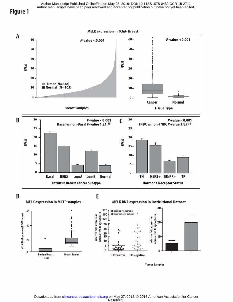
MELK is more highly expressed in breast tumors compared to normal breast tissue and is
enriched in basal-like and triple-negative breast cancers
Our previous work using gene expression profiling to identify differentially expressed kinases
between ER-positive and ER-negative tumors identified MELK as one of the most differentially
expressed kinases in ER-negative breast cancer (9). In this study we sought to further examine
the association between MELK expression and the intrinsic subtypes of breast cancer by
interrogating the Cancer Genome Atlas (TCGA) breast dataset (26-28). MELK RNA expression
is significantly increased in human breast tumors compared to normal breast tissue with little to
no expression identified in normal tissues (Figure 1A). Additionally, serial analysis of gene
expression (SAGE) analysis demonstrated markedly elevated levels of MELK RNA expression
in breast cancers (as well as certain GU malignancies) and absent to near absent expression in
normal tissues suggesting a potentially favorable therapeutic index (Supplementary Figure 1)
when treating with MELK inhibitors. While MELK expression was nearly absent in normal
breast tissues, MELK expression was heterogeneous across breast tumors with approximately
20% of human tumors demonstrating markedly elevated levels of expression. Further analysis of
the TCGA dataset demonstrates that MELK expression is significantly elevated in the basal-like,
HER2-amplified, and Luminal B subtypes when compared to either the Luminal A or normal-
like subtypes (Figure 1B). Furthermore, MELK RNA expression was highest in the triple-
negative subtype of breast tumors (Figure 1C). We validated the increased expression in tumor
specimens compared to normal breast tissue (from an institutionally assembled dataset of breast
Research. on May 27, 2018. © 2016 American Association for Cancerclincancerres.aacrjournals.org Downloaded from
Author manuscripts have been peer reviewed and accepted for publication but have not yet been edited. Author Manuscript Published OnlineFirst on May 25, 2016; DOI: 10.1158/1078-0432.CCR-15-2711
MELK 5-3-2016
11
reduction mammoplasties and breast tumors) and in ER-negative breast tumors compared to ER-
positive tumors (Figure 1D-E).
MELK is more highly expressed in ER-negative breast cancer cell lines
The association of MELK expression and basal-like breast cancer was further supported in data
derived from 51 in vitro cultured human breast cancer cell lines (25). Using this data, MELK
expression was significantly higher in the largely ER-negative (Basal A and Basal B) breast
cancer cell lines compared to the ER-positive luminal breast cancer cell lines (Supplementary
Figure 2A-B). As this gene expression data indicated that MELK was more highly expressed in
ER-negative breast cancer cell lines, we chose twelve ER-positive or ER-negative breast cancer
cell lines and measured the expression of MELK RNA under basal growth conditions using qRT-
PCR. MELK expression (RNA) was again found to be significantly elevated in the ER-negative
breast cancer cell lines (P-value 0.007) as compared to the ER-positive breast cancer cell lines
(Supplementary Figure 2C).
We next verified that the protein levels of MELK were also increased in ER-negative
breast cancer cell lines. Using western blot analysis, we demonstrated that MELK was more
highly expressed in ER-negative breast cancer cell lines compared to ER-positive breast cancer
cell lines (P-value 0.02) (Supplementary Figure 3A-C). Furthermore, total MELK protein and
RNA expression levels were significantly correlated across all breast cancer cell lines with a
correlation coefficient of 0.88, P-value <0.01 (Supplementary Figure 3D). This RNA and
protein expression data was used to identify cell lines for future experimentation in these studies.
MELK expression is correlated with radioresistance in vitro
Research. on May 27, 2018. © 2016 American Association for Cancerclincancerres.aacrjournals.org Downloaded from
Author manuscripts have been peer reviewed and accepted for publication but have not yet been edited. Author Manuscript Published OnlineFirst on May 25, 2016; DOI: 10.1158/1078-0432.CCR-15-2711

MELK 5-3-2016
12
Recognizing that triple-negative breast cancers demonstrate increased levels of radiation
resistance clinically, we sought to determine what, if any, role MELK played in the
radioresistance phenotype in human tumors. We began by performing clonogenic survival
assays on 21 breast cancer cells lines chosen to represent the heterogeneity common in human
breast cancer. Doses of radiation between 1 and 6 Gy were utilized and the area under the
clonogenic survival curve (AUC) were calculated for each of the 21 breast cancer cell lines.
Higher AUC values are associated with increasing radiation resistance as higher doses of
radiation are necessary to elicit and equieffective cell killing. Additionally, MELK expression
was assessed in the same 21 breast cancer cell lines and correlation coefficients were calculated
between MELK expression levels and radiation sensitivity as assessed by clonogenic survival
assays (AUC value). This analysis demonstrated a significant correlation between MELK RNA
expression and the intrinsic radiosensitivity of the breast cancer cell lines with increasing MELK
RNA expression in the breast cancer cell lines significantly correlated with increasing
radioresistance (R2: 0.62, p-value <0.005-Figure 2A). Similarly, MELK protein expression was
even more significantly correlated with increasing radioresistance (R2: 0.88, p-value <0.001-
Figure 2B).
MELK knockdown confers radiosensitivity in triple-negative breast cancer cells
We then more fully explored the role of MELK in regulating radioresistance in ER-
negative breast cancer using two independent ER-negative breast cancer cell lines found to have
high MELK expression. Clonogenic survival assays were performed on cell lines with high
MELK expression (MDA-MD-231 and BT-549) using scrambled control siRNA
oligonucleotides or 2 independent siRNA oligonucleotides designed to inhibit MELK
Research. on May 27, 2018. © 2016 American Association for Cancerclincancerres.aacrjournals.org Downloaded from
Author manuscripts have been peer reviewed and accepted for publication but have not yet been edited. Author Manuscript Published OnlineFirst on May 25, 2016; DOI: 10.1158/1078-0432.CCR-15-2711
MELK 5-3-2016
13
expression. Clonogenic survival curves show potent and consistent radiosensitization with
MELK knockdown using these 2 independent MELK siRNA oligonucleotides in MDA-MB-231
cells with enhancement ratios of 1.55-1.62 using MELK knockdown alone (Figure 2C). For
comparison, the well-characterized radiosensitizing drug cisplatin demonstrates enhancement
ratios of 1.2-1.3 in cancer cell lines (29, 30). There was also a significant difference in the
surviving fraction after 2 Gy (the dose of daily radiation used clinically for patients treated for
breast cancer) in these cells (Supplementary Figure 4A). Confirmation in the independent
triple-negative breast cancer cell line BT-549 again showed significant radiosensitiziation
(enhancement ratios ranged from 1.48-1.52) with MELK knock-down using the same control and
MELK siRNA oligonucleotides (Figure 2D). MELK knockdown was confirmed at both the
RNA and protein level (protein levels shown in Figure 2E-F) with limited toxicity with MELK
knockdown alone in both cell lines with a significant difference in the surviving fraction after 2
Gy values (Supplementary Figure 4A-C). This radiation sensitization was also confirmed
using 2 additional siRNA contructs designed against different exons of MELK to confirm
specificity (Supplementary Figure 4D-E). To confirm that this effect was not transient and in
preparation for future in vivo xenograft experiments, shRNA constructs were generated and cell
lines were transduced to make stable, doxycycline-inducible shMELK cell lines. Utilizing these
transduced, stable MDA-MB-231 cell lines with inducible shMELK, clonogenic survival assays
were again performed after induction of MELK knockdown by doxycycline induction. As in the
siMELK experiments, MELK knockdown showed significant radiosensitization utilizing two
independent shMELK constructs with radiation enhancement ratios ranging from 1.44 and 1.52.
There was no effect on radiosensitization with doxycline alone or induction of a control non-
targeted shRNA construct (shNT- Supplementary Figure 5A).
Research. on May 27, 2018. © 2016 American Association for Cancerclincancerres.aacrjournals.org Downloaded from
Author manuscripts have been peer reviewed and accepted for publication but have not yet been edited. Author Manuscript Published OnlineFirst on May 25, 2016; DOI: 10.1158/1078-0432.CCR-15-2711

MELK 5-3-2016
14
To further investigate whether MELK kinase function, not just protein level, was
necessary for this radiosensitization phenotype, the recently published MELK inhibitor
OTSSP167 was used in clonogenic survival assays (31). As with genetic manipulation of MELK
expression using si- and shMELK constructs, pharmacologic inhibition of MELK kinase
function significantly radiosensitized MDA-MB-231 breast cancer cell lines with radiation
enhancement ratios of 1.61 and 1.68 at OTSSP167 concentrations of 100 nM and 1 µM,
respectively (Supplementary Figure 5B). Similar findings were demonstrated using the MELK
inhibitor in an independent triple-negative cell line BT549 (data not shown). Thus, not only was
MELK expression necessary for radioresistance, its kinase function was needed to confer
resistance to ionizing radiation.
MELK overexpression confers radioresistance in ER-positive breast cancer cells with low
baseline MELK expression
To demonstrate causality, we next explored whether overexpression of MELK protein in
a breast cancer cell line with low MELK expression, in this case the ER-positive breast cancer
cell lines MCF-7, would confer radioresistance. As compared to the triple-negative breast cancer
cell lines MDA-MB-231 and BT-549, MCF-7 cells are significantly more sensitive to the effects
of ionizing radiation at baseline and are considered a radiation sensitive cell line.
Overexpression of MELK protein conferred radioresistance in MCF-7 cells (Figure 2G) with a
subsequent significant increase in the SF 2Gy values. MELK protein and RNA overexpression
was confirmed using western blot and qRT-PCR analysis (Figure 2H-I). Thus, knockdown of
MELK protein expression and inhibition of MELK kinase function using the si- and shMELK
constructs and the MELK inhibitor OTSSP167 in vitro was sufficient to confer significant
Research. on May 27, 2018. © 2016 American Association for Cancerclincancerres.aacrjournals.org Downloaded from
Author manuscripts have been peer reviewed and accepted for publication but have not yet been edited. Author Manuscript Published OnlineFirst on May 25, 2016; DOI: 10.1158/1078-0432.CCR-15-2711

MELK 5-3-2016
15
radiosensitization in 2 independent radioresistant cell lines with high MELK expression.
Similarly, MELK overexpression in a radiosensitive cell line with low MELK expression was
sufficient to confer radioresistance. To confirm that the effects were consistent with
radiosensitization by MELK inhibition and were not solely a function of decreased proliferation
or increased apoptosis, we assessed the effects of MELK inhibition (genetic or pharmacologic)
or overexpression on the growth and apoptotic rates of breast cancer cell lines (Supplementary
Figure 6-8). These data indicate that MELK knockdown or inhibition had at best a modest effect
on proliferation and little effect on apoptosis rates in most cell lines examined.
GSEA analysis identifies DNA damage repair strongly as being strongly associated with
MELK expression
To gain insight into the potential mechanisms whereby MELK was contributing to
radioresistance, we performed gene set enrichment analysis (GSEA) to identify concepts
associated with MELK expression across all published gene expression datasets. MELK gene
expression was correlated to every sequenced gene in the TCGA breast dataset and genes that
were significantly positively or negatively correlated with MELK expression were retained
within the gene set. These gene lists were then inputted into GSEA analysis as previously
described (32). GSEA analysis identified that of the top 10 nominated positively-associated
concepts, 3 were DNA damage related and 4 were cell cycle regulation related (Supplementary
Figure 9A-negatively associated concepts in Supplementary Figure 9B). These include
concepts related to response to radiation induced DNA damage at 6 and 24 hours
(Supplementary Figure 9 C,D). Additionally, molecular concept mapping was performed and
demonstrates DNA damage repair and cell cycle regulation as the two most strongly nominated
Research. on May 27, 2018. © 2016 American Association for Cancerclincancerres.aacrjournals.org Downloaded from
Author manuscripts have been peer reviewed and accepted for publication but have not yet been edited. Author Manuscript Published OnlineFirst on May 25, 2016; DOI: 10.1158/1078-0432.CCR-15-2711
MELK 5-3-2016
16
biological concepts (Supplementary Figure 10A). Furthermore, Ingenuity® Pathway analysis
(IPA) identified the role of BRCA1 in DNA damage response and the G2/M DNA damage repair
checkpoint as the most significantly correlated canonical pathways (Supplementary Figure
10B) suggesting the critical role of MELK in repair of ionizing radiation induced DNA damage.
Additionally, many of the genes associated with DNA repair mediated by homologous
recombination were significantly and positively correlated MELK expression with the BRCA1-
mediated DNA repair pathway genes, including FANC family proteins, CHK1, CHK2, PLK1,
and complex B proteins most significantly involved (Supplementary Figure 10B).
dsDNA repair is inhibited by MELK knockdown or inhibition
Having identified DNA damage repair as the most significant concept in GSEA analysis,
we interrogated the role of MELK in DNA damage repair. As ionizing radiation confers lethality
through the introduction of double stranded DNA (dsDNA) breaks, we sought to assess what, if
any, role MELK played in double stranded DNA damage repair using gamma H2AX foci
formation assays. Gamma H2AX assays were performed and quantitated using two independent
methods that included manual counting of gamma H2AX foci (Figure 3A-C) or using flow
cytometry and sorting for phospho-H2AX positive cells (Figure 3D,E). MELK knockdown itself
did not significantly impact dsDNA break formation (Figure 3B,D). As expected, dsDNA
damage was significantly increased with radiation treatment alone (2 Gy) and this damage
persisted significantly longer (4 and 16 hours) in the cells with MELK knock-down (using
siRNA) compared to control transfected cells (Figure 3B,D). Thus MELK expression
significantly impacted the degree and rate of dsDNA break repair. Similarly, when we used the
previously developed targeted MELK inhibitor OTSSP167, dsDNA damage persisted
Research. on May 27, 2018. © 2016 American Association for Cancerclincancerres.aacrjournals.org Downloaded from
Author manuscripts have been peer reviewed and accepted for publication but have not yet been edited. Author Manuscript Published OnlineFirst on May 25, 2016; DOI: 10.1158/1078-0432.CCR-15-2711

MELK 5-3-2016
17
significantly longer and to a greater extent in the MELK inhibitor treated cells compared to
control vehicle treated cells at 4, 16, and 24 hours (Figure 3C,E), suggesting the kinase function
of MELK was critical for repair and resolution of dsDNA breaks.
MELK knockdown or inhibition significantly delays xenograft tumor growth in
combination with RT
Having demonstrated that MELK inhibition results in radiosensitization of multiple
breast cancer cell lines, we sought to validate these findings in a mouse xenograft model. Using
inducible shMELK constructs under doxycycline control, SCID mice were injected with
transduced MDA-MB-231 cells. After tumors reached sufficient size, shMELK expression was
induced by doxycycline in the experimental arm. There were four treatment groups that included
a control group, MELK knockdown alone, RT alone, and combination treatment (see
supplementary methods section for full details). Knock-down of MELK alone or radiation alone
resulted in decreased tumor volume growth in the xenograft model (Figure 4A) but the
combination of radiation and MELK knock-down resulted in a statistically significant
(p<0.0001) reduction in tumor growth compared to radiation alone, and a tumor volume
doubling time nearly four times as long as treatment with radiation alone (Figure 4B).
Furthermore, analysis of the enhanced combination effect at different time points demonstrated
considerable synergism between the radiation and MELK knockdown treatments
(Supplementary Table 1). The indices (R) for combination therapy with MELK inhibition and
radiation were >1, indicating a synergistic interaction between the drugs. To confirm that MELK
kinase function, and not merely expression level, was necessary for radiosensitization in vivo,
similar experiments were performed using wild type MDA-MB-231 cells injected into the
Research. on May 27, 2018. © 2016 American Association for Cancerclincancerres.aacrjournals.org Downloaded from
Author manuscripts have been peer reviewed and accepted for publication but have not yet been edited. Author Manuscript Published OnlineFirst on May 25, 2016; DOI: 10.1158/1078-0432.CCR-15-2711
MELK 5-3-2016
18
bilateral flanks of SCID mice. These mice were treated with the MELK inhibitor OTSSP167
daily via oral gavage after tumors reached sufficient size with inhibitor treatment initiated 24
hours before radiation administration. As with the MELK knockdown experiment, MELK
inhibition with the oral inhibitor OTSSP167 led to a similar synergism with radiation treatment
and marked radiosensitization (Figure 4C, Supplementary Table 1) and delay of tumor
doubling time (Figure 4D). MELK expression was assessed by qRT-PCR from xenograft
tumors harvested during the fourth week of the experiment to confirm effective targeting in the
shMELK group (Figure 4E). Additionally, treatment with the MELK inhibitor OTSSP167 did
not affect MELK expression levels in the harvested xenograft tumors (Figure 4E) suggesting
that the radiosensitivity conferred by the MELK inhibitor was related to inhibition of MELK
kinase function. Additionally, treatment with MELK inhibitor did not result in significant
toxicity in the mice, a toxicity profile consistent with reports from other groups (31). An outline
of the experimental design is depicted in Figure 4F.
MELK expression is prognostic in breast cancer and predictive of local recurrence
While our studies identified MELK as being implicated in radioresistance in vitro and in
vivo, we wanted to determine whether MELK expression was a predictor of response to ionizing
radiation and associated with poorer prognosis in breast cancer patients treated with ionizing
radiation. For these studies we analyzed the local recurrence-free survival data from several
different, publically available datasets, with patient and dataset characteristics listed in
Supplementary tables 2-3. All datasets had a minimum of 12 year follow up and local
recurrences had been tracked in these cohorts. Additionally, MELK expression levels were
available for all tumor samples. The Wang dataset included patients with lymph-node negative
Research. on May 27, 2018. © 2016 American Association for Cancerclincancerres.aacrjournals.org Downloaded from
Author manuscripts have been peer reviewed and accepted for publication but have not yet been edited. Author Manuscript Published OnlineFirst on May 25, 2016; DOI: 10.1158/1078-0432.CCR-15-2711
MELK 5-3-2016
19
breast cancer who were treated with breast conserving surgery (BCS-219 pts) or modified radical
mastectomies (MRM-67 pts) from 1980–95. These patients also received radiotherapy when
indicated (87%), but most did not receive systemic chemotherapy. In this data set, we first
divided tumors by median level of MELK expression. Kaplan-Meier analysis of the local
recurrence-free survival between the different groups (higher than median versus lower than
median expression) showed that women who had higher MELK expression had a significantly
increased risk of local recurrence, even after radiation treatment (Figure 5A). In addition, when
expression was ordered in descending order and patients were divided into quartiles based on the
level of MELK expression, a step-like increase in local recurrence was noted as the level of
MELK expression increased (Figure 5B). Similarly in the Servant dataset, which consisted of
343 early stage node-negative patients managed with BCS and treated with radiation the same
pattern of increased local recurrence after ionizing radiation was found amongst those patients
whose tumors had higher than median expression of MELK (Figure 5C). Again, when the
cohort was divided into quartiles, the increasing rates of local recurrence were noted as the levels
of MELK expression rose (Figure 5D). Thus, in keeping with our preliminary in vitro and in
vivo data, MELK expression levels were significantly and repeatedly associated with increased
rates of local recurrence after ionizing radiation.
Though Kaplan-Meier analysis in multiple datasets suggested that MELK expression may
in itself be prognostic of overall survival and predictive of response to ionizing radiation, we
performed uni- and multivariate Cox proportional hazards analysis using local recurrence as an
endpoint to determine if MELK expression was independently prognostic, including all available
clinical and biological characteristics of the tumor that may affect local control in the model.
This analysis was performed on the Servant dataset as it had the most complete and updated
Research. on May 27, 2018. © 2016 American Association for Cancerclincancerres.aacrjournals.org Downloaded from
Author manuscripts have been peer reviewed and accepted for publication but have not yet been edited. Author Manuscript Published OnlineFirst on May 25, 2016; DOI: 10.1158/1078-0432.CCR-15-2711

MELK 5-3-2016
20
clinical, pathological, and local recurrence-specific information. As expected, univariate analysis
identified multiple factors significantly associated with local recurrence risk, but in multivariable
analysis, MELK expression level, analyzed as a continuous variable, outperformed all other
prognostic clinical or pathologic variables, including the intrinsic breast cancer subtype (Table
1). Significance increased, as does the hazard ratio, when analyzed as an ordinal variable.
Discussion:
In this report we identify maternal embryonic leucine zipper kinase (MELK) as one of the most
highly differentially expressed kinases in ER-negative breast cancers as compared to ER-positive
breast cancers. Additionally, MELK is overexpressed in triple-negative breast cancer and tumors
that give rise to local recurrences, including a disproportionately high number of radiation
refractory tumors. Further analysis revealed that MELK is not normally expressed at appreciable
levels in most normal tissues, including normal breast tissue, but is more highly expressed in
breast tumors, especially ER-negative tumors, demonstrating a potentially favorable therapeutic
index when translated clinically. MELK expression is strongly correlated with sensitivity to
radiation, and inhibition of MELK expression or function leads to significant radiosensitization
in vitro and in vivo through impaired DNA damage repair. Finally, increasing MELK expression
is significantly associated with increasing rates of local failure after radiation therapy across
multiple data sets and multivariable analysis identifies MELK expression as the strongest factor
associated with poor local control. These results suggest that women whose tumors have high
MELK expression have a poor prognosis and derive less benefit from radiation therapy and
therefore may benefit from more aggressive treatment. In addition, this study identifies MELK
itself as a potential target for the treatment in triple-negative breast cancer.
Research. on May 27, 2018. © 2016 American Association for Cancerclincancerres.aacrjournals.org Downloaded from
Author manuscripts have been peer reviewed and accepted for publication but have not yet been edited. Author Manuscript Published OnlineFirst on May 25, 2016; DOI: 10.1158/1078-0432.CCR-15-2711
MELK 5-3-2016
21
Triple-negative breast cancer has consistently been shown to portend a poorer response to
radiation therapy, increased rates of local recurrence, and an overall poorer prognosis (6, 33).
Recent efforts have sought to identify actionable targets in triple-negative breast cancer,
including those that may radiosensitize these tumors. A number of inhibitors of peptide growth
factor pathways, such as epidermal growth factor receptor (EGFR), the insulin-like growth factor
receptor (IGFR), fibroblast growth factor receptors (FGFR), vascular endothelial growth factor
(VEGF) pathways, have been studied in numerous clinical trials with limited success (34-36).
Additional targeted therapies, including inhibitors of PI3-kinase, PARP-1, CHK1, AR, and Src,
are in various stages of clinical trials in breast cancer. Though these therapies may prove
effective in treating subsets of women with breast cancer, it is clear that additional therapies are
critically needed. Many of these therapies have side effects that limit their clinical utility, and
the problem of drug resistance remains a substantial limitation to their use. Additionally, though
these targets hold promise for the treatment of tumors that express the aforementioned markers,
many ER-negative tumors do not express any of these targets. These studies credential MELK as
a possible additional target for the more effective treatment, with an expression pattern that
suggests a favorable therapeutic index.
While these results show radiosensitization of TN and basal-like breast cancers with the
inhibition of MELK both in vitro and in vivo, the mechanism of radiosensitization remains to be
fully elucidated. Previous groups have demonstrated that in gliomas, MELK knockdown leads to
cellular senescence, cell cycle arrest, and increased replicative stress secondary to the increase in
dsDNA breaks (37). This is mediated, in part, by p21 whose expression is increased by MELK
depletion. This, in turn, activates ATM, Chk2, and p53 sequentially causing cell cycle arrest and
accumulation of DNA damage at stalled replication forks. This same group subsequently
Research. on May 27, 2018. © 2016 American Association for Cancerclincancerres.aacrjournals.org Downloaded from
Author manuscripts have been peer reviewed and accepted for publication but have not yet been edited. Author Manuscript Published OnlineFirst on May 25, 2016; DOI: 10.1158/1078-0432.CCR-15-2711
MELK 5-3-2016
22
demonstrated a similar mechanism when treating with a novel MELK inhibitor (upregulation of
p21 leading to activation of ATM, Chk2, and p53) allowing the cancerous cells to continue
proliferation in the presence of replicative stress (37, 38). It is unclear what, if any, role this
mechanism plays in radioresistance of triple-negative and basal-like breast cancers as the vast
majority of these tumors, and all of the cell lines used in this study, harbor p53 mutations. More
interestingly, however, is the observation that MELK knockdown or inhibition leads to cell cycle
arrest in early S phase or in G2/M (39). As these phases correspond to time when cells are most
sensitive to the effects of ionizing radiation, this may be a mechanism whereby MELK inhibition
leads to radiosensitization. Furthermore it remains to be seen, what, if any role MELK plays on
impacting non homologous end joining or homologous recombination to impact DNA damage
repair. A more complete understanding of the mechanism by which MELK contributes to the
radioresistance phenotype will allow for the rationale design of strategies to interfere with this
resistance and is the subject of ongoing investigation.
Given the lack of targeted agents for triple-negative disease and their relative radiation
insensitivity, as evidenced by their increased locoregional recurrence risk, it is clear that
additional targets for radiosensitization are critically needed, including those that are selective
for triple-negative breast cancers. This report identifies MELK as one such targetable kinase.
MELK represents an ideal molecular target as it demonstrates many of the characteristics
necessary for effective targeted therapies. MELK protein is expressed in cancerous tissue but not
normal tissue suggesting an ideal candidate with a broad therapeutic window. Its kinase function,
which in this report was shown to be necessary for the effective repair of radiation-induced DNA
damage, is imminently targetable as the crystal structure of the MELK has already been
established (40, 41). Furthermore, MELK inhibitors have already been developed and have
Research. on May 27, 2018. © 2016 American Association for Cancerclincancerres.aacrjournals.org Downloaded from
Author manuscripts have been peer reviewed and accepted for publication but have not yet been edited. Author Manuscript Published OnlineFirst on May 25, 2016; DOI: 10.1158/1078-0432.CCR-15-2711
MELK 5-3-2016
23
shown efficacy in in vitro and in vivo model systems, and additional inhibitors are currently in
various stages of development (31). Additionally, MELK expression is high enough in breast
cancers, especially triple-negative breast cancers, to be clinically relevant, and as demonstrated
in this report, it serves as both a prognostic and predictive biomarker in predicting response to
radiation treatment. Finally, MELK functionally plays a role in several of the processes that are
‘hallmarks’ of cancer including proliferation, migration, invasion, cell cycle regulation, and
DNA damage repair. Thus, while there has been a relative absence of clinically available
radiation sensitizers in women with treatment refractory or triple-negative breast cancer, MELK
represents a novel therapeutic target that holds promise for the more effective treatment of this
deadly disease.
Acknowledgements:
We would also like to acknowledge Steven Kronenberg for his expertise in preparing the figures
for publication.
Funding: This work was supported in part by grants from the RSNA resident research
fellowship RR1349, ASCO Conquer Cancer Foundation Young Investigator Award in Memory
of Evelyn H. Lauder, Fashion Footwear Charitable Foundation of New York/QVC Presents
Shoes on Sale ™, and the Breast Cancer Research Foundation (BCRF).
Conflicts: The authors declare no conflict of interest in these studies.
Research. on May 27, 2018. © 2016 American Association for Cancerclincancerres.aacrjournals.org Downloaded from
Author manuscripts have been peer reviewed and accepted for publication but have not yet been edited. Author Manuscript Published OnlineFirst on May 25, 2016; DOI: 10.1158/1078-0432.CCR-15-2711
MELK 5-3-2016
24
References
1. Darby S, McGale P, Correa C, Taylor C, Arriagada R, Clarke M, et al. Effect of radiotherapy after breast-conserving surgery on 10-year recurrence and 15-year breast cancer death: meta-analysis of individual patient data for 10,801 women in 17 randomised trials. Lancet. 2011;378:1707-16. 2. Clarke M, Collins R, Darby S, Davies C, Elphinstone P, Evans V, et al. Effects of radiotherapy and of differences in the extent of surgery for early breast cancer on local recurrence and 15-year survival: an overview of the randomised trials. Lancet. 2005;366:2087-106. 3. Nguyen PL, Taghian AG, Katz MS, Niemierko A, Abi Raad RF, Boon WL, et al. Breast cancer subtype approximated by estrogen receptor, progesterone receptor, and HER-2 is associated with local and distant recurrence after breast-conserving therapy. J Clin Oncol. 2008;26:2373-8. 4. Kyndi M, Sorensen FB, Knudsen H, Overgaard M, Nielsen HM, Overgaard J. Estrogen receptor, progesterone receptor, HER-2, and response to postmastectomy radiotherapy in high-risk breast cancer: the Danish Breast Cancer Cooperative Group. J Clin Oncol. 2008;26:1419-26. 5. Wang Y, Yin Q, Yu Q, Zhang J, Liu Z, Wang S, et al. A retrospective study of breast cancer subtypes: the risk of relapse and the relations with treatments. Breast Cancer Res Treat. 2011;130:489-98. 6. von Minckwitz G, Untch M, Blohmer JU, Costa SD, Eidtmann H, Fasching PA, et al. Definition and impact of pathologic complete response on prognosis after neoadjuvant chemotherapy in various intrinsic breast cancer subtypes. J Clin Oncol. 2012;30:1796-804. 7. Cortazar P, Zhang L, Untch M, Mehta K, Costantino JP, Wolmark N, et al. Pathological complete response and long-term clinical benefit in breast cancer: the CTNeoBC pooled analysis. The Lancet. 2014;384:164-72. 8. Lowery AJ, Kell MR, Glynn RW, Kerin MJ, Sweeney KJ. Locoregional recurrence after breast cancer surgery: a systematic review by receptor phenotype. Breast Cancer Res Treat. 2012;133:831-41. 9. Speers C, Tsimelzon A, Sexton K, Herrick AM, Gutierrez C, Culhane A, et al. Identification of novel kinase targets for the treatment of estrogen receptor-negative breast cancer. Clin Cancer Res. 2009;15:6327-40. 10. Lizcano JM, Goransson O, Toth R, Deak M, Morrice NA, Boudeau J, et al. LKB1 is a master kinase that activates 13 kinases of the AMPK subfamily, including MARK/PAR-1. EMBO J. 2004;23:833-43. 11. Wang Y, Lee YM, Baitsch L, Huang A, Xiang Y, Tong H, et al. MELK is an oncogenic kinase essential for mitotic progression in basal-like breast cancer cells. eLife. 2014;3:e01763. 12. Suzuki A, Kusakai G, Kishimoto A, Lu J, Ogura T, Esumi H. ARK5 suppresses the cell death induced by nutrient starvation and death receptors via inhibition of caspase 8 activation, but not by chemotherapeutic agents or UV irradiation. Oncogene. 2003;22:6177-82. 13. Kato K, Ogura T, Kishimoto A, Minegishi Y, Nakajima N, Miyazaki M, et al. Critical roles of AMP-activated protein kinase in constitutive tolerance of cancer cells to nutrient deprivation and tumor formation. Oncogene. 2002;21:6082-90. 14. Nakano I, Paucar AA, Bajpai R, Dougherty JD, Zewail A, Kelly TK, et al. Maternal embryonic leucine zipper kinase (MELK) regulates multipotent neural progenitor proliferation. J Cell Biol. 2005;170:413-27.
Research. on May 27, 2018. © 2016 American Association for Cancerclincancerres.aacrjournals.org Downloaded from
Author manuscripts have been peer reviewed and accepted for publication but have not yet been edited. Author Manuscript Published OnlineFirst on May 25, 2016; DOI: 10.1158/1078-0432.CCR-15-2711
MELK 5-3-2016
25
15. Pickard MR, Green AR, Ellis IO, Caldas C, Hedge VL, Mourtada-Maarabouni M, et al. Dysregulated expression of Fau and MELK is associated with poor prognosis in breast cancer. Breast Cancer Res. 2009;11:R60. 16. Komatsu M, Yoshimaru T, Matsuo T, Kiyotani K, Miyoshi Y, Tanahashi T, et al. Molecular features of triple negative breast cancer cells by genome-wide gene expression profiling analysis. Int J Oncol. 2013;42:478-506. 17. Rajkumar T, Sabitha K, Vijayalakshmi N, Shirley S, Bose MV, Gopal G, et al. Identification and validation of genes involved in cervical tumourigenesis. BMC Cancer. 2011;11:80. 18. Lin ML, Park JH, Nishidate T, Nakamura Y, Katagiri T. Involvement of maternal embryonic leucine zipper kinase (MELK) in mammary carcinogenesis through interaction with Bcl-G, a pro-apoptotic member of the Bcl-2 family. Breast Cancer Res. 2007;9:R17. 19. Gray D, Jubb AM, Hogue D, Dowd P, Kljavin N, Yi S, et al. Maternal embryonic leucine zipper kinase/murine protein serine-threonine kinase 38 is a promising therapeutic target for multiple cancers. Cancer Res. 2005;65:9751-61. 20. Lu C, Speers C, Zhang Y, Xu X, Hill J, Steinbis E, et al. Effect of epidermal growth factor receptor inhibitor on development of estrogen receptor-negative mammary tumors. J Natl Cancer Inst. 2003;95:1825-33. 21. Matar P, Rojo F, Cassia R, Moreno-Bueno G, Di Cosimo S, Tabernero J, et al. Combined epidermal growth factor receptor targeting with the tyrosine kinase inhibitor gefitinib (ZD1839) and the monoclonal antibody cetuximab (IMC-C225): superiority over single-agent receptor targeting. Clin Cancer Res. 2004;10:6487-501. 22. Wei D, Li H, Yu J, Sebolt JT, Zhao L, Lawrence TS, et al. Radiosensitization of human pancreatic cancer cells by MLN4924, an investigational NEDD8-activating enzyme inhibitor. Cancer Res. 2012;72:282-93. 23. Servant N, Bollet MA, Halfwerk H, Bleakley K, Kreike B, Jacob L, et al. Search for a gene expression signature of breast cancer local recurrence in young women. Clin Cancer Res. 2012;18:1704-15. 24. Wang Y, Klijn JG, Zhang Y, Sieuwerts AM, Look MP, Yang F, et al. Gene-expression profiles to predict distant metastasis of lymph-node-negative primary breast cancer. Lancet. 2005;365:671-9. 25. Neve RM, Chin K, Fridlyand J, Yeh J, Baehner FL, Fevr T, et al. A collection of breast cancer cell lines for the study of functionally distinct cancer subtypes. Cancer Cell. 2006;10:515-27. 26. Comprehensive molecular portraits of human breast tumours. Nature. 2012;490:61-70. 27. Prat A, Perou CM. Deconstructing the molecular portraits of breast cancer. Molecular Oncology. 2011;5:5-23. 28. Perou CM, Sorlie T, Eisen MB, van de Rijn M, Jeffrey SS, Rees CA, et al. Molecular portraits of human breast tumours. Nature. 2000;406:747-52. 29. Skov K, Macphail S. Interaction of platinum drugs with clinically relevant X-ray doses in mammalian cells: A comparison of cisplatin, carboplatin, iproplatin, and tetraplatin. International Journal of Radiation Oncology*Biology*Physics. 1991;20:221-5. 30. Zhang X, Yang H, Gu K, Chen J, Rui M, Jiang G-L. In vitro and in vivo study of a nanoliposomal cisplatin as a radiosensitizer. International Journal of Nanomedicine. 2011;6:437-44.
Research. on May 27, 2018. © 2016 American Association for Cancerclincancerres.aacrjournals.org Downloaded from
Author manuscripts have been peer reviewed and accepted for publication but have not yet been edited. Author Manuscript Published OnlineFirst on May 25, 2016; DOI: 10.1158/1078-0432.CCR-15-2711
MELK 5-3-2016
26
31. Chung S, Suzuki H, Miyamoto T, Takamatsu N, Tatsuguchi A, Ueda K, et al. Development of an orally-administrative MELK-targeting inhibitor that suppresses the growth of various types of human cancer. Oncotarget. 2012;3:1629-40. 32. Zhao S, Chang SL, Linderman JJ, Feng FY, Luker GD. A Comprehensive Analysis of CXCL12 Isoforms in Breast Cancer. Translational oncology. 2014. 33. Dent R, Trudeau M, Pritchard KI, Hanna WM, Kahn HK, Sawka CA, et al. Triple-negative breast cancer: clinical features and patterns of recurrence. Clin Cancer Res. 2007;13:4429-34. 34. Fong PC, Boss DS, Yap TA, Tutt A, Wu P, Mergui-Roelvink M, et al. Inhibition of poly(ADP-ribose) polymerase in tumors from BRCA mutation carriers. N Engl J Med. 2009;361:123-34. 35. Tutt A, Robson M, Garber JE, Domchek SM, Audeh MW, Weitzel JN, et al. Oral poly(ADP-ribose) polymerase inhibitor olaparib in patients with BRCA1 or BRCA2 mutations and advanced breast cancer: a proof-of-concept trial. Lancet. 2010;376:235-44. 36. O'Shaughnessy J, Schwartzberg L, Danso MA, Miller KD, Rugo HS, Neubauer M, et al. Phase III study of iniparib plus gemcitabine and carboplatin versus gemcitabine and carboplatin in patients with metastatic triple-negative breast cancer. J Clin Oncol. 2014;32:3840-7. 37. Kig C, Beullens M, Beke L, Van Eynde A, Linders JT, Brehmer D, et al. Maternal embryonic leucine zipper kinase (MELK) reduces replication stress in glioblastoma cells. J Biol Chem. 2013;288:24200-12. 38. Beke L, Kig C, Linders JT, Boens S, Boeckx A, van Heerde E, et al. MELK-T1, a small-molecule inhibitor of protein kinase MELK, decreases DNA-damage tolerance in proliferating cancer cells. Bioscience reports. 2015;35. 39. Jiang P, Zhang D. Maternal embryonic leucine zipper kinase (MELK): a novel regulator in cell cycle control, embryonic development, and cancer. International journal of molecular sciences. 2013;14:21551-60. 40. Cho YS, Kang Y, Kim K, Cha YJ, Cho HS. The crystal structure of MPK38 in complex with OTSSP167, an orally administrative MELK selective inhibitor. Biochem Biophys Res Commun. 2014;447:7-11. 41. Cho YS, Yoo J, Park S, Cho HS. The structures of the kinase domain and UBA domain of MPK38 suggest the activation mechanism for kinase activity. Acta crystallographica Section D, Biological crystallography. 2014;70:514-21.
Research. on May 27, 2018. © 2016 American Association for Cancerclincancerres.aacrjournals.org Downloaded from
Author manuscripts have been peer reviewed and accepted for publication but have not yet been edited. Author Manuscript Published OnlineFirst on May 25, 2016; DOI: 10.1158/1078-0432.CCR-15-2711
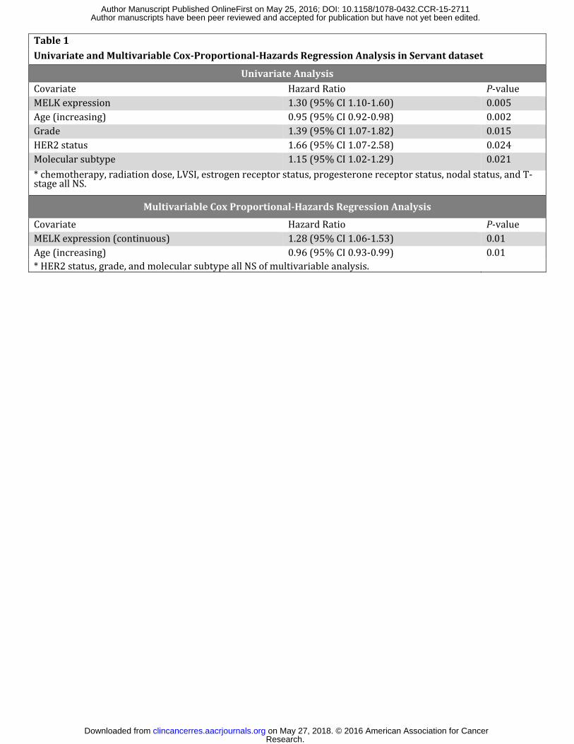
Table 1 Univariate and Multivariable Cox-Proportional-Hazards Regression Analysis in Servant dataset
Univariate Analysis Covariate Hazard Ratio P-value MELK expression 1.30 (95% CI 1.10-1.60) 0.005 Age (increasing) 0.95 (95% CI 0.92-0.98) 0.002 Grade 1.39 (95% CI 1.07-1.82) 0.015 HER2 status 1.66 (95% CI 1.07-2.58) 0.024 Molecular subtype 1.15 (95% CI 1.02-1.29) 0.021 * chemotherapy, radiation dose, LVSI, estrogen receptor status, progesterone receptor status, nodal status, and T-stage all NS. Multivariable Cox Proportional-Hazards Regression Analysis Covariate Hazard Ratio P-value MELK expression (continuous) 1.28 (95% CI 1.06-1.53) 0.01 Age (increasing) 0.96 (95% CI 0.93-0.99) 0.01 * HER2 status, grade, and molecular subtype all NS of multivariable analysis.
Research. on May 27, 2018. © 2016 American Association for Cancerclincancerres.aacrjournals.org Downloaded from
Author manuscripts have been peer reviewed and accepted for publication but have not yet been edited. Author Manuscript Published OnlineFirst on May 25, 2016; DOI: 10.1158/1078-0432.CCR-15-2711

1
Figure 1: MELK is more highly expressed in cancerous tissue and triple-negative breast
cancer. Analysis of the cancer genome atlas (TCGA) breast dataset demonstrates MELK
expression is significantly higher in breast tumors (in red) compared to normal breast tissue (in
green) with FPKM values on the y-axis and individual tumor samples from cancer vs. normal on
the x-axis (A) Error bars represent +/- SD. MELK expression is also significantly elevated in
basal-like and triple-negative breast cancers in the TCGA dataset (B and C). Error bars represent
+/- SEM. The expression of MELK in 180 breast normal and tumor samples (22 reduction
mammoplasty normal and 158 tumors) was measured using RNA-sequencing analysis from the
University of Michigan Translational Pathology databank (D). Data is depicted as absolute
RPKM values. Differential expression between ER-negative and ER-positive human breast
tumors was also confirmed in an institutional breast tumor database using qRT-PCR analysis (E).
Expression is depicted normalized to control with error bars representing +/- SEM.
Figure 2: MELK expression is associated with radioresistance. Intrinsic radiosensitivity of
21 breast cancer cell lines (as measured by clonogenic survival assay area under the survival
curve-AUC) was assessed and correlated to MELK RNA expression using Pearson’s correlation.
Each dot represents an individual cell line with colors corresponding to intrinsic subtype. Mean
centered log2 RNA expression is depicted on the x-axis and the survival AUC from clonogenic
survival assays are depicted on the y-axis (A). Intrinsic radiosensitivity was also correlated to
MELK protein expression in 15 breast cancer cell lines with MELK expression relative to the
expression in MCF-7 cells as determined by western blotting (B). Using siRNA knockdown of
MELK expression in a radiation resistant breast cancer cell lines with high baseline MELK
expression (MDA-MB-231 and BT549), radiation sensitivity was assessed using clonogenic
Research. on May 27, 2018. © 2016 American Association for Cancerclincancerres.aacrjournals.org Downloaded from
Author manuscripts have been peer reviewed and accepted for publication but have not yet been edited. Author Manuscript Published OnlineFirst on May 25, 2016; DOI: 10.1158/1078-0432.CCR-15-2711
2
survival assays. Knock down of MELK expression confers radiation sensitivity with limited
toxicity with an enhancement ratio (ER) of 1.55-1.62 in MDA-MD-231 cells (C) and 1.48-1.52
in BT549 cells (D). MELK knockdown was confirmed in both experiments using western
blotting for MELK expression (E, F). MELK overexpression in the radiosensitive ER-positive
breast cancer cell line MCF-7, with low baseline MELK expression and high radiosensitivity
confers radioresistance (G). MELK overexpression was confirmed at the protein and RNA level
(H,I). All experiments were repeated in triplicate with error bars +/- SEM.
Figure 3: MELK knock-down significantly delays repair of double stranded DNA (dsDNA)
breaks after ionizing radiation. Representative images of γH2AX foci at 16 hrs. are depicted
in (A). Using siRNA directed against MELK, the effect of MELK knock down on γH2AX foci
formation (B) or fluorescence staining by flow cytometry (D) was evaluated at various times (30
min, 4 hr., 16 hr., 24 hr.) after 2 Gy of ionizing radiation. The effect on dsDNA break repair
caused by inhibition of MELK kinase function using the MELK inhibitor OTSSP167 was also
assessed in time course by foci formation (C) or flow cytometry (E). Each experiment was run
in triplicate three independent times. Similar results were found using the cell line BT549 (data
not shown). Error bars represent SD.
Figure 4: MELK inhibition significantly reduces xenograft tumor doubling time compared
to radiation alone. Knock-down of MELK alone or radiation alone resulted in decreased tumor
volume growth in the xenograft model (A) but the combination of radiation and MELK knock-
down resulted in a synergistic and statistically significant (p<0.01) reduction in tumor growth
compared to radiation alone, and a tumor volume doubling time nearly four times as long as
Research. on May 27, 2018. © 2016 American Association for Cancerclincancerres.aacrjournals.org Downloaded from
Author manuscripts have been peer reviewed and accepted for publication but have not yet been edited. Author Manuscript Published OnlineFirst on May 25, 2016; DOI: 10.1158/1078-0432.CCR-15-2711

3
treatment with radiation alone (B). A similar experiment was performed using wild type MDA-
MB-231 cells injected as above but this time treatment was with the MELK inhibitor OTSSP167
treated at 10 mg/kg daily by oral gavage. While radiation and MELK inhibitor alone did delay
tumor doubling time slightly, combination therapy was significantly more effective at delay
tumor growth and doubling time (C-D). MELK expression was assessed by qRT-PCR from
xenograft tumors harvested during the fourth week of the experiment (E). A depiction of the
experimental design is shown (F). Error bars represent +SEM.
Figure 5: MELK expression is associated with increased risk of local recurrence. Kaplan
Meier local recurrence free survival analysis in the Wang dataset demonstrates that patients
whose tumors have higher than median expression of MELK have significantly higher rates of
local recurrence after radiation and an overall poorer prognosis than patients with lower than
median expression of MELK (HR for local recurrence 2.22, p-value <0.001 (A). Even when
divided into quartiles, increasing levels of MELK expression are associated with increased risk
of LR (B). Similarly, in the Servant dataset, Kaplan Meier local recurrence free survival analysis
demonstrates that patients whose tumors have high expression of MELK have significantly
higher rates of LR (C). Again, quartile expression of MELK demonstrates increasing rates of LR
with increasing MELK expression (D).
Table 1- Univariate and Multivariable Analysis identifies MELK as the variable most
strongly associated with local recurrence in early stage breast cancer patients treated with
adjuvant radiation. Univariate analysis identifies several clinical and pathologic factors
associated with local recurrence. In multivariable cox proportional-hazards regression analysis of
Research. on May 27, 2018. © 2016 American Association for Cancerclincancerres.aacrjournals.org Downloaded from
Author manuscripts have been peer reviewed and accepted for publication but have not yet been edited. Author Manuscript Published OnlineFirst on May 25, 2016; DOI: 10.1158/1078-0432.CCR-15-2711
4
all patients, only MELK expression (continuous variable) remained significantly associated with
worse local recurrence-free (LRF) survival. Hazards ratios and 95% confidence intervals were
calculated for all analyses and are listed.
Research. on May 27, 2018. © 2016 American Association for Cancerclincancerres.aacrjournals.org Downloaded from
Author manuscripts have been peer reviewed and accepted for publication but have not yet been edited. Author Manuscript Published OnlineFirst on May 25, 2016; DOI: 10.1158/1078-0432.CCR-15-2711
AMELK expression in TCGA- Breast
FPKM
0
10
20
30
40
50
60
Tumor (N=838)Normal (N=105)
0
10
20
30
40
50
60
FPKM
Cancer Normal
0
5
10
15
20
25
30
FPKM
Basal HER2 LumA LumB Normal
P-value <0.001
0
5
10
15
20
25
30FP
KM
TN HER2+ ER/PR+ TP
P-value <0.001
Intrinsic Breast Cancer Subtype Hormone Receptor Status
P-value <0.001 P-value <0.001
Breast Samples Tissue Type
TNBC vs non-TNBC P-value 3.85-31B
D
CBasal vs non-Basal P-value 1.21-45
60
40
20
0Benign Breast
TissueBreast Tumor
MELK
RNA E
xpre
ssion
(RPK
M va
lues
)
MELK expression in MCTP samples
0
10
20
30
rela
tive
fold
exp
ress
ion
norm
alize
d to
cyclo
phili
n
05
1015204570
125150175
MELK RNA expression in Institutional Dataset
rela
tive
fold
exp
ress
ion
norm
alize
d to
cyclo
phili
n
ER-Positive ER-Negative
Tumor Samples
ER-positive = 52 samplesER-negative = 46 samples
E
Figure 1
Research. on May 27, 2018. © 2016 American Association for Cancerclincancerres.aacrjournals.org Downloaded from
Author manuscripts have been peer reviewed and accepted for publication but have not yet been edited. Author Manuscript Published OnlineFirst on May 25, 2016; DOI: 10.1158/1078-0432.CCR-15-2711
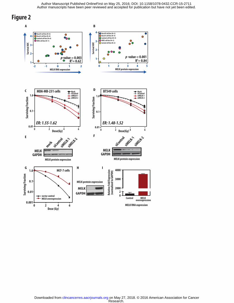
Figure 2A
4
3
2
10-1-2 1 2
R2= 0.62p-value = 0.003
MELK RNA expression
Surv
ival
AUC
Basal B cell line (N=6)Basal A cell line (N=8)Luminal cell line (N=6)HER2 cell line (N=1)
MELK protein expression
G H I
MELKGAPDH
02
2000
3000
4000
Rela
tive F
old
Expr
essio
n(n
orm
alize
d to
GAP
DH)
Control MELK overexpression
MELK RNA expressionDose (Gy)
Surv
ivin
g Fr
actio
n
MCF-7 cells
0 2 4 6
1.0
0.1
0.001
0.01
MELK overexpressionvector control
Dose(Gy)
Surv
ivin
g Fr
actio
n
ER: 1.48-1.52
BT549 cells1.0
0.1
0.010 2 4 6
siControlsiMELK-1siMELK-2
Mock
mock
siMELK-1
GAPDHMELK
siContro
l
siMELK-2
siMELK-1
GAPDHMELK
siContro
l
siMELK-2
C
E
D
FDose(Gy)
Surv
ivin
g Fr
actio
n
ER: 1.55-1.62
MDA-MB-231 cells1.0
0.1
0.010 2 4 6
siControlsiMELK-1siMELK-2
Mock
4
3
2
1
210 3 4
p -value = 0.001
MELK protein expression
Surv
ival
AUC
Basal B cell line (N=3)Basal A cell line (N=4)Luminal cell line (N=4)HER2 cell line (N=2)
B
5
normal cell line (N=2)
MELK protein expression MELK protein expression
R2= 0.84
Research. on May 27, 2018. © 2016 American Association for Cancerclincancerres.aacrjournals.org Downloaded from
Author manuscripts have been peer reviewed and accepted for publication but have not yet been edited. Author Manuscript Published OnlineFirst on May 25, 2016; DOI: 10.1158/1078-0432.CCR-15-2711
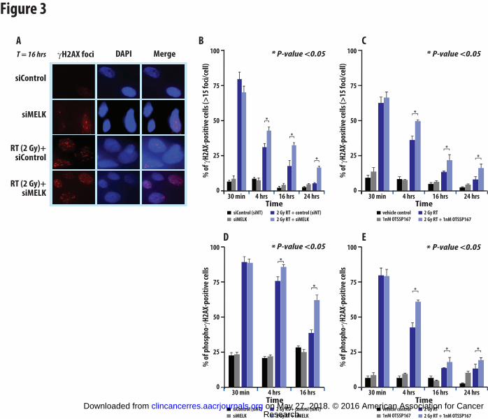
Figure 3
siControl
siMELK
RT (2 Gy)+siControl
RT (2 Gy)+siMELK
γH2AX foci DAPI MergeT = 16 hrs
A
100
75
50
25
0
100
75
50
25
04 hrs 16 hrs
% o
f ph
osph
o-γH
2 AX-
posi
tive
cel
ls
% o
f ph
osph
o-γH
2 AX-
posi
tive
cel
ls
siControl (siNT)
siMELK
2 Gy RT + control (siNT)
2 Gy RT + siMELK
Time Timevehicle control
1nM OTSSP167
2 Gy RT
2 Gy RT + 1nM OTSSP167
30 min 30 min 4 hrs 16 hrs 24 hrs
*
D E
*
**
*
100
75
50
25
0
Timevehicle control
1nM OTSSP167
2 Gy RT
2 Gy RT + 1nM OTSSP167
30 min 4 hrs 16 hrs 24 hrs
C
% o
f γH
2AX -
posi
tive
cel
ls (>
15 fo
ci/c
ell)
% o
f γH
2AX -
posi
tive
cel
ls (>
15 fo
ci/c
ell)
siControl (siNT)
siMELK
2 Gy RT + control (siNT)
2 Gy RT + siMELK
Time
*
*
*
100
75
50
25
0
* P-value <0.05
30 min 4 hrs 16 hrs 24 hrs
B
*
*
*
* P-value <0.05
* P-value <0.05
* P-value <0.05
Research. on May 27, 2018. © 2016 American Association for Cancerclincancerres.aacrjournals.org Downloaded from
Author manuscripts have been peer reviewed and accepted for publication but have not yet been edited. Author Manuscript Published OnlineFirst on May 25, 2016; DOI: 10.1158/1078-0432.CCR-15-2711

Figure 4
A
Perc
ent o
f tum
ors t
hat
have
not
dou
bled
in si
ze
0 10 20 30 40
100
80
60
40
20
0
Control
RTMELK Inhibitor
Combination
Median time to tumor volume doubling (days)
6.715.1
8.425.2
*p< 0.0001
*
Rela
tive
tum
or vo
lum
efr
om in
itial
(%)
Time (days)0 5 10 15 20 25 30 35
0
1000
2000
3000
4000
40 45
Control shMELK -doxRTMELK knock-downCombination
* *
*
*p< 0.01
B
Perc
ent o
f tum
ors t
hat
have
not
dou
bled
in si
ze
0 10 20 30 40 50
100
80
60
40
20
0
Control
RTMELK knock-down
Combination
Median time to tumor volume doubling (days)
6.714.3
7.929.1
*p< 0.0001
*
E F
Treat with 6 fractions of 2 Gy
Inject transduced MDA-MB-231 cells containing inducible shMELK inder dox control
Continue dox treatment or MELK inhibition and measure
tumor growth
Induce exp. arms with dox 48 hrsTreat with MELK inhibitor 24 hrs
Allow tumors to grow to 100 mm3
1.5
1.0
0.5
0
Rela
tive F
old
Chan
ge
MELK RNA expresion level(qRT-PCR)
doxradiation (2 Gy)
OTSSP167
---
+ - - --
-- - -
+++
+++
- -
Control shNT +dox
C DControl placeboRTMELK inhibitorCombination
Time (days)0 5 10 15 20 25
* *
30 350
1000
2000
3000
4000
40 45
*p< 0.01
Rela
tive
tum
or vo
lum
efr
om in
itial
(%)
*
Research. on May 27, 2018. © 2016 American Association for Cancerclincancerres.aacrjournals.org Downloaded from
Author manuscripts have been peer reviewed and accepted for publication but have not yet been edited. Author Manuscript Published OnlineFirst on May 25, 2016; DOI: 10.1158/1078-0432.CCR-15-2711
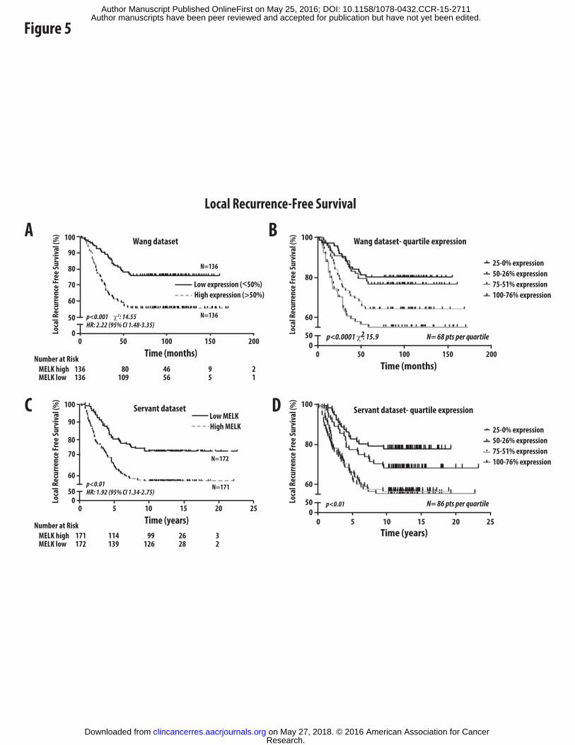
Figure 5
Local Recurrence-Free Survival
A B
C D
MELK highMELK low
)%( lavivruS eerF ecnerruceR lacoL
Servant dataset
1050 15 20 250
50
60
80
100
Time (years)
High MELKLow MELK
N=172
N=171p<0.01HR: 1.92 (95% CI 1.34-2.75)
N=136
N=136
p<0.001 χ2: 14.55HR: 2.22 (95% CI 1.48-3.35)
p<0.0001 χ2: 15.9
Number at Risk171172
114139
99126
26 28
32
MELK highMELK low
Number at Risk136136
80109
4656
95
21
)%( lavivruS eerF ecnerruceR lacoL
Servant dataset- quartile expression
p<0.01
0 5 10 15 20 250
50
60
80
100
Time (years)
N= 86 pts per quartile
100-76% expression75-51% expression50-26% expression25-0% expression
)%( lavivruS eerF ecnerruceR lacoL
100500 150 2000
50
60
80
100
90
70
90
70
Time (months)
Wang dataset
High expression (>50%)Low expression (<50%)
Wang dataset- quartile expression
)%( lavivruS eerF ecnerruceR lacoL
050
60
80
100
Time (months)
N= 68 pts per quartile
100-76% expression75-51% expression50-26% expression25-0% expression
100500 150 200
Research. on May 27, 2018. © 2016 American Association for Cancerclincancerres.aacrjournals.org Downloaded from
Author manuscripts have been peer reviewed and accepted for publication but have not yet been edited. Author Manuscript Published OnlineFirst on May 25, 2016; DOI: 10.1158/1078-0432.CCR-15-2711
Published OnlineFirst May 25, 2016.Clin Cancer Res Corey Speers, Shuang G. Zhao, Vishal Kothari, et al. cancermediator and biomarker of radioresistance in human breast Maternal embryonic leucine zipper kinase (MELK) as a novel
Updated version
10.1158/1078-0432.CCR-15-2711doi:
Access the most recent version of this article at:
Material
Supplementary
http://clincancerres.aacrjournals.org/content/suppl/2016/05/25/1078-0432.CCR-15-2711.DC1
Access the most recent supplemental material at:
Manuscript
Authoredited. Author manuscripts have been peer reviewed and accepted for publication but have not yet been
E-mail alerts related to this article or journal.Sign up to receive free email-alerts
Subscriptions
Reprints and
To order reprints of this article or to subscribe to the journal, contact the AACR Publications
Permissions
Rightslink site. Click on "Request Permissions" which will take you to the Copyright Clearance Center's (CCC)
.http://clincancerres.aacrjournals.org/content/early/2016/05/25/1078-0432.CCR-15-2711To request permission to re-use all or part of this article, use this link
Research. on May 27, 2018. © 2016 American Association for Cancerclincancerres.aacrjournals.org Downloaded from
Author manuscripts have been peer reviewed and accepted for publication but have not yet been edited. Author Manuscript Published OnlineFirst on May 25, 2016; DOI: 10.1158/1078-0432.CCR-15-2711