Embed Size (px)
Citation preview

Methods xxx (2014) xxx–xxx
Contents lists available at ScienceDirect
Methods
journal homepage: www.elsevier .com/locate /ymeth
Membrane association of the PTEN tumor suppressor:Neutron scattering and MD simulations reveal the structureof protein–membrane complexes
http://dx.doi.org/10.1016/j.ymeth.2014.10.0141046-2023/� 2014 Elsevier Inc. All rights reserved.
⇑ Corresponding author at: Department of Physics, Carnegie Mellon University,5000 Forbes Ave., Pittsburgh, PA 15213-3890, USA. Fax: +1 (412) 268 8252.
E-mail address: [email protected] (M. Lösche).
Please cite this article in press as: H. Nanda et al., Methods (2014), http://dx.doi.org/10.1016/j.ymeth.2014.10.014
Hirsh Nanda a,c, Frank Heinrich a,c, Mathias Lösche a,b,c,⇑a Department of Physics, Carnegie Mellon University, Pittsburgh, PA 15213, USAb Department of Biomedical Engineering, Carnegie Mellon University, Pittsburgh, PA 15213, USAc NIST Center for Neutron Research, National Institute of Standards and Technology, Gaithersburg, MD 20899, USA
a r t i c l e i n f o a b s t r a c t
Article history:Received 31 August 2014Received in revised form 10 October 2014Accepted 14 October 2014Available online xxxx
Keywords:PTENProtein–membrane complexPhosphatidylinositolphosphatePhosphatidylserineNeutron reflectionTethered bilayer lipid membranesMolecular dynamics simulations
Neutron reflection (NR) from planar interfaces is an emerging technology that provides unique and other-wise inaccessible structural information on disordered molecular systems such as membrane proteinsassociated with fluid bilayers, thus addressing one of the remaining challenges of structural biology.Although intrinsically a low-resolution technique, using structural information from crystallography orNMR allows the construction of NR models that describe the architecture of protein–membrane com-plexes at high resolution. In addition, a combination of these methods with molecular dynamics (MD)simulations has the potential to reveal the dynamics of protein interactions with the bilayer in atomisticdetail. We review recent advances in this area by discussing the application of these techniques to thecomplex formed by the PTEN phosphatase with the plasma membrane. These studies provide insightsin the cellular regulation of PTEN, its interaction with PI(4,5)P2 in the inner plasma membrane and thepathway by which its substrate, PI(3,4,5)P3, accesses the PTEN catalytic site.
� 2014 Elsevier Inc. All rights reserved.
1. Introduction
High-resolution structural studies of membrane-associated pro-teins that reside in or peripherally interact with disorderedphospholipid bilayers are rarely performed because these systemsare difficult to handle and characterize. Nevertheless, biologicalmembranes – exquisitely complex, tightly controlled systems com-posed of hundreds of lipid species and an even greater number ofproteins – determine selective streams of energy and information,as well as nutrients and wastes, across the physical barriers theyform between the inside and outside of each cell and betweenorganelles. Moreover, cellular membranes are also mediators ofsignaling within the cell, as a two-dimensional matrix at whichregulatory pathways are organized to allow the multitude of essen-tial protein–protein interactions. Thereby, they constitute high-value targets for methods development in structural biology.
Surface reflection techniques using X-rays or neutronswith fluid-immersed biomimetic samples provide structuralinformation that is inaccessible to the workhorses of structural
biology – X-ray crystallography, NMR spectroscopy and cryo-electron microscopy, albeit typically at lower than atomic-scaleresolution. This lack of detail can be offset by employing MD sim-ulations which offer tunable resolution, depending on grain sizeand parameterization – all the way down to atomic resolution inall-atom simulations. However, they often lack cross-referencingto experimental results, in particular because of the lack of suitableexperimental tools. Here, we describe a combination of neutronreflectometry and computational simulation that complementeach other to provide a structural characterization of membrane-associated proteins embedded in fluid phospholipid bilayers.While this methodology is generic and can potentially benefitmany investigations of relevant membrane proteins and proteincomplexes, we limit our view selectively to the membranestructure of the PTEN tumor suppressor which is as of yet thebest-studied system in our lab.
In order to obtain high-resolution information by any structuraltechnique, it is mandatory to isolate the system under study. X-raycrystallography requires the purification of a protein or proteincomplex in a detergent solution, lipid nanodisc or lipidic cubicphase. NMR requires highly purified protein in solution or in ori-ented membrane samples at extremely high concentration. Cryo-electron microscopy requires a homogenous protein or protein/

Fig. 1. Kinematic scheme of the reflection of a collimated neutron beam from a structured interface (A), and experimental realizations for studies of protein interactions withmodel membranes: (B) Langmuir phospholipid monolayer on an open buffer surface; (C) single lipid bilayer tethered to a solid substrate; (D) floating lipid bilayer on solidsubstrate; (E) substrate-supported multibilayer stack.
1 This equation – the Born approximation – is approximate because it neglectsmultiple scattering that occurs at small q.
2 H. Nanda et al. / Methods xxx (2014) xxx–xxx
membrane preparation, albeit at low concentration, that is injectedinto an effective cryogenic agent. Similarly, in recent years we opti-mized a synthetic lipid bilayer model that facilitates structuralstudies of membrane-associated proteins with NR. These lipidmembranes are surface-stabilized, planar, and fluid. They are com-prised of just one single lipid bilayer which is analyzed in multiplesteps, such that protein reorganization following external stimuli,such as pH changes, ligand binding events, sequential protein–protein interactions, etc., can be recorded and analyzed. Proteinstructures can be determined with confidence from samples thatcontain only 1011–1012 individual molecules at the bilayer. As inother areas of structural biology, structural models gain signifi-cantly if one can integrate complementing information, for exam-ple, X-ray crystal or NMR structures. Even without reference to anMD simulation, one can routinely locate such structures with aprecision of ±1 Å in a one-dimensional structural model, and orien-tations of the surface-associated proteins can be measured withina few degrees if the protein lacks radial symmetry. However, thisassumes that the protein structure is conserved in the mem-brane-bound state, which is somewhat alleviated by the fact thatNR does not have the intrinsic resolution to characterize smallchanges in protein organization. On the other hand, augmentingsuch experimentally determined structures with MD simulationsovercomes this limitation and, as we discuss below for the high-resolution structure of membrane-bound PTEN, may determinesubtle protein reorganizations induced by interactions with thebilayer.
2. Methods [1]
2.1. The neutron reflection method
NR is a scattering technique that is particularly well suited toinvestigate the structure of stratified interfaces (Fig. 1). In contrastto the well-established (X-ray or neutron) small-angle scatteringtechniques, SAXS and SANS, interaction of the probe beam withthe sample occurs at a planar interface (Fig. 1A) which dictates thatthe scattering vector q
!is normal to that interface and the strongest
scattering intensity is observed in the specular direction. Directdata inversion [2] or simultaneous model fitting of multiple reflec-tivity curves recorded with isotopically distinct bulk solvents [3]then provides an unambiguous, highly detailed structural profilenormal to the interface. Distinct from solution small angle scatter-ing, NR does not obtain information about the in-plane direction.However, three-dimensional structure reconstruction from small-angle scattering data is typically at low detail because of the isotro-pic averaging of the signal due to the random orientation of eachscattering center.
Please cite this article in press as: H. Nanda et al., Methods (2014), http://dx.d
Practical considerations for obtaining high-resolution models ofsuch interfaces require that the supporting substrate is planar andsmooth on the atomic length scale. This is easiest to achieve byreflecting from a fluid surface, however, solid surfaces, such as Siwafers, can also be routinely prepared to meet these requirements.To obtain information on protein interaction with lipid mem-branes, two sample formats have been established: Floating lipidmonolayers on aqueous sub-phases (Fig. 1B), considered a modelof ‘‘half a bilayer’’, and full bilayers attached to solid surfaces(Fig. 1C) in a way that leaves the membrane intact and conformalwith a near-perfect substrate.
Using neutron reflectometry, structural information along thebilayer normal, z, is obtained by analyzing the specular reflection,in which the incident angle of the neutron beam equals the exitangle, hin = hout � h (Fig. 1A). A reflectivity curve is obtained byrecording the reflectivity, R, which is the ratio of the reflectedintensity over the incident intensity of the neutron beam, as afunction of the momentum transfer, qz, of the neutron. Themomentum transfer is strictly along z and has the magnitude:
q ¼ qz ¼4pk
sin h ð1Þ
where k is the neutron wavelength. Eq. (1) shows that q can bescanned by either varying k at constant h or by varying h at constantk. The first scheme is typically used at neutron spallation sourceswhich create a bright polychromatic neutron beam pulse whilethe second scheme is typically applied at reactor-based neutronsources which provide a continuous beam of neutrons.
The scattering length density (SLD) of a material is a measure ofthe coherent scattering cross section, i.e., a quantity describing thelikelihood that a neutron is scattered by any of the atoms in thematerial. The measured reflectivity, R = R(qz), of a structured inter-face is related to the SLD profile across the interface, q = q(z),through a Fourier transform and can be approximated1 by Eq. (2)[4]:
RðqzÞ ¼RFðqzÞDq2
ZðdqðzÞ=dzÞexpðiqzzÞdz
��������
2
ð2Þ
RF – the Fresnel reflectivity – is the reflectivity originating from asingle interface between the two semi-infinite media, for example,a silicon wafer and the adjacent aqueous bulk phase, and Dq is theirdifference in SLD. Eq. (2) shows that large gradients in q(z), broughtabout by molecular structures assembled at the interface, contrib-ute greatly to deviations from the Fresnel reflectivity. Stratifiedsamples then give rise to characteristic oscillations in the reflectiv-
oi.org/10.1016/j.ymeth.2014.10.014
H. Nanda et al. / Methods xxx (2014) xxx–xxx 3
ity curves. A connection between the chemical composition of themolecular species assembled at the interface and the SLD profileis provided by their chemical content and the molecular volumesassociated with it:
qV ¼1V
XV
biniðVÞ ð3Þ
where n is a number density and b is the scattering length of amolecular segment, such as a lipid headgroup or a particular aminoacid (AA) sidechain, and the index i enumerates all species of thesefunctional segments in a microscopic volume V.
Eq. (2) also shows that R(qz) cannot be directly inverted into anSLD profile, q(z), because it depends on the square of the Fourierintegral – an exemplification of the phase problem in scattering.The typical solution in neutron reflectometry is to define an appro-priate structural model and refine its parameters until the reflec-tivity computed from the model matches the data withinexperimental errors. Making use of prior information greatlyreduces the number of appropriate models and the parameterspace that has to be searched for a solution to the scattering prob-lem [5].
Neutron scattering has the distinct advantage over X-ray scat-tering that two structurally and chemically identical samples thatdiffer only in their isotopic composition, e.g., after exchanging 1Hby 2H, give rise to substantially different scattering. Therefore forexample, if one succeeds in preparing samples in which all H2Ois exchanged for D2O, without changing the interfacial structure,one can determine a specific molecular configuration with muchhigher precision from the combined data set than from a single iso-topic configuration. Taking advantage of this possibility can also tosome extent alleviate the phase problem.
Multiple isomorphic isotopic contrasts give rise to as many SLDprofiles [6] that are based upon one unique molecular configura-tion which can be presented as a component volume occupancy
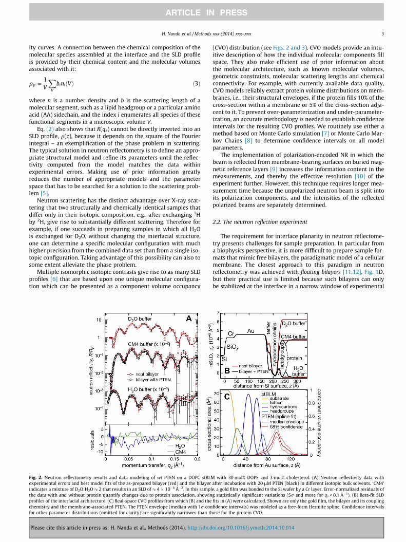
Fig. 2. Neutron reflectometry results and data modeling of wt PTEN on a DOPC stBLMexperimental errors and best model fits of the as-prepared bilayer (red) and the bilayerindicates a mixture of D2O:H2O � 2 that results in an SLD of � 4 � 10�6 �2. In this samplthe data with and without protein quantify changes due to protein association, showinprofiles of the interfacial architecture. (C) Real-space CVO profiles from which (B) and thechemistry and the membrane-associated PTEN. The PTEN envelope (median with 1r confor other parameter distributions (omitted for clarity) are significantly narrower than th
Please cite this article in press as: H. Nanda et al., Methods (2014), http://dx.d
(CVO) distribution (see Figs. 2 and 3). CVO models provide an intu-itive description of how the individual molecular components fillspace. They also make efficient use of prior information aboutthe molecular architecture, such as known molecular volumes,geometric constraints, molecular scattering lengths and chemicalconnectivity. For example, with currently available data quality,CVO models reliably extract protein volume distributions on mem-branes, i.e., their structural envelopes, if the protein fills 10% of thecross-section within a membrane or 5% of the cross-section adja-cent to it. To prevent over-parameterization and under-parameter-ization, an accurate methodology is needed to establish confidenceintervals for the resulting CVO profiles. We routinely use either amethod based on Monte Carlo simulation [7] or Monte Carlo Mar-kov Chains [8] to determine confidence intervals on all modelparameters.
The implementation of polarization-encoded NR in which thebeam is reflected from membrane-bearing surfaces on buried mag-netic reference layers [9] increases the information content in themeasurements, and thereby the effective resolution [10] of theexperiment further. However, this technique requires longer mea-surement time because the unpolarized neutron beam is split intoits polarization components, and the intensities of the reflectedpolarized beams are separately determined.
2.2. The neutron reflection experiment
The requirement for interface planarity in neutron reflectome-try presents challenges for sample preparation. In particular froma biophysics perspective, it is more difficult to prepare sample for-mats that mimic free bilayers, the paradigmatic model of a cellularmembrane. The closest approach to this paradigm in neutronreflectometry was achieved with floating bilayers [11,12], Fig. 1D,but their practical use is limited because such bilayers can onlybe stabilized at the interface in a narrow window of experimental
with 30 mol% DOPS and 3 mol% cholesterol. (A) Neutron reflectivity data withafter incubation with 20 lM PTEN (black) in different isotopic bulk solvents. ‘CM4’e, a gold film was bonded to the Si wafer by a Cr layer. Error-normalized residuals ofg statistically significant variations (5r and more for qz < 0.1 Å�1). (B) Best-fit SLDfits in (A) were calculated. Shown are only the gold film, the bilayer and its couplingfidence intervals) was modeled as a free-form Hermite spline. Confidence intervalsose for the protein CVO.
oi.org/10.1016/j.ymeth.2014.10.014

Fig. 3. Neutron reflectometry results and data modeling of wt PTEN on a DOPC stBLM with 29 mol% DOPS, 3.5% PI(4,5)P2 and 3 mol% cholesterol. Other details as in Fig. 2. Thegold film in this sample was bonded to the Si wafer by a permalloy layer.
4 H. Nanda et al. / Methods xxx (2014) xxx–xxx
conditions. Stacked multi-bilayer samples [13], shown in Fig. 1E,can be formed with high reproducibility under a wide range ofexperimental conditions. They consist of several thousand bilayerswith an inter-bilayer spacing of 10–20 Å, depending on osmoticpressure, temperature and level of hydration [14–18], and havebeen extensively studied with diffraction techniques. Such multib-ilayers can also prepared in the presence of (small) peptides andutilized in studies of peptide organization in membranes [19,20].A flexible tool to investigate peptide and protein interactions witha membrane surfaces is the floating Langmuir monolayer (Fig. 1B).Lipid monolayers have been extensively investigated for their lipidphase structures [21,22] and have served as a matrix for proteincrystallization [23] and as a simple model system to developparametrization schemes for lipid structure in membranes[24,25]. Their lateral lipid density can be varied over a large rangeto determine the dependence of protein adsorption on lipid status[26]. On the other hand, since Langmuir monolayers lack the twinmonolayer sheet, there is a residual pressure that is not compen-sated. The question which monolayer pressure is equivalent tothe state of lipid leaflets within a bilayer membrane [27] maytherefore be ill-posed.
The system that is most flexible, and has been most extensivelystudied in the context of biological relevance, are single bilayermembranes adsorbed to a solid substrate [28,29]. Such systemsin which the lipid membrane is separated from its solid supportby an ultrathin ‘cushion’ layer have been proposed as general vehi-cles to mimic biophysical processes relevant to membrane biology[30], and many types of this general scheme have been imple-mented [31–47]. A large number of adsorbed, chemisorbed andtethered membrane architectures were thoroughly investigatedwith reflection techniques [37,48–50]. We developed a membranearchitecture in which a short poly(ethylene oxide) chain tethers adual-chain lipid to a gold surface via thiol chemistry [51], and sim-ilar systems have been explored by others [44,45,47,52]. b-Mercap-toethanol (bME) serves as a ‘back-filler’ molecule to laterally spacethe membrane tether that anchors a single phospholipid bilayer tothe atomically flat, 100–1000 Å thick gold-film on a glass or Si
Please cite this article in press as: H. Nanda et al., Methods (2014), http://dx.d
surface [7,53,54]. The membrane is either precipitated from sol-vent [43] or formed by vesicle fusion [55] to complete the mem-brane structure, which thus forms a sparsely-tethered bilayerlipid membrane (stBLM) [53]. Such stBLMs are in-plane fluid withdiffusional mobilities of the lipids that are close to that in freebilayers [56]. Therefore, they are exquisitely suited to mimic thelipid component of biological membranes which has beenexploited in numerous studies [51,57–60]. Importantly, stBLMsprepared by rapid solvent exchange [43] show extremely lowdefect densities [53] that can be exploited for studies of bilayerconductivity with high sensitivity [57,58] and precise measure-ments of protein association with the membrane by surface plas-mon resonance (SPR) [61] without the need of defect blocking. Inthe context of NR, these systems are particularly attractive becauseof their long-term stability [62] which permits measurements ofthe membrane in different states of completion [53], prior to orafter protein adsorption or incorporation [58] and under distinctbuffer contrast [51]. As shown for a wide range of protein systems[51,57,58,60,61,63,64], this can be advantageously exploited tocharacterize the structure of membrane-associated proteins athigh out-of-plane and orientational resolution.
The quality of the CVO profile is limited by the maximummomentum transfer for which neutron reflectometry data can becollected at an acceptable signal-to-noise ratio [65]. While the sta-tistical quality of the data is improved by increasing the neutronflux, this does not improve the signal-to-noise ratio, which is, how-ever, the critical quantity, in particular at high q where backgroundscattering from the sample dominates. Therefore, improvements ofdata quality are better achieved through the reduction of the back-ground by eliminating dispensable sources of scattering. In addi-tion, the signal amplitude can be increased by minimizing theinterfacial roughness of the supporting substrate, and conse-quently, of the interfacial molecular architecture [66]. At the NISTCenter for Neutron Research (NCNR), a background-optimizedsample flow cell [67] that exposes a 100 lM thin aqueous reservoirto the neutron beam is used to minimize background. This aqueousreservoir is sandwiched between two Si wafers. The surface of the
oi.org/10.1016/j.ymeth.2014.10.014

H. Nanda et al. / Methods xxx (2014) xxx–xxx 5
sample wafer is polished to Å-scale residual roughness and termi-nated with an optimized metal film (functionalized gold layer) thatkeeps the membrane-bearing surface at an RMS roughness <7 Å.
The NCNR liquid flow cell is equipped with fluid inlets thatallow for in situ sample preparation and manipulation. In thecourse of a typical experiment, the as-prepared bilayer is charac-terized at least in two different isotopic bulk solvent contrasts.Thereafter, protein is added and measured using again multiplecontrasts. The membrane-bound protein may then be manipulatedin situ and the structural consequence of this manipulation deter-mined in further measurements. The properties of the protein, inparticular its membrane-binding kinetics and aggregation behav-ior, determine whether a measurement is better carried out whilethe protein remains in solution or following incubation and rins-ing. Data collection typically takes a few hours for a reflectivitycurve at a single contrast [68]. Complementary techniques, suchas surface plasmon resonance (SPR) are therefore indispensableto characterize the protein–membrane system before a neutronexperiment is attempted. The entire set of neutron reflectometrydata is analyzed in a simultaneous fit that shares model parame-ters between the individual reflectivity curves, for example thosedescribing the invariant substrate.
2.3. Molecular dynamics simulations of membrane proteins
Integrative methods that invoke MD are gaining more and moretraction in structural biology [69,70]. While the simultaneous fit-ting of complementary NR data sets and the incorporation of com-plementing information, such as volumetric data or chemicalconnectivity, boosts the resolution afforded by molecular modelsappreciably – as shown above – connecting these models withMD simulations has the potential to reveal atomistic details. Giventhe low intrinsic resolution of scattering experiments and thedependence of MD results on the precision of the underlyingparameterization, one might argue that in such a procedure theblindman assists the lame. Nevertheless, substantial progress hasbeen made in both directions: High-quality data on the experimen-tally determined structure of substrate-supported bilayers [71]were used to improve the quality of lipid parameter sets in MD[72], and MD simulations that were entirely independent fromexperiment reproduced with confidence the structural propertiesof the PTEN–membrane complex determined with NR [61]. In thatsense, scattering data and MD results have been successfully com-bined to cross-validate each other [64]. In this spirit, combiningMD with scattering approaches has the potential to provide atom-ically resolved structures of otherwise inaccessible systems. Thesestructures then represent our best guess, given all information athand.
In solution and in association with fluid lipid membranes, pro-teins are structurally dynamic molecules whose conformationalstates are inherently tied to their biological function. In addition,many proteins incorporate intrinsically disordered regions. Ratherthan adopting a single rigid structure, proteins thereby formensembles of varied conformational states that are governed bycomplex potential energy surfaces. NR determines time-averageddensity distributions of these ensembles of conformational statesprojected on the membrane normal. MD simulations provide amethod to interpret these results and assess the dynamic intercon-version between conformational states that underlies theseensemble averages. However, the capability to represent thepotential energy landscapes and sample thermally accessible con-formational states depends on the accuracy of the empiricallyderived force fields. Recent refinements of all-atom force fieldshave increased the accuracy of secondary structure predictionand stability in protein folding simulations [73,74]. Updated lipidparameters have also improved the predictive power for structural
Please cite this article in press as: H. Nanda et al., Methods (2014), http://dx.d
properties of membrane simulations [72], including lipid groupscontaining poly-unsaturated tails and unconventional headgroupssuch as those of phosphatidylinositol phosphates [75]. With com-putational resources growing ever more powerful [76], compari-sons between experimental results and molecular simulations ofcomplex protein–lipid systems have greatly gained traction andare becoming routine [63,77].
3. The PTEN–membrane complex – a paradigm for peripheralprotein association with the lipid bilayer
Lipid-mediated signaling utilizes the chemical diversity ofphosphatidylinositolphosphates (PIPs) to control vital cell func-tions by spatially and temporally organizing chemical patternson cellular membrane interfaces. Interconversion by kinases andphosphatases of lipidic PIPs in which the 3, 4 and 5 positions onthe inositol ring show distinct phosphorylation patterns form thechemical ‘‘hardware’’ for various signaling pathways [78], such asthe PI3K/Akt pathway [79,80], which regulates a wide spectrumof processes, including cell survival, proliferation, cell architectureand metabolism and presents an exquisite example for lipid-med-iated signaling with molecular selectivity and spatiotemporal con-trol. Within the pathway, the PTEN phosphatase acts as a PI3Kantagonist that controls PI(3,4,5)P3 levels in the inner leaflet ofthe plasma membrane (PM), which, if unchecked, leads to uncon-ditional cell growth and survival. This critical function makes PTENone of the most frequently mutated genes in human cancer [81].While the PTEN phosphatase also fulfills critical roles in chromo-some maintenance in the nucleus [82] and was recently reportedto associate with intracellular membranes [83], its role inPI(3,4,5)P3 dephosphorylation and the cellular control of PTENPM association are the aspects of PTEN function that are best char-acterized. Here we review how surface-sensitive characterizationtechniques, most notably NR, sheds light on PTEN membrane asso-ciation and the structure of the PTEN–membrane complex.
3.1. Why study membrane proteins in artificial settings?
Neutron reflection studies as those described above are largelylimited to artificial systems because interpretation of SLD profilesin terms of molecular compositions, and thus CVO profiles, is onlyachieved in well-defined molecular settings. While the PM ofwhole cells can be characterized with surface-sensitive scatteringtechniques at engineered surfaces [84], information on membranecomposition and membrane constitution is limited in such exper-iments because the SLD distribution is hard to decompose intoindividual molecular contributions. Moreover, cell membranesare inevitably heterogeneous in-plane, which further complicatesa molecular interpretation of scattering experiments.
While investigations in well-defined synthetic sample formats,on the other hand, are limited in their biological relevance, suchexperiments provide important ancillary information. The PTENphosphatase is known to undergo substantial post-translationalmodification, most prominently, phosphorylation of its C-terminaltail [85,86]. In addition, PTEN engages in a large set of protein–protein interactions, and covalent protein modifiers were recentlyproposed as obligate cofactors for PTEN membrane association andPIP dephosphorylation [87,88]. Our recent studies of bacteriallyexpressed PTEN on stBLMs show clearly that PTEN does not requirepost-translational modifications or cellular cofactors to associatewith the lipid bilayer surface, with or without PIP lipids. Moreover,SPR investigations showed clearly the roles of various lipid compo-nents in the bilayer in recruiting the phosphatase to the membrane[61]: The phosphatidylserine (PS) component of the inner PMprovides an electrostatic background that drives PTEN adsorption
oi.org/10.1016/j.ymeth.2014.10.014

6 H. Nanda et al. / Methods xxx (2014) xxx–xxx
to the membrane surface (equilibrium dissociation constant,Kd � 10 lmol/L). However, the affinity to PI(4,5)P2-containingmembranes devoid of PS is considerably higher (Kd � 0.4 lmol/L).It increases by yet another order of magnitude if both PS andPI(4,5)P2 are present in the membrane (Kd � 40 nmol/L). Thisexample shows clearly how synergetic contributions of membranecomponents to protein binding can be disentangled in in vitrosettings.
3.2. NR investigations of the PTEN–membrane complex
The interaction of wild-type (wt) PTEN with stBLMs preparedfrom lipid mixtures of (A) DOPC containing 30% DOPS and 3% cho-lesterol, and (B) DOPC containing 29% DOPS, 3.5% PI(4,5)P2 and 3%cholesterol was structurally characterized using NR [61]. Followingan initial characterization of the neat lipid bilayer using three iso-topic solvent contrasts, protein was added at a concentration of20 lM in both cases. After incubation for 6 h, the protein wasrinsed off and reflectivity curves were collected using two isotopiccontrasts for each sample (Fig. 2A: DOPC:DOPS:chol, Fig. 3A: DOPC:DOPS:PI(4,5)P2:chol). Both data sets show large differencesbetween the reflectivity curves collected before and after proteinaddition, exceeding five standard deviations at qz < 0.1 �1. Forvery small proteins or proteins at a low surface coverage, the dif-ferences are often smaller, and in some cases do not exceed twostandard deviations. Nevertheless, protein envelopes can still bereliably determined in those cases. Figs. 2B and 3B show thebest-fit nSLD profiles that constitute a stage of data evaluationintermediate between the fitting of the reflectivity curve and thereal-space modeling of the interfacial structure using CVOs.
The final CVO profiles are shown in Figs. 2C and 3C, respectively.The stBLM was parameterized using an established model [5] andthe associated protein was described as a free-form Hermite spline[89]. Model parameter uncertainties were determined in a MonteCarlo Markov Chain procedure [8]. For both samples, the proteinenvelope extends �50 Å from the membrane surface and isanchored in the substrate-distal lipid headgroups without pene-trating the hydrocarbon chains. The shape of the envelopes isasymmetric, showing a peak density �20 Å away from the mem-brane and a trailing shoulder. The protein forms a dense layer witha peak volume occupancy of �40% in both samples. The dimen-sions of the protein in solution support the interpretation thatthe interfacial layer of PTEN at the membrane is a monomolecularlayer. Plugging independent structural information such as the
Table 1A selection of biological relevant parameters from the component volume occupancymodeling. Reported are median values and 68% confidence limits determined using aMonte Carlo Markov Chain.
wt PTEN onPC:PS:cholstBLM
wt PTEN onPC:PS:chol:PI(4,5)P2
stBLM
Lipid hydrocarbon thicknessInner lipid leaflet 16.3 ± 1.0 Å 19.1 ± 1.1 ÅOuter lipid leaflet 12.8 ± 1.1 Å 10.5 ± 0.8 ÅChange upon protein incubation +0.1 ± 0.5 Å +0.9 ± 0.25 Å
Membrane area per lipidAs prepared 75 ± 6 Å3 91 ± 7 Å3
After protein incubation 74 ± 6 Å3 82 ± 7 Å3
Completeness of lipid bilayerAs prepared 99 ± 2% 96 ± 3%After protein incubation 98 ± 2% 97 ± 3%
Amount of surface-associatedprotein (volume surfacedensity)
8.3 ± 1.4 Å3/Å2 7.4 ± 0.7 Å3/Å2
PTEN penetration into bilayer 10.1 ± 2.3 Å 9.7 ± 2.3 Å
Please cite this article in press as: H. Nanda et al., Methods (2014), http://dx.d
(partial) X-ray crystal structure [90] (or an NMR structure) intothe model can reveal critical information on the protein orientationat the membrane and the conformation of disordered protein seg-ments not included in the crystal structure [61]. However, this andmore detailed structural information on the protein–membranecomplex can be more precisely derived from MD simulations, asshown below.
Table 1 shows biologically relevant parameters of the structuralmodels of the two samples. Both stBLMs are essentially coveringthe interface completely, i.e., they are low in defect density. Thelatter shows a thickness of the outer lipid leaflet that is slightlylower than expected, which is often observed for membranes thatare less than 100% complete. Protein incubation does not affectmembrane completion or lipid leaflet thickness despite the highsurface volume density of associated PTEN. Protein penetrationinto the bilayer is low: The PTEN phosphatase dips into the bilayersurface merely to the headgroup/hydrocarbon interface of the sub-strate-distal leaflet.
3.3. Refinement of PTEN models by MD simulations validated throughNR results
MD simulations of a protein–membrane complex entail gener-ating equilibrated structures of the two components and combin-ing them into a single, fully solvated system. For peripheralmembrane proteins such as PTEN, the protein is initially placeddistant from the membrane surface and allowed to dock with thebilayer over time. The simulations described below were set upusing NAMD 2.9 [91] with the CHARMM22 CMAP correction [92]and CHARMM36 [72,93] force field parameters to describe proteinand lipids, respectively. To generate the membrane composition ofinterest, pre-equilibrated DOPC patches were stitched together toform a bilayer that consisted of 720 lipids (360 lipids per leaflet).DOPC molecules were randomly mutated to DOPS or PIPs to gener-ate the desired compositions. Stearoylarachidinoylphosphatidyl-inositol was substituted with phosphates on the inositol ring togenerate PI(4,5)P2 and PI(3,4,5)P3. At neutral pH, the phosphategroups are 50–60% protonated [94]; however, protons do notexchange between the phosphate groups in MD simulations.Therefore, the 50 phosphate was chosen as the protonated groupon PI(4,5)P2 while both the 30 and 50 phosphates were protonatedon PI(3,4,5)P3 [95]. NaCl was added to neutralize the system andestablish a concentration of 100 mM. The new bilayer systemswere equilibrated for approximately 30 ns before PTEN wasintroduced.
The truncated PTEN crystal structure (PDB ID: 1D5R) [90] wassupplemented with AAs 1–13, 282–312 and 352–411 for the sim-ulation of the full-length protein with a C-terminal His-tag. UsingSASSIE [96], extended conformations of the unstructured proteinstretches were generated as starting configurations for the all-atom MD runs. Simulated annealing under elevated temperatures[64] was used to relax backbone torsion and steric clashes. Afterfurther 9 ns simulation at room temperature, the equilibratedfull-length PTEN structure was combined with the equilibratedDOPC/DOPS or DOPC/DOPS/PIP membrane. The protein was cen-tered with respect to the bilayer and placed away from its surfaceto form a �10 Å water layer between the protein and the lipidheadgroups. An extra 40 Å depth of water was added along thebilayer normal to provide sufficient space for PTENs flexible tail.The simulation box then contained �104,000 water molecules.The final equilibration of the system was achieved by holding theentire protein structure fixed for 5 ns, followed by 5 ns under a3 kcal/mol harmonic force constraint and another 5 ns under a1 kcal/mol constraint. Finally, all constraints on the protein werereleased and the production run initiated. PTEN docked to themembrane surface after 60–100 ns of simulation time [64]. All
oi.org/10.1016/j.ymeth.2014.10.014

Fig. 4. Comparison of COV profiles from NR and MD simulations. (A) Full-length wt PTEN on a DOPC:DOPS (7:3) bilayer. (B) PTEN on a PI(4,5)P2-containing bilayer. Themembrane composition was DOPC:DOPS:PI(4,5)P2 = 67.5:29:3.5 in NR experiments and 70:26:4 in MD simulations. The experimental PTEN CVO profile is shown as a 68%confidence band. CVO contributions of different protein regions determined from MD are also shown. Profiles from MD are averaged over 300 ns.
H. Nanda et al. / Methods xxx (2014) xxx–xxx 7
subsequent analysis of the PTEN–membrane complex was per-formed after the docking event occurred.
Comparisons of PTEN–membrane complex structures from MDand NR were made in terms of their CVO profiles. Fig. 4A showsfull-length PTEN on a DOPC:DOPS = 7:3 membrane [64]. The CVOprofile of PTEN, shown in blue, is an average over �300 ns of thesimulation trajectory and fits well within the 68% confidence bandsof the experimental profile (red band). Both the phosphatasedomain (PD) and C2 domain associate closely with the membranesurface and give rise to the peak density observed in the NR CVOattributed to the protein. This region was also well approximatedby the crystal structure that contains the two core domains [61].Some excess protein CVO density distal to the membrane is notwell accounted for by the X-ray structure. It was therefore postu-lated that this region contains contributions from the tail [61],which represents the major portion of the protein clipped for crys-tallization. The overall envelope profile determined from MD wasdissected into individual distributions of different molecularregions, as shown in Fig. 4A. During the simulation, the initiallyextended, highly acidic tail (excess charge:�10e) collapsed quicklyinto a dynamic coil structure and remained at a distance from theacidic membrane surface during the entire simulation, apparentlydue to electrostatic repulsion. The resulting organization of the tailin connection with the clipped loop in the C2 domain (AAs 286–309) indeed fills the observed excess density that is not accountedfor by placing the crystal structure into the experimental CVOprofile.
In a recent set of simulations, we examined the interaction ofPTEN with PIPs. PI(4,5)P2, a key component for high affinity mem-brane binding and plasma membrane specificity, and PI(3,4,5)P3,the catalytic target of PTEN, were studied in DOPC/DOPS mem-branes. A long simulation was conducted of a DOPC membranecontaining 29 mol% DOPS and 3.5 mol% PI(4,5)P2 to mimic experi-mental NR measurements. CVO profiles derived from MD simula-tions and NR experiments are compared in Fig. 4B. Withoutknowledge of the MD-derived structure, the PTEN crystal structurecan be placed to describe the peak protein density near the mem-brane surface satisfactorily, as for the DOPC/DOPS membrane, andagain this protein fragment alone misses the CVO distribution dis-tal to the bilayer. On the other hand, the MD-derived CVO profile,which accounts for the entire protein, matches the experimentalconfidence bands and places the tail and the C2 loop distant fromthe membrane surface.
While so far only protein segment organization at the mem-brane surface was discussed, MD simulations provide significantlymore detail, and the excellent low-resolution agreement with theNR results provides confidence that these more detailed MD resultsare adequate descriptions of the structure of the PTEN/membranecomplex. For example, it should be expected that PI(4,5)P2, in
Please cite this article in press as: H. Nanda et al., Methods (2014), http://dx.d
low abundance in the inner PM, may cluster in the vicinity ofadsorbed PTEN due to electrostatic attraction. To investigate lipidredistribution, a thorough sampling of lipid dynamics and organi-zation required longer simulation times. Sampling was extendedan order of magnitude longer than in our previous work [64], to4 ls, using the Anton supercomputer [76]. Because we were pri-marily interested in the membrane binding interface, the tail wastruncated in the longer simulation to reduce system size. In addi-tion, one of the PI(4,5)P2 molecules in the simulation was modifiedto a PI(3,4,5)P3 to further investigate interactions of the enzymatictarget with PTENs PD domain.
The AA residues that comprise the membrane binding interfacewere determined by measuring orientation and penetration depthof PTEN on the membrane. The orientation was defined in terms ofthe inclination of the second longest principal axes against themembrane normal (‘tilt’) and the rotation of the longest principalaxis about the tilt axis. Fig. 5 shows the dynamic fluctuations inPTEN orientation (panel A) and its averaged distribution (panelB). PTEN inclination is fairly stable, rocking only about 10� fromits average position, while the rotation is more dynamic. However,because the tilt is generally <30�, PTEN rotation about its tilt axisdoes not alter the membrane binding interface significantly overtime.
There is controversy in the literature as to which PTEN residuesare critical for membrane binding. For example, it was reportedthat SUMOylation of K254 or K266, in the CBR3 membrane-bindingmotif of the C2 domain, is required for efficient PTEN membranebinding [87]. This is at odds with the experimental finding thatrecombinant PTEN binds membranes in vitro [61,90] and in vivo[97], as evidenced by enzymatic activity. To identify AAs importantfor membrane anchoring, lipid residence times in contact withindividual PTEN AA residues were calculated. In Fig. 6A, we com-pare the results for PS in the PIP-free membrane with those forPS and PIP in the PIP-containing membrane. Clusters of contactsare revealed on both C2 and the PD for both lipid types. Contactswith PIPs are much longer than those with PS, reflecting a strongerelectrostatic interaction with these highly charged lipid head-groups. A mapping of these residues onto PTENs crystal structureis shown in Fig. 6B. The CBR3 loop (PTEN260–269) with its basicAAs (K260, K263, K266, K267 and K269) binds strongly to PS andPIPs, and its hydrophobic L265 side chain snorkels into the hydro-phobic membrane core [64]. In PIP-containing membranes, K221and K223 and surrounding residues that line the cleft betweenthe PD and C2 domain also coordinate PIP. In the PD, a group ofAAs around K128 forms prolonged contacts with PI(3,4,5)P3. Infact, once PI(3,4,5)P3 binds to this ligation site on the surface ofthe PD domain, it is held there for the remaining duration of thesimulation. The coordination of PI(3,4,5)P3 to K128 is reinforcedby K163, R161, R47 and the N-terminal Met (see Fig. 7), which is
oi.org/10.1016/j.ymeth.2014.10.014

Fig. 5. Dynamics and average orientation of PTEN bound to a DOPC:DOPS:PIP2 membrane. The reference frame for protein orientation uses the principal axes of thecrystallized protein core (PD and C2) [90], with the longest principal axis aligned with x and the second longest principal axis aligned with z (the membrane normal). (A)Fluctuations of PTEN orientation are small (±15�) over the entire 4 ls trajectory. At this small inclination of the axis against the membrane normal, PTEN rotates about the z0
axis (the local tilt axis) with only small variation in its membrane binding interface. (B) Left: Contour plot of the averaged tilt and rotation showing the well-defined proteinorientation on the membrane surface. Right: Superpositions of the two longest principal axes of PTEN from several simulation snapshots visualize the degree of rocking andspinning of the protein.
Fig. 6. Dynamics of lipid binding to PTEN residues that form the membrane binding interface. (A) Residence times of lipids at specific AAs extracted from the DOPC:DOPS andthe DOPC:DOPS:PIP2:PIP3 simulations. Residence times <5 ns are omitted. Top: A comparison of DOPS residence times shows how PIP association with AAs on PTENsmembrane binding interface suppresses PS protein associations that take place in the absence of PIPs. Bottom: A comparison of PIP association with that of DOPS in theabsence of PIPs illustrations how much stronger the inositolphosphates bind to the protein than PS. The emergence of lipid contacts near residues 163 and 330, only observedin the PIP-containing membrane, indicates that the phosphatase is slightly deeper immersed into the bilayer surface when bound to PIPs than in the PIP-free membrane. (B)Map of lipid contacts on the PTEN crystal structure. The color code for lipids associated with PTEN AA residues is the same in both panels.
Fig. 7. Time-averaged density distributions of PI(4,5)P2 and PI(3,4,5)P3 ligated by PTEN in the DOPC:DOPS:PIP2:PIP3 simulation. (A) Stereogram of PTEN with the distributionof associated PIPs. The PTEN protein is shown with the PD on the left and the C2 domain on the right. The PIP distribution depicted under the PD is that of PI(3,4,5)P3 bound toits ligation site below the catalytic pocket. Several PI(4,5)P2 molecules that interchange at a slow rate form the density shown below C2. (B) Magnified view of the PI(3,4,5)P3
coordination on the PD, rotated from the view in panel (A) by �180� about the membrane normal. Residues that form the PI(3,4,5)P3 ligation site are shown in space fillingand color-coded according to residue type (blue – basic, red – acidic, green – polar, white – hydrophobic). C124 in the catalytic site, also shown in space filling, is locatedabove and to the right of the bound PI(3,4,5)P3. R47, K128 and K163 bind electrostatically to PI(3,4,5)P3 and form the blue contours surrounding the lipid headgroup. The N-terminal Met is shown as a white contour on the left. M1 snorkels its hydrophobic side chain towards the lipid chains and points its charged amine group towards theinositolphosphates, thus forming both hydrophobic and electrostatic interactions with PI(3,4,5)P3.
8 H. Nanda et al. / Methods xxx (2014) xxx–xxx
close to K128. The PD also intermittently captures a PI(4,5)P2 atR41 and nearby residues, including R47, with a loop, locatedbetween pb2 and pa1, that dips into the bilayer surface.
The time-averaged PIP organization within the protein footprinton the membrane is visualized in Fig. 7A, and overlaid with a snap-shot of lipid configurations. An accumulation of PI(4,5)P2 is
Please cite this article in press as: H. Nanda et al., Methods (2014), http://dx.d
observed at the C2 domain. It populates the CBR3 loop and the cleftbetween the C2 and PD. This region accommodates two to threePI(4,5)P2 that exchange dynamically between each other and freemembrane PIPs on a time scale of hundreds of ns. The PD hastwo distinct PIP binding sites: a cluster of basic residues betweenpb2 and pa1, and the PIP3 binding pocket. Fig. 7B shows the
oi.org/10.1016/j.ymeth.2014.10.014

H. Nanda et al. / Methods xxx (2014) xxx–xxx 9
residue surfaces, encoded by residue type, that form the PI(3,4,5)P3
ligation site and their sidechain arrangements around the lipidheadgroup. The site is formed by four basic residues that providecharge complementarity to the anionic lipid and the positivelycharged N-terminus of M1. In addition, the hydrophobic M1 side-chain snorkels into the membrane and interacts with thePI(3,4,5)P3 hydrocarbon chain. The enzymatic site centered atC124 is located only a few Ångstrom above the PIP3 ligation site.While direct interaction of PIP3 with C124 was not observed duringthe simulation run, a translation of the lipid by �5 Å would deliverthe substrate into the catalytic site. Thus the ligation of PI(3,4,5)P3
on the PD just outside of the catalytic pocket observed in this sim-ulation is conceivably an intermediate step in the PIP3 hydrolysispathway.
4. Discussion and outlook
Neutron reflection measurements in conjunction with molecu-lar dynamics simulations provide a new and unique window intothe structural biology of protein–membrane complexes. Reviewingrecent investigations of the membrane association of the PTENphosphatase, we demonstrate here the level of detail that can bededuced from such combined studies. With the knowledge of thepartial crystal structure, NR was used to characterize the overallstructure of the PTEN–membrane complex [61]. In this work, theprecise determination of the protein orientation on the membranein combination with the penetration depth into the bilayer definedthe membrane binding interface of the phosphatase. In addition,NR showed that the disordered tail is repelled form the bilayerupon membrane binding. The biological function of PTEN as aphosphatase at the plasma membrane depends strongly on itsinteractions with PI(4,5)P2. Binding measurements on stBLMsshowed that a physiologically small concentration of PI(4,5)P2 inthe membrane increases membrane affinity significantly and, inconjunction with PS, leads to a strong association of PTEN withthe target membrane. To understand how PIP-specificity isachieved in the binding event, the AA residues that comprise thePTENs membrane binding interface were identified by combiningNR with MD simulations.
As shown above, NR provides structural information of mem-brane proteins associated with fluid lipid bilayers in their physio-logically relevant aqueous environment. Utilizing the exquisitesensitivity of NR for low-atomic number materials (i.e., protein,lipid and solvent), we developed models of the biomimetic inter-face in the form of CVO profiles of the membrane components.As shown in Figs. 2 and 3, we observed that PTEN is peripherallybound to the bilayer where it only inserts into the lipid headgroupregion. The width of the peak protein density near the membranesurface (�35 Å) suggested that PTEN binds the membrane withboth the C2 domain and PD collectively forming the membranebinding interface. A superposition of the truncated crystal struc-ture onto the CVO profile of the protein supported this interpreta-tion (Fig. 3A and B). However, excess protein contributions to thescattering were not accounted for by the X-ray structure end weretentatively attributed to PTENs unstructured regions, i.e., the tailand the C2 loop. MD simulations of the full-length protein, set upto mimic the experimental conditions of the NR measurements,reproduced the CVO profiles well. This agreement between thetwo independent methods validated the simulation results andverified the tentative localization of the proteins unstructuredregions.
A more detailed analysis of the MD results provided a detailedaccount of the molecular interactions of PTEN with the membranelipids. Comparing the PIP-free and the PIP-containing membranesimulations we observed very similar binding orientations of the
Please cite this article in press as: H. Nanda et al., Methods (2014), http://dx.d
protein. Many of the residues that formed strong interactions withDOPS in the PIP-free bilayers also formed prolonged contacts withPIPs. However, one also observes three AA clusters that arerecruited into lipid associations by the PIPs (Fig. 6A), i.e., they onlyform lipid associations on the PIP-containing bilayer. These clus-ters are located at the N-terminal tail, around R161/K163/K164and at the ca2 motif around N329/K330. The appearance of thesenew lipid contact suggests that the penetration of the protein intothe bilayer is slightly increased by its enhanced interaction withthe PIP-containing membrane. This incrementally deeper penetra-tion of the bilayer is not resolved by NR (Table 1), as such level ofdetail is clearly beyond the capabilities of scattering experiments.
Average lipid residence times, and reciprocally koff rates, of PIPswere an order of magnitude longer than those of PS. This canaccount for the smaller Kd values observed in SPR binding studiesof PTEN to PIP-containing membranes. In the simulations, PIPsinteract primarily with protein regions rich in Lys and Arg residues.Other AA residues reinforce this association by forming hydrogenbonds with inositol hydroxyl and phosphate groups, and in somecases hydrophobic sidechains snorkel into the membrane core.The CBR3 loop on C2 as well as the cleft between the C2 and PDform basic patches that cluster 2–3 PIPs which exchange dynami-cally on the of timescale of hundreds of ns. A ligation site close tothe catalytic pocket of the PD captures a PI(3,4,5)P3 and holds themolecule for several ls. This unusually long dwell time and, hence,strong association may contribute to the observed orientationalstability of PTEN on the membrane and could be functionally rele-vant for the delivery of the substrate to the catalytic site.
As a model for membrane-associated proteins with intrinsicallydisordered regions, PTEN violates the paradigm of a simple struc-ture–function relationship and poses a challenge to structuralcharacterization. For example, PTENs tail has been reported to reg-ulate membrane binding [98–100]. In combination, the localizationof the tail by NR distal from the membrane surface and the moredetailed assessment of its organization by MD show that the tail,while undergoing constant conformational rearrangement, isstrongly repelled by the acidic membrane surface. On the otherhand, sampling tail configurations in solution showed that it canwrap around PTENs core domain, and thus block the membraneaccessibility of the CBR3 loop [64]. More extensive, microsec-onds-long MD simulations confirm these results (not shown). Itsintrinsic flexibility enables the tail to sample blocking and non-blocking conformations subject to a delicate energetic balance.Modifications, such as the phosphorylation of tail residues, maydisturb this balance and favor one conformation over the other.Indeed, it is well established that the phosphorylation of a clusterof residues (S380, T382, T383 and S385) downregulates membranebinding and thus, PTEN activity [99].
Combining NR characterization with MD simulations has beenhighly informative for several systems of membrane-associatedproteins, including HIV-1 Gag and Nef [59,63,77], as well as thePTEN protein discussed here. Sampling of protein conformationsin simulations enables comparisons with the experimental resultsthat can validate the MD methodology and, if successful at low res-olution, provides novel and otherwise inaccessible structuraldetail. For membrane simulations that accommodate the size ofPTEN (105 atoms), MD simulations on the microsecond timescaleare required to equilibrate the system and sufficiently sample theequilibrated state. In simulations of Nef, we used steered MD sim-ulations [101] to apply forces that pulled the protein into an openconfiguration suggested by NR experiments and then sampled theopen state free of biasing potentials. Thereby, the protein reachesmore relevant areas of conformation space and requires signifi-cantly less equilibration time. However, this whole field still needsfurther methodological development, for example, of rigorousprocedures based upon the CVO profiles derived from NR. An
oi.org/10.1016/j.ymeth.2014.10.014

10 H. Nanda et al. / Methods xxx (2014) xxx–xxx
experimentally validated biasing potential could thus be used notonly to rapidly equilibrate a protein–membrane system but alsoto determine an ensemble of protein configurations that strictlymatch the NR data. In addition, such methodologies can beexpanded to implement constraints from multiple sources of infor-mation [102,103], such as fluorescence resonance energy transferor electron paramagnetic resonance, to be integrated in refinedstructural models of membrane-associated proteins. For structur-ally disordered systems, the combination of multiple sparse datasets promises to be a powerful method for relating conformationaldynamics and flexibility to function.
Acknowledgments
We thank Dr. Alonzo Ross for critically reading the manuscriptand valuable suggestions. This work was supported by the U.S.Dept. of Commerce (70NANB13H009, 70NANB11H8139 andthrough the NIST IMS program ‘‘Precision Measurements for Inte-gral Membrane Proteins’’) and the NIH (1R01 GM101647), and per-formed in parts at the NIST Center for Neutron Research and theNIST Center for Nanoscale Science and Technology.
References
[1] Certain commercial materials, equipment, and instruments are identified inthis manuscript in order to specify the experimental procedure as completelyas possible. In no case does such identification imply a recommendation orendorsement by the National Institute of Standards and Technology, nor doesit imply that the materials, equipment, or instruments identified arenecessarily the best available for the purpose.
[2] C.F. Majkrzak, N.F. Berk, Physica B 336 (2003) 27–38.[3] C.F. Majkrzak, N.F. Berk, P. Kienzle, U. Perez-Salas, Langmuir 25 (2009) 4154–
4161.[4] J. Als-Nielsen, K. Kjaer, in: T. Riste, D. Sherrington (Eds.), Phase Transitions in
Soft Condensed Matter, Plenum Press, New York, 1989, pp. 113–138.[5] P. Shekhar, H. Nanda, M. Lösche, F. Heinrich, J. Appl. Phys. 110 (2011) 102216.[6] J. Ankner, C.F. Majkrzak, Proc. SPIE 1738 (1992) 260–269.[7] F. Heinrich, T. Ng, D.J. Vanderah, P. Shekhar, M. Mihailescu, H. Nanda, M.
Lösche, Langmuir 25 (2009) 4219–4229.[8] B.J. Kirby, P.A. Kienzle, B.B. Maranville, N.F. Berk, J. Krycka, F. Heinrich, C.F.
Majkrzak, Curr. Opin. Colloid Interface Sci. 17 (2012) 44–53.[9] S.A. Holt, A.P. Le Brun, C.F. Majkrzak, D.J. McGillivray, F. Heinrich, M. Lösche,
J.H. Lakey, Soft Matter 5 (2009) 2576–2586.[10] M.C. Wiener, S.H. White, Biophys. J. 59 (1991) 162–173.[11] G. Fragneto, T. Charitat, F. Graner, K. Mecke, L. Perino-Galice, E. Bellet-
Amalric, Europhys. Lett. 53 (2001) 100–106.[12] T. Charitat, S. Lecuyer, G. Fragneto, Biointerphases 3 (2008) FB3–FB15.[13] D.L. Worcester, N.P. Franks, J. Mol. Biol. 100 (1976) 359–378.[14] S. Tristram-Nagle, J.F. Nagle, Chem. Phys. Lipids 127 (2004) 3–14.[15] M.C. Wiener, G.I. King, S.H. White, Biophys. J. 60 (1991) 568–576.[16] M.C. Wiener, S.H. White, Biophys. J. 61 (1992) 428–433.[17] M.C. Wiener, S.H. White, Biophys. J. 61 (1992) 434–447.[18] J.F. Nagle, S. Tristram-Nagle, Biochim. Biophys. Acta 1469 (2000) 159–195.[19] K. Hristova, C.E. Dempsey, S.H. White, Biophys. J. 80 (2001) 801–811.[20] S. Tristram-Nagle, R. Chan, E. Kooijman, P. Uppamoochikkal, W. Qiang, D.P.
Weliky, J.F. Nagle, J. Mol. Biol. 402 (2010) 139–153.[21] V.M. Kaganer, G. Brezesinski, H. Möhwald, P.B. Howes, K. Kjaer, Phys. Rev. E
59 (1999) 2141–2152.[22] V.M. Kaganer, H. Möhwald, P. Dutta, Rev. Mod. Phys. 71 (1999) 779–819.[23] M. Weygand, M. Schalke, P.B. Howes, K. Kjaer, J. Friedmann, B. Wetzer, D.
Pum, U.B. Sleytr, M. Lösche, J. Mater. Chem. 10 (2000) 141–148.[24] M. Schalke, M. Lösche, Adv. Colloid Interface Sci. 88 (2000) 243–274.[25] M. Schalke, P. Krüger, M. Weygand, M. Lösche, Biochim. Biophys. Acta 1464
(2000) 113–126.[26] P. Calvez, S. Bussieres, E. Demers, C. Salesse, Biochimie 91 (2009) 718–733.[27] A. Blume, Biochim. Biophys. Acta 557 (1979) 32–44.[28] A.A. Brian, H.M. McConnell, Proc. Natl. Acad. Sci. U.S.A. 81 (1984) 6159–6163.[29] L.K. Tamm, H.M. McConnell, Biophys. J. 47 (1985) 105–113.[30] M. Tanaka, E. Sackmann, Nature 437 (2005) 656–663.[31] M. Kühner, R. Tampé, E. Sackmann, Biophys. J. 67 (1994) 217–226.[32] R. Naumann, A. Jonczyk, R. Kopp, J. Vanesch, H. Ringsdorf, W. Knoll, P. Gräber,
Angew. Chem. Int. Ed. Engl. 34 (1995) 2056–2058.[33] X. Lu, A. Leimannova-Ottova, H. Ti Tien, Bioelectrochem. Bioenerg. 39 (1996)
285–289.[34] C. Steinem, A. Janshoff, W.P. Ulrich, M. Sieber, H.J. Galla, Biochim. Biophys.
Acta 1279 (1996) 169–180.[35] B. Wetzer, D. Pum, U.B. Sleytr, J. Struct. Biol. 119 (1997) 123–128.[36] J.T. Groves, S.G. Boxer, H.M. McConnell, Proc. Natl. Acad. Sci. U.S.A. 94 (1997)
13390–13395.
Please cite this article in press as: H. Nanda et al., Methods (2014), http://dx.d
[37] J. Majewski, J.Y. Wong, C.K. Park, M. Seitz, J.N. Israelachvili, G.S. Smith,Biophys. J. 75 (1998) 2363–2367.
[38] M.L. Wagner, L.K. Tamm, Biophys. J. 79 (2000) 1400–1414.[39] U. Rädler, J. Mack, N. Persike, G. Jung, R. Tampé, Biophys. J. 79 (2000) 3144–
3152.[40] R. Kügler, W. Knoll, Bioelectrochemistry 56 (2002) 175–178.[41] C.A. Naumann, O. Prucker, T. Lehmann, J. Rühe, W. Knoll, C.W. Frank,
Biomacromolecules 3 (2002) 27–35.[42] H.P. Vacklin, F. Tiberg, R.K. Thomas, Biochim. Biophys. Acta 1668 (2005) 17–
24.[43] B.A. Cornell, V.L.B. Braach-Maksvytis, L.B. King, P.D.J. Osman, B. Raguse, L.
Wieczorek, R.J. Pace, Nature 387 (1997) 580–583.[44] R. Naumann, S.M. Schiller, F. Giess, B. Grohe, K.B. Hartman, I. Karcher, I. Köper,
J. Lubben, K. Vasilev, W. Knoll, Langmuir 19 (2003) 5435–5443.[45] A. Kibrom, R.F. Roskamp, U. Jonas, B. Menges, W. Knoll, H. Paulsen, R.L.C.
Naumann, Soft Matter 7 (2011) 237–246.[46] F. Giess, M.G. Friedrich, J. Heberle, R.L. Naumann, W. Knoll, Biophys. J. 87
(2004) 3213–3220.[47] S.M. Schiller, R. Naumann, K. Lovejoy, H. Kunz, W. Knoll, Angew. Chem. Int.
Ed. Engl. 42 (2003) 208–211.[48] A.V. Hughes, S.J. Roser, M. Gerstenberg, A. Goldar, B. Stidder, R. Feidenhans’l, J.
Bradshaw, Langmuir 18 (2002) 8161–8171.[49] U. Perez-Salas, K. Faucher, C.F. Majkrzak, N. Berk, S. Krueger, E. Chaikof,
Langmuir 19 (2003) 7688–7694.[50] S. Krueger, C.W. Meuse, C.F. Majkrzak, J.A. Dura, N.F. Berk, M. Tarek, A.L. Plant,
Langmuir 17 (2001) 511–521.[51] G. Valincius, D.J. McGillivray, W. Febo-Ayala, D.J. Vanderah, J.J. Kasianowicz,
M. Lösche, J. Phys. Chem. B 110 (2006) 10213–10216.[52] V. Atanasov, P.P. Atanasova, I.K. Vockenroth, N. Knorr, I. Köper, Bioconjug.
Chem. 17 (2006) 631–637.[53] D.J. McGillivray, G. Valincius, D.J. Vanderah, W. Febo-Ayala, J.T. Woodward, F.
Heinrich, J.J. Kasianowicz, M. Lösche, Biointerphases 2 (2007) 21–33.[54] R. Budvytyte, M. Mickevicius, D.J. Vanderah, F. Heinrich, G. Valincius,
Langmuir 29 (2013) 4320–4327.[55] T. Baumgart, M. Kreiter, H. Lauer, R. Naumann, G. Jung, A. Jonczyk, A.
Offenhäusser, W. Knoll, J. Colloid Interface Sci. 258 (2003) 298–309.[56] S. Shenoy, R. Moldovan, J. Fitzpatrick, D.J. Vanderah, M. Deserno, M. Lösche,
Soft Matter 6 (2010) 1263–1274.[57] G. Valincius, F. Heinrich, R. Budvytyte, D.J. Vanderah, D.J. McGillivray, Y.
Sokolov, J.E. Hall, M. Lösche, Biophys. J. 95 (2008) 4845–4861.[58] D.J. McGillivray, G. Valincius, F. Heinrich, J.W.F. Robertson, D.J. Vanderah, W.
Febo-Ayala, I. Ignatjev, M. Lösche, J.J. Kasianowicz, Biophys. J. 96 (2009)1547–1553.
[59] H. Nanda, S.A.K. Datta, F. Heinrich, M. Lösche, A. Rein, S. Krueger, J.E. Curtis,Biophys. J. 99 (2010) 2516–2524.
[60] F. Heinrich, H. Nanda, H.Z. Goh, C. Bachert, M. Lösche, A.D. Linstedt, J. Biol.Chem. 289 (2014) 9683–9691.
[61] S. Shenoy, P. Shekhar, F. Heinrich, M.-C. Daou, A. Gericke, A.H. Ross, M. Lösche,PLoS ONE 7 (2012) e32591.
[62] I.K. Vockenroth, C. Ohm, J.W.F. Robertson, D.J. McGillivray, M. Lösche, I. Köper,Biointerphases 3 (2008) FA68–FA73.
[63] S.A.K. Datta, F. Heinrich, S. Raghunandan, S. Krueger, J.E. Curtis, A. Rein, H.Nanda, J. Mol. Biol. 406 (2011) 205–214.
[64] S. Shenoy, H. Nanda, M. Lösche, J. Struct. Biol. 180 (2012) 394–408.[65] C.M. Majkrzak, N.F. Berk, S. Krueger, U.A. Perez-Salas, in: J. Fitter, T. Gutberlet,
J. Katsaras (Eds.), Neutron Scattering in Biology, Springer, New York, 2006, pp.225–264.
[66] C.F. Majkrzak, E. Carpenter, F. Heinrich, N.F. Berk, J. Appl. Phys. 110 (2011)102212.
[67] C.F. Majkrzak, N.F. Berk, S. Krueger, J.A. Dura, M. Tarek, D. Tobias, V. Silin, C.W.Meuse, J. Woodward, A.L. Plant, Biophys. J. 79 (2000) 3330–3340.
[68] J.F. Ankner, X. Tao, C.E. Halbert, J.F. Browning, S.M. Kilbey II, O.A. Swader, M.S.Dadmun, E. Kharlampieva, S.A. Sukhishvili, Neutron News 19 (2008)14–16.
[69] F. Alber, S. Dokudovskaya, L.M. Veenhoff, W. Zhang, J. Kipper, D. Devos, A.Suprapto, O. Karni-Schmidt, R. Williams, B.T. Chait, M.P. Rout, A. Sali, Nature450 (2007) 683–694.
[70] G. Zhao, J.R. Perilla, E.L. Yufenyuy, X. Meng, B. Chen, J. Ning, J. Ahn, A.M.Gronenborn, K. Schulten, C. Aiken, P. Zhang, Nature 497 (2013) 643–646.
[71] R.W. Benz, H. Nanda, F. Castro-Roman, S.H. White, D.J. Tobias, Biophys. J. 91(2006) 3617–3629.
[72] J.B. Klauda, R.M. Venable, J.A. Freites, J.W. O’Connor, D.J. Tobias, C.Mondragon-Ramirez, I. Vorobyov, A.D. MacKerell, R.W. Pastor, J. Phys.Chem. B 114 (2010) 7830–7843.
[73] R.B. Best, X. Zhu, J. Shim, P.E.M. Lopes, J. Mittal, M. Feig, A.D. MacKerell, J.Chem. Theory Comput. 8 (2012) 3257–3273.
[74] R.B. Best, J. Mittal, M. Feig, A.D. MacKerell, Biophys. J. 103 (2012) 1045–1051.[75] E. Hatcher, O. Guvench, A.D. MacKerell, J. Chem. Theory Comput. 5 (2009)
1315–1327.[76] D.E. Shaw, P. Maragakis, K. Lindorff-Larsen, S. Piana, R.O. Dror, M.P. Eastwood,
J.A. Bank, J.M. Jumper, J.K. Salmon, Y. Shan, W. Wriggers, Science 330 (2010)341–346.
[77] B. Akgun, S. Satija, H. Nanda, G.F. Pirrone, X. Shi, J.R. Engen, M.S. Kent,Structure 21 (2013) 1822–1833.
[78] M.P. Wymann, R. Schneiter, Nat. Rev. Mol. Cell Biol. 9 (2008) 162–176.[79] Y.R. Chin, A. Toker, Cell. Signal. 21 (2009) 470–476.
oi.org/10.1016/j.ymeth.2014.10.014

H. Nanda et al. / Methods xxx (2014) xxx–xxx 11
[80] A. Toker, L.C. Cantley, Nature 387 (1997) 673–676.[81] L. Li, A.H. Ross, J. Cell. Biochem. 102 (2007) 1368–1374.[82] C. Bassi, J. Ho, T. Srikumar, R.J.O. Dowling, C. Gorrini, S.J. Miller, T.W. Mak, B.G.
Neel, B. Raught, V. Stambolic, Science 341 (2013) 395–399.[83] A. Bononi, M. Bonora, S. Marchi, S. Missiroli, F. Poletti, C. Giorgi, P.P. Pandolfi,
P. Pinton, Cell Death Differ. 20 (2013) 1631–1643.[84] L. Pocivavsek, A. Junghans, N. Zebda, K. Birukov, J. Majewski, Am. J. Physiol.
Lung Cell. Mol. Physiol. 306 (2014) L1–L9.[85] M. Rahdar, T. Inoue, T. Meyer, J. Zhang, F. Vazquez, P.N. Devreotes, Proc. Natl.
Acad. Sci. U.S.A. 106 (2009) 480–485.[86] F. Vazquez, S. Ramaswamy, N. Nakamura, W. Sellers, Mol. Cell. Biol. 20 (2000)
5010–5018.[87] J. Huang, J. Yan, J. Zhang, S. Zhu, Y. Wang, T. Shi, C. Zhu, C. Chen, X. Liu, J.
Cheng, T. Mustelin, G.-S. Feng, G. Chen, J. Yu, Nat. Commun. 3 (2012)911–922.
[88] K. Zmajkovicova, V. Jesenberger, F. Catalanotti, C. Baumgartner, G. Reyes, M.Baccarini, Mol. Cell 50 (2013) 43–55.
[89] F. Heinrich, M. Lösche, Biochim. Biophys. Acta 1838 (2014) 2341–2349.[90] J.O. Lee, H. Yang, M.M. Georgescu, A. Di Cristofano, T. Maehama, Y. Shi, J.E.
Dixon, P.P. Pandolfi, N.P. Pavletich, Cell 99 (1999) 323–334.[91] J.C. Phillips, R. Braun, W. Wang, J. Gumbart, E. Tajkhorshid, E. Villa, C. Chipot,
R.D. Skeel, L. Kalé, K. Schulten, J. Comput. Chem. 26 (2005) 1781–1802.
Please cite this article in press as: H. Nanda et al., Methods (2014), http://dx.d
[92] A. MacKerell, D. Bashford, M. Bellott, R.L. Dunbrack, J. Evanseck, M. Field, S.Fischer, J. Gao, H. Guo, S. Ha, D. Joseph-McCarthy, L. Kuchnir, K. Kuczera, F.Lau, C. Mattos, S. Michnick, T. Ngo, D. Nguyen, B. Prodhom, W. Reiher, B. Roux,M. Schlenkrich, J. Smith, R. Stote, J. Straub, M. Watanabe, J. Wiorkiewicz-Kuczera, D. Yin, M. Karplus, J. Phys. Chem. B 102 (1998) 3586–3616.
[93] J.B. Klauda, V. Monje, T. Kim, W. Im, J. Phys. Chem. B 116 (2012) 9424–9431.[94] E.E. Kooijman, K.E. King, M. Gangoda, A. Gericke, Biochemistry 48 (2009)
9360–9371.[95] D.R. Slochower, P.J. Huwe, R. Radhakrishnan, P.A. Janmey, J. Phys. Chem. B 117
(2013) 8322–8329.[96] J.E. Curtis, S. Raghunandran, H. Nanda, S. Krueger, Comput. Phys. Commun.
183 (2012) 382–389.[97] F. Liu, S. Wagner, R.B. Campbell, J.A. Nickerson, C.A. Schiffer, A.H. Ross, J. Cell.
Biochem. 96 (2005) 221–234.[98] F. Vazquez, S.R. Grossman, Y. Takahashi, M.V. Rokas, N. Nakamura, W.R.
Sellers, J. Biol. Chem. 276 (2001) 48627–48630.[99] L. Odriozola, G. Singh, T. Hoang, A.M. Chan, J. Biol. Chem. 282 (2007) 23306–
23315.[100] J. Torres, R. Pulido, J. Biol. Chem. 276 (2001) 993–998.[101] S. Park, K. Schulten, J. Chem. Phys. 120 (2004) 5946–5961.[102] A.B. Ward, A. Sali, I.A. Wilson, Science 339 (2013) 913–915.[103] E. Karaca, A.M.J.J. Bonvin, Methods 59 (2013) 372–381.
oi.org/10.1016/j.ymeth.2014.10.014