Embed Size (px)
Citation preview
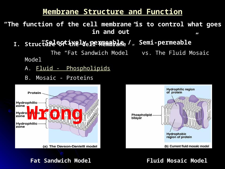
Membrane Structure and Function
“The function of the cell membrane is to control what goes in and out”
“Selectively permeable / Semi-permeable”
I. Structure of the Cell Membrane
The “Fat Sandwich Model” vs. The Fluid Mosaic Model
A. Fluid - Phospholipids
B. Mosaic - Proteins
Fat Sandwich Model Fluid Mosaic Model
Wrong
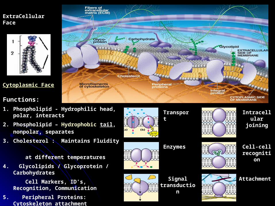
Functions:
1. Phospholipid - Hydrophilic head, polar, interacts
2. Phospholipid – Hydrophobic tail, nonpolar, separates
3. Cholesterol : Maintains Fluidity
at different temperatures
4. Glycolipids / Glycoprotein / Carbohydrates
Cell Markers, ID’s, Recognition, Communication
5. Peripheral Proteins: Cytoskeleton attachment
6. Integral Proteins Many Functions
Cytoplasmic Face
ExtraCellular Face
Transport
Enzymes
Signal transduction
Intracellular joining
Cell-cell recognition
Attachment

II. Molecules can pass through Membranes if:
A. Small enough
Example: Fast - O2, CO2, H2O
Slow - C6H12O6
Can’t – polypetides (proteins) polysaccarides (starches)
B. Non-Polar enough
Example: O2, CO2, Steroids, Lipids
Note: Ions do not move through membranes very well (Na+, Cl-, H+, ) due hydration shell
C. Concentrated enough
Example: H2O, All living things are 70-80% water
D. Helped by Transport Proteins Enough
Transport Proteins provide unique
environments in the membrane to allow
passage of specific molecules
E. Any combination of the above Example - Water

III. What energy “makes" molecules move through membranes?
A. Molecular motion (Kinetic Energy) Diffusion
1. Definition: Movement of molecules from a high concentration
to a low concentration
2. Characteristics
a. Follows a concentration gradient
b. Does not require additional energy
c. “Passive Transport”
3. How cell uses diffusion
Environment CellHigh Oxygen
Cell Respiration
Low Carbon Dioxide

IV. Osmosis The Special Case of Water
A. Definition: The diffusion of water across a semi-permeable membrane
B. Why does water have its own unique word to describe how it diffuses?
1. All organism are 70-80% water - The highest of all concentration gradients
2. Membranes are completely permeable to water (The membrane can not “just say no”)
Environment Environment EnvironmentCell CellCellH2O – 98%
DS -- 2%
H2O – 90%
DS -- 10%
H2O – 98%
DS -- 2%
H2O – 80%
DS -- 20%
H2O – 95%
DS -- 5%H2O – 98%
DS -- 2%
Water Flow
Tonicity Environment: Hypotonic
Cell: Hypertonic
JargonPlant: Turgor Pressure
Animal: Cytolysis
“Hydrostatic Skeleton”
Environment: Hypertonic
Cell: Hypotonic
Isotonic
Plasmolysis
Crenation
Summary
Flaccid
Osmotic Balance
Tonicity Tonicity
Cell Cell Cell
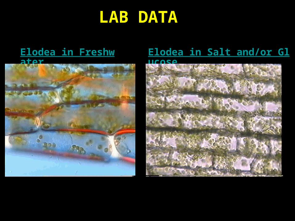
Elodea in Freshwater Elodea in Salt and/or Glucose
LAB DATA
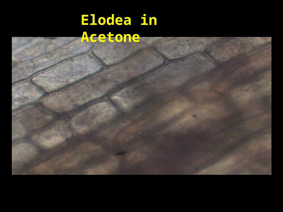
Elodea in Acetone

V. Facilitated Diffusion
A. Definition: Movement of specific molecules from a high to a low concentration with the help of specific transport proteins
B. Characteristics of transport protein
1. No Extra energy requirement High Low gradient
2. Can change shape
3. Shape change may open or close channels
4. Works like an enzyme
Ion Channel Transport Glucose Transport
Inside Cell
GlucoseInsulin
Transport Protein
Open Channel
Examples: Ion Channels, Glucose transport, Aquaporins
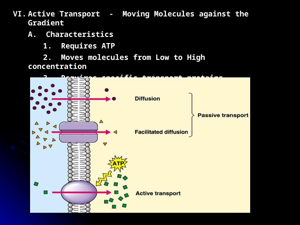
VI. Active Transport - Moving Molecules against the Gradient
A. Characteristics
1. Requires ATP
2. Moves molecules from Low to High concentration
3. Requires specific transport proteins

B. Active Transport of Small Molecules
1. Usually involves the transport of Ions (why?)
2. Examples
a) Sodium potassium pump
b) Proton (Hydrogen) pump Electrogenic pumps
c) Co-Transport pumps - Active transport of H+ coupled
with the passive transport of a different molecule
C. Active Transport of Large Molecules (Bulk Transport)
-All require fusion of membranes
1. Exocytosis - Bulk transport outside the cell
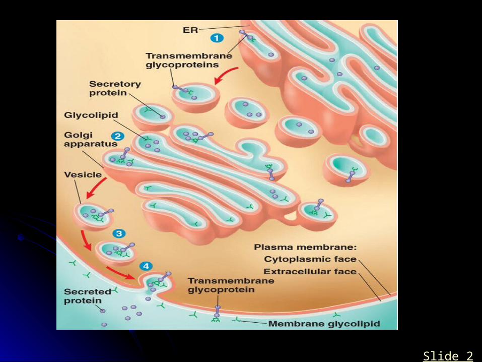
a) Golgi Body Secretions
b) Contractile Vacuoles “The Protozoan Sump Pump”07-16-EndomembraneSystem.mov
Cell 80% H20 Environment 99% H20
H20
Contractile VacuoleH20
Paramecium
Active transport of water to the outside of the cell
How the pump actually works
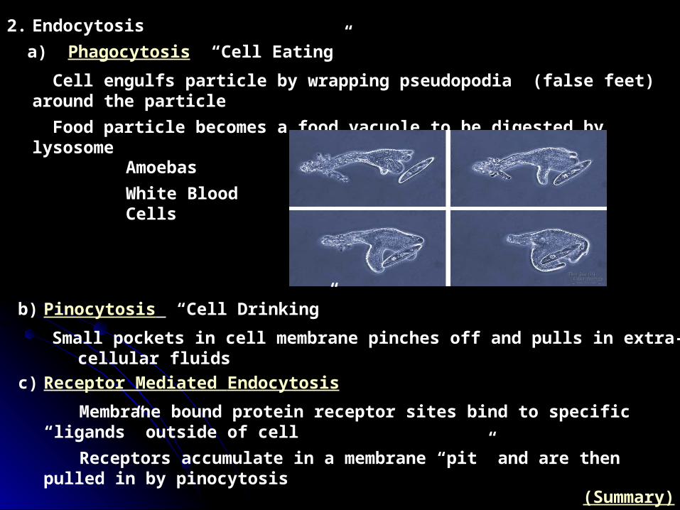
2. Endocytosis
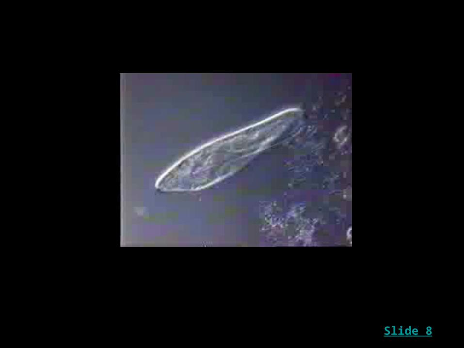
a) Phagocytosis “Cell Eating”
Cell engulfs particle by wrapping pseudopodia (false feet) around the particle
Food particle becomes a food vacuole to be digested by lysosome
Amoebas
White Blood Cells
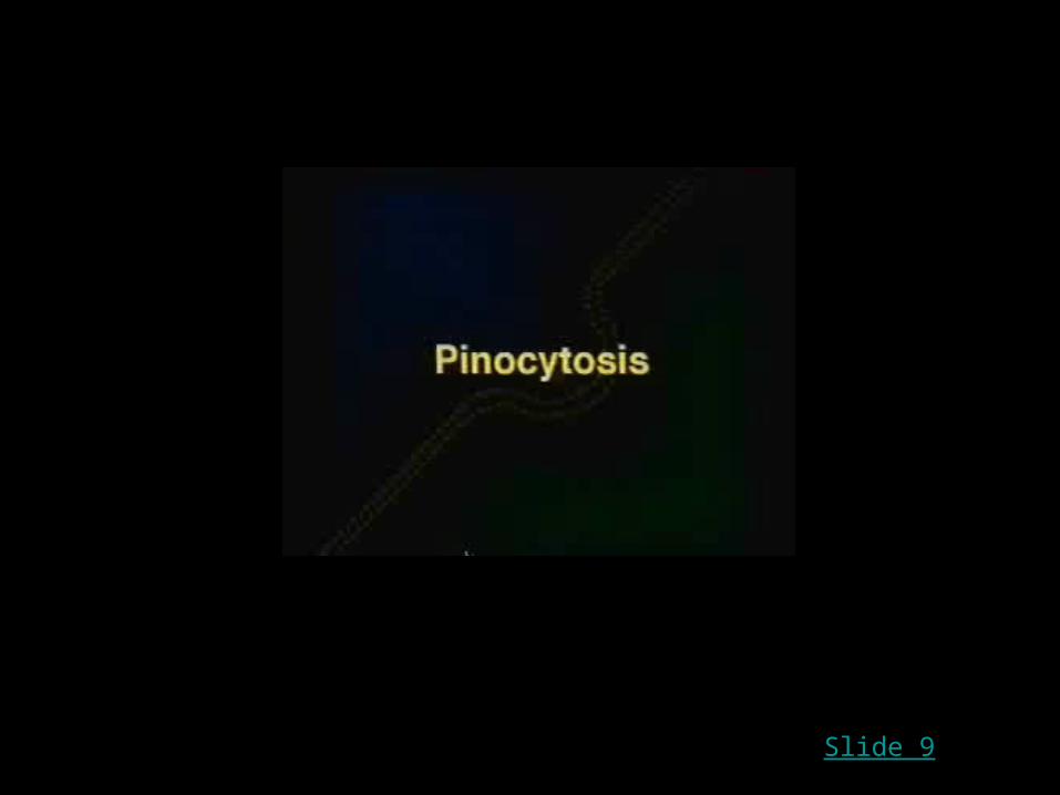
b) Pinocytosis “Cell Drinking”
Small pockets in cell membrane pinches off and pulls in extra-cellular fluids
c) Receptor Mediated Endocytosis
Membrane bound protein receptor sites bind to specific “ligands” outside of cell
Receptors accumulate in a membrane “pit” and are then pulled in by pinocytosis
(Summary)


A Small Phospholipid Drop In Water
“Miscelle”
Phospholipid bilayer forms when larger quantities are immersed in water
“Amphipathic Molecule”
Slide 2
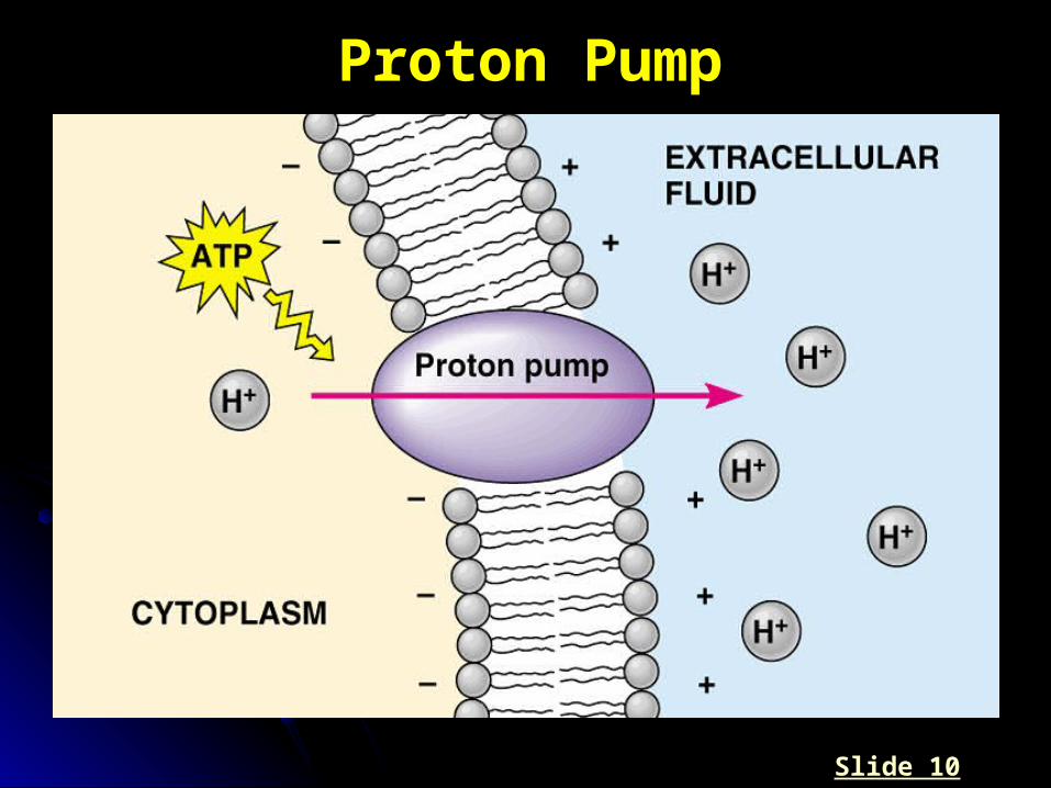
Proton Pump
Slide 10

Proton Pump (active transport) coupled with a cotransport protein (passive)
Slide 10
Slide 2
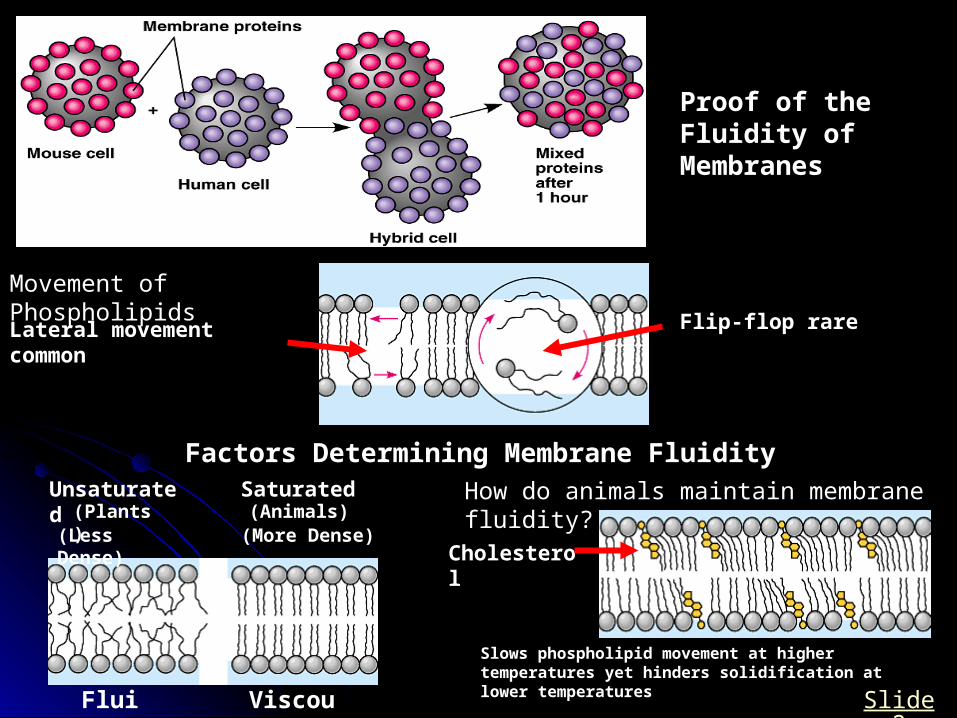
Proof of the Fluidity of Membranes
Movement of Phospholipids
Lateral movement common Flip-flop rare
Factors Determining Membrane FluidityUnsaturated Saturated
(Less Dense) (More Dense)
Fluid Viscous
(Plants) (Animals)How do animals maintain membrane fluidity?
Cholesterol
Slows phospholipid movement at higher temperatures yet hinders solidification at lower temperatures
Slide 2
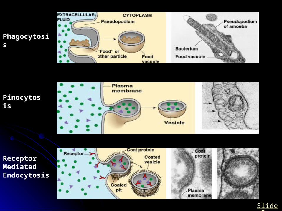
Phagocytosis
Pinocytosis
Receptor Mediated Endocytosis
Slide 9
Slide 1

Slide 3“Porin” an Example of a Transport Channel
Slide 8
Slide 9
Slide 9

Slide 9
Receptor-mediated Endocytosis (pinocytosis)

Slide 5
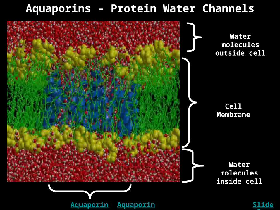
Aquaporins – Protein Water Channels
Water molecules outside cell
Water molecules inside cell
Aquaporin I
Cell Membrane
Slide 8Aquaporin II

The Sodium Potassium Pump in Action
Slide 10
Inside the Cell
Outside the Cell