Embed Size (px)
Citation preview

This is an English translation from Japanese of: Harada, M., et al., 2011. Mercury Pollution in First Nations Groups in Ontario, Canada: 35 years of Canadian Minamata Disease. Journal of Minamata Studies 3: 3-30.
Mercury Poisoning in First Nations Groups in Ontario, Canada 35 years of Minamata Disease in Canada
Masazumi Harada, Masanori Hanada, Masami Tajiri, Yukari Inoue, Nobuyuki Hotta,
Tadashi Fujino, Shigeru Takaoka, and Keishi Ueda
Abstract In 1969, it was revealed that a river system in Ontario, Canada had been contaminated with mercury emitted by a caustic soda factory located upstream. The residents in the 2 Indigenous communities along the river near Kenora, Ontario were poisoned by eating fish contaminated with mercury. In 1975, we proved that the mercury pollution was affecting the residents’ health through our clinical research on the contaminated residents. In 2002 and 2004 we carried out follow-up research. This paper discusses the results of the most recent clinical research, conducted in March 2010. Our target group consisted of 73 residents from Grassy Narrows (Asubpeeschoseewagong) and 87 from White Dog (Wabaseemoong), totaling 160 who were over 20 years old. Various subjective symptoms were confirmed, such as difficulty seeing (38.1%), insomnia (37.5%), exhaustion (37.5%), visual disturbances (37.5%), fatigue (36.1%), and numbness in the limbs (35.6%). From the previous survey, numbness (72.0%) and pain in the limbs (61.8%) remained unchanged. Neurological symptoms including sensory disturbances on the limbs were recorded at 43.7%, identical to the previous survey, while sensory disturbances through the entire body were 16.2%, and sensory disturbances around the mouth were 16.8%. The survey also recorded hearing impairment (35.0%), concentric constriction of the visual field (16.2%), tremors (28.1%), and ataxia (15.0%). Furthermore, adiadochokinesia was shown in 17.5%, difficulty walking a straight line in 39.3%, finger-to-nose test disabilities in 20.6%, knee-to-heel test disabilities in 9.3%, speech impairment in 9.3%, and disturbed ocular movement (Saccadic movement) in 6.8%. There were many difficult cases to diagnose, because the symptoms continually changed. It was especially hard to diagnose the sensory disturbances through the entire body, and the difficulties standing and balancing as they were extremely changeable. On the other hand, the research shows clear symptoms of mental impairment, such as intellectual disabilities (11.8%), as well as emotional and motivational disturbances (20.6%). Above all, depression (13.1%) was more noticeable than in the last research. The causes for these mental symptoms may be: the long period of time passed, little official recognition of the disease, little compensation made, and low understanding of the medical tests. Under our diagnostic criteria used in Minamata City, 33.7% of the target group would be diagnosed as Minamata Disease patients; and 25.0% would be suspected Minamata

Disease patients with light or changing symptoms. A total of 58.7% was affected by mercury. This reinforces the conclusion that the residents suffer from the effects of Minamata Disease. However, only 15% of those who were tested have been given some compensation. The effect of mercury contamination on fetuses remains an area for future study.
Introduction Canadian mercury pollution surfaced as an issue due to the mercury pollution of the Great Lakes in the USA. In 1969 N. Fimreit investigated mercury pollution throughout Canada and pointed to the basins of Wabigoon and English Rivers in Ontario as being particularly polluted (Showa 44).1 The mercury levels in the fish and waterfowls of these areas were shown to be high.1,2,3,4,5 The pollution source was found to be Dryden Chemical’s caustic soda factory upstream. The pulp industry is one of the main industries in Canada and caustic soda is used to bleach the pulp. Mercury was used as a catalyst to purify the caustic soda. Eugene Smith, an internationally acclaimed photojournalist, and his wife Aileen Smith communicated this news to us which led us to investigate Canadian mercury pollution.6 The fields in question were two Indigenous communities named Grassy Narrows (GN) and Whitedog (WD) near Kenora, Ontario.
The first investigation took place in March 1975 (Showa 50) with Harada (then at Kumamoto University, Institute of Constitutional Medicine), Ken-ichi Miyamoto (then at Osaka City University, School of Economics), Jun Ui (then at Tokyo University, Faculty of Engineering), Kiyoshi Karaki (Chunichi Shimbun newspaper), and Aileen Smith. As we grasped the gravity of situation, we followed-up with an in-depth, multimodal investigation in August of the same year with Harada, Miyamoto, Ui, as well as Tadashi Fujino (then at Minamata Kyouritsu Hospital), Taketoshi Akagi (then at Kumamoto University, School of Medicine), Junko Nakanishi (then at Tokyo University, Faculty of Engineering), and Nobuko Iijima (then at Tokyo University, Department of Public Health. We kept in touch with Minamata thereon at the levels of public administration, medicine, and victims. From the clinical and epidemiological investigations in 1975, we reported the confirmation of organic mercury poisoning (Minamata Disease) to the Canadian public administration, researchers, and site residents. 7, 8, 9, 10 Ten years later, in 1986 (Showa 61), the corporation responsible for the pollution, Provincial government, and Federal government pooled funds to found the Mercury Disability Board, and are paying a certain amount of compensation to the patients.11 However, the government claims that it “has not admitted that [the condition suffered

by the patients] is Minamata Disease. The relief is in place because there has been mercury pollution, and there are residents of the polluted area who exhibit symptoms.” 11, 12, 13 The Board’s standard criteria of acknowledgement includes: sensory impairment, narrowed visual field, hearing difficulty, ataxia, hearing impairment, tremor, decrease or loss of tendon reflex. Points are then assigned based on these criteria to rank the monetary benefit. Even so, the Head of the Acknowledgement Committee insists: “it is not Minamata Disease. Because there are residents who suffer from various health issues in the mercury pollution district, this policy is to relieve them.” Later, in August 2002, we conducted a field study in GN with Dr. Fujino (then at Sakuragaoka Hospital). In August 2004, we did a field study in GN and WD with Dr. Fujino, Dr. Kazuhito Tsuruta (then at Koga Hospital Internal Neurology), Dr. Akira Fukuhara (Idemizu Hospital), Masanori Hanada, Takashi Miyakita (Kumamoto Gakuen University, Faculty of Social Welfare Studies), Chihito Araki, Masami Tajiri, Itsuka Nagano (then at Kumamoto Gakuen University, Faculty of Social Welfare Studies). For the most recent research, we conducted a field study between March 22 to and March 30, 2010 (Heisei 22) in the two reserves. 14, 15 As in Minamata District, the mercury levels in fish has declined, and resident hair mercury levels have also declined; however, influence of methyl-mercury is clearly visible even in those residents who were not officially acknowledged. This draws a parallel with the issue of tardy acknowledgement in Japan. It is unfortunate that no data exists on fetal Minamata Disease, and no investigation has taken place. Cerebral palsy patients were also acknowledged as a target of compensation, but situations surrounding it were not clarified.
1. 1975 Research 1.1 Environment & Way of Life There are many large and small lakes in the English-Wabigoon River system, a complex river system, which can make it hard to distinguish which way the current runs. Some locals even say it would take 100 years for the water to reach the ocean. The river is very wide and the current is slow. We need to consider how this fact affects the contamination.
GN and WD are located on this river system. GN is located 100 miles and WD is 200 miles from the pulp factory. The people along this river system made a living by commercial fishing, hunting, guiding, forestry, and gathering wild rice. The area of their activity is extensive, from Clay Lake, Indian Lake, Sydney Lake and Ball Lake to Tetu Lake (see Figure 1; notes 7, 8, 9 ).
Local people still eat a lot of fish. During the winter, the consumption of fish decreases, but fishing continues. After 1970, fish consumption deceased. However one-third of the

people interviewed confirmed that they were “still eating fish.” The amount of consumption is between a half pound and 2 pounds per day.
In April 1970, the Ontario Government banned commercial fishing in the English-Wabigoon River system. Since the main local income sources were commercial fishing and guiding for sports fishing, Indigenous people were hit badly. For example, there were 66 people on public assistance in 1968 in GN, but by 1973, that number increased to 346 people (80% of the population in that area). They were paid only $35 per month (1973). According to statistics, an Indigenous person's average annual income was only a quarter of the lowest income of a white person.12
People in the tourism business were also damaged by the prohibition of commercial fishing and thus, took their case to court. Indigenous people were only partially compensated for the loss of commercial fishing, and not adequately. They were required to submit the payment of tax records and purchase orders from brokers.11, 12
Figure 1: Map of the Area
From Fimreite, N. and Reynolds, L. M. J. of Wildlife Management 37 (1973), 63
1.2 Environmental Contamination by Mercury
In May 1975, there was a case of mercury contamination caused by a pulp mill in Manitoba. Although the Government of Manitoba paid compensated for commercial fishing, it was found not guilty in court. The reason cited was: “not enough evidence.”

The pulp factory in Dryden responsible for the contamination is located in a thinly populated region of Ontario, Canada. As in Minimata, there are few factories in the area. The factory told our research group: “Our factory is regulated by the government and we follow the rules of the government. If there is a problem, they should be compensated by the government.” It was calculated that there were at least 7 tons of mercury released into the river system (compared with 200-600 tons in Minamata.) According to a Canadian scientist's calculation, when the sample was dried 8.4 ppm of mercury was found within 2cm of the surface of Clay Lake’s lake bed and 7.8 ppm within 6cm. This is a high amount (Armstrong, F.A. 1973). Approximately 2,000 kg of mercury remain in the whole of Clay lake. 7, 11, 17
Mercury contamination was very serious at that time and there is plenty of data about contaminated fish from these river systems. Since 1970 - when contaminated fish were found in the Wabigoon River with a mercury level of 16 µg/g - the highest level of mercury in fish reached 27.8 ppm at the peak of contamination.1, 3, 5, 7, 8
Furthermore, in 1973, 19.71 ppm was the highest rate of mercury found in fish from Ball Lake and 9.12 ppm was the highest rate found in fish from Clay Lake. This reveals the extent to which fish in this river system had been contaminated by mercury. The rate of mercury upstream from the factory in Dryden was much less: 0.1 ppm. Further downstream, past the initial settling area of mercury, the rate also decreases. This indicates that the factory in Dryden is the source of mercury contamination. After contamination in Minamata, dead fish appeared on the surface of the sea. Then cats seemed to go crazy, birds fell from the sky, and chickens, dogs, pigs, and weasels showed symptoms of Minamata Disease. Around the 2 Ontario reserves, mink (Mustela vison) and otter (Lutra vison) disappeared before 1970. Domesticated cats are very rare in this area. Then it was observed that the flight of turkey vultures was affected. They flew strangely. The rate of mercury contamination in turkey vultures was 96.7 ppm in the liver and 17.4 ppm in the breast meat. A high rate of mercury was also found in waterfowl liver, muscle, and eggs. In spite of this obvious evidence, the government and citizens did not take this seriously and were not alarmed. Adolf Kizikas lives in WD. His cats delivered 3 kittens on May 10, 1973. One kitten died right after it was born. Since August 10, 1973, a surviving kitten started to show a strange way of walking by turning in circles, increased salivation, and convulsions. The cat’s body was examined on February 20, 1974. This cat’s walking was recorded on 8mm film. It was obviously showing ataxic walking symptoms.

The cat’s brain was sent to Dr. Tadao Takeuchi at Kumamoto University. It was confirmed that it had the cat version of Minamata Disease. The cat’s mercury rates in each part of the body were: 12.4 ppm in the brain, 17.8 ppm in the blood, 67.1 ppm in the liver, 13.4 ppm in the kidneys, and 592.0 ppm in hair. In addition, we went to the Food Research Laboratories, Protection Branch, in Ottawa. We found that an experiment they conducted succeeded in recreating the cat version of Minamata Disease by administering contaminated fish from Clay Lake into the cat’s food.
1.3 Hair Mercury Levels and Clinical Symptoms of the Residents The measurement of hair mercury level in Minamata Disease was begun by Shouji Kitamura (Kumamoto Univ. School of Public Health) and used as reference for diagnosis.4 In Canada too, hair mercury levels have been measured since 1970. 18. 19 The highest hair mercury level measured in 1970 was 95.77 ppm in GN and 198 ppm in WD.7 Since banning fish consumption, the hair mercury levels of the residents has clearly declined. In our investigation in 1975 (Showa 50), the hair mercury level of those who answered “not eating fish since 1970” were low, and those of people still eating fish, like tour guides, were high. The maximum value was 80.3 ppm, and 23 out of 71 examinees exceeded 30 ppm. At the same time, Clarkson et al. from the USA also measured a maximum hair mercury level of 105 ppm.7, 8, 22, 23, 24 Furthermore, the residents consume more fish in the summer because the lakes are frozen during winter. Considering this and cutting the residents’ long hair into 3cm sections, the sections that sprouted in the summer had high mercury levels, and those that sprouted in the winter had low levels. In conclusion, the residents are poisoned by mercury during the summer due to higher fish consumption (see Figure 3).7, 8, 22, 23, 24 The blood mercury levels of residents from both reserves assessed by Canada’s Environmental Health Service Branch exhibited a maximum of 385 ppb. But this is on a global decline since 1973.9, 23, 24 However, residents with exceptional blood levels above 100~200 ppb are also being found. Such findings indicate that a few people have not stopped consuming fish while the resident population as a whole is walking away from fish consumption. Many of these people were tour guides, indicating that while occupational fishery has been discontinued, tour guides are still consuming fish at work alongside the tourists.8, 9, 15
Figure 3: Hair Mercury Level by Season
In August of 1975, we randomly selected 89 residents (mainly consisting of tour guides and fishers) and conducted hair mercury level analysis and health surveying. Popularly self-reported symptoms included: limb pain (40 cases), numbness/tingling (28 cases), and leg cramps (16 cases). These symptoms are also seen in organic mercury poisoning (Minamata Disease). Neurological symptoms included: 12 cases of eye movement disturbance, 15 cases of sensory impairments in the extremities, 5 cases of perioral sensory impairment, 9 cases of loss of peripheral vision, 21 cases of tremor, 8 cases of ataxia, 5 cases of speech impairment, and 40 cases of hearing impairment.7, 16 Given these findings, a conservative diagnosis resulted in “at least 7 cases of suspected organic mercury poisoning (Minamata Disease),” excluding other concurrent illnesses and regarding family and social situations. However, expanding the scope beyond the examinees, there are other suspected patients: “it is inevitable to conclude that methyl-mercury influence is indeed starting to manifest in the human body, epidemiologically regarding the fact that many residents exhibit neurological symptoms, and that narrowed visual field and cognitive impairments are found in families.” 7, 12, 13 However, such a conservative approach towards diagnosis undeniably contributed to the Canadian government’s hesitation to diagnose Minamata Disease.
Mer
cury
Lev
el
Hair length
X axis: The numbers on the first two rows denote length from root, ~3 ~6 ~9…cm The first row denotes hair collected in Aug. 1975. The second row denotes hair collected in Mar. 1975. The numbers on the bottom row are months: Jun, Mar, Dec, Sep, Jun, Mar, … of years 1975, 1974, 1973, 1972. Below the diagram: Analysis by Mr. Susumu Nishigaki, Tokyo Metropolitan Institute of Public Health

1.4 Destruction of Social Culture Both reserves were almost forcefully relocated from their ancestral living grounds due to road and dam constructions. WD has had electricity since 1968, but GN still did not have electricity in March 1975 when we first visited it. GN did have electricity in August 1975, but residents were made to buy washing machines and refrigerators that were of no practical use. Tap water and sewage were available only at schools and teachers’ homes. Most toilets freely discharged contents to the ground or in vaults. Most residents were fishers and hunters, with tour guiding (for fishing and hunting for white customers) as a side job. Amidst the destruction of their traditional life, customs, and culture, the fishing ban and declining tourist population due to mercury pollution exacerbated the situation. Many who lost jobs fell victim to alcoholism.11, 12
We were asked to meet the Kenora district police officers at our first visit. As we were wondering what happened, the chief officer asked us out of the blue whether Minamata had a high crime rate. He kept speaking to my confused face: “Doesn’t Minamata Disease make people drink alcohol and become vicious?” When I answered no, he showed us a dataset, commenting: “perhaps, then, the high crime rate amongst the First Nations people in the mercury-polluted district of Kenora is not related to mercury pollution?” According to the dataset, there had been 189 accidental deaths of Indigenous people in this district in 3.5 years since 1970. We were surprised not only by its number and rate of deaths, but also by its contents.
With accidental deaths, there were twice as many males as females (see Table 1). 24 cases involved people less than 10 years old and were categorized as parental violence, negligence, and freezing from abandonment (or perhaps forgetting to take the children back home). The breakdown of deaths by causes revealed drowning, gunshots, stabbing, strangling, burning, and child abandonment as the main causes. These statistics no doubt present a dreadful condition. The criminal arrest rate was also high. The male rate was 110.6 out of 1,000 and the female rate was 55.6 out of 1,000. This was more than twice as much as the second worst and any other reserve. On the one had we could understand the Kenora physicians’ words: “I am amazed that you even go to such dangerous place.” On the other hand, we were dumbfounded by the strong bias and contempt by the physicians who commented that “there was only alcoholism and no mercury poisoning” or by the sarcastic remarks such as: “it will be a great place for alcoholism study for a neuropsyciatrist like you.” Indeed, the scheme became apparent that, be it alcohol dependency or mercury poisoning, it happens where discrimination exists.10. 11

Table 1: Causes of Accidental Deaths on the Reserves (1970-1973)
Male Female Total
Vehicle accidents 9 7 16
Burning to death 22 8 30
Drowning 35 7 42
Railroad accident 7 3 10
Child accident 2 0 2
Child abandonment 10 15 25
Gunshot, stabbing, strangling 26 12 38
Battery 3 1 4
Alcohol poisoning 6 6 12
Unknown 6 4 10
At the time, alcohol imports were banned on the reserves, which, contrary to the aim exacerbated the situation. According to Dr. Newbury, a Quaker physician devoted to working in the reserve, once the residents craved alcohol, any material thought to contain alcohol, from hair spray to perfume to pesticides—unbelievably—went into their mouths. We used to understand that poverty and discrimination occurred as a result of pollution, but indeed it was the opposite: pollution occurred where there was poverty and discrimination. 8, 11, 12, 13
2. 2002 and 2004 Research Results 2.1 Governmental Policies that Followed Inspired by our research, the Canadian Government hired a U.S. research team to investigate the two reserves. Their research results were almost the same as ours (ie: hair mercury levels etc). Dr. Clarkson, a U.S. team member from Rochester University, wrote a report that warned of fetal prenatal organic mercury poisoning -- Minamata Disease -- particularly. However, because Dr. Clarkson was not a clinician, his data on mercury poisoning remained 'unofficial.' After Clarkson's reports were provided, the provincial government set-up long-term monitoring of hair mercury levels found amongst residents in the contaminated area.14,15,23
In 1986 (Showa 61), a system to pay compensation to patients with particular symptoms was established. Seed money was paid by polluting industries and the provincial and federal governments. It was explained to us that contributions were made because the polluting industry was responsible for mercury contamination; the provincial government was responsible for careless monitoring of industry; and the federal government has the responsibility to protect Indigenous people. The Mercury Disability Board decides whether to pay compensation as well as what amount of compensation. The Board was formed with a chairperson, two doctors, one person from each of the two reserves, and two discussion committees consisting of governmental officials and residents. The medical examiner is only permitted to report the examination results (clinical symptoms) to decide whether or not patients qualify for compensation. The board scores each symptom. It issues $250 to $800 per month per person according to symptomatic scores. Essentially the scores are given according to sensory impairment of peripheral limb dominance, constriction of the visual field, ataxia, speech impariment, hearing impairment, tremors, and the loss of tendon reflex. All of the above symptoms are identical to symptoms of Minamata Disease. Though this is clear, the government insisted on proof that neurological symptoms found in the community were not attributable instead to diabetes or other conditions with symptoms similar to those found in Minamata. he doctor commissioned to the case by the Canadian government still refuses to officially accept the scientific fact of Minamata Disease. In a sense, it was clearly a political resolution. 2.2 Clinical Symptoms 2.2.1 The Survey We conducted two medical examinations in GN and WD. One was from August 31 to September 3, 2002 with for 57 people in GN (Heisei 14). The other was from August 27 to September 2, 2004 with 156 people total (Heisei 16). (There were 26 duplicate examinees from 2002 in both of the reserves). In 2005, we made a report in English about the 57 examinees in GN. The report was as follows:
Self-Reported Symptoms: numbness (66.7%), Limb pain (45.6%), and limb cramps (42.1%).
Neurological Symptoms: sensory impairment in the limbs (54.4%), walking balance disorder (36.8%), tremors (21.1%), perioral dysesthesia (15.8%), and mental disability (15.8%).
Then we completed another set of examinations in GN and WD in 2004. Excluding the duplicate examinees from 2002, there were 187 people surveyed -- 108 males and 79 females. 12 people were between one and ten years old; 12 were teenagers; 12 were in

their twenties; 35 were in their thirties; 27 were in their forties; 40 were in their fifties; 30 were in their sixties; 13 were in their seventies; and 6 were in their eighties. The oldest examinee was 90 years old. However, some people were unsure of their precise ages. 2.2.2 Self-Reported Symptoms Self-reported symptoms were indefinite for examinees under ten years old, leaving a remaining sample population of 175. The most common symptoms were numbness (126 people or 72%). The second most common was pain in the limbs, joints, and back (107 people or 61.1%). Other symptoms included: decreased vision (70 people or 40%), impaired hearing (66 people or 37.7%), cramps in the limbs (59 people or 33.7%), dizziness (47 people or 26.8%), tendency to fall (39 people or 22.2%), forgetfulness (39 people or 22.2%), impaired finger movement (27 people or 15.4%), tremor (21 people or 12%), Dysesthesia speech impairment (18 people or 12%). These self-reported symptoms are recognized as the same symptoms as Minamata Disease. 2.2.3 Neurological Symptoms According to our findings, there should be no doubt that the sensory impairment is a result of methylmercury contamination. Symptoms included: peripheral limbs dysesthesia (114 people or 65.1%), perioral dysesthesia (35 people or 20.9%), and systemic dysesthesia (32 cases or 18.2%). The above results confirmed our assumption of Minamata Disease (though the totals won't match because of some duplicate examinees): Ataxia (44 people or 23.7%), standing / gait disorder (36 people or 19.2%), hearing impairment (47 cases or 25.1%), tremor (37 people or 19.7%), constriction of the visual field (19 people or 10.1%), oculomotor disorder (17 people or 9%), intellectual disability [including dementia] (114 cases), seizures (6 cases), syncope (5 cases), muscular dystrophy (3 cases) were confirmed. 2.2.4 Complications There are many complications because the contaminated area has relatively poor medical access. Therefore, it is difficult to track the effects of methylmercury dysesthesia. As follows, there were diabetes (42 cases), heart disease (19 cases), stroke (13 cases), thyroid dysfunction (7 cases), Kennedy-Alter-Sung-Syndrome (5 cases) [we could not confirm muscular dystrophy].

Other symptoms are as follows: Dystrophy, impaired spinal column, traumatic fracture, cancer, tuberculosis, thinner intoxication, liver disease, kidney disease, Burgers disease. There was especially high incidence of diabetes among patients (24% excluding examinees under 20 years old, and 25.7% between 20 and 30 years old). 2.3 Diagnosis 60 people (excluding 34.2% under ten years old) were deemed to have 'Minamata Disease', confirmed with perioral and limbs dysesthesia, ataxia, oculomotor disorder, standing balance disorder, constriction of the visual field, and aphasia. 54 cases (30.8%) were deemed to have 'Minamata Disease + Complications' because they have complications and other diseases on top of Minamata Disease. 25 cases (14.2%) were deemed with 'Suspicion of Minamata Disease' because their Minamata Disease symptoms fluctuated or were not entirely clear. We confirmed 7 cases of pediatric cerebral palsy and intellectual disabilities among children under ten years old. However, it was difficult to confirm these afflictions as congenital Minamata Disease. We are therefore putting these cases on hold. From our subjected examinees, 54 people (28.8%) were certified as having Minamata Disease. 77 people (41.1%) were rejected. The rest of the people (7) were put on hold or were otherwise not claimed. The certification standard was higher than we had previously assumed. Only 21 cases out of 60 people (35%) who had been diagnosed with 'Minamata Disease' were certified in our examination. 27 cases (50%) out of 54 people who were diagnosed with 'Minamata Disease + Complications' were certified. 5 people out of 25 who had 'Suspicion of Minamata Disease' were certified. These results indicate the difficulty in Canada in certifying symptoms as absolutely attributable to Minamata Disease. We re-examined 27 people who were examined in 1975 (Showa 50). Their symptoms were clearly getting worse. 21 cases (77.7%) out of those 24 cases (88.8%) were certified. For those re-examined, we considered the relationship with hair mercury levels, but no one had a hair mercury level higher than 50ppm. This result suggests long-term mercury contamination can generate Minamata Disease even among people with hair mercury levels lower than 50ppm. 2.4 Hair Mercury Levels We re-tested for hair mercury levels during our examination. We tested 200 total mercury samples from 151 people (61 female, 90 male) -- 83 GN cases and 68 WD cases. Workers from the contaminated mill in Dryden wanted to have their hair mercury levels analyzed. Their hair mercury levels were lower than we had predicted, but two people's levels were 25ppm and 11ppm in Grassy Narrows.

Case 1: 69 year-old male, born April 1935 (Showa 10) -- hair mercury level 25 ppm He has some self-reported symptoms such as numbness, headaches, malaise, exhaustion, cramping, and tremors. Other symptoms include perioral and limbs dysesthesia, tremor, adiadokokinesis, nose finger test disorder, standing/gait disturbance, intellectual disabilities of memory and memorizing, heart disease etc. He was certified. Case 2: 50 year-old male, born February 1954 (Showa 29) -- hair mercury level 11 ppm He claimed numbness, ringing in the ears, hearing impairment, fingertip dysesthesia, and tremors. He also had blindness in the left eye, standing imbalance, loss of leg muscle strength, and head and limb dysesthesia. Despite these symptoms, he was not certified. No one in WD showed a hair mercury level greater than 10 ppm. However, in GN a woman born in 1977 (Showa 52) had 9.1 ppm; a male born in 1923 (Taisho 12) had 7.5 ppm; a female born in 1931 (Showa 6) had 7.4 ppm; a male born in 1935 (Showa 10) had 7.3 ppm; a male born in 1950 (Showa 25) had 7.3ppm; and a male born in 1949 (Showa 24) had 7.1 ppm. They were all certified. However, the majority in both reserves showed low levels (less than 2ppm). The workers from the Dryden mill showed between 0.18 ppm and 2.9 ppm. In 1975, we found clear seasonal variations in hair mercury levels (Figure 3). We applied mercury analysis to every 3cm of long hair in 2004. There were no significant variations. Therefore, it is difficult to say whether the two reserves' residents are in danger of mercury poisoning. However, this does not deny their current symptoms as occasioned by mercury poisoning. If one considers that effects are the result of past contamination, these could signify delayed onset Minamata Disease, or oversight, or potential Minamata Disease.
3. 2010 Survey Results It has been 35 years since we first went to the two reserves in Ontario, Canada to investigate their mercury pollution. As it was clear in the cases of Minamata and Niigata, resolution is not easy once the environment is polluted with mercury. We have experienced that, even if the pollution itself could come to an end, the impact on the health and socio-economic life of the people throughout the area is immense.12, 16 Therefore, we organized an investigation team centered on The Open Research Center for Minamata Studies at Kumamoto Gakuen University, and conducted a third on-site clinical survey with the collaboration of the residents and the band office.
3.1 Date & Time, Methods, Investigation Team
The survey was done in the two reserves: GN and WD. Interviews and neurological/psychiatric tests were done on March 25 and 26, 2010 at GN and on March

28 and 29, 2010 at WD. The methods are the same as the diagnostic method used in Japan for Minamata Disease, and have not changed since 1975. In addition, the diagnosticians are experienced in Minamata screening, notably with Harada and Fujino being involved since 1975. In total the team included 5 neurologists/psychiatrists and internal medicine specialists in neurology, 2 nurses, 1 sociologist, 1 analyst, 2 supporters, and 3 on-site supporters. The Chiefs as well as many residents kindly provided us with screening, boarding, and other sites. 3.2 Examinees
In GN, 73 people - 34 males and 39 females - were surveyed. In WD, 87 people - 41 males and 46 females - were surveyed. In total, there were 160 examinees: 75 males and 85 females (see Table 2). There were 14 (19.1%) patients in GN and 33 (37.9%) patients in WD who had family members who were certified patients (eligible for governmental support), making a total of 29.3%. Thus, nearly a third of all examinees are said to have family members who are certified. There were 11 people in GN and 14 in WD who were screened but excluded from the analysis due to being less than 20 years of age. Analysis was limited to 20-year-old or older examinees based on the assumption that mercury pollution ceased 20 years ago. This criterion is only provisional and must be examined again in the future. Excluded examinees consisted of one person under 1 year-old, 10 people between 1 and 5 years-old, 5 people between 5 and 10 years-old, and 9 people between 10 and 20 years-old. That totals 25 people. (See Section 3.3.7 for more). In addition, 5 examinees in GN and 6 in WD did not finish the screening. Table 2: Examinees in Terms of Age and Gender)
GN WD Total
Total M F Total M F Total M F
# Examinees 73 34 39 87 41 46 160 75 85
Ages 20-30 9 4 5 12 6 6 21 10 11
31-40 8 3 5 21 9 12 29 12 17
41-50 21 13 8 22 9 13 43 22 21
51-60 23 9 14 20 11 9 43 20 23
61-70 8 2 6 10 5 5 18 7 11
70- 4 3 1 2 1 1 6 4 2

3.3 Clinical Symptoms
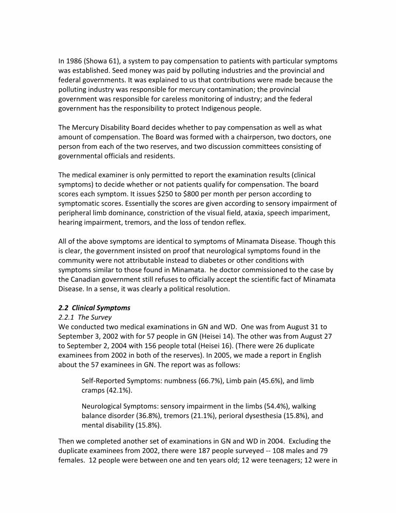
3.3.1 Self-Reported Symptoms Various self-reported symptoms were present (see Table 3). Many were common to Minamata Disease. However, some self-reported symptoms were slightly different from those of residents at the polluted districts of Minamata. In addition, there were differences between GN and WD districts. Possible reasons are social factors in a broad sense, such as changes in the disease image, socio-psychological factors, and the combination with other illnesses. Details must wait for future research. Overall, the most prevalent self-reported symptom was "impaired vision" (38.1%). This was followed by "sleeplessness," "loss of [muscle] strength," and "headache" (37.5%). Then followed "lethargy" (36.1%), "numbness of hands and feet" (35.6%), "impaired hearing" and "leg cramps" (31.2%), "dull sensation in hands and feet," (30.6%), "narrowed visual field" (28.7%), and "tremor" (27.5%). In WD, "impaired vision" was the most prevalent at 43.6% reported, followed by "lethargy" (41.3%), "headache" (40.2%), and "tremor" (39.8%). Then followed "loss of [muscle] strength" (39.0%), "sleeplessness" (37.8%), "narrowed visual field" (34.4%), "numbness in hands and feet" (33.3%), "impaired hearing" (32.1%), and "stiff shoulders" (31.0%). As seen here, the self-reported symptoms in WD were quite original with distinct characteristics. In GN, "numbness of hands and feet" was the most prevalent at 38.3% reported. This was followed by "loss of [muscle] strength" (35.6%), "headache" and "lethargy" (34.2%), "dull sensation in hands and feet," "impaired vision," "impaired hearing," (31.5%), "leg cramps" (28.7%), "sleeplessness" (28.7%), "tinnitus" (26.0%), "upset mood" (26.0%), "tremor" (24.6%), "tendency to fall" (23.2%), and "narrowed visual field," "limb pain", "impaired word production," "anhedonia or apathy," and "irritability" (21.9%). GN examinees shared many common symptoms with one another, and also with residents of Minamata district (see Table 3).
Table 3: Self-Reported Symptoms
GN (73 cases) WD (87 cases) Total (160 cases)
Self-Reported Sx Case count (%) Case count (%) Case count (%)
Numbness of hands & feet 28 (38.3) 29 (33.3) 57 (35.6)
Dull sensation of hands & feet 23 (31.5) 26 (29.8) 49 (30.6)
Dropping things from hands 6 (9.2) 19 (21.8) 25 (15.6)
Perioral numbness (tingling) 6 (9.2) 9 (10.3) 15 (9.3)
Limb pain 16 (21.9) 18 (20.6) 34 (21.2)
Loss of strength 26 (35.6) 34 (39.0) 60 (37.5)
Stiff shoulders 11 (15.0) 27 (31.0) 38 (23.7)
Tremor (hands, feet, etc) 18 (24.6) 26 (39.8) 44 (27.5)
Difficulty producing words 16 (21.9) 22 (25.2) 38 (23.7)
Tendency to stumble 17 (23.2) 14 (15.0) 31 (19.3)
Impaired sight 23 (31.5) 38 (43.6) 61 (38.1)
Narrowed visual field 16 (21.9) 30 (34.4) 46 (28.7)
Impaired hearing 23 (31.5) 28 (32.1) 51 (31.8)
Tinnitus 19 (26.0) 13 (13.9) 32 (20.0)
Impaired sense of taste 13 (17.8) 15 (17.2) 28 (17.5)
Impaired sense of smell 16 (21.9) 20 (22.9) 36 (22.5)
Leg cramps 21 (28.7) 29 (33.3) 50 (31.2)
Headache 25 (34.2) 35 (40.2) 60 (37.5)
Dizziness 7 (9.3) 16 (18.3) 23 (14.3)
Forgetfulness 12 (16.4) 26 (29.8) 38 (23.7)
Anhedonia / apathy 16 (21.9) 15 (17.2) 31 (19.3)
Irritability 16 (21.9) 22 (25.2) 38 (23.7)
Anxiety 19 (26.0) 24 (27.5) 43 (26.8)
Sleeplessness 27 (26.9) 33 (37.8) 60 (37.5)
Depression 14 (19.1) 17 (19.5) 31 (19.3)
Lethargy 25 (34.2) 36 (41.3) 61 (36.1)
3.3.2 Neurological Symptoms Neurological symptoms were highly prevalent in the examinees (because people with some active symptoms tended to sign up for the study). While it was not surprising to find many symptoms common to organic mercury poisoning (Minamata Disease) by the virtue of prior mercury pollution, there were also clinical characteristics that slightly differed from the symptoms found in the (Japanese) Minamata Disease (as discussed
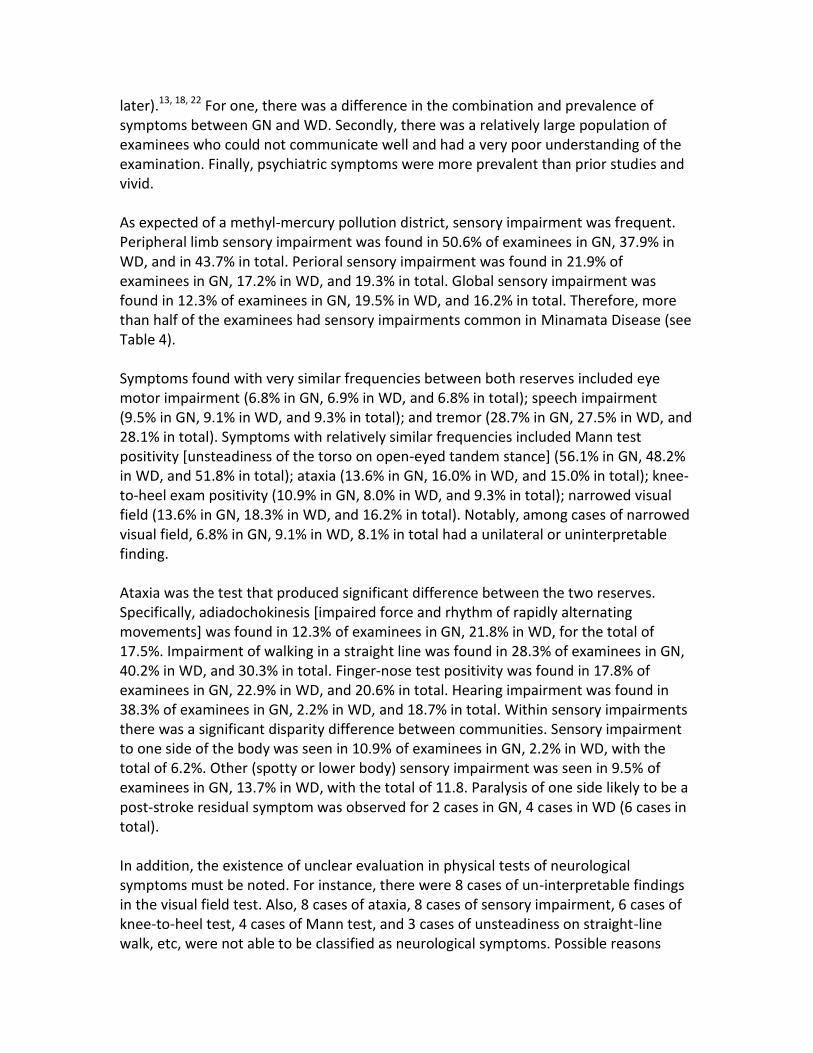
later).13, 18, 22 For one, there was a difference in the combination and prevalence of symptoms between GN and WD. Secondly, there was a relatively large population of examinees who could not communicate well and had a very poor understanding of the examination. Finally, psychiatric symptoms were more prevalent than prior studies and vivid. As expected of a methyl-mercury pollution district, sensory impairment was frequent. Peripheral limb sensory impairment was found in 50.6% of examinees in GN, 37.9% in WD, and in 43.7% in total. Perioral sensory impairment was found in 21.9% of examinees in GN, 17.2% in WD, and 19.3% in total. Global sensory impairment was found in 12.3% of examinees in GN, 19.5% in WD, and 16.2% in total. Therefore, more than half of the examinees had sensory impairments common in Minamata Disease (see Table 4). Symptoms found with very similar frequencies between both reserves included eye motor impairment (6.8% in GN, 6.9% in WD, and 6.8% in total); speech impairment (9.5% in GN, 9.1% in WD, and 9.3% in total); and tremor (28.7% in GN, 27.5% in WD, and 28.1% in total). Symptoms with relatively similar frequencies included Mann test positivity [unsteadiness of the torso on open-eyed tandem stance] (56.1% in GN, 48.2% in WD, and 51.8% in total); ataxia (13.6% in GN, 16.0% in WD, and 15.0% in total); knee-to-heel exam positivity (10.9% in GN, 8.0% in WD, and 9.3% in total); narrowed visual field (13.6% in GN, 18.3% in WD, and 16.2% in total). Notably, among cases of narrowed visual field, 6.8% in GN, 9.1% in WD, 8.1% in total had a unilateral or uninterpretable finding. Ataxia was the test that produced significant difference between the two reserves. Specifically, adiadochokinesis [impaired force and rhythm of rapidly alternating movements] was found in 12.3% of examinees in GN, 21.8% in WD, for the total of 17.5%. Impairment of walking in a straight line was found in 28.3% of examinees in GN, 40.2% in WD, and 30.3% in total. Finger-nose test positivity was found in 17.8% of examinees in GN, 22.9% in WD, and 20.6% in total. Hearing impairment was found in 38.3% of examinees in GN, 2.2% in WD, and 18.7% in total. Within sensory impairments there was a significant disparity difference between communities. Sensory impairment to one side of the body was seen in 10.9% of examinees in GN, 2.2% in WD, with the total of 6.2%. Other (spotty or lower body) sensory impairment was seen in 9.5% of examinees in GN, 13.7% in WD, with the total of 11.8. Paralysis of one side likely to be a post-stroke residual symptom was observed for 2 cases in GN, 4 cases in WD (6 cases in total). In addition, the existence of unclear evaluation in physical tests of neurological symptoms must be noted. For instance, there were 8 cases of un-interpretable findings in the visual field test. Also, 8 cases of ataxia, 8 cases of sensory impairment, 6 cases of knee-to-heel test, 4 cases of Mann test, and 3 cases of unsteadiness on straight-line walk, etc, were not able to be classified as neurological symptoms. Possible reasons
included poor understanding for the test and psychiatric symptoms (hysteria), which was plausible considering the high frequency of psychiatric symptoms (see next section). Table 4: Neurological Symptoms
GN (73 cases) WD (87 cases) Total (160 cases)
Symptom Case count (%) Case count (%) Case count (%)
Eye motor impairment 5 (6.8) 6 (6.9) 11 (6.8)
Hearing impairment
- (bilateral) 28 (38.3) 2 (2.2) 30 (18.7)
- (unilateral) 6 (9.2) 9 (10.3) 15 (9.3)
Speech impairment 7 (9.5) 8 (9.1) 15 (9.3)
Narrowed visual field (bilateral) 10 (13.6) 16 (18.3) 26 (16.2)
Uninterpretable, unilateral 5 (6.8) 8 (9.1) 13 (8.1)
Tremor 21 (28.7) 24 (27.5) 45 (28.1)
Adiadochokinesis 9 (12.3) 19 (21.8) 28 (17.5)
Straight-line walking impairment 28 (28.3) 35 (40.2) 63 (39.3)
Mann test positivity 41 (56.1) 42 (48.2) 83 (51.8)
Finger-nose test positivity 13 (17.8) 20 (22.9) 33 (20.6)
Knee-heel test positivity 8 (10.9) 7 (8.0) 15 (9.3)
Ataxia (total) 10 (13.6) 14 (16.0) 24 (15.0)
Sensory impairment
- (extremeties) 37 (50.6) 33 (37.9) 70 (43.7)
- (perioral) 16 (21.9) 15 (17.2) 31 (19.3)
- (extremeties + perioral) 14 (19.1) 13 (14.9) 27 (16.8)
- global 9 (12.3) 17 (19.5) 26 (16.2)
- one side of the body 8 (10.9) 2 (2.2) 10 (6.2)
- others 7 (9.5) 12 (13.7) 19 (11.8)
One side paralysis (stroke) 2 (2.7) 4 (4.5) 6 (3.7)
Acute attacks 2 (2.7) 5 (5.7) 7 (4.3)
Compared to 2002 (Heisei 14) and 2004 (Heisei 16), despite the decreased frequency of sensory disturbance and ataxia, there was an increase in straight-line walking impairment, Mann positivity, global sensory impairment, and narrowed visual field, to name a few. Especially, the increase in un-interpretable findings (to be re-examined) in tests of narrowness of visual field etc. was characteristic of this round of investigation.14,
20

3.3.3 Psychiatric Symptoms Psychiatric symptoms were difficult to interpret. Especially for intellectual impairment, only the most definite cases were counted due to the language barrier. Because of it, tests were limited to basic assessments such as the current month and date, day of the week, birth year/month/date, age, and comprehension of test directions. Positive findings were seen in 13.6% of examinees in GN, 10.3% in WD, to the total of 11.8%. In contrast, it was relatively uncomplicated to interpret emotional impairments such as those of mood, intension, and volition from the examinee’s facial expression, behavior, exchanges during the screening, and subjective complaints. As a result, 20% were thought to have emotional and mental disturbance. Additionally, we observed 21 cases (13.1%) of depression, 8 cases of anxiety, 4 cases of neurotic tendency with hypochondria, and 9 cases of decreased volition as seen in stiff facial expression or stolid, apathetic, passive tendencies (see Table 5). Self-injury to the wrist was found in 1 male and 3 females in GN, and 1 male in WD, all in the 20-30 age group. It was characteristic of this round of investigation to see these eminent psychiatric symptoms. This was probably influenced in part by the prolonged course of incidence resolution and frustration brought by the lack of new acknowledgement other than for children with severe cerebral palsy. Table 5: Psychiatric Symptoms
GN (73 cases) WD (87 cases) Total (160 cases)
Symptom Case count (%) Case count (%) Case count (%)
Intellectual disturbance 10 (13.6) 9 (10.3) 19 (11.8)
Emotional disturbance 15 (20.5) 18 (20.6) 33 (20.6)
- depression 11 (15.0) 10 (11.4) 21 (13.1)
- anxiety 2 6 8 (0.5)
- neurosis 3 1 4
- decreased volition 2 7 9
- euphoria 2 1 3
3.3.4 Diagnosis of Minamata Disease The purpose of this investigation was to clarify part of the long-term effects on the residents in the polluted districts and the resulting administrative reaction (not discussed in this document) since the studied districts were polluted with mercury. Therefore, the purpose of this round of investigation was also to assess how much influence of mercury poisoning was present in the examinees (residents of polluted districts). In other words, it was to clarify the degree of influence of methyl-mercury in the examined residents based on the diagnostic experience of Minamata Disease in Minamata, Japan.

Here, a diagnosis of Minamata Disease was made for patients who clearly exhibited distal limb sensory impairment, perioral sensory impairment, and global sensory impairment, as well as those who exhibited narrowed visual field, coordinative motor impairment, or speech sound production impairment. There were 30 such cases (41.0%) in GN and 24 cases (27.5%) in WD. Within those who exhibited limb or global sensory impairments, those who also had notable psychiatric symptoms, had conflicting findings with other symptoms, or those with whom it was difficult confirming the symptoms due to other illnesses, were deemed “suspected of Minamata Disease.” There were 12 such cases (16.4%) in GN and 28 cases (32.1%) in WD. Within them, there were 5 cases of 20 years or older examinees suspected of fetal exposure. In all 5 cases, symptoms reminiscent of developmental disorders were confirmed. Within the subjects who were diagnosed or suspected of Minamata Disease, there were 14 cases (19.1% of examinees) in GN and 10 cases (11.4% of examinees) in WD who were officially approved for compensatory fund. It means that 33.3% in GN and 19.2% in WD of those whom we diagnosed or deemed suspicious of Minamata Disease were officially approved. Within those whom we diagnosed or deemed suspected of Minamata Disease, 28 cases (66.6%; 38.3% of all examinees) in GN and 42 cases (80.7%; 48.2% of all examinees) in WD were not yet officially approved, indicating the strictness of approval process - especially in WD (see Table 6). There was one officially approved case with clear indication against Minamata Disease. There were only a few cases in which at least one of the examinee’s family members was an officially approved patient of Minamata Disease: 14 cases (19.1% of examinees) in GN, 33 cases (37.9% of examinees) in WD, with the total of 47 cases (29.3% of all examinees). However, the government does not recognize Minamata Disease while entitling the approved individuals for compensation (as discussed above).

Table 6: Diagnosis of Minamata Disease
GN (73 cases) WD (87 cases) Total (160 cases)
Diagnosis Case count (%) Case count(%) Case count (%)
Minamata Disease 30 (41.0) 24 (27.5) 54 (33.7)
Suspected Minamata Disease
12 (16.4) 28 (32.1) 40 (25.0)
- Total 42 (57.5) 52 (59.7) 94 (58.7)
- (officially approved)
14 (33.3) 10 (19.2) 24 (25.5)
- (fetal) 1 4 5
- (not yet approved) 28 (66.6) 42 (80.7) 70 (74.4)
- (denied MD) 1 0 1
Has approved family member(s)
14 (19.1) 33 (37.9) 47 (29.3)
(The percentage in “officially approved,” “fetal,” and “not yet approved” are out of the number we diagnosed or deemed suspected of Minamata Disease.)
Individuals in their 40s-50s predominated the diagnosed or suspected group. Those in their 20s were present as well, although fewer in number (see Table 7). However, this round of results could not determine how long the pollution lasted and until how late it gave rise to illness. To answer these questions, it is necessary to cover all the residents (the polluted population). The 20s cohort had 21 examinees, out of which 6 individuals or 28.5% had (suspected) Minamata Disease. Similarly, 16 out of 29 cases (55.1%) in their 30s were either diagnosed or suspected of having Minamata Disease. In the 40s cohort it was 31 out of 43 (72.0%). It was 25 out of 43 (58.1%) in the 50s cohort, 12 out of 18 (66.6%) in the 60s cohort, and 4 out of 6 (66.6%) in the 70s cohort. Therefore, the ratio of Minamata Disease patients was high in the 30s cohort and above. This suggests that the influence of organic mercury was notable in ages above 30 (from within our subjects). Across all ages, the majority (58.7%) were diagnosed with or suspected of having Minamata Disease.
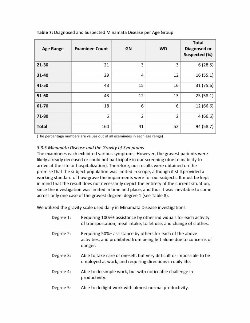
Table 7: Diagnosed and Suspected Minamata Disease per Age Group
Age Range Examinee Count GN WD Total
Diagnosed or Suspected (%)
21-30 21 3 3 6 (28.5)
31-40 29 4 12 16 (55.1)
41-50 43 15 16 31 (75.6)
51-60 43 12 13 25 (58.1)
61-70 18 6 6 12 (66.6)
71-80 6 2 2 4 (66.6)
Total 160 41 52 94 (58.7)
(The percentage numbers are values out of all examinees in each age range)
3.3.5 Minamata Disease and the Gravity of Symptoms The examinees each exhibited various symptoms. However, the gravest patients were likely already deceased or could not participate in our screening (due to inability to arrive at the site or hospitalization). Therefore, our results were obtained on the premise that the subject population was limited in scope, although it still provided a working standard of how grave the impairments were for our subjects. It must be kept in mind that the result does not necessarily depict the entirety of the current situation, since the investigation was limited in time and place, and thus it was inevitable to come across only one case of the gravest degree: degree 1 (see Table 8). We utilized the gravity scale used daily in Minamata Disease investigations:
Degree 1: Requiring 100%± assistance by other individuals for each activity of transportation, meal intake, toilet use, and change of clothes.
Degree 2: Requiring 50%± assistance by others for each of the above activities, and prohibited from being left alone due to concerns of danger.
Degree 3: Able to take care of oneself, but very difficult or impossible to be employed at work, and requiring directions in daily life.
Degree 4: Able to do simple work, but with noticeable challenge in productivity.
Degree 5: Able to do light work with almost normal productivity.

Degree 6: Almost no impediment in regular tasks. Table 8: Degrees of Gravity
GN (73 cases) WD (87 cases) Total (160 cases)
Degree Case count (%) Case count (%) Case count (%)
1 0 1 1
2 4 (5.4) 2 (2.2) 6 (3.7)
3 18 (24.5) 20 (22.9) 38 (23.7)
4 8 (10.9) 11 (12.6) 19 (11.8)
5 21 (28.7) 30 (34.4) 51 (31.8)
6 22 (30.1) 23 (26.4) 45 (28.1)
Total 73 (100) 87 (100) 160 (100)
There were 6 cases (3.7%) of Degree 2, 38 (23.7%) of Degree 3, 19 (11.8%) of Degree 4, 51 (31.8%) of Degree 5, and 45 (28.1%) of Degree 6. This indicates that the examined population was skewed towards lighter gravity.
Since examinees were also hopefuls [i.e. examinees voluntarily signed up], it was expected that the sample population were skewed towards people with various health concerns. So we next analyzed the correlation between the examinees’ diagnosis status and their degrees of gravity (see Table 9). Table 9: Minamata Disease vs. Degree of Gravity
GN (73 cases) WD (87 cases) Total (160 cases)
Degree Diagnosed Suspected Diagnosed Suspected Diagnosed Suspected
1 0 0 1 0 1 0
2 3 1 2 0 5 1
3 11 1 8 6 19 7
4 6 2 6 4 12 6
5 6 7 7 16 13 23
6 4 1 0 2 4 3
Total 30 12 24 28 54 40
In GN, there were no cases of Degree 1 severity. There were 4 cases of Degree 2, which included both diagnosed or suspected cases. All three of the Degree 1 and 2 cases in WD were diagnosed cases. Within the diagnosed and suspected examinees in GN, Degrees 3 and 5 were the two most prevalent (12 and 13 cases each). The more definite the diagnosis (i.e. diagnosed, rather than suspected), the graver was the degree. In other
words, in GN, Degree 3 had only 1 suspected case, while Degree 5 had 7. In WD, Degree 5 contained the largest number of diagnosed or suspected cases at 23 cases, followed by Degree 3, containing 14 cases. In addition, in GN, Degree 5 had the largest number of suspected cases. Therefore, graver Degrees coincided with more diagnosed cases by our diagnostic standard, and lighter Degrees coincided with an increase in suspected cases. This trend also indicates a difficulty diagnosing cases with relatively light symptoms. One characteristic of this follow-up investigation was, as mentioned before, a high percentage of Suspected Minamata Disease at 40 cases (25.0% of total examinees; 42.5% of Diagnosed plus Suspected), nearing that of Diagnosed Minamata Disease at 54 cases (33.7% of total examinees). In previous (2002, 2004) investigations, Suspected cases were 25 out of 139 cases (17.8%). Thus, there were more cases with difficult interpretation of symptoms. There were other cases rendering difficult decisions. For instance, conflicting findings in visual field evaluation were observed in 9 cases, in which the test was either not understood well, produced widely vacillating results, or indicated extremely restricted visual fields. Motor paralysis evaluation also had 8 cases with extreme impairment at the test despite smooth movement in daily functions. Sensory impairment evaluation was also riddled with wide vacillations, including 8 cases with the widest discrepancy, in which tests indicated global loss of total sensory input despite daily activities indicating otherwise. These cases were deemed to require further evaluation, and removed from analysis. Such a situation can in part be attributed to an increasing number of conditions evoking psychogenic reaction: for instance, that the target population consisted of younger individuals and those with lighter symptoms, and that governmental approval criteria became stricter over the years, rendering fewer newly approved patients.
3.3.6 Concurrent Conditions Many concurrent conditions were confirmed, but they were all from interview results and thus had their limitations.14 The most prevalent condition was hypertension (HTN), with 60 cases (37.5%) from GN and WD together. The next most prevalent was diabetes mellitus (DM), with 30 cases (18.7%) (see Table 10). Since the purpose of screening was focused on the (especially neurological) influence of organic mercury, it limited the thoroughness of information gathering and tests on internal medical conditions. Although inputs were restricted to self-reports, HTN and DM still stood out. This is an issue that remains to be addressed in the near future. (While DM was also an issue in the 2002-2004 investigations, it was due in part to the uneven diagnostic standards).
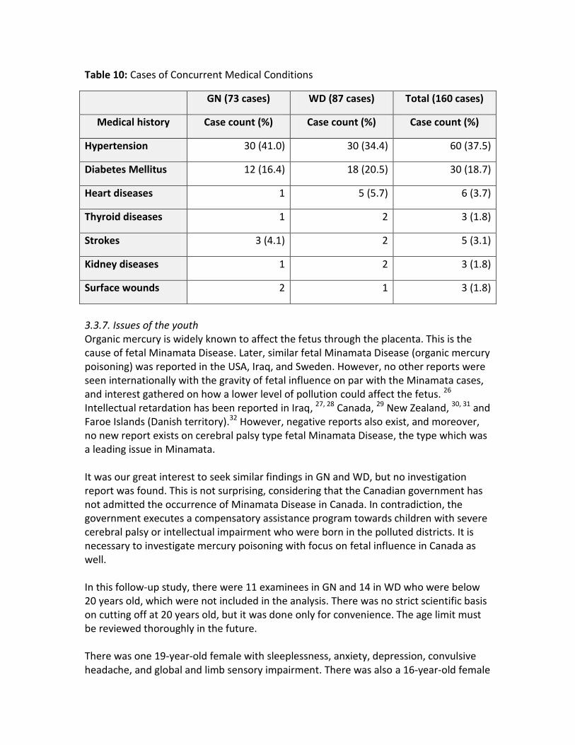
Table 10: Cases of Concurrent Medical Conditions
GN (73 cases) WD (87 cases) Total (160 cases)
Medical history Case count (%) Case count (%) Case count (%)
Hypertension 30 (41.0) 30 (34.4) 60 (37.5)
Diabetes Mellitus 12 (16.4) 18 (20.5) 30 (18.7)
Heart diseases 1 5 (5.7) 6 (3.7)
Thyroid diseases 1 2 3 (1.8)
Strokes 3 (4.1) 2 5 (3.1)
Kidney diseases 1 2 3 (1.8)
Surface wounds 2 1 3 (1.8)
3.3.7. Issues of the youth Organic mercury is widely known to affect the fetus through the placenta. This is the cause of fetal Minamata Disease. Later, similar fetal Minamata Disease (organic mercury poisoning) was reported in the USA, Iraq, and Sweden. However, no other reports were seen internationally with the gravity of fetal influence on par with the Minamata cases, and interest gathered on how a lower level of pollution could affect the fetus. 26 Intellectual retardation has been reported in Iraq, 27, 28 Canada, 29 New Zealand, 30, 31 and Faroe Islands (Danish territory).32 However, negative reports also exist, and moreover, no new report exists on cerebral palsy type fetal Minamata Disease, the type which was a leading issue in Minamata.
It was our great interest to seek similar findings in GN and WD, but no investigation report was found. This is not surprising, considering that the Canadian government has not admitted the occurrence of Minamata Disease in Canada. In contradiction, the government executes a compensatory assistance program towards children with severe cerebral palsy or intellectual impairment who were born in the polluted districts. It is necessary to investigate mercury poisoning with focus on fetal influence in Canada as well.
In this follow-up study, there were 11 examinees in GN and 14 in WD who were below 20 years old, which were not included in the analysis. There was no strict scientific basis on cutting off at 20 years old, but it was done only for convenience. The age limit must be reviewed thoroughly in the future.
There was one 19-year-old female with sleeplessness, anxiety, depression, convulsive headache, and global and limb sensory impairment. There was also a 16-year-old female

complaining of lethargy and leg cramps and presenting with intellectual impairment, unsteady gait (ataxia), and suspected global sensory impairment. Both females were approved as recipients of compensatory funds.
Another 2-year-old female displayed cognitive functional disorder (hyperkinetic), epileptic seizure, visual impairment, hearing impairment, and language impairment, all of which were severe. She was also governmentally approved.
A 16-year-old female with an approved patient in her family exhibited depressive and anxious conditions as well as tremor. In addition, epileptic seizure was found in an 11-year-old male and a 5-year-old male.
Thirteen cases had no noticeable symptoms and were screened for precautionary purpose.
Fetal influence remains to be studied, and no trace of investigation was found. There is no plan of investigation as of now.
4. Lessons from the Canadian Minamata Disease Incident
4.1 Background of Mercury Pollution on Canadian Reserves
We first visited the mercury-polluted reserves in Kenora district, Ontario, during the March and August of 1975 (Showa 50). The Indigenous people living in this district are called the Ojibwe people, a tribe which inhabited a wide region from around Lakes Superior and Huron (two of the Great Lakes) to the northeastern US. After the foundation of the State of Canada, the government encouraged its white subjects to move into Indigenous districts in order to shape the country. This caused disputes between the Indigenous people and the settlers (whites) across the country. In response, the federal government signed treaties with the Indigenous peoples starting in 1870 (Meiji 3), in an effort to keep the whites and Indigenous people living in separate districts. The Ojibwe tribe in GN and WD, whom we studied, were the third tribe to sign a treaty with the white settlers, so they used to be called “Peoples of treaty three.” We were shown the treaty document in 1975, but there was only an X in the place of a signature. The mercury pollution incident happened in such a background.10,11,12,16 This incidence of Canadian mercury pollution was discovered in 1970 (Showa 45). It was exposed to the public eye by the discovery of a fish containing 16 ppm of mercury from the Wabigoon River, Ontario. It soon became evident that the source of contamination was a caustic soda factory established in parallel to a pulp factory.1,2,3,6 The pulp industry was one of the crucial fundamental industries in Canada, and the Indigenous people were a minority in the face of a mammoth fundamental industry. Just as the minority fishers in Minamata were disregarded in the face of rapid economic growth, there was a
common structure here, in which the minority contracts damage. At the root of the issue was the discrimination of whites against Indigenous people. At the wake of the incidence, physicians’ associations and the police who interviewed the victims spoke words of blatent discrimination: “They are alcoholics,” and “There is no such thing as organic mercury poisoning.” Such words render it undoubtable that pollution occurs where discrimination exists, instead of discrimination occurring as a result of pollution. 10,12,16
Indeed, at our first visit in 1975, we observed a surprising prevalence of alcohol intoxication and crime. However, we came to believe that these were a consequence and not a cause, as we comprehended the Indigenous philosophy and history. They lived in harmony with nature and believed that all living beings harbored the spirits of their ancestors and were reincarnations of their ancestors. Therefore, animals were killed for the sole need of food, and thought of as taking their ancestors’ life for the purpose of living. However, the settling whites massacred animals for their pelts and for sportive entertainment. We learned such facts at the center of Canadian mercury poisoning. Therefore, we came to believe firmly that pollution occurs where discrimination exists, not that discrimination ensues as a consequence of pollution.12
4.2 Canadian Minamata Disease
The diagnosis of Minamata Disease is still very much disputed in Japan. Indeed the disease still harbors medically unknown aspects. However, the disease is not so mysterious that policymakers are prevented from adequately supporting patients. At least by analyzing the pollution profile (epidemiologic condition) and gathering circumstantial evidence, correct diagnosis is possible with a high acuity. In the beginning, in Kumamoto, diagnosis was difficult because the etiology was unknown, and evidence crucial for diagnosis, such as hair mercury level and blood mercury level, were not obtained. Thus, circumstantial evidence was necessary for diagnosis thereon. Also, global or peripheral sensory disturbance was a characteristic symptom of Minamata Disease with a high probability. However, the government administration refused to rely on such evidence for diagnosis. For this reason, patient identification process became complicated and prolonged.
On the other hand, diagnosis in the Canadian case should have benefited from hair and blood mercury level because of the ongoing pollution. If the effect of pollution at a relatively low concentration of organic mercury on human body were clarified, it would have influenced the diagnosis in Japan. However, Japan neither conducted a field investigation of mercury pollution districts in Canada and elsewhere nor screened their inhabitants, instead obstinately sticking to our nation's preexisting diagnostic standard, thus creating a barrier against resolution. Moreover, such behavior was detrimental to the worldwide investigation into the true state of mercury poisoning. Owing to such behavior, the Canadian administrators refuse to acknowledge the existence of Minamata Disease to this day (as discussed previously). 12, 14, 15

In polluted districts in Canada, as in Minamata, it was confirmed that distal peripheral, perioral, and global sensory disturbances were the basic symptoms of organic mercury poisoning (Minamata Disease). Although gloves-and-stocking and perioral sensory disturbances have other etiologies, it is very characteristic of Minamata Disease and is of central nervous system origin. It is worthwhile to note that the same findings were observed in places other than Minamata and Niigata. However, both Japan and Canada are barely aware of this finding. A Canadian investigators' organization (including physicians) visited Minamata and inquired after our opinions as well as examined patients, but there is no trace of Japanese physicians' or administrators' visiting the Canadian site. Canada continues to perform remedy measures based on the Minamata Disease diagnostic standard, perhaps in compensation for previously neglecting what Minamata could have contributed greatly. Yet, Canada refuses to admit the occurrence of Minamata Disease officially.
Furthermore, it is regretful that the influence of organic mercury poisoning on fetus has not been clarified.
4.3 Diagnosis of Canadian Minamata Disease
As discussed previously, we have continued to investigate the health of inhabitants at sites of Canadian mercury pollution 1975, especially on whether they had Minamata Disease. As a result, we confirmed the existence of symptoms in the inhabitants that were almost identical to Minamata Disease. The symptom structure (characteristics) of the government compensation program also matched almost perfectly.7, 14, 25 Namely, patients are given numerical scores for distal peripheral sensory disturbance, constriction of visual field, akinesia, hearing loss, tremor, and decreased tendon reflex. They are recorded if they satisfy a certain threshold score, ranked in the order of severity of symptoms, and paid compensations accordingly (see Section 2.1). At first in Minamata, sensory disturbances were thought to be peripheral with decreased tendon reflex being a defining clinical characteristic (and thus many patients were deemed malingerers). The fact that this misconception is reflected in their scoring scheme demonstrates that the Canadians copied the Japanese diagnostic criteria. However, the Canadian criteria are more reasonable and practical, in that symptoms are numerically scored, and that they were evaluated not only by medical scientists but also by victim representatives. Japan has much to learn from this, for it not only facilitates understanding by the victims while basing the criteria on medical decisions, but it also improves the transparency of compensation scheme. This is a staggering contrast from the theme of "purely medical viewpoint" often quoted by the Japanese administrators. Nevertheless, such seemingly advanced scheme does not necessarily satisfy the victims on site. Indeed, clinical severity of symptoms does not necessarily match the decline in the quality of life. For instance, pain on the limbs interfere with our daily life much more severely than a mild visual field constriction. Therefore, the variability of symptoms does

not necessarily match the degree of difficulty in carrying out daily life (which is not limited to Minamata Disease).
In any case, it is an undoubtable fact that Minamata Disease occurred in the corresponding districts in Canada, based on our long-term investigation result. The source of organic mercury pollution here is caustic soda factories. In the case of Chisso, organic mercury was already produced and disposed from within the factories, but the Canadian case makes an important first example in which inorganic mercury was thought to become organic in the environment. There exist caustic soda factories worldwide that still use mercury. Considering that inorganic mercury is being used in gold mining in Brazil and elsewhere, this case is far from being closed.36, 37
At any rate, it was a significant finding to confirm that the least (i.e. most frequent and basic) symptom to diagnose organic mercury poisoning (Minamata Disease) is a characteristic sensory disturbance, based on the examples at Minamata,18, 38 Niigata,39 and Canada15. This finding can be deemed with a great significance to the advancement of diagnosing organic mercury poisoning.
4.4 Fetal Influence
Postnatal Minamata Disease has been studied in depth, as discussed earlier (despite the administrators' reluctance to admit its existence). Studies on the influence of exposure in utero, in contrary, has been pervasively delayed. In Japan, severe fetal Minamata Disease was so prevalent that only the severe manifestation of their cerebral palsy subtype caught attention. In contrast, the onset of cerebral palsy type fetal Minamata Disease, namely its influence in the fetal period when motor paralysis may not be apparent, has been overlooked. Accordingly, its detail has not been clarified in Canada either. Severe cerebral palsy itself, however, is certified in the scheme of the administrative remedy. While that in itself is desirable, it is quite regrettable that fetal influence was never investigated in a place where pollution condition was more understood than in Minamata.
Internationally, reports from Iraq,27, 28 Canada,27 New Zealand, 30, 31 and the Faroe Islands (Denmark) exist on the fetal influence of even lower levels of mercury pollution. According to them, certain fetal influence is present when the mother's hair mercury level is 10 to 20 ppm. It is regrettable to think that a systematic investigation in Canada's GN and WD would have yielded a precious resource for humanity. Clarkson et al., who investigated in 1975, also made a similar criticism, but such investigation has not been realized to this day.24
4.5 About Naming Minamata Disease
Minamata Disease is organic mercury poisoning. Therefore, there is an opinion that it shall be named organic mercury poisoning rather than Minamata Disease. Indeed, usually, many of the diseases named after a region are considered an endemic (a

disease specific to the region). Therefore, naming it as Minamata Disease harbors a risk that it may be interpreted as a disease endemic to the Minamata area.
Organic mercury poisoning can be traced back to the 19th Century. However, those cases were all direct poisoning through occupational exposure or accidents. Minamata Disease is a form of organic mercury poisoning which originated from environmental pollution mediated by the food chain, an unprecedented experience in the history of mankind. The peculiarity of its origin will be obscured if it is termed simply as organic mercury poisoning. Therefore, it must be named Canadian Minamata Disease.
This study has been supported in part by the Supportive Funding for Scientific Research by Ministry of Education, Culture, Sports, Science, and Technology (MEXT), categorized under Basic Research (B) Topic 20330118. "Reevaluation of the reality of damage throughout fifty decades of Minamata Disease and its social consequences" has been funded by the Open Research Fund by MEXT. We cordially thank Mr. Tadashi Ohrui and supporters on site, whose efforts greatly supported the study. We pray that this document will be able to contribute in any form to the problem resolution on site.


![Biological Methods of Polluted Soil Remediation for an ......of soil remediation technologies [6]. Soil metal(oid) contamination often results from agricultural, mining and metallurgical](https://img.pdfslide.net/doc/110x75/60b76a2e6272575a6a3d037c/biological-methods-of-polluted-soil-remediation-for-an-of-soil-remediation.jpg)