Embed Size (px)
Citation preview

CChhaapptteerr 44
MMeerriisstteemmss aass eexxppllaannttss ffoorr
ssoommaattiicc eemmbbrryyooggeenneessiiss

CHAPTER 4 80
INTRODUCTION
A deeper understanding of induction, initiation and development of somatic embryos is
crucial for better regulation of these processes towards various experimental or practical
objectives. Knowledge of regeneration mechanisms, including the initiation sites of
regeneration and the cells/tissues involved in embryo formation is of high importance for
choosing optimum strategy in a particular technology (Griga 2002). The location of
initiation sites within the explant as well as the mode of embryo initiation and the
cells/tissues involved may be affected by a number of factors, the most important of
which include, explant type, its physiological state and position on agar medium, type and
concentration of auxin used, and duration of auxin treatment (Hartweck et al. 1988;
Yeung 1995).
Protocols for somatic embryogenesis in peanut have been developed using explants
including immature embryo axes (Hazra et al. 1989; Ozias-Akins et al. 1989; Roja Rani
and Padmaja 2005) and immature cotyledons (Eapen and George 1993; Baker et al.
1994). Keeping in view the limitations (i.e., obtaining plant material at the correct
developmental stage and contamination) in using immature zygotic embryo derived
initial explants, alternative methods for in vitro regeneration of peanuts via somatic
embryogenesis have been developed using various explants including seedling-derived
leaflets (Baker and Wetzstein 1992; Venkatachalam et al. 1999b), mature zygotic embryo
axis (Mckently 1991; Baker et al. 1995), mature zygotic embryo derived leaflets
(Chengalrayan et al. 1994) and hypocotyl (Venktachalam et al.1997). The majority of
these protocols are inefficient possibly due to morphological abnormalities in the apical
meristem of the somatic embryos as demonstrated in the cultivar JL-24 (Chengalrayan et
al. 1997, 2001). The abnormalities in somatic embryos lead to low frequency of embryo
conversion and plant recovery in medium devoid of growth regulator. This limitation is
overcome by several in vitro manipulations, which involves time and labor to recover the
plants while maintaining sterility. In an attempt to regenerate peanut transgenics an
elaborate method involving repeated culturing of the embryos in various media
formulations was adopted by Joshi et al. (2005). Such protocols are not only time
consuming but also pose the risk of microbial contamination during in vitro
manipulations. Thus, for transformation of peanut, reducing the time to transgenic plant
recovery remains an important goal for both biological and direct DNA delivery methods

CHAPTER 4 81
(Joshi et al. 2005). Therefore continuous efforts are being made to develop efficient
protocols for in vitro regeneration of this important oil seed crop. In a recent study,
attempt was made to induce normal embryo differentiation by culturing the EMs in
embryo development medium containing 2,4-D and various concentrations of TDZ (Joshi
et al. 2008). However, somatic embryo development was restricted in the presence of
TDZ. In an earlier study, TDZ was used for conversion of abnormal somatic embryos
developed from mature zygotic embryo-derived leaflets (Chengalrayan et al. 1997).
Somatic embryogenesis system is developed in peanut using embryo axis-derived
explants from harvested, dry, stored seeds (Baker et al.1995). The explants were cultured
horizontally in the medium. In this study, embryos developed on the epicotyl portion of
the embryo axes, primarily on the young, expanding leaves. Somatic embryogenesis from
the leaf explants is reported (Chengalrayan et al. 1994). Various explants have been
tested for somatic embryogenesis in peanut except the meristematic cells, which have the
potential to differentiate into shoot.
In the present investigation, our objective was to test the embryogenic potential of the
determined meristematic cells as explant, presuming that somatic embryos developing
from the cells programmed and determined to form shoot may give rise to embryos with
potent plumule. Until date, there is no report on development of somatic embryos from
defined caulogenic meristems in peanut. Development of somatic embryos from the
existing meristems was confirmed histologically. Moreover, regeneration of plants via
somatic embryogenesis from the axillary buds may reduce the possibility of appearance
of somaclonal variation. Culturing the embryo axis-derived explants horizontally lead to
induction of embryos from the leaves (Baker et al.1995) differentiated from the
meristems. Leaf differentiation from the meristems was restricted by culturing the
explants vertically. To test the effect of varied 2,4-D exposure on the process of
embryogenesis and embryo conversion, the primary explants were cultured in 2,4-D for
varying periods. In addition to the above experiments, the effect of silver nitrate on
peanut somatic embryogenesis and embryo conversion was also investigated.

CHAPTER 4 82
To explore the possibilities of obtaining normal embryos with well-defined plumule and
to understand the phenomenon of the origin of somatic embryos the present investigation
was designed and conducted in three parts:
1) Mature zygotic embryo axis-derived meristems as explants for somatic
embryogenesis
2) Axenic shoot culture-derived meristems as explants for somatic embryogenesis
3) Effect of silver nitrate on somatic embryogenesis
4.1 MATURE ZYGOTIC EMBRYO AXIS DERIVED MERISTEMS AS
EXPLANTS FOR SOMATIC EMBRYOGENESIS
Regeneration of plant via somatic embryogenesis from the axillary buds has additional
advantages. For instance, developing somatic embryos directly from this explant reduces
the possibility of appearance of somaclonal variation. A reliable protocol for regeneration
of peanut plants via somatic embryogenesis from the existing meristems will be useful
for genetic transformation using direct DNA delivery approach. This approach has been
used for genetic transformation of Tylophora indica by injection of agrobacterial
suspension in the nodal part (Chaudhuri et al. 2005).
4.1.1 MATERIALS AND METHODS
Plumule end of embryo axis having three meristems including the shoot tip and a pair of
nodal meristems was dissected and used as explant (Fig.4.1a, b). Dissection was done
carefully from below the pair of nodal meristems. These were cultured vertically in agar-
gelled MS basal medium (Murashige and Skoog 1962) following the same method as
mentioned in Chapter 3 (Section 3.1.2) for the standardization of 2,4-D exposure in EM
induction medium. In each petridish 10-12 explants were cultured in 10 ml medium in 55
mm petridishes. The experiment was repeated thrice with 30-40 replicates in each repeat.
All data were subjected to ANOVA analysis.

CHAPTER 4 83
Histology:
Histological studies were carried out with the seed derived embryo axis soaked over night
in distilled water and, with the explants with developing somatic embryos in the axillary
nodes. These were fixed in FAA (formaldehyde: glacial acetic acid: alcohol, 5:5:90, v/v)
for 48h at room temperature and were dehydrated using graded concentrations of tertiary
butyl alcohol. The tissues were embedded in paraffin wax using the procedure described
(Sharma and Sharma 1980). Serial sections of 10 μm were cut using a rotary microtome.
Sections were double stained with haematoxylin-eosin and mounted with DPX (Loba
Chemie, Mumbai, India) prior to examination under microscope.
4.1.2 RESULTS AND DISCUSSION
All three meristems could be identified in the explant on isolation from the de-
embryonated embryo axes (Fig.4.1a, b). On culturing these explants in medium devoid of
growth regulator, the meristems turned green (Fig.4.1c) within a few days. Shoot
differentiated from the shoot apex confirming the viability of the explants.
After 2 wks in the PM (MS + 90.5 μM of 2,4-D) EM like structures appeared in the
axillary meristems (Fig.4.1d) of 89% of the cultures (Table 4.1). These masses (75%)
grew in size with days of culture in PM and were obvious in 20-25 days. This reduction in
response frequency from 89% to 75% on extended incubation for 2 wks in the PM was
due to dedifferentiation of some of the EMs to calli. Apical meristem of the plumule in
these explants was swollen and remained white in all the explants tested (Fig.4.1d).
Absence of shoot differentiation in the apical meristem indicates 2,4-D induced
suppression of shoot differentiation in the tip. On transfer to SM-1, after 2 wks of culture
in the PM, 22.8% (Table 4.1) of the explants responded and somatic embryos developed
from the mass in the cotyledon node. At the end of the incubation period of 4 wks in this
medium the embryos were well developed and both the shoot and root poles could be
distinguished (Fig.4.1e). These well-differentiated, bipolar embryos were loosely attached
to the explants. Occasionally the shoot tip meristem of the explant that was suppressed
under the influence of 2,4-D, differentiated on withdrawal of the growth regulator to form
the main shoot whereas somatic embryos developed from the axillary meristems
(Fig.4.1e). It is assumed that 2 wks exposure in PM was not enough for total suppression
of differentiation of the determined meristematic cells of the apical meristem. Thus, the
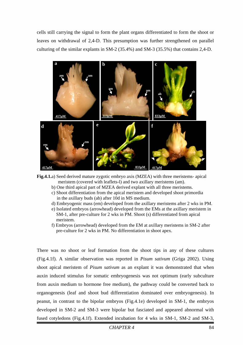
CHAPTER 4 84
cells still carrying the signal to form the plant organs differentiated to form the shoot or
leaves on withdrawal of 2,4-D. This presumption was further strengthened on parallel
culturing of the similar explants in SM-2 (35.4%) and SM-3 (35.5%) that contains 2,4-D.
Fig.4.1.a) Seed derived mature zygotic embryo axis (MZEA) with three meristems- apical meristem (covered with leaflets-l) and two axillary meristems (am).
b) One third apical part of MZEA derived explant with all three meristems. c) Shoot differentiation from the apical meristem and developed shoot primordia
in the axillary buds (ab) after 10d in MS medium. d) Embryogenic mass (em) developed from the axillary meristems after 2 wks in PM. e) Isolated embryos (arrowhead) developed from the EMs at the axillary meristem in SM-1, after pre-culture for 2 wks in PM. Shoot (s) differentiated from apical meristem. f) Embryos (arrowhead) developed from the EM at axillary meristems in SM-2 after pre-culture for 2 wks in PM. No differentiation in shoot apex.
There was no shoot or leaf formation from the shoot tips in any of these cultures
(Fig.4.1f). A similar observation was reported in Pisum sativum (Griga 2002). Using
shoot apical meristem of Pisum sativum as an explant it was demonstrated that when
auxin induced stimulus for somatic embryogenesis was not optimum (early subculture
from auxin medium to hormone free medium), the pathway could be converted back to
organogenesis (leaf and shoot bud differentiation dominated over embryogenesis). In
peanut, in contrast to the bipolar embryos (Fig.4.1e) developed in SM-1, the embryos
developed in SM-2 and SM-3 were bipolar but fasciated and appeared abnormal with
fused cotyledons (Fig.4.1f). Extended incubation for 4 wks in SM-1, SM-2 and SM-3,
c
ab ab
833µM
a
l
417µM
am am
b
313µM
am am
d
em em
417µM
f
417µM
e
s
833µM

CHAPTER 4 85
number of embryos developed from each explant increased marginally (4.0 and 5.0%).
On transferring the embryos from these cultures to medium devoid of growth regulator,
5.8-13.9% of the embryos germinated and converted into plantlets.
Table 4.1 Effect of 2,4-D exposure on somatic embryogenesis in embryo axis derived axillary meristems.
Exposure period in PM
Response (EM + embryos) (%)
Secondary Medium (MS + Different conc. of 2,4-D) (μM)
Response (EM + embryo) mean±sd* (%)
Average no. of embryo after 2wks in SM mean±sd
Average no. of embryo after 4wks in SM mean±sd
Conversion after 1st subculture in MS mean±sd** (%)
Conversion after 2nd subculture in MS mean±sd** (%)
Conversion after 3rd subculture in MS mean±sd (%)
SM-1
22.8 ± 5.1 (75)
2.8 ± 1.0 4.0 ± 2.0
00 ± 00 (56)
00 ± 00
5.8 ± 5.6
SM-2
35.4 ± 10.7 (83)
3.0 ± 0.9 5.0 ± 2.2
00 ± 00 (75)
00 ± 00
6.6 ± 3.6
SM-3
35.5 ± 13.2 (83)
1.5 ± 1.3 4.1 ± 2.0
00 ± 00 (46)
2.4 ± 4.1
13.9 ± 9.1
2 weeks 89
ANOVA NS NS NS NS NS NS
SM-1
37.3 ± 17.9 (74)
3.9 ± 0.6 4.4 ± 0.2
00 ± 00 (54)
0.9 ± 1.6
4.0 ± 4.5
SM-2
39.7 ± 13.7 (74)
4.9 ± 0.5 5.5 ± 0.3
00 ± 00 (62)
7.9 ± 1.0
11.0 ± 2.5
SM-3
42.1 ± 14.5 (54)
2.6 ± 1.1 3.6 ± 2.8
00 ± 00 (60)
00 ± 00
7.3 ± 4.9
4 weeks 75
ANOVA NS S 5% NS NS S1% NS
SM-1
56.6 ± 3.7 (99)
3.9 ± 0.7 4.3 ± 0.6
3.4 ± 2.1 (223)
10.0 ± 3.0 15.4 ± 3.2
SM-2
59.9 ± 8.6 (92)
6.5 ± 0.7 7.5 ± 1.4
12.2 ± 6.0 (347)
20.2 ± 7.6 32.3 ± 3.9
SM-3
47.2 ± 10.6 (92)
3.9 ± 1.2 3.8 ± 1.2
1.4 ± 1.6 (193)
8.2 ± 3.8 14.2 ± 6.0
6 weeks 79
ANOVA NS S 5% S5% S5% NS S1%
SM-1
60.8 ± 4.13 (97)
4.1 ± 0.9 4.2 ± 0.9
6.3 ± 2.4 (227)
6.2 ± 0.9 17.4 ± 3.6
SM-2
61.5 ± 2.3 (98)
5.7 ± 0.4 6.6 ± 0.3
7.3 ± 5.8 (305)
10.7 ± 9.5 26.0 ± 4.7
SM-3
54.3 ± 2.6 (91)
4.0 ± 0.4 4.6 ± 0.8
0.3 ± 0.5 (233)
4.3 ± 0.4 12.9 ± 2.8
8 weeks 79
ANOVA NS S 5% S5% NS NS S5%
* Three repeats-each with 30-40 replicates, ** Figures in parenthesis indicate total number of embryos tested

CHAPTER 4 86
On extending the culture period to 4 wks in PM containing 90.5 µM 2,4-D, the masses in
the axils were more prominent (Fig.4.2a). Occasionally embryo development was noted
in some cultures in PM before transferring to secondary media. Appearance of somatic
embryos never occurred in the shoot tip of the plumule. However, the bases of the leaf
primordia surrounding the apex were swollen. On transferring these cultures to SM-1
shoot differentiation never occurred from the apex. This was in contrast to the observation
(Fig.4.1e) in the cultures in SM-1 after 2 wks pre-culture in PM. This suggests silencing
of the apical meristem due to longer exposure in PM. In SM-1, SM-2 and SM-3 the
frequencies of embryogenic responses were 37.3, 39.4 and 42% respectively, after 4wks
pre-culture in PM. In these cultures somatic embryos appeared from the EMs of the
axillary meristems in multiples (Fig.4.2b, c). On culturing the 4 wks exposed primary
explants in SM-1, somatic embryos appeared from the EM of the axillary meristems in
multiples. Some of these embryos developed into singular, bipolar structures (Fig.4.2b)
and the rest were fasciated. In SM-2 and SM-3 somatic embryos developed from EMs of
the axillary meristems appeared in clusters. Embryos (Fig.4.2c) were fasciated and
appeared morphologically abnormal with fused cotyledons. No single embryos were
identified in these clusters.
From these results, it appears that 2,4-D at high concentration, induced transition of the
organogenic cells of the axillary meristems to embryogenic cells. Longer exposure (4
wks) in this growth regulator suppressed the caulogenic ability of the shoot apex
irreversibly and supported proliferation of the embryogenic cells to develop large masses
(Fig.4.1d) in the axils. It affected the process of embryogenesis adversely resulting in the
formation of fasciated embryos (Fig.4.1f and Fig.4.2c). When the plumule explants were
cultured vertically, the axillary meristems differentiated predominantly over the apical
meristems, resulting in the formation of embryos. The degree of suppression of the shoot
apex varied with the exposure in 2,4-D. The results of this experiment suggest that by
monitoring the exposure in 2,4-D and the orientation of the plumule explant it is possible
to obtain both types of morphogenetic activity in two parts of the same explant (Fig.4.1e).
Embryos developed in SM-1 (Fig.4.2b) were greener compared to the embryos developed
in SM-2 and SM-3 (Fig.4.2c). The average number of embryos developed after 2 wks in
SM-1, SM-2 and SM-3 were 3.9, 4.9 and 2.6, which marginally increased to 4.4, 5.5 and
3.6 respectively, on incubation for further 2 wks. Conversion of somatic embryos (4%-

CHAPTER 4 87
11%) developed after 4 wks in all the three SMs was achieved in MS medium without
PGR (Table 4.1).
When explants were incubated for more than 4 wks (6 and 8 wks) in PM, somatic
embryos started developing in EMs itself before getting transferred to SMs. This could be
due to gradual dehydration of the medium and less availability of 2,4-D to the explants.
But embryos developed in any of the SMs after 6 (Fig.4.2d, e) and 8 wks (Fig.4.2f) of
incubation in PM were morphologically abnormal and highly fused.
Fig.4.2a) Embryogenic masses (em) in axillary meristems grew larger on incubation for 4 wks in PM.
b) Both single (arrowhead) and fused types of embryos developed from the axillary meristem after 4 wks in SM-1, in explants pre-treated in PM for 4 wks. c) Bipolar embryos (arrowhead) with fused cotyledons developed from axillary meristem after 4 wks in SM-2. Explants were in PM for 4 wks. d) Embryos (e) with fused cotyledons developed from axillary meristem after 4 wks in SM-1. Explants were in PM for 6 wks. (P-plumular end) e) Embryos (e) with fused cotyledons developed from axillary meristem after 4 wks in SM-2. Explants were in PM for 6 wks. f) Embryos (e) with fused cotyledons developed from axillary meristem after 4 wks in SM-2. Explants were in PM for 8 wks.
Repetitive embryogenesis (Fig.4.3a) was noted occasionally in explants developed in SM-
2 and SM-3 after pre-culture for 6 and 8 wks in PM. Isolating apical part of the embryo
axis from close to the axillary buds led to the development of embryos from the apical
c
417µM
a em em
417µM
b
454µM
e
625µM
ee
f
625µM
e
d
625µM
e e p

CHAPTER 4 88
meristem (Fig.4.3b) but these embryos were very small in size in contrast to embryos
obtained from axillary buds and their growth was very slow. Culturing in MS medium for
conversion, embryos get detached very easily from the explants (Fig.4.3c) developed in
any of the SMs.
In PM after 6 wks and 8 wks embryogenic response varied from 47-59.9% and 54.3-
61.5%, respectively (Table 4.1). Extended incubation (6 wks and 8 wks) in PM altered the
number of embryos/explant significantly. The number of embryos/explant increased after
incubation for 2 wks in all the SMs after 6 (3.9-6.5%) and 8 (4-5.7%) wks pre-treatment
in PM (Table 4.1). Optimum average number of embryos developed after 2 wks in SM-2
following 6 wks pre-culture in PM was 6.5, which increased to 7.5 on extended
incubation in SM-2 for further 2 wks (Table 4.1). On the other hand, the optimum average
number of embryos was 6.6 after 4 wks in SM-2 following 8 wks pre-culturing in PM
(Table 4.1).
Fig.4.3 a) Secondary embryos (se) developed in SM-2 after 4wks. The explants were pre-cultured in PM for 6 wks.(pe-primary embryo).
b) Bipolar embryos (arrow) with fused cotyledons developed from apical meristem (ap) after 4wks in SM-1. Explants were pre-cultured in PM for 6 wks. c) Isolated embryos. d) Radicle emergence from the embryos in MS basal medium.

CHAPTER 4 89
From the results it is apparent that the frequency of embryogenic response and the
number of embryos developed/explant in secondary medium (SM1, SM2 and SM-3),
varied with exposure (2, 4, 6 and 8 wks) in the PM (Table 4.1). Frequency of
embryogenic response increased with increasing incubation period in PM. It was
optimum (61.5%) in the explants pre-cultured in PM for 8 wks (Table 4.1) and then
transferred to SM-2 and minimum (22%) in SM-1 in the explants pre-cultured for 2 wks
in PM (Table 4.1). The average number of embryos increased with increasing incubation
period in PM till 6 wks and afterwards it started decreasing. It was optimum (7.5/explant)
in explants cultured in SM-2, after 6 wks pre-culturing in PM.
Embryos germinated (Fig.4.3d) and shoot formation (Fig.4.4a) started in MS basal
medium. The optimum conversion frequency was 32% in the embryos developed in SM-2
after 6 wks pre-culturing in PM (Table 4.1) and 25% in the embryos developed in SM-2
after 8 wks pre-culturing in PM (Table 4.1). From the results it is apparent that the
conversion frequency was higher for the embryos developed in SM-2 than that for the
embryos developed in SM-1 and SM-3 after 4, 6, and 8 wks pre-treatment in PM
(Fig.4.5). Thus among all the three SMs, SM-2 is more efficient in facilitating the
development of embryos, irrespective of the duration (4, 6 and 8 wks) of pre-culturing in
PM, which results in optimum conversion. While the results of the present experiment
demonstrate the effect of 2,4-D concentration on somatic embryogenesis, a report by
Baker et al. (1995) indicates that the concentration of 2,4-D in the medium does not affect
embryogenic responses when the apical portion of mature zygotic embryo axis was
cultured horizontally, in which case the embryos developed from young expanded leaves
of the epicotyl. Chengalrayan et al. (1998) evaluated the embryogenic responses and
conversion frequency in 16 genotypes of peanut by applying a single protocol
(Chengalrayan et al. 1994, 1997) initially optimized for peanut cv. JL-24 with MZEDLs.
They reported only 4% conversion frequency in this genotype (SB-11) compared to the
high conversion frequency (32%) obtained in the present experiment by using meristems
as explants. The converted plants acclimatized in plastic cups and hardened (Fig.4.4b) in
green house successfully. Pods (Fig.4.4c) were collected during harvesting.

CHAPTER 4 90
Fig.4.4 a) Converted plantlets.
b) Plants hardened and flowered successfully in greenhouse. c) Seeds collected from harvested plants.
Fig.4.5 Conversion frequency of embryos developed in SM-1, SM-2,
and SM-3 after pre-treatment in PM for 2, 4, 6, and 8 wks.
In the present study, development of somatic embryos occurred specifically from the
axillary meristems while the growth of shoot apex seized. McKently (1991) cultured
mature zygotic embryo axes of peanut and noted embryo development from a 2-mm band
of hypocotyledonary tissue surrounding epicotyls.
The role of 2,4-D in plant tissue culture has been reviewed extensively by Feher et al.
(2003). It has been suggested that 2,4-D has a dual effect in culture above a certain
concentration: it acts as an auxin directly through endogenous IAA metabolism or as a
stressor. It has also been suggested that 2,4-D affects electrical patterns, membrane
permeability, IAA binding to the auxin-binding protein (Deshpande and Hall 2000) and
photosynthesis of algae (Fargasova 1994). The protocol for direct somatic
a b c

CHAPTER 4 91
embryogenesis from the existing meristems of peanut provides a suitable system to
study some of these phenomena described in algae under the influence of 2,4-D.
Wetzstein and Baker (1993) observed that the concentration of 2,4-D in the induction
medium had little effect on embryo morphology and no effect on conversion of somatic
embryos in peanut. However, the influence of 2,4-D exposure was not tested. Filippov
et al. (2006) showed that with extended exposure to 2,4-D, there was an increase in the
rate of somatic embryogenesis and number of regenerated wheat plants. Appropriate
time duration for mature embryo formation in Arabidopsis was determined (Raghavan
2005). Exposure to 2,4-D for more than 10 days showed no indication of increase in the
number of mature stage somatic embryos formed during subsequent growth in the basal
medium.
Histological analysis of development of embryos from axillary meristems
Two axillary meristems (Fig.4.6a) were obvious in isolated mature zygotic embryo axis.
Histological investigation of these initial explants (Fig.4.6b) confirmed the presence of
three meristems (two axillary meristems and one apical meristem). Leaf initials and leaf
primordia surrounded each bud (Fig.4.6b). Culturing the 1/3rd apical portion of embryo
axis in MS basal medium without PGR led to the differentiation of plumular meristem
into shoot and development of axillary meristems to axillary buds (Fig.4.6c, d).
Vasculatures of axillary buds connected with main vascular strand of the explant were
noted (Fig.4.6d).
Five days old explants in PM, showed only swelling at the axillary meristems (Fig.4.6e).
Histological studies of these explants showed a dense tissue at the axillary meristems
(Fig.4.6f). Culturing in PM for 10 days led to the development of more prominent EM
like structures from the axillary meristems of the explants (Fig.4.6g). Histological studies
demonstrated the absence of vascular connection (Fig.4.6h) of EMs to the main vascular
system of the 10 days old explants. These EMs appeared meristematic and preliminary
leaves were present at the base of these EMs (Fig.4.6f, h).

CHAPTER 4 92
Fig.4.6a) Mature zygotic embryo axis with three meristems. Apical meristem covered with leaflets and two axillary meristems (am). Dotted line (d) is the line of incision to isolate the plumule explant with 3 meristems.
b) Cellular morphology of mature zygotic embryo axis with apical shoot meristem (ap) and two axillary meristems (am). Well-developed vasculature (v) present till cut ends of the removed cotyledons (rc).
c) Shoot differentiation from the apical meristem (ap) and development of shoot primordia in the axillary buds (sb) after 10 d in MS basal medium.
d) Cellular morphology of the explant after 10 d in MS basal medium. Vasculature (arrow) of differentiating apical meristem and axillary bud were connected.
e) Swelling (sl) observed at the axillary meristem after 5d in PM. f) Histology of the 5d old explant in PM, showing slight connectivity of axillary
meristem with apical meristem via vasculature (v). g) More prominent EM (em) observed at the axillary meristem after 10d in PM. h) Absence of vasculature strands in axillary buds of the10d old explant in PM.
Vasculature (v) present in the mother explant.
a
am am
d
417µM
amam ap
rc d
v
rc
312µM
b
d
312µM
v
sbap
c
417µM
sb sb
g
417µM
em em
l
h
227µM cc
lp
lp
em
vv
ap
f
208µM
v
lplp
sl
e
417µM
sl
sl
l

CHAPTER 4 93
Origin of EMs from the meristematic cells of axils could be seen in 2 wks old explants in
PM (Fig.4.7a, b). Morphology of the leaflets adjacent to the meristematic buds remained
unaffected confirming that the EMs developed specifically from the meristematic cells.
Transition of the meristem to mass of cells was obvious and some partially
dedifferentiated leaflets and cells at the tips of the masses could be seen. Size of EMs
increased with increased incubation in PM.
Fig.4.7 a) Developed EM (em) like structure from axillary meristems in PM from 2wks old explant.
b) Morphology after 2 wks in PM shows disintegration of the vasculature (dv), restriction of differentiation of apical meristem (ap), transition of axillary meristem to embryogenic mass (em). Leaf primordia (lp) maintained the integrity. Remaining part of cotyledon (c) is seen at the base of the section.
c) Developed somatic embryos (e) from the EM in the axils. Explants were in SM-1 for 2 wks after pre-culturing in PM for 2 wks. d) Vasculature (v) and well-developed epidermis (arrow) observed in somatic embryos (e) with broad suspensor after 2 wks in SM-1. Explant pre-cultured in PM for 2 wks. (Leaf primordia -lp). e) Developed somatic embryos (e) from the EM in the axils. Explants were in SM-2 for 2 wks following 2 wks pre-culture in PM. f) Cellular morphology, showing dome (d) shaped structure with well- developed epidermis (arrow) from the axils of 2 wks old explants in SM-2 and pre-cultured in PM for 2 wks. Apical meristem (ap) and leaf primordia (lp) maintained the integrity.
e
417µM
p
e
e
f
227µM cc
ap
v
lp
lp
d
lp
e e
d
227µM
ap
vv
cc
lplp
c
227µM
ee
c
b
ap
227µM
c
lp
lp dv
lp
emem
a
em
227µM
em

CHAPTER 4 94
Histological studies of the cultures in SMs after 2 wks in PM
Observation of two wks old cultures in SM-1 (Fig.4.7c, d) and in SM-2 (Fig.4.7e, f), pre-
cultured in PM for 2 wks confirmed direct somatic embryogenesis in the axillary
meristems. It showed that EMs originated from the meristematic cells (Fig.4.7e, f) of the
axils, which further led to the development of somatic embryos from these EMs.
Epidermis of the somatic embryos developed in SM-1 was much clearer and these
embryos attached with mother explant with broad suspensor (Fig.4.7d). Development of
vascular strands could be seen in these embryos. Preliminary leaves were present at the
base of somatic embryos (Fig.4.7d). Meristematic cells were present at the base of the
plumular preliminary leaves. Near the cut of the embryo axis where the cotyledon
attached, cells were loosely arranged. In explants, which were in SM-2, embryos were not
much developed as compared to SM-1 explants. Leaf primordia, surrounding the
meristems demonstrated meristematic activity. Distinct epidermis was observed around
the meristematic domes (Fig.4.7f) of the explants, which were in SM-2.
Culturing for further 2 wks (4 wks in total) in SMs (SM-1 and SM-2) resulted in well-
developed embryos with distinct vascular strands and epidermis (Fig.4.8a-d). Both fused
and single types of embryos were present in the same explant (Fig.4.8d). Many vascular
strands were visible in the fused embryos. Fused embryos were attached to the parent
explant with broad suspensor (Fig.4.8b, d). Isolated embryos (bipolar embryo) were noted
to have only single vascular strands and the embryos were connected to the parent plant
with a well-developed narrow suspensor (Fig.4.8d). This result confirms multicellular
origin of somatic embryos. At the base of the embryos, callus was also visible (Fig.4.8b,
d). At the base of the last preliminary leaf of the caulogenic bud, meristematic swelling
was observed (Fig.4.8d). Presence of meristematic mass like structure (Fig.4.8b) at the
base of the preliminary leaf of plumule was also noted.
Histological studies of the cultures in PM for 4 wks
Extended exposure of explants to PM, till 4 wks led to the formation of embryos from the
caulogenic buds (Fig.4.8e, f). Meristematic structure was also observed in the plumule.
Preliminary leaves still maintained their integrity. Development of embryos was noted in
PM itself (Fig.4.8f).

CHAPTER 4 95
Fig.4.8 a) Somatic embryos (e) developed from the EM in the axils. Explants were in SM-1 for 4 wks following 2 wks pre-culture in PM. b) Fused embryos (fe) with vasculature (v) developed from the axil after 4 wks in SM-1 following 2 wks pre-culture in PM. c) Somatic embryos (e) developed from the EM in the axils after 4wks in SM-2 following 2 wks pre-culture in PM. d) Bipolar (bp) and fused (fe), both types of embryos with well-formed suspensor (s), epidermis (arrow) and vasculature (v) developed from axillary meristem after 4 wks in SM-2. Explants pre-treated in PM for 2 wks. e) Somatic embryos (e) started developing from EM (em) after 4 wks in PM.
f) Embryos (e) developed from axillary meristem after 4 wks in PM. Apical meristem (am) was clearly visible. Leaf primordia (lp) maintained the integrity.
Histological studies of the cultures in SMs after 4 wks in PM
Transferring the cultures to SMs (SM-1 and SM-2) after 4 wks pre-treatment in PM led to
the development of well-developed somatic embryos with well-developed cotyledons and
vascular strands and these embryos were attached to the mother explants with broad
suspensors (Fig.4.9a-d). These structures had a continuous epidermis with the parental
a
417µM
e
b
312µM
v
fe lp
c
417µM
e
e
d
fe
s
312µM
v
bp
e
em em
417µM
p
ee
f
312µM
e
e
am
lp lp

CHAPTER 4 96
tissue, which could be due to sub-epidermal cell division of the explants, demonstrating
the multicellular origin of the somatic embryos. In SM-2, repetitive embryos were noted
from the base of the somatic embryos as well as from the meristematic cells present in the
plumule (Fig.4.9e, f), which were also multicellular in origin. On the contrary, repetitive
embryogenesis was not observed in SM-1, which implies that the presence of 2,4-D in the
previous case (SM-2) could be responsible for the development of secondary embryos.
Fig.4.9 a) Fused (fe) and bipolar (bp), both types of somatic embryos developed from the EM in the axils after 2 wks in SM-1 following 4 wks pre-culture in PM.
b) Well-developed somatic embryos (e) attached to the mother explant with broad base. Meristematic (m) structure was present at plumular end. Leaf primordia (lp) maintained the integrity. c) A cluster of somatic embryos (e) developed from the EM (em) in the axils after 2 wks in SM-2 following 4 wks pre-culture in PM. d) Somatic embryos (e) developed from the EM in the axils after 2 wks in SM-2. Meristematic (m) swelling was present at the base of plumular leaves. e) Secondary embryo (se) developed from the plumular part of the primary embryo (pe). Obvious vasculature (v) in primary embryo. f) Secondary somatic embryo (se) developed from the base of the primary embryo (pe). Meristem (m) is clearly visible in primary embryo.
We observed that the meristem at the shoot tip did not demonstrate any morphological
change after 2 wks exposure in PM (90.5 μM 2,4-D). This explained our observation
(Fig.4.1e) of shoot apex differentiation into shoots and the development of somatic
f
m
se
156µM
e
v
se
pe
se
156µM
c
em e
eem
417µM
d
e
eelp
m
312µM
bm
e
e
lp
312µM
a
bp
417µM
fe em

CHAPTER 4 97
embryos from the axils. However, 4 wks exposure (Fig.4.8f) to PM followed by exposure
to SM-1 (Fig.4.9b) and SM-2 (Fig.4.9d), were effective in inducing the meristematic
activity in the bases of the leaf primordia surrounding the apical meristem (Fig.4.1f). This
resulted in the swelling of the shoot apex with a broad base. Nevertheless, the
development of somatic embryos was noted only from the EMs present at the axils.
Maheswaran and Williams (1985) suggested that the origin of somatic embryos from one
to number of adjacent cells is possible depending on the synchrony of their internal pre-
embryogenic determined states and their ability to interact as a group as opposed to
individual cells. In the present system, it appears that direct somatic embryogenesis could
be of multicellular origin. The suspensors connecting the somatic embryos to the parent
tissue were noted to be of both, broad (Fig.4.8d and Fig.4.9b, d) and narrow (Fig.4.8d)
types. A similar result was obtained by Hu and Sussex (1971) and Fernando et al. (2001)
in papaya. According to Maheswaran and Williams (1985), the embryos attached by a
narrow suspensor like organ appeared to have arisen superficially, whereas those attached
by a broader suspensor might have arisen from meristematic regions within the buds. In
the cultures used in the present experiment, direct somatic embryogenesis is associated
with suppression of the main zygotic embryo axis. The relationship appears analogous to
that between apical and lateral buds. According to Maheswaran and Williams (1985), the
growth suppression of the main embryo axis is presumably associated with the
breakdown of integration of the cells as a single embryonic group, and escape of
individual cells or smaller groups to function autonomously.
4.1.3 CONCLUSION
Our results demonstrate:
(i) The effect of 2,4-D exposure on the frequency and morphology of the
peanut embryos.
(ii) It also demonstrates that by altering the orientation of the explant on an
appropriate medium, the determined organogenic cells of an explant can be
made embryogenic.
(iii) By culturing the determined meristems vertically and by altering the auxin
exposure both pathways of morphogenesis can be demonstrated in the same
explant.

CHAPTER 4 98
(iv) Histological studies confirmed that the development of somatic embryo
occurred specifically from the axillary meristems.
To the best of our knowledge this is the first systematic study on the influence of 2,4-D
on the determined meristems of peanut explants at a particular orientation and on control
of two morphogenetic pathways simultaneously in the same explant. This protocol will
not only be useful for in situ studies on understanding the pathways of morphogenesis and
signal transduction but also for genetic transformation using either direct DNA delivery
approach or by infection with Agrobacterium mediated transformation. The possibility of
extending this approach for genetic transformation in in vivo system through direct DNA
delivery or Agrobacterium injection in meristems can also be explored.
A part of this work has been communicated for publication Somatic embryogenesis from the axillary meristems of peanut (Arachis hypogaea L.)
CHAPTER 4 99
4.2 AXENIC SHOOT CULTURE-DERIVED MERISTEMS AS EXPLANTS FOR
SOMATIC EMBRYOGENESIS
In the above experiment we used determined meristematic cells of mature zygotic embryo
axis with three meristems as explants for somatic embryogenesis and observed that
embryogenesis occurred from the axillary meristems. To test the embryogenic potential of
the other meristems of the same species and of a wild species, we extended the protocol
to the axenic shoot cultures derived meristems of Arachis hypogaea (cv. SB-11) and in
Arachis duranensis (wild species).
The wild genotypes of peanut are a valuable source of resistant genes against several
pests and pathogens besides having high oil and protein contents. The growing concern
over the collection, rescue, conservation, multiplication and characterization of the wild
species of Arachis germplasm relies on the fact that they contain useful genes for the
genetic improvement of peanut (Gagliardi et al. 2000). However, the multiplication and
maintenance of wild Arachis germplasm is very labour-intensive and involves specific
protocols because many accessions are grown mostly under green house conditions. Even
with optimum storage practices, seed germinability and germplasm losses are inevitable
(Dunbar et al. 1993), since the seeds display a sub-orthodox behavior due to their high
lipid contents and thin seed coat, which result in short viability (Vasquez-Yanes and
Arechiga 1996). Therefore, there is a limited supply of wild germplasm from the gene
bank and it becomes difficult to maintain wild species of Arachis for its use in breeding
programme. Consequently, in vitro germplasm conservation constitutes a viable option
for their preservation. In vitro protocols for plant regeneration have been reported in
different Arachis species via indirect way of organogenesis (Gagliardi et al. 2000; Rey et
al. 2000 and 2006; Vijaya Laxmi and Giri 2003). Indirect way of somatic embryogenesis
was reported in A. paraguariensis (Sellars et al. 1990), A. pintoi (Rey et al. 2000 and
2006) and A. glabrata (Vidoz et al. 2004). Both direct and indirect ways of somatic
embryogenesis were reported in A. pintoi (Rey et al. 2006) by using leaflets. Several wild
groundnut species have been successfully regenerated from seed explants including
cotyledon, mature and immature leaflets and embryo axes (Rani and Reddy 1996; Vidoz
et al. 2004; Rey et al. 2000; Rey and Mroginski 2006; Gagliardi et al. 2000), but studies
on the morphogenetic potential of nodal buds from in vitro plants are not explored. The
methods based on the activation of pre-formed meristems (shoot tips and axillary buds),

CHAPTER 4 100
which retain the potential to recover true-to-type plants, is desirable for in vitro
conservation programs.
Micropropagation through direct somatic embryogenesis would help in the mass scale
propagation of the wild species and also facilitate germplasm conservation in vitro. In
addition, the protocol can be exploited for generating new genetic variability in peanut by
somatic hybridization through protoplast fusion. This is an attempt to study the effect of
growth regulators on morphogenic response of axenic shoot culture derived shoot apices
and axillary buds.
4.2.1 MATERIALS AND METHODS
Seeds of cultivated species, Arachis hypogaea (cv. SB-11) were procured from local
market and wild species, Arachis duranensis (ICG No. 8200) were obtained from
ICRISAT (International Crops Research Institute for the Semi-Arid Tropics), Hyderabad.
Axenic shoot cultures of both the species were maintained in in vitro condition for 5 years
to preserve the germplasm. Shoot cultures of A. hypogaea were maintained in MS basal
medium containing 2.22 μM BAP and 2.32 μM KN and 2% sucrose. Shoot cultures of A.
duranensis were maintained in MS basal medium containing 4.44 μM BAP and 2%
sucrose. The cultures were incubated in light.
Cluster of axenic shoots (A. hypogaea and A. duranensis) was removed from the culture
bottles and the shoots were separated aseptically. Leaves were removed (Fig.4.11a1, a2).
Explants (shoot tip and nodes) of 3-4 mm (Fig.4.11a3) were dissected from the axenic
shoots of both the species (A. hypogaea and A. duranensis). The length of the shoots
varied. Therefore the number of explants obtained from each shoot varied from 2-5 with
the length of the axenic shoot. From the shorter shoot only 2 nodal explants could be
obtained in addition to the shoot tip. Explants carrying the apical meristem with axillary
meristem (shoot tip with 1st node) and explants carrying only a single axillary meristem
(node) were cultured vertically in the primary medium (PM) composed of MS (Murashige
and Skoog 1962) basal salt with 90.5 μM 2,4-D and 6% sucrose (Chengalrayan et al.
1994) in 85 mm petridishes. To determine the difference in potential if any, in the axillary
buds due to their relative position from the shoot apex, the explants were arranged in
sequence as in the axenic shoots. The cultures of both the species (A. hypogaea and A.
CHAPTER 4 101
duranensis) were divided into two groups. One group was incubated in light and the other
group was incubated in dark for 4 wks. Embryogenic response was recorded. Afterwards
the cultures were transferred to secondary medium (SM) composed of MS basal salt
supplemented with 13.6 μM 2,4-D and 6% sucrose. The explants were cultured in the
same order as it was in the PM and incubated for 4 wks in their respective culture
conditions. Number of embryos was noted. Thereafter all the cultures (including those
incubated in light as well as in dark) were transferred to MS medium without PGR and
incubated in light. Experiments were repeated twice with 72 and 74 explants in SB-11
and 70 and 75 explants in A. duranensis.
The same set of experiments was carried out for both the species, by extending the
incubation period of cultures in PM to 8 wks without sub-culturing. Embryogenic
response was recorded after incubating the cultures for 8 wks in PM and after 4 wks in
SM. The experiment was repeated 5 times with 40-50 replicates in each repeat.
Both the experiments were repeated with medium in which 2,4-D was substituted by
picloram and the concentration of sucrose was reduced to 3%. Picloram has been chosen
for this study because it has been reported earlier for embryogenesis in immature zygotic
embryos (Ozias-Akins et al. 1993) and MZEDLs (Joshi 2003) of peanut. Various
concentrations (2.07, 4.14, 8.28, 12.42, 16.56 and 20.71 μM) of picloram were tested for
induction of somatic embryos from the meristematic explants of both the species (A.
hypogaea and A. duranensis). Similar to the experiment with 2,4-D, the cultures were
divided into two groups and one group was incubated in light and the other one was
incubated in dark. Cultures of A. hypogaea were incubated for 4 and 8 wks and the
observations were noted. The explants of A. duranensis were incubated initially for 4 wks
in both light and dark conditions and the responses were noted. Incubation period was
extended for next 4 wks (total 8 wks) in case of explants incubated in light, as they did
not show embryogenic response in the first 4 wks. After incubation in different
concentrations of picloram, all the cultures (embryos) were transferred to 2.07 μM
picloram for the next 4 wks after which they were transferred to MS medium devoid of
PGR in light for conversion. These embryos were further transferred twice to MS medium
at intervals of 4 wks. The experiments were repeated thrice with 12 replicates each.

CHAPTER 4 102
The pH of all the media was adjusted to 5.8 prior to the addition of (0.7%) agar. The
media were autoclaved at 120°C under 15psi for 20 minutes and distributed in 85mm
petridishes. Cultures were incubated in 16h photoperiod at 25 ± 2°C under diffuse cool
white fluorescent lights (32μEm-2sec-1) and in dark condition at 25 ± 2°C. All data were
subjected to ANOVA analysis.
4.2.2 RESULTS AND DISCUSSION
We did not observe any EM like structures in meristem explants in PM. Development of
somatic embryos were observed only in some explants of 1st axillary node (near to shoot
tip) of SB-11 cultured in SM for 4 wks after pre-treatment with PM for 4 wks and
incubation in light, whereas the shoot tip and other nodes turned brown. Embryogenic
response was only 4.5% and average number of embryos per explant was 1.5 (Fig.4.10).
These embryos germinated successfully in PGR free media but shoot development was
restricted.
Fig.4.10 Embryogenic response from axenic shoot culture derived meristem explants of A. hypogaea, incubated for 4 and 8 wks in PM.
Extended incubation in PM for 8 wks in light and then 4 wks in SM, led to embryogenic
response in all the axillary buds tested with varying frequency (Fig.4.10) except in 6th
axillary node. Embryogenic response was 4% in shoot tip and it was optimum (15%) in

CHAPTER 4 103
1st axillary node (Fig.4.10). In 2nd (4.4%) and 3rd (4.9%) axillary node, embryogenic
response was almost similar. In 4th and 5th axillary node, embryogenic response was
reduced. The average number of embryos per explant was 1.9 in 2nd axillary node
(Fig.4.10).
Fig.4.11 Embryogenic response in axenic culture-derived meristems of Arachis hypogaea. a) Axenic shoot culture (1), shoot without leaves (2), isolated nodal parts (3) with nodal bud from different nodes of axenic shoot culture. b) Embryos (e) developed from shoot apical meristems in SM, pre-treated in PM for 8 wks. Primary leaves (arrow head) turned brown. c) Embryos (e) developed from 1st nodal meristems in SM, pre-treated in PM for 8 wks started rooting (r). Shoot tips were covered with primary leaves. d) Single fused embryo (fe) developed from 2nd nodal meristems (n) in SM, pre-treated in PM for 8 wks. e) Embryos rooted in MS media without PGR.
Shoot tip and 1st axillary node rarely responded together. Mostly either shoot tip
(Fig.4.11b) or 1st nodal bud (Fig.4.11c, d) was showing embryogenic potential. Embryos
were both of bipolar (Fig.4.11c) and fused types (Fig.4.11b, d). Embryos developed from
different axillary nodes and from shoot tip were easily separated from the explants.
Somatic embryos germinated (Fig.4.11e) successfully in PGR free media. Germination
frequency of embryos was 100%, whereas conversion frequency varied with origin of the
1 2 3
a b
e
e
e
833µM
c
Shoot tip
e
re
417µM
d
fe
n
417µM
e

CHAPTER 4 104
embryo with respect to the position of the nodal buds. Somatic embryo obtained from the
1st and 2nd axillary nodal buds converted into plantlet (Fig.4.12a). Conversion frequency
was optimum (9%) in embryos derived from 1st nodal buds (Table 4.2). The converted
plantlets hardened successfully (Fig.4.12b). Embryos developed from other nodal buds
and from shoot tips did not convert into plantlets in PGR free media.
Table 4.2 Conversion frequency of embryos in MS medium, developed in SM after 8 wks exposure in PM.
Fig.4.12 a) Conversion of somatic embryo (developed from axillary nodal bud) into plantlet. b) Hardened plant in green house. c) Induction of embryos (arrowhead) from 2nd nodal bud in 12.42 μM picloram in dark. Rest of the explant became dark brown. Increasing exposure duration of PM led to an increase in embryogenic response in the
present experiment. Four wks exposure rarely led to the development of somatic embryos
that too only in the 1st node (Fig.4.10), whereas 8 wks exposure to PM resulted in
embryogenesis from shoot tip to 5th nodal buds with varying frequency (Fig.4.10). This
confirms our earlier observation (Section 4.1) that culturing the meristem explants
vertically led to the formation of somatic embryos from the axillary buds.
Incubation of explants in dark with PM (irrespective of incubation period) was ineffective
in A. hypogaea (cv. SB-11) for embryogenesis and only callus was noted. On the other
hand, in the case of picloram containing media, all cultures became dark brown after 4
wks exposure in all concentrations of picloram (2.07, 4.14, 8.28, 12.42, 16.56 and
Explant Shoot tip Node 1 Node 2 Node 3 Node 4 Node 5 Conversion mean±sd*
(%)
0±0 (40)
8.8±8.4 (47)
1.7±3.7 (22)
0±0 (10)
0±0 (4)
0±0 (1)
* Figures in parenthesis indicate total number of embryos tested
a b
c
625µM

CHAPTER 4 105
20.71μM), both in light and dark conditions. Embryogenic response was hardly visible (2
out of 36 explants) in explants incubated in dark condition in 12.42 μM picloram for 8
wks but they never grew further (Fig. 4.12c).
Experiments on induction of somatic embryogenesis using axenic culture-derived
meristem explants for SB-11 were extended to A. duranensis -a wild species, to test the
applicability of the protocol for other species.
In contrast to the effect noted in A. hypogaea (cv. SB-11), in A. duranensis, culturing the
explants in the media containing various concentrations of picloram and incubation in
dark was highly effective and led to the induction of somatic embryos (Fig.4.13a, b) from
axillary buds within 2 wks. Embryogenic response frequency (Table 4.3) was varying in
all the concentrations of picloram tested. Within 4 wks repetitive embryogenesis
(Fig.4.13c, d) was also apparent. After 4 wks all the cultures were transferred to MS basal
media composed of 2.07 μM picloram for maturation of developing embryos. With
respect to the distance from the shoot tip, in 8.28, 12.4, 16.56 and 20.71 μM picloram,
embryogenic response frequency increased till 2nd node and then started decreasing
(Table 4.3). Embryogenic response in the 2nd nodal explants cultured in 12.4 μM and
16.56 μM picloram was 44% and 39 %, respectively (Table 4.3). Next to 2nd nodal
axillary buds, the 3rd nodal axillary buds showed higher embryogenic response in 8.28,
12.4, 16.56 μM of picloram (Table 4.3).
The average number of embryos with respect to the explants position on stem, in various
concentration of picloram reveals that, in all nodal buds (1st, 2nd, 3rd, 4th and 5th), average
number of embryos was optimum in explants cultured in 16.56 μM picloram (Table 4.3).
Within 16.56 μM picloram, the average number of embryos was optimum (13.4) in 4th
node followed by 3rd, 2nd and 1st node (Table 4.3). In 12.42 μM of picloram, it was
optimum in 2nd node, whereas in the remaining concentrations of picloram, the average
number of embryos was not significantly different.
After culturing all the embryos in MS medium without PGR for conversion and
incubation in light, the plumular part of embryos turned green (Fig.4.13e) within 4 wks.
Embryos could be easily isolated. They showed different types of morphology though all
of them appeared morphologically abnormal (Fig.4.14a).

CHAPTER 4 106
Table 4.3 Embryogenic responses in different concentration of picloram in different nodes of axenic shoots culture of A. duranensis.
Explants 2.07 μM PIC mean±sd
4.14 μM PIC mean±sd
8.28 μM PIC mean±sd
12.42 μM PIC Mean±sd
16.56 μM PIC mean±sd
20.71 μM PIC mean±sd
Embryogenic response frequency (%)*
Shoot tip 0.0 ± 0.0 (36) 5.6 ± 9.6 (36) 5.6 ± 4.8 (36) 0.0 ± 0.0 (36) 2.8 ± 4.8 (36) 13.9 ± 12.7 (36)
Node 1 2.8 ± 4.8 (36) 30.6 ± 45.9 (36) 11.1 ± 12.7 (36) 22.2 ± 9.6 (36) 27.8 ± 9.6 (36) 19.4 ± 17.3 (36)
Node 2 22.2± 19.2 (36) 11.1 ± 19.2 (36) 30.6 ± 25.6 (36) 44.4 ± 17.4 (36) 38.9 ± 4.8 (36) 30.6 ± 24.1 (36)
Node 3 14.7± 13.8 (27) 9.5 ± 16.5 (25) 30.1 ± 16.2 (26) 30.2 ± 2.8 (23) 37.8 ± 20.9 (27) 12.6 ± 14.6 (26)
Node 4 15.3± 16.8 (16) 23.3 ± 25.2 (13) 6.7 ± 11.6 (5) 8.3 ± 14.4 (10) 37.5 ± 12.5 (16) 16.7 ± 28.9 (15)
Node 5 16.7 ± 28.9 (2) 0.0 ± 0.0 (9) 0.0 ± 0.0 (4) 0.0 ± 0.0 (5) 19.1 ± 32.9 (8) 0.0 ± 0.0 (5)
ANOVA NS NS NS S1% NS NS
Average number of embryos per explant
Shoot tip 0.0 ± 0.0 3.8 ± 5.8 8.0 ± 7.0 0.0 ± 0.0 0.3 ± 0.6 3.5 ± 3.3
Node 1 1.3 ± 2.3 3.0 ± 5.3 1.4 ± 1.7 3.7 ± 1.3 7.3 ± 1.9 2.1 ± 2.0
Node 2 1.3 ± 1.4 4.8 ± 8.4 3.8 ± 2.2 5.0 ± 2.0 9.3 ± 3.0 6.4 ± 3.6
Node 3 3.7 ± 3.2 1.2 ± 2.0 4.0 ± 3.5 3.3 ± 1.9 12.5 ± 6.5 7.5 ± 7.5
Node 4 1.7 ± 2.1 1.0 ± 1.7 0.7 ± 1.2 1.7 ± 2.9 13.4 ± 5.7 1.7 ± 2.9
Node 5 1.3 ± 2.3 0.0 ± 0.0 0.0 ± 0.0 0.0 ± 0.0 1.9 ± 3.3 0.0 ± 0.0
ANOVA NS NS NS S5% S1% NS
Germination frequency (%)**
Shoot tip 0.0 ± 0.0 (0) 4.8 ± 8.3 (22) 5.6 ± 4.9 (24) 0.0 ± 0.0 (0) 0.0 ± 0.0 (1) 5.3 ± 4.6 (25)
Node 1 8.3 ± 14.4 (4) 9.2 ± 15.9 (91) 10.0 ± 17.3 (11) 11.4 ± 10.3 (27) 8.9 ± 5.0 (77) 8.9 ± 7.8 (23)
Node 2 3.0 ± 5.3 (16) 12.1± 20.9 (58) 14.4 ± 13.7 (43) 12.8 ± 2.5 (88) 13.8 ± 3.1 (132) 12.3 ± 13.8 (91)
Node 3 12.2± 10.7 (21) 4.8 ± 8.3 (7) 40.3 ± 8.7 (34) 3.0 ± 5.3 (22) 11.2 ± 1.9 (106) 11.1 ± 10.2 (30)
Node 4 8.3 ± 14.4 (9) 6.7 ± 11.6 (8) 0.0 ± 0.0 (2) 0.0 ± 0.0 (5) 10.5 ± 4.3 (82) 0.0 ± 0.0 (5)
Node 5 0.0 ± 0.0 (4) 0.0 ± 0.0 (0) 0.0 ± 0.0 (0) 0.0 ± 0.0 (0) 2.9 ± 5.02 (23) 0.0 ± 0.0 (0)
ANOVA NS NS NS S5% S1% NS
Conversion frequency (%)**
Shoot tip 00 ± 00 (0) 0.0 ± 0.0 (22) 0.0 ± 0.0 (24) 0.0 ± 0.0 (0) 0.0 ± 0.0 (1) 0.0 ± 0.0 (25)
Node 1 0.0 ± 0.0 (4) 0.7 ± 1.3 (91) 3.3 ± 5.8 (11) 0.0 ± 0.0 (27) 3.9 ± 4.1 (77) 0.0 ± 0.0 (23)
Node 2 0.0 ± 0.0 (16) 0.6 ± 0.9 (58) 4.8 ± 4.6 (43) 5.7 ± 5.1 (88) 7.7 ± 2.4 (132) 7.4 ± 9.5 (91)
Node 3 0.0 ± 0.0 (21) 0.0 ± 0.0 (7) 35 ± 16.8 (34) 0.0 ± 0.0 (22) 5.5 ± 0.6 (106) 0.0 ± 0.0 (30)
Node 4 0.0 ± 0.0 (9) 0.0 ± 0.0 (8) 0.0 ± 0.0 (2) 0.0 ± 0.0 (5) 2.7 ± 2.5 (82) 0.0 ± 0.0 (5)
Node 5 0.0 ± 0.0 (4) 0.0 ± 0.0 (0) 0.0 ± 0.0 (0) 0.0 ± 0.0 (0) 0.0 ± 0.0 (23) 0.0 ± 0.0 (0)
ANOVA NS NS S1% S5% S1% NS
* Three repeats with 12 replicates ** Figures in parenthesis indicate total number of embryos tested

CHAPTER 4 107
Fig.4.13 Development of somatic embryos (e) in the nodal axillary meristems of Arachis duranensis. a) Induction of direct somatic embryos (e) from 2nd nodal bud after 2wks in 12.42 μM of picloram in dark. (n-position of nodal bud). b) Induction of direct somatic embryos from 3rd nodal bud after 2wks in 16.56 μM of picloram in dark. (g-globular embryos). c) Induction of repetitive embryos from 3rd nodal bud after 4wks in16.56 μM
of picloram in dark. d) Repetitive embryogenesis showing primary (p), secondary (s) and tertiary (t) embryos in the same culture.
e) Embryos started differentiating and became partially green in MS medium in light.
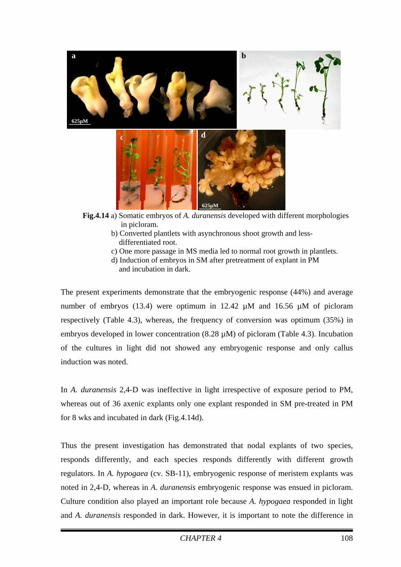
After 2nd transfer in MS medium, the embryos started germinating and converted into
plantlets (Fig.4.14b). Optimum germination (40%) and conversion frequency (35%) was
obtained in 3rd nodal axillary bud- derived embryos, developed in 8.28 μM picloram.
There was no pattern in germination and conversion frequency (Table 4.3) with respect to
the position of nodal buds on the axenic shoot. In 16.56 μM picloram 1st to 4th nodal bud-
derived embryos converted into plantlets with varying frequencies and it was optimum
(8%) in 2nd axillary bud-derived embryos. The conversion frequency of somatic embryos,
developed in the remaining concentrations of picloram, was quite low (Table 4.3) in MS
basal medium without PGR. In 2.07 μM picloram, none of the embryos converted into
plants developed from any of the nodal buds. The roots of converted embryos were very
thin and short (Fig.4.14b) till 2nd passage in MS medium. Root growth of emblings
became better in 3rd passage of PGR free medium (Fig.4.14c).
e
625µM
d
p
s
ss t
227µM
c
g g
g
156µM
a
417µM
ne
b
313µM
n
e
CHAPTER 4 108
Fig.4.14 a) Somatic embryos of A. duranensis developed with different morphologies in picloram. b) Converted plantlets with asynchronous shoot growth and less- differentiated root. c) One more passage in MS media led to normal root growth in plantlets. d) Induction of embryos in SM after pretreatment of explant in PM and incubation in dark.
The present experiments demonstrate that the embryogenic response (44%) and average
number of embryos (13.4) were optimum in 12.42 µM and 16.56 µM of picloram
respectively (Table 4.3), whereas, the frequency of conversion was optimum (35%) in
embryos developed in lower concentration (8.28 µM) of picloram (Table 4.3). Incubation
of the cultures in light did not showed any embryogenic response and only callus
induction was noted.
In A. duranensis 2,4-D was ineffective in light irrespective of exposure period to PM,
whereas out of 36 axenic explants only one explant responded in SM pre-treated in PM
for 8 wks and incubated in dark (Fig.4.14d).
Thus the present investigation has demonstrated that nodal explants of two species,
responds differently, and each species responds differently with different growth
regulators. In A. hypogaea (cv. SB-11), embryogenic response of meristem explants was
noted in 2,4-D, whereas in A. duranensis embryogenic response was ensued in picloram.
Culture condition also played an important role because A. hypogaea responded in light
and A. duranensis responded in dark. However, it is important to note the difference in
ba
625µM
c d
625µM

CHAPTER 4 109
MS media composition used to propagate and maintain (for 5 years) the axenic shoot
cultures in vitro viz., MS media containing 4.44 μM BAP for A. duranensis and MS
media containing 22.2 μM BAP and 2.32 μM KIN for A. hypogaea, which could be
responsible for the differences in embryogenic response between the two species.
In A. pintoi (Rey et al.2000) and A. correntina (Vidoz et al. 2004), culturing the leaf
explants in 2,4-D produced only shoots, whereas in all the accessions of A. glabrata
(Vidoz et al. 2006) tested neither somatic embryogenesis nor organogenesis were
observed when leaf were cultured in 2,4-D instead of picloram. In contrast, somatic
embryos were obtained in cultivated varieties of peanut in 2,4-D (Baker and Wetsztein
1995; Chengalrayan et al. 1994; Little et al. 2000). These reports support the observations
of the present investigation that 2,4–D is effective only in cultivated variety and hardly
responded to wild variety.
Dudits et al. (1995) reported the involvement of an early stage dedifferentiation of the
induced cells in in vitro plant regeneration pathways, which leads to a total
reprogramming of differentiated cells at the molecular level. One of the most extreme
examples of this plasticity in plant development is the capability of several cell types to
initiate embryogenic development. Our result is one of the examples of extreme plasticity
because caulogenic nodal buds, which were pre-determined for shoot formation converted
into somatic embryos by changing the culture condition and orientation of the explants.
4.2.3 CONCLUSION
The present experiment confirmed that the axillary meristem explants could be worked
out for embryogenesis in different species of peanut. It is also confirmed that
embryogenic response in peanut tissues relies on genotype, auxin type, concentration,
exposure, culture conditions and orientation of the explants. This is the first report on
somatic embryogenesis from caulogenic buds of A. hypogaea (cv. SB-11) and A.
duranensis. This protocol will not only be useful for in situ studies on understanding the
pathways of morphogenesis but also for genetic transformation. These protocols aid in
preserving the wild and cultivated varieties in vitro. At the same time, it is not required to
depend on the availability of seeds from the market (Shweta et al. Manuscript under
preparation).

CHAPTER 4 110
4.3 EFFECT OF SILVER NITRATE ON SOMATIC EMBRYOGENESIS Regeneration through somatic embryogenesis is one of the most widely employed
methods in transformation techniques. Induction and regeneration of somatic embryos are
very sensitive to culture conditions such as the composition of the medium, the physical
environment of the culture, the genotype and explant sources. An important in vitro
factor, unexplored in peanut, is the role of silver nitrate in modifying the effect of
ethylene accumulation in the gaseous phase of culture vessels. The involvement of
ethylene in plant tissue growth and differentiation has been widely investigated.
Application of ethylene precursors and/or inhibitors has shown that ethylene may often
have diverse effects in similar tissue culture systems (Fuentes et al. 2000). As ethylene
production appears to be as universal in cell and tissue culture as it is in intact plants, the
ethylene that accumulates in the vessel atmosphere can influence explant growth and
morphogenesis (Santos et al. 1997). Several researchers have attempted to elucidate the
possible influence of ethylene on plant tissue culture. For many plant regeneration
systems, ethylene may act as an inhibitor. However, it remains difficult to make
generalizations regarding its effects because ethylene may also have promotive effects in
certain species. For example, the addition of silver nitrate (AgNO3), a potent inhibitor of
ethylene action (Beyer 1976) was shown to promote regeneration in Brassica campestris
(Palmer 1992) and Helianthus annuus (Chraibi et al. 1991). Similarly, AgNO3 improved
somatic embryogenesis in Hevea brasiliensis (Auboiron et al.1990), Solanum tuberosum
(Tiainen 1992), Hordeum vulgare (Evans and Batty 1994) and Picea glauca (Kong and
Yeung 1994). On the other hand, somatic embryogenesis has been shown to be
stimulated by ethylene in Coffea canephora (Hatanaka et al. 1995). In Daucus carota
somatic embryogenesis, ethylene may act either as an inhibitor (Roustan et al. 1992) or a
stimulator (Nissen 1994).
In this connection and in continuation with the demonstration of meristems as explants
for somatic embryogenesis in the previous sections of this chapter, here we proceed
further by investigating the effect of silver nitrate on embryogenic responses and
conversion frequency of somatic embryos developed from axillary meristems (1/3rd apical
portion of mature zygotic embryo axis). More specifically, the present experiment
involves the addition of AgNO3 to the PM and the resulting embryos were monitored for
their ability to develop into normal plants.

CHAPTER 4 111
4.3.1 MATERIALS AND METHODS
To study the effect of silver nitrate on somatic embryogenesis, filter-sterilized AgNO3
was added to the autoclaved PM aseptically at concentrations of 0, 25, 50, 75 and 100
μM. Embryo axis-derived plumule explants were cultured in these media for 6 wks and
incubated in light. The embryogenic responses were scored before transferring these
cultures to SM-2 for 4 wks and incubated in light. Number of embryos were scored in
both PM (with/without AgNO3) and SM-2 medium and mean number of embryos per
explant was determined. Obtained embryos were cultured in MS basal medium for
conversion into plantlets. The experiment was repeated thrice with 60 replicates in each
repeat. All data were subjected to ANOVA analysis.
The pH of all the media was adjusted to 5.8 prior to addition of agar. Media were
autoclaved at 120°C under 15 psi for 20 minutes and distributed in 55mm petridishes. In
each petridish 10-12 explants were cultured. Cultures were incubated in 16h photoperiod
at 25 ± 2°C under diffuse cool white fluorescent lights (32 μEm-2sec-1).
4.3.2 RESULTS AND DISCUSSION
The media containing AgNO3 turned brown due to photo-oxidation of the latter. While
the presence of AgNO3 markedly enhanced the production of somatic embryos, there was
no effect on the embryogenic response frequency in PM. The average number of embryos
in SM-2 from the explants pre-cultured in PM containing 50 μM AgNO3 for 6 wks (Table
4.4) was higher (6.7) than that obtained from the control cultures. However, at other
concentrations (25, 75, 100 μM) of AgNO3, the average number of embryos was lesser
than that from control cultures. Embryos induced in SM-2 from the explants pre-cultured
in PM incorporated with 25 μM AgNO3 were morphologically bigger (Fig.4.15b) and
more isolated than the embryos developed in other concentrations (Fig.4.15a-e). Embryos
were not well developed (Fig.4.15e) and mostly EM like structure appeared at the axillary
meristem in SM-2 from the explants pre-cultured in PM with 100 μM AgNO3.
Transferring all the embryos (developed from explants pre-cultured in PMs with various
concentrations of AgNO3) from SM-2 to MS basal medium for germination and
conversion to plantlets. All the embryos germinated (Fig.4.15f) successfully. Optimum

CHAPTER 4 112
(32%) conversion frequency was obtained from embryos developed from cultures pre-
treated in PM with 25 μM of AgNO3 (Fig.4.15g). On the other hand, AgNO3 at higher
levels inhibited the development of normal embryos (Fig.4.15e) and further conversion of
these embryos into plantlets (Table 4.4). Thus the present results indicate a stimulatory
effect at low concentration and inhibitory effect at higher concentrations of AgNO3.
Table 4.4 Effect of AgNO3 on peanut somatic embryogenesis
These results could be compared with the literature reports on the stimulatory effect of
low concentrations (30-60 μM) of silver nitrate on somatic embryo formation in Coffea
canephora and the inhibitory effect at higher concentrations on the regenerative capacity
(Fuentes et al. 2000). In another report, increase in frequency of secondary embryo
formation in C. arabica was observed in presence of 40µM silver nitrate (Giridhar et al.
2004a). Incorporation of AgNO3 at 10–70 µM concentration in the culture medium
enhanced the direct somatic embryogenesis of both C. arabica and C. canephora coffee
(Giridhar et al. 2004b). Pullman et al. (2003) improved the embryogenic response with 20
μM of AgNO3 in pine.
Induction Medium (PM+ different conc. Of AgNO3) (μM)
Response after 6 wks in PM (EM + embryo) mean±sd* (%)
Average no. of embryo (after 6 wks in PM) mean±sd
Average no. of embryo (after 4wks in SM) mean±sd
Conversion mean±sd**
(%)
PM (control) 45.4 ± 9.4 (272) 4.7 ± 1.0 5.3 ± 1.2 27.4 ± 6.5(280) PM + 25 Ag 38.2 ± 16.1(279) 4.3 ± 0.0 4.8 ± 1.1 32.3 ± 12.7(289) PM + 50 Ag 45.0 ± 7.7(284) 4.9 ± 0.9 6.7 ± 1.4 11.9 ± 3.1(204) PM + 75 Ag 36.6 ± 14.5(291) 3.9 ± 0.5 4.7 ± 1.1 8.4 ± 1.5(218)
PM + 100 Ag 42.3 ± 16.9(292) 4.1 ± 0.8 4.5 ± 0.9 8.9 ± 2.2(225) ANOVA NS NS S5% S1%
*Five repeats-each with 60 explants, ** Figures in parenthesis indicate number of embryos tested

CHAPTER 4 113
Fig.4.15 a) Embryos (e) developed in SM-2 from the axillary meristems in pre-cultured in PM (90.5 µM 2, 4-D) (control). (p-plumule of the embryo axis)
b) Embryos (e) developed in SM-2 from the cultures pre-treated in PM with 25 μM AgNO3. The embryos were morphologically more isolated than control. c) Embryos (e) developed in SM-2 from the cultures pre-treated in PM with 50 μM AgNO3. The embryos were highly fused and smaller in size. d) Embryos (e) developed in SM-2 from the cultures pre-treated in PM with 75 μM AgNO3. The embryos were highly fused. e) Prominent swelling (sl) noted at the axillary meristems in SM-2 from the cultures pre-treated in PM with 100 μM AgNO3. At this concentration embryos were not well developed. f) Embryos (e) rooted successfully in MS basal media.
g) Converted plantlets.
Zhang et al. (2001) reported that, the addition of AgNO3 to the regeneration medium
improved the regeneration frequency and reduced callus formation in all tested cultivars
of cassava. They also observed that the optimum concentration of AgNO3 were cultivar-
dependent. Addition of the ethylene antagonist, AgNO3, into callus induction medium
significantly enhanced embryogenic callus production in buffalograss (Fei et al. 2000).
Study in white spruce has demonstrated that even higher concentration (100 μM) of
AgNO3 stimulates embryo formation by influencing endogenous ABA levels (Kong and
f g
a
417µM
p
e
e
b
417µM
e
ep
c
417µM
p e
d
417µM
p e
e
e
sl
sl
p
417µM

CHAPTER 4 114
Yeung 1995). In black spruce, the addition of AgNO3 to the maturation medium did not
affect either ethylene concentration or somatic embryo production. It was concluded that
ethylene accumulation during maturation has no effect on somatic embryo production in
black spruce (Meskaoui and Tremblay 1999) and no differences were observed in the
germination of embryos obtained from different treatments compared to the control.
Cho and Kasha (1992) hypothesized that ethylene may be beneficial during the induction
phase of somatic embryogenesis in barley but detrimental in subsequent stages. Also,
Roustan et al. (1992) reported that AgNO3 did not affect the induction of somatic
embryogenesis in Daucus carota, but enhanced the differentiation of somatic embryos, as
we had observed in the present study.
4.3.3 CONCLUSION
In the present investigation, we demonstrated the stimulatory effect of AgNO3 at low
concentration (25 μM) added to the PM, on direct somatic embryogenesis and conversion
of the embryos into plantlets. The results were discussed with respect to the hypothesis
that AgNO3 acts as a direct inhibitor of ethylene action, thereby regulating the availability
of ethylene in the culture vessel during specific stages of peanut embryogenesis.
Nevertheless, it would be necessary to measure ethylene concentration in different stages
during embryogenesis to confirm this assumption. Although the concentration of ethylene
was not determined in this study, the results emphasize the importance of studying the
effect of AgNO3 concentrations on induction stages of embryogenesis and its further
development of embryo and its conversion into plantlets.