Embed Size (px)
Citation preview
International Journal of
Molecular Sciences
Review
Mesenchymal Stem Cell Secretome as an EmergingCell-Free Alternative for Improving Wound Repair
Parinaz Ahangar 1,2 , Stuart J. Mills 1 and Allison J. Cowin 1,*1 Future Industries Institute, University of South Australia, Adelaide, SA 5000, Australia;
[email protected] (P.A.); [email protected] (S.J.M.)2 Clinical and Health Sciences, University of South Australia, Adelaide, SA 5000, Australia* Correspondence: [email protected]; Tel.: +61-8-8302-5018
Received: 3 September 2020; Accepted: 23 September 2020; Published: 24 September 2020 �����������������
Abstract: The use of mesenchymal stem cells (MSC) for the treatment of cutaneous wounds iscurrently of enormous interest. However, the broad translation of cell therapies into clinical use ishampered by their efficacy, safety, manufacturing and cost. MSCs release a broad repertoire of trophicfactors and immunomodulatory cytokines, referred to as the MSC secretome, that has considerablepotential for the treatment of cutaneous wounds as a cell-free therapy. In this review, we outline thecurrent status of MSCs as a treatment for cutaneous wounds and introduce the potential of the MSCsecretome as a cell-free alternative for wound repair. We discuss the challenges and provide insightsand perspectives for the future development of the MSC secretome as well as identify its potentialclinical translation into a therapeutic treatment.
Keywords: mesenchymal stem cells; secretome; wound healing
1. Introduction
Despite advances in our understanding of the mechanisms involved in acute and chronic woundrepair, non-healing wounds remain a cause of morbidity and mortality worldwide and are a hugeeconomic burden to our society [1]. Generally, cutaneous wounds heal through an intricate cascade ofphases in which the interactions of different cell types alongside local and systemic factors replaceinjured tissues and re-establish supportive structures [2]. However, when these processes fail toprogress normally and in conjunction with an underlying disease state, chronic non-healing woundsmay eventuate [3]. The best practice in wound management is aimed at promoting healing andpreventing complications, such as scarring. However, despite the plethora of wound products availableon the market, there remains a significant number of wounds that either fail to heal or heal withscarring. There is, therefore, a clear need for the development of alternative wound therapies thatpromote healing and reduce scar formation.
It has been suggested that cell-based therapies have great potential for the treatment of wounds.Stem cells have been shown to accelerate the healing process, and it has been proposed that these cellscan induce regenerative healing rather than the repair mechanisms that result in scar formation [4].Direct incorporation into regenerating tissues and differentiation to parenchymal cells has beenhypothesised to be the main mechanism by which mesenchymal stem cells (MSC) exert their beneficialeffects [5,6]. However, it has been shown more recently that the rate of MSC survival, engraftment andthe number of newly generated cells, by cell fusion or differentiation, seems to be too low to explain thesignificant effects achieved by MSCs [7,8]. Proteomic analysis of MSC conditioned media (MSC-CM),containing MSC secretome (MSC-S), shows that stem cells secrete a broad range of biologically activemolecules, including cytokines, mRNAs, growth factors and active lipids with vital roles in skin tissueregeneration [9]. Hence, paracrine signalling of MSCs has been suggested as the main mechanism
Int. J. Mol. Sci. 2020, 21, 7038; doi:10.3390/ijms21197038 www.mdpi.com/journal/ijms
Int. J. Mol. Sci. 2020, 21, 7038 2 of 15
of action [10]. This breakthrough in the field of MSCs has motivated researchers to investigate theapplication of the MSC-S on wound healing to overcome the challenges of using live cells. This reviewdescribes the potential effects of MSC-S on cutaneous wound healing and additionally discusses thechallenges in translating its use into a therapeutic treatment.
2. MSCs as A Cell Therapy for Cutaneous Wound Healing
MSCs are non-hematopoietic and plastic-adherent cells that exhibit a fibroblast-like phenotype [11].They are a heterogeneous population that was first discovered in the bone marrow (BM-MSCs) [12],but later, they were obtained from various adult tissues, such as adipose tissues (ADSCs) [13], placenta(p-SCs) [14], dental pulp (DPSCs) [15] and umbilical cord (UC-MSCs) [16]. MSCs are able to renewthemselves and differentiate into various tissue-forming cell lineages, such as chondrocytes, adipocytes,osteocytes, liver epithelium, endothelial cells, smooth muscle cells and keratinocytes [17–19]. MSCsstain positive for cluster of differentiation 44 (CD44), CD90, CD105 and CD73, and negative for CD11b orCD14, CD19 or CD79α, CD34, CD45 and HLA-DR [20,21]. MSCs are considered as immune-privilegedcells since they do not express the major histocompatibility complex (MHC) II and costimulatorymolecules, such as CD86, CD40 or CD80 and express a low level of MHC I [22]. MSCs also possessimmunomodulatory properties that can alter the function of T cells, B cells, natural killer (NK) cellsand monocytes/macrophages [23]. Overall, these properties suggest that MSCs could potentiallyrevolutionise cell therapies for the regeneration of damaged tissue in many different systems, such ascardiac, bone, kidney and lung [24,25].
Treatment of wounds with MSCs has been shown to have beneficial effects, including theacceleration of wound closure [26]. MSCs differentiate into various tissue-specific cell types, whichcan promote angiogenesis, suppress the immune system, and secrete and remodel the extracellularmatrix (ECM) [8,25]. MSCs exhibit reparative, regenerative and immunomodulatory effects throughparacrine signalling, pointing towards the promising therapeutic potential of these cells [27]. Indeed,numerous studies have shown that the administration of MSCs to cutaneous wounds enhancesthe healing of skin injuries, including acute and diabetic wounds and burns in mice, rats and pigs.MSCs that are derived from different tissues possess differences, which are mainly reflected in theexpression of marker genes, proliferation rate, differentiation capacity, secreted cytokine profile andimmunomodulatory capacity [28,29]. Treatment with MSCs supports wound healing by acceleratingre-epithelialisation, improving granulation tissue formation, stimulating angiogenesis and diminishinginflammation [30]. These consistent and promising results have led to the use of MSCs in clinical trialsas human wound therapies. In these trials, autologous BM-MSCs have been administered to chroniccutaneous ulcerations [31], diabetic foot ulcers [32], presser ulcers [33], radiation burns [34], resultingin accelerated wound closure and improved healing properties. All these findings from preclinical andclinical studies demonstrate that MSCs could be a promising resource for regenerative therapy [35].
3. Development of MSC Secretome as An Alternative Cell-Free Therapy for Cutaneous Wounds
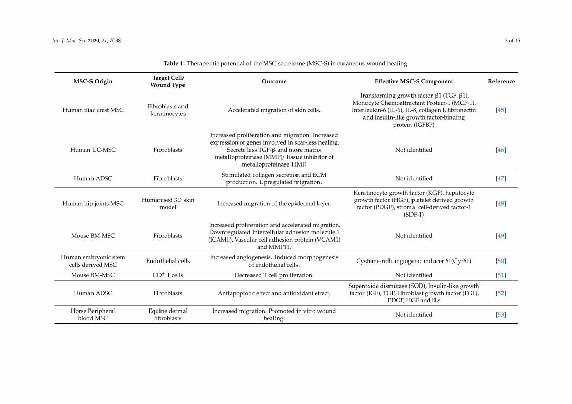
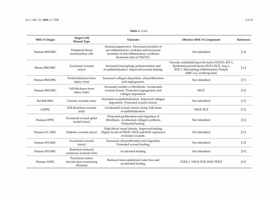
Recent studies have suggested that the main therapeutic benefits of MSCs are not limited solely totheir cell-to-cell interactions [36–39]. MSCs secrete a broad range of bioactive molecules, includingproteins, nucleic acids, proteasomes, exosomes, microRNA and membrane vesicles, collectively knownas the secretome, in response to the surrounding environment [40,41]. The MSC secretome (MSC-S)then influences neighbouring cells and regulates multiple biological processes [42]. Currently, paracrineor trophic properties are considered as the primary means of the therapeutic effect of MSCs [26,40,43].Although MSCs derived from different organs share phenotypic and regenerative characteristics, theirsecretome is different and depends on their origin, which consequently can lead to different therapeuticpotentials [44]. MSC-S from various origins has been used to assess its effect on skin cell functionalityas well as its effects on wound healing using in vitro and in vivo models (summarised in Table 1).
Int. J. Mol. Sci. 2020, 21, 7038 3 of 15
Table 1. Therapeutic potential of the MSC secretome (MSC-S) in cutaneous wound healing.
MSC-S Origin Target Cell/Wound Type Outcome Effective MSC-S Component Reference
Human iliac crest MSC Fibroblasts andkeratinocytes Accelerated migration of skin cells.
Transforming growth factor β1 (TGF-β1),Monocyte Chemoattractant Protein-1 (MCP-1),Interleukin-6 (IL-6), IL-8, collagen I, fibronectin
and insulin-like growth factor-bindingprotein (IGFBP)
[45]
Human UC-MSC Fibroblasts
Increased proliferation and migration. Increasedexpression of genes involved in scar-less healing.
Secrete less TGF-β and more matrixmetalloproteinase (MMP)/ Tissue inhibitor of
metalloproteinase TIMP.
Not identified [46]
Human ADSC Fibroblasts Stimulated collagen secretion and ECMproduction. Upregulated migration. Not identified [47]
Human hip joints MSC Humanised 3D skinmodel Increased migration of the epidermal layer.
Keratinocyte growth factor (KGF), hepatocytegrowth factor (HGF), platelet derived growthfactor (PDGF), stromal cell-derived factor-1
(SDF-1)
[48]
Mouse BM-MSC Fibroblasts
Increased proliferation and accelerated migration.Downregulated Intercellular adhesion molecule 1(ICAM1), Vascular cell adhesion protein (VCAM1)
and MMP11.
Not identified [49]
Human embryonic stemcells derived MSC Endothelial cells Increased angiogenesis. Induced morphogenesis
of endothelial cells. Cysteine-rich angiogenic inducer 61(Cyr61) [50]
Mouse BM-MSC CD+ T cells Decreased T cell proliferation. Not identified [51]
Human ADSC Fibroblasts Antiapoptotic effect and antioxidant effect.Superoxide dismutase (SOD), Insulin-like growthfactor (IGF), TGF, Fibroblast growth factor (FGF),
PDGF, HGF and ILs[52]
Horse Peripheralblood MSC
Equine dermalfibroblasts
Increased migration. Promoted in vitro woundhealing. Not identified [53]
Int. J. Mol. Sci. 2020, 21, 7038 4 of 15
Table 1. Cont.
MSC-S Origin Target Cell/Wound Type Outcome Effective MSC-S Component Reference
Human BM-MSC Peripheral bloodmononuclear cells
Immunosuppressive. Decreased secretion ofpro-inflammatory cytokines and increasedsecretion of anti-inflammatory cytokines.
Increased ratio of Th2/Th1.
Not identified [54]
Mouse BM-MSC Excisional wounds(mice)
Increased macrophage polymerisation andre-epithelialisation. Improved wound healing.
Vascular endothelial growth factor (VEGF), IGF-1,Epidermal growth factor (EGF), KGF, Ang-1,
SDF-1, Macrophage Inflammatory Protein(MIP-1α), erythropoietin
[36]
Human BM-MSC Partial-thickness burninjury (rats)
Increased collagen deposition, cell proliferationand angiogenesis. Not identified [37]
Human BM-MSC Full-thickness burninjury (rats)
Increased number of fibroblasts. Acceleratedwound closure. Promoted angiogenesis and
collagen deposition.bFGF [38]
Rat BM-MSC Chronic wounds (rats) Increased re-epithelialisation. Improved collagendeposition. Promoted wound closure. Not identified [39]
ADPSC Full-thickness wounds(rats)
Accelerated wound closure along with fasterre-epithelialisation. VEGF, EGF [55]
Human DPSC Excisional wound splintmodel (mice)
Promoted proliferation and migration offibroblasts. Accelerated collagen synthesis.
Promoted healing.Not identified [56]
Human UC-MSC Diabetic wounds (mice)High blood vessel density. Improved healing.
Higher levels of PDGF, VEGF and KGF expressionin treated wounds.
Not identified [57]
Human WJ-MSC Excisional wounds(mice)
Increased cell proliferation and migration.Promoted wound healing. Not identified [58]
Human WJ-MSC Radiation-inducedcutaneous wounds (rats) Accelerated healing. Not identified [59]
Human ADSCFractional carbon
dioxide laser resurfacing(Human)
Reduced trans-epidermal water loss andaccelerated healing. TGFβ-1, VEGF, FGF, HGF, PDGF [60]
Int. J. Mol. Sci. 2020, 21, 7038 5 of 15
4. Potential Mechanism of Action of MSC-S
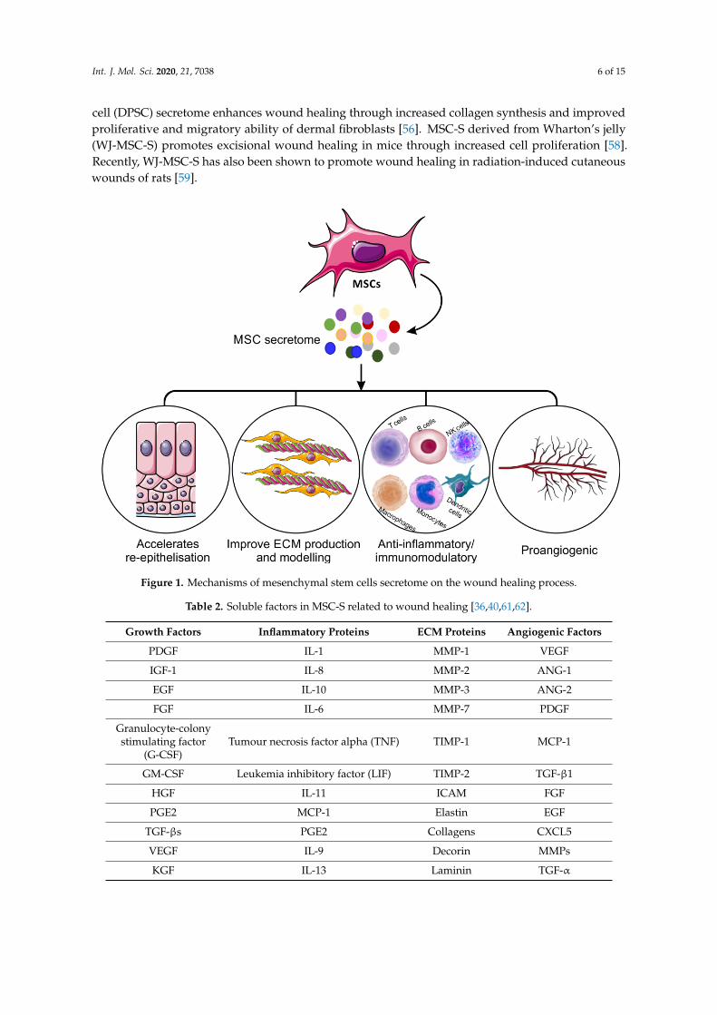
The mechanism of action of the MSC-S must be elucidated before it can be widely introduced as apotential new therapy in the clinic. Recent advances in cell and molecular biology have offered insightsinto multiple mechanisms, and it has been proposed that MSC-S can contribute to wound healing(Figure 1). Dissection of the MSC-S shows a large repertoire of proteins known to be involved in skininflammation, haemostasis and wound repair (Table 2). The biochemical pathways and mechanism ofaction of these proteins have been shown previously [36,40,61,62].
The MSC-S is, therefore, a complex mixture of bioactive factors and has been shown to havesignificant positive effects in the treatment of inflammatory disorders of nervous, cardiovascular,respiratory and skeletal systems [63–65]. Although it is believed that the anti-inflammatory effectsof cells rely on direct cell–cell interactions, several studies have demonstrated that the interactionbetween MSCs and immune cells can be attributed to MSC-secreted cytokines [66]. For example,MSC-secreted interleukin-1 receptor antagonist (IL1-RA) inhibits B cell differentiation [67]. HumanMSC-derived galectin-1 also has inhibitory impacts on the proliferation of alloreactive CD4+ andCD8+ T cells [68]. MSCs also secrete programmed death-ligand 1 (PD-L1), which suppresses T cellactivation and increases T cell apoptosis [69,70]. Furthermore, MSC-secreted Prostaglandin E2 (PGE2),TGF-β1, IL-6 and nitric oxide, all provide inhibitory effects on T cells, macrophages, neutrophils andNK cells [71,72]. MSC-S as a whole has been shown to exert immunosuppressive effects throughmodulating proliferation and activation of immune cells in vitro [51]. Treatment of peripheral bloodmononuclear cells with MSC-S led to a reduction in pro-inflammatory cytokine production andan increase in anti-inflammatory cytokines [54]. BM-MSC-S injected into the margins of excisionalwounds in mice promotes wound healing through diminished inflammation mediated by macrophagepolymerisation [36]. This beneficial effect of MSC-S is significantly higher than the equivalent treatmentwith fibroblast secretome [36].
Increased angiogenesis has been proposed as another of the main mechanisms of action forMSC-S in different types of wounds supported by in vitro treatment of endothelial cells with MSC-Senhancing their proliferation and migration. [37,38]. This impact of MSC-S on angiogenesis issuggested to be mediated by the secretion of Cyr61 from MSCs [50]. Pro-angiogenic proteins secretedby MSCs, such as Ang-1, Ang-2, VEGF, angiostatin, CXCL16, EGF, FGF, PDGF, granulocyte-macrophagecolony-stimulating factor (GM-CSF), HGF, MCP-1, MMP-8 and MMP-9, also contribute to vascularformation and stability [73]. In preclinical studies, BM-MSC-S treatment improved partial-thicknessburn injury repair in rats, which was mediated by increased blood vessel formation [37]. In another study,topical administration of BM-MSC-S cream to full-thickness burns of rats resulted in increased numbersof fibroblasts and improved angiogenesis as well as accelerated wound closure [38]. Subcutaneousinjection of umbilical cord-derived MSC secretome (UC-MSC-S) to wounds of diabetic mice led toaccelerated wound closure and high capillarity density in wound areas [57].
MSC-S from different origins (such as iliac crest, bone marrow, adipose, Wharton’s jelly, umbilicalcords) have been shown to enhance the migratory and proliferative abilities of dermal fibroblastsand epidermal keratinocytes in vitro [45–47,74]; MSC-S alters the expression of genes involved inre-epithelialisation and angiogenesis and increases re-epithelialisation in human 3D skin models [48,49].The secretome from adipose tissue-derived MSCs (ADSC-S) has been shown to protect dermal fibroblastsfrom oxidative stress-mediated apoptosis and accelerate wound closure with stimulatory effects onfibroblast migration in in vitro models [52,53]. The beneficial effect of MSC-S on skin cells is believedto be mediated by growth factors (such as IGF-1, EGF, FGF-2, KGF, TGF-β, HGF, PDGF, VEGF, SDF-1,erythropoietin) and chemokines (such as IL-6, IL-8, MCP-1 and RANTES) (Table 1) [36,40,61,62,74–76].Treatment of wounds with MSC-S significantly accelerates new tissue formation, collagen deposition andre-epithelialisation in treated wounds [36,49,55,56]. Application to chronic rat wounds of BM-MSC-Sdelivered in a fibrin vehicle also increases re-epithelialisation and collagen deposition [39]. In anotherstudy, excisional wounds of rats topically treated with ADSC secretome displayed accelerated woundclosure along with faster re-epithelialisation [55]. It has further been demonstrated that dental pulp stem
Int. J. Mol. Sci. 2020, 21, 7038 6 of 15
cell (DPSC) secretome enhances wound healing through increased collagen synthesis and improvedproliferative and migratory ability of dermal fibroblasts [56]. MSC-S derived from Wharton’s jelly(WJ-MSC-S) promotes excisional wound healing in mice through increased cell proliferation [58].Recently, WJ-MSC-S has also been shown to promote wound healing in radiation-induced cutaneouswounds of rats [59].
Int. J. Mol. Sci. 2020, 21, 7038 6 of 17
cell (DPSC) secretome enhances wound healing through increased collagen synthesis and improvedproliferative and migratory ability of dermal fibroblasts [56]. MSC-S derived from Wharton’s jelly(WJ-MSC-S) promotes excisional wound healing in mice through increased cell proliferation [58].Recently, WJ-MSC-S has also been shown to promote wound healing in radiation-induced cutaneouswounds of rats [59].
Figure 1. Mechanisms of mesenchymal stem cells secretome on the wound healing process.
Table 2. Soluble factors in MSC-S related to wound healing [36,40,61,62].
Growth Factors Inflammatory Proteins ECM Proteins Angiogenic Factors
PDGF IL-1 MMP-1 VEGF
IGF-1 IL-8 MMP-2 ANG-1
EGF IL-10 MMP-3 ANG-2
FGF IL-6 MMP-7 PDGF
Granulocyte-colonystimulating factor
(G-CSF)Tumour necrosis factor alpha (TNF) TIMP-1 MCP-1
GM-CSF Leukemia inhibitory factor (LIF) TIMP-2 TGF-β1
HGF IL-11 ICAM FGF
PGE2 MCP-1 Elastin EGF
TGF-βs PGE2 Collagens CXCL5
VEGF IL-9 Decorin MMPs
KGF IL-13 Laminin TGF-α
5. Advantages of MSC-S over Other Cell-Based Products
The use of cell-based therapies and products is not new. Indeed, skin substitutes, platelet-richplasmas, recombinant growth factors and cytokines have been around for decades [1]. However,despite promising preclinical datasets and successful clinical trials, there remains the need forimproved cell-based solutions, as evidenced by the spiraling increase in chronic wounds worldwide.Currently, skin substitutes containing living fibroblasts, keratinocytes or both, include TransCyte, [77],Dermagraft [78], Apligraf, [79] and OrCel [80]. These cell-based skin graft substitutes have shownpromising results in promoting faster wound closure (Transcyte), improved rate of re-epithelialisation
Figure 1. Mechanisms of mesenchymal stem cells secretome on the wound healing process.
Table 2. Soluble factors in MSC-S related to wound healing [36,40,61,62].
Growth Factors Inflammatory Proteins ECM Proteins Angiogenic Factors
PDGF IL-1 MMP-1 VEGF
IGF-1 IL-8 MMP-2 ANG-1
EGF IL-10 MMP-3 ANG-2
FGF IL-6 MMP-7 PDGF
Granulocyte-colonystimulating factor
(G-CSF)Tumour necrosis factor alpha (TNF) TIMP-1 MCP-1
GM-CSF Leukemia inhibitory factor (LIF) TIMP-2 TGF-β1
HGF IL-11 ICAM FGF
PGE2 MCP-1 Elastin EGF
TGF-βs PGE2 Collagens CXCL5
VEGF IL-9 Decorin MMPs
KGF IL-13 Laminin TGF-α
Int. J. Mol. Sci. 2020, 21, 7038 7 of 15
5. Advantages of MSC-S over Other Cell-Based Products
The use of cell-based therapies and products is not new. Indeed, skin substitutes, platelet-richplasmas, recombinant growth factors and cytokines have been around for decades [1]. However,despite promising preclinical datasets and successful clinical trials, there remains the need forimproved cell-based solutions, as evidenced by the spiraling increase in chronic wounds worldwide.Currently, skin substitutes containing living fibroblasts, keratinocytes or both, include TransCyte, [77],Dermagraft [78], Apligraf, [79] and OrCel [80]. These cell-based skin graft substitutes have shownpromising results in promoting faster wound closure (Transcyte), improved rate of re-epithelialisation(Dermagraft), and superior vascularity, pigmentation, wound height and scar scores (Apligraf) [81].However, they are expensive, require specific storage conditions, have the potential risks oftumorigenicity, infection and rejection, and are difficult to use within the community [82]. Recombinantgrowth factors were postulated to be the solution to impaired healing as it was hypothesised thatchronic non-healing wounds lacked specific growth factors and cytokines [83]. Numerous clinicaltrials were undertaken to investigate the growth factor therapies including EGF, PDGF, GM-CSF,KGF therapies, but despite appearing to be efficacious in many animal models of wound repair,translation into clinical products has been limited due to the significant amounts of growth factorsrequired for treatment, the expense of manufacture and the lack of clinically relevant improvementsin healing [84–87]. To date, only PDGF has received FDA approval for the treatment of diabetic footulcers, and its use is limited due to the need for dressing replacement and the potential increased riskof malignancy [88,89]. Administering single growth factors and/or cytokines has potential limitationsas wounds are complex environments, and multiple factors may be required to stimulate healingresponses. MSC-S contains a vast array of proteins at physiological and balanced levels, includingcytokines, growth factors and chemokines (Table 2), that potentially makes it a superior alternative toexpensive cytokine and growth factor therapies that are limited to delivering only one or two proteinsto the wounds.
The delivery of live cells to cutaneous wounds presents a unique and specific set of challenges [90].The injection of cells through a syringe or needle has been shown to decrease cell viability to only1–32% and can cause irreversible and sometimes fatal damage to the cell membrane [91,92]. Not onlydoes this negate the potential benefit of the cell therapy but the introduction of a large populationof apoptotic or necrotic cells may serve to elicit an immune response, which could be detrimental tothe healing process. MSC-S therapy avoids the difficulties associated with live-cell administrationin stem cells as well as advantages of ease of mass production, packaging and transportation [62].These advantageous factors have led to the growing potential of MSC-S use as a treatment for tissueregeneration and various disorders [40].
6. Challenges with the MSC-S as a Wound Therapy
6.1. Secretome Characterisation
Although MSC-S may be a promising medical product, it has been very challenging to define itsbiochemical composition or measure the activity and half-life of all of its components [62]. In additionto proteins, MSC-S also contains exosomes and extracellular vesicles [93]. Exosomes can containmiRNA, lipids and long noncoding RNAs, which regulate multiple signalling pathways related toinflammation [94]. The identification and characterisation of all the biomolecules that constitute thesecretome are difficult to achieve but will improve the understanding of the secreted factor profile andprovide information about its function, regulation and clinical use [95]. Further research on the MSC-Susing high throughput genetic and chemical screenings and next-generation metabolomics-drivenapproaches is required to clarify all of the key metabolic and signalling pathways that are mediatingrobust new tissue formation, dampened inflammation and enhanced wound closure.
Int. J. Mol. Sci. 2020, 21, 7038 8 of 15
6.2. Inconsistency in Preparation of Secretome
It has been reported that the isolation and culture methods, as well as donor health conditionand age, can affect the quality of MSC products [96]. Inconsistency in secretome harvesting in termsof MSC heterogenicity, inter-donor differences, cell number and time interval is another part of thecurrent challenge regarding the clinical use of secretome. Production of MSC-S under pharmaceuticalstandards and according to good manufacturing practice (GMP) is a vital step to use MSC-S as atherapeutic agent in the clinic. Compliance with well-defined good manufacturing protocols (GMP)will improve batch-to-batch consistency and the reproducible efficacy of MSC-S [97].
6.3. Potential Side Effects of MSC-S
Although there are limited reports of the negative effects of secretome, there are always potentialrisks using exogenous biological molecules, although these risks are reduced when compared tocell-based therapies. A comprehensive analysis is needed before MSC-S transplantation to specificniches in different tissues. For example, MSC-S contains MSC-derived exosomes and extracellularvesicles that can be immunogenic; however, the immunogenicity of exosomes has been shown to be lessthan their parent MSCs [98]. On the other hand, immunosuppressive properties of MSC-S have beenreported in several studies and have been hypothesised to be one of the main mechanisms of action ofMSC-S when treating autoimmune diseases [99]. However, the use of secretome may diminish theimmune system, which may increase the risk of infection, immunodeficiency and tumour growth intreated patients [100]. Thus, an optimal amount of secretome should be clearly defined with an aim tofind the right balance between safety and effectiveness of any secretome based therapy.
6.4. Limitation of Secretome Resources and Instability of Secretome Components
The number of MSCs that are required to produce sufficient quantities of secretome for anequivalent effect on acute wounds is about 10–25 times higher than directly administered live cells [43].These elevated numbers of cells impact the costs of derivation and validation because the biologicalproperties and activity of these cells may change with repeated passages. However, with increasedproduction and improvement in cell factories and bioreactors, the impact of this drawback may beminimised. Another major concern in secretome therapy is the instability and short half-life of proteins.One of the successful strategies to address these drawbacks is preconditioning cells to stimulate theparacrine production of the secretome. Preconditioning is also useful to control the composition ofthe secretome to avoid the toxicity caused by upregulated cytokines [95]. Hence, it is important tofirst elevate the production of desirable factors and downregulate the detrimental ones, and secondto achieve an appropriate balance between stimulatory and inhibitory factors produced by thesecells. There are different pre-treatment methods for MSCs, for example, subjecting cells to hypoxiaor anoxia has been reported to increase the secretion of cytokines and growth factors in transplantedstem cells [101]. Genetic manipulation of cells using transgenes can also alter specific gene expressionwith the aim of controlling the MSC-S post-transplantation [102]. Another promising approach forpre-treating stem cells before transplantation involves small molecules, such as inflammatory cytokinesand growth factors [103]. For example, treating MSCs with inflammatory cytokines increases theirsecretion of anti-inflammatory biomolecules and improves their immunosuppressive function [104].Preconditioning through cell–cell interactions is another strategy to improve the secretion of favourablebiomolecules. For example, Potapova et al. (2007) reported that MSCs in 3D spheroids are able tosecrete higher levels of paracrine biomolecules, such as IL-11, VEGF, FGF-2 and angiogenin, comparedto MSCs in monolayers [105]. This tailoring of the MSC-S could potentially lead to numerous off theshelf products specifically designed for the treatment of specific conditions or wound types.
Int. J. Mol. Sci. 2020, 21, 7038 9 of 15
7. Conclusions
Even though advances in the field of stem cell therapy have grown significantly, there are stillpractical and clinical hurdles to overcome before they can be routinely used for the treatment ofwounds. Poor engraftment and survival of cells in damaged areas, immunogenicity, tumorigenicityand lack of efficiency are notable limitations for clinical stem cell therapies. The use of MSC-S as apotential alternative to MSCs is of enormous interest and has significant clinical potential, given thetrophic properties of many of the secreted factors. MSC-S therapy avoids the use of live cells andcan limit biological variability, therefore, leading to the potential development of efficient and safetherapeutic approaches. While the mechanism of action of the MSC-S is still to be fully determined,the development of a cell-free therapy for the treatment of cutaneous wounds holds great promise.
Author Contributions: Original draft preparation, P.A.; review and editing, S.J.M., A.J.C. All authors have readand agreed to the published version of the manuscript.
Funding: A.J.C is supported by a National Health and Medical Research Council (NHMRC) Senior ResearchFellowship (GNT#1002009).
Conflicts of Interest: The authors declare no conflict of interest.
Abbreviations
ADSC Adipose-derived stem cellsAng AngiopoietinBM-MSC Bone marrow mesenchymal stem cellCD Cluster of differentiationDPSCs Dental pulp derived stem cellsECM Extracellular matrixEGF Epidermal growth factorFGF-2 Fibroblast growth factor-2G-CSF Granulocyte - colony stimulating factorGM-CSF Granulocyte-macrophage colony-stimulating factorHGF Hepatocyte growth factorICAM Intercellular adhesion molecule 1IFN-γ Interferon-gammaIGF Insulin-like growth factorIL InterleukinIL1-RA Interleukin-1 receptor antagonistKGF Keratinocyte growth factorLIF Leukemia inhibitory factorMCP-1 Monocyte chemoattractant protein-1MHC Major histocompatibility complexMMP Matrix metalloproteinaseMSCs Mesenchymal stem cellsMSC-S Mesenchymal stem cells secretomePDGF Platelet derived growth factorPD-L1 Programmed death-ligand 1SDF-1 Stromal cell-derived factor-1TGF Transforming growth factorTh1 Type 1 T helper cellTh2 Type 2 T helper cellTIMP-1 Tissue inhibitor of metalloproteinaseTNF Tumour necrosis factor alphaUC-MSCs Umbilical cord-derived mesenchymal stem cellsVCAM Vascular cell adhesion proteinVEGF Vascular endothelial growth factor
Int. J. Mol. Sci. 2020, 21, 7038 10 of 15
References
1. Ahangar, P.; Woodward, M.; Cowin, A.J. Advanced wound therapies. Wound Pract. Res. 2018, 26, 58–68.2. Gonzalez, A.C.D.O.; Freire, T.F.C.; Andrade, Z.D.A.; Medrado, A.P. Wound healing—A literature review.
An. Bras. Dermatol. 2016, 91, 614–620. [CrossRef] [PubMed]3. Han, G.; Ceilley, R. Chronic wound healing: A review of current management and treatments. Adv. Ther.
2017, 34, 599–610. [CrossRef]4. Johnson, R.M.; Richard, R. Partial-thickness burns: Identification and management. Adv. Skin Wound Care
2003, 16, 178–187. [PubMed]5. Simpson, D.; Liu, H.; Fan, T.H.M.; Nerem, R.; Dudley, S.C., Jr. A tissue engineering approach to progenitor
cell delivery results in significant cell engraftment and improved myocardial remodeling. Stem Cells 2007, 25,2350–2357. [CrossRef]
6. Jackson, K.A.; Majka, S.M.; Wang, H.; Pocius, J.; Hartley, C.J.; Majesky, M.W.; Entman, M.L.; Michael, L.H.;Hirschi, K.K.; Goodell, M.A. Regeneration of ischemic cardiac muscle and vascular endothelium by adultstem cells. J. Clin. Investig. 2001, 107, 1395–1402. [CrossRef]
7. Pérez-Ilzarbe, M.; Agbulut, O.; Pelacho, B.; Ciorba, C.; José-Eneriz, E.S.; Desnos, M.; Hagege, A.; Aranda, P.;Andreu, E.J.; Menasché, P.; et al. Characterization of the paracrine effects of human skeletal myoblaststransplanted in infarcted myocardium. Eur. J. Heart Fail. 2008, 10, 1065–1072. [CrossRef]
8. Picinich, S.C.; Mishra, P.J.; Mishra, P.J.; Glod, J.; Banerjee, D. The therapeutic potential of mesenchymal stemcells. Expert Opin. Biol. Ther. 2007, 7, 965–973. [CrossRef]
9. Park, S.R.; Kim, J.W.; Jun, H.S.; Roh, J.Y.; Lee, H.Y.; Hong, I.S. Stem cell secretome and its effect on cellularmechanisms relevant to wound healing. Mol. Ther. 2018, 26, 606–617. [CrossRef]
10. Blüguermann, C.; Wu, L.; Petrigliano, F.; McAllister, D.; Miriuka, S.; Evseenko, D. Novel aspects ofparenchymal-mesenchymal interactions: From cell types to molecules and beyond. Cell Biochem. Funct. 2013,31, 271–280. [CrossRef]
11. Galipeau, J.; Sensébé, L. Mesenchymal stromal cells: Clinical Challenges and therapeutic opportunities.Cell Stem Cell 2018, 22, 824–833. [CrossRef]
12. Charbord, P. Bone Marrow Mesenchymal stem cells: Historical overview and concepts. Hum. Gene Ther.2010, 21, 1045–1056. [CrossRef]
13. Strioga, M.; Viswanathan, S.; Darinskas, A.; Slaby, O.; Michalek, J. Same or not the same? Comparison ofadipose tissue-derived versus bone marrow-derived mesenchymal stem and stromal cells. Stem Cells Dev.2012, 21, 2724–2752. [CrossRef]
14. Pelekanos, R.A.; Sardesai, V.S.; Futrega, K.; Lott, W.B.; Kuhn, M.; Doran, M.R. Isolation and expansion ofmesenchymal stem/stromal cells derived from human placenta tissue. J. Vis. Exp. 2016, 10, e54204. [CrossRef][PubMed]
15. Sharpe, P. Dental mesenchymal stem cells. Development 2016, 143, 2273–2280. [CrossRef] [PubMed]16. Lee, O.K.; Kuo, T.K.; Chen, W.M.; Lee, K.D.; Hsieh, S.L.; Chen, T.H. Isolation of multipotent mesenchymal
stem cells from umbilical cord blood. Blood 2004, 103, 1669–1675. [CrossRef]17. Sasaki, M.; Abe, R.; Fujita, Y.; Ando, S.; Inokuma, D.; Shimizu, H. Mesenchymal stem cells are recruited into
wounded skin and contribute to wound repair by transdifferentiation into multiple skin cell type. J. Immunol.2008, 180, 2581–2587. [CrossRef] [PubMed]
18. Orlic, D.; Kajstura, J.; Chimenti, S.; Jakoniuk, I.; Anderson, S.M.; Li, B.; Pickel, J.; McKay, R.; Nadal-Ginard, B.;Bodine, D.M.; et al. Bone marrow cells regenerate infarcted myocardium. Nature 2001, 410, 701–705.[CrossRef]
19. Pittenger, M.F. Multilineage potential of adult human mesenchymal stem cells. Science 1999, 284, 143–147.[CrossRef]
20. Bourin, P.; Bunnell, B.A.; Casteilla, L.; Dominici, M.; Katz, A.J.; March, K.L.; Redl, H.; Rubin, J.P.; Yoshimura, K.;Gimble, J.M. Stromal cells from the adipose tissue-derived stromal vascular fraction and culture expandedadipose tissue-derived stromal/stem cells: A joint statement of the International Federation for AdiposeTherapeutics and Science (IFATS) and the International Society for Cellular Therapy (ISCT). Cytotherapy 2013,15, 641–648. [CrossRef]
21. Lv, F.J.; Tuan, R.S.; Cheung, K.M.C.; Leung, V.Y.L. Concise review: The surface markers and identity ofhuman mesenchymal stem cells. Stem Cells 2014, 32, 1408–1419. [CrossRef] [PubMed]
Int. J. Mol. Sci. 2020, 21, 7038 11 of 15
22. Oh, W.; Kim, D.S.; Yang, Y.S.; Lee, J.K. Immunological properties of umbilical cord blood-derived mesenchymalstromal cells. Cell. Immunol. 2008, 251, 116–123. [CrossRef] [PubMed]
23. Németh, K.; Leelahavanichkul, A.; Yuen, P.S.T.; Mayer, B.; Parmelee, A.; Doi, K.; Robey, P.G.;Leelahavanichkul, K.; Koller, B.H.; Brown, J.M.; et al. Bone marrow stromal cells attenuate sepsis viaprostaglandin E2–dependent reprogramming of host macrophages to increase their interleukin-10 production.Nat. Med. 2009, 15, 42–49. [CrossRef]
24. Meirelles, L.D.S.; Fontes, A.M.; Covas, D.T.; Caplan, A.I. Mechanisms involved in the therapeutic propertiesof mesenchymal stem cells. Cytokine Growth Factor Rev. 2009, 20, 419–427. [CrossRef] [PubMed]
25. Caplan, A.I. Why are MSCs therapeutic? New data: New insight. J. Pathol. 2009, 217, 318–324. [CrossRef]26. Lee, D.E.; Ayoub, N.; Agrawal, D.K. Mesenchymal stem cells and cutaneous wound healing: Novel methods
to increase cell delivery and therapeutic efficacy. Stem Cell Res. Ther. 2016, 7, 37. [CrossRef]27. Wang, M.; Yuan, Q.; Xie, L. Mesenchymal stem cell-based immunomodulation: Properties and clinical
application. Stem Cells Int. 2018, 2018, 3057624. [CrossRef]28. Kehl, D.; Generali, M.; Mallone, A.; Heller, M.; Uldry, A.C.; Cheng, P.F.; Gantenbein, B.; Hoerstrup, S.P.;
Weber, B. Proteomic analysis of human mesenchymal stromal cell secretomes: A systematic comparison ofthe angiogenic potential. NPJ Regen. Med. 2019, 4, 8. [CrossRef]
29. Mattar, P.; Bieback, K. Comparing the immunomodulatory properties of bone marrow, adipose tissue, andbirth-associated tissue mesenchymal stromal cells. Front. Immunol. 2015, 6, 560. [CrossRef]
30. Hocking, A.M.; Gibran, N.S. Mesenchymal stem cells: Paracrine signaling and differentiation duringcutaneous wound repair. Exp. Cell Res. 2010, 316, 2213–2219. [CrossRef]
31. Badiavas, E.V.; Ford, D.; Liu, P.; Kouttab, N.; Morgan, J.; Richards, A.; Maizel, A. Long-term bone marrowculture and its clinical potential in chronic wound healing. Wound Repair Regen. 2007, 15, 856–865. [CrossRef]
32. Ravari, H.; Hamidi-Almadari, D.; Salimifar, M.; Bonakdaran, S.; Parizadeh, M.R.; Koliakos, G. Treatmentof non-healing wounds with autologous bone marrow cells, platelets, fibrin glue and collagen matrix.Cytotherapy 2011, 13, 705–711. [CrossRef] [PubMed]
33. Sarasúa, J.G.; López, S.P.; Viejo, M.Á; Basterrechea, M.P.; Rodríguez, A.F.; Gutiérrez, A.F.; Gala, J.G.;Menéndez, Y.M.; Augusto, D.E.; Arias, A.P.; et al. Treatment of pressure ulcers with autologous bone marrownuclear cells in patients with spinal cord injury. J. Spinal Cord Med. 2011, 34, 301–307. [CrossRef] [PubMed]
34. Lataillade, J.J.; Doucet, C.; Bey, E.; Carsin, H.; Huet, C.; Clairand, I.; Bottollier-Depois, J.; Chapel, A.; Ernou, I.;Gourven, M.; et al. New approach to radiation burn treatment by dosimetry-guided surgery combined withautologous mesenchymal stem cell therapy. Regen. Med. 2007, 2, 785–794. [CrossRef]
35. Kanji, S.; Das, H. Advances of stem cell therapeutics in cutaneous wound healing and regeneration. Mediat.Inflamm. 2017, 2017, 5217967. [CrossRef]
36. Chen, L.; Tredget, E.E.; Wu, P.Y.G.; Wu, Y. Paracrine factors of mesenchymal stem cells recruit macrophagesand endothelial lineage cells and enhance wound healing. PLoS ONE 2008, 3, e1886. [CrossRef]
37. Aryan, A.; Bayat, M.; Bonakdar, S.; Taheri, S.; Haghparast, N.; Bagheri, M.; Piryaei, A.; Abdollahifar, M.A.Human bone marrow mesenchymal stem cell conditioned medium promotes wound healing in deepsecond-degree burns in male rats. Cells Tissues Organs 2019, 206, 317–329. [CrossRef] [PubMed]
38. Padeta, I.; Nugroho, W.S.; Kusindarta, D.L.; Fibrianto, Y.H.; Budipitojo, T. Mesenchymal stem cell-conditionedmedium promote the recovery of skin burn wound. Asian J. Anim. Veter. Adv. 2017, 12, 132–141. [CrossRef]
39. Mehanna, R.; Nabil, I.; Attia, N.; Bary, A.A.; Razek, K.A.; Ahmed, T.A.E.; Elsayed, F. The effect of bonemarrow-derived mesenchymal stem cells and their conditioned media topically delivered in fibrin glue onchronic wound healing in rats. BioMed Res. Int. 2015, 2015, 846062. [CrossRef]
40. Ferreira, J.R.; Teixeira, G.Q.; Santos, S.G.; Barbosa, M.A.; Almeida-Porada, G.; Gonçalves, R.M. Mesenchymalstromal cell secretome: Influencing therapeutic potential by cellular pre-conditioning. Front. Immunol. 2018,9, 2837. [CrossRef]
41. Wang, S.Y.; Hong, Q.; Zhang, C.Y.; Yang, Y.J.; Cai, G.; Chen, X.M. miRNAs in stem cell-derived extracellularvesicles for acute kidney injury treatment: Comprehensive review of preclinical studies. Stem Cell Res. Ther.2019, 10, 281–287. [CrossRef] [PubMed]
42. Caplan, A.I.; Dennis, J.E. Mesenchymal stem cells as trophic mediators. J. Cell. Biochem. 2006, 98, 1076–1084.[CrossRef] [PubMed]
Int. J. Mol. Sci. 2020, 21, 7038 12 of 15
43. Ahangar, P.; Mills, S.J.; Smith, L.E.; Strudwick, X.L.; Ting, A.E.; Vaes, B.; Cowin, A.J. Human multipotentadult progenitor cell-conditioned medium improves wound healing through modulating inflammation andangiogenesis in mice. Stem Cell Res. Ther. 2020, 11, 299. [CrossRef]
44. Vieira, N.M.; Zucconi, E.; Bueno, C.R., Jr.; Secco, M.; Suzuki, M.F.; Bartolini, P.; Vainzof, M.; Zatz, M. Humanmultipotent mesenchymal stromal cells from distinct sources show different in vivo potential to differentiateinto muscle cells when injected in dystrophic mice. Stem Cell Rev. Rep. 2010, 6, 560–566. [CrossRef]
45. Walter, M.; Wright, K.; Fuller, H.R.; MacNeil, S.; Johnson, W. Mesenchymal stem cell-conditioned mediumaccelerates skin wound healing: An in vitro study of fibroblast and keratinocyte scratch assays. Exp. Cell Res.2010, 316, 1271–1281. [CrossRef]
46. Li, M.R.; Luan, F.X.; Zhao, Y.; Hao, H.; Liu, J.; Dong, L.; Fu, X.; Han, W. Mesenchymal stem cell-conditionedmedium accelerates wound healing with fewer scars. Int. Wound J. 2017, 14, 64–73. [CrossRef] [PubMed]
47. Kim, W.S.; Park, B.S.; Sung, J.H.; Yang, J.M.; Park, S.B.; Kwak, S.J.; Park, J.S. Wound healing effect ofadipose-derived stem cells: A critical role of secretory factors on human dermal fibroblasts. J. Dermatol. Sci.2007, 48, 15–24. [CrossRef] [PubMed]
48. Al-Shaibani, M.B.; Dickinson, A.; Wang, X.N.; Tulah, A.S.; Lovat, P.E. Effect of conditioned media frommesenchymal stem cells (MSC-CM) on wound healing using a prototype of a fully humanised 3D skin model.Cytotherapy 2017, 19, e23–e24. [CrossRef]
49. Smith, A.N.; Willis, E.; Chan, V.T.; Muffley, L.A.; Isik, F.F.; Gibran, N.S.; Hocking, A.M. Mesenchymal stemcells induce dermal fibroblast responses to injury. Exp. Cell Res. 2010, 316, 48–54. [CrossRef]
50. Estrada, R.; Li, N.; Sarojini, H.; An, J.; Lee, M.J.; Wang, E. Secretome from mesenchymal stem cells inducesangiogenesis via Cyr61. J. Cell. Physiol. 2009, 219, 563–571. [CrossRef]
51. Kay, A.G.; Long, G.; Tyler, G.; Stefan, A.; Broadfoot, S.J.; Piccinini, A.M.; Middleton, J.; Kehoe, O. Mesenchymalstem cell-conditioned medium reduces disease severity and immune responses in inflammatory arthritis.Sci. Rep. 2017, 7, 18019. [CrossRef] [PubMed]
52. Kim, W.S.; Park, B.S.; Kim, H.K.; Park, J.S.; Kim, K.J.; Choi, J.S.; Chung, S.J.; Kim, D.D.; Sung, J.H. Evidencesupporting antioxidant action of adipose-derived stem cells: Protection of human dermal fibroblasts fromoxidative stress. J. Dermatol. Sci. 2008, 49, 133–142. [CrossRef] [PubMed]
53. Bussche, L.; Harman, R.; Syracuse, B.A.; Plante, E.L.; Lu, Y.C.; Curtis, T.; Ma, M.; Van De Walle, G.R.Microencapsulated equine mesenchymal stromal cells promote cutaneous wound healing in vitro. Stem CellRes. Ther. 2015, 6, 66. [CrossRef]
54. Chen, W.; Huang, Y.; Han, J.; Yu, L.; Li, Y.; Lu, Z.; Li, H.; Liu, Z.; Shi, C.; Duan, F.; et al. Immunomodulatoryeffects of mesenchymal stromal cells-derived exosome. Immunol. Res. 2016, 64, 831–840. [CrossRef]
55. Tarcisia, T.; Damayanti, L.; Antarianto, R.D.; Moenadjat, Y.; Pawitan, J.A. Adipose derived stem cellconditioned medium effect on proliferation phase of wound healing in Sprague Dawley rat. Med. J. Indones.2018, 26, 239–245. [CrossRef]
56. Nishino, Y.; Ebisawa, K.; Yamada, Y.; Okabe, K.; Kamei, Y.; Ueda, M. Human deciduous teeth dental pulpcells with basic fibroblast growth factor enhance wound healing of skin defect. J. Craniofacial Surg. 2011, 22,438–442. [CrossRef] [PubMed]
57. Shrestha, C.; Zhao, L.; Chen, K.; He, H.; Mo, Z. Enhanced healing of diabetic wounds by subcutaneousadministration of human umbilical cord derived stem cells and their conditioned media. Int. J. Endocrinol.2013, 2013, 10. [CrossRef]
58. Arno, A.I.; Amini-Nik, S.; Blit, P.H.; Al-Shehab, M.; Belo, C.; Herer, E.; Tien, H.C.; Jeschke, M.G. HumanWharton’s jelly mesenchymal stem cells promote skin wound healing through paracrine signaling. Stem CellRes. Ther. 2014, 5, 28. [CrossRef]
59. Sun, J.; Zhang, Y.; Song, X.; Zhu, J.; Zhu, Q. The healing effects of conditioned medium derived frommesenchymal stem cells on radiation-induced skin wounds in rats. Cell Transplant. 2019, 28, 105–115.[CrossRef]
60. Zhou, B.-R.; Xu, Y.; Guo, S.-L.; Xu, Y.; Wang, Y.; Zhu, F.; Permatasari, F.; Wu, D.; Yin, Z.-Q.; Luo, D. The effectof conditioned media of adipose-derived stem cells on wound healing after ablative fractional carbon dioxidelaser resurfacing. BioMed Res. Int. 2013, 2013, 519126. [CrossRef]
Int. J. Mol. Sci. 2020, 21, 7038 13 of 15
61. Harrell, C.R.; Fellabaum, C.; Jovicic, N.; Djonov, V.; Arsenijevic, N.; Volarevic, V. Molecular mechanismsresponsible for therapeutic potential of mesenchymal stem cell-derived secretome. Cells 2019, 8, 467.[CrossRef]
62. Vizoso, F.J.; Eiró, N.; Cid, S.; Schneider, J.; Perez-Fernandez, R. Mesenchymal stem cell secretome: Towardcell-free therapeutic strategies in regenerative medicine. Int. J. Mol. Sci. 2017, 18, 1852. [CrossRef]
63. Xin, H.; Li, Y.; Buller, B.; Katakowski, M.; Zhang, Y.; Wang, X.; Shang, X.; Zhang, Z.G.; Chopp, M.Exosome-mediated transfer of miR-133b from multipotent mesenchymal stromal cells to neural cellscontributes to neurite outgrowth. Stem Cells 2012, 30, 1556–1564. [CrossRef]
64. Zhang, S.; Chu, W.C.; Lai, R.C.; Lim, S.K.; Hui, J.H.P.; Toh, W.S. Exosomes derived from human embryonicmesenchymal stem cells promote osteochondral regeneration. Osteoarthr. Cartil. 2016, 24, 2135–2140.[CrossRef]
65. Yu, B.; Kim, H.W.; Gong, M.; Wang, J.; Millard, R.W.; Wang, Y.; Ashraf, M.; Xu, M. Exosomes secretedfrom GATA-4 overexpressing mesenchymal stem cells serve as a reservoir of anti-apoptotic microRNAs forcardioprotection. Int. J. Cardiol. 2014, 182, 349–360. [CrossRef]
66. Weiss, A.R.R.; Dahlke, M.H. Immunomodulation by mesenchymal stem cells (MSCs): Mechanisms of actionof living, apoptotic, and dead MSCs. Front. Immunol. 2019, 10, 1191. [CrossRef]
67. Luz-Crawford, P.; Djouad, F.; Toupet, K.; Bony, C.; Franquesa, M.; Hoogduijn, M.J.; Jorgensen, C.; Noël, D.Mesenchymal stem cell-derived interleukin 1 receptor antagonist promotes macrophage polarization andinhibits B cell differentiation. Stem Cells 2016, 34, 483–492. [CrossRef]
68. Gieseke, F.; Böhringer, J.; Bussolari, R.; Dominici, M.; Handgretinger, R.; Müller, I. Human multipotentmesenchymal stromal cells use galectin-1 to inhibit immune effector cells. Blood 2010, 116, 3770–3779.[CrossRef]
69. Beyth, S.; Borovsky, Z.; Mevorach, D.; Liebergall, M.; Gazit, Z.; Aslan, H.; Galun, E.; Rachmilewitz, J.Human mesenchymal stem cells alter antigen-presenting cell maturation and induce T-cell unresponsiveness.Blood 2005, 105, 2214–2219. [CrossRef]
70. Davies, L.C.; Heldring, N.; Kadri, N.; Le Blanc, K. Mesenchymal stromal cell secretion of programmeddeath-1 ligands regulates T cell mediated immunosuppression. Stem Cells 2017, 35, 766–776. [CrossRef]
71. Deng, Y.; Zhang, Y.; Ye, L.; Zhang, T.; Cheng, J.; Chen, G.; Zhang, Q.; Yang, Y. Umbilical cord-derivedmesenchymal stem cells instruct monocytes towards an IL10-producing phenotype by secreting IL6 andHGF. Sci. Rep. 2016, 6, 37566. [CrossRef]
72. Lin, L.; Du, L. The role of secreted factors in stem cells-mediated immune regulation. Cell. Immunol. 2018,326, 24–32. [CrossRef]
73. Watt, S.M.; Gullo, F.; Van Der Garde, M.; Markeson, D.; Camicia, R.; Khoo, C.P.; Zwaginga, J.J. The angiogenicproperties of mesenchymal stem/stromal cells and their therapeutic potential. Br. Med. Bull. 2013, 108, 25–53.[CrossRef]
74. Lee, C.S.; Burnsed, O.A.; Raghuram, V.; Kalisvaart, J.F.; Boyan, B.D.; Schwartz, Z. Adipose stem cellscan secrete angiogenic factors that inhibit hyaline cartilage regeneration. Stem Cell Res. Ther. 2012, 3, 35.[CrossRef]
75. Wu, Y.; Chen, L.; Scott, P.G.; Tredget, E.E. Mesenchymal Stem Cells Enhance Wound Healing ThroughDifferentiation and Angiogenesis. Stem Cells 2007, 25, 2648–2659. [CrossRef]
76. Hsiao, S.T.-F.; Asgari, A.; Lokmic, Z.; Sinclair, R.; Dusting, G.J.; Lim, S.Y.; Dilley, R.J. Comparative analysis ofparacrine factor expression in human adult mesenchymal stem cells derived from bone marrow, adipose,and dermal tissue. Stem Cells Dev. 2012, 21, 2189–2203. [CrossRef]
77. Bello, Y.M.; Falabella, A.F.; Eaglstein, W.H. Tissue-engineered skin. Current status in wound healing. Am. J.Clin. Dermatol. 2001, 2, 305–313. [CrossRef]
78. Hansen, S.L.; Voigt, D.W.; Wiebelhaus, P.; Paul, C.N. Using skin replacement products to treat burns andwounds. Adv. Skin Wound Care 2001, 14, 37–46. [CrossRef]
79. Eaglstein, W.H.; Iriondo, M.; Laszlo, K. A composite skin substitute (graftskin) for surgical wounds. A clinicalexperience. Dermatol. Surg. 1995, 21, 839–843. [CrossRef]
80. Martin, L.K.; Kirsner, R.S. Use of a meshed bilayered cellular matrix to treat a venous ulcer. Adv. Skin WoundCare 2002, 15, 260–264. [CrossRef]
81. Waymack, P.; Duff, R.G.; Sabolinski, M. The effect of a tissue engineered bilayered living skin analog, overmeshed split-thickness autografts on the healing of excised burn wounds. Burns 2000, 26, 609–619. [CrossRef]
Int. J. Mol. Sci. 2020, 21, 7038 14 of 15
82. Alrubaiy, L.; Al-Rubaiy, K.K. Skin substitutes: A brief review of types and clinical applications. Oman Med. J.2009, 24, 4–6. [CrossRef]
83. Barrientos, S.; Brem, H.; Stojadinovic, O.; Tomic-Canic, M. Clinical application of growth factors and cytokinesin wound healing. Wound Repair Regen. 2014, 22, 569–578. [CrossRef]
84. Da Costa, R.M.; Jesus, F.M.; Aniceto, C.; Mendes, M. Double-blind randomized placebo-controlled trial ofthe use of granulocyte-macrophage colony-stimulating factor in chronic leg ulcers. Am. J. Surg. 1997, 173,165–168. [CrossRef]
85. Heldin, C.-H.; Westermark, B. Mechanism of action and in vivo role of platelet-derived growth factor. Physiol.Rev. 1999, 79, 1283–1316. [CrossRef]
86. Lin, H.; Chen, B.; Sun, W.; Zhao, W.; Zhao, Y.; Dai, J. The effect of collagen-targeting platelet-derived growthfactor on cellularization and vascularization of collagen scaffolds. Biomaterials 2006, 27, 5708–5714. [CrossRef][PubMed]
87. Krishnaswami, S.; Ly, Q.P.; Rothman, V.L.; Tuszynski, G.P. Thrombospondin-1 promotes proliferative healingthrough stabilization of PDGF. J. Surg Res. 2002, 107, 124–130. [CrossRef]
88. Mast, B.A.; Schultz, G. Interactions of cytokines, growth factors, and proteases in acute and chronic wounds.Wound Repair Regen. 1996, 4, 411–420. [CrossRef]
89. Papanas, D.; Maltezos, E. Benefit-risk assessment of becaplermin in the treatment of diabetic foot ulcers.Drug Saf. 2010, 33, 455–461. [CrossRef]
90. Kirby, G.T.S.; Mills, S.J.; Cowin, A.J.; Smith, L.E. Stem cells for cutaneous wound healing. BioMed Res. Int.2015, 2015, 285869. [CrossRef]
91. Zhang, M.; Methot, D.; Poppa, V.; Fujio, Y.; Walsh, K.; Murry, C.E. Cardiomyocyte grafting for cardiac repair:Graft cell death and anti-death strategies. J. Mol. Cell. Cardiol. 2001, 33, 907–921. [CrossRef]
92. Wahlberg, B.; Ghuman, H.; Liu, J.R.; Modo, M. Ex vivo biomechanical characterization of syringe-needleejections for intracerebral cell delivery. Sci. Rep. 2018, 8, 1–17. [CrossRef] [PubMed]
93. Krek, A.; Grün, D.; Poy, M.N.; Wolf, R.; Rosenberg, L.; Epstein, E.J.; MacMenamin, P.; Da Piedade, I.;Gunsalus, K.C.; Stoffel, M.; et al. Combinatorial microRNA target predictions. Nat. Genet. 2005, 37, 495–500.[CrossRef]
94. Gao, F.; Yu, L.; Zhang, N.; Zhang, Y.; Wang, R.; Zhao, J. Long noncoding RNAs and their regulatory network:Potential therapeutic targets for adult moyamoya disease. World Neurosurg. 2016, 93, 111–119. [CrossRef]
95. Ranganath, S.H.; Levy, O.; Inamdar, M.S.; Karp, J.M. Harnessing the mesenchymal stem cell secretome forthe treatment of cardiovascular disease. Cell Stem Cell 2012, 10, 244–258. [CrossRef]
96. Lukomska, B.; Stanaszek, L.; Zuba-Surma, E.; Łegosz, P.; Sarzynska, S.; Drela, K. Challenges and controversiesin human mesenchymal stem cell therapy. Stem Cells Int. 2019, 2019, 9628536. [CrossRef] [PubMed]
97. De Sousa, P.; Downie, J.; Tye, B.; Bruce, K.; Dand, P.; Dhanjal, S.; Serhal, P.; Harper, J.; Turner, M.; Bateman, M.Development and production of good manufacturing practice grade human embryonic stem cell lines assource material for clinical application. Stem Cell Res. 2016, 17, 379–390. [CrossRef]
98. Gowen, A.; Shahjin, F.; Chand, S.; Odegaard, K.E.; Yelamanchili, S.V. Mesenchymal stem cell-derivedextracellular vesicles: Challenges in clinical applications. Front. Cell Dev. Biol. 2020, 8, 149. [CrossRef]
99. Zhao, Q.; Ren, H.; Han, Z. Mesenchymal stem cells: Immunomodulatory capability and clinical potential inimmune diseases. J. Cell. Immunother. 2016, 2, 3–20. [CrossRef]
100. Bascones-Martinez, A.; Mattila, R.; Gomez-Font, R.; Meurman, J.H. Immunomodulatory drugs: Oral andsystemic adverse effects. Med. Oral Patol. Oral Cir. Bucal. 2014, 19, e24–e31. [CrossRef] [PubMed]
101. Lee, E.Y.; Xia, Y.; Kim, W.S.; Kim, M.H.; Kim, T.H.; Kim, K.J.; Park, B.S.; Sung, J.H. Hypoxia-enhancedwound-healing function of adipose-derived stem cells: Increase in stem cell proliferation and up-regulationof VEGF and bFGF. Wound Repair Regen. 2009, 17, 540–547. [CrossRef] [PubMed]
102. Mangi, A.A.; Noiseux, N.; Kong, D.; He, H.; Rezvani, M.; Ingwall, J.S.; Dzau, V.J. Mesenchymal stem cellsmodified with Akt prevent remodeling and restore performance of infarcted hearts. Nat. Med. 2003, 9,1195–1201. [CrossRef] [PubMed]
103. Afzal, M.R.; Haider, H.K.; Idris, N.M.; Jiang, S.; Ahmed, R.P.; Ashraf, M. Preconditioning promotes survivaland angiomyogenic potential of mesenchymal stem cells in the infarcted heart via NF-κB signaling. Antioxid.Redox Signal 2009, 12, 693–702. [CrossRef] [PubMed]
Int. J. Mol. Sci. 2020, 21, 7038 15 of 15
104. Baldari, S.; Di Rocco, G.; Piccoli, M.; Pozzobon, M.; Muraca, M.; Toietta, G. Challenges and strategies forimproving the regenerative effects of mesenchymal stromal cell-based therapies. Int. J. Mol. Sci. 2017, 18,2087. [CrossRef] [PubMed]
105. Potapova, I.A.; Gaudette, G.R.; Brink, P.R.; Robinson, R.B.; Rosen, M.R.; Cohen, I.S.; Doronin, S.V.Mesenchymal stem cells support migration, extracellular matrix invasion, proliferation, and survivalof endothelial cells in vitro. Stem Cells 2007, 25, 1761–1768. [CrossRef]
© 2020 by the authors. Licensee MDPI, Basel, Switzerland. This article is an open accessarticle distributed under the terms and conditions of the Creative Commons Attribution(CC BY) license (http://creativecommons.org/licenses/by/4.0/).