Embed Size (px)
DESCRIPTION
Metabolic bone disease. Biochemistry. PTH Vitamin D Calcitonin. Hypercalcemic states. Causes Hyperparathyroidism : presentations symptoms “ stones,bones,abdominal groans&psychic moans ” Impact on bones : osteporosis - PowerPoint PPT Presentation
Citation preview
Metabolic bone disease
Metabolic bone disease
Biochemistry• PTH• Vitamin D• Calcitonin

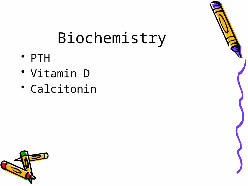
Hypercalcemic states• Causes• Hyperparathyroidism : presentations symptoms “stones,bones,abdominal
groans&psychic moans”Impact on bones : osteporosisImpact on kidney : renal stonesNon-specific features : sometimes asymptomaticDiagnosisTreatment

Primary hyperparathyroidism
• Calcium is high• Phosphorus is low• PTH is high

Other hypercalcemic states
• Sarcoidosis• Thyrotoxicosis• Adrenal insufficiency• Thiazides• Hypervitaminosis D&A• Immobilization• MALIGNANCY
Treatment of hypercalcemia
• Remove cause• Hydration• Calcitonin\bisphosphnates• Steroids• In primary hyperparathyroidism :
removal of the adenoma.

Hypocalcemia• Causes : hypoparathyroidism , surgical ,
hypomagnesimia • Pseudohypoparathyroidism : type 1A
autosomal dominant . Resistance to PTH+ somatic features. Type 1B : isolated resistance
• Clinical presentations : acute vs chronic.• Eye , CNS ( EXTRAPYRAMIDAL),CARDIAC
• Treatment

Hypoparathyroidism• Low calcium• High phosphorus• Cause : surgical• auto immune• severe vitamin D
deficiency

Clinical presentation• Numbness• If severe
hypocalcemia : tetany
• Trosseau sign• Chovstek sign
Treatment of hypocalcemia
• Calcium and vitamin D supplements
• If severe with tetany : give 10 cc of 10% calcium gluconate slowly ( careful in patients on digoxin )
OsteoporosisDEFINITIONDIFFERNTIATIING OSTEOPOROSIS
FROM OSTEOMALACIA CAUSESDIAGNOSISPREVENTIONTREATMENT
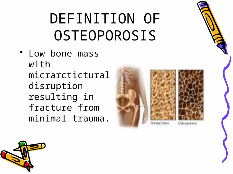
DEFINITION OF OSTEOPOROSIS
• Low bone mass with micrarctictural disruption resulting in fracture from minimal trauma.

Causes of osteoporosis• Menopause• Old age• Calcium and vitamin D deficiency• Estrogen deficiency• Use of steroids

Diagnosis of osteoporosis
• Plain x-ray : not very sensitive• Dual-energy x-ray absoptiometry (
DXA) measuring bone minaeral density (BMD) and comparing it to BMD of a healthy woman
• More than -2.5 SD below average : osteoporosis


Treatment of osteoporosis
• Prevention• Public awareness• Adequate calcium and vitamin D
supplements• Bisphphosnates : reducing bone
breakdown

Steroid induced osteoporosis
• Major impact on ? : axial bone

Effects• Steroids for several days causes
bone loss more on axial bones ( 40 %) than on peripheral bones ( 20%).
• Muscle weakness• Prednisolone more than 5 mg /day
for long time

Mechanisms• Renal Ca loss• Inhibition of intestinal Ca
absorption• In animals : increase osteoclast
and inhibition of osteoblast activity
• Suppression of gonadotropin secretion ( high dose)

Management• Use smallest possible dose • Shortest possible duration• Physical activity• Calcium and vitamin D• Pharmacologic treatment:
bisphosphontaes , ? PTH
Osteomalacia

Definition of osteomalacia
• Reduced mineralization of bone
• Rickets occurs in growing bone
Causes of osteomalacia

• Vitamin D deficoency• Ca deficiency• Phosphate deficiency• Liver disease• Renal disease• Malabsorption• Hereditary forms• ( intestinal and gastric surgery)• drugs
Clinical presentation
• Bony aches and pains• Muscle weakness

LAB.
• Low serum vitamin D• High PTH• High serum alkaline phosphatase
labCa levelPo4 levelAlk phosphPTHVitamin D level

Radiology• X-ray: growing
bones vs mature bones. Subperiosteal resorption , looser”s zones ( pathognomonic).
• Bone scan
Treatment of osteomalacia

• Calcium and vitamin D supplements
• Sun exposure• Results of treatment is usually
very good.

Paget’s disease of bone
Clinical presentation

• Two thirds of patients are asymptomatic• Incidental radiological finding• Unexplained high alk phosph• Large skull,frontal bossing,bowing of
legs, deafness,erythema, bony tenderness
• Fracture tendency: verteberal crush fractures , tibia or femur. Healing is rapid.
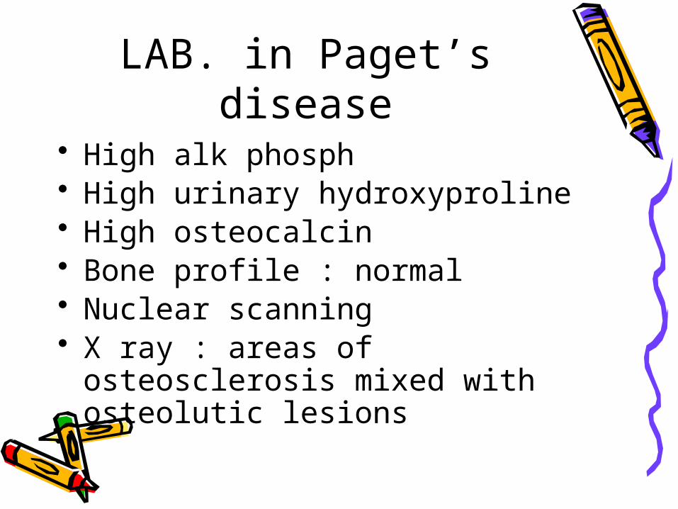
LAB. in Paget’s disease• High alk phosph• High urinary hydroxyproline• High osteocalcin• Bone profile : normal• Nuclear scanning• X ray : areas of osteosclerosis
mixed with osteolutic lesions

Complications• Sensory deafness• Spinal stenosis• Osteoarthritis & gout• Osteosarcoma• Hypercalcemia( immobilization)• urolithiasis

Treatment of Paget’s disease
• Calcitonon• Bisphphosphonates• Plicamycin( rarely used)

Renal Osteodystrophy• pathogenesis• Clinical presentations:Osteitis fibrosaOsteomalaciaLow serum calciumHigh phosphorusHigh alkaline phosphHigh PTH 2ry →3ry
hyperparathyroidism( hypercalcemia)
How is vitamin D carried in blood ?

What is VDR? • Clinical applications ?• Vitamin D-dependent rickets type
2 ( lack of functioning VDR. 1,25 (OH)2 D3 IS VERY HIGH.

Extrarenal production of 1,25 (OH)2 D3
• Macrophages : cause of hypercalcemia in sarcoidosis , lymphoma and other granulomatous disease ( regulated by cytokines &TNF).

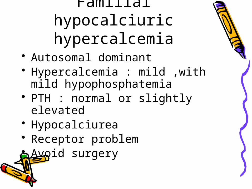
Familial hypocalciuric hypercalcemia
• Autosomal dominant• Hypercalcemia : mild ,with mild
hypophosphatemia• PTH : normal or slightly elevated• Hypocalciurea• Receptor problem• Avoid surgery

Mechanisms

Management