Embed Size (px)
Citation preview

Metabolic diseases of joints
or
Crystal induced arthropathy
By:Dr Zahra Rezaieyazdi
Zahra Rezaieyazdi
Rheumatologist
Rheumatic Diseases Research Center
Ghaem hospital
Mashhad university of medical sciences
19/3/1389
• Gout
•Pseudogout •Other crystals
By:Dr Zahra Rezaieyazdi

Objectives:
• To review the etiology and
pathophysiology of gout
• To recognize predisposing factors for gout
• To review diagnostic criteria and
evaluation for gout
• To select appropriate treatment for a
patient presenting with gout
By:Dr Zahra Rezaieyazdi
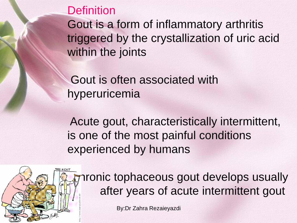
Definition
Gout is a form of inflammatory arthritis
triggered by the crystallization of uric acid
within the joints
Gout is often associated with
hyperuricemia
Acute gout, characteristically intermittent,
is one of the most painful conditions
experienced by humans
Chronic tophaceous gout develops usually
after years of acute intermittent gout
By:Dr Zahra Rezaieyazdi

EPIDEMIOLOGY
Gout occurs predominantly among men and
postmenopausal women
The prevalence of gout is approximately 2.7%
Serum urate concentrations in men are about 1 mg/dL higher than in
women
The incidence of primary gout, has doubled over the past 20 years in
both sexes
Diet and lifestyle trends, increasing frequencies of obesity, metabolic
syndrome, hypertension, organ transplantation, and increasing use of
certain medications (e.g., low dose salicylate and diuretics)
By:Dr Zahra Rezaieyazdi
PATHOGENESIS OF
HYPERURICEMIA AND GOUT
Humans are the only mammals
who are known to develop gout
The absence of uricase,
combined with extensive
reabsorption of filtered urate,
results in urate levels in human
plasma that are approximately 10
times than those of most other
mammals
By:Dr Zahra Rezaieyazdi

Solubility of Urate
Uric acid is a weak acid (pKa = 5.8) that exists largely
as urate, the ionized form, at physiological pH.
In general, the risk of supersaturation and crystal
formation rises in parallel with the concentration of urate
in physiologic fluids.
The solubility of urate in joint fluids is influenced by
other factors including temperature, pH, concentration,
hydration state, and the presence of nucleating agents
around which urate crystals may coalesce (e.g.,
nonaggregated proteoglycans, insoluble collagens, and
chondroitin sulfate).
(1) podagra (caused by the lower temperature at this
peripheral body site);
(2) tendency to occur in osteoarthritic joints (because
such joints contain nucleating debris);
(3) the frequency of nocturnal onset (the result of
intra-articular dehydration that may occur at night)
By:Dr Zahra Rezaieyazdi

Urate Metabolism
The amount of urate in the body depends on
the balance between dietary intake,
synthesis, and excretion of this molecule.
Hyperuricemia results from the
overproduction of urate (10%), underexcretion
of urate (90%), or often a combination of the
two.
The purine precursors come from
exogenous (dietary) sources or endogenous
metabolism (synthesis and cell turnover)
By:Dr Zahra Rezaieyazdi

Urate Production Pathways
endogenous overproduction of urate:
-increased cell turnover in proliferative and
inflammatory disorders (e.g., hematologic
malignancies and psoriasis)
-pharmacologic intervention resulting in increased
urate production (e.g., chemotherapy)
-tissue hypoxia.
- inborn error of metabolism (10%) such as
superactivity of 5'-phosphoribosyl 1-pyrophosphate
(PRPP) synthetase or deficiency of hypoxanthine-
guanine phosphoribosyl transferase (HPRT)=(Lesch-
Nyhan syndrome)
both of these enzyme defects are X-linked traits,
homozygous males are affected.
postmenopausal gout and urinary tract stones can
occur in carrier females.
By:Dr Zahra Rezaieyazdi

Alcohol and Gout
Ethanol administration increases uric acid production by net
ATP degradation to AMP.
Decreased urinary excretion associated with dehydration and
metabolic acidosis
alcoholic beverage: beer confers a larger risk
By:Dr Zahra Rezaieyazdi
Adiposity, Insulin Resistance, and Hyperuricemia
body mass index, waist-to-hip ratio, and weight gain are all associated
with gout
Insulin may enhance renal urate reabsorption in renal proximal tubule
leptin and adenosine may contribute to hyperuricemia
By:Dr Zahra Rezaieyazdi

RENAL TRANSPORT OF URATE
(1) glomerular filtration, (2) nearly complete
reabsorption of the filtered urate, (3) subsequent
secretion, and (4) postsecretory reabsorption in the
remaining proximal tubule
URAT1 (SLC22A12), a novel transporter expressed
at the apical brush border of the proximal nephron
uricosuric compounds (e.g., probenecid,
benzbromarone, sulfinpyrazone, and losartan) directly
inhibit URAT1
Urate retention is provoked also by a reduction in
extracellular fluid volume
and by excesses of angiotensin II, insulin, and
parathyroid hormone
Biphasic effects on urate excretion, that is, anti-
uricosuria at low dose and uricosuria at high dose, for
salicylate
By:Dr Zahra Rezaieyazdi

URATE CRYSTAL-INDUCED INFLAMMATION
Urate crystals in joint fluid : rupture of preformed synovial deposits or de novo
Urate crystals initiate, amplify, and sustain intense inflammatory attacks by release
of humoral and cellular mediators
Urate crystals 1.activate phagocytosis 2.directly lipid membrane activation
crystal-induced interleukin (IL) 8 expression in monocytic cells, which plays a key
role in the neutrophil accumulation
Toll-like receptors (TLR) 2 and 4 , trigger receptor expressed on myeloid cells 1
(TREM-1) induced innate immune response to amplification of acute gouty
inflammation
By:Dr Zahra Rezaieyazdi

URATE CRYSTAL-INDUCED INFLAMMATION
monocytes and mast cells participate during the early phase of inflammation,
neutrophilic infiltrates occur later
monocytes play a central role: In undifferentiated monocytes, induction of
proinflammatory cytokines [tumor necrosis factor alpha (TNF-alpha), IL-1 beta, IL-6,
IL-8, and cyclooxygenase-2 (COX-2)] and endothelial cell activation occur after
urate crystal phagocytosis.
In response to C3a, C5a, and IL-1, mast cells release histamine and other
inflammatory mediators
The vasodilatation, increased vascular permeability, and pain so
characteristic of gout are also mediated by kinins, complement cleavage
peptides, and other vasoactive prostaglandins
Neutrophil influx is believed to be promoted by endothelial-neutrophil
adhesion, triggered by IL-1, TNF-alpha, IL-8, the neutrophil chemoattractant
protein-1 (MCP-1), and other cytokines and chemokines
Among these factors, IL-8 and growth-related gene chemokines play a central
role in neutrophil invasion
By:Dr Zahra Rezaieyazdi

URATE CRYSTAL-INDUCED INFLAMMATION
Several processes contribute to the self-limited nature of acute gout.
Clearance of urate crystals by differentiated macrophages
Neutrophil apoptosis
upregulation of IL-10
apolipoprotein B and E
Inactivation of inflammatory mediators by proteolytic cleavage,
desensitization of receptors for chemokines, release of lipoxins, IL-1 receptor
antagonist
By:Dr Zahra Rezaieyazdi

By:Dr Zahra Rezaieyazdi

URATE CRYSTAL-INDUCED INFLAMMATION
Chronic gouty arthritis leading to chronic synovitis, cartilage loss, and bone
erosion
Tophi may contribute to chondrolysis despite adequate treatment of both
hyperuricemia and acute gouty attacks
microcrystals produce active metalloproteinases. leading to cartilage
destruction
The crystals can also suppress the 1,25-dihydroxycholecalciferol-induced
activity of alkaline phosphatase and osteocalcin. Thus, crystals can alter the
osteoblast phenotype by reducing their anabolic effects that may contribute to
damage to the juxta-articular bone
By:Dr Zahra Rezaieyazdi

Causes of hyperuricemia Uric acid overproduction
HGRT deficiency, PRPP synthetase overactivity
Increased cell turnover Myeloproliferative and lymphoproliferative
disorders, polycythemia vera, malignant diseases, hemolysis, psoriasis
Purine-rich foods
Obesity
Accelerated ATP degradation Ethanol, fructose, severe tissue
hypoxemia or muscle exertion, glycogen storage diseases,
Urate Increasing Agents Cytotoxic drugs
Uric acid underexcretion
Renal failure, hypertension, metabolic syndrome, obesity
Lead nephropathy, polycystic kidney disease, medullary cystic kidney
disease
Agents increasing urate reabsorption through transstimulation of
URAT1
Pyrazinamide, salicylate (low dose), nicotinate, lactate, beta-
hydroxybutyrate, acetoacetate
Agents decreasing renal urate excretion, maybe through
URAT1 or other mechanisms Diuretics, ethambutol, insulin, beta-
blockers
By:Dr Zahra Rezaieyazdi

By:Dr Zahra Rezaieyazdi

• Heredity
• Drug usage
• Renal failure
• Hematologic Disease
• Trauma
• Alcohol use
• Psoriasis
• Poisoning
• Obesity
• Hypertension
• Organ transplantation
• Surgery
Predisposing Factors
By:Dr Zahra Rezaieyazdi

• Asymptomatic hyperuricemia – Very common biochemical abnormality
– Defined as 2 SD above mean value
– Majority of people with hyperuricemia never develop symptoms of uric acid excess
• Acute Intermittent Gout (Gouty Arthritis) – Episodes of acute attacks. Symptoms may be confined to a single joint or patient
may have systemic symptoms.
• Intercritical Gout – Symptom free period interval between attacks. May have hyperuricemia and
MSU crystals in synovial fluid
• Chronic Tophaceous Gout – Results from established disease and refers to stage of deposition of urate,
inflammatory cells and foreign body giant cells in the tissues. Deposits may be in tendons or ligaments.
– Usually develops after 10 or more years of acute intermittent gout.
Stages of Classic Gout
By:Dr Zahra Rezaieyazdi

• Systemic: fever rare but patients may have fever, chills and malaise
• Musculoskeletal: Acute onset of monoarticular joint pain. First MTP most common. Usually affected in 90% of patients with gout. Other joints knees, foot and ankles. Less common in upper extremities
• Skin: warmth, erythema and tenseness of skin overlying joint. May have pruritus and desquamation
• GU: Renal colic with renal calculi formation in patients with hyperuricemia
Presenting Symptoms
By:Dr Zahra Rezaieyazdi

• Abrupt onset of severe joint inflammation, often with onset
in the night
• 75% of initial attacks in first MTP joint
• Usually monarticular, may be polyarticular
• Attack subsides in 3-10 days
• Urate crystals present in synovial fluid
• Hyperuricemia may or may not be present
Acute gouty arthritis
By:Dr Zahra Rezaieyazdi

• Trauma
• Infections – septic arthritis, gonococcal arthritis, cellulitis
• Inflammatory – Rheumatic arthritis, Reiter’s syndrome, Psoriatic
arthritis
• Metabolic – pseudogout
• Miscellaneous – Osteoarthrtis
Differential Diagnosis
By:Dr Zahra Rezaieyazdi

• Trauma
• Infections – septic arthritis, gonococcal arthritis, cellulitis
• Inflammatory – Rheumatic arthritis, Reiter’s syndrome, Psoriatic
arthritis
• Metabolic – pseudogout
• Miscellaneous – Osteoarthrtis
Differential Diagnosis
By:Dr Zahra Rezaieyazdi

By:Dr Zahra Rezaieyazdi

By:Dr Zahra Rezaieyazdi

By:Dr Zahra Rezaieyazdi

By:Dr Zahra Rezaieyazdi

By:Dr Zahra Rezaieyazdi

By:Dr Zahra Rezaieyazdi

• Definitive diagnosis only possible by aspirating and inspecting synovial fluid or tophaceous material and demonstrating MSU crystals
• Polarized microscopy, the crystals appear as bright birefringent crystals that are yellow (negatively birefringent)
Diagnosis
By:Dr Zahra Rezaieyazdi

• Definitive diagnosis only possible by aspirating and inspecting synovial fluid or tophaceous material and demonstrating MSU crystals
• Polarized microscopy, the crystals appear as bright birefringent crystals that are yellow (negatively birefringent)
Diagnosis
By:Dr Zahra Rezaieyazdi

• Definitive diagnosis only possible by aspirating and inspecting synovial fluid or tophaceous material and demonstrating MSU crystals
• Polarized microscopy, the crystals appear as bright birefringent crystals that are yellow (negatively birefringent)
By:Dr Zahra Rezaieyazdi

Synovial Fluid Findings
• Needle shaped
crystals of
monosodium urate
monohydrate that
have been engulfed
by neutrophils
By:Dr Zahra Rezaieyazdi
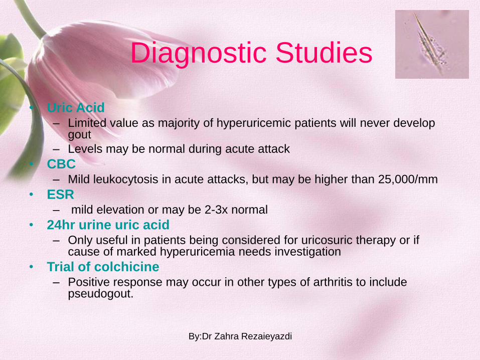
• Uric Acid – Limited value as majority of hyperuricemic patients will never develop
gout
– Levels may be normal during acute attack
• CBC – Mild leukocytosis in acute attacks, but may be higher than 25,000/mm
• ESR – mild elevation or may be 2-3x normal
• 24hr urine uric acid – Only useful in patients being considered for uricosuric therapy or if
cause of marked hyperuricemia needs investigation
• Trial of colchicine – Positive response may occur in other types of arthritis to include
pseudogout.
Diagnostic Studies
By:Dr Zahra Rezaieyazdi

• Gout can be treated without complications.
• Therapeutic goals include
– terminating attacks
– providing control of pain and inflammation
– preventing future attacks
– preventing complications such as renal stones, tophi, and destructive arthropathy
Treatment Goals
By:Dr Zahra Rezaieyazdi

Treatment Goals
nonsteroidal antiinflammatory drugs (NSAIDs) are considered first-line therapy for acute gout
Systemic glucocorticoids, also effective therapy for acute gout, are very useful for patients in whom NSAIDs are contraindicated Intra-articular glucocorticoid injections may be effective if only one or two joints are affected by acute gout
By:Dr Zahra Rezaieyazdi

Treatment Goals
Colchicine Asymptomatic hyperuricemia does not require treatment allopurinol must be decreased in the setting of renal insufficiency Febuxostat, a relative newcomer to gout therapy, also achieves its effects through the inhibition of xanthine oxidase, albeit through a different mechanism than allopurinol.
By:Dr Zahra Rezaieyazdi

• Renal Failure
– ARF can be caused by
hyperuricemia, chronic
urate nephropathy
• Nephrolithiasis
• Joint deformity
• Recurrent Gout
Complications
By:Dr Zahra Rezaieyazdi

• Calcium phosphate (hydroxyapatite and others)
– Calcific periarthritis
– Destructive arthropathy (Milwaukee shoulder, inflammatory osteoarthritis)
• Calcium oxalate
– Chronic renal failure
• Cholesterol
– Chronic inflammatory effusions
• Corticosteroid
– Post intraarticular steroid injection
• Lipid
– Intraarticular fracture, monarthritis, “Maltese Cross”
Crystals found in synovial fluid
By:Dr Zahra Rezaieyazdi

Pseudogout: synovial fluid, calcium pyrophosphate dihydrate crystals
(compensated polarized light microscopy)
• Hyperparathyroidism
• Hemachromatosis
• Osteoarthritis
• Hypomagnesemia
• Familial chondrocalcinosis
• Hypophosphatasia
By:Dr Zahra Rezaieyazdi

• Acute synovitis (pseudogout)
• Chronic arthropathy
– Atypical osteoarthritis
– Atypical spondyloarthropathy
– Pseudo-rheumatoid arthritis
– Pseudo-neuropathic arthropathy
• Radiographic (chondrocalcinosis)
Calcium pyrophosphate dihydrate deposition
disease (CPPD): Presentations
By:Dr Zahra Rezaieyazdi

By:Dr Zahra Rezaieyazdi

Thank you of your attention By:Dr Zahra Rezaieyazdi


![the role of the facet in whiplash 2[1] · 2013. 6. 14. · Clinical Features of Cervical Facet Arthropathy Joints are deep to posterior cervical musculature. Can not exam with specificity](https://img.pdfslide.net/doc/110x75/60a6205b3f6c893fd016f7bd/the-role-of-the-facet-in-whiplash-21-2013-6-14-clinical-features-of-cervical.jpg)


![Inflammatory Bowel Disease Arthropathy[1]](https://img.pdfslide.net/doc/110x75/577d21e21a28ab4e1e9619b3/inflammatory-bowel-disease-arthropathy1.jpg)