Embed Size (px)
Citation preview

APPLIED AND ENVIRONMENTAL MICROBIOLOGY, Apr. 1993, p. 1092-10980099-2240/93/041092-07$02.00/0
Metabolic Pathways in Methanococcus jannaschii andOther Methanogenic Bacteriat
G. DENNIS SPROTT,* IRENA EKIEL,2 AND GIRISHCHANDRA B. PATEL'
Institute for Biological Sciences, National Research Council of Canada, 100 Susse-x Drive,Ottawa, Ontario KIA OR6,1 and Biotechnology Research Institute, National
Research Council of Canada, Montreal, Quebec H4P 2R2,2 Canada
Received 2 November 1992/Accepted 26 January 1993
Eleven strains of methanogenic bacteria were divided into two groups on the basis of the directionality(oxidative or reductive) of their citric acid pathways. These pathways were readily identified for mostmethanogens from the patterns of carbon atom labeling in glutamate, following growth in the presence of[2-13Clacetate. All used noncyclic pathways, but members of the family Methanosarcinaceae were the onlymethanogens found to use the oxidative direction. Methanococcusjannaschii failed to incorporate carbon fromacetate despite transmembrane equilibration comparable to other weak acids. This organism was devoid ofdetectable activities of the acetate-incorporating enzymes acetyl coenzyme A synthetase, acetate kinase, andphosphotransacetylase. However, incorporation of [1-_3CJ-, [2-13CJ-, or 13-_3Cjpyruvate during the growth ofM. jannaschii was possible and resulted in labeling patterns indicative of a noncyclic citric acid pathwayoperating in the reductive direction to synthesize amino acids. Carbohydrates were labeled consistent withglucogenesis from pyruvate. Leucine, isoleucine, phenylalanine, lysine, formate, glycerol, and mevalonate wereincorporated when supplied to the growth medium. Lysine was preferentially incorporated into the lipidfraction, suggesting a role as a phytanyl chain precursor.
"4C-labeling and enzyme analysis has shown that Metha-nobactenum thermoautotrophicum and Methanosarcinabarken assimilate the carbon of acetate via incomplete TCA(tricarboxylic acid) pathways which differ in their directionof operation. The pathway functions in a reductive directionto generate glutamate in M. thermoautotrophicum (5, 13,36), whereas an oxidative direction is used in M. barken(33).
13C nuclear magnetic resonance (NMR) methods haveconfirmed and extended these results to provide pathwayinformation for the synthesis of most of the amino acids ofMethanospinillum hungatei (7), Methanococcus voltae (6),and Methanosaeta concilii (9). The basic premise requiredfor the NMR method to succeed is that during growth onCO%-H2, the methanogen incorporates '3C labels, usually[1-1 C]- or [2-'3C]acetate. Long-term labeling is permittedbecause of the noncyclic nature of the biosynthetic path-ways, often leading to relatively low scrambling of label inthe cell metabolites. (The extent of scrambling, as used inthis article, is the degree of incorporation of exogenous labelinto carbon positions in the cell metabolites where label isunexpected based on the central metabolic pathways in use.)Despite biosynthetic dilution of [13C]acetate, known to oc-cur in methanogens capable of synthesizing acetyl coenzymeA from two CO2 molecules (32), sufficient label is stillincorporated to obtain good NMR spectra starting with aslittle as 50 mg (dry weight) of '3C-enriched cells (ca. 100-mlcultures). It is possible, therefore, to use the 13C-NMRmethod to rapidly screen these bacteria for reductive versusoxidative pathways, to ascertain from scrambling levelswhether pathways are cyclic, and to exclude pathwaysinconsistent with the labeling patterns.Methanococcus jannaschii is a methanogen isolated from
* Corresponding author.t NRCC publication number 34314.
a sample collected from the base of a white smoker chimney(19). The bacterium grew most rapidly at 83°C, at a pH near6.0, and a Na+ concentration of about 0.5 M. CO2 plus H2was the only substrate found to support growth (19). Themetabolic pathways in M. jannaschii, and most other meth-anogens, have not been reported. Here we determine thatassimilation of [13C]pyruvate by M. jannaschii provides aneffective means to monitor metabolism by 13C-NMR, andcompare the pathways in M. jannaschii to those in severalother methanogens labeled by [13C]acetate.
MATERIALS AND METHODS
Growth of methanogens. Organisms were obtained fromthe sources described by Sprott et al. (30). Growth was in100-ml aliquots of medium dispensed in 1-liter capped bottleswhich were shaken at 120 rpm. Incubation was at 35'Cexcept for M. jannaschii, grown at 65'C, and M. thernoau-totrophicum, grown at 62°C. Methanobactenum thermo-lithotrophicum labeled by [2-'3C]acetate was a gift from R.Sparling, University of Manitoba. Defined medium JM (17)was used for most methanogens; exceptions were severaldefined media specifically designed for Methanobactenumespanolae (24), M. concilii (23), M. jannaschii (12), M.voltae (34), and Methanothermusfervidus (30), and complexBalch 3 medium (2) for Methanosarcina mazei. Substratesfor growth were C02-H2 (20/80, vol/vol) with the exceptionsof M. hungatei (CO2-H2 plus acetate), M. concilii (acetate,N2 gas phase), and M. mazei (methanol, N2 gas phase).Cultures metabolizing C02-H2 were pressurized daily to 70kPa; M. jannaschii was pressurized twice daily and received0.4 mM cysteine and 0.26 mM NaS 9H20 daily.To label with 13C-compounds, we omitted growth sub-
strates from the media except for inclusion of the desired gasphase. Aqueous 20% (vol/vol) methanol (M. mazei, 125 mM)and filter-sterilized 13C-compounds were added after auto-
1092
Vol. 59, No. 4

METABOLIC PATHWAYS IN METHANOCOCCUS JANNASCHII 1093
claving. Additions of sodium [13C]acetate were 100 mg/100ml, and sodium ['3C]pyruvate was added at 30 mg/100 ml.
Cell fractionation. In general, cells from a 100-ml culturewere harvested and broken by French pressure cell treat-ment in 8 ml of 20 mM Tris-HCI (pH 7.5)-10 mM magnesiumacetate-30 mM NH4C1-6 mM 2-mercaptoethanol buffer (7).Lysis in water (6 ml per pellet derived from a 100-ml cellculture) was used for M. jannaschii and M. voltae. Lysatesincubated with DNase 1 (0.5 mg) and RNase I (0.5 mg) for 1h at room temperature were then clarified by centrifugationat 12,000 x g for 30 min.
Proteins were precipitated from the supernatant withethanol (70% final) at 4°C for 1 h and collected by centrifu-gation at 10,500 x g for 15 min. The protein pellets, washedonce with 9 ml of 80% ethanol, were hydrolyzed with 6 NHCI in vacuo at 110°C for 48 h. Protein hydrolysates weredried in vacuo and dissolved in D20 at pH 0.8.
Lysate pellets were extracted for lipid by the neutral Blighand Dyer (3) method. Lipids were dried and dissolved inbenzene-d6/methanol-d4 (7:2, vol/vol). Carbohydrates wererecovered from the lipid-depleted cell residue by hydrolyz-ing it for 4 h at 110°C with 2 M H2SO4. The acidic extract wasneutralized with BaCO3 and clarified at 10,500 x g for 15min, and the supernatant was passed through cation- andanion-exchange columns (7). Carbohydrates were lyophi-lized twice from water and resuspended in D20.NMR analyses. Spectra were recorded with a Bruker AM
500 spectrometer operating at room temperature and 75MHz. Tetramethylsilane was used as an internal chemicalshift reference.
Distribution of weak acids. M. jannaschii cells were har-vested anaerobically under CO2-H2 at 7,400 x g for 5 minand resuspended into 5-ml aliquots of fresh medium (7.5 to9.0 mg [dry weight] of cells). Internal and external spacesand distribution of weak acids were determined as describedpreviously (31) in packed cell pellets (7,400 x g for 10 min)following 15-min incubations at 62°C in the presence of14C-compounds. Penetrations were compared with total wa-ter space (gravimetric) for the following: glucose, 40 p,M, 4.1pCi/4mol; taurine, 3.0 FM, 56 ,uCi/,umol; inulin, 0.56 mg, 2,Ci/mg; and urea, 100 ,M, 0.15 ,Ci/p,mol. Sodium salts ofweak acids were used at the following concentrations: 5,5-dimethyl-2,4-oxazolidinedione (DMO), 8.7 FM, 46 pCi/pmol; pyruvate, 0.4 mM, 0.25 pCi/4tmol; acetate, 0.2 mM,2.5 p,Ci/,mol; propionate, 32 ,M, 6.3 pCi/jxmol; and bu-tyrate, 22 FM, 13.4 pCi/4mol. Cytoplasmic pH was calcu-lated using a pK. of 6.0 for DMO, extrapolated to 62°C fromthe data of Addanki et al. (1). Other pK. values wereobtained from Kaback (20) or from a biochemistry hand-book.
Incorporation of 14C-compounds. The 14C-compound wasadded to 10 ml of growth medium at the concentrationindicated just prior to inoculation with M. jannaschii (2%,vol/vol). Uptake was determined as the counts retained uponfiltration of 0.5-ml samples on 0.45-p,m-pore-size membranefilters washed with 5 ml of fresh growth medium.To determine the distribution of 14C, we labeled M.
jannaschii by growth (2% inoculum) in 100 ml of mediumcontaining [2- 4C]pyruvate (200 puM, 0.25 p,Ci/p,mol),[U-14CJglycerol (100 p,M, 1.0 p.Ci/plmol), L-[U-'4C]lysine (50p.M, 0.2 ,uCi/p,mol), or [2-14C]mevalonate (2.13 p.M, 47p.Ci/pmol). Cells were harvested and washed twice with 8-mlaliquots of 0.1 M HEPES (N-2-hydroxyethylpiperazine-N'-2-ethanesulfonic acid, pH 6.0) containing 0.05 M KCl, 0.05M MgCl2, and 0.4 M sucrose. Cell fractions were obtainedby treating sequentially with cold TCA (cell pool), ethanol
and diethyl ether (lipid), and hot TCA (nucleic acid), leavingprotein as the residue (14).Enzyme assays. M. jannaschii, corresponding to 30 mg of
dry weight in the late logarithmic growth phase, was har-vested (4,400 x g for 10 min) under an atmosphere ofH2-CO2 and washed with fresh growth medium in 15-mlCorex centrifuge tubes fitted with serum vial closures.Complete lysis was achieved by resuspending the cell pelletinto 4 ml of anaerobic, 0.1 M Tricine-KOH buffer (pH 7.9) inthe presence of 0.5 mg of DNase. The lysed suspension wasclarified by centrifuging as before and transferred with an02-free syringe to a serum vial filled with H2. The cell extractwas stored on ice and used within 4 h. Protein was deter-mined by the Coomassie blue method with bovine serumalbumin as the standard (4). Buffers were prereduced bystorage under H2 in the presence of 0.1 mM Na2S 9H20.Acetyl coenzyme A synthetase (EC 6.2.1.1) and acetate
kinase (EC 2.7.2.1) were assayed at 50°C in Tricine-KOHbuffer, and phosphotransacetylase (EC 2.3.1.8) was assayedat 50°C in Tris-HCl (0.1 M, pH 7.3) by the method ofOberlies et al. (22). No oxidation to pink coloration wasobserved when resazurin was included to monitor anaerobi-osis. Reaction mixtures containing cell extract with noactivities were retested after spiking the assay mixture withthe respective commercial enzyme.
Reproducibility. Experiments were repeated at least twice,and representative data are shown.
Materials. 14C-compounds were uniformly labeled exceptfor [3-14C]projionate [1-l4C]butyrate, [2-'4C]DMO, [2-'4C]pyruvate, [2-1 C]mevalonate (used as the dibenzoylethylene-diamine salt), and [1,5-14C]citrate. Radioactive compoundswere purchased from Dupont Canada Ltd., Mississauga,Ontario, except for L-serine and L-malate (Amersham Can-ada Ltd., Oakville, Ontario). Sodium salts of [1_13C]pyru-vate (99 atom% '3C), [2-13CJpyruvate (99 atom%), [3- 3C]pyruvate (99.1 atom%), [1-13C]acetate (99 atom%), [2-13C]acetate (99 atom%), and L-[U-_3C]iysine (93 atom%) wereproducts of MSD Isotopes, Montreal, Quebec, Canada.Acetyl coenzyme A synthetase (baker's yeast), acetatekinase (Bacillus stearothermophilus), and phosphotrans-acetylase (Leuconostoc mesenteroides) were purchasedfrom Sigma Chemical Co., St. Louis, Mo.
RESULTS
TCA pathway in methanogens. Six genera of methanogenswere tested, and five of these incorporated the [2-13C]acetatelabel into only the C-3 and C-4 positions of glutamate,indicating a reductive TCA pathway (Table 1). In contrast,glutamate was labeled in positions C-2 and C-4 in themembers of the family Methanosarcinaceae tested. Thelabeling patterns of the glutamate and aspartate families ofamino acids from M. mazei were the same as reportedpreviously for M. barkeri (9), suggesting that the oxidativeTCA pathway may be a general phenomenon of Methano-sarcina spp.
Biosynthesis in M.jannaschii. Label from [13C]acetate wasnot incorporated into cell components at detectable amountsduring the growth ofM. jannaschii. Comparison of uptake of[14C]pyruvate and [14C]acetate into growing cells confirmedthat acetate was not taken up appreciably (Fig. 1) andsuggested that [13C]pyruvate would be an appropriate alter-nate substrate for the NMR study.Amino acids. Signals were assigned from earlier results
with similar amino acid hydrolysates from M. hungatei (7).Hydrolysates derived from M. jannaschii grown on [2-13C]_
VOL. 59, 1993

APPL. ENVIRON. MICROBIOL.
TABLE 1. Directionality of partial TCA pathways in methanogenic bacteria, as determined by l3C-NMRa
Methanogen Strain Directionality Reference orsource
Methanococcus jannaschii JAL-1 Reductive This studyMethanococcus voltae PS Reductive 6Methanothermus fervidus V245 Reductive This studyMethanospirillum hungatei GP1 Reductive 7
JF1 Reductive This studyMethanobrevibacter smithii PS Reductive This studyMethanobrevibacter arboniphilus DH1 Reductive This studyMethanobacterium bryantii M.o.H. Reductive 6
M.o.H.G. Reductive This studyMethanobacterium thermoautotrophicum AH Reductive 11, this studyMethanobacterium strain G2R Reductive This studyMethanobacterium thermolithotrophicum SN1 Reductive 27, this studyMethanobacterium espanolae GP9 Reductive This studyMethanogenium cariaci Reductive 27Methanosarcina barkeri MS Oxidative 9Methanosarcina mazei S6 Oxidative This studyMethanosaeta concilii GP6 Oxidative 9Methanohalophilus sp. FDF1 Oxidative 26
a Cells were labeled by growth in media containing [2-13Clacetate, under an atmosphere of H2-C02 (4:1, vol/vol). Exceptions were M. mazei, grown on[2-13C]acetate plus 0.4% (vol/vol) unlabeled methanol in an N2 atmosphere, and M. concilii, grown on [2-'3C]acetate in an N2 atmosphere.
and [3-13C]pyruvate indicated that the label was scramblednot more than 10 to 15% (Fig. 2). Part of this scrambling(degree not assessed) is expected via pyruvate synthesisfrom the natural abundance of 13C02 in the CO2 supplied forgrowth. Scrambling was most extensive with [1- C]pyru-vate labeling, in which most unassigned signals appear to beaccounted for by scrambling at the level of pyruvate fromcarbon 1 into carbons 2 and 3. Although dilution of [13C]pyruvate probably occurs from de novo synthesis of pyru-vate from C02, this was insufficient to cause difficulty indetecting most signals excluding histidine and methionine.
Glutamate, proline, and arginine. Label from [3-'3C]pyru-vate was randomized in these amino acids into positions C-3and C-4, indicating a reductive TCA pathway (Fig. 2; Table2). This pattern is comparable to the glutamate family ofamino acids in M. hungatei labeled with [2-13Clacetate (7).Similarly, [1-13C]pyruvate labeled those carbon positionswhich in M. hungatei are derived from CO2 via pyruvate
EC
0
cotD
-i
E
0
CO)w
0
C
w
I-a.
ACETATE
o 0.5 1 1.5 2 2.5 3 3.5 4 4.5 5 5.5 6
TIME (DAYS)FIG. 1. Uptake of [2-'4C]pyruvate and [1,2-14C]acetate during
growth of M. jannaschii. Concentrations of pyruvate (1.60 ,uCi/pLmol) and acetate (0.94 p.Ci/I.mol) were 100 ,uM.
synthesis from acetyl coenzyme A, whereas [2-'3C]pyruvatecompared to labeling with [1-'3C]acetate in M. hungatei (7).
Aspartate, threonine, and methionine. Incorporation of theC-1, C-2, and C-3 of pyruvate into the first three carbonatoms of aspartate (Fig. 2; Table 2) shows clearly thataspartate is synthesized from pyruvate via the reductiveTCA pathway. Threonine and methionine labeling patternsare consistent with their formation from aspartate. Methi-onine, however, was present in insufficient amount to detectmost signals.
Isoleucine. The labeling patterns for isoleucine (Table 2)corresponded to those found for other methanogens (6-9),clearly indicating synthesis by the citramalate pathway.Other amino acids. Labeling patterns for other amino acids
shown in Table 2 were consistent with the biosyntheticroutes common to most bacteria and reported for M. hun-gatei (7). Lysine was made by the diaminopimelic acidpathway. Histidine labeling was difficult to determine be-cause of the small amounts of this amino acid present in thehydrolysates.
Carbohydrates. Hydrolysis of the lipid-depleted particu-late cell material produced a monosaccharide fraction com-posed predominantly of glucose, galactose, and mannose.Growth in the presence of [3-13C]pyruvate labeled the ano-meric carbons (Fig. 3A) and C-6 positions (data not shown)of hexoses. Labeling with [2-13C]pyruvate resulted in incor-poration of 13C into C-2 and C-5 of hexoses (Fig. 3B).
Lipids. The phytanyl chains of C20 20-diether lipids werelabeled by [2-13C]pyruvate in positions 1, 3, 5, 7, 9, 11, 13,and 15, whereas [3-13C]pyruvate was incorporated into po-sitions 2, 4, 6, 8, 10, 12, 14, and 16 and the methyl groups atcarbons 17, 18, 19, and 20. Macrocyclic diethers and tetra-ether lipid chains were labeled as expected from the abovepattern.
Acetate assimilation. Incubation for 15 min at 62°C withvarious weak acids resulted generally in a distribution (in/out) of the compounds across the cell membrane indicativeof the magnitude of the imposed pH gradient (Table 3) and oftheir relative PKa values. Pyruvate, with the lowest pKa,may have produced the lowest estimate of cytoplasmic pH
1094 SPROTT ET AL.

METABOLIC PATHWAYS IN METHANOCOCCUS JANNASCHII
1
9
3 64I li
10
f I
Is
l
so9 m4
________________________ I, I A- 'IA l
160 140 120 100 PPM 60 50 40 30 20 10
t i, 1 , JL. 1I1l.iCi,,l ......... .~~~21 2
B12-"'CJuva.e9 .123~~~~~~~~~~~~~1
160 140 120 100 80 PPM'60 S0 40 30 20 10
C[1-3C)pyruwate
0i L _.L.0.12 5~~~~~~~~
. .1
160 140 120 100 80 60 50
PPM40 30 20 10
FIG. 2. 13C-NMR spectra of amino acid hydrolysates from M. jannaschii grown on ['3C]pyruvate and C02-H2. (A) [3-13C]pyruvate;signals: 1, Tyr C-5'; 2, Phe C-5'; 3, Thr C-3; 4, Ser C-3; 5, Ile C-2; 6, Leu C-2; 7, Phe C-3; 8, Tyr C-3; 9, Asp C-3; 10, Glu C-4; 11, Lys C-5;12, Pro C-3; 13, Arg C-3; 14, Lys C-3; 15, Glu C-3; 16, Arg C-4; 17, Pro C-4; 18 and 19, Leu C-5 and Leu 4-CH3; 20 and 21, Val C-4 and Val3-CH3; 22, Ala C-3; 23, Ile 3-CH3; 24, Ile C-5. (B) [2-13C]pyruvate; signals: 1, Leu C-1; 2, Ile C-1; 3, Phe C-6'; 4, Tyr C-6'; 5, Thr C-2; 6, ValC-2; 7, Ser C-2; 8, Phe + Tyr C-2; 9, Lys C-2; 10, Met C-2; 11, Asp C-2; 12, Ala C-2; 13, Gly C-2; 14, Lys C-6; 15, Leu C-3; 16, Ile C-3; 17,Glu C-4; 18, Val C-3; 19, Pro C-3; 20, Arg C-3; 21, Glu C-3; 22, Ile C-4; 23, Leu C-4; 24, Arg C-4; and 25, Pro C-4. (C) [1-_3C]pyruvate; signals:1, Glu C-5; 2, Tyr 7' or 8'; 3 and 4, Phe 7' and 8'; 5, Tyr 7' or 8'; 6, Pro C-2; 7, Arg C-2; 8, Glu C-2; 9, Pro C-5; 10, Arg C-5; and 11, HisC-3. *, unassigned major signals; C-1, unassigned carboxyl signals.
because of partial quenching of the pH gradient by therelatively high concentration of pyruvate used.
Acetate penetrated equally well compared to propionateand butyrate, giving a cytoplasmic pH typical of othermethanogens (18). Since acetate freely penetrated the cellsand CO2 served as the sole carbon source for growth, theinability to incorporate exogenous acetate is predicted toresult from an inability to generate acetyl coenzyme A forbiosynthesis from acetate. Phosphotransacetylase, acetylcoenzyme A synthetase, and acetate kinase were not de-tected in assays with up to 0.42, 0.84, or 1.1 mg of protein,respectively. In all cases, the assay conditions were con-firmed to be valid by registering a positive enzyme analysisupon the addition of the respective commercial enzyme.
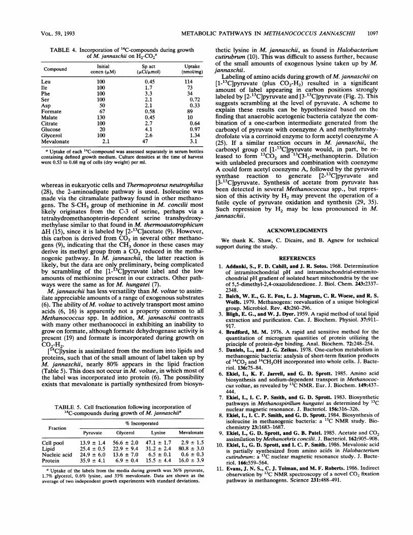
Incorporation of other compounds. 14C label from several
hydrophobic amino acids, formate, and malate was incorpo-rated into biomass during growth (Table 4). Lesser amountsof mevalonate, glycerol, and lysine were taken up (Tables 4and 5). Fractionation of the cells revealed that pyruvate wasdistributed among proteins, nucleic acids, and lipids. Glyc-erol and mevalonate labels were found largely in the lipidfraction, as expected, whereas lysine was incorporatedrelatively more into lipids than into proteins. The possibilitythat lysine might serve as a phytanyl chain precursor wastested by growing cells (100-ml culture) in the presence of 10mg of L-[U-13C]lysine. Unfortunately, the low extent ofuptake (Table 5) prevented detection of 13C signals in theextracted lipid fraction. Similarly, no difference was de-tected in signal intensity from phytanyl chain carbons la-beled by growth with 2.7 mM [2-13C]- or [3-13C]pyruvate
A 13-'3Clpyruvate1a l IIt
VOL. 59, 1993 1095
1. II

1096 SPROTTI ET AL.
TABLE 2. Origin of carbon atoms from ["3C]pyruvate in aminoacids synthesized by mixotrophic growth of M. jannaschii
on C02-H2 and pyruvatea
Amino Amino acid carbon atom:acid C_lb C-2" C-3b C-4b C-5b C-6_ Otherb
Ala, Ser (1)c 2 3Gly (1)C 2Asp, Thr (1)c 2 3 UPhe, Tyr (1)c 2 3 C-4', (2)C; C-6', 2
C-5', 3; C-9', (3)CC-7', 1; C-8', 1
Glu, Pro U 1 2, 3 2, 3 1Arg U 1 2, 3 2, 3 1 C-7, ULeu 2 3 2 2 3 4-CH3, 3Val (1)c 2 2 3 3-CH3, 3Ile 2 3 2 2 3 3-CH3, 3His (3)c (2)c 1 (1)c 2 C-7, (3)cLys (1)c 2 3 U 3 2Met (1)c 2 (3)c U S-CH3, (U)c
a Grown on C02-H2 with either [1-13C]-, [2-13C]-, or [3-13C]pyruvate. Thistable is the composite of separate labelings with each 13C label. U, unlabeled(CO2).
b Source of the 13C label from pyruvate carbon 1, 2, or 3 or from CO2.c Signal either not detected or assigned; the theoretical labeling patterns
shown in parentheses are based on the pathways described in M. hungatei (7).
regardless of whether 5.4 mM L-lysine was included in thegrowth medium (data not shown).
Glucose, citrate, aspartate, and serine were not assimi-lated during growth. Lack of glucose assimilation suggeststhat glucose does not cross the cell membrane, supporting itsuse as an indicator of extracellular volume (Table 3) in M.jannaschii.
DISCUSSION
Metabolism in methanogens is known to proceed via citricacid cycle enzymes, which function in an incomplete path-way in either oxidative or reductive directions (Table 1) (13,27, 31). Growth of most methanogens on H2 plus CO2 inmedia containing [2-'3C]acetate resulted in the incorporationof 13C into cell carbon. Methanogens grown autotrophicallyor mixotrophically (CO2-H2 being one substrate) could besubdivided into those that require acetate for growth onC02-H2 and synthesize amino acids relatively highly en-riched in 13C, those that are capable of autotrophic growthresulting in dilution of [13C]acetate incorporation, and thosesuch as M. jannaschii which grow autotrophically but fail toincorporate sufficient [13C]acetate for detection.13C-NMR analyses of total protein hydrolysates from
labeled cells readily detected signals derived, for example,from enrichment of the diagnostic al (oxidative) versus ,B(reductive) carbons of the glutamate family of amino acids(6, 7). Thermophilicity, halophilicity, or substrate utilizationrange does not appear to correlate with pathway direction-ality. However, strains of the same species or species of thesame genus had, without exception, the same type of partialTCA pathway, albeit the sampling size is small for general-izations. On the basis of our present data, the oxidative-partial TCA pathway is characteristic of the family Metha-nosarcinaceae (Table 1).
Acetic acid diffused into cells of M. jannaschii and accu-mulated in response to the transmembrane pH gradient.Acetyl coenzyme A synthetase, acetate kinase, and phos-photransacetylase were not detected in extracts of M. jann-
A [3-"qCJpyrutate fl-Gal (C-i)
fi-Glc (C-1)
a-Man (C-l) a-Gal (C-i)
*,I -. r99.0 98.0 97.0 96.0 95.0 94.0 93.0 92.0 91.0
PPM
B 12-"3C'pyruvate
fl-Gal
fl-Glc (C-5) f-Gilc(C-5) (C-2)
fl-Man(C-5)
fl-Gal(C-2)
789.0 77.0 76.0 75.0 74'.0 730.PPM
a-Gal
(C-2)
72.0 71.0 70.0 69.0
FIG. 3. 13C-NMR spectra of the carbohydrate fraction from M.jannaschii. (A) Spectrum expanded to show the signals from ano-meric carbons of monosaccharides labeled by [3-'3C]pyruvate. (B)Spectrum of monosaccharides following labeling by [2-13C]pyru-vate. Assignments are shown to the right of each signal.
aschii, explaining why the bacterium is defective in acetateassimilation. Since M. jannaschii grows autotrophically, itclearly has the capability of forming acetyl coenzyme A fromCO2. In contrast, acetate was assimilated from the growthmedium in other methanogens (Table 1), indicating activa-tion of acetate in these strains either by acetyl coenzyme Asynthetase (22) or by the combination of acetate kinase andphosphotransacetylase (21).
It was possible to deduce the amino acid biosyntheticpathways found in M. jannaschii from '3C labeling patternswhen cells were grown on C02-H2 and either [1-13C]-,[2-1C]-, or [3-13C]pyruvate. Lysine was synthesized by thediaminopimelic acid route found in other methanogens (6, 9),
TABLE 3. Distribution of weak acids in M. jannaschii (62'C)
Organicaaacid pHoa In/out pHia ApH
Pyruvic 5.84 4.65 6.51 0.67Acetic 5.85 10.25 6.89 1.04Propionic 5.83 12.44 6.97 1.14Butyric 5.90 11.56 6.99 1.09DMO 5.86 3.68 6.73 0.87
a pH., pH of the environment; pHi, cytoplasmic pH.
APPL. ENVIRON. MICROBIOL.
METABOLIC PATHWAYS IN METHANOCOCCUS JANNASCHII 1097
TABLE 4. Incorporation of "4C-compounds during growthof M. jannaschii on H2-CO2a
Compound Initial Sp act UptakeCompound ~concn (p.M) (~LCi/p.mol) (nmol/mg)
Leu 100 0.45 114Ile 100 1.7 73Phe 100 3.3 34Ser 100 2.1 0.72Asp 50 2.1 0.33Formate 67 0.58 89Malate 130 0.45 10Citrate 100 2.7 0.64Glucose 20 4.1 0.97Glycerol 100 2.6 1.34Mevalonate 2.1 47 3.1
a Uptake of each 14C-compound was assessed separately in serum bottlescontaining defined growth medium. Culture densities at the time of harvestwere 0.53 to 0.68 mg of cells (dry weight) per ml.
whereas in eukaryotic cells and Thermoproteus neutrophilus(28), the 2-aminoadipate pathway is used. Isoleucine wasmade via the citramalate pathway found in other methano-gens. The S-CH3 group of methionine in M. concilii mostlikely originates from the C-3 of serine, perhaps via atetrahydromethanopterin-dependent serine transhydroxy-methylase similar to that found in M. thermoautotrophicumAH (15), since it is labeled by [2-'3C]acetate (9). However,this carbon is derived from CO2 in several other methano-gens (9), indicating that the CH3 donor in these cases mayderive its methyl group from a CO2 reduced in the metha-nogenic pathway. In M. jannaschii, the latter reaction islikely, but the data are only preliminary, being complicatedby scrambling of the [1- 3C]pyruvate label and the lowamounts of methionine present in our extracts. Other path-ways were the same as for M. hungatei (7).M. jannaschii has less versatility than M. voltae to assim-
ilate appreciable amounts of a range of exogenous substrates(6). The ability ofM. voltae to actively transport most aminoacids (6, 16) is apparently not a property common to allMethanococcus spp. In addition, M. jannaschii contrastswith many other methanococci in exhibiting an inability togrow on formate, although formate dehydrogenase activity ispresent (19) and formate is incorporated during growth onC02-H2-
["'C]lysine is assimilated from the medium into lipids andproteins, such that of the small amount of label taken up byM. jannaschii, nearly 80% appears in the lipid fraction(Table 5). This does not occur in M. voltae, in which most ofthe label was incorporated into protein (6). The possibilityexists that mevalonate is partially synthesized from biosyn-
TABLE 5. Cell fractionation following incorporation of"4C-compounds during growth of M. jannaschiiP
% IncorporatedFraction
Pyruvate Glycerol Lysine Mevalonate
Cell pool 13.9 ± 1.4 56.6 ± 2.0 47.1 + 1.7 2.9 ± 1.5Lipid 25.4 + 0.5 22.9 ± 9.4 31.2 + 2.4 80.8 + 3.0Nucleic acid 24.9 ± 6.0 13.6 ± 7.0 6.5 + 0.1 0.6 + 0.3Protein 35.9 ± 4.1 6.9 + 0.4 15.5 ± 4.4 16.0 ± 3.9
a Uptake of the labels from the media during growth was 36% pyruvate,1.7% glycerol, 0.6% lysine, and 33% mevalonate. Data are shown as theaverage of two independent growth experiments with standard deviations.
thetic lysine in M. jannaschii, as found in Halobactenumcutirubnum (10). This was difficult to assess further, becauseof the small amounts of exogenous lysine taken up by M.jannaschii.
Labeling of amino acids during growth ofM. jannaschii on[1-'3C]pyruvate (plus C02-H2) resulted in a significantamount of label appearing in carbon positions stronglylabeled by [2-'3C]pyruvate and [3-'3C]pyruvate (Fig. 2). Thissuggests scrambling at the level of pyruvate. A scheme toexplain these results can be hypothesized based on thefinding that anaerobic acetogenic bacteria catalyze the com-bination of a one-carbon intermediate generated from thecarboxyl of pyruvate with coenzyme A and methyltetrahy-drofolate via a corrinoid enzyme to form acetyl coenzyme A(25). If a similar reaction occurs in M. jannaschii, thecarboxyl group of [1-'3C]pyruvate would, in part, be re-leased to form 13C02 and '3CH3-methanopterin. Dilutionwith unlabeled precursors and combination with coenzymeA could form acetyl coenzyme A, followed by the pyruvatesynthase reaction to generate [2-'3C]pyruvate and[3-'3C]pyruvate. Synthesis of acetate from pyruvate hasbeen detected in several Methanococcus spp., but repres-sion of this activity by H2 may prevent the operation of afutile cycle of pyruvate oxidation and synthesis (29, 35).Such repression by H2 may be less pronounced in M.jannaschii.
ACKNOWLEDGMENTS
We thank K. Shaw, C. Dicaire, and B. Agnew for technicalsupport during the study.
REFERENCES1. Addanki, S., F. D. Cahill, and J. R. Sotos. 1968. Determination
of intramitochondrial pH and intramitochondrial-extramito-chondrial pH gradient of isolated heart mitochondria by the useof 5,5-dimethyl-2,4-oxazolidenedione. J. Biol. Chem. 243:2337-2348.
2. Balch, W. E., G. E. Fox, L. J. Magrum, C. R. Woese, and R. S.Wolfe. 1979. Methanogens: reevaluation of a unique biologicalgroup. Microbiol. Rev. 43:260-296.
3. Bligh, E. G., and W. J. Dyer. 1959. A rapid method of total lipidextraction and purification. Can. J. Biochem. Physiol. 37:911-917.
4. Bradford, M. M. 1976. A rapid and sensitive method for thequantitation of microgram quantities of protein utilizing theprinciple of protein-dye binding. Anal. Biochem. 72:248-254.
5. Daniels, L., and J. G. Zeikus. 1978. One-carbon metabolism inmethanogenic bacteria: analysis of short-term fixation productsof 14CO2 and 14CH3OH incorporated into whole cells. J. Bacte-riol. 136:75-84.
6. Ekiel, I., K. F. Jarrell, and G. D. Sprott. 1985. Amino acidbiosynthesis and sodium-dependent transport in Methanococ-cus voltae, as revealed by 13C NMR. Eur. J. Biochem. 149:437-444.
7. Ekiel, I., I. C. P. Smith, and G. D. Sprott. 1983. Biosyntheticpathways in Methanospirillum hungatei as determined by 13Cnuclear magnetic resonance. J. Bacteriol. 156:316-326.
8. Ekiel, I., I. C. P. Smith, and G. D. Sprott. 1984. Biosynthesis ofisoleucine in methanogenic bacteria: a 13C NMR study. Bio-chemistry 23:1683-1687.
9. Ekiel, I., G. D. Sprott, and G. B. Patel. 1985. Acetate and CO2assimilation byMethanothrix concilii. J. Bacteriol. 162:905-908.
10. Ekiel, I., G. D. Sprott, and I. C. P. Smith. 1986. Mevalonic acidis partially synthesized from amino acids in Halobacteriumcutirubrum: a 13C nuclear magnetic resonance study. J. Bacte-riol. 166:559-564.
11. Evans, J. N. S., C. J. Tolman, and M. F. Roberts. 1986. Indirectobservation by 13C NMR spectroscopy of a novel CO2 fixationpathway in methanogens. Science 231:488-491.
VOL. 59, 1993

APPL. ENVIRON. MICROBIOL.
12. Ferrante, G., J. C. Richards, and G. D. Sprott. 1990. Structuresof polar lipids from the thermophilic, deep-sea archaeobacte-rium Methanococcus jannaschii. Biochem. Cell Biol. 68:274-283.
13. Fuchs, G., and E. Stupperich. 1982. Autotrophic CO2 fixationpathway in Methanobacterium thermoautotrophicum. Zen-tralbl. Bakteriol. Mikrobiol. Hyg. 1 Abt. Orig. C 3:277-288.
14. Hanson, R. S., and J. A. Phillips. 1981. Chemical composition,p. 328-364. In P. Gerhardt (ed.), Manual of methods for generalbacteriology. American Society for Microbiology, Washington,D.C.
15. Hoyt, J. C., A. Oren, J. C. Escalante-Semerena, and R. S. Wolfe.1986. Tetrahydromethanopterin-dependent serine transhy-droxymethylase from Methanobacterium thermoautotrophi-cum. Arch. Microbiol. 145:153-158.
16. Jarrell, K. F., S. E. Bird, and G. D. Sprott. 1984. Sodium-dependent isoleucine transport in the methanogenic archaebac-terium Methanococcus voltae. FEBS Lett. 166:357-361.
17. Jarrell, K. F., J. R. Colvin, and G. D. Sprott. 1982. Spontaneousprotoplast formation in Methanobacterium bryantii. J. Bacte-riol. 149:346-353.
18. Jarrell, K. F., and G. D. Sprott. 1981. The transmembraneelectrical potential and intracellular pH in methanogenic bacte-ria. Can. J. Microbiol. 27:720-728.
19. Jones, W. J., J. A. Leigh, F. Mayer, C. R. Woese, and R. S.Wolfe. 1983. Methanococcus jannaschii sp. nov., an extremelythermophilic methanogen from a submarine hydrothermal vent.Arch. Microbiol. 136:254-261.
20. Kaback, H. R. 1977. Molecular biology and energetics ofmembrane transport, p. 598-625. In G. Semenza and E. Carafoli(ed.), Biochemistry of membrane transport. Springer-Verlag,New York.
21. Kenealy, W., and J. G. Zeikus. 1982. One-carbon metabolism inmethanogens: evidence for synthesis of a two-carbon interme-diate and unification of catabolism and anabolism in Methanosa-rcina barkeri. J. Bacteriol. 151:932-941.
22. Oberlies, G., G. Fuchs, and R. K. Thauer. 1980. Acetatethiokinase and the assimilation of acetate in Methanobacteriumthermoautotrophicum. Arch. Microbiol. 128:248-252.
23. Patel, G. B., and G. D. Sprott. 1991. Cobalt and sodiumrequirements for methanogenesis in washed cells of Methano-saeta concilii. Can. J. Microbiol. 37:110-115.
24. Patel, G. B., G. D. Sprott, and J. E. Fein. 1990. Isolation andcharacterization of Methanobacterium espanolae sp. nov., amesophilic, moderately acidophilic methanogen. Int. J. Syst.Bacteriol. 40:12-18.
25. Pezacka, E., and H. G. Wood. 1984. Role of carbon monoxidedehydrogenase in the autotrophic pathway used by acetogenicbacteria. Proc. Natl. Acad. Sci. USA 81:6261-6265.
26. Roberts, M. F., M.-C. Lai, and R. P. Gunsalus. 1992. Biosyn-thetic pathways of the osmolytes N-acetyl-,-glutamine andbetaine in Methanohalophilus strain FDF1 suggested by nuclearmagnetic resonance analysis. J. Bacteriol. 174:6688-6693.
27. Robertson, D. N., D. Noll, and M. F. Roberts. 1992. Free aminoacid dynamics in marine methanogens. J. Biol. Chem. 276:14893-14901.
28. Schafer, S., T. Paalme, R. Vilu, and G. Fuchs. 1989. 13C-NMRstudy of acetate assimilation in Thermoproteus neutrophilus.Eur. J. Biochem. 186:695-700.
29. Shieh, J., and W. B. Whitman. 1987. Pathway of acetateassimilation in autotrophic and heterotrophic methanococci. J.Bacteriol. 169:5327-5329.
30. Sprott, G. D., I. Ekiel, and C. Dicaire. 1990. Novel, acid-labile,hydroxydiether lipid cores in methanogenic bacteria. J. Biol.Chem. 265:13735-13740.
31. Sprott, G. D., K. M. Shaw, and K. F. Jarrell. 1985. Methano-genesis and the K' transport system are activated by divalentcations in ammonia-treated cells of Methanospirillum hungatei.J. Biol. Chem. 260:9244-9250.
32. Stupperich, E., and G. Fuchs. 1984. Autotrophic synthesis ofactivated acetic acid from two CO2 in Methanobacterium ther-moautotrophicum. II. Evidence for different origins of acetatecarbon atoms. Arch. Microbiol. 139:14-20.
33. Weimer, P. J., and J. G. Zeikus. 1979. Acetate assimilationpathway of Methanosarcina barkeri. J. Bacteriol. 137:332-339.
34. Whitman, W. B., E. Ankwanda, and R. S. Wolfe. 1982. Nutritionand carbon metabolism of Methanococcus voltae. J. Bacteriol.149:852-863.
35. Yang, Y.-L., J. Ladapo, and W. B. Whitman. 1992. Pyruvateoxidation by Methanococcus spp. Arch. Microbiol. 158:271-275.
36. Zeikus, J. G., G. Fuchs, W. Kenealy, and R. K. Thauer. 1977.Oxidoreductases involved in cell carbon synthesis of Methano-bacterium thermoautotrophicum. J. Bacteriol. 132:604-613.
1098 SPROTT ET AL.