Embed Size (px)
Citation preview
University of New OrleansScholarWorks@UNO
University of New Orleans Theses and Dissertations Dissertations and Theses
Summer 8-4-2011
Metabolism and cryo-sensitivity of domestic cat(Felis catus) and cheetah (Acinonyx jubatus)spermatozoaKimberly TerrellUniversity of New Orleans, [email protected]
Follow this and additional works at: https://scholarworks.uno.edu/td
Part of the Evolution Commons, and the Other Ecology and Evolutionary Biology Commons
This Dissertation is brought to you for free and open access by the Dissertations and Theses at ScholarWorks@UNO. It has been accepted for inclusionin University of New Orleans Theses and Dissertations by an authorized administrator of ScholarWorks@UNO. The author is solely responsible forensuring compliance with copyright. For more information, please contact [email protected].
Recommended CitationTerrell, Kimberly, "Metabolism and cryo-sensitivity of domestic cat (Felis catus) and cheetah (Acinonyx jubatus) spermatozoa"(2011). University of New Orleans Theses and Dissertations. 138.https://scholarworks.uno.edu/td/138
Metabolism and cryo-sensitivity of domestic cat (Felis catus) and cheetah (Acinonyx jubatus) spermatozoa
A Dissertation
Submitted to the graduate faculty of the University of New Orleans in partial fulfillment of the
requirements for the degree of
Doctor of Philosophy in
Conservation Biology
by
Kimberly Ann Terrell
B.S. Tulane University, 2005 B.A. Tulane University, 2005
August, 2011
ii
Acknowledgments
This dissertation would not exist without a team of remarkable individuals who dedicated
their time and energy to my research and served as thesis committee members. In particular, Dr.
Adrienne Crosier worked closely with me for five years and was a valuable source of
information about cheetah biology and in situ conservation. I am deeply indebted to Dr. Barry
Bavister, who helped me to overcome my fear of all things biochemical and whose determined
efforts resulted in the creation of the University of New Orleans – Smithsonian Conservation
Biology Institute research partnership. I am very grateful to Dr. David Wildt, who always kept
me focused on the ‘big picture’ and whose intensive, ‘Kool-Aid’ editing helped dramatically
improve my scientific writing. Dr. Nicola Anthony deserves special thanks – her passion for
science and ability to remain neutral in research politics are continued sources of inspiration to
me. Through his almost super-human attention to detail, Dr. Stanley Leibo helped correct many
errors in these manuscripts, and I owe him my gratitude. He has the rare ability to impart
lifelong knowledge to others, and I will always remember the benefit of purchasing frozen
turkeys. Dr. Bernard Rees greatly facilitated this project, through both his understanding of
cellular metabolism and his role as graduate student coordinator. The humor and affability he
brought to this position helped me to feel connected to our university, despite living far from
New Orleans.
Several individuals outside my thesis committee also made valuable contributions to this
project. Dr. Budhan Pukazhenthi provided thoughtful feedback related to the project design and
study results and was always happy to answer my endless calls for help with laboratory
equipment. Dr. Nucharin Songsasen is both a professional and personal role model to me, and
has been a valuable source of career advice. Her multidisciplinary approach to science is an
iii
instructive research paradigm for any aspiring conservation physiologist. Dr. Pierre Comizzoli
provided helpful advice and support throughout this project, as well as motivation to reopen my
high school French textbook. Dr. Linda Penfold reviewed each of these manuscripts, but also
taught me that high fashion is perfectly compatible with wildlife research. Drs. Louis Padilla,
Katharine Hope, and Carlos Sanchez contributed veterinary support, and Lisa Ware, Copper
Aitken-Palmer, Laurie Marker, and Marianne de Jonge provided valuable technical assistance. I
am grateful to Jenny Santiestevan who, in addition to technical support, provided friendship and
commiseration over the tedium of metabolic assays. I especially thank the late Dr. JoGayle
Howard for her contribution to the study design and for her pioneering research that made this
work possible.
This collaborative project was made possible through support from the National Science
Foundation Graduate Research Fellowship Program. Funding also was provided by the
Audubon Center for Research of Endangered Species, Louisiana State University, the University
of New Orleans Department of Biological Sciences, the Ohrstrom Family Foundation, the
William H. Donner Foundation, Inc., the Smithsonian Predoctoral Fellowship Program, and the
Association for Women in Science. My research relied on technical and veterinary support from
the staff of collaborating institutions, specifically, Cheetah Conservation Fund, White Oak
Conservation Center, The Wilds, San Diego Wild Safari Park, the Philadelphia Zoo, and the
Cleveland Metroparks Zoo.
This acknowledgment would not be complete without recognizing the incredible support
I have received from my friends and family. In particular, my friend and brilliant scientist, Dr.
Wayne Buck, catalyzed my decision to pursue a graduate education. Pamela Thompson, Yamile
Molina, and I have shared an incredible journey that began as Tulane University freshmen and
iv
will soon culminate in a biology Ph.D. trifecta. Lisa Ware, Amy Johnson, and Jennifer Buff
continually inspire me to get outside and learn more about the natural world. Kari Morfeld
reminds me that any challenge can be overcome. Lacey Braun, Sarah Putman, Jessica Kordell,
Laura Linn, Ivonne Garzon, Andrea Liebl, Kimberly Fernald, Christine Facella, Amanda Norris,
and Nick Terrell help me remember there is a world outside of science. My partner, Rosamond
Dietrich, put his career on hold for my education and is an infinite source of support and
inspiration to me. Finally, I’d like to thank my parents, particularly my mom, who endowed me
with her strong work ethic and relentless frugality – key traits of a successful conservationist.
v
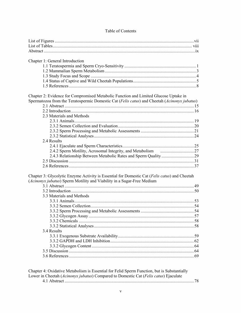
Table of Contents
List of Figures ................................................................................................................................ vii List of Tables ................................................................................................................................ viii Abstract ........................................................................................................................................... ix Chapter 1: General Introduction 1.1 Teratospermia and Sperm Cryo-Sensitivity .................................................................. 1 1.2 Mammalian Sperm Metabolism .................................................................................... 3 1.3 Study Focus and Scope ................................................................................................. 4 1.4 Status of Captive and Wild Cheetah Populations .......................................................... 5 1.5 References ..................................................................................................................... 8 Chapter 2: Evidence for Compromised Metabolic Function and Limited Glucose Uptake in Spermatozoa from the Teratospermic Domestic Cat (Felis catus) and Cheetah (Acinonyx jubatus) 2.1 Abstract ....................................................................................................................... 15 2.2 Introduction ................................................................................................................. 16 2.3 Materials and Methods 2.3.1 Animals .............................................................................................................. 19 2.3.2 Semen Collection and Evaluation ...................................................................... 20 2.3.2 Sperm Processing and Metabolic Assessments ................................................. 21 2.3.2 Statistical Analyses ............................................................................................ 24 2.4 Results 2.4.1 Ejaculate and Sperm Characteristics .................................................................. 25 2.4.2 Sperm Motility, Acrosomal Integrity, and Metabolism ............................... 27 2.4.3 Relationship Between Metabolic Rates and Sperm Quality .............................. 29 2.5 Discussion ................................................................................................................... 31 2.6 References ................................................................................................................... 37 Chapter 3: Glycolytic Enzyme Activity is Essential for Domestic Cat (Felis catus) and Cheetah (Acinonyx jubatus) Sperm Motility and Viability in a Sugar-Free Medium 3.1 Abstract ....................................................................................................................... 49 3.2 Introduction ................................................................................................................. 50 3.3 Materials and Methods 3.3.1 Animals .............................................................................................................. 53 3.3.2 Semen Collection ............................................................................................... 54 3.3.2 Sperm Processing and Metabolic Assessments ................................................. 54 3.3.2 Glycogen Assay ................................................................................................. 57 3.3.2 Chemicals .......................................................................................................... 58 3.3.2 Statistical Analyses ............................................................................................ 58 3.4 Results 3.3.1 Exogenous Substrate Availability ...................................................................... 59 3.3.2 GAPDH and LDH Inhibition ............................................................................. 62 3.3.2 Glycogen Content .............................................................................................. 64 3.5 Discussion ................................................................................................................... 64 3.6 References ................................................................................................................... 69 Chapter 4: Oxidative Metabolism is Essential for Felid Sperm Function, but is Substantially Lower in Cheetah (Acinonyx jubatus) Compared to Domestic Cat (Felis catus) Ejaculate 4.1 Abstract ....................................................................................................................... 78
vi
4.2 Introduction ................................................................................................................. 79 4.3 Materials and Methods 4.3.1 Animals .............................................................................................................. 81 4.3.2 Semen Collection ............................................................................................... 82 4.3.3 Sperm Processing ............................................................................................... 83 4.3.4 Inhibition of Oxidative Phosphorylation ........................................................... 83 4.3.5 Mitochondrial Membrane Potential ................................................................... 85 4.3.6 Sperm Morphometrics ....................................................................................... 86 4.3.7 Statistical Analyses ............................................................................................ 86 4.4 Results 4.3.1 Inhibition of Oxidative Phosphorylation ........................................................... 87 4.3.2 Mitochondrial Membrane Potential and Sperm Morphometrics ....................... 90 4.5 Discussion ................................................................................................................... 94 4.6 References ................................................................................................................. 100 Chapter 5: Different Patterns of Metabolic Cryo-Damage in Domestic Cat (Felis catus) and Cheetah (Acinonyx jubatus) Spermatozoa 5.1 Abstract ..................................................................................................................... 112 5.2 Introduction ............................................................................................................... 113 5.3 Materials and Methods 5.3.1 Animals ............................................................................................................ 115 5.3.2 Semen Collection ............................................................................................. 116 5.3.3 Sperm Processing and Metabolic Assessments ............................................... 116 5.3.4 Sperm Cryopreservation .................................................................................. 118 5.3.5 Comparison of Post-Thaw Processing Methods .............................................. 118 5.3.6 Accudenz Gradient Optimization for Domestic Cat Spermatozoa .................. 121 5.3.7 Chemicals ........................................................................................................ 122 5.3.8 Statistical Analyses .......................................................................................... 122 5.4 Results 5.4.1 Accudenz Gradient Optimization for Domestic Cat Spermatozoa .................. 123 5.4.2 Comparison of Post-Thaw Processing Methods .............................................. 124 5.5 Discussion ................................................................................................................. 129 5.6 References ................................................................................................................. 132 Chapter 6: General Discussion 6.1 Metabolic Profiles of Cat and Cheetah Spermatozoa ................................................ 140 6.2 Metabolic Indicators of Sperm Function ................................................................... 141 6.3 Influence of Species Physiology ............................................................................... 142 6.4 Influence of Teratospermia ....................................................................................... 143 6.5 Influence of Sperm Cryopreservation ....................................................................... 144 6.6 Conclusions and Recommendations .......................................................................... 145 6.7 References ................................................................................................................. 146 Appendices Appendix A: Supplemental Figure 2.1 ............................................................................ 148 Appendix B: Supplemental Figure 5.1 ............................................................................ 149 Supplemental Figure 5.2............................................................................ 150 Vita ............................................................................................................................................... 151
vii
List of Figures
Chapter 1 Figures Fig. 1.1 ................................................................................................................................ 6 Fig. 1.2 ................................................................................................................................ 6 Fig. 1.3 ................................................................................................................................ 7 Chapter 2 Figures Fig. 2.1 .............................................................................................................................. 27 Fig. 2.2 .............................................................................................................................. 28 Fig. 2.3 .............................................................................................................................. 31 Fig. 2.4 .............................................................................................................................. 33 Chapter 3 Figures Fig. 3.1 .............................................................................................................................. 60 Fig. 3.2 .............................................................................................................................. 61 Fig. 3.3 .............................................................................................................................. 62 Fig. 3.4 .............................................................................................................................. 63 Fig. 3.5 .............................................................................................................................. 65 Chapter 4 Figures Fig. 4.1 .............................................................................................................................. 88 Fig. 4.2 .............................................................................................................................. 89 Fig. 4.3 .............................................................................................................................. 90 Fig. 4.4 .............................................................................................................................. 91 Fig. 4.5 .............................................................................................................................. 92 Fig. 4.6 .............................................................................................................................. 93 Chapter 5 Figures Fig. 5.1 ............................................................................................................................ 124 Fig. 5.2 ............................................................................................................................ 125 Fig. 5.3 ............................................................................................................................ 126 Fig. 5.4 ............................................................................................................................ 128 Chapter 6 Figures Fig. 6.1 ............................................................................................................................ 143
viii
List of Tables Chapter 2 Tables Table 2.1 ............................................................................................................................ 26 Table 2.2 ............................................................................................................................ 29 Table 2.3 ............................................................................................................................ 30 Chapter 4 Tables Table 4.1 ............................................................................................................................ 94 Chapter 5 Tables Table 5.1 .......................................................................................................................... 123 Table 5.2 .......................................................................................................................... 127 Table 5.3 .......................................................................................................................... 127
ix
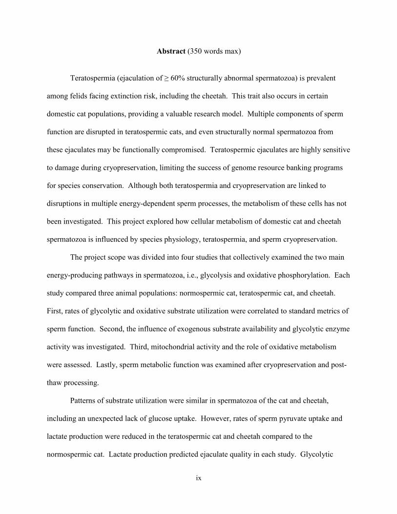
Abstract (350 words max)
Teratospermia (ejaculation of ≥ 60% structurally abnormal spermatozoa) is prevalent
among felids facing extinction risk, including the cheetah. This trait also occurs in certain
domestic cat populations, providing a valuable research model. Multiple components of sperm
function are disrupted in teratospermic cats, and even structurally normal spermatozoa from
these ejaculates may be functionally compromised. Teratospermic ejaculates are highly sensitive
to damage during cryopreservation, limiting the success of genome resource banking programs
for species conservation. Although both teratospermia and cryopreservation are linked to
disruptions in multiple energy-dependent sperm processes, the metabolism of these cells has not
been investigated. This project explored how cellular metabolism of domestic cat and cheetah
spermatozoa is influenced by species physiology, teratospermia, and sperm cryopreservation.
The project scope was divided into four studies that collectively examined the two main
energy-producing pathways in spermatozoa, i.e., glycolysis and oxidative phosphorylation. Each
study compared three animal populations: normospermic cat, teratospermic cat, and cheetah.
First, rates of glycolytic and oxidative substrate utilization were correlated to standard metrics of
sperm function. Second, the influence of exogenous substrate availability and glycolytic enzyme
activity was investigated. Third, mitochondrial activity and the role of oxidative metabolism
were assessed. Lastly, sperm metabolic function was examined after cryopreservation and post-
thaw processing.
Patterns of substrate utilization were similar in spermatozoa of the cat and cheetah,
including an unexpected lack of glucose uptake. However, rates of sperm pyruvate uptake and
lactate production were reduced in the teratospermic cat and cheetah compared to the
normospermic cat. Lactate production predicted ejaculate quality in each study. Glycolytic
x
enzyme activity was essential for sperm function, but, unexpectedly, the importance of this
pathway appeared to be linked to glycerol rather than glucose metabolism. Sperm oxidative
metabolism was severely compromised in the cheetah, and comparison with the teratospermic
cat proved this defect to be species-specific. Spermatozoa from both species experienced
metabolic damage during cryopreservation. Post-thaw processing recovered a metabolically-
normal sperm subpopulation in the cat, but cheetah spermatozoa remained functionally
compromised. Collectively, these studies provided key insight into metabolism and cryo-
sensitivity of felid spermatozoa and highlighted the importance of domestic animal models for
wildlife research.
Keywords: Cryopreservation, Teratospermia, Teratozoospermia, Felid, Sperm, Glucose, Pyruvate, Lactate, Mitochondria, Glycolysis
1
CHAPTER 1: GENERAL INTRODUCTION
1.1 Teratospermia and Sperm Cryo-Sensitivity
Felidae comprises nearly 40 species and is one of the most phylogenetically diverse
carnivore families in existence [1]. Teratospermia (ejaculation of ≥ 60% structurally abnormal
spermatozoa) is remarkably prevalent in this taxon, and nearly all (~90%) felid species studied to
date consistently ejaculate high proportions (> 40%) of malformed spermatozoa [2]. In addition
to sperm pleiomorphisms, teratospermic ejaculates generally contain low numbers of
spermatozoa, and these cells are highly susceptible to damage during cryopreservation [2, 3].
Severe teratospermia (> 85% structurally abnormal cells) is linked to reduced fecundity and
infertility in certain wild felids [4-6]. Because most (70%) wild felids are at risk of extinction
and nearly half of the non-threatened species are in decline [7], understanding the physiology of
this intriguing reproductive phenomenon is important both for conservation efforts and to
manage genetically valuable populations ex situ. Such knowledge also would benefit human
health, as teratospermia is nearly ubiquitous in man (representing a significant source of male
infertility [3]) and is common among domestic cat (Felis catus) lineages studied as models of
human disease [8, 9].
Teratospermia has been linked to a loss of genetic diversity through opportunistic studies
of rare felids, including the cheetah (Acinonyx jubatus) [10, 11], lion (Panthera leo) [4, 12],
Florida panther (Puma concolor coryi) [5, 13], and leopard cat (Felis bengalensis) [4]. In each
case, ejaculates containing high proportions of structurally abnormal spermatozoa, low sperm
numbers, and/or poor motility were associated with a lack of heterozygosity from an ancient
population bottleneck (cheetah [14, 15]), recent population decline (lion [12] and Florida panther
[13]), or captive inbreeding (leopard cat [4]). Prospective inbreeding in the domestic cat
2
confirmed the link between ejaculate quality and genetic diversity [2]. After a single generation
of incestuous mating, proportions of malformed spermatozoa ejaculated increased to > 85%
(compared to < 45% in outbred males). In contrast to the low numbers of spermatozoa
previously reported in teratospermic ejaculates, inbred toms experienced an ~80% increase in
total sperm production compared to normospermic cats [2]. High numbers of ejaculated
spermatozoa were linked to an increase in testicular volume and a greater spermatogenic/Sertoli
cell ratio [16]. This physiological response presents the intriguing possibility that certain
individuals or populations may possess a compensatory mechanism to maintain fertility despite
abnormal spermatogenesis [2].
The ability to empirically test the link between teratospermia and genetic diversity in the
domestic cat highlights the value of this species as a model for wildlife conservation and human
health. Comparative studies of domestic cat populations that produce different proportions of
pleiomorphic spermatozoa have yielded insight into the extreme cryo-sensitivity and potential
for decreased fertility of teratospermic ejaculates. Specifically, spermatozoa from these
ejaculates exhibit increased susceptibility to osmotic stress [17] and cold-induced acrosome
damage [18], as well as delayed capacitation [19], impaired acrosomal function [19], reduced
levels of protein tyrosine phosphorylation [20], chromatin instability [21], decreased zona
pellucida penetration [22], and compromised fertilization ability in vitro [22]. Importantly, many
of these cellular functions also are impaired in structurally normal spermatozoa from
teratospermic ejaculates [19, 20, 22]. In contrast to these detailed studies, there is a lack of
knowledge about cellular metabolism in felid spermatozoa, including the possible influence of
teratospermia or cryopreservation. Given the spermatozoon’s extraordinarily high ATP demands
and the disruption of multiple energy-dependent processes (e.g., motility, viability, protein
3
tyrosine phosphorylation) in teratospermic ejaculates, cellular metabolism is a priority research
focus. Identifying the primary biochemical pathway responsible for sperm energy production
and understanding its role in teratospermia/cryo-sensitivity could help improve the success of
assisted reproductive technologies (e.g., sperm cryopreservation, in vitro fertilization) for
wildlife management and human fertility [23].
1.2 Mammalian Sperm Metabolism
Mammalian spermatozoa produce energy in the form of ATP almost exclusively by two
pathways: glycolysis and oxidative phosphorylation (OXPHOS) [24]. Each pathway’s relative
importance varies substantially among the small number of species studied, which includes the
human, mouse, boar, bull, ram, rabbit, and dog (reviewed in [25]). Sperm energy sources are
equally diverse, likely reflecting species variation in metabolic substrate availability within the
female tract [24]. Potential energy substrates include exogenous hexoses (primarily glucose) and
monocarboxylates (pyruvate and lactate) [24], as well as endogenous lipid [26, 27] and even
glycogen [28, 29]. The localization of mitochondria to the midpiece and glycolytic enzymes to
the flagellum results in a compartmentalized mode of sperm energy production [24]. Therefore,
the relative contribution of each pathway is dictated by its capacity to fulfill local energy
demands and/or by the operation of intracellular ATP transporters [30]. This
compartmentalization likely explains the essential role of glycolysis in human [31] and mouse
[32] spermatozoa, despite extremely inefficient rates of ATP production relative to OXPHOS.
Both glycolytic [33] and oxidative [34-36] pathways may be disrupted by sperm
cryopreservation, although substantial variation in susceptibility to metabolic damage can exist,
even within a species [37]. Given the remarkable differences in mechanisms of energy
4
production among mammalian spermatozoa, comparative investigations of multiple species
and/or populations are essential for generating reliable conclusions about these unstudied
processes in felids.
1.3 Study Focus and Scope
The overall aim of this project was to understand how metabolism of felid spermatozoa is
influenced by teratospermia, sperm cryopreservation, and species physiology. To achieve this
goal, each study comparatively assessed three felid populations: 1) normospermic domestic cat,
2) teratospermic domestic cat, and 3) cheetah (an entirely teratospermic species). The cheetah
population included wild- and captive-born individuals of the southern African subspecies
Acinonyx jubatus jubatus housed at the Cheetah Conservation Fund (Otjiwarongo, Namibia) or
in North American institutions within the Association of Zoos and Aquarium’s Species Survival
Plan (SSP).
To assess the importance of glycolytic and oxidative metabolism in felid spermatozoa,
and to elucidate the mechanisms of sperm cryo-sensitivity, the specific foci of this project
included:
1) Glycolytic versus oxidative substrate utilization and metabolic indicators of cellular
function (Chapter 2).
Published manuscript:
Terrell KA, Wildt DE, Anthony NM, Bavister BD, Leibo SP, Penfold LM, Marker
LL, Crosier AE. Evidence for compromised metabolic function and limited glucose
uptake in spermatozoa from the teratospermic domestic cat (Felis catus) and cheetah
(Acinonyx jubatus). Biol Reprod 2010; 83: 833-841.
5
2) The role of exogenous substrate availability and glycolytic enzyme activity in sperm
function (Chapter 3).
Published manuscript:
Terrell KA, Wildt DE, Anthony NM, Bavister BD, Leibo SP, Penfold LM, Marker
LL, Crosier AE. Glycolytic enzyme activity is essential for domestic cat (Felis catus)
and cheetah (Acinonyx jubatus) sperm motility and viability in a sugar-free medium.
Biol Reprod 2011; 84: 1198-1206.
3) The importance of oxidative phosphorylation for sperm function (Chapter 4).
Published manuscript:
Terrell KA, Wildt DE, Anthony NM, Bavister BD, Leibo SP, Penfold LP, Crosier
AE. Oxidative metabolism is essential for felid sperm function, but is substantially
lower in cheetah (Acinonyx jubatus) compared to domestic cat (Felis catus) Ejaculate.
Biol Reprod 2011; 83: 833-841.
4) Changes in metabolic function resulting from cryopreservation and/or post-thaw
processing (Chapter 5).
Manuscript in review:
Terrell KA, Wildt DE, Anthony NM, Bavister BD, Leibo SP, Penfold LP, Crosier
AE. Different patterns of metabolic cryo-damage in domestic cat (Felis catus) and
cheetah (Acinonyx jubatus) spermatozoa. Cryobiology.
1.4 Status of Wild and Captive Cheetah Populations
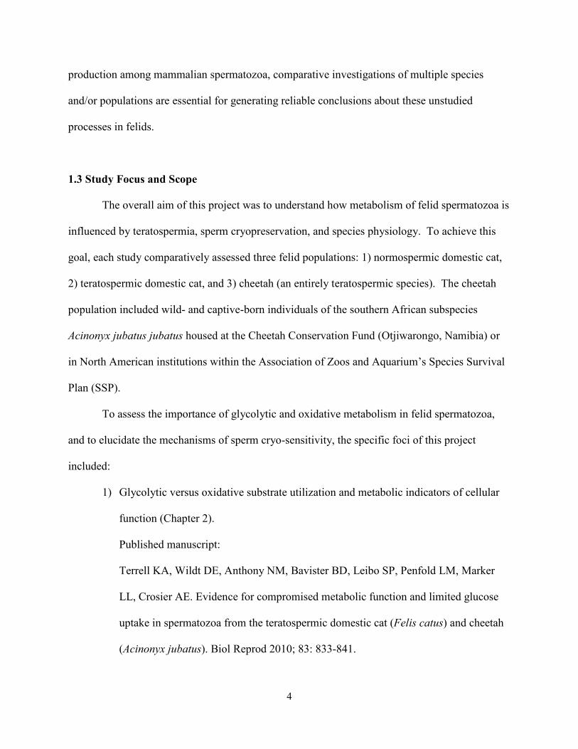
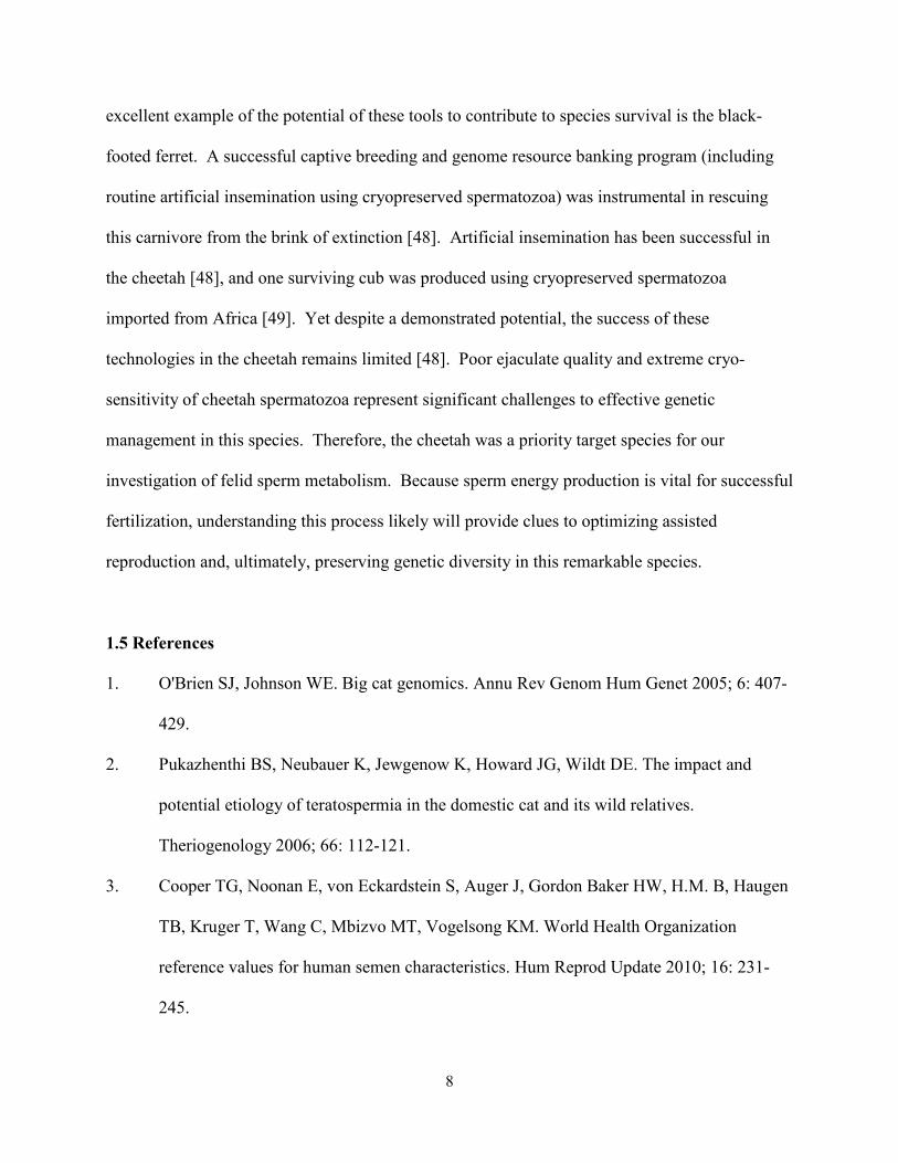
The cheetah (Fig. 1.1) is a species well known to consistently ejaculate high (~80%)
proportions of abnormal spermatozoa, regardless of geographic range, season, age, or
6
environment (captive versus wild) [11, 38]. This trait has been
linked to an extreme lack of genetic diversity from a severe
population contraction ~10,000 years ago [14, 15, 39],
coincident with the widespread extinctions of Pleistocene
megafauna [40]. Subsequent analysis revealed a second
population bottleneck in the twentieth century due to a ~90%
decline in wild populations from poaching and habitat loss [41]. Today fewer than 10,000
cheetahs remain in nature, although the effective population size may be less than half of that
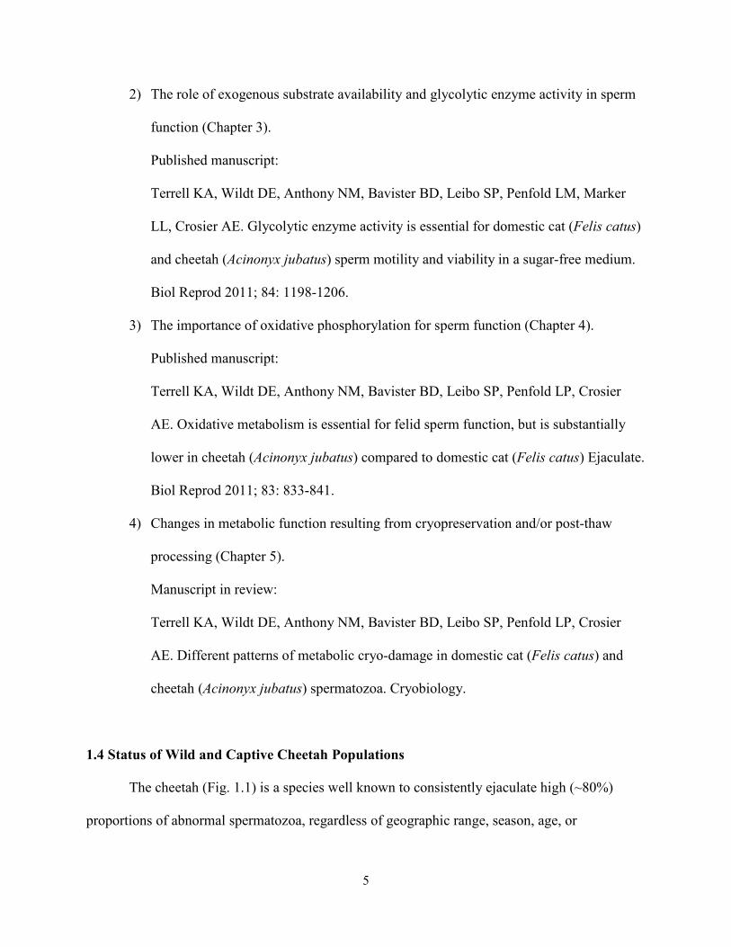
number [42]. The species has disappeared from ~80% of its historic African range [42].
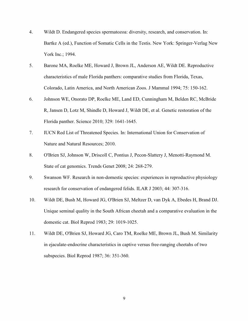
Surviving cheetahs exist in geographically isolated populations throughout the continent, with a
very small number of individuals remaining in Iran (Fig. 1.2) [42]. The largest extant cheetah
population (~4,500 adults) occurs in southern Africa, primarily in Namibia and Botswana [43].
Since the vast majority of these individuals inhabit private farmland, shooting or trapping by
farmers (due to the perceived threat of livestock predation) continues to pose a major threat to
the species’ survival [44]. For example, ~7,000
Namibian cheetah were killed or brought into
captivity during the 1980s [45], approximately
three times the number of free-ranging individuals
in Namibia today [42].
Due to the ongoing decline in wild cheetah
populations (suspected to be ≥ 30% over the past
three generations), the species is considered by the
IUCN (International Union for the Conservation of Figure 1.2. Current range (red shading) of the cheetah [7].
Figure 1.1. The cheetah.
7
Nature and Natural Resources) to be ‘vulnerable’ to extinction [42]. Captive cheetahs have the
potential to contribute to the survival of wild populations by providing 1) a long-term reservoir
of genetic diversity, 2) an opportunity for increasing basic knowledge about a species that is
challenging to study in the wild, and 3) ‘ambassador’ animals that educate the public, engage
non-scientists in biodiversity conservation, and generate funds to support in situ conservation.
However, cheetahs reproduce poorly in captivity (likely due to suboptimal management), and
these populations are not self-sustaining. As a result, ~30% of individuals in the global captive
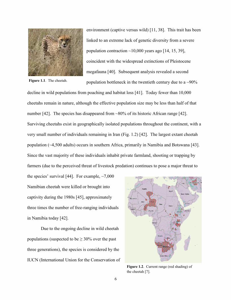
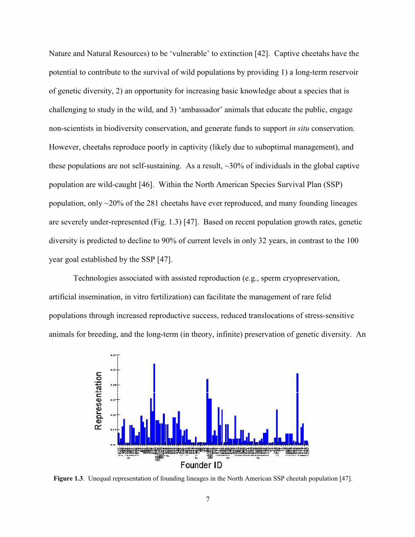
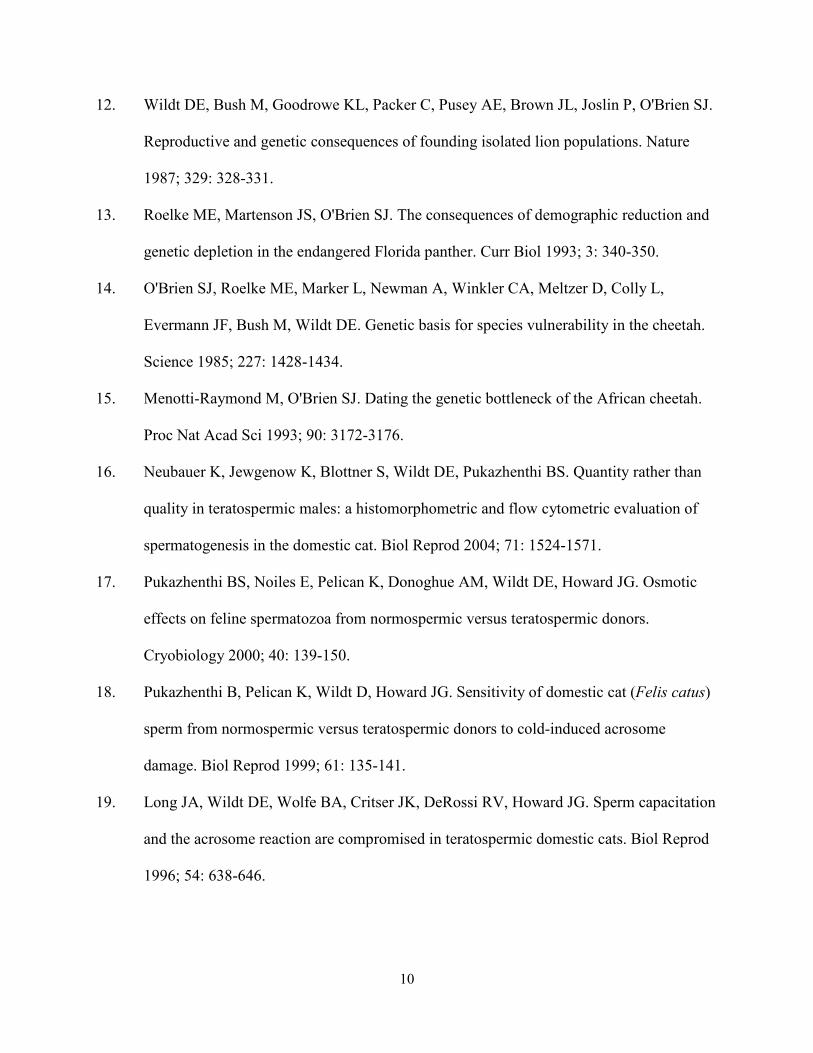
population are wild-caught [46]. Within the North American Species Survival Plan (SSP)
population, only ~20% of the 281 cheetahs have ever reproduced, and many founding lineages
are severely under-represented (Fig. 1.3) [47]. Based on recent population growth rates, genetic
diversity is predicted to decline to 90% of current levels in only 32 years, in contrast to the 100
year goal established by the SSP [47].
Technologies associated with assisted reproduction (e.g., sperm cryopreservation,
artificial insemination, in vitro fertilization) can facilitate the management of rare felid
populations through increased reproductive success, reduced translocations of stress-sensitive
animals for breeding, and the long-term (in theory, infinite) preservation of genetic diversity. An
Figure 1.3. Unequal representation of founding lineages in the North American SSP cheetah population [47].
8
excellent example of the potential of these tools to contribute to species survival is the black-
footed ferret. A successful captive breeding and genome resource banking program (including
routine artificial insemination using cryopreserved spermatozoa) was instrumental in rescuing
this carnivore from the brink of extinction [48]. Artificial insemination has been successful in
the cheetah [48], and one surviving cub was produced using cryopreserved spermatozoa
imported from Africa [49]. Yet despite a demonstrated potential, the success of these
technologies in the cheetah remains limited [48]. Poor ejaculate quality and extreme cryo-
sensitivity of cheetah spermatozoa represent significant challenges to effective genetic
management in this species. Therefore, the cheetah was a priority target species for our
investigation of felid sperm metabolism. Because sperm energy production is vital for successful
fertilization, understanding this process likely will provide clues to optimizing assisted
reproduction and, ultimately, preserving genetic diversity in this remarkable species.
1.5 References
1. O'Brien SJ, Johnson WE. Big cat genomics. Annu Rev Genom Hum Genet 2005; 6: 407-
429.
2. Pukazhenthi BS, Neubauer K, Jewgenow K, Howard JG, Wildt DE. The impact and
potential etiology of teratospermia in the domestic cat and its wild relatives.
Theriogenology 2006; 66: 112-121.
3. Cooper TG, Noonan E, von Eckardstein S, Auger J, Gordon Baker HW, H.M. B, Haugen
TB, Kruger T, Wang C, Mbizvo MT, Vogelsong KM. World Health Organization
reference values for human semen characteristics. Hum Reprod Update 2010; 16: 231-
245.
9
4. Wildt D. Endangered species spermatozoa: diversity, research, and conservation. In:
Bartke A (ed.), Function of Somatic Cells in the Testis. New York: Springer-Verlag New
York Inc.; 1994.
5. Barone MA, Roelke ME, Howard J, Brown JL, Anderson AE, Wildt DE. Reproductive
characteristics of male Florida panthers: comparative studies from Florida, Texas,
Colorado, Latin America, and North American Zoos. J Mammal 1994; 75: 150-162.
6. Johnson WE, Onorato DP, Roelke ME, Land ED, Cunningham M, Belden RC, McBride
R, Jansen D, Lotz M, Shindle D, Howard J, Wildt DE, et al. Genetic restoration of the
Florida panther. Science 2010; 329: 1641-1645.
7. IUCN Red List of Threatened Species. In: International Union for Conservation of
Nature and Natural Resources; 2010.
8. O'Brien SJ, Johnson W, Driscoll C, Pontius J, Pecon-Slattery J, Menotti-Raymond M.
State of cat genomics. Trends Genet 2008; 24: 268-279.
9. Swanson WF. Research in non-domestic species: experiences in reproductive physiology
research for conservation of endangered felids. ILAR J 2003; 44: 307-316.
10. Wildt DE, Bush M, Howard JG, O'Brien SJ, Meltzer D, van Dyk A, Ebedes H, Brand DJ.
Unique seminal quality in the South African cheetah and a comparative evaluation in the
domestic cat. Biol Reprod 1983; 29: 1019-1025.
11. Wildt DE, O'Brien SJ, Howard JG, Caro TM, Roelke ME, Brown JL, Bush M. Similarity
in ejaculate-endocrine characteristics in captive versus free-ranging cheetahs of two
subspecies. Biol Reprod 1987; 36: 351-360.
10
12. Wildt DE, Bush M, Goodrowe KL, Packer C, Pusey AE, Brown JL, Joslin P, O'Brien SJ.
Reproductive and genetic consequences of founding isolated lion populations. Nature
1987; 329: 328-331.
13. Roelke ME, Martenson JS, O'Brien SJ. The consequences of demographic reduction and
genetic depletion in the endangered Florida panther. Curr Biol 1993; 3: 340-350.
14. O'Brien SJ, Roelke ME, Marker L, Newman A, Winkler CA, Meltzer D, Colly L,
Evermann JF, Bush M, Wildt DE. Genetic basis for species vulnerability in the cheetah.
Science 1985; 227: 1428-1434.
15. Menotti-Raymond M, O'Brien SJ. Dating the genetic bottleneck of the African cheetah.
Proc Nat Acad Sci 1993; 90: 3172-3176.
16. Neubauer K, Jewgenow K, Blottner S, Wildt DE, Pukazhenthi BS. Quantity rather than
quality in teratospermic males: a histomorphometric and flow cytometric evaluation of
spermatogenesis in the domestic cat. Biol Reprod 2004; 71: 1524-1571.
17. Pukazhenthi BS, Noiles E, Pelican K, Donoghue AM, Wildt DE, Howard JG. Osmotic
effects on feline spermatozoa from normospermic versus teratospermic donors.
Cryobiology 2000; 40: 139-150.
18. Pukazhenthi B, Pelican K, Wildt D, Howard JG. Sensitivity of domestic cat (Felis catus)
sperm from normospermic versus teratospermic donors to cold-induced acrosome
damage. Biol Reprod 1999; 61: 135-141.
19. Long JA, Wildt DE, Wolfe BA, Critser JK, DeRossi RV, Howard JG. Sperm capacitation
and the acrosome reaction are compromised in teratospermic domestic cats. Biol Reprod
1996; 54: 638-646.
11
20. Pukazhenthi BS, Wildt DE, Ottinger MA, Howard JG. Compromised sperm protein
phosphorylation after capacitation, swim-up, and zona pellucida exposure in
teratospermic domestic cats. J Androl 1996; 17: 409-419.
21. Penfold LM, Jost L, Evenson DP, Wildt DE. Normospermic versus teratospermic
domestic cat sperm chromatin integrity evaluated by flow cytometry and intracytoplasmic
injection. Biol Reprod 2003; 69: 1730-1735.
22. Howard JG, Donoghue AM, Johnston LA, Wildt DE. Zona pellucida filtration of
structurally abnormal spermatozoa and reduced fertilization in teratospermic cats. Biol
Reprod 1993; 49: 131-139.
23. Wildt DE, Comizzoli P, Pukazhenthi B, Songsasen N. Lessons from biodiversity-the
value of nontraditional species to advance reproductive science, conservation, and human
health. Mol Reprod Dev 2010; 77: 397-409.
24. Ford WCL, Rees JM. The bioenergetics of mammalian sperm motility. In: Gagnon C
(ed.), Controls of Sperm Motility: Biological and Clinical Aspects. Boca Raton, FL: CRC
Press; 1990: 175-202.
25. Storey BT, Kayne FJ. Energy metabolism of spermatozoa. VII. Interactions between
lactate, pyruvate, and malate as oxidative substrates for rabbit sperm mitochondria. Biol
Reprod 1978; 18: 527-536.
26. Jones AR, Milmlow D. Endogenous energy production by mature boar spermatozoa. J
Reprod Fertil 1997; 111: 285-290.
27. Lardy HA, Phillips PH. Phospholipids as a source of energy for motility of bull
spermatozoa. Am J Physiol 1941; 134: 542-548.
12
28. Ballester J, Fernández-Novell JP, Rutllant J, García-Rocha M, Palomo MJ, Mogas T,
Peña A, Rigau T, Guinovart JJ, Rodríguez-Gil JE. Evidence for a functional glycogen
metabolism in mature mammalian spermatozoa. Mol Reprod Dev 2000; 56: 207-219.
29. Albarracín JL, Fernández-Novell JP, Ballester J, Rauch MC, Quintero-Moreno A, Peña
A, Mogas T, Rigau T, Yañez A, Guinovart JJ, Slebe JC, Concha II, et al.
Gluconeogenesis-linked glycogen metabolism is important in the achievement of in vitro
capacitation of dog spermatozoa in a medium without glucose. Biol Reprod 2004; 71:
1437-1445.
30. Ford WCL. Glycolysis and sperm motility: does a spoonful of sugar help the flagellum
go round? Hum Reprod Update 2006; 12: 269-274.
31. Williams AC, Ford WC. The role of glucose in supporting motility and capacitation in
human spermatozoa. J Androl 2001; 22: 680-695.
32. Travis AJ, Tutuncu L, Jorgez C, Ord T, Jones B, Kopf GS, Williams C. Requirements for
glucose beyond sperm capacitation during in vitro fertilization in the mouse. Biol Reprod
2004; 71: 139-145.
33. Sancho S, Casas I, Ekwall H, Saravia F, Rodriguez-Martinez H, Rodriguíz-Gil JE, Flores
E, Pinart E, Briz M, Garcia-Gil N, Bassols J, Pruneda A, et al. Effects of cryopreservation
on semen quality and the expression of sperm membrane hexose transporters in the
spermatozoa of Iberian pigs. Reproduction 2007; 134: 111-121.
34. Henry MA, Noiles EE, Gao D, Mazur P, Critser JK. Cryopreservation of human
spermatozoa. IV. The effects of cooling rate and warming rate on the maintenance of
motility, plasma membrane integrity, and mitochondrial function. Fertil Steril 1993; 60:
911-918.
13
35. O'Connell M, McClure N, Lewis SEM. The effects of cryopreservation on sperm
morphology, motility, and mitochondrial function. Hum Reprod 2002; 17: 704-709.
36. Schober D, Aurich C, Nohl H, Gille L. Influence of cryopreservation on mitochondrial
functions in equine spermatozoa. Theriogenology 2007; 68: 745-754.
37. Garrett LJA, Revell SG, Leese HJ. Adenosine triphosphate production by bovine
spermatozoa and its relationship to semen fertilizing ability J Androl 2008; 29: 449-458.
38. Crosier AE, Marker L, Howard JG, Pukazhenthi BS, Henghali JN, Wildt DE. Ejaculate
traits in the Namibian cheetah (Acinonyx jubatus): influence of age, season, and captivity.
Reprod Fertil Dev 2007; 19: 370-382.
39. O'Brien SJ, Wildt DE, Goldman D, Merril CR, Bush M. The cheetah is depauperate in
genetic variation. Science 1983; 221: 459-462.
40. Martin PS. Prehistoric overkill: a global model. In: P.S. M, Klein RG (eds.), Quaternary
Extinctions: A Prehistoric Revolution. Tucson, AZ: Univ. Arizona Press; 1989: 354-404.
41. O'Brien SJ, Wildt DE, Bush M, Caro TM, FitzGibbon C, Aggundey I, Leakey RE. East
African cheetahs: evidence for two population bottlenecks? Proc Natl Acad Sci 1987; 84:
508-511.
42. Durant S, Marker L, Purchase N, Belbachir F, Hunter L, Packer C, Breitenmoser-
Wursten C, Sogbohossou E, Bauer H. Acinonyx jubatus. In: IUCN 2010 Red List of
Threatened Species, Version 2010.4 ed; Downloaded on 21 April 2011.
43. Purchase G, Marker L, Marnewick K, Klein R, Williams S. Regional assessment of the
status, distribution, and conservation needs of the cheetah in southern Africa. Cat News
2007; 3: 44-46.
14
44. Marker-Kraus L, Kraus D. The Namibian free-ranging cheetah. Envir Conserv 1995; 21:
369-370.
45. CITES. Quotas for trade in specimens of cheetah. In: Eighth Conference of the Parties of
the Convention on International Trade in Endangered Species of Wild Fauna and Flora;
1992: 1-5.
46. Marker LL. Aspects of cheetah (Acinonyx jubatus) biology, ecology, and conservation
strategies on Namibian farmlands. University of Oxford; 2002.
47. Grisham J. Population analysis and breeding transfer plan. In: Cheetah (Acinonyx
jubatus) AZA Species Survival Plan Program; 2010.
48. Howard JG, Wildt DE. Approaches and efficacy of artificial insemination in felids and
mustelids. Theriogenology 2009; 71: 130-148.
49. Howard JG, Marker L, Pukazhenthi BS, Roth TL, Swanson WF, Grisham J, Wildt DE.
Genome resource banking and successful artificial insemination with cryopreserved
sperm in the cheetah. Spermatology 2002: 70.
15
CHAPTER 2: EVIDENCE FOR COMPROMISED METABOLIC FUNCTION AND LIMITED GLUCOSE UPTAKE IN SPERMATOZOA
FROM THE TERATOSPERMIC DOMESTIC CAT (FELIS CATUS) AND CHEETAH (ACINONYX JUBATUS)
2.1 Abstract
Cheetahs and certain other felids consistently ejaculate high proportions (≥ 60%) of
malformed spermatozoa, a condition known as teratospermia that is prevalent in humans. Even
normal-appearing spermatozoa from domestic cat teratospermic ejaculates have reduced
fertilizing capacity. To understand the role of sperm metabolism in this phenomenon, we
conducted a comparative study in the normospermic domestic cat versus the teratospermic cat
and cheetah with the general hypothesis that sperm metabolic function is impaired in males
producing predominantly pleiomorphic spermatozoa. Washed ejaculates were incubated in
chemically-defined medium containing glucose and pyruvate. Uptake of glucose and pyruvate,
and production of lactate were assessed using enzyme-linked fluorescence assays. Spermatozoa
from domestic cats and cheetahs exhibited similar metabolic profiles, with minimal glucose
metabolism and approximately equimolar rates of pyruvate uptake and lactate production.
Compared to normospermic counterparts, pyruvate and lactate metabolism were reduced in
teratospermic cat and cheetah ejaculates, even when controlling for sperm motility. Rates of
pyruvate and lactate (but not glucose) metabolism were correlated positively with sperm
motility, acrosomal integrity, and normal morphology. Collectively, our findings revealed that
pyruvate uptake and lactate production were reliable, quantitative indicators of sperm quality in
these two felid species, and that metabolic function was impaired in teratospermic ejaculates.
Furthermore, patterns of substrate utilization were conserved between these species, including
the unexpected lack of exogenous glucose metabolism. Because glycolysis is required to support
16
sperm motility and capacitation in certain other mammals (including the dog), the activity of this
pathway in felid spermatozoa is a target for future investigation.
2.2 Introduction
An interesting trait of certain felid species and genotypes is the production of unusually
high proportions of sperm malformations. Species, populations, or individuals that express this
condition are considered teratospermic [1]. This phenomenon is especially common in species
or subspecies that have low levels of gene diversity (cheetah [2-4]; Florida panther [5, 6]; Asia
lion [7]) and in domestic cats that have been purposefully inbred [8]. Teratospermia (defined
here as the production of ≥ 60% structurally-abnormal spermatozoa) also is common among
men. A recent meta-analysis of semen characteristics by the World Health Organization
revealed that > 95% of men can be classified as teratospermic under this definition [9].
Researchers at the Smithsonian Conservation Biology Institute have used certain felid species
and genetic lineages to better understand the impact and etiology of teratospermia. Various
studies have revealed that spermatozoa from teratospermic ejaculates demonstrate delayed
capacitation [10], compromised acrosomal function [10], disrupted protein tyrosine
phosphorylation [11, 12], increased osmotic sensitivity [13, 14], reduced zona penetration ability
[15], and increased sensitivity to cooling [16] and cryopreservation [17]. These mechanisms no
doubt contribute to the reduced fertilizing ability of teratospermic ejaculates in vitro, even after
processing to isolate structurally-normal spermatozoa for insemination [15].
Some of these physiological impairments (e.g., tyrosine phosphorylation) could be
related to a diminished capacity for energy production in malformed spermatozoa, but there is
currently no knowledge of gamete metabolism in felids. Studies of mammalian sperm energy

17
production, although conducted since the 1940s, have generally been confined to humans and
fewer than 10 domesticated species [18]. Yet as Storey detailed in a recent review [18], there are
considerable differences in metabolic function of male gametes, even within this small group of
species. It is known that spermatozoa are capable of generating energy in the form of adenosine
triphosphate (ATP) through glycolysis and/or oxidative phosphorylation. However, the relative
importance of each pathway to sperm functions, such as motility and capacitation, varies among
species [18-25]. Oxidative phosphorylation is 18 times more efficient than anaerobic glycolysis
and provides a significant proportion of the ATP supply in spermatozoa of most species [21].
The notable exception is the human, whose sperm appear to rely entirely on glycolysis for
motility and hyperactivation [26]. Despite the efficiency of oxidative metabolism, its ability to
fulfill energy demands in the distal flagellum is questionable [23, 24, 27] as mitochondria are
confined to the sperm midpiece. Therefore, glycolysis may be an important supplemental source
of ATP to fuel sperm motility, and glycolytic enzymes have been localized along the fibrous
sheath of the flagellum in the boar, bull, rat, stallion, human and mouse [28, 29]. Sperm
production of lactate (presumably by glycolysis) is correlated positively with motility, normal
morphology, acrosomal integrity, and osmotic resistance in the boar and donkey [30, 31]. One
of the latter studies has suggested that these relationships are more than casual in that litter size
in the pig is enhanced after artificial insemination using sperm producing high lactate
concentrations [30].
In contrast to livestock species, there is a lack of information on sperm metabolism in
carnivores. Gamete metabolism has been fairly well-studied in the domestic dog [19, 32-39],
but, to our knowledge, these pathways have not been investigated in any other carnivore species.
The dog apparently is uniquely capable of sperm gluconeogenesis [19], which is surprising given
18
that glucose synthesis requires three times more ATP than is produced by glycolysis [40].
Utilization of this pathway may explain how dog spermatozoa are able to maintain motility and
achieve capacitation in a medium without glucose [19, 38]. This is especially interesting as
glucose is required for capacitation in the mouse [41] and human [26], but inhibits this process in
the bull [42] and guinea pig [43].
Felids are attractive models for studying gamete metabolism. The availability of multiple
species within the family Felidae provides opportunities for comparative studies to understand
the conservation (or diversification) of physiological processes. Furthermore, the existence of
teratospermia in certain species or genetic lineages provides the opportunity to explore linkage
between a complex biological phenomenon and potential causative factors. Our aim in this study
was to determine the relationship between rates of glycolytic and oxidative sperm metabolism
and conventional indices of cellular function (i.e., structural morphology, motility, and
acrosomal integrity). The approach was unique because we took advantage of two domestic cat
populations that consistently produce differing proportions of pleiomorphic spermatozoa. To
increase the robustness of our findings, we conducted a cross-species comparison using the
cheetah, a species that is well known to be teratospermic regardless of season or living in free-
ranging versus captive conditions [2-4]. Our hypotheses were that: 1) metabolic rates are useful
indicators of sperm quality in felids; and 2) metabolic function is compromised in spermatozoa
from teratospermic ejaculates compared to normospermic counterparts. We expected that
elucidating the pathways of felid sperm energy production not only would provide insight into
the physiological basis of teratospermia, but also might yield a reliable, quantitative indicator of
ejaculate quality. The latter information has potential applied benefits. For example, identifying
metabolic substrate requirements would be highly informative for enhancing the use of certain
19
types of assisted reproductive technologies for genetically managing wild felid species [44] or
domestic cat lineages studied as models of human disorders [45].
2.3 Materials and Methods
2.3.1 Animals
Electroejaculates were collected from adult domestic cats (ages, 1.5 – 8 years) that were
known to consistently produce either normospermic (≥ 60% normal sperm/ejaculate, n = 3
males) or teratospermic (<40% normal sperm/ejaculate, n = 3 males) ejaculates. A total of 15
ejaculates was collected from normospermic males (3 – 8 per individual) and 19 ejaculates from
teratospermic males (3 – 9 per individual). Males were housed individually in 2.7 m3 indoor
cages at the Smithsonian Conservation Biology Institute (Front Royal, VA), maintained on a
14:10 h light:dark cycle and provided dry, commercial cat food (Purina Cat Chow; Ralston
Purina Co., St. Louis, MO) and water ad libitum.
Electroejaculates (1 per male, 22 males) were collected from adult cheetahs (ages, 2.5 –
10 years) housed at the Cheetah Conservation Fund (CCF, Otjiwarongo, Namibia; n = 18), White
Oak Conservation Center (WOCC, Yulee, FL; n = 3), or the Smithsonian’s National Zoological
Park (NZP, Washington, D.C.; n = 1). Males at CCF were wild-born and housed as described
previously [46]. Males at WOCC were wild-born and housed together (in a group of 3) in a
2,000 m2 outdoor enclosure and fed a commercially-produced Nebraska Carnivore diet (Central
Nebraska Packaging Inc, North Platte, NE). The single male at NZP was captive-born and
housed on exhibit with two other males in a 1,400 m2 outdoor enclosure and fed a commercially-
produced carnivore diet (Carnivore - 10; Natural Balance Pet Foods Inc., Pacoima, CA).
20
2.3.2 Semen Collection and Evaluation
A surgical plane of anesthesia was induced in domestic cats and cheetahs according to
protocols determined by institutional veterinarians and similar to those previously utilized for
semen collection in these two species [10, 15, 46]. All animal procedures were approved by
NZP’s Animal Care and Use Committee (ACUC) and the WOCC-ACUC. Methods for semen
collection/evaluation were similar to those described in previous studies [10, 15, 46]. A rectal
probe of 1 cm (domestic cat) or 1.9 cm (cheetah) in diameter with three longitudinal electrodes
and an electrostimulator (P.T. Electronics, Boring, OR) were used to deliver 80 stimuli (at a low
voltage of 2 – 5 V) over a 30 min interval [47]. Ejaculates (n = 56 total) were collected in sterile,
pre-warmed vials as previously described [4, 47].
The volume of each ejaculate was measured using a pipette, and 3 µl of ejaculate were
immediately assessed visually for sperm percentage motility and forward progressive status (FPS
or speed of forward progression; scale = 0 – 5, with a rating of 5 equivalent to most rapid,
straightforward progress [47]). A sperm motility index was calculated using the formula
(percent motility + (FPS × 20) ÷ 2 [48]. A 5 µl sample of raw semen was fixed in 0.3%
glutaraldehyde in phosphate-buffered saline (pH 7.4, 340 mOsm) for subsequent assessment of
sperm morphology [13]. For each sample, 100 spermatozoa were assessed (1,000×
magnification) and classified as normal or having (in order of precedence) a head, acrosomal,
midpiece, flagellar, or other abnormality, as previously described [47]. For all ejaculates, a
second 5 µl aliquot of raw semen was fixed in 4% paraformaldehyde to evaluate acrosomal
integrity. Fixed samples were processed using a modified Coomassie Blue G-250 (Fisher
Biotech, Springfield, NJ) staining technique, as described earlier [49, 50], and 200 spermatozoa
from each sample were evaluated (1,000×) and classified as having an intact or non-intact
21
acrosome. Spermatozoa with an intact acrosome exhibited a uniform blue staining pattern
overlying the acrosomal region, whereas non-intact cells had clear or patchy staining over this
region [50].
2.3.3 Sperm Processing and Metabolic Assessments
Each ejaculate was diluted immediately with an equal volume of a chemically-defined
and protein-free, modified mouse tubal fluid medium (cMTF) [51] supplemented with 2%
polyvinyl alcohol (PVA) [52]. The final cMTF medium (pH 7.45) contained 98.4 mM NaCl,
4.78 mM KCl, 1.19 mM MgSO4, 1.19 mM KH2PO4, 25 mM NaHCO3, 1.71 mM CaCl2, 1 mM
glucose, 1 mM Na-pyruvate, 25 mM 3-(N-morpholino) propanesulfonic acid (MOPS) buffer, and
0.02 mg/mL phenol red. All reagents were purchased from Sigma Aldrich (St. Louis, MO). The
cMTF medium was prepared fresh daily from five concentrated stock solutions containing: 1)
NaHCO3 and phenol red; 2) CaCl2; 3) glucose and pyruvate; 4) MOPS and phenol red; and 5) all
remaining reagents. All stock solutions were kept at 4oC and discarded after 2 weeks (stocks 1 –
3) or 3 months (stocks 4 and 5). PVA was added, and the medium was sterilized through a 0.22
µm syringe filter immediately prior to use. Osmolality of the final working medium (295 – 341
mOsm) was determined using a vapor pressure osmometer (Wescor, Inc. Logan, UT) and was
within 10% of the physiological value of domestic cat semen (323 mOsm [53]).
Diluted ejaculates (maintained at ambient temperature, 19 – 24oC) were washed by
centrifugation (8 min; × 300 g for domestic cat; × 200 g for cheetah) and resuspended in fresh
cMTF at a concentration of 3 × 10⁶ motile sperm/ml. Sperm concentration was determined using
a Nucleocounter SP-100 (Chemometec, Denmark) [54]. Sperm samples (0.5 – 1.0 ml) were
incubated (37oC) in 1.5 ml centrifuge tubes under oil (200 µl) to prevent evaporation. Based on

22
rates of sperm oxygen consumption in the dog and fox [55], we estimated that dissolved oxygen
in cat/cheetah sperm samples would decrease by < 1% after 24 h. Because sperm respiration is
not limited until 90% of oxygen is depleted from the medium (starting at the atmospheric value)
[56], hypoxia due to culture under oil was not of concern. A sample (130 µl) of sperm
suspension was taken at 0, 1, 3, 7, and 24 h of incubation, and cells were removed by
centrifugation (3 min; × 1,000 g) through a CoStar Spin-X 0.22 µm nylon filter tube (Corning
Incorporated, Corning, NY). The filter was removed from the tube, and the sperm-free medium
was stored at -80oC until analysis. Acrosomal membrane integrity and sperm motility were
assessed at 0, 1, 3, 7, and 24 h as described above, and are reported as average values over each
time interval to facilitate comparison with metabolic rates.
Samples of sperm-free medium were analyzed for glucose, pyruvate, and lactate
concentrations using enzyme-linked fluorescence assays [51, 57]. Each assay is linked to the
oxidative status of the coenzyme NADP (glucose) or NAD (pyruvate and lactate). The reduced
forms of these coenzymes (NADPH and NADH) fluoresce at 445 nm when excited at 340 nm,
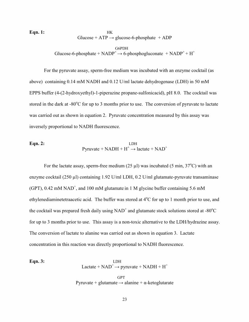
while the oxidized forms do not. For the glucose assay, sperm-free medium (10 µl) was
incubated (5 min, 37oC) with an enzyme cocktail (200 µl) containing 0.42 mM dithiothreitol, 3.1
mM MgSO4, 0.42 mM ATP, 1.25 mM NADP, and 0.1 U/ml hexokinase/glucose-6-phosphate
dehydrogenase (HK/G6PDH) in 50 mM EPPS buffer, pH 8.0. The cocktail was stored in the
dark at -80oC for up to 3 months prior to use. The conversion of glucose to 6-phosphogluconate
was carried out as shown in equation 1. Glucose concentration determined by this assay was
directly proportional to NADPH fluorescence.
23
Eqn. 1: HK Glucose + ATP → glucose-6-phosphate + ADP
G6PDH
Glucose-6-phosphate + NADP+ → 6-phosphogluconate + NADP+ + H+
For the pyruvate assay, sperm-free medium was incubated with an enzyme cocktail (as
above) containing 0.14 mM NADH and 0.12 U/ml lactate dehydrogenase (LDH) in 50 mM
EPPS buffer (4-(2-hydroxyethyl)-1-piperazine propane-sulfonicacid), pH 8.0. The cocktail was
stored in the dark at -80oC for up to 3 months prior to use. The conversion of pyruvate to lactate
was carried out as shown in equation 2. Pyruvate concentration measured by this assay was
inversely proportional to NADH fluorescence.
Eqn. 2: LDH
Pyruvate + NADH + H+ → lactate + NAD+
For the lactate assay, sperm-free medium (25 µl) was incubated (5 min, 37oC) with an
enzyme cocktail (250 µl) containing 1.92 U/ml LDH, 0.2 U/ml glutamate-pyruvate transaminase
(GPT), 0.42 mM NAD+, and 100 mM glutamate in 1 M glycine buffer containing 5.6 mM
ethylenediaminetetraacetic acid. The buffer was stored at 4oC for up to 1 month prior to use, and
the cocktail was prepared fresh daily using NAD+ and glutamate stock solutions stored at -80oC
for up to 3 months prior to use. This assay is a non-toxic alternative to the LDH/hydrazine assay.
The conversion of lactate to alanine was carried out as shown in equation 3. Lactate
concentration in this reaction was directly proportional to NADH fluorescence.
Eqn. 3: LDH
Lactate + NAD+ → pyruvate + NADH + H+
GPT Pyruvate + glutamate → alanine + α-ketoglutarate

24
Enzymes (LDH, product # HK/G6PDH and GPT) were purchased from Roche Applied Science
(Indianapolis, IN), and all other assay reagents were obtained from Sigma Aldrich (St. Louis,
MO). Fluorescence was analyzed using a Spectra Max Gemini XPS fluorescent plate reader
(Molecular Devices, Sunnyvale, CA) and SoftMax Pro 5 software (Molecular Devices,
Sunnyvale, CA). Metabolic rates were calculated as the change in substrate concentration over
time, divided by sperm concentration and are reported in nmol/106 sperm/h.
2.3.4 Statistical Analyses
Data were analyzed with statistical analysis software (SAS) version 9.1 (SAS Institute,
Cary, NC), and percentage data were arcsine-transformed before evaluation. Differences in
ejaculate characteristics and sperm morphology among animal groups (normospermic domestic
cat, teratospermic domestic cat, cheetah) were assessed using SAS General Linear Model
Procedures (GLM) [58]. To evaluate changes in sperm motility index (SMI), acrosomal integrity
(% IA) and metabolic rate over time, data were analyzed using a separate GLM for each animal
group [58]. Within each domestic cat group (normospermic and teratospermic), there was no
interaction (P > 0.05) between individual and time as well as no main effect of individual (P >
0.05) on SMI, % IA, or metabolism; thus, these variables were omitted from the final model.
Differences in SMI, % IA, and metabolic rate among animal groups were assessed using a
separate GLM for each time interval [58]. To determine if variation in sperm motility was
responsible for differences in metabolic rates among animal groups, data from all individuals and
time points were combined and analyzed using a GLM, with percent motile spermatozoa
included as a covariate. When a significant (P < 0.05) F-statistic was measured in any GLM,
differences among means were assessed using Duncan’s multiple-range test. Pearson’s
25
correlation was used to evaluate the relationships between metabolic rate and sperm morphology,
SMI and % IA within and across animal groups. Results were considered significant at P < 0.05.
2.4 Results
2.4.1 Ejaculate and Sperm Characteristics
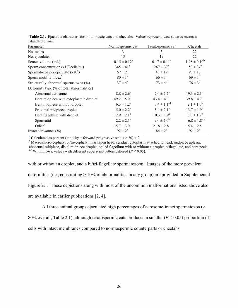
Semen volume and sperm concentration were similar (P > 0.05) in normospermic and
teratospermic domestic cats (Table 2.1). Cheetah ejaculates were less (P < 0.05) concentrated
than those from domestic cats, but due to larger (P < 0.05) seminal volumes, the total number of
spermatozoa per ejaculate did not (P > 0.05) differ among the three animal groups (Table 2.1).
The average sperm motility index and percentage of structurally-normal spermatozoa were
similar (P > 0.05) between teratospermic domestic cats and cheetahs, both of which were less (P
< 0.05) than in normospermic cats (Table 2.1). A bent midpiece encompassing a cytoplasmic
droplet was the most prevalent deformity observed in each animal group, and constituted ~45%
of all abnormalities (Table 2.1). This was followed by acrosomal abnormalities and proximal
droplets, which were more (P < 0.05) common in the cheetah (19% and 14% of all deformities,
respectively) compared to the domestic cat (~8% and 5%). A bent flagellum encircling a
cytoplasmic droplet was a less (P < 0.05) frequent deformity in the cheetah (3%) than domestic
cat (~12%). In both species, spermatids and midpiece bends (without a droplet) constituted <
10% of all abnormalities. More than a dozen other deformities were observed rarely (≤ 5%) in
each group, but collectively comprised a significant proportion (~15%) of total anomalies.
These malformations were classified as ‘other’ and included macro/micro-cephaly, bi/tri-
cephaly, a misshapen head, residual cytoplasm attached to the head, a bent neck, partial or
complete midpiece aplasia, a distal midpiece droplet, a misshapen midpiece, a coiled flagellum
26
with or without a droplet, and a bi/tri-flagellate spermatozoon. Images of the more prevalent
deformities (i.e., constituting ≥ 10% of abnormalities in any group) are provided in Supplemental
Figure 2.1. These depictions along with most of the uncommon malformations listed above also
are available in earlier publications [2, 4].
All three animal groups ejaculated high percentages of acrosome-intact spermatozoa (>
80% overall; Table 2.1), although teratospermic cats produced a smaller (P < 0.05) proportion of
cells with intact membranes compared to normospermic counterparts or cheetahs.
Parameter Normospermic cat Teratospermic cat Cheetah No. males 3 3 22 No. ejaculates 15 19 22 Semen volume (mL) 0.15 ± 0.12a 0.17 ± 0.11a 1.98 ± 0.10b
Sperm concentration (x106 cells/ml) 345 ± 41a 267 ± 37a 50 ± 34b Spermatozoa per ejaculate (x106) 57 ± 21 48 ± 19 93 ± 17 Sperm motility index* 80 ± 1a 66 ± 1b 69 ± 1b
Structurally-abnormal spermatozoa (%) 37 ± 4a 73 ± 4b 76 ± 3b Deformity type (% of total abnormalities)
Abnormal acrosome 8.8 ± 2.6a 7.0 ± 2.2a 19.3 ± 2.1b Bent midpiece with cytoplasmic droplet 49.2 ± 5.0 43.4 ± 4.7 39.8 ± 4.7 Bent midpiece without droplet 6.3 ± 1.2a 3.4 ± 1.1a,b 2.1 ± 1.0b Proximal midpiece droplet 5.0 ± 2.2a 5.4 ± 2.1a 13.7 ± 1.9b Bent flagellum with droplet 12.9 ± 2.1a 10.3 ± 1.9a 3.0 ± 1.7b Spermatid 2.2 ± 2.1a 9.0 ± 2.0b 6.8 ± 1.8a,b Other* 15.7 ± 3.0 21.8 ± 2.8 15.4 ± 2.5
Intact acrosomes (%) 92 ± 2a 84 ± 2b 92 ± 2a * Calculated as percent (motility + forward progressive status × 20) ÷ 2. † Macro/micro-cephaly, bi/tri-cephaly, misshapen head, residual cytoplasm attached to head, midpiece aplasia, abnormal midpiece, distal midpiece droplet, coiled flagellum with or without a droplet, biflagellate, and bent neck. a,b Within rows, values with different superscript letters differed (P < 0.05).
Table 2.1. Ejaculate characteristics of domestic cats and cheetahs. Values represent least-squares means ± standard errors.
27
a aa
a
bb
b
b
bb
b
b
0
20
40
60
80
100
0-1 >1-3 >3-7 >7-24
Sper
m m
otili
ty in
dex
Time (h)
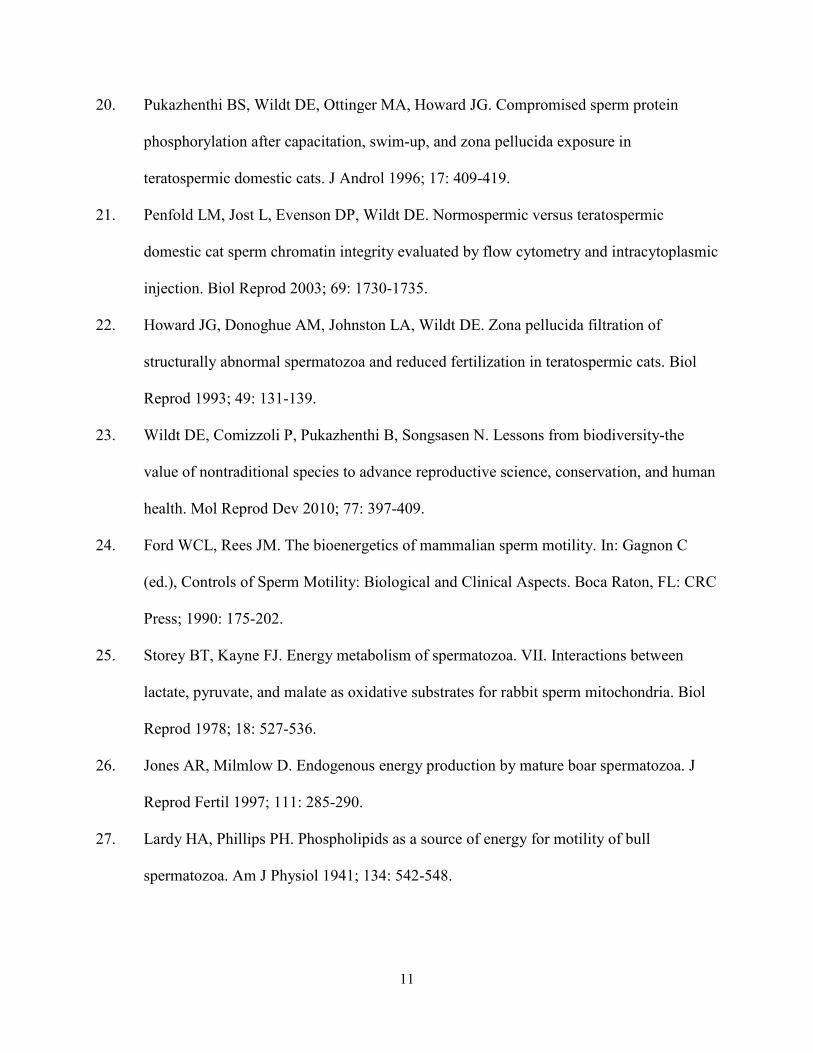
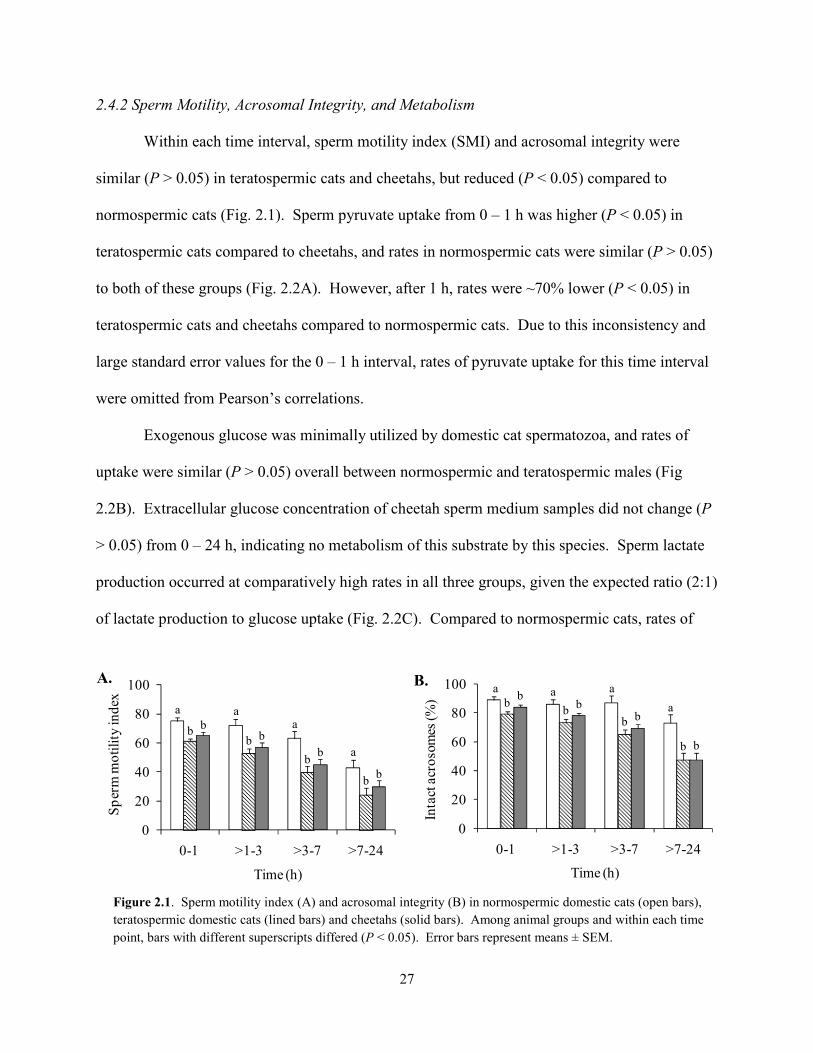
2.4.2 Sperm Motility, Acrosomal Integrity, and Metabolism
Within each time interval, sperm motility index (SMI) and acrosomal integrity were
similar (P > 0.05) in teratospermic cats and cheetahs, but reduced (P < 0.05) compared to
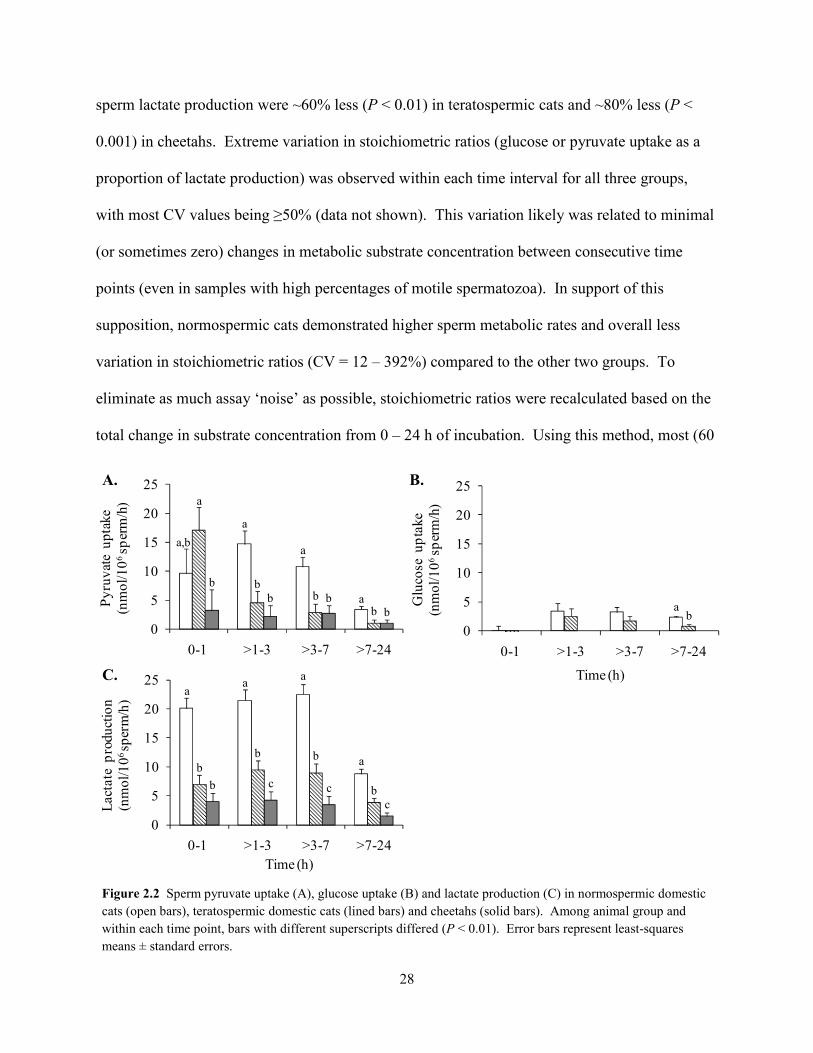
normospermic cats (Fig. 2.1). Sperm pyruvate uptake from 0 – 1 h was higher (P < 0.05) in
teratospermic cats compared to cheetahs, and rates in normospermic cats were similar (P > 0.05)
to both of these groups (Fig. 2.2A). However, after 1 h, rates were ~70% lower (P < 0.05) in
teratospermic cats and cheetahs compared to normospermic cats. Due to this inconsistency and
large standard error values for the 0 – 1 h interval, rates of pyruvate uptake for this time interval
were omitted from Pearson’s correlations.
Exogenous glucose was minimally utilized by domestic cat spermatozoa, and rates of
uptake were similar (P > 0.05) overall between normospermic and teratospermic males (Fig
2.2B). Extracellular glucose concentration of cheetah sperm medium samples did not change (P
> 0.05) from 0 – 24 h, indicating no metabolism of this substrate by this species. Sperm lactate
production occurred at comparatively high rates in all three groups, given the expected ratio (2:1)
of lactate production to glucose uptake (Fig. 2.2C). Compared to normospermic cats, rates of
a a a
abb
b
b
bb
b
b
0
20
40
60
80
100
0-1 >1-3 >3-7 >7-24
Inta
ct a
cros
omes
(%)
Time (h)
Figure 2.1. Sperm motility index (A) and acrosomal integrity (B) in normospermic domestic cats (open bars), teratospermic domestic cats (lined bars) and cheetahs (solid bars). Among animal groups and within each time point, bars with different superscripts differed (P < 0.05). Error bars represent means ± SEM.
B. A.
28
sperm lactate production were ~60% less (P < 0.01) in teratospermic cats and ~80% less (P <
0.001) in cheetahs. Extreme variation in stoichiometric ratios (glucose or pyruvate uptake as a
proportion of lactate production) was observed within each time interval for all three groups,
with most CV values being ≥50% (data not shown). This variation likely was related to minimal
(or sometimes zero) changes in metabolic substrate concentration between consecutive time
points (even in samples with high percentages of motile spermatozoa). In support of this
supposition, normospermic cats demonstrated higher sperm metabolic rates and overall less
variation in stoichiometric ratios (CV = 12 – 392%) compared to the other two groups. To
eliminate as much assay ‘noise’ as possible, stoichiometric ratios were recalculated based on the
total change in substrate concentration from 0 – 24 h of incubation. Using this method, most (60
aa a
abb b
bb c cc
0
5
10
15
20
25
0-1 >1-3 >3-7 >7-24
Lact
ate
prod
uctio
n
(n
mol
/106
sper
m/h
)
Time (h)
a,b
a
a
a
a
bb
b
bb b
b0
5
10
15
20
25
0-1 >1-3 >3-7 >7-24
Pyru
vate
upt
ake
(nm
ol/1
06sp
erm
/h)
ab
0
5
10
15
20
25
0-1 >1-3 >3-7 >7-24
Glu
cose
upt
ake
(nm
ol/1
06sp
erm
/h)
Time (h)
Figure 2.2 Sperm pyruvate uptake (A), glucose uptake (B) and lactate production (C) in normospermic domestic cats (open bars), teratospermic domestic cats (lined bars) and cheetahs (solid bars). Among animal group and within each time point, bars with different superscripts differed (P < 0.01). Error bars represent least-squares means ± standard errors.
B. A.
C.
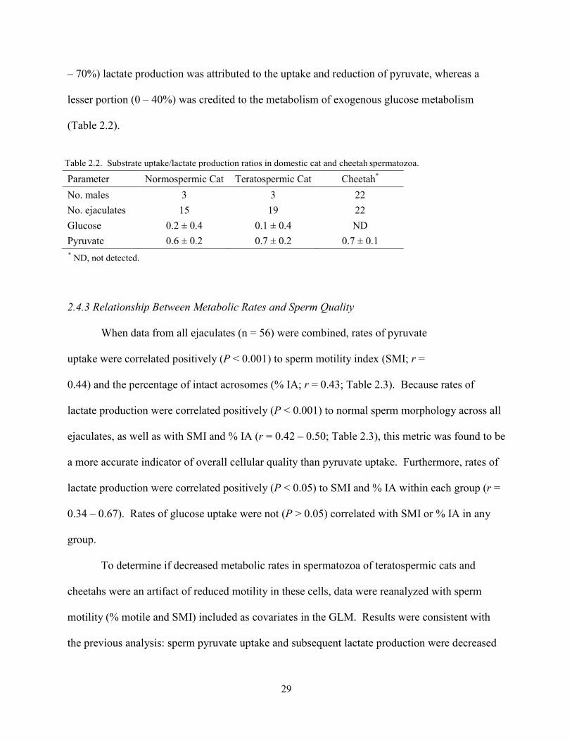
29
– 70%) lactate production was attributed to the uptake and reduction of pyruvate, whereas a
lesser portion (0 – 40%) was credited to the metabolism of exogenous glucose metabolism
(Table 2.2).
Parameter Normospermic Cat Teratospermic Cat Cheetah*
No. males 3 3 22 No. ejaculates 15 19 22 Glucose 0.2 ± 0.4 0.1 ± 0.4 ND Pyruvate 0.6 ± 0.2 0.7 ± 0.2 0.7 ± 0.1
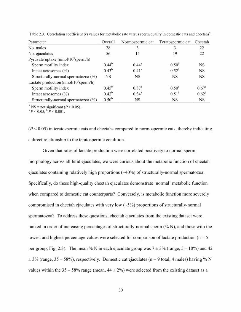
2.4.3 Relationship Between Metabolic Rates and Sperm Quality
When data from all ejaculates (n = 56) were combined, rates of pyruvate
uptake were correlated positively (P < 0.001) to sperm motility index (SMI; r =
0.44) and the percentage of intact acrosomes (% IA; r = 0.43; Table 2.3). Because rates of
lactate production were correlated positively (P < 0.001) to normal sperm morphology across all
ejaculates, as well as with SMI and % IA (r = 0.42 – 0.50; Table 2.3), this metric was found to be
a more accurate indicator of overall cellular quality than pyruvate uptake. Furthermore, rates of
lactate production were correlated positively (P < 0.05) to SMI and % IA within each group (r =
0.34 – 0.67). Rates of glucose uptake were not (P > 0.05) correlated with SMI or % IA in any
group.
To determine if decreased metabolic rates in spermatozoa of teratospermic cats and
cheetahs were an artifact of reduced motility in these cells, data were reanalyzed with sperm
motility (% motile and SMI) included as covariates in the GLM. Results were consistent with
the previous analysis: sperm pyruvate uptake and subsequent lactate production were decreased
Table 2.2. Substrate uptake/lactate production ratios in domestic cat and cheetah spermatozoa.
* ND, not detected.
30
(P < 0.05) in teratospermic cats and cheetahs compared to normospermic cats, thereby indicating
a direct relationship to the teratospermic condition.
Given that rates of lactate production were correlated positively to normal sperm
morphology across all felid ejaculates, we were curious about the metabolic function of cheetah
ejaculates containing relatively high proportions (~40%) of structurally-normal spermatozoa.
Specifically, do these high-quality cheetah ejaculates demonstrate ‘normal’ metabolic function
when compared to domestic cat counterparts? Conversely, is metabolic function more severely
compromised in cheetah ejaculates with very low (~5%) proportions of structurally-normal
spermatozoa? To address these questions, cheetah ejaculates from the existing dataset were
ranked in order of increasing percentages of structurally-normal sperm (% N), and those with the
lowest and highest percentage values were selected for comparison of lactate production (n = 5
per group; Fig. 2.3). The mean % N in each ejaculate group was 7 ± 3% (range, 5 – 10%) and 42
± 3% (range, 35 – 58%), respectively. Domestic cat ejaculates (n = 9 total, 4 males) having % N
values within the 35 – 58% range (mean, 44 ± 2%) were selected from the existing dataset as a
Parameter Overall Normospermic cat Teratospermic cat Cheetah
No. males 28 3 3 22 No. ejaculates 56 15 19 22 Pyruvate uptake (nmol/106sperm/h)
Sperm motility index 0.44b 0.44a 0.50b NS Intact acrosomes (%) 0.43b 0.41a 0.52b NS Structurally-normal spermatozoa (%) NS NS NS NS
Lactate production (nmol/106sperm/h)
Sperm motility index 0.45b 0.37a 0.50b 0.67b
Intact acrosomes (%) 0.42b 0.34a 0.51b 0.62b Structurally-normal spermatozoa (%) 0.50b NS NS NS
Table 2.3. Correlation coefficient (r) values for metabolic rate versus sperm quality in domestic cats and cheetahs*.
* NS = not significant (P > 0.05). a P < 0.05; b P < 0.001.
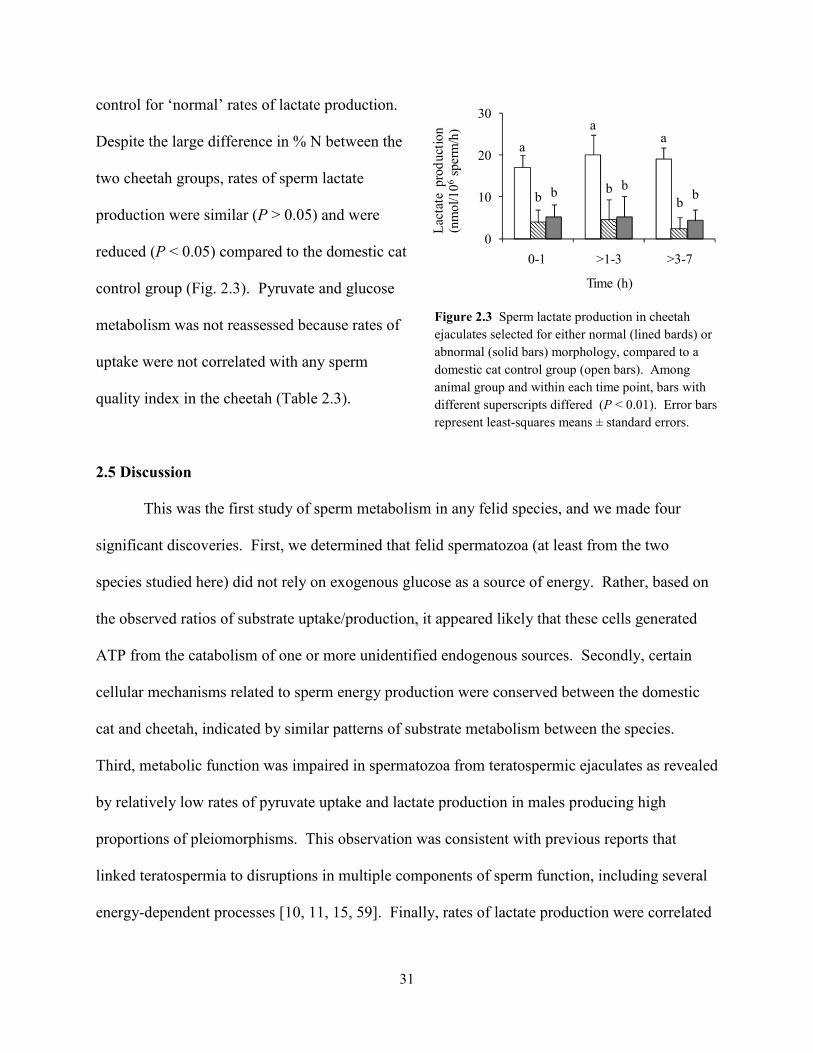
31
control for ‘normal’ rates of lactate production.
Despite the large difference in % N between the
two cheetah groups, rates of sperm lactate
production were similar (P > 0.05) and were
reduced (P < 0.05) compared to the domestic cat
control group (Fig. 2.3). Pyruvate and glucose
metabolism was not reassessed because rates of
uptake were not correlated with any sperm
quality index in the cheetah (Table 2.3).
2.5 Discussion
This was the first study of sperm metabolism in any felid species, and we made four
significant discoveries. First, we determined that felid spermatozoa (at least from the two
species studied here) did not rely on exogenous glucose as a source of energy. Rather, based on
the observed ratios of substrate uptake/production, it appeared likely that these cells generated
ATP from the catabolism of one or more unidentified endogenous sources. Secondly, certain
cellular mechanisms related to sperm energy production were conserved between the domestic
cat and cheetah, indicated by similar patterns of substrate metabolism between the species.
Third, metabolic function was impaired in spermatozoa from teratospermic ejaculates as revealed
by relatively low rates of pyruvate uptake and lactate production in males producing high
proportions of pleiomorphisms. This observation was consistent with previous reports that
linked teratospermia to disruptions in multiple components of sperm function, including several
energy-dependent processes [10, 11, 15, 59]. Finally, rates of lactate production were correlated
Figure 2.3 Sperm lactate production in cheetah ejaculates selected for either normal (lined bards) or abnormal (solid bars) morphology, compared to a domestic cat control group (open bars). Among animal group and within each time point, bars with different superscripts differed (P < 0.01). Error bars represent least-squares means ± standard errors.
a
aa
bb
bb b
b
0
10
20
30
0-1 >1-3 >3-7
Lac
tate
pro
duct
ion
(nm
ol/1
06sp
erm
/h)
Time (h)

32
positively to multiple measures of sperm function in both the domestic cat and cheetah.
Therefore, this substrate may prove to be a valuable indicator of ejaculate quality.
The lack of glucose uptake by domestic cat and cheetah spermatozoa is unexpected,
given that glycolysis is required to support sperm motility [21, 26, 34] and capacitation [19, 26,
60, 61] in the human and domestic dog. This finding perhaps could result from low hexokinase
activity in felids compared to other species or, in the case of the cheetah, the complete absence of
this enzyme. Reduced hexokinase activity would limit NADPH production, a key component of
the glutathione-mediated defense system that protects cellular membranes against lipid
peroxidation damage [62]. This is an intriguing possibility, as spermatozoa from teratospermic
felids (including the domestic cat and cheetah) are unusually susceptible to membrane damage
[13, 16, 46]. However, it also remains possible that felid spermatozoa possess fully functional
hexokinase, but metabolize glucose at modest rates relative to oxidative substrates. We currently
are using the glycolytic inhibitor α-chlorohydrin to more thoroughly understand the role of
glycolysis in felid sperm cellular function.
Given the observed lack of glucose uptake by cat and cheetah spermatozoa, we were
surprised to discover that these cells consistently produced lactate, which is an end-product of
glycolysis [63]. Under our experimental conditions, we consider there to be three possible
sources of lactate: 1) endogenous glycogen; 2) endogenous phospholipid; and/or 3) imported
pyruvate. These potential mechanisms of lactate production are not mutually exclusive and may
also contribute to varying degrees in the generation of NADH and ATP (Fig. 2.4). In each case,
lactate formation could occur in cytosol or mitochondria as sperm-specific lactate dehydrogenase
(LDHC) has been found in these compartments [64-67]. Lactate and NADH production also
could occur in separate cellular locations, since reducing equivalents can be transferred between
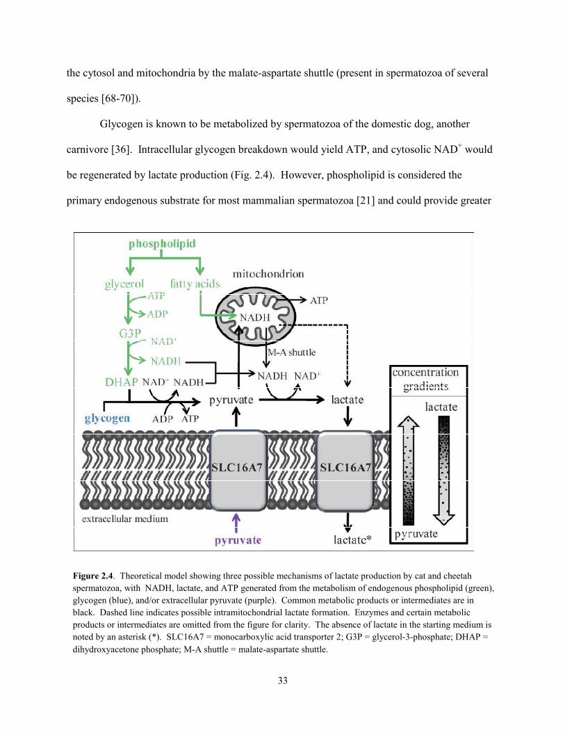
33
the cytosol and mitochondria by the malate-aspartate shuttle (present in spermatozoa of several
species [68-70]).
Glycogen is known to be metabolized by spermatozoa of the domestic dog, another
carnivore [36]. Intracellular glycogen breakdown would yield ATP, and cytosolic NAD+ would
be regenerated by lactate production (Fig. 2.4). However, phospholipid is considered the
primary endogenous substrate for most mammalian spermatozoa [21] and could provide greater
Figure 2.4. Theoretical model showing three possible mechanisms of lactate production by cat and cheetah spermatozoa, with NADH, lactate, and ATP generated from the metabolism of endogenous phospholipid (green), glycogen (blue), and/or extracellular pyruvate (purple). Common metabolic products or intermediates are in black. Dashed line indicates possible intramitochondrial lactate formation. Enzymes and certain metabolic products or intermediates are omitted from the figure for clarity. The absence of lactate in the starting medium is noted by an asterisk (*). SLC16A7 = monocarboxylic acid transporter 2; G3P = glycerol-3-phosphate; DHAP = dihydroxyacetone phosphate; M-A shuttle = malate-aspartate shuttle.
34
amounts of cellular ATP. Phospholipid hydrolysis would yield glycerol and fatty acids (Fig.
2.4). Glycerol would enter the glycolytic pathway via conversion to dihydroxyacetone
phosphate (DHAP) and would be metabolized to produce lactate, NADH, and ATP [71].
Mitochondrial oxidation of fatty acids would provide substantial amounts of NADH, which
could contribute to ATP and/or lactate production (Fig. 2.4).
Because we used a protein-free, chemically defined medium, the only possible exogenous
source of lactate in our study was by the uptake and reduction of extracellular pyruvate.
Although pyruvate reduction would require a NADH source and would not generate ATP [63],
these molecules could be provided by endogenous substrate metabolism (as described above).
Pyruvate uptake likely would occur via the facultative transporter SLC16A7 (monocarboxylic
acid transporter 2, previously known as MCT2), the primary monocarboxylate transporter in
mature spermatozoa of species studied to date [72-74]. Given the starting composition of our
cMTF medium (1 mM pyruvate, 0 mM lactate) and the kinetic properties of SLC16A7 and LDH
[75-77], rapid pyruvate uptake and reduction theoretically should occur independently of sperm
energy production. Examination of stoichiometric ratios revealed that 60 to 70% of produced
lactate could have been explained on the basis of pyruvate uptake and reduction (in contrast to
only 0 – 40% generated from exogenous glucose catabolism). These are important observations
given that lactate production (measured by enzyme-linked fluorescence) has been used as an
indicator of glycolytic metabolism for other species [25, 30, 31]. Ongoing research in our
laboratory supports this mechanism of lactate production in felid spermatozoa. Initial findings
determined that sperm lactate production was approximately four times greater in the presence of
exogenous pyruvate compared to equimolar amounts of exogenous glucose. Furthermore, in the
absence of both substrates in the culture medium no lactate was produced. Collectively, these
35
observations suggested that mammalian spermatozoa may produce lactate independently of
glycolysis, implying that the functional importance of this pathway could have been
misinterpreted in earlier studies. More detailed studies are in progress in our laboratory using
chemical inhibitors of oxidative and glycolytic metabolism to identify the primary energy
substrates for felid spermatozoa.
Although the source of lactate was unclear, rates of production provided a consistent
indicator of sperm motility and acrosomal function in both felid species. Intriguingly, lactate
production also was correlated to proportions of structurally-normal spermatozoa among
domestic cat, but not cheetah, ejaculates. This finding may be related to previous reports that
even structurally-intact cells from teratospermic ejaculates can be functionally-compromised.
Specifically, these spermatozoa may demonstrate increased osmotic sensitivity, delayed
acrosome reaction, or reduced zona penetration ability [10, 11, 15]. Therefore, rates of lactate
production may provide an accurate indicator of ejaculate quality in both species, and may reveal
disrupted cellular physiology in apparently-normal cheetah spermatozoa. However, lactate
production should be validated against a more direct measure of sperm fertilizing ability such as
the zona pellucida assay, particularly given its unclear relationship to ATP generation. Because
spermatozoa must be capable of multiple energy-dependent processes to achieve successful
fertilization, a zona penetration assay could provide a more robust test of the functional
relevance of lactate production than any single measure of sperm function (e.g., motility).
Understanding the source and biological significance of lactate produced by felid spermatozoa
could yield a quantitative, field-friendly indicator of ejaculate quality, which would significantly
facilitate developing and refining reproductive technologies for improving felid management and
conservation.
36
Because so little is known about gamete metabolism in felids, this taxon is an excellent
target for more detailed investigation in this area of study. The existence of >30 species in the
family Felidae (including several with the teratospermic phenotype [78]) also provides important
opportunities to determine the etiology and evolution of certain physiological attributes
influencing reproductive success. Indeed, direct comparisons across species are fundamental to
identifying differences in the mechanisms involving energy production in mammalian gametes,
particularly given the ambiguities of published findings relating to sperm metabolism. For
example, Storey et al. have reported that mouse spermatozoa metabolize endogenous oxidative
substrates to remain motile for >4 h in vitro in the absence of supplemental energy sources [79].
Yet Mukai and Okuno have found that mouse spermatozoa become non-motile within 30 min in
a substrate-free medium and have presented evidence for an obligate role of glycolysis in
supporting cellular motility [80]. Contradictory findings also have been reported regarding the
need for exogenous glucose to achieve capacitation in dog spermatozoa [61, 81]. Such
equivocations often can be avoided and more confidence generated by comparatively evaluating
two taxonomically-related species using a standardized experimental protocol. In the present
study, we found similar patterns of substrate uptake/production in the domestic cat and cheetah,
suggesting that spermatozoa of both species relied on the same energy source(s). Relatively low
rates of substrate uptake/production in cheetah ejaculates revealed that sperm metabolic
efficiency was compromised in this species. More importantly, comparison with the
teratospermic cat model then allowed us to link this finding to ejaculate phenotype. Still, we
uncovered a subtle, but key physiological difference between these species. Specifically, in
contrast to the domestic cat, even apparently ‘high-quality’ cheetah ejaculates (i.e., containing
higher proportions of structurally-normal spermatozoa) demonstrated compromised metabolic
37
function. Thus, while the collective results confirmed that the teratospermic cat was an excellent
model for expanding knowledge about gamete physiology in wild felids, findings revealed
important functional differences between these two related species.
In conclusion, study results underscored the importance of species diversity in
fundamental reproductive phenomena, as has been emphasized recently in the contexts of
biological conservation and human health [82]. We predict that understanding the unique
mechanisms of energy production in felid spermatozoa will facilitate increased efficiency of
assisted reproductive technologies that have been used for producing offspring in both wild
felids [83, 84] and domestic cats, including at least eight models of human genetic disease
(Swanson, W.F., personal communication) [85]. For example, such information would be
important for optimizing medium composition to maximize sperm survival for artificial
insemination, in vitro culture (including for IVF), and cryopreservation. Furthermore, we
observed that metabolic efficiency reflected other sperm quality metrics across both individuals
and species, thus providing the first objective, field-friendly indicator of gamete function in
felids. Such findings also offer new insights for improving reproduction in small populations of
endangered species or rare biomedical genotypes. Indeed, a basic understanding of the
reproductive uniqueness of a previously unstudied species has been critical to recovery or
development of self-sustaining populations [82]. The opportunity to conduct these fundamental
studies is one of the invaluable contributions of ex situ (hedge) felid populations to the
conservation of Earth’s biodiversity [86].
2.6 References
1. Pukazhenthi BS, Wildt DE, Howard JG. The phenomenon and significance of
teratospermia in felids. J Reprod Fertil Suppl 2001; 57: 423-433.
38
2. Wildt DE, Bush M, Howard JG, O'Brien SJ, Meltzer D, van Dyk A, Ebedes H, Brand DJ.
Unique seminal quality in the South African cheetah and a comparative evaluation in the
domestic cat. Biol Reprod 1983; 29: 1019-1025.
3. Wildt DE, Brown JL, Bush M, Barone MA, Cooper KA, Grisham J, Howard JG.
Reproductive status of cheetahs (Acinonyx jubatus) in North American zoos: the benefits
of physiological surveys for strategic planning. Zoo Biol 1993; 12: 45-80.
4. Crosier AE, Marker L, Howard JG, Pukazhenthi BS, Henghali JN, Wildt DE. Ejaculate
traits in the Namibian cheetah (Acinonyx jubatus): influence of age, season, and captivity.
Reprod Fertil Dev 2007; 19: 370-382.
5. Barone MA, Roelke ME, Howard J, Brown JL, Anderson AE, Wildt DE. Reproductive
characteristics of male Florida panthers: comparative studies from Florida, Texas,
Colorado, Latin America, and North American Zoos. J Mammal 1994; 75: 150-162.
6. Roelke ME, Martenson JS, O'Brien SJ. The consequences of demographic reduction and
genetic depletion in the endangered Florida panther. Curr Biol 1993; 3: 340-350.
7. Wildt DE, Bush M, Goodrowe KL, Packer C, Pusey AE, Brown JL, Joslin P, O'Brien SJ.
Reproductive and genetic consequences of founding isolated lion populations. Nature
1987; 329: 328-331.
8. Neubauer K, Jewgenow K, Blottner S, Wildt DE, Pukazhenthi BS. Quantity rather than
quality in teratospermic males: a histomorphometric and flow cytometric evaluation of
spermatogenesis in the domestic cat. Biol Reprod 2004; 71: 1524-1571.
9. Cooper TG, Noonan E, von Eckardstein S, Auger J, Gordon Baker HW, H.M. B, Haugen
TB, Kruger T, Wang C, Mbizvo MT, Vogelsong KM. World Health Organization
39
reference values for human semen characteristics. Hum Reprod Update 2010; 16: 231-
245.
10. Long JA, Wildt DE, Wolfe BA, Crister JK, DeRossi RV, Howard JG. Sperm capacitation
and the acrosome reaction are compromised in teratospermic domestic cats. Biol Reprod
1996; 54: 638-646.
11. Pukazhenthi BS, Wildt DE, Ottinger MA, Howard JG. Compromised sperm protein
phosphorylation after capacitation, swim-up, and zona pellucida exposure in
teratospermic domestic cats. J Androl 1996; 17: 409-419.
12. Pukazhenthi BS, Long JA, Wildt DE, Ottinger MA, Armstrong DL, Howard JG.
Regulation of sperm function by protein tyrosine phosphorylation in diverse wild felid
species. J Androl 1998; 19: 675-685.
13. Pukazhenthi BS, Noiles E, Pelican K, Donoghue AM, Wildt DE, Howard JG. Osmotic
effects on feline spermatozoa from normospermic versus teratospermic donors.
Cryobiology 2000; 40: 139-150.
14. Pukazhenthi BS, Spindler RE, Wildt DE, Bush LM, Howard JG. Osmotic properties of
spermatozoa from felids producing different proportions of pleiomorphisms: influence of
adding and removing cryoprotectant. Cryobiology 2002; 44: 288-300.
15. Howard JG, Donoghue AM, Johnston LA, Wildt DE. Zona pellucida filtration of
structurally abnormal spermatozoa and reduced fertilization in teratospermic cats. Biol
Reprod 1993; 49: 131-139.
16. Pukazhenthi B, Pelican K, Wildt D, Howard JG. Sensitivity of domestic cat (Felis catus)
sperm from normospermic versus teratospermic donors to cold-induced acrosome
damage. Biol Reprod 1999; 61: 135-141.
40
17. Pukazhenthi B, Santymire R, Crosier A, Howard JG, Wildt DE. Challenges in
cryopreserving endangered mammal spermatozoa: morphology and the value of
acrosomal integrity as markers of cryo-survival. In: Roldan ERS, Gomendio M (eds.),
Spermatology, vol. 65. Nottingham: Nottingham University Press; 2008: 433-444.
18. Storey B. Mammalian sperm metabolism: oxygen and sugar, friend and foe. Int J Dev
Biol 2008; 52: 427-437.
19. Albarracín JL, Fernández-Novell JP, Ballester J, Rauch MC, Quintero-Moreno A, Peña
A, Mogas T, Rigau T, Yañez A, Guinovart JJ, Slebe JC, Concha II, et al.
Gluconeogenesis-linked glycogen metabolism is important in the achievement of in vitro
capacitation of dog spermatozoa in a medium without glucose. Biol Reprod 2004; 71:
1437-1445.
20. Marín S, Chiang K, Bassilian S, Lee W-NP, Boros L, Fernandáz-Novell JM, Centelles JJ,
Medrano A, Rodriguíz-Gil JE, Cascante M. Metabolic strategy of boar spermatozoa
revealed by a metabolomic characterization. FEBS Lett 2003; 554: 342-346.
21. Ford WCL, Rees JM. The bioenergetics of mammalian sperm motility. In: Gagnon C
(ed.) Controls of Sperm Motility: Biological and Clinical Aspects. Boca Raton, FL: CRC
Press; 1990: 175-202.
22. Nascimento JM, Shi LZ, Tam J, Chandsawangbhuwana C, Durrant B, Botvinick EL,
Berns MW. Comparison of glycolysis and oxidative phosphorylation as energy sources
for mammalian sperm motility, using the combination of fluorescence imaging, laser
tweezers and real-time automated tracking and trapping. J Cell Physiol 2008; 217: 745-
751.
41
23. Ford WCL. Glycolysis and sperm motility: does a spoonful of sugar help the flagellum
go round? Hum Reprod Update 2006; 12: 269-274.
24. Ruiz-Pesini E, Díez-Sánchez C, López-Pérez MJ, Enríquez JA. The role of the
mitochondrion in sperm function: is there a place for oxidative phosphorylation or is this
a purely glycolytic process? Curr Top Dev Biol 2007; 77: 3-19.
25. Garrett LJA, Revell SG, Leese HJ. Adenosine triphosphate production by bovine
spermatozoa and its relationship to semen fertilizing ability J Androl 2008; 29: 449-458.
26. Williams AC, Ford WC. The role of glucose in supporting motility and capacitation in
human spermatozoa. J Androl 2001; 22: 680-695.
27. Turner R. Tales from the tail: what do we really know about sperm motility? J Androl
2003; 24: 790-803.
28. Westhoff D, Kamp G. Glyceraldehyde 3-phosphate dehydrogenase is bound to the
fibrous sheath of mammalian spermatozoa. J Cell Sci 1997; 110: 1821-1829.
29. Travis AJ, Foster JA, Rosenbaum NA, Visconti PE, Gerton GL, Kopf GS, Moss SB.
Targeting of a germ cell-specific type 1 hexokinase lacking a porin-binding domain to the
mitochondria as well as to the head and fibrous sheath of murine spermatozoa. Mol Cell
Biol 1998; 9: 263-276.
30. Rigau T, Piedrafita J, Reverter A, Canal M, Rodríguez-Gil JE. The rate of L-lactate
production: a feasible parameter for the fresh diluted boar semen quality analysis. Anim
Reprod Sci 1996; 43: 161-172.
31. Miró J, Lobo V, Quintero-Moreno A, Medrano A, Peňa A, Rigau T. Sperm motility
patterns and metabolism in Catalonian donkey semen. Theriogenology 2005; 63: 1706-
1716.
42
32. Songsasen N, Spindler RE, Wildt DE. Requirement for, and patterns of, pyruvate and
glutamine metabolism in the domestic dog oocyte in vitro. Mol Reprod Dev 2007; 74:
870-877.
33. Palomo MJ, Fernández-Novell JP, Peña A, Guinovart JJ, Rigau T, Rodríguez-Gil JE.
Glucose- and fructose-induced dog-sperm glycogen synthesis shows specific changes in
the location of the sperm glycogen deposition. Mol Reprod Dev 2003; 64: 349-359.
34. Rigau T, Farré M, Ballester J, Mogas T, Peña A, Rodríguez-Gil JE. Effects of glucose
and fructose on motility patterns of dog spermatozoa from fresh ejaculates
Theriogenology 2001; 56: 801-815.
35. Rigau T, Rivera M, Palomo MJ, Fernández-Novell JM, Mogas T, Ballester J, Peña A,
Otaegui PJ, Guinovart JJ, Rodríguez-Gil JE. Differential effects of glucose and fructose
on hexose metabolism in dog spermatozoa. Reproduction 2002; 123: 579-591.
36. Ballester J, Fernández-Novell JP, Rutllant J, García-Rocha M, Palomo MJ, Mogas T,
Peña A, Rigau T, Guinovart JJ, Rodríguez-Gil JE. Evidence for a functional glycogen
metabolism in mature mammalian spermatozoa. Mol Reprod Dev 2000; 56: 207-219.
37. Fernández-Novell JM, Ballester J, Medrano A, Otaegui PJ, Rigau T, Guinovart JJ,
Rodríguez-Gil JE. The presence of a high-Km hexokinase activity in dog, but not in boar,
sperm. FEBS Lett 2004; 570: 211-216.
38. Albarracín JL, Mogas T, Palomo MJ, Peña A, Rigau T, Rodríguez-Gil JE. In vitro
capacitation and acrosome reaction of dog spermatozoa can be feasibly attained in a
defined medium without glucose. Reprod Dom Anim 2004; 39: 129-135.
43
39. Bucci D, Isani G, Spinaci M, Tamanini C, Mari G, Zambelli D, Galeati G. Comparative
immunolocalization of GLUTs 1, 2, 3, and 5 in boar, stallion and dog spermatozoa.
Reprod Domest Anim 2010; 45: 315-322.
40. Pilkis SJ, Granner DK. Molecular physiology of the regulation of hepatic
gluconeogenesis and glycolysis. Annu Rev Physiol 1992; 54: 885-909.
41. Hoppe PC. Glucose requirement for mouse sperm capacitation in vitro. Biol Reprod
1976; 15: 39-45.
42. Parrish JJ, Susko-Parrish J, First NL. Capacitation of bovine sperm by heparin: inhibitory
role of glucose and role of intracellular pH. Biol Reprod 1989; 38: 683-689.
43. Rogers BJ, Yanagimachi R. Retardation of guinea pig sperm acrosome reaction by
glucose: the possible importance of pyruvate and lactate metabolism in capacitation and
the acrosome reaction. Biol Reprod 1975; 13: 568-575.
44. Wildt DE. Assisted reproduction for managing and conserving threatened felids. Int Zoo
Yb 1997; 35: 164-172.
45. O'Brien SJ, Johnson W, Driscoll C, Pontius J, Pecon-Slattery J, Menotti-Raymond M.
State of cat genomics. Trends Genet 2008; 24: 268-279.
46. Crosier A, Henghali J, Howard JG, Pukazhenthi BS, Terrell KA, Marker LL, Wildt DE.
Improved quality of cryopreserved cheetah (Acinonyx jubatus) spermatozoa after
centrifugation through Accudenz. J Androl 2009; 30: 298-308.
47. Howard JG. Semen collection and analysis in carnivores. In: Fowler ME (ed.) Zoo and
Wild Animal Medicine: Current Therapy III. Philadelphia: WB Saunders Co.; 1993: 390-
399.
44
48. Howard JG, Brown JL, Bush M, Wildt DE. Teratospermic and normospermic domestic
cats: ejaculate traits, pituitary-gonadal hormones, and improvement of spermatozoal
motility and morphology after swim-up processing. J Androl 1990; 11: 204-215.
49. Larson JL, Miller DJ. Simple histochemical stain for acrosomes on sperm from several
species. Mol Reprod Dev 1999; 52: 445-449.
50. Crosier AE, Pukazhenthi BS, Henghali JN, Howard JG, Dickman AJ, Marker L, Wildt
DE. Cryopreservation of spermatozoa from wild-born Namibian cheetahs (Acinonyx
jubatus) and influence of glycerol on cryosurvival. Cryobiology 2006; 52: 169-181.
51. Gardner DK, Leese HJ. Concentrations of nutrients in mouse oviduct fluid and their
effects on embryo development and metabolism in vitro. J Reprod Fertil 1990; 88: 361-
368.
52. Bavister BD. Substitution of a synthetic polymer for protein in a mammalian gamete
culture system. J Exp Zool 1981; 217: 45-51.
53. Johnston SD, Osborne CA, Lipowitz AJ. Characterization of seminal plasma, prostatic
fluid, and bulbourethral gland secretions in the domestic cat. In: Proceedings of the 11th
International Congress on Animal Reproduction and Artificial Insemination. Dublin;
1988: 4:560.
54. Shah D, Naciri M, Clee P, Al-Rubeai M. NucleoCounter: an efficient technique for the
determination of cell number and viability in animal cell culture processes.
Cytotechnology 2006; 51: 39-44.
55. Bishop DW. Oxygen consumption of fox sperm. Biol Bull 1942; 83: 353-362.
56. Christen R, Schackmann RW, Shapiro BM. Metabolism of sea urchin sperm. J Biol
Chem 1983; 258: 5392-5399.
45
57. Passonneau JV, Lowry OH. Enzymatic Analysis: A Practical Guide. Totowa, NJ:
Humana Press; 1993.
58. SAS. SAS/STAT User's Guide: Statistics. Version 9.1. Cary, NC: Statistical Analysis
System Institute, Inc; 2002.
59. Penfold LM, Jost L, Evenson DP, Wildt DE. Normospermic versus teratospermic
domestic cat sperm chromatin integrity evaluated by flow cytometry and intracytoplasmic
injection. Biol Reprod 2003; 69: 1730-1735.
60. Mahadevan MM, Miller MM, Moutos DM. Absence of glucose decreases human
fertilization and sperm movement characteristics in vitro. Hum Reprod 1997; 12: 119-
123.
61. Mahi CA, Yanagimachi R. Capacitation, acrosome reaction, and egg penetration by
canine spermatozoa in a simple defined medium. Gamete Res 1978; 1: 101-109.
62. Voet D, Voet JG. Other pathways of carbohydrate metabolism. In: Harris D, Fitzgerald P
(eds.), Biochemistry, 3rd ed. Hoboken, NJ: John Wiley & Sons, Inc.; 2004: 843-870.
63. Voet D, Voet JG. Glycolysis. In: Harris D, Fitzgerald P (eds.), Biochemistry, 3rd ed.
Hoboken, NJ: John Wiley & Sons, Inc.; 2004: 581-624.
64. Storey BT, Kayne FJ. Energy metabolism of spermatozoa VI. Direct intramitochondrial
lactate oxidation by rabbit sperm mitochondria. Biol Reprod 1977; 16: 549-556.
65. Gallina FG, Deburgos NMG, Burgos C, Coronel CE, Blanco A. The lactate/pyruvate
shuttle in spermatozoa: operation in vitro. Arch Biochem Biophys 1994; 308: 515-519.
66. Wang SX, Luo AM, Liang ZG, Song JF, Wang HA, Chen YX. Preparation and
characterization of monoclonal antibodies against sperm-specific lactate dehydrogenase
C4. J Androl 1990; 11: 319-324.
46
67. Burgos C, Maldonado C, Gerez de Burgos NM, Aoki A, Blanco A. Intracellular
localization of the testicular and sperm-specific lactate dehydrogenase isozyme C4 in
mice. Biol Reprod 1995; 53: 84-92.
68. Blanco A. On the functional significance of LDH X. Johns Hopkins Med J 1980; 146:
231-235.
69. Calvin J, Tubbs PK. Mitochondrial transport processes and oxidation of NADH by
hypotonically-treated boar spermatozoa. Eur J Biochem 1978; 89: 315-320.
70. Voet D, Voet JG. Electron transport and oxidative phosphorylation. In: Harris D,
Fitzgerald P (eds.), Biochemistry, 3rd ed. Hoboken, NJ: John Wiley & Sons, Inc.; 2004:
797-842.
71. Voet D, Voet JG. Lipid metabolism. In: Harris D, Fitzgerald P (eds.), Biochemistry, 3rd
ed. Hoboken, NJ: John Wiley & Sons, Inc.; 2004: 909-984.
72. Merezhinskaya N, Fishbein WN. Monocarboxylate transporters: past, present, and future.
Histol Histopathol 2009; 24: 243-264.
73. Garcia CK, Brown MS, Pathak RK, Goldstein JL. CDNA cloning of MCT2, a second
monocarboxylate transporter expressed in different cells than MCT1 J Biol Chem 1995;
270: 1843-1849.
74. Halestrap AP, Price NT. The proton-linked monocarboxylate transporter (MCT) family:
structure, function, and regulation. Biochem J 1999; 343: 281-299.
75. Brooks GA. Intra- and extra-cellular lactate shuttles. Med Sci Sports Exerc 2000; 32:
790-799.
76. Brooks GA. Mammalian fuel utilization during sustained exercise. Comp Biochem
Physiol B Biochem Mol Biol 1998; 120: 89-107.
47
77. Brooks GA, Dubouchaud H, Brown M, Sicurello JP, Butz CE. Role of mitochondrial
lactate dehydrogenase and lactate oxidation in the intracellular lactate shuttle. Proc Natl
Acad Sci USA 1999; 96: 1129-1134.
78. Pukazhenthi BS, Neubauer K, Jewgenow K, Howard JG, Wildt DE. The impact and
potential etiology of teratospermia in the domestic cat and its wild relatives.
Theriogenology 2006; 66: 112-121.
79. Carey JE, Oldsclarke P, Storey BT. Oxidative metabolism of spermatozoa from inbred
and random bred mice. J Exp Zool 1981; 216: 285-292.
80. Mukai C, Okuno M. Glycolysis plays a major role for adenosine triphosphate
supplementation in mouse sperm flagellar movement. Biol Reprod 2004; 71: 540-547.
81. Rota A, Peña AI, Linde-Forsberg C, Rodríguez-Martínez H. In vitro capacitation of
fresh, chilled, and frozen-thawed dog spermatozoa assessed by the chlorotetracycline
assay and changes in motility patterns. Anim Reprod Sci 1999; 57: 199-215.
82. Wildt DE, Comizzoli P, Pukazhenthi B, Songsasen N. Lessons from biodiversity-the
value of nontraditional species to advance reproductive science, conservation, and human
health. Mol Reprod Dev 2010; 77: 397-409.
83. Howard JG, Wildt DE. Approaches and efficacy of artificial insemination in felids and
mustelids. Theriogenology 2009; 71: 130-148.
84. Swanson WF. Application of assisted reproduction for population management in felids:
the potential and reality for conservation of small cats. Theriogenology 2006; 66: 49-58.
85. Swanson WF. Research in nondomestic species: experiences in reproductive physiology
research for conservation of endangered felids. ILAR J 2003; 44: 307-316.
48
86. Wildt DE, Swanson W, Brown J, Sliwa A, Vargas A. Felids ex situ: managed
programmes, research, and species recovery. In: Macdonald DW, Loveridge AJ (eds.),
Biology and Conservation of Wild Felids: Oxford University Press; 2010: 217-235.
49
CHAPTER 3: GLYCOLYTIC ENZYME ACTIVITY IS ESSENTIAL FOR DOMESTIC CAT (FELIS CATUS) AND CHEETAH (ACINONYX JUBATUS) SPERM MOTILITY
AND VIABILITY IN A SUGAR-FREE MEDIUM
3.1 Abstract
We previously have reported a lack of glucose uptake in domestic cat and cheetah
spermatozoa, despite observing that these cells produce lactate at rates that correlate positively
with sperm function. To elucidate the role of glycolysis in felid sperm energy production, we
conducted a comparative study in the domestic cat and cheetah, with the hypothesis that sperm
motility and viability are maintained in both species in the absence of glycolytic metabolism and
are fueled by endogenous substrates. Washed ejaculates were incubated in chemically defined
medium in the presence/absence of glucose and pyruvate. A second set of ejaculates was
exposed to a chemical inhibitor of either lactate dehydrogenase (sodium oxamate) or
glyceraldehyde-3-phosphate dehydrogenase (α-chlorohydrin). Sperm function (motility and
acrosomal integrity) and lactate production were assessed, and a subset of spermatozoa was
assayed for intracellular glycogen. In both the cat and cheetah, sperm function was maintained
without exogenous substrates and following lactate dehydrogenase inhibition. Lactate
production occurred in the absence of exogenous hexoses, but only if pyruvate was present.
Intracellular glycogen was not detected in spermatozoa from either species. Unexpectedly,
glycolytic inhibition by α-chlorohydrin resulted in an immediate decline in sperm motility,
particularly in the domestic cat. Collectively, our findings revealed an essential role of the
glycolytic pathway in felid spermatozoa that was unrelated to hexose metabolism or lactate
formation. Instead, glycolytic enzyme activity could have been required for the metabolism of
50
endogenous lipid-derived glycerol, with fatty acid oxidation providing the primary energy source
in felid spermatozoa.
3.2 Introduction
Glycolysis is widely considered to be a key pathway in mammalian sperm energy
production [1-7]. In the bull, ram, dog, and mouse, glycolysis can fully support sperm motility
when oxidative metabolism is blocked [1, 8-12]. In the human and rhesus macaque, glycolysis is
an essential source of ATP, and motility cannot be maintained by respiration alone [5, 12-15].
Glucose is considered to be the primary metabolic sugar in mammalian spermatozoa [4], but
these cells also can utilize fructose, mannose, and maltose as substrates for glycolysis [7, 12, 16].
Although producing < 6% of the ATP generated by oxidative metabolism [17], the
disproportionate importance of the relatively inefficient glycolytic pathway has been linked to
the compartmentalization of sperm energy production [18-20]. Because active microtubule
sliding occurs in the distal flagellum, far from the site of mitochondrial activity [2], glycolysis
may be an obligate energy source for cellular motility. Consistent with this idea, glycolytic
enzymes are tightly bound to structural elements of the sperm flagellum in several species,
including the rabbit, boar, bull, rat, stallion, human, mouse, and fox [18-21]. Furthermore, even
in species capable of sustaining motility by oxidative metabolism alone (e.g., mouse), glycolysis
may be required for sperm capacitation [11, 22, 23], hyperactivation [24, 25], the acrosome
reaction [25], zona binding [26], or fusion with the oocyte plasma membrane [26].
We recently described sperm metabolic profiles of the domestic cat and cheetah, the first
knowledge about gamete energy production in any felid species [27]. One key finding was that
the condition of teratospermia (where males produce ≥ 60% structurally abnormal spermatozoa)
51
was linked to remarkably reduced rates of sperm lactate production. This observation suggests
that sperm ATP synthesis is impaired in teratospermic felids – an intriguing idea, since metabolic
disruption could explain many of the functional abnormalities observed in these ejaculates,
including reduced sperm motility [27], delayed capacitation [28], compromised acrosomal
function [28], disrupted protein tyrosine phosphorylation [29, 30], reduced zona penetration
ability [31] and decreased fertilization success in vitro [31]. Although occurring at reduced rates
in teratospermic ejaculates, lactate production is positively correlated to other components of
felid sperm function (motility and acrosomal integrity [27]), suggesting a key role of anaerobic
glycolysis in these cells. Yet regardless of ejaculate quality, our earlier study [27] demonstrated
that felid spermatozoa experience a surprising lack of glucose uptake, even in the absence of
other glycolyzable substrates [27]. There are three possible explanations for these observed
metabolic profiles. First, glucose may be imported and metabolized at very low rates (below the
threshold of detection), but at a level still required for supporting sperm motility. Second,
endogenous glucose could be present in the form of glycogen, as in the domestic dog where this
carbohydrate is localized to the sperm head and midpiece [32] and can provide an energy source
for capacitation in a hexose-free medium [6]. Finally, felids may be unusual among mammals in
that glycolysis is an insignificant source of sperm energy production. In this case, we would
expect the oxidative metabolism of endogenous lipid to support sperm function, since our
previous study detected little uptake of any extracellular substrates by cat or cheetah
spermatozoa. In several species, including the mouse [33], bull [34], and boar [35], sperm
motility can be maintained through endogenous phospholipid or di-/tri-glyceride metabolism,
and it is conceivable that this mechanism exists in felids.
52
The cheetah and domestic cat are excellent models for studying sperm function in
teratospermic species, including humans [36] and several endangered felids [37]. Cheetahs are
well-known to consistently produce ejaculates containing at least 70% malformed spermatozoa
[27, 38, 39], while domestic cats may exhibit either the normospermic or teratospermic
phenotype [27, 40, 41]. Certain other physiological traits that influence fertility also are
common between humans and domestic cats or cheetahs, including oligospermia [36, 38, 42] and
sperm chromatin abnormalities [4, 43]. Thus understanding the mechanisms driving sperm
energy production in the teratospermic domestic cat and cheetah could provide insight into male
reproductive abnormalities across a range of species. The present study extended our recent
comparative findings on sperm metabolism in the cat and cheetah [27] to elucidate the
contribution of endogenous substrates and glycolytic metabolism to basic sperm function. The
hypothesis was that sperm motility and viability are maintained in the absence of glycolytic
metabolism and are supported by endogenous energy sources, regardless of ejaculate phenotype
(i.e., normospermic or teratospermic). Studying the domestic cat and cheetah, our objectives
were to determine: 1) the influence of exogenous glucose and pyruvate availability on sperm
motility and viability; 2) the impact of blocking glycolytic ATP synthesis versus NAD+
regeneration using chemical inhibitors of glyceraldehyde-3-phosphate dehydrogenase (GAPDH)
and lactate dehydrogenase (LDH), respectively; and 3) differences (or similarities) in sperm
metabolic function related to species physiology or ejaculate phenotype.
53
3.3 Materials and Methods
3.3.1 Animals
Electroejaculates were collected from adult domestic cats (ages, 1.5 – 8 yr) that were
known to consistently produce either normospermic or teratospermic ejaculates (n = 3 males per
group). A total of 10 ejaculates was collected from normospermic males (1 – 5 per individual)
and 10 ejaculates from teratospermic males (2 – 4 per individual). Males were housed
individually in 2.7-m3 indoor cages at the Smithsonian Conservation Biology Institute (SCBI;
Front Royal, VA), maintained on a 14:10 h light:dark cycle, and provided dry, commercial cat
food (Purina Cat Chow; Ralston Purina Co., St. Louis, MO) and water ad libitum.
Electroejaculates (1 per male, 17 males) were collected from adult cheetahs (ages, 2.5 –
10 yr) housed at the Cheetah Conservation Fund (CCF; Otjiwarongo, Namibia; n = 7), White
Oak Conservation Center (WOCC; Yulee, FL; n = 6), SCBI (n = 3), or the Philadelphia Zoo
(Philadelphia, PA; n = 1). Males at CCF were wild-born and housed as described previously
[44]. The six males at WOCC represented three sibling pairs, and two of these pairs were
captive-born. Each pair was housed separately in 2,500-m2 outdoor enclosures off exhibit and
fed a mixed diet of two commercial products (Toronto Zoo Feline Diet; Milliken Meat Products
Ltd., Scarborough, Ontario, Canada; and Carnivore Diet 10; Natural Balance Pet Foods Inc.,
Pacoima, CA). Males at SCBI were captive born and housed together (in a group of three) off
exhibit in a 2,200-m2 outdoor enclosure and fed a commercially produced carnivore diet
(Carnivore Diet 10; Natural Balance Pet Foods Inc., Pacoima, CA). The single male in
Philadelphia was captive-born and housed on exhibit in a sibling group with two other males in a
1,500-m2 outdoor enclosure and fed a commercially produced carnivore diet (Carnivore Diet 10;
Natural Balance Pet Foods Inc., Pacoima, CA).
54
3.3.2 Semen Collection
A surgical plane of anesthesia was induced in domestic cats and cheetahs according to
protocols determined by institutional veterinarians and similar to those previously utilized for
semen collection in these two species [28, 44]. All animal procedures were approved by the
National Zoological Park’s Animal Care and Use Committee and similar committees of the
WOCC and Philadelphia Zoo. Semen was collected using a rectal probe of 1 cm (domestic cat)
or 1.9 cm (cheetah) diameter and an electrostimulator (P.T. Electronics, Boring, OR), as
described previously [27, 45]. A sample of raw semen containing ~2 × 10⁵ spermatozoa was
fixed in 0.3% glutaraldehyde in phosphate-buffered saline for assessment of sperm morphology,
as described previously [27, 45, 46].
3.3.3 Sperm Processing and Metabolic Assessments
Each ejaculate was diluted immediately with an equal volume of a chemically defined,
protein-free, modified mouse tubal fluid medium (cMTF) [47] supplemented with 2% polyvinyl
alcohol (PVA) [48]. The cMTF medium was prepared as described previously [27] and
contained 98.4 mM NaCl, 4.78 mM KCl, 1.19 mM MgSO4, 1.19 mM KH2PO4, 25 mM
NaHCO3, 1.71 mM CaCl2, 1 mM glucose, 1 mM Na-pyruvate, 25 mM 3-(N-morpholino)
propanesulfonic acid (MOPS) buffer, and 0.02 mg/mL phenol red. Sperm concentration was
determined using a Nucleocounter SP-100 (Chemometec, Allerød, Denmark) [49].
Each diluted ejaculate (maintained at ambient temperature, 19 – 22oC) was washed by
centrifugation (8 min; × 300 g for domestic cat; × 100 g for cheetah) and resuspended in a
modification of cMTF corresponding to each treatment. To evaluate the influence of exogenous
substrate availability, four aliquots from a given, well-mixed ejaculate (n = 3 normospermic cat,
55
n = 3 teratospermic cat, n = 8 cheetah) were incubated in cMTF containing: 1) no metabolic
substrates; 2) pyruvate only; 3) glucose only; or 4) both substrates (i.e., complete medium,
representing the positive control). To determine the effect of blocking glycolysis, sperm aliquots
from a second set of ejaculates (n = 6 normospermic cat, n = 6 teratospermic cat, n = 8 cheetah)
were exposed to 50 mM α-chlorohydrin (a GAPDH inhibitor) and incubated in glucose-free
cMTF in parallel with negative controls in complete cMTF. This α-chlorohydrin concentration
has been used to study sperm metabolism in other species [50] and was the minimum required to
impair domestic cat sperm function in a preliminary dose-dependent trial (data not shown). To a
lesser extent, α-chlorohydrin also inhibits triose phosphate isomerase (TIM), the enzyme
immediately preceding GAPDH in the glycolytic pathway [51]. Glucose was omitted from the
α-chlorohydrin treatment medium to prevent the accumulation of cytotoxic glycolytic
intermediates [52]. Finally, to determine the influence of blocking lactate production, aliquots of
individual ejaculates (n = 3 normospermic cat, n = 2 teratospermic cat, n = 5 cheetah) were
exposed to 50 mM sodium oxamate (a specific LDH inhibitor) and incubated in parallel with
negative inhibitor-free controls in complete cMTF. This inhibitor concentration also was chosen
on the basis of a preliminary dose-dependent trial (data not shown). Some of the ejaculates in
the sodium oxamate group were also represented in the α-chlorohydrin treatment group (n = 2
normospermic cat, n = 1 teratospermic cat, n = 4 cheetah). Medium osmolality was maintained
in GAPDH and LDH-inhibited samples by adjusting NaCl concentration. Osmolality of all final
working media (300 – 345 mOsm) was determined using a vapor pressure osmometer (Wescor,
Inc, Logan, UT) and was within 10% of the physiological value of domestic cat semen (323
mOsm).
56
All sperm samples were incubated (37oC) at a concentration of 3 × 10⁶ motile cells per
milliliter in microcentrifuge tubes under oil to prevent evaporation, as described previously [27].
Assessments of sperm percentage motility (% M), forward progression (FPS), and acrosomal
integrity (% IA) were made at 0, 1, 3, and 7 h of incubation by a single investigator who was
blind to each treatment. Motility was assessed visually (200 ×), and FPS was rated on a 0 to 5
scale, with a rating of 5 equivalent to most rapid, linear progress [45]. Spermatozoa (~2 × 10⁵
cells) were fixed in 4% paraformaldehyde and stained with Coomassie Blue G-250 (Fisher
Biotech, Springfield, NJ) to evaluate acrosomal integrity, as described previously [42, 53].
Spermatozoa with an intact acrosome exhibited a uniform blue staining pattern overlying the
acrosomal region, whereas nonintact cells had clear or patchy staining over this region [42].
Rates of pyruvate uptake and lactate production were also assessed over each time interval
(except in the LDH-inhibited group, as these assays utilize LDH-linked fluorescence for
substrate quantification). Rates of glucose uptake were assessed over each time interval in the
first treatment group (exogenous substrate availability). Although we previously had determined
that sperm glucose uptake was minimal in these species [27], it was possible that the absence of
oxidative substrates in the medium would stimulate glucose uptake. To determine pyruvate,
lactate, and glucose concentrations, all medium samples were centrifuged (8 min; × 1,000 g)
through a CoStar Spin-X 0.22-µm nylon filter tube (Corning Incorporated, Corning, NY) and
stored at –80oC until analysis using a LDH/GPT or HK/G6PDH-linked fluorescence assay
described previously [27, 47, 54]. Fluorescence was analyzed using a Spectra Max Gemini XPS
fluorescent plate reader (340 nm excitation, 445 nm emission) and SoftMax Pro 5 software
(Molecular Devices, Sunnyvale, CA). In a subset of control samples, sperm cells were removed
by centrifugation (8 min; × 1,000 g) prior to medium filtration for analysis of glycogen content
57
as described below. Rates of lactate production were calculated as the change in medium
substrate concentration over time, divided by sperm concentration and were reported in nmol/106
sperm/h. All data were normalized to control values for presentation.
3.3.4 Glycogen Assay
Glycogen content was measured in control group sperm samples (3 x 10⁵ spermatozoa; n
= 5 domestic cat, n = 5 cheetah) taken at 0 h, as described above. To address the possibility that
the glycogen content of these samples might be below an unknown threshold of detection, we
also prepared highly concentrated domestic cat epididymal samples for this assay. Testes (n = 6
males; ages 1 – 3 yr) were harvested at local veterinary clinics, transported in PBS to the
laboratory within 6 h of orchiectomy, and dissected in 500 µl cMTF at room temperature.
Testicular cell suspensions were combined (2 males per tube) and centrifuged (8 min; 300 × g) to
obtain pellets (n = 3) containing ~1 x 10⁸ sperm. All samples (ejaculated and epididymal) were
stored frozen (-80oC) until analysis. Sperm extracts were prepared for analysis of glycogen
content using a modified protocol of Ballester et al. [32]. Briefly, sperm pellets were thawed and
homogenized by sonication with 300 µl of KOH on ice. Homogenates were incubated at 100oC
for 15 min, then incubated (37oC) 1:1 with a glycogen hydrolysis buffer (0.3 U/ml α-
amyloglucosidase, 50 mM sodium acetate, pH 4.6) for 30 min. Standards of known
concentration (0.00, 0.06, 0.13, 0.25, 0.50, and 1.00 mM) and controls (domestic cat sperm with
1 mM glycogen added, n = 3) were prepared using the same protocol. Standards and unknowns
were assayed for glycogen content using a HK/G6PDH-linked assay described previously [27]
with a 1:5 ratio of sample to reaction volume. Fluorescence was quantified as described above.
58
3.3.5 Chemicals
All reagents were purchased from Sigma Aldrich (St. Louis, MO) unless otherwise noted.
Enzymes (LDH, GPT, α-amyloglucosidase, HK, and G6PDH) were obtained from Roche
Applied Science (Indianapolis, IN).
3.3.6 Statistical Analyses
Data were analyzed with statistical analysis software (SAS) version 9.1 (SAS Institute,
Cary, NC), and percentage data were arcsine-transformed before evaluation. The interaction
between treatment and domestic cat group (normospermic and teratospermic) was assessed using
SAS General Linear Model (GLM) Procedures [55] with % M, FPS, % IA, and lactate
production included as response variables. Treatment and domestic cat group were considered
class variables, and time was included as a covariate. Because there was no interaction (P >
0.05) between treatment and group, all domestic cat samples were combined for subsequent
analysis. The interaction between treatment and species (cat and cheetah) was assessed using a
GLM as described above. Within species, treatment effects were analyzed separately for each
time point using paired Student’s t-tests (treatment vs. control). Pearson’s correlation was used
to evaluate the relationships between sperm morphology and changes in sperm motility, FPS, %
IA, and lactate production (relative to controls) at the end of incubation. Fluorescence of
glycogen samples was analyzed using a GLM. Results were considered significant at P < 0.05
and were reported as least-squares means ± SEM.
59
3.4 Results
3.4.1 Exogenous Substrate Availability
To facilitate comparisons between domestic cats and cheetahs, data for all treatment
groups are presented as normalized to control values. Absolute values for control samples are
provided in Figure 3.1. Overall, the absence of either glucose or pyruvate from the culture
medium did not (P > 0.05) influence any sperm functional metrics (% M, FPS, or % IA) in the
domestic cat or cheetah (Fig. 3.2A-C). The only exception was a decrease (P < 0.05) in % M in
cheetah ejaculates after 7 h of incubation in a pyruvate-free medium. After 7 h incubation in
substrate-free medium, sperm percent motility was decreased (P < 0.05) only in the cat (Fig.
3.2A), whereas FPS was reduced (P < 0.05) ~50% in the cat and ~20% in the cheetah (Fig 3.2B).
Acrosomal integrity was not (P > 0.05) influenced by the lack of substrates in either species (Fig.
3.2C). In both cats and cheetahs, spermatozoa produced lactate in the absence of glucose, but
only if pyruvate was present in the culture medium (Fig. 3.2D). Conversely, rates of lactate
production were reduced (P < 0.05) by ~80% or more in the absence of pyruvate. Consistent
with previous findings [27], sperm glucose uptake was minimal in both species and was not
influenced (P > 0.05) by the absence of exogenous oxidative substrates (Sup. Fig. 3.1A).
Likewise, rates of pyruvate uptake were not affected (P > 0.05) by the absence of exogenous
glycolytic substrates (Sup. Fig. 3.1B). For all response variables, there was no interaction (P >
0.05) between substrate availability and species, or between substrate availability and domestic
cat ejaculate phenotype (i.e., normospermic vs. teratospermic).
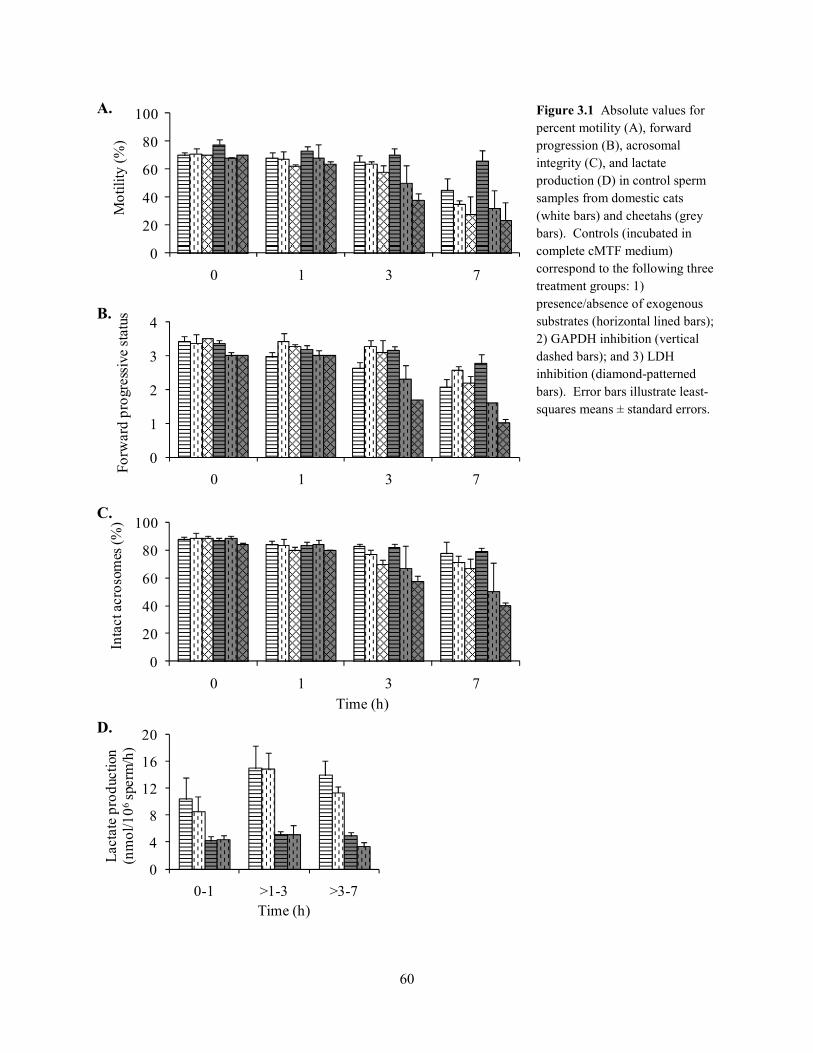
60
0
20
40
60
80
100
0 1 3 7
Inta
ct a
cros
omes
(%)
Time (h)
0
4
8
12
16
20
0-1 >1-3 >3-7
Lac
tate
pro
duct
ion
(nm
ol/1
06sp
erm
/h)
Time (h)
0
20
40
60
80
100
0 1 3 7
Mot
ility
(%)
0
1
2
3
4
0 1 3 7
Forw
ard
prog
ress
ive
stat
usFigure 3.1 Absolute values for percent motility (A), forward progression (B), acrosomal integrity (C), and lactate production (D) in control sperm samples from domestic cats (white bars) and cheetahs (grey bars). Controls (incubated in complete cMTF medium) correspond to the following three treatment groups: 1) presence/absence of exogenous substrates (horizontal lined bars); 2) GAPDH inhibition (vertical dashed bars); and 3) LDH inhibition (diamond-patterned bars). Error bars illustrate least-squares means ± standard errors.
A.
B.
C.
D.
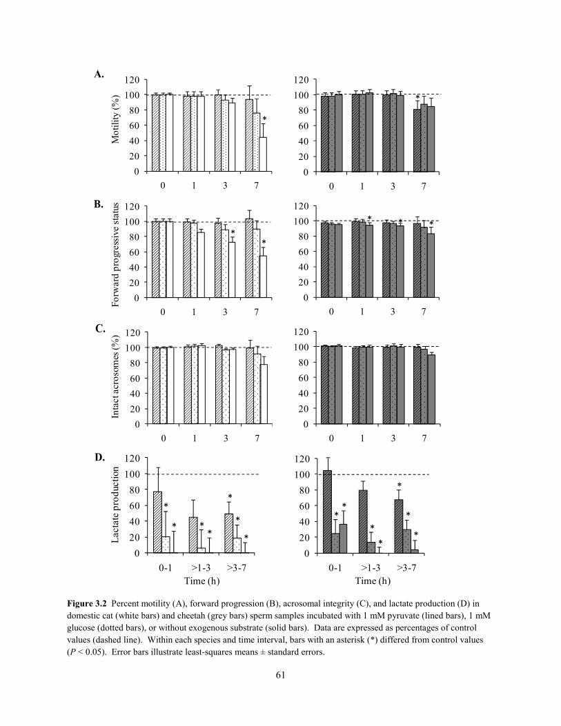
61
0
20
40
60
80
100
120
0 1 3 7
* **
0
20
40
60
80
100
120
0 1 3 7
Mot
ility
(%)
*
0
20
40
60
80
100
120
0 1 3 7
Forw
ard
prog
ress
ive
stat
us
**
0
20
40
60
80
100
120
0 1 3 7
Inta
ct a
cros
omes
(%)
0
20
40
60
80
100
120
0-1 >1-3 >3-7
Lac
tate
pro
duct
ion
Time (h)
**
*
*
**
*
0
20
40
60
80
100
120
0 1 3 7
*
0
20
40
60
80
100
120
0 1 3 7
0
20
40
60
80
100
120
0-1 >1-3 >3-7
**
*
*
*
*
*
Figure 3.2 Percent motility (A), forward progression (B), acrosomal integrity (C), and lactate production (D) in domestic cat (white bars) and cheetah (grey bars) sperm samples incubated with 1 mM pyruvate (lined bars), 1 mM glucose (dotted bars), or without exogenous substrate (solid bars). Data are expressed as percentages of control values (dashed line). Within each species and time interval, bars with an asterisk (*) differed from control values (P < 0.05). Error bars illustrate least-squares means ± standard errors.
C.
D.
A.
B.
0-1 >1-3 >3-7Time (h)

62
3.4.2 GAPDH and LDH Inhibition
The influence of glyceraldehyde-3-phosphate dehydrogenase (GAPDH) inhibition was
consistent between species and resulted in impaired (P < 0.05) sperm function (% M, FPS, and
% IA; Fig. 3.3). Losses in motility (% M and FPS) became more severe (P < 0.05) over time
(Fig. 3.3A&B), whereas acrosomal integrity was not impaired until 7 h (Fig. 3.3C). An unusual
motility pattern that involved rapid flagellar beating, but little forward progression (i.e., vigorous
twitching) often was observed within 1 h of exposure to the GAPDH inhibitor, prior to declines
in % M. Within each species, rates of lactate production were positively correlated (r = 0.30 –
0.44, P < 0.05) to sperm function (% M, FPS and % IA) and declined (P < 0.05) after 1 h of
Figure 3.3 Percent motility (A), forward progression (B), acrosomal integrity (C), and lactate production (D) in domestic cat (white bars) and cheetah (grey bars) sperm samples incubated with 50 mM α-chlorohydrin to inhibit GAPDH. Data are expressed as percentages of control values (dashed line). Within each species and time interval, bars with an asterisk (*) differed from control values (P < 0.05). Error bars illustrate least-squares means ± standard errors.
0
20
40
60
80
100
120
0 1 3 7
Forw
ard
prog
ress
ive
stat
us
* *
**
**
*
0
20
40
60
80
100
120
0 1 3 7
Mot
ility
(%)
Domestic cat Cheetah
*
*
*
*
*
0
20
40
60
80
100
120
0 1 3 7
Inta
ct a
cros
omes
(%)
Time (h)
*
*
020406080
100120140
0-1 >1-3 >3-7
Lac
tate
pro
duct
ion
**
*
*
B.
C.
A.
D.
0 1 3 7Time (h)
0 1 3 7
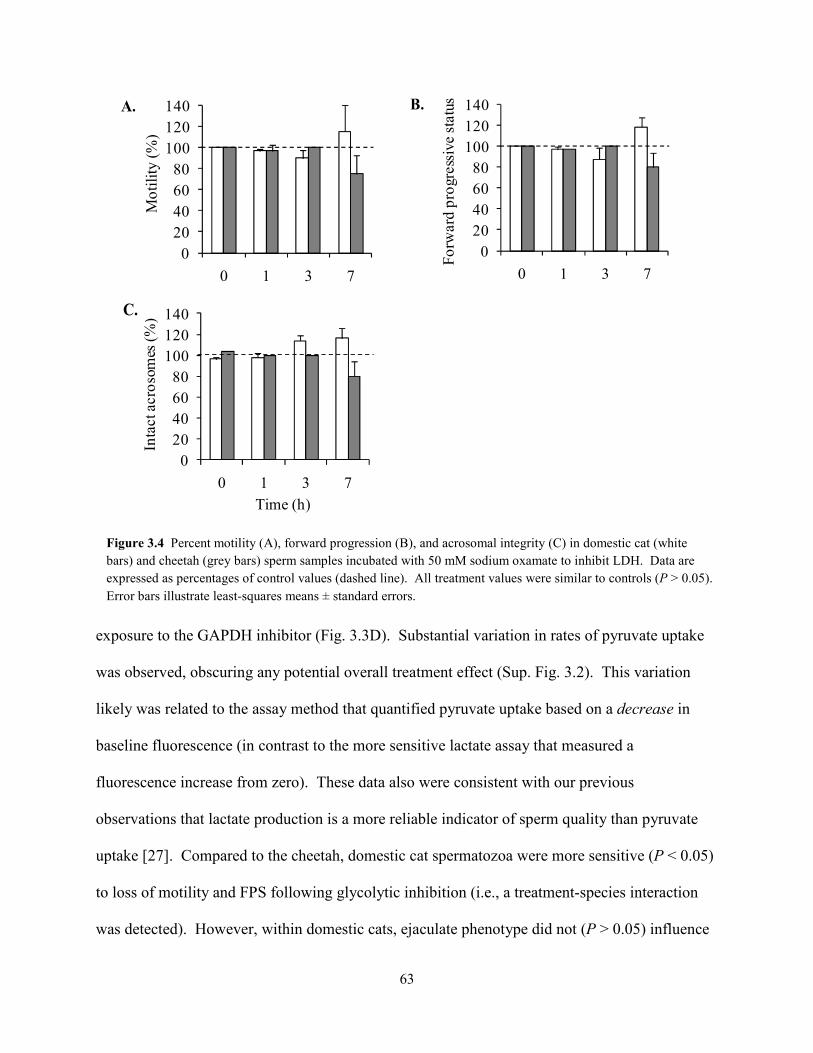
63
exposure to the GAPDH inhibitor (Fig. 3.3D). Substantial variation in rates of pyruvate uptake
was observed, obscuring any potential overall treatment effect (Sup. Fig. 3.2). This variation
likely was related to the assay method that quantified pyruvate uptake based on a decrease in
baseline fluorescence (in contrast to the more sensitive lactate assay that measured a
fluorescence increase from zero). These data also were consistent with our previous
observations that lactate production is a more reliable indicator of sperm quality than pyruvate
uptake [27]. Compared to the cheetah, domestic cat spermatozoa were more sensitive (P < 0.05)
to loss of motility and FPS following glycolytic inhibition (i.e., a treatment-species interaction
was detected). However, within domestic cats, ejaculate phenotype did not (P > 0.05) influence
Figure 3.4 Percent motility (A), forward progression (B), and acrosomal integrity (C) in domestic cat (white bars) and cheetah (grey bars) sperm samples incubated with 50 mM sodium oxamate to inhibit LDH. Data are expressed as percentages of control values (dashed line). All treatment values were similar to controls (P > 0.05). Error bars illustrate least-squares means ± standard errors.
020406080
100120140
0 1 3 7
Inta
ct a
cros
omes
(%)
Time (h)
020406080
100120140
0 1 3 7
Mot
ility
(%)
020406080
100120140
0 1 3 7
Forw
ard
prog
ress
ive
stat
us B.
C.
A.
64
sensitivity to glycolytic inhibition. None of the sperm functional metrics (% M, FPS, or % IA)
was influenced (P > 0.05) by the inhibition of lactate production (LDH) in either species (Fig.
3.4).
3.4.3 Glycogen Content
Glycogen digest was effective across all standards (R2 ≥ 0.945). The concentration of
glycogen in spiked controls was similar (P > 0.05) to the corresponding 1-mM standard.
However, glycogen content of all samples (ejaculated or epididymal; domestic cat or cheetah)
did not differ (P > 0.05) from the 0-mM standard.
3.5 Discussion
Results from our comparative investigation of sperm metabolism in the domestic cat and
cheetah supported our hypothesis that these cells relied primarily on endogenous substrates to
fuel motility and viability, although surprisingly GAPDH activity was essential for these
functions. Because (1) sperm motility was maintained in a medium without glycolyzable
substrates, and (2) there was no evidence of intracellular glycogen stores, we concluded that the
obligate role of the glycolytic pathway was unrelated to sugar metabolism. Furthermore, since
sperm motility was impaired following GAPDH, but not LDH inhibition, we surmised that the
role of this pathway was related to ATP production, rather than NAD+ regeneration. Based on
these collective results we propose a new model of energy production in felid spermatozoa,
whereby ATP is produced primarily by the oxidation of endogenous lipid, and the main function
of the glycolytic pathway is to metabolize lipid-derived glycerol (Fig. 3.5). In this model, fatty
acids hydrolyzed from intracellular lipid (i.e., phospholipid or di-/tri-glycerides) would be
oxidized by the mitochondria to generate ATP, whereas glycerol would enter the glycolytic
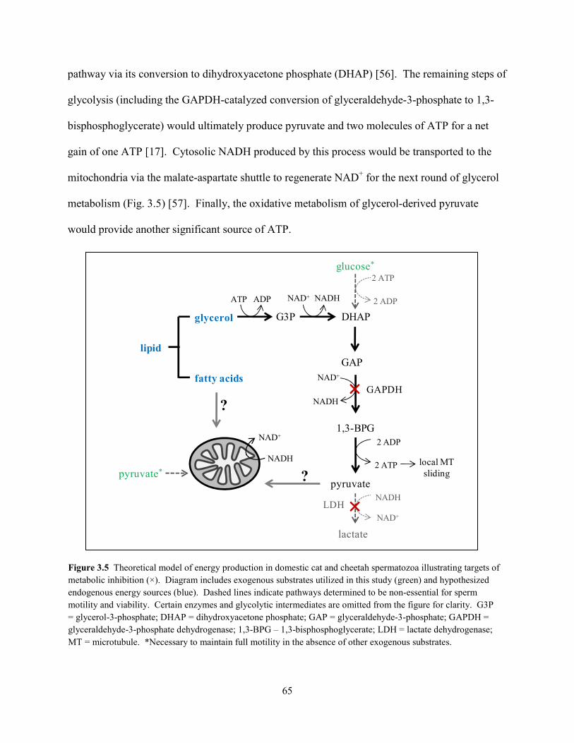
65
pathway via its conversion to dihydroxyacetone phosphate (DHAP) [56]. The remaining steps of
glycolysis (including the GAPDH-catalyzed conversion of glyceraldehyde-3-phosphate to 1,3-
bisphosphoglycerate) would ultimately produce pyruvate and two molecules of ATP for a net
gain of one ATP [17]. Cytosolic NADH produced by this process would be transported to the
mitochondria via the malate-aspartate shuttle to regenerate NAD+ for the next round of glycerol
metabolism (Fig. 3.5) [57]. Finally, the oxidative metabolism of glycerol-derived pyruvate
would provide another significant source of ATP.
Figure 3.5 Theoretical model of energy production in domestic cat and cheetah spermatozoa illustrating targets of metabolic inhibition (×). Diagram includes exogenous substrates utilized in this study (green) and hypothesized endogenous energy sources (blue). Dashed lines indicate pathways determined to be non-essential for sperm motility and viability. Certain enzymes and glycolytic intermediates are omitted from the figure for clarity. G3P = glycerol-3-phosphate; DHAP = dihydroxyacetone phosphate; GAP = glyceraldehyde-3-phosphate; GAPDH = glyceraldehyde-3-phosphate dehydrogenase; 1,3-BPG – 1,3-bisphosphoglycerate; LDH = lactate dehydrogenase; MT = microtubule. *Necessary to maintain full motility in the absence of other exogenous substrates.
glycerol G3P DHAP
GAP
1,3-BPG
pyruvate
lactate
GAPDH
2 ATPglucose*
ATP ADP NAD+ NADH
lipid
fatty acids
2 ADP
NAD+
NADH
2 ADP
2 ATP
NAD+
NADH
NADH
NAD+
local MT sliding
LDH
pyruvate*
?
?
×
×
66
Based on this model, we identified three possible mechanisms by which GAPDH
inhibition could have compromised sperm function. First, although perhaps least likely,
glycolytic inhibition could have caused cytotoxic pathway intermediates to accumulate within
the spermatozoa [52]. We are doubtful of this possibility, as evidence in the ram and boar
suggests that α-chlorohydrin does not generate cytotoxic intermediates in the absence of
exogenous sugars, even when endogenous lipid is actively metabolized [58]. Secondly, the
glycolytic pathway may have been an important source of mitochondrial substrate, analogous to
the mechanism of sperm energy production in the boar [35]. Although actively metabolizing
endogenous lipid, boar spermatozoa do not appear to oxidize fatty acids [59] and instead
generate oxidative substrates from the glycolytic metabolism of glycerol [35]. However, because
exogenous pyruvate was available to GAPDH-inhibited spermatozoa, we believe it is unlikely
that the role of this enzyme is related to oxidative substrate production. Finally, GAPDH could
have maintained active microtubule sliding along the length of the flagellum via local glycerol
metabolism. This pathway is less efficient than oxidative phosphorylation with a net yield of
only one ATP per molecule of glycerol. Nonetheless, glycerol metabolism could support sperm
motility if mitochondrial energy cannot reach the distal flagellum, analogous to the role of
compartmentalized glucose metabolism in other species [11]. Given the abnormal pattern of
sperm motility observed in the domestic cat and cheetah immediately after exposure to the
inhibitor (i.e., vigorous twitching), we predict that the importance of GAPDH activity was
related to the need for localized ATP production along the flagellum.
The importance of lipid metabolism in our model is supported by other studies that
indicate that this substrate is endogenously present in the form of phospholipid or di/tri-
glycerides and is a significant source of ATP in bull [34], ram [60], boar [35], mouse [33], and
67
rhesus macaque [61] spermatozoa. Lipid can provide up to six times the energy of an equal
weight of glycogen [56] and, therefore, is an ideal energy source for spermatozoa that have an
inherently high ATP demand and tightly-packed, economical structure. Yet this endogenous
energy supply appears to be limited in felid spermatozoa, given our observation of a gradual loss
in progressive motility in substrate-free medium. In contrast, spermatozoa maintained motility
with glucose or pyruvate as the sole exogenous substrate, thereby implying a capacity of these
cells to alter their metabolic strategy in response to a changing microenvironment. Such an
adaptive mechanism of energy production would improve chances for in vivo longevity and
successful fertilization by allowing spermatozoa to metabolize substrates within the female tract
while utilizing an endogenous energy supply when needed.
Because the domestic cat routinely produces higher quality ejaculates compared to the
cheetah [27, 38], we were surprised to discover that spermatozoa from the former were more
susceptible to motility declines in the absence of exogenous substrates or when GAPDH was
inhibited. This unexpected finding could indicate that ATP demand is unusually low in cheetah
spermatozoa, and may be related to earlier observations that multiple energy-driven processes
(e.g., motility and cellular viability [27, 39]) consistently are impaired in ejaculates from this
species. One of the strengths of our study design was that sperm metabolic function could be
comparatively assessed not only between species, but also in domestic cat ejaculates containing a
high versus low proportion of structurally malformed spermatozoa. This remarkable phenotypic
difference in the cat has had a significant influence in earlier studies on sperm motility [27, 31],
chilling sensitivity [62], osmotic stress [46], protein tyrosine phosphorylation [29], capacitation
[28], acrosomal function [28], zona penetration [31], and fertilizing ability [31]. Yet, the
incidence of teratospermia among domestic cats in the present study had no influence on sperm

68
sensitivity to glycolytic inhibition or substrate availability. In contrast, we previously found that
rates of sperm pyruvate uptake and lactate production were greatly decreased in teratospermic
cats and cheetahs compared to normospermic cats [27]. Given the proposed model in Figure 3.5,
we predict that disruptions in sperm physiology associated with teratospermia (including
substrate uptake/production) are driven primarily by reduced capacity for oxidative energy
production. This is the focus of a current investigation in our laboratory.
In conclusion, our findings emphasized the value of studies that compare biological
phenomena in non-traditional species (the cheetah) to more commonly used models (the
domestic cat) [63]. This approach revealed conserved mechanisms of glycolytic metabolism and
substrate utilization in domestic cat and cheetah spermatozoa. The consistency of our findings
was noteworthy, given the significant genetic and phenotypic differences between these two felid
species, and provided strong support for our proposed model of sperm energy production. Yet
while domestic cat and cheetah spermatozoa shared a requirement for GAPDH activity,
differences were observed regarding the severity of sperm functional declines following
exposure to the GAPDH inhibitor. These differences suggested that sperm energy demand may
be greater in the domestic cat compared to the teratospermic cheetah. If confirmed, this new
finding could be mechanistically linked to previous observations that multiple energy-driven
processes are disrupted in felid ejaculates with the teratospermic phenotype [28, 29, 31, 43].
Understanding the fundamental cause of these physiological disruptions could provide clues to
developing methods to enhance sperm fertilizing ability, for example, through exposure to
metabolic stimulants [64]. As high (>40%) proportions of pleiomorphic spermatozoa have been
documented in at least 18 of the 21 wild felid species studied [41, 65] and are nearly ubiquitous
69
in man [36], cross-species comparisons that provide insight into teratospermia can help improve
the success of assisted reproduction for biodiversity preservation and human fertility.
3.6 References
1. Mukai C, Okuno M. Glycolysis plays a major role for adenosine triphosphate
supplementation in mouse sperm flagellar movement. Biol Reprod 2004; 71: 540-547.
2. Turner R. Tales from the tail: what do we really know about sperm motility? J Androl
2003; 24: 790-803.
3. Miki K, Qu W, Goulding EH, Willis WD, Bunch DO, Strader LF, Perreault SD, Eddy
EM, O'Brien DA. Glyceraldehyde 3-phosphate dehydrogenase-S, a sperm-specific
glycolytic enzyme, is required for sperm motility and male fertility. Proc Natl Acad Sci
USA 2004; 101: 16501-16506.
4. Ford WCL, Rees JM. The bioenergetics of mammalian sperm motility. In: Gagnon C
(ed.) Controls of Sperm Motility: Biological and Clinical Aspects. Boca Raton, FL: CRC
Press; 1990: 175-202.
5. Williams AC, Ford WC. The role of glucose in supporting motility and capacitation in
human spermatozoa. J Androl 2001; 22: 680-695.
6. Albarracín JL, Fernández-Novell JP, Ballester J, Rauch MC, Quintero-Moreno A, Peña
A, Mogas T, Rigau T, Yañez A, Guinovart JJ, Slebe JC, Concha II, et al.
Gluconeogenesis-linked glycogen metabolism is important in the achievement of in vitro
capacitation of dog spermatozoa in a medium without glucose. Biol Reprod 2004; 71:
1437-1445.
70
7. Leese HJ, Astley NR, Lambert D. Glucose and fructose utilization by rat spermatozoa
within the uterine lumen. J Reprod Fertil 1981; 61: 435-437.
8. Lardy HA, Phillips PH. Inhibition of sperm respiration and reversibility of the effects of
metabolic inhibitors. J Biol Chem 1943; 148: 333-341.
9. Storey B. Mammalian sperm metabolism: oxygen and sugar, friend and foe. Int J Dev
Biol 2008; 52: 427-437.
10. Lardy HA, Winchester B, Phillips PH. The respiratory metabolism of ram spermatozoa.
Arch Biochem 1945; 6: 33-40.
11. Travis AJ, Jorgez CJ, Merdiushev T, Jones BH, Dess DM, Diaz-Cueto L, Storey BT,
Kopf GS, Moss SB. Functional relationships between capacitation-dependent cell
signaling and compartmentalized metabolic pathways in murine spermatozoa. J Biol
Chem 2001; 276: 7630-7636.
12. Macleod J. The effect of glycolysis inhibitors and of certain substrates on the metabolism
and motility of human spermatozoa. Endocrinology 1941; 29: 583-591.
13. Hung P, Miller MG, Meyers SA, VandeVoort CA. Sperm mitochondrial integrity is not
required for hyperactivated motility, zona binding, or acrosome reaction in the rhesus
macaque. Biol Reprod 2008; 79: 367-375.
14. MacLeod J. The metabolism of human spermatozoa. Am J Physiol 1941; 132: 0193-
0201.
15. Rigau T, Farré M, Ballester J, Mogas T, Peña A, Rodríguez-Gil JE. Effects of glucose
and fructose on motility patterns of dog spermatozoa from fresh ejaculates.
Theriogenology 2001; 56: 801-815.
71
16. Rigau T, Rivera M, Palomo MJ, Fernández-Novell JM, Mogas T, Ballester J, Peña A,
Otaegui PJ, Guinovart JJ, Rodríguez-Gil JE. Differential effects of glucose and fructose
on hexose metabolism in dog spermatozoa. Reproduction 2002; 123: 579-591.
17. Voet D, Voet JG. Glycolysis. In: Harris D, Fitzgerald P (eds.), Biochemistry, 3rd ed.
Hoboken, NJ: John Wiley & Sons, Inc.; 2004: 581-624.
18. Westhoff D, Kamp G. Glyceraldehyde 3-phosphate dehydrogenase is bound to the
fibrous sheath of mammalian spermatozoa. J Cell Sci 1997; 110: 1821-1829.
19. Travis AJ, Foster JA, Rosenbaum NA, Visconti PE, Gerton GL, Kopf GS, Moss SB.
Targeting of a germ cell-specific type 1 hexokinase lacking a porin-binding domain to the
mitochondria as well as to the head and fibrous sheath of murine spermatozoa. Mol Cell
Biol 1998; 9: 263-276.
20. Storey B, Kayne FJ. Energy metabolism of spermatozoa. V The Embden-Meyerhof
pathway of glycolysis: activities of the pathway enzymes in hypertonically treated rabbit
epididymal spermatozoa. Fertil Steril 1975; 26: 1257-1265.
21. Bradley M, Geelan A, Leitch V, Goldberg E. Cloning sequencing and characterization of
LDH-C-4 from a fox testis cDNA library. Mol Reprod Dev 1996; 44: 452-459.
22. Fraser LR, Herod JE. Expression of capacitation-dependent changes in chlorotetracycline
fluorescence patterns in mouse spermatozoa requires a suitable glycolysable substrate. J
Reprod Fertil 1990; 88: 611-621.
23. Hoppe PC. Glucose requirement for mouse sperm capacitation in vitro. Biol Reprod
1976; 15: 39-45.
24. Cooper T. The onset and maintenance of hyperactivated motility of spermatozoa from the
mouse. Gamete Res 1984; 9: 55-74.
72
25. Fraser L, Quinn P. A glycolytic product is obligatory for initiation of the sperm acrosome
reaction and whiplash motility required for fertilization in the mouse J Reprod Fertil
1981; 61: 25-35.
26. Travis AJ, Tutuncu L, Jorgez C, Ord T, Jones B, Kopf GS, Williams C. Requirements for
glucose beyond sperm capacitation during in vitro fertilization in the mouse. Biol Reprod
2004; 71: 139-145.
27. Terrell KA, Wildt DE, Anthony NM, Bavister BD, Leibo SP, Penfold LM, Marker LL,
Crosier AE. Evidence for compromised metabolic function and limited glucose uptake in
spermatozoa from the teratospermic domestic cat (Felis catus) and cheetah (Acinonyx
jubatus). Biol Reprod 2010; 83: 833-841.
28. Long JA, Wildt DE, Wolfe BA, Crister JK, DeRossi RV, Howard JG. Sperm capacitation
and the acrosome reaction are compromised in teratospermic domestic cats. Biol Reprod
1996; 54: 638-646.
29. Pukazhenthi BS, Wildt DE, Ottinger MA, Howard JG. Compromised sperm protein
phosphorylation after capacitation, swim-up, and zona pellucida exposure in
teratospermic domestic cats. J Androl 1996; 17: 409-419.
30. Pukazhenthi BS, Long JA, Wildt DE, Ottinger MA, Armstrong DL, Howard JG.
Regulation of sperm function by protein tyrosine phosphorylation in diverse wild felid
species. J Androl 1998; 19: 675-685.
31. Howard JG, Donoghue AM, Johnston LA, Wildt DE. Zona pellucida filtration of
structurally abnormal spermatozoa and reduced fertilization in teratospermic cats. Biol
Reprod 1993; 49: 131-139.
73
32. Ballester J, Fernández-Novell JP, Rutllant J, García-Rocha M, Palomo MJ, Mogas T,
Peña A, Rigau T, Guinovart JJ, Rodríguez-Gil JE. Evidence for a functional glycogen
metabolism in mature mammalian spermatozoa. Mol Reprod Dev 2000; 56: 207-219.
33. Carey JE, Oldsclarke P, Storey BT. Oxidative metabolism of spermatozoa from inbred
and random bred mice. J Exp Zool 1981; 216: 285-292.
34. Lardy HA, Phillips PH. Phospholipids as a source of energy for motility of bull
spermatozoa. Am J Physiol 1941; 134: 542-548.
35. Jones AR, Milmlow D. Endogenous energy production by mature boar spermatozoa. J
Reprod Fertil 1997; 111: 285-290.
36. Cooper TG, Noonan E, von Eckardstein S, Auger J, Gordon Baker HW, H.M. B, Haugen
TB, Kruger T, Wang C, Mbizvo MT, Vogelsong KM. World Health Organization
reference values for human semen characteristics. Hum Reprod Update 2010; 16: 231-
245.
37. Pukazhenthi BS, Wildt DE, Howard JG. The phenomenon and significance of
teratospermia in felids. J Reprod Fertil Suppl 2001; 57: 423-433.
38. Wildt DE, Bush M, Howard JG, O'Brien SJ, Meltzer D, van Dyk A, Ebedes H, Brand DJ.
Unique seminal quality in the South African cheetah and a comparative evaluation in the
domestic cat. Biol Reprod 1983; 29: 1019-1025.
39. Crosier AE, Marker L, Howard JG, Pukazhenthi BS, Henghali JN, Wildt DE. Ejaculate
traits in the Namibian cheetah (Acinonyx jubatus): influence of age, season, and captivity.
Reprod Fertil Dev 2007; 19: 370-382.
74
40. Howard JG, Brown JL, Bush M, Wildt DE. Teratospermic and normospermic domestic
cats: ejaculate traits, pituitary-gonadal hormones, and improvement of spermatozoal
motility and morphology after swim-up processing. J Androl 1990; 11: 204-215.
41. Pukazhenthi BS, Neubauer K, Jewgenow K, Howard JG, Wildt DE. The impact and
potential etiology of teratospermia in the domestic cat and its wild relatives.
Theriogenology 2006; 66: 112-121.
42. Crosier AE, Pukazhenthi BS, Henghali JN, Howard JG, Dickman AJ, Marker L, Wildt
DE. Cryopreservation of spermatozoa from wild-born Namibian cheetahs (Acinonyx
jubatus) and influence of glycerol on cryosurvival. Cryobiology 2006; 52: 169-181.
43. Penfold LM, Jost L, Evenson DP, Wildt DE. Normospermic versus teratospermic
domestic cat sperm chromatin integrity evaluated by flow cytometry and intracytoplasmic
injection. Biol Reprod 2003; 69: 1730-1735.
44. Crosier A, Henghali J, Howard JG, Pukazhenthi BS, Terrell KA, Marker LL, Wildt DE.
Improved quality of cryopreserved cheetah (Acinonyx jubatus) spermatozoa after
centrifugation through Accudenz. J Androl 2009; 30: 298-308.
45. Howard JG. Semen collection and analysis in carnivores. In: Fowler ME (ed.) Zoo and
Wild Animal Medicine: Current Therapy III. Philadelphia: WB Saunders Co.; 1993: 390-
399.
46. Pukazhenthi BS, Noiles E, Pelican K, Donoghue AM, Wildt DE, Howard JG. Osmotic
effects on feline spermatozoa from normospermic versus teratospermic donors.
Cryobiology 2000; 40: 139-150.
75
47. Gardner DK, Leese HJ. Concentrations of nutrients in mouse oviduct fluid and their
effects on embryo development and metabolism in vitro. J Reprod Fertil 1990; 88: 361-
368.
48. Bavister BD. Substitution of a synthetic polymer for protein in a mammalian gamete
culture system. J Exp Zool 1981; 217: 45-51.
49. Shah D, Naciri M, Clee P, Al-Rubeai M. NucleoCounter: an efficient technique for the
determination of cell number and viability in animal cell culture processes.
Cytotechnology 2006; 51: 39-44.
50. Ford S, Jones AR. The effect of alpha-chlorohydrin on the oxidation of fructose by rabbit
spermatozoa in vitro. Contraception 1983; 28: 565-573.
51. Ford WCL. Glycolysis and sperm motility: does a spoonful of sugar help the flagellum
go round? Hum Reprod Update 2006; 12: 269-274.
52. Ford W, Harrison A. The concerted effect of alpha-chlorohydrin and glucose on the ATP
concentration in spermatozoa is associated with the accumulation of glycolytic
intermediates. J Reprod Fertil 1986; 77: 537-545.
53. Larson JL, Miller DJ. Simple histochemical stain for acrosomes on sperm from several
species. Mol Reprod Dev 1999; 52: 445-449.
54. Passonneau JV, Lowry OH. Enzymatic Analysis: A Practical Guide. Totowa, NJ:
Humana Press; 1993.
55. SAS. SAS/STAT User's Guide: Statistics. Version 9.1. Cary, NC: Statistical Analysis
System Institute, Inc; 2002.
56. Voet D, Voet JG. Lipid metabolism. In: Harris D, Fitzgerald P (eds.), Biochemistry, 3rd
ed. Hoboken, NJ: John Wiley & Sons, Inc.; 2004: 909-984.
76
57. Voet D, Voet JG. Electron transport and oxidative phosphorylation. In: Harris D,
Fitzgerald P (eds.), Biochemistry, 3rd ed. Hoboken, NJ: John Wiley & Sons, Inc.; 2004:
797-842.
58. Ford W, Harrison A. The presence of glucose increases the lethal effect of a-chlorohydrin
on ram and boar spermatozoa in vitro. J Reprod Fertil 1985; 73: 197-206.
59. Jones AR, Chantrill LA. Oxidative metabolic activity of boar spermatozoa: a system for
assessing anti-glycolytic activity of potential inhibitors in vitro. Reprod Fert Dev 1989; 1:
357-367.
60. Tash J, Mann T. Adenosine 3',5'-cyclic monophosphate in relation to motility and
senescence of spermatozoa. Proc R Soc Lond B Biol Sci 1973; 184: 109-114.
61. Hoskins D, Patterson D. Metabolism of rhesus monkey spermatozoa. J Reprod Fertil
1968; 16: 183-195.
62. Pukazhenthi B, Pelican K, Wildt D, Howard JG. Sensitivity of domestic cat (Felis catus)
sperm from normospermic versus teratospermic donors to cold-induced acrosome
damage. Biol Reprod 1999; 61: 135-141.
63. Wildt DE, Comizzoli P, Pukazhenthi B, Songsasen N. Lessons from biodiversity-the
value of nontraditional species to advance reproductive science, conservation, and human
health. Mol Reprod Dev 2010; 77: 397-409.
64. Stachecki JJ, Ginsburg KA, Armant DR. Stimulation of cryopreserved epididymal
spermatozoa of the domestic cat using the motility stimulants caffeine, pentoxifylline,
and 2'-deoxyadenosine. J Androl 1994; 15: 157-164.
65. Herrick JR, Campbell M, Levens G, Moore T, Benson K, D'Agostino J, West G, Okeson
DM, Coke R, Portacio SC, Leiske K, Kreider C, et al. In vitro fertilization and sperm
77
cryopreservation in the black-footed cat (Felis nigripes) and sand cat (Felis margarita).
Biol Reprod 2010; 82: 552-562.
78
CHAPTER 4: OXIDATIVE METABOLISM IS ESSENTIAL FOR FELID SPERM FUNCTION, BUT IS SUBSTANTIALLY LOWER IN CHEETAH (ACINONYX JUBATUS) COMPARED TO DOMESTIC CAT (FELIS CATUS) EJACULATE
4.1 Abstract
Compared to the normospermic domestic cat, sperm metabolic function is compromised
in the teratospermic cat and cheetah, but the pathway(s) involved in this deficiency are unknown.
Glycolysis is essential for sperm motility, yet appears to function normally in spermatozoa of
either species regardless of structural morphology. We conducted a comparative study to further
understand the mechanisms of energy production in felid spermatozoa, with the hypothesis that
oxidative metabolism is required for normal sperm function and is impaired in teratospermic
ejaculates. Ejaculates from both species were stained with MitoTracker® to quantify
mitochondrial membrane potential (MMP) or were incubated to assess changes in sperm function
(motility, acrosomal integrity, and lactate production) after mitochondrial inhibition with
myxothiazol. Sperm midpiece dimensions also were quantified. Sperm mitochondrial
fluorescence (directly proportional to MMP) was ~95% lower in the cheetah compared to the
normospermic and teratospermic cat, despite the former having a 10% longer midpiece. In both
species, MMP was increased 5-fold in spermatozoa with retained cytoplasm compared to
structurally normal cells. Inhibition of oxidative phosphorylation impaired sperm function, but a
100-fold higher inhibitor concentration was required in the cat compared to the cheetah.
Collectively, findings revealed that oxidative phosphorylation was required for sperm function in
the domestic cat and cheetah. This pathway of energy production appeared markedly less active
in the cheetah, indicating a species-specific vulnerability to mitochondrial dysfunction. The
unexpected, cross-species link between retained cytoplasmic droplets and elevated MMP may
have reflected increased concentrations of metabolic enzymes or substrates in these structures.
79
4.2 Introduction
Oxidative phosphorylation is an active pathway of sperm energy production in all
mammalian species studied to date, including the bull, ram, boar, rabbit, mouse, and man
(reviewed in [1]). Although human spermatozoa were once considered to be exclusively
glycolytic [1], cellular respiration now is known as a significant source of ATP [2] and is
required to maintain motility in these cells [2-6]. Human infertility and/or asthenospermia are
linked to various indicators of impaired oxidative phosphorylation in spermatozoa, including
reduced midpiece length [7], abnormal mitochondrial organization [7-9], reduced oxygen
consumption [10], and decreased mitochondrial enzyme activity [11]. However, the relationship
between mitochondrial ATP production and fertilizing ability in species other than the human is
less clear. For example, although oxidative phosphorylation supplies the majority of ATP in
bovine spermatozoa [12], bull fertility can be predicted by sperm motility values, but not by
mitochondrial membrane potential [13, 14]. Conversely, although motility and capacitation of
murine spermatozoa are unaffected by chemical inhibition of oxidative phosphorylation [15, 16],
Smcp-/- (sperm mitochondrion-associated cysteine-rich protein) knock-out mice are infertile and
experience reduced sperm motility and zona penetration ability [17].
Little is known about the mechanisms of sperm energy production in the domestic cat or
its wild relatives. The cat itself is a valuable model for understanding gamete physiology,
because there are two readily available phenotypes, normospermic versus teratospermic
(ejaculating > 60% structurally abnormal spermatozoa) [18, 19]. Because teratospermia is highly
prevalent in men [20] and considered ubiquitous in certain rare felid species (most notably the
cheetah, clouded leopard [Neofelis nebulosa], and Florida panther [Puma concolor coryi]) [19],
understanding the physiological consequences of this condition has application to both human
80
fertility and wildlife management/conservation. We recently determined that rates of sperm
lactate production are reduced in the teratospermic domestic cat and cheetah compared to the
normospermic domestic cat [21]. In both species, lactate production is positively correlated to
sperm motility, acrosomal integrity, and normal morphology, yet appears to be indirectly related
to cellular energy production. Glucose uptake by these cells is minimal or absent [21], and the
majority of lactate is instead produced by the reduction of exogenous pyruvate [22]. This
process does not appear to serve any physiological purpose, as sperm motility and acrosomal
integrity are unaffected when lactate dehydrogenase is chemically inhibited or when pyruvate is
absent from the culture medium [22]. Although having a questionable role in felid sperm
fertilizing ability [22], lactate production may indirectly indicate NADH availability that, in turn,
is important in maintaining REDOX potential [23] and in ATP generation via oxidative
phosphorylation [24]. If this prediction is correct, reduced rates of lactate production may reflect
mitochondrial dysfunction in teratospermic domestic cat and cheetah ejaculates. Alternately, the
physiological mechanisms causing impaired lactate production may differ between the two
species. Regardless, it is likely that a mitochondrial deficiency would significantly impair
energy generation by the felid spermatozoon, particularly since our previous investigations
suggest a key role of oxidative phosphorylation in these cells [21, 22]. Although cat and cheetah
spermatozoa metabolize little or no exogenous glucose, we have discovered that the glycolytic
enzyme glyceraldehyde-3-phosphate dehydrogenase (GAPDH) is required to maintain sperm
progressive motility in both species [22]. We predict that the importance of GAPDH in felid
spermatozoa relates to its role in glycerol breakdown, and that these cells rely primarily on the
metabolism of endogenous phospholipid to support motility and viability. Such a mechanism
would explain why felid spermatozoa remain motile for at least 7 h in a substrate-free medium
81
[22], and would suggest that mitochondrial metabolism (of fatty acids and/or glycerol-derived
pyruvate) is a substantial source of cellular ATP.
To better understand the mechanisms of cellular metabolism in felid spermatozoa, we
conducted a comparative assessment of sperm oxidative phosphorylation in the normospermic
versus teratospermic domestic cat and the teratospermic cheetah. We tested the hypotheses that
oxidative phosphorylation is required for sperm motility and viability in the cat and cheetah, and
that mitochondrial function is impaired in the teratospermic phenotype. We anticipated that
knowledge of these mechanisms could yield insight into the disruptions of various energy-
dependent processes usually associated with teratospermia in felids, including sperm motility
[21], protein tyrosine phosphorylation [25], and the acrosome reaction [26]. Elucidation of these
functional defects could provide clues for improving the success of reproductive technologies
(e.g., sperm cryopreservation) useful to improving both human fertility and wildlife
management/conservation.
4.3 Materials and Methods
Animals
Electroejaculates were collected from adult (1.5 – 8 yr old) domestic cats previously
determined to be normospermic (n = 2 males, n = 6 total ejaculates) or teratospermic (n = 5
males, n = 8 total ejaculates) [21, 22]. Management protocols for this species at the Smithsonian
Conservation Biology Institute (SCBI; Front Royal, VA) have been described in detail [21].
Electroejaculates (1 per male, 16 males) were collected from adult cheetahs (ages, 2 – 10
yr) housed at the Cheetah Conservation Fund (CCF; Otjiwarongo, Namibia; n = 1), White Oak
Conservation Center (WOCC; Yulee, FL; n = 4), the SCBI (n = 5), the Philadelphia Zoo (PHL;
82
Philadelphia, PA; n = 3), The Wilds (TW; Cumberland, OH; n = 2), and the Cleveland
Metroparks Zoo (CLE; Cleveland, OH; n = 1). The male at CCF was wild-born and housed
singly, as described previously [27]. Males at WOCC, SCBI, and PHL were captive-born and
managed as explained earlier [26], and represented one (PHL) or two (WOCC, SCBI) sibling
groups at each institution. Both males at TW were captive-born, one of which was maintained
singly in a 5,000-m2 outdoor enclosure and the other with two unrelated males in a 500-m2
outdoor enclosure. The male at CLE was captive-born and managed on exhibit with a male
sibling in a 900-m2 outdoor enclosure. Males at TW and CLE were fed a commercially
produced diet (Premium Beef Feline Diet; Central Nebraska Packaging Inc., North Platte, NE),
and diets for this species in all other institutions have been reported previously [21, 22]. There
was no statistical influence of location on any sperm quality metric (P ≥ 0.27), and we previously
have determined that ejaculate quality is similar between wild-born Namibian cheetahs and
captive individuals at North American institutions [28, 29]. Reagents and semen collection
equipment were transported from SCBI to each study site, and all samples were shipped back to
our laboratory for analyses of metabolic rates, mitochondrial membrane potential, sperm
morphology, and acrosomal integrity.
Semen Collection
A surgical plane of anesthesia was induced in domestic cats and cheetahs for semen
collection according to protocols developed by institutional veterinarians and were the same as
we have described previously [26, 27]. All animal procedures were approved by National
Zoological Park’s Animal Care and Use Committee (ACUC) and similar committees of
collaborating institutions. Semen was collected using a rectal probe of 1 cm (domestic cat) or
83
1.9 cm (cheetah) in diameter and an electrostimulator (P.T. Electronics, Boring, OR) as
described earlier [21, 30].
Sperm Processing
Immediately after collection, a 10 µl aliquot of raw semen containing ~2 × 10⁵
spermatozoa was fixed in 0.3% glutaraldehyde in phosphate-buffered saline for assessment of
sperm morphology [21, 30, 31]. Each ejaculate then was diluted with an equal volume of a
chemically defined, protein-free, modified mouse tubal fluid medium (cMTF) [32] supplemented
with 2% polyvinyl alcohol (PVA) [33]. The cMTF medium was prepared as described earlier
[21] and contained 98.4 mM NaCl, 4.78 mM KCl, 1.19 mM MgSO4, 1.19 mM KH2PO4, 25 mM
NaHCO3, 1.71 mM CaCl2, 1 mM glucose, 1 mM Na-pyruvate, 25 mM 3-(N-morpholino)
propanesulfonic acid (MOPS) buffer, and 0.02 mg/mL phenol red. All reagents were purchased
from Sigma Aldrich (St. Louis, MO) unless otherwise noted. Osmolality of the final working
medium (315 – 345 mOsm) was determined using a vapor pressure osmometer (Wescor, Inc,
Logan, UT) and was within 10% of the physiological value of domestic cat semen (323 mOsm
[34]). Sperm concentration was determined using a Nucleocounter SP-100 (Chemometec,
Allerød, Denmark) [35].
Inhibition of Oxidative Phosphorylation
Each diluted ejaculate (maintained at ambient temperature, 19–22oC) was washed by
centrifugation (8 min; × 300 g for domestic cat; × 100 g for cheetah) and resuspended in cMTF.
To determine the influence of inhibition of oxidative phosphorylation, a sperm sample (3 × 10⁶
motile sperm/ml) from each ejaculate (n = 6 normospermic cat, n = 6 teratospermic cat, n = 8
84
cheetah) was exposed to 160 nM myxothiazol (known to block transfer of electrons from
complex ΙΙΙ to cytochrome C [5]; included in the resuspension medium) and incubated in parallel
with negative controls. This myxothiazol concentration has been used previously to study
mammalian sperm metabolism [12] and was the minimum required to disrupt cheetah sperm
motility in dose-dependent trials (0.16 nM, 16 nM, and 160 nM; data not shown). Because
domestic cat spermatozoa were not influenced by the 160 nM myxothiazol treatment, a subset of
these ejaculates (n = 2 normospermic, n = 2 teratospermic) also were incubated in higher
inhibitor concentrations (1.6 µM and 16 µM).
Sperm samples were cultured (37oC) in microcentrifuge tubes under oil to prevent
evaporation, as described previously [21]. Assessments of sperm percentage motility (% M),
forward progression (FPS), and acrosomal integrity (% IA) were made at 0, 1, 3, and 7 h of
incubation. Motility was assessed visually (200 ×), and FPS was rated on a 0 to 5 scale, with a
rating of 5 equivalent to most rapid, linear progress [30]. Spermatozoa (~2 × 10⁵ cells) were
fixed in 4% paraformaldehyde and stained with Coomassie Blue G-250 (Fisher Biotech,
Springfield, NJ) to evaluate acrosomal integrity using previously described methods [36, 37].
Spermatozoa with an intact acrosome exhibited a uniform blue staining pattern overlying the
acrosomal region, whereas nonintact cells had clear or patchy staining over this region [37].
Because we discovered in an earlier study that there was a positive correlation between lactate
production and sperm motility, acrosomal integrity, and normal morphology in both the cat and
cheetah [19], we monitored rates of lactate production (∆ L) in each sample during the 7 h
incubation period [21]. To determine lactate concentration, medium samples were centrifuged (8
min; × 1,000 g) through a CoStar Spin-X 0.22-µm nylon filter tube (Corning Incorporated,
Corning, NY) and stored at –80oC until analysis using a LDH/GPT-linked fluorescence assay
85
[21, 32, 38]. Assay enzymes were purchased from Roche Applied Science (Indianapolis, IN)
and fluorescence analyzed using a Spectra Max Gemini XPS fluorescent plate reader (340 nm
excitation, 445 nm emission) and SoftMax Pro 5 software (Molecular Devices, Sunnyvale, CA).
The ∆ L was calculated as the change in medium substrate concentration over time, divided by
sperm concentration and reported in nmol/106 sperm/h. All data were normalized to control
values for presentation.
Mitochondrial Membrane Potential
To assess mitochondrial membrane potential (MMP), samples of ejaculates (n = 5
domestic cat, n = 7 cheetah) were incubated (45 min; ambient temperature, 19–22oC) in the dark
with 0.5 nM MitoTracker® Red CMXRos (Molecular Probes, Inc., Eugene, OR) in a volume of
100 µl at a concentration of 3 × 10⁶ motile sperm/ml. To assess MMP, 100
spermatozoa/ejaculate were individually analyzed (400 ×) using a BX40 fluorescence
microscope (Olympus America Inc., Center Valley, PA; 555 nm excitation), and fluorescence
was quantified using a Sensicam qe high performance camera (Cooke Corp., Romulus, MI) and
IP Lab v4.04 software (BD Biosciences, Rockville, MD). The presence of sperm structural
abnormalities was recorded (as described previously [21]), except for acrosomal deformities,
which were not reliably detected at this magnification. Spermatids and retained cytoplasmic
droplets represented the majority (~80%) of sperm malformations in each species. For data
analysis, each spermatozoon was classified into one of the following five major morphotypes:
structurally normal, midpiece droplet, flagellar droplet, spermatid, and ‘other’ [21]. The
category of ‘midpiece droplet’ primarily represented a spermatozoon with a bent midpiece
encircling residual cytoplasm, but also included spermatozoa with a proximal droplet attached to
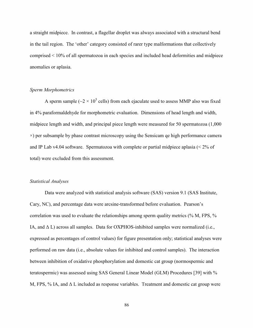
86
a straight midpiece. In contrast, a flagellar droplet was always associated with a structural bend
in the tail region. The ‘other’ category consisted of rarer type malformations that collectively
comprised < 10% of all spermatozoa in each species and included head deformities and midpiece
anomalies or aplasia.
Sperm Morphometrics
A sperm sample (~2 × 10⁵ cells) from each ejaculate used to assess MMP also was fixed
in 4% paraformaldehyde for morphometric evaluation. Dimensions of head length and width,
midpiece length and width, and principal piece length were measured for 50 spermatozoa (1,000
×) per subsample by phase contrast microscopy using the Sensicam qe high performance camera
and IP Lab v4.04 software. Spermatozoa with complete or partial midpiece aplasia (< 2% of
total) were excluded from this assessment.
Statistical Analyses
Data were analyzed with statistical analysis software (SAS) version 9.1 (SAS Institute,
Cary, NC), and percentage data were arcsine-transformed before evaluation. Pearson’s
correlation was used to evaluate the relationships among sperm quality metrics (% M, FPS, %
IA, and ∆ L) across all samples. Data for OXPHOS-inhibited samples were normalized (i.e.,
expressed as percentages of control values) for figure presentation only; statistical analyses were
performed on raw data (i.e., absolute values for inhibited and control samples). The interaction
between inhibition of oxidative phosphorylation and domestic cat group (normospermic and
teratospermic) was assessed using SAS General Linear Model (GLM) Procedures [39] with %
M, FPS, % IA, and ∆ L included as response variables. Treatment and domestic cat group were
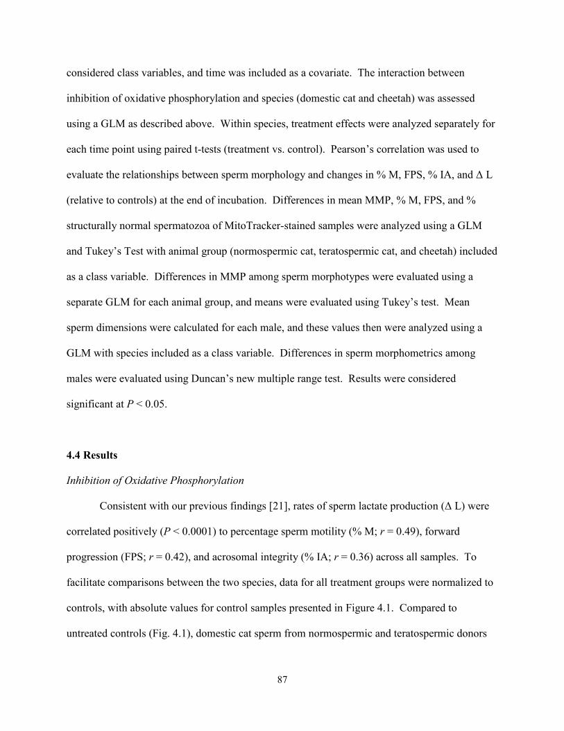
87
considered class variables, and time was included as a covariate. The interaction between
inhibition of oxidative phosphorylation and species (domestic cat and cheetah) was assessed
using a GLM as described above. Within species, treatment effects were analyzed separately for
each time point using paired t-tests (treatment vs. control). Pearson’s correlation was used to
evaluate the relationships between sperm morphology and changes in % M, FPS, % IA, and ∆ L
(relative to controls) at the end of incubation. Differences in mean MMP, % M, FPS, and %
structurally normal spermatozoa of MitoTracker-stained samples were analyzed using a GLM
and Tukey’s Test with animal group (normospermic cat, teratospermic cat, and cheetah) included
as a class variable. Differences in MMP among sperm morphotypes were evaluated using a
separate GLM for each animal group, and means were evaluated using Tukey’s test. Mean
sperm dimensions were calculated for each male, and these values then were analyzed using a
GLM with species included as a class variable. Differences in sperm morphometrics among
males were evaluated using Duncan’s new multiple range test. Results were considered
significant at P < 0.05.
4.4 Results
Inhibition of Oxidative Phosphorylation
Consistent with our previous findings [21], rates of sperm lactate production (∆ L) were
correlated positively (P < 0.0001) to percentage sperm motility (% M; r = 0.49), forward
progression (FPS; r = 0.42), and acrosomal integrity (% IA; r = 0.36) across all samples. To
facilitate comparisons between the two species, data for all treatment groups were normalized to
controls, with absolute values for control samples presented in Figure 4.1. Compared to
untreated controls (Fig. 4.1), domestic cat sperm from normospermic and teratospermic donors
88
fffffffffffffffffffffffffffffffffffffffffffffffffffffffffffffffffffffffffffffffffffffffffffffffffffffffffffffffffffff
ffffff
were unaffected (P ≥ 0.05) for sperm %M, % IA, and ∆ L when exposed to 0.16 µM
myxothiazol (Fig. 4.2A, C-D); there was a modest, but significant (~20%; P < 0.05) loss in FPS
(Fig. 4.2B). By contrast, there was an immediate and marked decline (P < 0.05) in cheetah
sperm % M and FPS, with a ~50% reduction (P < 0.05) in % IA and ∆ L after 7 h of incubation
(Fig. 4.2A-D). This species-treatment interaction was highly significant (P < 0.0001) for % M,
FPS, and % IA, but represented a trend (P = 0.08) for ∆ L. There was no interaction (P ≥ 0.05)
between domestic cat sperm phenotype (normospermic versus teratospermic) and myxothiazol
treatment.
Figure 4.1 Absolute values for percent motility (A), forward progression (B), acrosomal integrity (C), and lactate production (D) in normospermic cat (white bar; n = 6), teratospermic cat (lined bar; n = 6), and cheetah (gray bar; n = 8) control sperm samples. Assessments at 0 h were made < 5 min after sperm washing and resuspension in cMTF medium. Error bars illustrate least-squares means ± standard errors..
B.
0.0
1.0
2.0
3.0
4.0
0 1 3 7
Forw
ard
prog
ress
ive
stat
us
0
20
40
60
80
100
0 1 3 7
Mot
ility
(%)
0
5
10
15
20
25
0-1 >1-3 >3-7
Lac
tate
pro
duct
ion
(nm
ol/1
06sp
erm
/h)
Time (h)
0
20
40
60
80
100
0 1 3 7
Inta
ct a
cros
omes
(%)
Time (h)
A.
C. D.
89
Substantial decreases in % M, FPS, % IA, and ∆ L were only observed (P < 0.05) in
domestic cat ejaculates exposed to the highest concentration (16 µM) of myxothiazol (Fig. 4.3A-
D). These declines became more severe (P < 0.05) over time, with the exception of ∆ L (due to
larger standard errors). Absolute values for control samples in this treatment group (data not
shown) were similar (P ≥ 0.05) to those from domestic cats in the previous treatment group (Fig.
1).
Figure 4.2 Percent motility (A), forward progression (B), acrosomal integrity (C), and lactate production (D) in normospermic cat (white bar; n = 6), teratospermic cat (lined bar; n = 6) and cheetah (gray bar; n = 8) sperm samples incubated with 0.16 µM myxothiazol. Assessments at 0 h were made < 5 min after sperm washing and resuspension in cMTF with myxothiazol. Data are expressed as percentages of control values (dashed line). Within each time point and animal group, bars with an asterisk (*) differ from controls (P < 0.05). Error bars illustrate least-squares means ± standard errors.
0
25
50
75
100
125
0 1 3 7
Inta
ct a
cros
omes
(%)
Time (h)
** *
0
40
80
120
160
200
0-1 >1-3 >3-7
Lact
ate
prod
uctio
n
Time (h)
* *
0
25
50
75
100
125
0 1 3 7
Mot
ility
(%)
**
*
*
0
25
50
75
100
125
0 1 3 7
Forw
ard
prog
ress
ive
stat
us
** *
*
*
*
*
*
* *
B. A.
C. D.
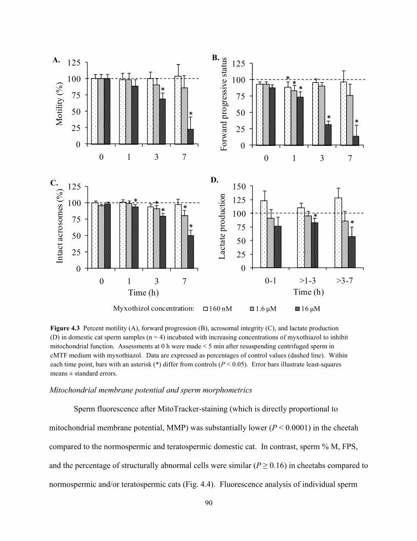
90
Mitochondrial membrane potential and sperm morphometrics
Sperm fluorescence after MitoTracker-staining (which is directly proportional to
mitochondrial membrane potential, MMP) was substantially lower (P < 0.0001) in the cheetah
compared to the normospermic and teratospermic domestic cat. In contrast, sperm % M, FPS,
and the percentage of structurally abnormal cells were similar (P ≥ 0.16) in cheetahs compared to
normospermic and/or teratospermic cats (Fig. 4.4). Fluorescence analysis of individual sperm
0
25
50
75
100
125
0 1 3 7
Mot
ility
(%)
*
*
0
25
50
75
100
125
0 1 3 7
Forw
ard
prog
ress
ive
stat
us
* *
**
*
0
25
50
75
100
125
150
0-1 >1-3 >3-7
Lact
ate
prod
uctio
n
Time (h)
**
0
25
50
75
100
125
0 1 3 7
Inta
ct a
cros
omes
(%)
Time (h)
*
****
B. A.
C. D.
Myxothizol concentration: 160 nM 1.6 µM 16 µM
Figure 4.3 Percent motility (A), forward progression (B), acrosomal integrity (C), and lactate production (D) in domestic cat sperm samples (n = 4) incubated with increasing concentrations of myxothiazol to inhibit mitochondrial function. Assessments at 0 h were made < 5 min after resuspending centrifuged sperm in cMTF medium with myxothiazol. Data are expressed as percentages of control values (dashed line). Within each time point, bars with an asterisk (*) differ from controls (P < 0.05). Error bars illustrate least-squares means ± standard errors.
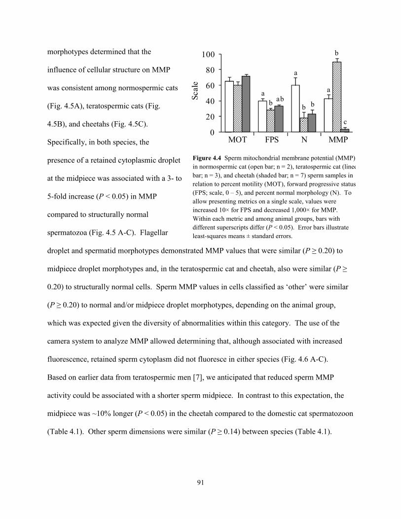
91
morphotypes determined that the
influence of cellular structure on MMP
was consistent among normospermic cats
(Fig. 4.5A), teratospermic cats (Fig.
4.5B), and cheetahs (Fig. 4.5C).
Specifically, in both species, the
presence of a retained cytoplasmic droplet
at the midpiece was associated with a 3- to
5-fold increase (P < 0.05) in MMP
compared to structurally normal
spermatozoa (Fig. 4.5 A-C). Flagellar
droplet and spermatid morphotypes demonstrated MMP values that were similar (P ≥ 0.20) to
midpiece droplet morphotypes and, in the teratospermic cat and cheetah, also were similar (P ≥
0.20) to structurally normal cells. Sperm MMP values in cells classified as ‘other’ were similar
(P ≥ 0.20) to normal and/or midpiece droplet morphotypes, depending on the animal group,
which was expected given the diversity of abnormalities within this category. The use of the
camera system to analyze MMP allowed determining that, although associated with increased
fluorescence, retained sperm cytoplasm did not fluoresce in either species (Fig. 4.6 A-C).
Based on earlier data from teratospermic men [7], we anticipated that reduced sperm MMP
activity could be associated with a shorter sperm midpiece. In contrast to this expectation, the
midpiece was ~10% longer (P < 0.05) in the cheetah compared to the domestic cat spermatozoon
(Table 4.1). Other sperm dimensions were similar (P ≥ 0.14) between species (Table 4.1).
0
20
40
60
80
100
Scal
e
aabb
b
b
a
a
b
c
MOT FPS N MMP
Figure 4.4 Sperm mitochondrial membrane potential (MMP) in normospermic cat (open bar; n = 2), teratospermic cat (lined bar; n = 3), and cheetah (shaded bar; n = 7) sperm samples in relation to percent motility (MOT), forward progressive status (FPS; scale, 0 – 5), and percent normal morphology (N). To allow presenting metrics on a single scale, values were increased 10× for FPS and decreased 1,000× for MMP. Within each metric and among animal groups, bars with different superscripts differ (P < 0.05). Error bars illustrate least-squares means ± standard errors.
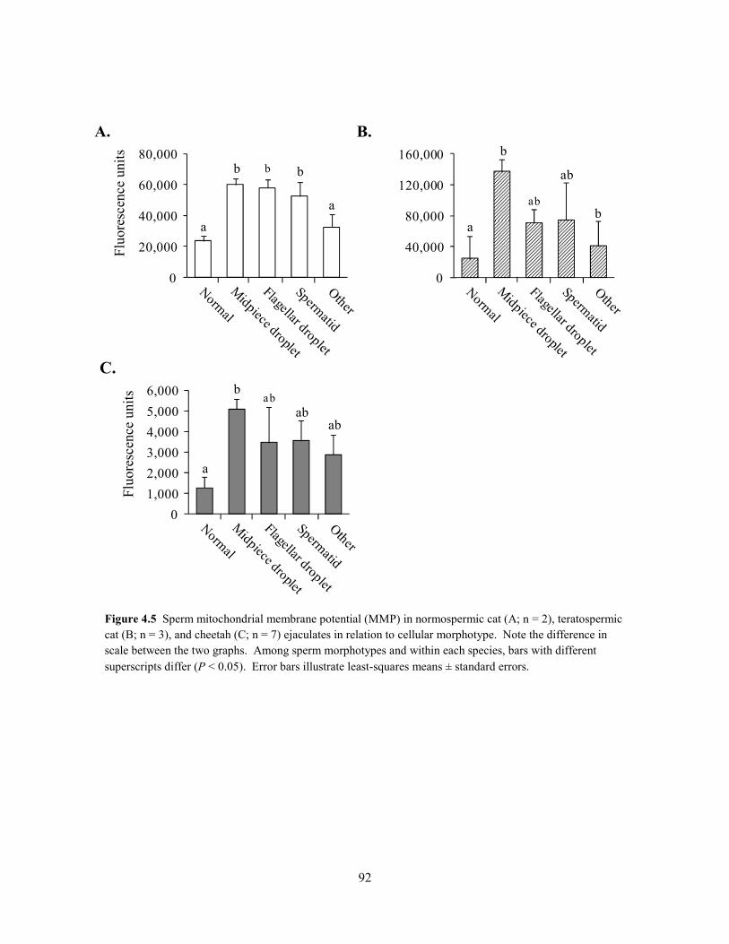
92
A.
0
20,000
40,000
60,000
80,000b
a
b
a
b
0
40,000
80,000
120,000
160,000 b
b
ab
a
ab
B.
0
1,000
2,000
3,000
4,000
5,000
6,000 b
abab
a
ab
C.
Figure 4.5 Sperm mitochondrial membrane potential (MMP) in normospermic cat (A; n = 2), teratospermic cat (B; n = 3), and cheetah (C; n = 7) ejaculates in relation to cellular morphotype. Note the difference in scale between the two graphs. Among sperm morphotypes and within each species, bars with different superscripts differ (P < 0.05). Error bars illustrate least-squares means ± standard errors.
Fl
uore
scen
ce u
nits
Fluo
resc
ence
uni
ts
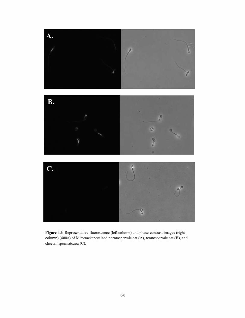
93
Figure 4.6 Representative fluorescence (left column) and phase-contrast images (right column) (400×) of Mitotracker-stained normospermic cat (A), teratospermic cat (B), and cheetah spermatozoa (C).
C.
B.
A.
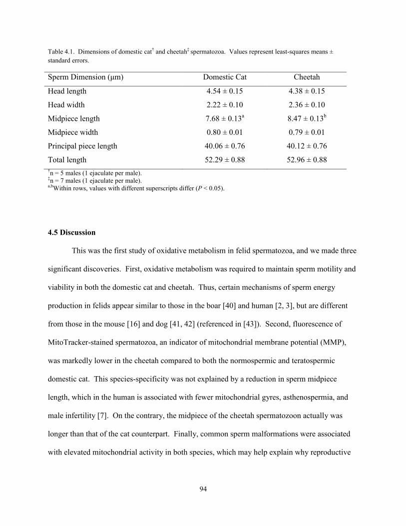
94
Table 4.1. Dimensions of domestic cat† and cheetah‡ spermatozoa. Values represent least-squares means ± standard errors.
Sperm Dimension (µm) Domestic Cat Cheetah
Head length 4.54 ± 0.15 4.38 ± 0.15
Head width 2.22 ± 0.10 2.36 ± 0.10
Midpiece length 7.68 ± 0.13a 8.47 ± 0.13b
Midpiece width 0.80 ± 0.01 0.79 ± 0.01
Principal piece length 40.06 ± 0.76 40.12 ± 0.76
Total length 52.29 ± 0.88 52.96 ± 0.88 †n = 5 males (1 ejaculate per male). ‡n = 7 males (1 ejaculate per male). a,bWithin rows, values with different superscripts differ (P < 0.05).
4.5 Discussion
This was the first study of oxidative metabolism in felid spermatozoa, and we made three
significant discoveries. First, oxidative metabolism was required to maintain sperm motility and
viability in both the domestic cat and cheetah. Thus, certain mechanisms of sperm energy
production in felids appear similar to those in the boar [40] and human [2, 3], but are different
from those in the mouse [16] and dog [41, 42] (referenced in [43]). Second, fluorescence of
MitoTracker-stained spermatozoa, an indicator of mitochondrial membrane potential (MMP),
was markedly lower in the cheetah compared to both the normospermic and teratospermic
domestic cat. This species-specificity was not explained by a reduction in sperm midpiece
length, which in the human is associated with fewer mitochondrial gyres, asthenospermia, and
male infertility [7]. On the contrary, the midpiece of the cheetah spermatozoon actually was
longer than that of the cat counterpart. Finally, common sperm malformations were associated
with elevated mitochondrial activity in both species, which may help explain why reproductive

95
physiologists have long been perplexed by the inability of certain traditional quality metrics (e.g.,
MMP, motility) to accurately predict sperm fertilizing ability [44].
An advantage of our research approach was the cross-species comparative assessment
that allowed us to discover a 100-fold increased sensitivity to electron transport inhibition and
evidence for a remarkably low MMP in the cheetah spermatozoon compared to the domestic cat.
While fluorescence of MitoTracker-stained spermatozoa is directly proportional to MMP, it is
important to note that this species difference also could be related to uncontrolled factors
influencing the dye’s accumulation in the mitochondrion, such as the degree of binding to
cellular structures, facilitated diffusion, or complex ion interactions [45]. However, given our
standardization of MitoTracker and sperm concentrations, extended incubation time, use of the
same cellular medium, and, particularly, the 20-fold magnitude of the MMP difference, we
believe that this finding indicated that there was a functional difference in membrane potential
between the species. Combined with the relative sensitivity to OXPHOS inhibition, these
observations provided evidence for mitochondrial dysfunction in cheetah spermatozoa. This
may well represent the mechanism underpinning the functional abnormalities observed in
teratospermic felid ejaculates, including reduced sperm longevity in vitro [46], poor motility
[47], disrupted protein tyrosine phosphorylation [25], delayed capacitation [26], compromised
acrosomal function [26], and low IVF success [48]. Interestingly, however, sperm MMP and
sensitivity to oxidative inhibition were similar between the normospermic and teratospermic
domestic cat, even though sperm lactate production was previously found to be compromised in
a large set of ejaculates from the latter group [21]. This observation may indicate that the
assemblage of genes involved in teratospermia differs between the domestic cat and cheetah
[49], a reasonable hypothesis given that the trait presumably arose in the former species after a
96
severe population contraction that occurred ~10,000 years ago [50], long after the evolutionary
divergence from other felids ~6,000,000 years ago [51].
In contrast to our previous study [21], lactate production of control sperm samples was
reduced by only 20% or less in teratospermic compared to normospermic domestic cats, a trend
that was insignificant. Although perhaps due to our overall smaller sample size in the present
study, we suspect that this observation reflected physiological differences among the males used
in the two investigations. Substantial variation in sperm motility and longevity in vitro
commonly occurs among teratospermic domestic cats, and it is likely that such variation is
driven by differences in metabolic function and, ultimately, the genes causing this condition.
Interestingly, analogous differences in ejaculate quality are commonly detected among
teratospermic cheetahs, even though we have observed little variation in metabolic function
within this genetically monomorphic species [21, 22].
It was noteworthy that, although sperm mitochondrial activity was ~95% lower in the
cheetah compared to the domestic cat, cellular percent motility and forward progression were
similar between species. This may have indicated that lower sperm MMP in the cheetah was
indeed ‘normal’ and not representing metabolic dysfunction. If so, glycolytic metabolism could
compensate for relatively low mitochondrial activity. In this context, our previous finding that
GAPDH inhibition causes less severe motility declines in cheetah versus cat spermatozoa [22]
could be interpreted as evidence that the glycolytic pathway is comparatively more robust in
cheetah ejaculates. It also is possible that energy demand was reduced in cheetah compared to
domestic cat spermatozoa, perhaps due to differences in the efficiency of microtubule sliding or
ATP transport along the flagellum [52]. However, we suspect that the most likely explanation
for similar sperm motility traits between species was that the low MMP of cheetah spermatozoa
97
was just above the threshold required to support sperm function, and that the rapid losses in
motility commonly observed in these ejaculates [21, 47, 53] resulted from unsustainable ATP
consumption. In this case, our previous observations of reduced sensitivity of cheetah ejaculates
to glycolytic inhibition [22] could indicate that this pathway is simply less active in the cells of
this species.
While determining that the low MMP in cheetah spermatozoa was unrelated to midpiece
length, we also had the opportunity to compare other cellular dimensions. Such data are rare for
wild felids, although we discovered that the cheetah spermatozoon had a shorter and narrower
(~30%) head than a previous report for the tiger (Panthera tigris) [54], and a shorter (~30%)
midpiece than for the leopard (Panthera pardus) [55], both of which are considered
normospermic species. No doubt the size metrics of the cheetah spermatozoon more closely
resembled those of the domestic cat because these species are more genetically similar compared
to the tiger or leopard [51].
Also related to sperm morphology was its interesting link to MMP, an association that
was remarkably consistent between the two species. A spermatozoon with retained cytoplasm is
a common pleiomorphism in the cat [18, 21] and cheetah [21, 28] as well as certain other
species/populations that experience teratospermia [46, 56-67]. We discovered in the present
study that this malformation was associated with substantially increased MMP. To a lesser, but
nonsignificant extent, increased MMP also was observed in spermatozoa with retained cytoplasm
in the flagellar region, suggesting that the proximity of the droplet to the midpiece influenced its
ultimate impact on MMP. The relationship between a retained droplet and enhanced
mitochondrial function was unexpected because in human, mouse, stallion, and boar, this
residual cytoplasm is rich in lysosomal enzymes (e.g., 15-lipoxygenase;15-LOX) that degrade
98
organelles, including mitochondria via phospholipid peroxidation [68, 69]. We have identified
three possible mechanisms by which retained cytoplasm could confer increased MMP, the first
being that there simply could be functional mitochondria in the residual droplet that causes the
fluorescence. Although such sperm cytoplasm in mammals generally does not contain
mitochondria [43, 68], a recent study documented intact mitochondria embedded in the retained
cytoplasm of sperm from Alox15 knock-out mice lacking the 15-LOX enzyme [58]. This
possibility was unlikely in our current study because cytoplasmic droplets did not fluoresce after
MMP staining, indicating an absence of functional mitochondria. Another alternative is that the
retained droplets may be a source of metabolic substrates. For example, in the bull [70], ram
[70], boar [71], rabbit [70], rat [72], and hamster [73], the droplet contains an abundance of
membranous elements, which may be remnants of Golgi bodies or the endoplasmic reticulum,
and could provide substantial energy in the form of lipid [68]. A final option is that the
preserved cytoplasm may contain factors that increase metabolic efficiency by directly
stimulating mitochondrial activity or increasing the availability of existing substrates [74]. For
example, glycolytic enzyme activity in free-lying (i.e., unattached to a spermatozoon)
cytoplasmic droplets from bull, ram, and boar ejaculates can be up to 5-fold higher than in the
spermatozoon itself [74]. If felid spermatozoa containing cytoplasmic droplets are enriched in
enzymes involved in lipid/glycerol metabolism, a higher concentration of oxidative substrate
may be available to the mitochondrion. Because the current study provides compelling evidence
that oxidative metabolism plays a key role in felid sperm motility, we predict that the link
between cytoplasmic droplets and increased MMP is related to the increased abundance or
availability of lipid as a mitochondrial substrate.
99
Our observation of an association between a retained cytoplasmic droplet and increased
MMP likely would confound mitochondrial-based estimates of sperm quality, as there is strong
evidence that mammalian sperm function is compromised by residual cytoplasm [48, 75-80].
Although normally shed soon after ejaculation [81], retained droplets are known to reduce
plasma membrane integrity [78], disrupt nuclear maturation [82], and decrease IVF success [78]
in humans. Cytoplasmic retention also reduces sperm-ovum interaction in vitro in the mouse
[79] and appears to perturb capacitation in the dog [80]. Domestic cat spermatozoa with retained
cytoplasm fail to penetrate the oocyte’s inner zona pellucida and, therefore, are incapable of
fertilization [48]. While felid spermatozoa with retained cytoplasm may demonstrate high MMP
soon after ejaculation, these values could quickly decrease as lysosomal enzymes assault
mitochondrial and plasma membranes, resulting in the rapid loss of viability. We suspect that
this scenario may occur in many felid species because residual cytoplasm is a highly common
anomaly in the Felidae taxon. Prevalence can range from ~10% to ~70% of ejaculated
spermatozoa in the domestic cat [21], cheetah [21, 28], puma (Puma concolor) [60], leopard cat
(Prionailurus bengalensis) [61], clouded leopard [62, 63], snow leopard (Panthera uncia) [64],
Iberian lynx (Lynx pardinus) [65], fishing cat (Prionailurus viverrinus) [46], and sand cat (Felis
margarita) [66]. Although several genes have been linked to cytoplasmic droplet retention using
mouse knockout models [43, 57, 83], spermatozoa from these individuals commonly have other
structural abnormalities (e.g., a hairpin bend in the neck or flagellum) that are rarely observed in
felids [21, 28, 46, 60-66]. It is unlikely that the 15-LOX gene is being disrupted in the cat or
cheetah, because we observed no functional mitochondria in spermatozoa with retained droplets.
Thus, we suspect that there remains a yet-to-be-determined genetic mechanism(s) by which
sperm cytoplasmic droplet migration can become compromised in felids.
100
In conclusion, the results of this study demonstrated the value of a comparative approach
to understanding sperm metabolic mechanisms related to distinctive species physiology versus
teratospermia. Oxidative phosphorylation appears to be a critical pathway for supporting sperm
motility in felids, yet the cheetah spermatozoon may operate at or below the minimum threshold
of aerobic metabolism required to maintain cellular function. We predict that identifying sperm
metabolic deficiencies that are driven by species-specific mechanisms or related to teratospermia
will be the first step to developing effective strategies for mitigation. Given the remarkable
differences in gamete physiology observed between these two closely-related taxa (domestic cat
and cheetah), it is clear that improving the success of assisted reproduction will require methods
tailored to the unique physiology of the target species. By understanding and addressing sperm
physiological disruptions at the fundamental level of energy production, we may be able to
simultaneously overcome multiple functional deficiencies (e.g., poor motility, delayed
capacitation) that arise from a common metabolic defect. This basic knowledge will provide a
valuable foundation for future studies on the importance of sperm metabolism and teratospermia,
with broad interest and, importantly, application to preserving fertility in humans and an array of
domestic and wildlife species [84, 85].
4.6 References
1. Storey B. Mammalian sperm metabolism: oxygen and sugar, friend and foe. Int J Dev
Biol 2008; 52: 427-437.
2. Ford WCL, Harrison A. The role of oxidative phosphorylation in the generation of ATP
in human spermatozoa. J Reprod Fertil 1981; 63: 271-278.

101
3. Weinberg J, Doty E, Bonaventura J, Haney A. Nitric oxide inhibition of human sperm
motility. Fertil Steril 1995; 64: 408-413.
4. Meseguer M, Garrido N, Martinez-Conejero JA, Simon C, Pellicer A, Remohi J.
Relationship between standard semen parameters, calcium, cholesterol contents, and
mitochondrial activity in ejaculated spermatozoa from fertile and infertile males. J Assist
Reprod Genet 2004; 21: 445-451.
5. Auger J, Ronot X, Dadoune JP. Human sperm mitochondrial function related to motility:
a flow and image cytometric assessment. J Androl 1989; 10: 439-448.
6. Ruiz-Pesini E, Lapeña AC, Díez C, Álvarez E, Enríquez JA, López-Pérez MJ. Seminal
quality correlates with mitochondrial functionality. Clin Chim Acta 2000; 300: 97-105.
7. Mundy A, Ryder T, Edmonds D. Asthenozoospermia and the human sperm midpiece.
Hum Reprod 1995; 10: 116-119.
8. Rawe VY, Hermes R, Nodar FN, Fiszbajn G, Chemes HE. Results of intracytoplasmic
sperm injection in two infertile patients with abnormal organization of sperm
mitochondrial sheaths and severe asthenoteratozoospermia. Fertil Steril 2007; 88: 649-
653.
9. Pelliccione F, Micillo A, Cordeschi G, D'Angeli A, Necozione S, Gandini L, Lenzi A,
Francavilla F, Francavilla S. Altered ultrastructure of mitochondrial membranes is
strongly associated with unexplained asthenozoospermia. Fertil Steril 2011; 95: 641-646.
10. Ferramosca A, Focarelli R, Piomboni P, Coppola L, Zara V. Oxygen uptake by
mitochondria in demembranated human spermatozoa: a reliable tool for the evaluation of
sperm respiratory efficiency. Int J Androl 2008; 31: 337-345.
102
11. Ruiz-Pesini E, Diez C, Lapeña C, Pérez-Martos A, Montoya J, Alvarez E, Arenas J,
López-Pérez MJ. Correlation of sperm motility with mitochondrial enzymatic activities.
Clin Chem 1998; 44: 1616-1620.
12. Garrett LJA, Revell SG, Leese HJ. Adenosine triphosphate production by bovine
spermatozoa and its relationship to semen fertilizing ability J Androl 2008; 29: 449-458.
13. Tanghe S, Van Soom A, Sterckx V, Maes D, de Kruif A. Assessment of different sperm
quality parameters to predict in vitro fertility of bulls. Reprod Domest Anim 2002; 37:
127-132.
14. Hallap T, Nagy S, Jaakma Ü, Johannisson A, Rodriguez-Martinez H. Mitochondrial
activity of frozen-thawed spermatozoa assessed by MitoTracker Deep Red 633.
Theriogenology 2005; 63: 2311-2322.
15. Mukai C, Okuno M. Glycolysis plays a major role for adenosine triphosphate
supplementation in mouse sperm flagellar movement. Biol Reprod 2004; 71: 540-547.
16. Travis AJ, Jorgez CJ, Merdiushev T, Jones BH, Dess DM, Diaz-Cueto L, Storey BT,
Kopf GS, Moss SB. Functional relationships between capacitation-dependent cell
signaling and compartmentalized metabolic pathways in murine spermatozoa. J Biol
Chem 2001; 276: 7630-7636.
17. Nayernia K, Adham IM, Burkhardt-Gottges E, Neesen J, Rieche M, Wolf S, Sancken U,
Kleene K, Engel W. Asthenozoospermia in mice with targeted deletion of the sperm
mitochondrion-associated cysteine-rich protein (Smcp) gene. Mol Cell Biol 2002; 22:
3046-3052.
103
18. Howard JG, Brown JL, Bush M, Wildt DE. Teratospermic and normospermic domestic
cats: ejaculate traits, pituitary-gonadal hormones, and improvement of spermatozoal
motility and morphology after swim-up processing. J Androl 1990; 11: 204-215.
19. Pukazhenthi BS, Neubauer K, Jewgenow K, Howard JG, Wildt DE. The impact and
potential etiology of teratospermia in the domestic cat and its wild relatives.
Theriogenology 2006; 66: 112-121.
20. Cooper TG, Noonan E, von Eckardstein S, Auger J, Gordon Baker HW, H.M. B, Haugen
TB, Kruger T, Wang C, Mbizvo MT, Vogelsong KM. World Health Organization
reference values for human semen characteristics. Hum Reprod Update 2010; 16: 231-
245.
21. Terrell KA, Wildt DE, Anthony NM, Bavister BD, Leibo SP, Penfold LM, Marker LL,
Crosier AE. Evidence for compromised metabolic function and limited glucose uptake in
spermatozoa from the teratospermic domestic cat (Felis catus) and cheetah (Acinonyx
jubatus). Biol Reprod 2010; 83: 833-841.
22. Terrell KA, Wildt DE, Anthony NM, Bavister BD, Leibo SP, Penfold LM, Marker LL,
Crosier AE. Glycolytic enzyme activity is essential for domestic cat (Felis catus) and
cheetah (Acinonyx jubatus) sperm motility and viability in a sugar-free medium. Biol
Reprod In press.
23. Garcia J, Han D, Sancheti H, Yap LP, Kaplowitz N, Cadenas E. Regulation of
mitochondrial glutathione redox status and protein glutathionylation by respiratory
substrates. J Biol Chem 2010; 285: 39646-39654.
104
24. Voet D, Voet JG. Electron transport and oxidative phosphorylation. In: Harris D,
Fitzgerald P (eds.), Biochemistry, 3rd ed. Hoboken, NJ: John Wiley & Sons, Inc.; 2004:
797-842.
25. Pukazhenthi BS, Wildt DE, Ottinger MA, Howard JG. Compromised sperm protein
phosphorylation after capacitation, swim-up, and zona pellucida exposure in
teratospermic domestic cats. J Androl 1996; 17: 409-419.
26. Long JA, Wildt DE, Wolfe BA, Crister JK, DeRossi RV, Howard JG. Sperm capacitation
and the acrosome reaction are compromised in teratospermic domestic cats. Biol Reprod
1996; 54: 638-646.
27. Crosier A, Henghali J, Howard JG, Pukazhenthi BS, Terrell KA, Marker LL, Wildt DE.
Improved quality of cryopreserved cheetah (Acinonyx jubatus) spermatozoa after
centrifugation through Accudenz. J Androl 2009; 30: 298-308.
28. Crosier AE, Marker L, Howard JG, Pukazhenthi BS, Henghali JN, Wildt DE. Ejaculate
traits in the Namibian cheetah (Acinonyx jubatus): influence of age, season, and captivity.
Reprod Fertil Dev 2007; 19: 370-382.
29. Wildt DE, O'Brien SJ, Howard JG, Caro TM, Roelke ME, Brown JL, Bush M. Similarity
in ejaculate-endocrine characteristics in captive versus free-ranging cheetahs of two
subspecies. Biol Reprod 1987; 36: 351-360.
30. Howard JG. Semen collection and analysis in carnivores. In: Fowler ME (ed.) Zoo and
Wild Animal Medicine: Current Therapy III. Philadelphia: WB Saunders Co.; 1993: 390-
399.
105
31. Pukazhenthi BS, Noiles E, Pelican K, Donoghue AM, Wildt DE, Howard JG. Osmotic
effects on feline spermatozoa from normospermic versus teratospermic donors.
Cryobiology 2000; 40: 139-150.
32. Gardner DK, Leese HJ. Concentrations of nutrients in mouse oviduct fluid and their
effects on embryo development and metabolism in vitro. J Reprod Fertil 1990; 88: 361-
368.
33. Bavister BD. Substitution of a synthetic polymer for protein in a mammalian gamete
culture system. J Exp Zool 1981; 217: 45-51.
34. Johnston SD, Osborne CA, Lipowitz AJ. Characterization of seminal plasma, prostatic
fluid and bulbourethral gland secretions in the domestic cat. In: Proceedings of the 11th
International Congress on Animal Reproduction and Artificial Insemination. Dublin;
1988: 4:560.
35. Shah D, Naciri M, Clee P, Al-Rubeai M. NucleoCounter: an efficient technique for the
determination of cell number and viability in animal cell culture processes.
Cytotechnology 2006; 51: 39-44.
36. Larson JL, Miller DJ. Simple histochemical stain for acrosomes on sperm from several
species. Mol Reprod Dev 1999; 52: 445-449.
37. Crosier AE, Pukazhenthi BS, Henghali JN, Howard JG, Dickman AJ, Marker L, Wildt
DE. Cryopreservation of spermatozoa from wild-born Namibian cheetahs (Acinonyx
jubatus) and influence of glycerol on cryosurvival. Cryobiology 2006; 52: 169-181.
38. Passonneau JV, Lowry OH. Enzymatic Analysis: A Practical Guide. Totowa, NJ:
Humana Press; 1993.
106
39. SAS. SAS/STAT User's Guide: Statistics. Version 9.1. Cary, NC: Statistical Analysis
System Institute, Inc; 2002.
40. Aalbers J, Mann T, Polge C. Metabolism of boar semen in relation to sperm motility and
survival. J Reprod Fertil 1961; 2: 42-53.
41. Koelliker A. Physiologische studien über die samenflüssigkeit. Z Wiss Zool 1856; 7: 201.
42. Ivanov E. Zur frage der energetik der spermatozoenbewegung. Z Zucht, B 1931; 20.
43. Bartlett D. Studies on dog semen II. Biochemical characteristics. J Reprod Fertil 1962; 3:
190-205.
44. Barratt CLR, Mansell S, Beaton C, Tardif S, Oxenham SK. Diagnostic tools in male
infertility - the question of sperm dysfunction. Asian J Androl 2011; 13: 53-58.
45. Dykens JA, Stout AK. Assessment of mitochondrial membrane potential in situ using
single potentiometric dyes and a novel fluorescence resonance energy transfer technique.
In: Pon LA, Schon EA (eds.), Mitochondria. San Diego, CA: Academic Press; 2001: 287.
46. Thiangtum K, Swanson WF, Howard J, Tunwattana W, Tongthainan D, Wichasilpa W,
Patumrattanathan P, Pinyopoommintr T. Assessment of basic seminal characteristics,
sperm cryopreservation and heterologous in vitro fertilization in the fishing cat
(Prionailurus viverrinus). Reprod Fert Dev 2006; 18: 373-382.
47. Donoghue AM, Howard JG, Byers AP, Goodrowe KL, Bush M, Blumer E, Lukas J,
Stover J, Snodgrass K, Wildt DE. Correlation of sperm viability with gamete interaction
and fertilization in vitro in the cheetah (Acinonyx jubatus). Biol Reprod 1992; 46: 1047-
1056.
107
48. Howard JG, Donoghue AM, Johnston LA, Wildt DE. Zona pellucida filtration of
structurally abnormal spermatozoa and reduced fertilization in teratospermic cats. Biol
Reprod 1993; 49: 131-139.
49. Matzuk MM, Lamb DJ. Genetic dissection of mammalian fertility pathways. Nat Cell
Biol 2002; 4: S41-S49.
50. O'Brien SJ, Roelke ME, Marker L, Newman A, Winkler CA, Meltzer D, Colly L,
Evermann JF, Bush M, Wildt DE. Genetic basis for species vulnerability in the cheetah.
Science 1985; 227: 1428-1434.
51. O'Brien SJ, Johnson WE. Big cat genomics. Annu Rev Genom Hum Genet 2005; 6: 407-
429.
52. Ford WCL, Rees JM. The bioenergetics of mammalian sperm motility. In: Gagnon C
(ed.), Controls of Sperm Motility: Biological and Clinical Aspects. Boca Raton, FL: CRC
Press; 1990: 175-202.
53. Wildt DE, Bush M, Howard JG, O'Brien SJ, Meltzer D, van Dyk A, Ebedes H, Brand DJ.
Unique seminal quality in the South African cheetah and a comparative evaluation in the
domestic cat. Biol Reprod 1983; 29: 1019-1025.
54. Cummins JM, Woodall P. On mammalian sperm dimensions. J Reprod Fertil 1985; 75:
153-175.
55. Gage MJG. Mammalian sperm morphometry. Proc R Soc Lond B Biol Sci 1998; 265: 97-
103.
56. Dahlbom M, Andersson M, Vierula M, Alanko M. Morphometry of normal and
teratozoospermic canine sperm heads using an image analyzer: Work in progress.
Theriogenology 1997; 48: 687-698.
108
57. Zheng H, Stratton CJ, Morozumi K, Jin J, Yanagimachi R, Yan W. Lack of Spem1 causes
aberrant cytoplasm removal, sperm deformation, and male infertility. Proc Nat Acad Sci
2007; 104: 6852-6857.
58. Moore K, Lovercamp K, Feng DY, Antelman J, Sutovsky M, Manandhar G, van Leyen
K, Safranski T, Sutovsky P. Altered epididymal sperm maturation and cytoplasmic
droplet migration in subfertile male Alox15 mice. Cell Tissue Res 2010; 340: 569-581.
59. Lovercamp KW, Safranski TJ, Fischer KA, Manandhar G, Sutovsky M, Herring W,
Sutovsky P. High resolution light microscopic evaluation of boar semen quality sperm
cytoplasmic droplet retention in relationship with boar fertility parameters. Arch Androl
2007; 53: 219-228.
60. Barone MA, Roelke ME, Howard J, Brown JL, Anderson AE, Wildt DE. Reproductive
characteristics of male Florida panthers: comparative studies from Florida, Texas,
Colorado, Latin America, and North American Zoos. J Mammal 1994; 75: 150-162.
61. Howard JG, Wildt DE. Ejaculate-hormonal traits in the leopard cat (Felis bengalensis)
and sperm function as measured by in vitro penetration of zona-free hamster ova and
zona-intact domestic cat oocytes. Mol Reprod Dev 1990; 26: 163-174.
62. Wildt DE, Howard JG, Hall LL, Bush M. Reproductive physiology of the clouded
leopard. I. Electroejaculates contain high proportions of pleiomorphic spermatozoa
throughout the year. Biol Reprod 1986; 34: 937-947.
63. Howard JG, Donoghue AM, Barone MA, Goodrowe KL, Blumer ES, Snodgrass K,
Starnes D, Tucker M, Bush M, Wildt DE. Successful induction of ovarian activity and
laparoscopic intrauterine artificial insemination in the cheetah (Acinonyx jubatus). J Zoo
Wildl Med 1992; 23: 288-300.
109
64. Johnston LA, Armstrong DL, Brown JL. Seasonal effects on seminal and endocrine traits
in the captive snow leopard (Panthera uncia). J Reprod Fertil 1994; 102: 229-236.
65. Gañán N, Sestelo A, Garde JJ, Martínez F, Vargas A, Sánchez I, Pérez-Aspa MJ, López-
Bao JV, Palomares F, Gomendio M, Roldan ERS. Reproductive traits in captive and free-
ranging males of the critically endangered Iberian lynx (Lynx pardinus). Reproduction
2010; 139: 275-285.
66. Herrick JR, Campbell M, Levens G, Moore T, Benson K, D'Agostino J, West G, Okeson
DM, Coke R, Portacio SC, Leiske K, Kreider C, et al. In vitro fertilization and sperm
cryopreservation in the black-footed cat (Felis nigripes) and sand cat (Felis margarita).
Biol Reprod 2010; 82: 552-562.
67. Brito LFC, Sertich PL, Stull GB, Rives W, Knobbe M. Sperm ultrastructure,
morphometry, and abnormal morphology in American black bears (Ursus americanus).
Theriogenology 2010; 74: 1403-1413.
68. Oko R, Clermont Y. Mammalian spermatozoa: structure and assembly of the tail. In:
Gagnon C (ed.), Controls of Sperm Motility: Biological and Clinical Aspects. Boca
Raton, FL: CRC Press; 1990: 4-21.
69. Fischer KA, Van Leyen K, Lovercamp KW, Manandhar G, Sutovsky M, Feng D,
Safranski T, Sutovsky P. 15-Lipoxygenase is a component of the mammalian sperm
cytoplasmic droplet. Reproduction 2005; 130: 213-222.
70. Bloom G, Nicander L. On the ultrastructure and development of the protoplasmic droplet
of spermatozoa. Z Zellforsch 1961; 55: 833-844.
110
71. Kaplan M, Russell LD, Peterson RN, Martan J. Boar sperm cytoplasmic droplets: Their
ultrastructure, their numbers in the epididymis and at ejaculation, and their removal
during isolation of sperm plasma membranes. Tissue Cell 1984; 16: 455-468.
72. Hermo L, Dworkin J, Oko R. Role of epithelial clear cells of the rat epididymis in the
disposal of the contents of cytoplasmic droplets detached from spermatozoa. Am J Anat
1988; 183: 107-124.
73. Cortadellas N, Durfort M. Fate and composition of cytoplasmic droplet of hamster
epididymal spermatozoa. J Morphol 1994; 221: 199-210.
74. Harrison R, White I. Glycolytic enzymes in the spermatozoa and cytoplasmic droplets of
bull, boar, and ram, and their leakage after shock. J Reprod Fertil 1972; 30: 105-115.
75. Bedford JM. Influence of abdominal temperature on epididymal function in the rat and
rabbit. Am J Anat 1978; 152: 509-521.
76. Cummins JM. Effects of artificial cryptorchidism in the rabbit on the transport and
survival of spermatozoa in the female reproductive tract. J Reprod Fertil 1973; 33: 469-
479.
77. Cummins JM, Glover TD. Artificial cryptorchidism and fertility in the rabbit. J Reprod
Fertil 1970; 23: 423-433.
78. Keating J, Grundy C, Fivey P, Elliott M, Robinson J. Investigation of the association
between the presence of cytoplasmic residues on the human sperm midpiece and
defective sperm function. J Reprod Fertil 1997; 110: 71-77.
79. Kawai Y, Hata T, Suzuki O, Matsuda J. The relationship between sperm morphology and
in vitro fertilization ability in mice. J Reprod Dev 2006; 52: 561-568.
111
80. Peña AI, Barrio M, Becerra JJ, Quintela LA, Herradón PG. Infertility in a dog due to
proximal cytoplasmic droplets in the ejaculate: investigation of the significance for sperm
functionality in vitro. Reprod Domest Anim 2007; 42: 471-478.
81. Cooper T, Yeung CH, Fetic S, Sobhani A, Nieschlag E. Cytoplasmic droplets are normal
structures of human spermatozoa, but are not well preserved by routine procedures for
assessing sperm morphology. Hum Reprod 2004; 19: 2283-2288.
82. Sati L, Ovari L, Bennett D, Simon SD, Demir R, Huszar G. Double probing of human
spermatozoa for persistent histones, surplus cytoplasm, apoptosis, and DNA
fragmentation. Reprod Biomed Online 2008; 16: 570-579.
83. Esaclier D. Knockout mouse models of sperm flagellum anomalies. Hum Reprod Update
2006; 12: 449-461.
84. Wildt DE, Comizzoli P, Pukazhenthi B, Songsasen N. Lessons from biodiversity-the
value of nontraditional species to advance reproductive science, conservation, and human
health. Mol Reprod Dev 2010; 77: 397-409.
85. Comizzoli P, Songsasen N, Wildt DE. Protecting and extending fertility for females of
wild and endangered mammals. In: Woodruff TK, Zoloth L, Campo-Engelstein L,
Rodriguez S (eds.), Oncofertility: Ethical, Legal, Social, and Medical Perspectives. New
York: Springer Science+Business Media, LLC; 2010: 87-102.
112
CHAPTER 5: DIFFERENT PATTERNS OF METABOLIC CRYO-DAMAGE IN DOMESTIC CAT (FELIS CATUS) AND CHEETAH (ACINONYX JUBATUS)
SPERMATOZOA
5.1 Abstract
Felid spermatozoa are sensitive to cryopreservation-induced damage, but functional
losses can be mitigated by post-thaw swim-up or density gradient processing methods that
selectively recover motile or structurally normal spermatozoa, respectively. Despite the
importance of sperm energy production to achieving fertilization, there is little knowledge about
the influence of cryopreservation or post-thaw processing on felid sperm metabolism. We
conducted a comparative study of domestic cat and cheetah sperm metabolism after
cryopreservation and post-thaw processing. We hypothesized that freezing/thawing impairs
sperm metabolism and that swim-up, but not density gradient centrifugation, recovers
metabolically-normal spermatozoa. Ejaculates were cryopreserved, thawed, and processed by
swim-up, Accudenz gradient centrifugation, or conventional washing (representing the
‘control’). Sperm glucose and pyruvate uptake, lactate production, motility, and acrosomal
integrity were assessed. Mitochondrial membrane potential (MMP) was measured in cat
spermatozoa. In both species, lactate production, motility, and acrosomal integrity were reduced
in post-thaw, washed samples compared to freshly-collected ejaculates. Glucose uptake was
minimal pre- and post-cryopreservation, whereas pyruvate uptake was similar between
treatments, due to variation in the data. In the cat, swim-up, but not Accudenz processing,
recovered spermatozoa with increased lactate production, pyruvate uptake, and motility
compared to controls. Although confounded by differences in non-specific fluorescence among
processing methods, MMP within treatments was positively correlated to sperm motility and
acrosomal integrity. Cheetah spermatozoa isolated by either selection method exhibited
113
improved motility and/or acrosomal integrity, but remained metabolically compromised.
Collectively, findings revealed a metabolically-robust subpopulation of cryopreserved cat, but
not cheetah, spermatozoa, recovered by selecting for motility rather than morphology.
5.2 Introduction
Despite extensive research to optimize sperm cryopreservation protocols for the domestic
cat [1] and various wild felids [2], freezing/thawing causes significant damage to these cells [3-
6]. The degree of cryo-injury is particularly severe in the cheetah and other species that produce
teratospermic ejaculates (i.e., those containing high proportions of sperm pleiomorphisms) [7-
12]. In such cases, thawed spermatozoa consistently exhibit reduced motility and decreased
proportions of cells with intact acrosomes [3-12]. This cellular damage is linked to disruption of
sperm membranes from osmotic stress during feezing, thawing, and/or cryoprotectant removal
[9, 13, 14]. It also is possible that the physiological stress experienced by spermatozoa during
cryopreservation disrupts cellular metabolism. For example, one recent investigation of
epididymal spermatozoa from the domestic cat determined that mitochondrial membrane
potential (MMP) declined rapidly after cryopreservation and post-thaw washing [15]. Sperm
MMP also is impaired in frozen-thawed ejaculates from the human [16], boar [17], ram [18],
stallion [19], bull [20], and elephant [21]. Such published observations reinforce the need to
explore the impact of cryopreservation on metabolic function of felid spermatozoa. Our recent
studies of the domestic cat and cheetah have provided an understanding of baseline sperm
metabolism in felids and have revealed intriguing species differences in metabolic function [22-
24]. In both species, sperm glucose metabolism was minimal, while rates of pyruvate uptake and
lactate production were correlated positively to cellular function [22]. Sperm MMP was

114
essential for lactate production [24], and these metabolic indicators were substantially reduced in
cheetah compared to domestic cat ejaculates [22, 24].
Although being a potential source of osmotic stress, post-thaw removal of sperm
cryoprotectant [25] provides an opportunity to isolate functionally-normal spermatozoa. For
example, sperm ‘swim-up’ into fresh medium increases the proportions of motile, structurally
intact cells in cat ejaculates [26, 27] and improves sperm morphology in cryopreserved cheetah
samples [28]. Evidence in other mammals indicates that, in addition to improved motility and/or
morphology, spermatozoa isolated by selective processing also exhibit high metabolic rates. In
the ram, density gradient processing of thawed ejaculates improves sperm MMP [18], whereas
swim-up processing has the same influence on bull sperm [20] and increases oxygen
consumption by ~20-fold in freshly-ejaculated human semen [29]. While either approach could
enhance metabolic function in thawed felid ejaculates, swim-up processing (targeting vigorous,
motile cells with high rates of ATP production) [30-32] might be expected to recover the most
metabolically-robust spermatozoa. By contrast, density gradient processing (targeting cells with
normal morphology) may be less effective, because it is known that metabolism can be impaired
in structurally normal cheetah spermatozoa [22].
Our general aim was to determine the impact of cryopreservation and post-thaw
processing on the metabolic function of felid spermatozoa. This novel approach compared two
species (domestic cat and cheetah) for which baseline patterns of sperm metabolism are well-
characterized [22, 23]. Because felid sperm metabolism is influenced both by teratospermia [22]
and species physiology [22-24], domestic cats producing low proportions of structurally normal
spermatozoa were included for comparison with the teratospermic cheetah. To assess the
influence of cellular function versus morphology, metabolism was assessed in thawed
115
spermatozoa selected on the basis of motility (swim-up) or normal morphology (density
gradient). We hypothesized that the metabolism of cat and cheetah spermatozoa, specifically
rates of pyruvate and lactate utilization, would be impaired by cryopreservation. Considering the
known differences in cryo-sensitivity and baseline metabolism in spermatozoa from these
species, we expected sperm metabolic cryo-damage to be more severe in the cheetah compared
to the domestic cat. We also predicted that a subpopulation of thawed spermatozoa in each
species would exhibit robust metabolic function (similar to baseline values) and would be
recovered more effectively by isolating motile rather than normal-appearing cells.
5.3 Materials and Methods
5.3.1 Animals
All animal procedures were approved by the National Zoological Park’s Animal Care and
Use Committee (ACUC) and similar committees of the WOCC and the SDZSP.
Electroejaculates were collected from domestic cats and cheetahs using methods described
below. For the former, a total of 16 semen samples was recovered from five adult males (ages,
1.5 – 8 yr) that were known to consistently produce either normospermic or teratospermic
ejaculates [22-24]. Teratospermia has been described previously in felids and is defined as the
ejaculation of < 40% structurally normal spermatozoa [33]. Three normospermic cats were used
to produce five ejaculates (1 – 2 per individual) and two teratospermic males produced five
samples (2 – 3 per individual). Three additional ejaculates were collected from one male in each
group for the optimization of Accudenz density gradients (described below). The management
protocol for domestic cats maintained at the Smithsonian Conservation Biology Institute (SCBI;
Front Royal, VA) has been described in detail [22].
116
Electroejaculates (1 per individual) were collected from eleven adult cheetahs (ages, 2.5 –
10 yr). These animals were managed at the Cheetah Conservation Fund (CCF; Otjiwarongo,
Namibia; n = 9), White Oak Conservation Center (WOCC; Yulee, FL; n = 1), or the San Diego
Zoo Safari Park (SDZSP; Escondido, CA; n = 1). Males at CCF and the WOCC were wild
caught and captive born, respectively, and maintained under previously described protocols [22,
28]. The male at SDZSP was captive born and managed with two male siblings off exhibit in a
1,300-m2 outdoor enclosure and fed a commercial carnivore diet (Natural Balance Pet Foods
Inc., Pacoima, CA).
5.3.2 Semen Collection
A surgical plane of anesthesia was induced in domestic cats and cheetahs according to
protocols determined by institutional veterinarians for these species [28, 34]. Semen was
collected using a rectal probe of 1 cm (domestic cat) or 1.9 cm (cheetah) in diameter and an
electrostimulator (P.T. Electronics, Boring, OR) as described previously [22, 35]. A sample of
raw semen containing ~2 × 10⁵ spermatozoa was fixed in 0.3% glutaraldehyde in phosphate-
buffered saline for assessment of sperm morphology according to previous descriptions for our
laboratory [9, 22, 35].
5.3.3 Sperm Processing and Metabolic Assessments
Each ejaculate was diluted immediately with an equal volume of a chemically-defined,
protein-free, modified mouse tubal fluid medium (cMTF) [36] supplemented with 2% polyvinyl
alcohol (PVA) [37]. The cMTF medium was prepared as described previously [22] and
contained 98.4 mM NaCl, 4.78 mM KCl, 1.19 mM MgSO4, 1.19 mM KH2PO4, 25 mM
117
NaHCO3, 1.71 mM CaCl2, 1 mM glucose, 1 mM Na-pyruvate, 25 mM 3-(N-morpholino)
propanesulfonic acid (MOPS) buffer and 0.02 mg/mL phenol red. Sperm concentration was
determined using a Nucleocounter SP-100 (Chemometec, Allerød, Denmark) [38]. Osmolality
of the final cMTF medium (300 – 345 mOsm) was determined using a vapor pressure osmometer
(Wescor, Inc, Logan, UT) and was within 10% of the physiological value of domestic cat semen
(323 mOsm).
A sample containing ~3 × 10⁶ motile spermatozoa was removed from each diluted
ejaculate, washed by centrifugation for 8 min (× 300 g for domestic cat; × 100 g for cheetah),
resuspended in fresh cMTF, and then incubated (37oC) in the dark in a microcentrifuge tube (3 ×
106 motile sperm/ml) under oil to prevent evaporation, as described previously [22].
Assessments of sperm percentage motility (% M), forward progression (FPS), and acrosomal
integrity were made at 0, 1, 3, 5, and 7 h of incubation. However, the low percentages of
spermatozoa recovered in samples processed by swim-up or gradient centrifugation (see below)
precluded sampling at more than three time points. Therefore, data obtained at 1 and 5 h were
omitted from analysis. Motility was assessed visually (200 ×), and FPS was rated on a 0 to 5
scale, with a rating of 5 equivalent to most rapid, linear progress [35]. A sperm motility index
was calculated using the formula (% M + [FPS × 20] ÷ 2) [27]. Spermatozoa (~2 × 10⁵ cells)
were fixed in 4% paraformaldehyde and stained with Coomassie Blue G-250 (Fisher Biotech,
Springfield, NJ) to evaluate acrosomal integrity, as described previously [7, 39].
Glucose and pyruvate uptake and lactate production also were assessed at each time point, and
metabolic rates were calculated from substrate concentrations at 0, 3, and 7 h of incubation. To
determine metabolic substrate concentrations, medium samples (110 µl) were centrifuged for 8
min (× 1,000 g) through a CoStar Spin-X 0.22-µm nylon filter tube (Corning Incorporated,
118
Corning, NY) and stored at –80oC until analysis using a HK/G6PDH (glucose), or LDH
(pyruvate and lactate) fluorescence assay, as described previously [22, 40]. Rates of substrate
utilization were calculated as the change in medium substrate concentration over time, divided
by sperm concentration.
5.3.4 Sperm Cryopreservation
After removing a sperm sample for metabolic assessment, the remainder of each diluted
ejaculate was cryopreserved in 4% glycerol as described previously [7, 28]. Briefly, each diluted
ejaculate was centrifuged for 8 min (× 300 g domestic cat; × 100 g cheetah), resuspended in
TEST-yolk buffer (TYB) refrigeration medium without glycerol (Irvine Scientific, Santa Ana,
CA), and cooled slowly (~3.5 h) in a water bath from ambient temperature (19 – 22oC) to 4oC.
After cooling, an equal volume of TYB containing 8% glycerol (prepared as a 1:2 mixture of
refrigeration medium and TYB with 12% glycerol; Irvine Scientific) was added in a step-wise
manner over 30 min (¼ volume at 0 min, ¼ volume at 15 min, and ½ volume at 30 min).
Samples were loaded into 0.25-ml plastic straws (Veterinary Concepts, Spring Valley, WI) with
a final sperm concentration of ~60 × 106 motile spermatozoa/ml, frozen in liquid nitrogen vapor
(1 min at ~7.5 cm and 1 min at ~2.5 cm above liquid nitrogen), and then plunged directly into
liquid nitrogen. Samples were stored in liquid nitrogen until thawing (2 days to 30 mo).
5.3.5 Comparison of Post-Thaw Processing Methods
Cryopreserved straws were thawed individually for 10 sec in air (19 – 22oC) followed by
immersion in a 37°C water bath for 30 sec [7, 28]. Each straw was dried, and its contents
emptied into a sterile microcentrifuge tube. After thorough mixing, sperm motility, acrosomal
119
integrity, and morphology were assessed as described above. Each sample was divided among
three processing treatments: 1) wash; 2) swim-up; and 3) Accudenz density gradient. For wash
and swim-up treatments, 100 µl of the thawed sample was diluted to 1 ml by the slow, drop-wise
addition of cMTF medium [28]. Each washed sample was centrifuged for 8 min (× 100 g), the
supernatant removed, and the pellet resuspended in 500 µl of cMTF medium. Each swim-up
sample was centrifuged as above, the supernatant removed, and the sperm pellet gently overlaid
with 100 µl of cMTF medium. These samples were maintained at ambient temperature for 45
min in the dark to allow motile spermatozoa to enter the supernatant. The top 90 µl of
supernatant was removed and diluted to 500 µl in fresh cMTF.
For the Accudenz treatment, 100 µl of thawed sperm solution was gently layered on top
of a 4% to 10% (wt/vol) discontinuous density gradient in cMTF (except for domestic cat
samples, described below) [28]. The Accudenz gradient was created by layering 100 µl of 10%
solution underneath 500 µl of 4% solution in a microcentrifuge tube. After adding the sperm
sample, each gradient was centrifuged for 8 min (× 100 g). The entire suspension formed three
distinct layers after centrifugation: 1) the top layer (predominantly composed of TYB); 2) the
interphase layer containing motile spermatozoa (except for domestic cat samples, described
below); and 3) the bottom layer containing non-motile cells. The interphase (~80 µl) was
removed and diluted to 500 µl. To recover sufficient Accudenz and swim-up processed
spermatozoa for metabolic assessments, treatments were replicated in separate microcentrifuge
tubes (3 – 5 replicates/treatment for each thawed ejaculate, depending on the ejaculate volume
available) and combined before determining sperm concentration. For each processing
treatment, the combined sample was diluted to a standard concentration (3 x 10⁶ motile
spermatozoa/ml), and final volumes ranged from 0.5 ml to 1.4 ml. Processed samples were
120
incubated (37°C) and motility, forward progression, acrosomal integrity, and metabolic rates
were evaluated at 0, 3, and 7 h, as described above. A volume of sample (containing ~2 × 10⁵
cells) was removed at the start of incubation for assessment of sperm morphology, as described
above.
Sperm mitochondrial membrane potential (MMP) of domestic cat samples was evaluated
immediately post-thaw and after processing at 0, 3, and 7 h of incubation. Cheetah sperm MMP
was not assessed because too few spermatozoa were recovered from swim-up and Accudenz
treatments. Additionally, a recent study from our laboratory demonstrated that MMP values of
fresh cheetah ejaculate are extremely low [24], suggesting that it would be impossible to detect
differences among post-thaw treatments for this species. At each time point, 48 µl of domestic
cat sperm suspension were removed, combined with 2 µl of 12.5 nM MitoTracker® Red
CMXRos (Molecular Probes, Inc., Eugene, OR) in cMTF (0.5 nM final concentration), and
incubated (1 h at ambient temperature) in the dark. To quantify MMP, 100 spermatozoa per
ejaculate were individually analyzed (400 ×) using a BX40 fluorescence microscope (Olympus
America Inc., Center Valley, PA; 555 nm excitation) and a Sensicam qe high performance
camera (Cooke Corp., Romulus, MI) and IP Lab v4.04 software (BD Biosciences, Rockville,
MD). Sperm with MMP values below the background fluorescence threshold were considered to
have non-functional mitochondria. The presence of sperm structural abnormalities in MMP
samples was recorded as described previously [24], with each spermatozoon classified as being
one of the following five morphotypes: structurally normal, midpiece droplet, flagellar droplet,
spermatid, and ‘other.’ The ‘other’ category included less common abnormalities (head
deformities, malformation of the midpiece or flagellum) that collectively represented ≤ 10% of
sperm in each species.
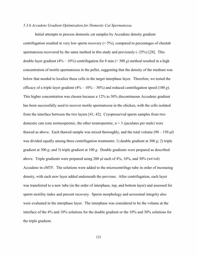
121
5.3.6 Accudenz Gradient Optimization for Domestic Cat Spermatozoa
Initial attempts to process domestic cat samples by Accudenz density gradient
centrifugation resulted in very low sperm recovery (< 5%), compared to percentages of cheetah
spermatozoa recovered by the same method in this study and previously (~25%) [28]. This
double layer gradient (4% – 10%) centrifugation for 8 min (× 300 g) method resulted in a high
concentration of motile spermatozoa in the pellet, suggesting that the density of the medium was
below that needed to localize these cells in the target interphase layer. Therefore, we tested the
efficacy of a triple layer gradient (4% – 10% – 30%) and reduced centrifugation speed (100 g).
This higher concentration was chosen because a 12% to 30% discontinuous Accudenz gradient
has been successfully used to recover motile spermatozoa in the chicken, with the cells isolated
from the interface between the two layers [41, 42]. Cryopreserved sperm samples from two
domestic cats (one normospermic, the other teratospermic, n = 3 ejaculates per male) were
thawed as above. Each thawed sample was mixed thoroughly, and the total volume (90 – 150 µl)
was divided equally among three centrifugation treatments: 1) double gradient at 300 g; 2) triple
gradient at 300 g; and 3) triple gradient at 100 g. Double gradients were prepared as described
above. Triple gradients were prepared using 200 µl each of 4%, 10%, and 30% (wt/vol)
Accudenz in cMTF. The solutions were added to the microcentrifuge tube in order of increasing
density, with each new layer added underneath the previous. After centrifugation, each layer
was transferred to a new tube (in the order of interphase, top, and bottom layer) and assessed for
sperm motility index and percent recovery. Sperm morphology and acrosomal integrity also
were evaluated in the interphase layer. The interphase was considered to be the volume at the
interface of the 4% and 10% solutions for the double gradient or the 10% and 30% solutions for
the triple gradient.
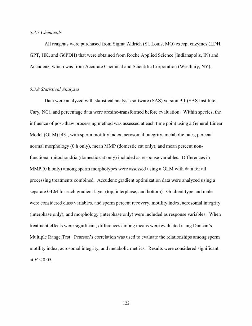
122
5.3.7 Chemicals
All reagents were purchased from Sigma Aldrich (St. Louis, MO) except enzymes (LDH,
GPT, HK, and G6PDH) that were obtained from Roche Applied Science (Indianapolis, IN) and
Accudenz, which was from Accurate Chemical and Scientific Corporation (Westbury, NY).
5.3.8 Statistical Analyses
Data were analyzed with statistical analysis software (SAS) version 9.1 (SAS Institute,
Cary, NC), and percentage data were arcsine-transformed before evaluation. Within species, the
influence of post-thaw processing method was assessed at each time point using a General Linear
Model (GLM) [43], with sperm motility index, acrosomal integrity, metabolic rates, percent
normal morphology (0 h only), mean MMP (domestic cat only), and mean percent non-
functional mitochondria (domestic cat only) included as response variables. Differences in
MMP (0 h only) among sperm morphotypes were assessed using a GLM with data for all
processing treatments combined. Accudenz gradient optimization data were analyzed using a
separate GLM for each gradient layer (top, interphase, and bottom). Gradient type and male
were considered class variables, and sperm percent recovery, motility index, acrosomal integrity
(interphase only), and morphology (interphase only) were included as response variables. When
treatment effects were significant, differences among means were evaluated using Duncan’s
Multiple Range Test. Pearson’s correlation was used to evaluate the relationships among sperm
motility index, acrosomal integrity, and metabolic metrics. Results were considered significant
at P < 0.05.

123
5.4 Results
5.4.1 Accudenz Density Gradient Optimization for Domestic Cat Spermatozoa
Motile spermatozoa were recovered from all layers of each gradient. However, most (≥
73%) of these cells in the gradients centrifuged at 300 × g were isolated from the pellet-
containing bottom layer (Table 5.1). Compared to these treatments, the percentage of
spermatozoa recovered from the target interphase layer of the 100 × g triple gradient was
increased (P < 0.05) ~3 to 20-fold (Table 5.1). Reduced centrifugation speed also was associated
with a ~50% improvement (P < 0.05) in sperm motility index (SMI) compared to the double
gradient (Table 5.1). Acrosomal integrity (% IA) and percentages of structurally normal sperm
were not different (P > 0.05) among gradient treatments (Table 5.1). Primary head abnormalities
Table 5.1. Characteristics of domestic cat* post-thaw sperm samples recovered from Accudenz density gradients. Values represent least-squares means ± standard errors.
Density gradient Parameter Double† Triple† Triple, low speed‡ Interphase (target layer)
Sperm recovered (%) 4 ± 7a 24 ± 7a 70 ± 7b
Sperm motility index 32 ± 5a 40 ± 5a,b 48 ± 4b
Acrosomal integrity (%) 34 ± 6 43 ± 6 43 ± 6
Normal sperm morphology (%) 54 ± 4 65 ± 4 52 ± 4
Macro/microcephaly (%) 12 ± 1a 4 ± 1b 3 ± 1b
Top layer
Sperm recovered (%) 2 ± 4a 3 ± 4a 18 ± 4b
Sperm motility index 35 ± 7 25 ± 7 39 ± 7
Bottom layer and sperm pellet
Sperm recovered (%) 94 ± 7a 73 ± 7a 12 ± 7b
Sperm motility index 53 ± 5 42 ± 5 44 ± 5 *n = 2 males, 3 ejaculates per male. †8 min at 300 g; ‡8 min at 100 g. a, bWithin rows, values with different superscripts differed (P < 0.05).
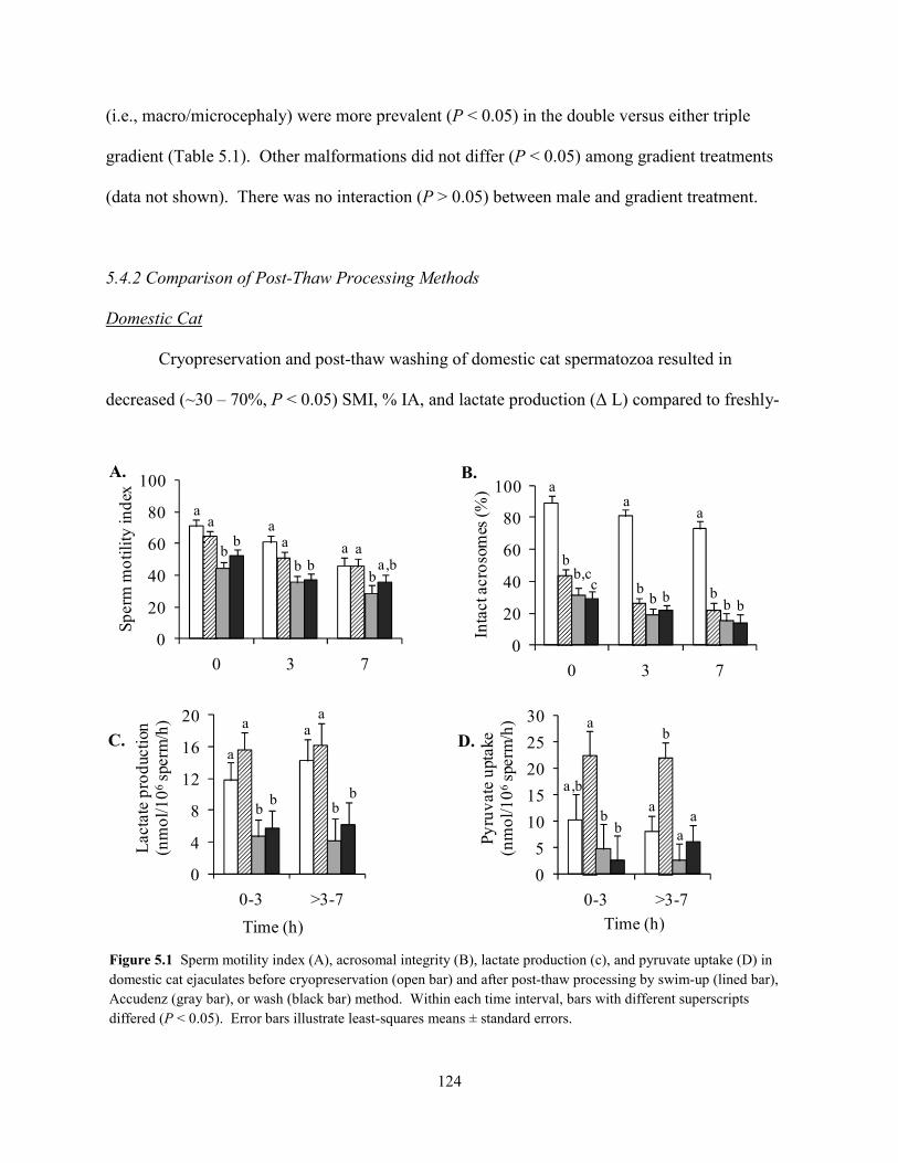
124
(i.e., macro/microcephaly) were more prevalent (P < 0.05) in the double versus either triple
gradient (Table 5.1). Other malformations did not differ (P < 0.05) among gradient treatments
(data not shown). There was no interaction (P > 0.05) between male and gradient treatment.
5.4.2 Comparison of Post-Thaw Processing Methods
Domestic Cat
Cryopreservation and post-thaw washing of domestic cat spermatozoa resulted in
decreased (~30 – 70%, P < 0.05) SMI, % IA, and lactate production (∆ L) compared to freshly-
0
4
8
12
16
20
0-3 >3-7
Lac
tate
pro
duct
ion
(nm
ol/1
06sp
erm
/h)
Time (h)
a
a
b
aa
bb
b
0
5
10
15
20
25
30
0-3 >3-7
Pyru
vate
upt
ake
(nm
ol/1
06sp
erm
/h)
Time (h)
a,b
a
ba
b
b aa
0
20
40
60
80
100
0 3 7
Inta
ct a
cros
omes
(%) a
b
cb,c
a
bb b
a
bb b
0
20
40
60
80
100
0 3 7
Sper
m m
otili
ty in
dex
aa
bb
aa
b ba a
a,bb
B. A.
C. D.
0-3 >3-7Time (h)
Figure 5.1 Sperm motility index (A), acrosomal integrity (B), lactate production (c), and pyruvate uptake (D) in domestic cat ejaculates before cryopreservation (open bar) and after post-thaw processing by swim-up (lined bar), Accudenz (gray bar), or wash (black bar) method. Within each time interval, bars with different superscripts differed (P < 0.05). Error bars illustrate least-squares means ± standard errors.
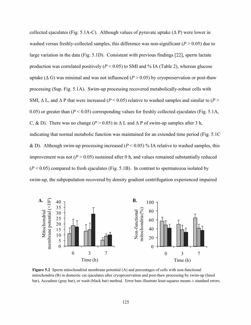
125
collected ejaculates (Fig. 5.1A-C). Although values of pyruvate uptake (∆ P) were lower in
washed versus freshly-collected samples, this difference was non-significant (P > 0.05) due to
large variation in the data (Fig. 5.1D). Consistent with previous findings [22], sperm lactate
production was correlated positively (P < 0.05) to SMI and % IA (Table 2), whereas glucose
uptake (∆ G) was minimal and was not influenced (P > 0.05) by cryopreservation or post-thaw
processing (Sup. Fig. 5.1A). Swim-up processing recovered metabolically-robust cells with
SMI, ∆ L, and ∆ P that were increased (P < 0.05) relative to washed samples and similar to (P >
0.05) or greater than (P < 0.05) corresponding values for freshly collected ejaculates (Fig. 5.1A,
C, & D). There was no change (P > 0.05) in ∆ L and ∆ P of swim-up samples after 3 h,
indicating that normal metabolic function was maintained for an extended time period (Fig. 5.1C
& D). Although swim-up processing increased (P < 0.05) % IA relative to washed samples, this
improvement was not (P > 0.05) sustained after 0 h, and values remained substantially reduced
(P < 0.05) compared to fresh ejaculates (Fig. 5.1B). In contrast to spermatozoa isolated by
swim-up, the subpopulation recovered by density gradient centrifugation experienced impaired
0
20
40
60
80
100
0 3 7
Non
-fun
ctio
nal
mito
chon
dria
(%)
Time (h)
05
10152025303540
0 3 7
Mito
chon
dria
l m
embr
ane
pote
ntia
l (×1
03)
Time (h)
B. A.
Figure 5.2 Sperm mitochondrial membrane potential (A) and percentages of cells with non-functional mitochondria (B) in domestic cat ejaculates after cryopreservation and post-thaw processing by swim-up (lined bar), Accudenz (gray bar), or wash (black bar) method. Error bars illustrate least-squares means ± standard errors.
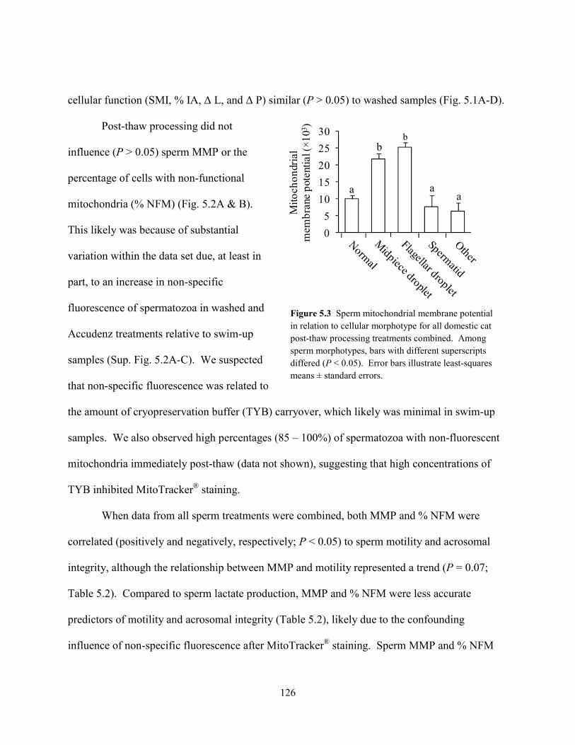
126
cellular function (SMI, % IA, ∆ L, and ∆ P) similar (P > 0.05) to washed samples (Fig. 5.1A-D).
Post-thaw processing did not
influence (P > 0.05) sperm MMP or the
percentage of cells with non-functional
mitochondria (% NFM) (Fig. 5.2A & B).
This likely was because of substantial
variation within the data set due, at least in
part, to an increase in non-specific
fluorescence of spermatozoa in washed and
Accudenz treatments relative to swim-up
samples (Sup. Fig. 5.2A-C). We suspected
that non-specific fluorescence was related to
the amount of cryopreservation buffer (TYB) carryover, which likely was minimal in swim-up
samples. We also observed high percentages (85 – 100%) of spermatozoa with non-fluorescent
mitochondria immediately post-thaw (data not shown), suggesting that high concentrations of
TYB inhibited MitoTracker® staining.
When data from all sperm treatments were combined, both MMP and % NFM were
correlated (positively and negatively, respectively; P < 0.05) to sperm motility and acrosomal
integrity, although the relationship between MMP and motility represented a trend (P = 0.07;
Table 5.2). Compared to sperm lactate production, MMP and % NFM were less accurate
predictors of motility and acrosomal integrity (Table 5.2), likely due to the confounding
influence of non-specific fluorescence after MitoTracker® staining. Sperm MMP and % NFM
0
5
10
15
20
25
30
Mito
chon
dria
l m
embr
ane
pote
ntia
l (×1
03)
b
aaa
b
Figure 5.3 Sperm mitochondrial membrane potential in relation to cellular morphotype for all domestic cat post-thaw processing treatments combined. Among sperm morphotypes, bars with different superscripts differed (P < 0.05). Error bars illustrate least-squares means ± standard errors.
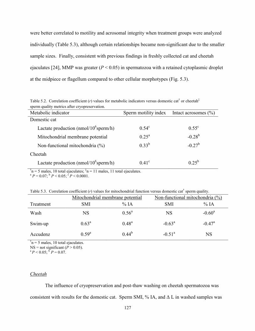
127
were better correlated to motility and acrosomal integrity when treatment groups were analyzed
individually (Table 5.3), although certain relationships became non-significant due to the smaller
sample sizes. Finally, consistent with previous findings in freshly collected cat and cheetah
ejaculates [24], MMP was greater (P < 0.05) in spermatozoa with a retained cytoplasmic droplet
at the midpiece or flagellum compared to other cellular morphotypes (Fig. 5.3).
Table 5.2. Correlation coefficient (r) values for metabolic indicators versus domestic cat† or cheetah‡ sperm quality metrics after cryopreservation.
Metabolic indicator Sperm motility index Intact acrosomes (%) Domestic cat
Lactate production (nmol/106sperm/h) 0.54c 0.55c
Mitochondrial membrane potential 0.25a -0.28b
Non-functional mitochondria (%) 0.33b -0.27b
Cheetah
Lactate production (nmol/106sperm/h) 0.41c 0.25b †n = 5 males, 10 total ejaculates; ‡n = 11 males, 11 total ejaculates. a P = 0.07; b P < 0.05; c P < 0.0001.
Table 5.3. Correlation coefficient (r) values for mitochondrial function versus domestic cat† sperm quality.
Mitochondrial membrane potential Non-functional mitochondria (%) Treatment SMI % IA SMI % IA
Wash NS 0.56a NS -0.60a
Swim-up 0.63a 0.48a -0.63a -0.47a
Accudenz 0.59a 0.44b -0.51a NS †n = 5 males, 10 total ejaculates. NS = not significant (P > 0.05). a P < 0.05; b P = 0.07.
Cheetah
The influence of cryopreservation and post-thaw washing on cheetah spermatozoa was
consistent with results for the domestic cat. Sperm SMI, % IA, and ∆ L in washed samples was
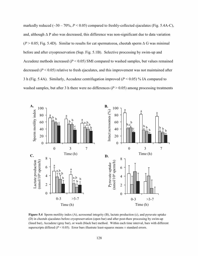
128
markedly reduced (~30 – 70%, P < 0.05) compared to freshly-collected ejaculates (Fig. 5.4A-C),
and, although ∆ P also was decreased, this difference was non-significant due to data variation
(P > 0.05; Fig. 5.4D). Similar to results for cat spermatozoa, cheetah sperm ∆ G was minimal
before and after cryopreservation (Sup. Fig. 5.1B). Selective processing by swim-up and
Accudenz methods increased (P < 0.05) SMI compared to washed samples, but values remained
decreased (P < 0.05) relative to fresh ejaculates, and this improvement was not maintained after
3 h (Fig. 5.4A). Similarly, Accudenz centrifugation improved (P < 0.05) % IA compared to
washed samples, but after 3 h there were no differences (P > 0.05) among processing treatments
0
2
4
6
8
0-3 >3-7
Pyru
vate
upt
ake
(nm
ol/1
06sp
erm
/h)
Time (h)
0
20
40
60
80
100
0 3 7
Inta
ct a
cros
omes
(%)
Time (h)
a
b,cb
c
a a
b bb
b bb
0
2
4
6
8
0-3 >3-7
Lact
ate
prod
uctio
n(n
mol
/106
sper
m/h
)
a
a,ba,b
b
a
a,bb
b
0
20
40
60
80
100
0 3 7
Sper
m m
otili
ty in
dex
Time (h)
ab b
c
a
b b
c
aa,b
a,bb
0-3 >3-7Time (h)
Figure 5.4 Sperm motility index (A), acrosomal integrity (B), lactate production (c), and pyruvate uptake (D) in cheetah ejaculates before cryopreservation (open bar) and after post-thaw processing by swim-up (lined bar), Accudenz (gray bar), or wash (black bar) method. Within each time interval, bars with different superscripts differed (P < 0.05). Error bars illustrate least-squares means ± standard errors.
B. A.
C. D.
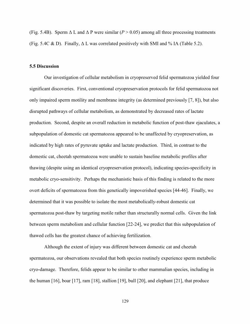
129
(Fig. 5.4B). Sperm ∆ L and ∆ P were similar (P > 0.05) among all three processing treatments
(Fig. 5.4C & D). Finally, ∆ L was correlated positively with SMI and % IA (Table 5.2).
5.5 Discussion
Our investigation of cellular metabolism in cryopreserved felid spermatozoa yielded four
significant discoveries. First, conventional cryopreservation protocols for felid spermatozoa not
only impaired sperm motility and membrane integrity (as determined previously [7, 8]), but also
disrupted pathways of cellular metabolism, as demonstrated by decreased rates of lactate
production. Second, despite an overall reduction in metabolic function of post-thaw ejaculates, a
subpopulation of domestic cat spermatozoa appeared to be unaffected by cryopreservation, as
indicated by high rates of pyruvate uptake and lactate production. Third, in contrast to the
domestic cat, cheetah spermatozoa were unable to sustain baseline metabolic profiles after
thawing (despite using an identical cryopreservation protocol), indicating species-specificity in
metabolic cryo-sensitivity. Perhaps the mechanistic basis of this finding is related to the more
overt deficits of spermatozoa from this genetically impoverished species [44-46]. Finally, we
determined that it was possible to isolate the most metabolically-robust domestic cat
spermatozoa post-thaw by targeting motile rather than structurally normal cells. Given the link
between sperm metabolism and cellular function [22-24], we predict that this subpopulation of
thawed cells has the greatest chance of achieving fertilization.
Although the extent of injury was different between domestic cat and cheetah
spermatozoa, our observations revealed that both species routinely experience sperm metabolic
cryo-damage. Therefore, felids appear to be similar to other mammalian species, including in
the human [16], boar [17], ram [18], stallion [19], bull [20], and elephant [21], that produce

130
ejaculates exhibiting impaired post-thaw metabolism. The exact origin of this disruption remains
to be elucidated. However, it was important to consider that cryopreservation-induced stress to
plasma membranes could cause leakage of sperm glycolytic enzymes [47]. Therefore, we
postulated that sperm metabolic rates (measured by changes in medium glucose, pyruvate, and
lactate concentrations) might falsely appear to be increased post-thawing due to greater activity
of enzymes released into the medium. Cytoplasmic droplets (a common abnormality in cat and
cheetah spermatozoa [22, 46]) are particularly susceptible to post-thaw LDH leakage [47], and
this could confound the use of lactate production as an indicator of cellular quality in thawed
ejaculate. Nonetheless, there was no evidence of glycolytic enzyme leakage from cat or cheetah
spermatozoa. On the contrary, rates of pyruvate and lactate metabolism were reduced (non-
significantly in the case of pyruvate), and glucose uptake remained minimal post-thaw.
Furthermore, rates of lactate production were correlated positively to sperm motility, acrosomal
integrity, and MMP. This finding provides strong support of our previous studies [22-24] and
indicates that lactate production is a reliable metric of sperm function in both the domestic cat
and cheetah.
Although rates of lactate production revealed metabolic cryo-damage in both the
domestic cat and cheetah, we also identified an important species difference in the extent of
cellular injury. Unlike the domestic cat, metabolic function in cheetah spermatozoa was
uniformly impaired by cryopreservation and not improved by selective processing via swim-up
or density gradient centrifugation. Therefore, future efforts to improve the post-thaw function of
cheetah spermatozoa should focus on preventing damage to the freshly-collected cells, perhaps
by testing alternative cryoprotectants (e.g., non-permeating sugars [48]) or preservation
approaches (e.g., vitrification [49]). Regardless, the revealed differences re-emphasize the value
131
of conducting cross-species comparisons, and, for felids, the importance of the domestic cat as a
research model [8, 22-24, 27, 34, 50, 51]. Besides determining what reproductive mechanisms
are conserved, this comparative approach can identify unique traits for a given species that
eventually may permit large-scale, systematic cryo-banking of germplasm. Because the present
study demonstrated that a proportion of domestic cat spermatozoa survived cryopreservation
with their metabolic machinery intact, this cellular subpopulation could be especially useful for
future research studies designed to understand the key biological factors that contribute to sperm
cryo-survival.
The relatively high rates of substrate utilization in cat ejaculates processed by swim-up
versus Accudenz centrifugation confirmed our suspicion that post-thaw sperm metabolic
function was more closely linked to cellular motility than morphology. Although impaired in
teratospermic felids, sperm lactate production also is compromised in freshly-collected cheetah
ejaculates that contain high proportions of structurally normal spermatozoa [22]. We have
speculated [22] that disrupted metabolism could explain why even structurally normal
spermatozoa from teratospermic ejaculates often are incapable of fertilization [26]. Furthermore,
we previously discovered important differences in sperm glycolytic [23] and oxidative
metabolism [24] between the cat and cheetah that were related to species physiology rather than
sperm morphology. For example, sperm MMP was ~95% lower in the cheetah compared to both
normospermic and teratospermic domestic cats [24]. Based on these collective observations, it
makes sense that metabolically-robust spermatozoa are recovered post-thaw by isolating cells on
the basis of motility rather than structural integrity. Furthermore, results of the present study
suggested that structurally normal cells were as susceptible to metabolic cryo-damage as their
abnormal counterparts. However, because only structurally intact spermatozoa are capable of
132
achieving fertilization [26], cellular morphology remains an important consideration for
optimizing post-thaw sperm fertilizing potential. Indeed, it now appears that the most effective
cryopreservation and processing protocols should yield post-thaw samples containing high
proportions of structurally normal, motile, and metabolically-robust cells.
In conclusion, our findings confirmed that important differences existed in gamete
physiology among felids and in the inherent ability of spermatozoa to retain metabolic function
after cryopreservation. In this case, cheetah spermatozoa, well known for a high incidence of
structural pleiomorphisms [44, 46, 52, 53], was particularly susceptible to metabolic cryo-
damage. In contrast, a proportion of domestic cat spermatozoa retained normal metabolism after
thawing, and perhaps this cellular subpopulation can provide clues for improving sperm cryo-
survival in other species or populations with less vigorous ejaculate quality. Finally, our results
confirmed that sperm metabolism, specifically the rate of lactate production, was a reliable
metric of cellular quality that can be applied to evaluate and enhance sperm cryo-survival.
5.6 References
1. Luvoni GC, Kalchschmidt E, Leoni S, Ruggiero C. Conservation of feline semen, part I:
cooling and freezing protocols. J Feline Med Surg 2003; 5: 203-208.
2. Swanson WF. Research in nondomestic species: experiences in reproductive physiology
research for conservation of endangered felids. ILAR J 2003; 44: 307-316.
3. Cocchia N, Ciani F, El-Rass R, Russo M, Borzacchiello G, Esposito V, Montagnaro S,
Avallone L, Tortora G, Lorizio R. Cryopreservation of feline epididymal spermatozoa
from dead and alive animals and its use in assisted reproduction. Zygote 2010; 18: 1-8.
133
4. Tebet JM, Martins MIM, Chirinea VH, Souza FF, Campagnol D, Lopes MD.
Cryopreservation effects on domestic cat epididymal versus electroejaculated
spermatozoa. Theriogenology 2006; 66: 1629-1632.
5. Herrick JR, Campbell M, Levens G, Moore T, Benson K, D'Agostino J, West G, Okeson
DM, Coke R, Portacio SC, Leiske K, Kreider C, et al. In vitro fertilization and sperm
cryopreservation in the black-footed cat (Felis nigripes) and sand cat (Felis margarita).
Biol Reprod 2010; 82: 552-562.
6. Baudi DLK, Jewgenow K, Pukazhenthi BS, Spercoski KM, Santos AS, Reghelin ALS,
Candido MV, Javorouski ML, Müller G, Morais RN. Influence of cooling rate on the
ability of frozen-thawed sperm to bind to heterologous zona pellucida, as assessed by
competitive in vitro binding assays in the ocelot (Leopardus pardalis) and tigrina
(Leopardus tigrinus). Theriogenology 2008; 69: 204-211.
7. Crosier AE, Pukazhenthi BS, Henghali JN, Howard JG, Dickman AJ, Marker L, Wildt
DE. Cryopreservation of spermatozoa from wild-born Namibian cheetahs (Acinonyx
jubatus) and influence of glycerol on cryosurvival. Cryobiology 2006; 52: 169-181.
8. Pukazhenthi B, Pelican K, Wildt D, Howard JG. Sensitivity of domestic cat (Felis catus)
sperm from normospermic versus teratospermic donors to cold-induced acrosome
damage. Biol Reprod 1999; 61: 135-141.
9. Pukazhenthi BS, Noiles E, Pelican K, Donoghue AM, Wildt DE, Howard JG. Osmotic
effects on feline spermatozoa from normospermic versus teratospermic donors.
Cryobiology 2000; 40: 139-150.
134
10. Pukazhenthi B, Laroe D, Crosier AE, Bush LM, Spindler R, Pelican KM, Bush M,
Howard JG, Wildt DE. Challenges in cryopreservation of clouded leopard (Neofelis
nebulosa) spermatozoa. Theriogenology 2006; 66: 1790-1796.
11. Ganan N, Gonzalez R, Garde JJ, Martinez F, Vargas A, Gomendio M, Roldan ERS.
Assessment of semen quality, sperm cryopreservation, and heterologous IVF in the
critically endangered Iberian lynx (Lynx pardinus). Reprod Fert Dev 2009; 21: 848-859.
12. Thiangtum K, Swanson WF, Howard J, Tunwattana W, Tongthainan D, Wichasilpa W,
Patumrattanathan P, Pinyopoommintr T. Assessment of basic seminal characteristics,
sperm cryopreservation, and heterologous in vitro fertilization in the fishing cat
(Prionailurus viverrinus). Reprod Fert Dev 2006; 18: 373-382.
13. Pukazhenthi BS, Spindler RE, Wildt DE, Bush LM, Howard JG. Osmotic properties of
spermatozoa from felids producing different proportions of pleiomorphisms: influence of
adding and removing cryoprotectant. Cryobiology 2002; 44: 288-300.
14. Mazur P, Leibo SP, Seidel GE, Jr. Cryopreservation of the germplasm of animals used in
biological and medical research: importance, impact, status, and future directions. Biol
Reprod 2008; 78: 2-12.
15. Thuwanut P, Chatdarong K, Johannisson A, Bergqvist AS, Soderquist L, Axner E.
Cryopreservation of epididymal cat spermatozoa: effects of in vitro antioxidative
enzymes supplementation and lipid peroxidation induction. Theriogenology 2010; 73:
1076-1087.
16. O'Connell M, McClure N, Lewis SEM. The effects of cryopreservation on sperm
morphology, motility, and mitochondrial function. Hum Reprod 2002; 17: 704-709.

135
17. Flores E, Fernandez-Novell JM, Pena A, Rigau T, Rodriguez-Gil JE. Cryopreservation-
induced alterations in boar spermatozoa mitochondrial function are related to changes in
the expression and location of midpiece mitofusin-2 and actin network. Theriogenology
2010; 74: 354-363.
18. Garcia-Alvarez O, Maroto-Morales A, Ramon M, del Olmo E, Montoro V, Dominguez-
Rebolledo AE, Bisbal A, Jimenez-Rabadan P, Perez-Guzman MD, Soler AJ. Analysis of
selected sperm by density gradient centrifugation might aid in the estimation of in vivo
fertility of thawed ram spermatozoa. Theriogenology 2010; 74: 979-988.
19. Schober D, Aurich C, Nohl H, Gille L. Influence of cryopreservation on mitochondrial
functions in equine spermatozoa. Theriogenology 2007; 68: 745-754.
20. Hallap T, Nagy S, Jaakma Ü, Johannisson A, Rodriguez-Martinez H. Mitochondrial
activity of frozen-thawed spermatozoa assessed by MitoTracker Deep Red 633.
Theriogenology 2005; 63: 2311-2322.
21. Sa-Ardrit M, Saikhun J, Thongtip N, Damyang M, Mahasawangkul S, Angkawanish T,
Jansittiwate S, Faisaikarm T, Kitiyanant Y, Pavasuthipaisit K, Pinyopummin A.
Ultrastructural alterations of frozen-thawed Asian elephant (Elephas maximus)
spermatozoa. Int J Androl 2006; 29: 346-352.
22. Terrell KA, Wildt DE, Anthony NM, Bavister BD, Leibo SP, Penfold LM, Marker LL,
Crosier AE. Evidence for compromised metabolic function and limited glucose uptake in
spermatozoa from the teratospermic domestic cat (Felis catus) and cheetah (Acinonyx
jubatus). Biol Reprod 2010; 83: 833-841.
23. Terrell KA, Wildt DE, Anthony NM, Bavister BD, Leibo SP, Penfold LM, Marker LL,
Crosier AE. Glycolytic enzyme activity is essential for domestic cat (Felis catus) and
136
cheetah (Acinonyx jubatus) sperm motility and viability in a sugar-free medium. Biol
Reprod 2011; 84: 1198-1206.
24. Terrell KA, Wildt DE, Anthony NM, Bavister BD, Leibo SP, Penfold LP, Crosier
AE. Oxidative metabolism is essential for felid sperm function, but is substantially
lower in cheetah (Acinonyx jubatus) compared to domestic cat (Felis catus) ejaculate.
Biol Reprod 2011; 83: 833-841.
25. McLaughlin EA, Ford WCL, Hull MGR. The contribution of the toxicity of a glycerol
egg-yolk citrate cryopreservative to the decline in human sperm motility during
cryopreservation. J Reprod Fertil 1992; 95: 749-754.
26. Howard JG, Donoghue AM, Johnston LA, Wildt DE. Zona pellucida filtration of
structurally abnormal spermatozoa and reduced fertilization in teratospermic cats. Biol
Reprod 1993; 49: 131-139.
27. Howard JG, Brown JL, Bush M, Wildt DE. Teratospermic and normospermic domestic
cats: ejaculate traits, pituitary-gonadal hormones, and improvement of spermatozoal
motility and morphology after swim-up processing. J Androl 1990; 11: 204-215.
28. Crosier A, Henghali J, Howard JG, Pukazhenthi BS, Terrell KA, Marker LL, Wildt DE.
Improved quality of cryopreserved cheetah (Acinonyx jubatus) spermatozoa after
centrifugation through Accudenz. J Androl 2009; 30: 298-308.
29. Stendardi A, Focarelli R, Piomboni P, Palumberi D, Serafini F, Ferramosca A, Zara V.
Evaluation of mitochondrial respiratory efficiency during in vitro capacitation of human
spermatozoa. Int J Androl 2011 (in press).
30. Bohnensack R, Halangk W. Control of respiration and of motility in ejaculated bull
spermatozoa. Biochim Biophys Acta 1986; 850: 72-79.
137
31. Minelli A, Moroni M, Castellini C, Lattaioli P, Mezzasoma I, Ronquist G. Rabbit
spermatozoa: a model system for studying ATP homeostasis and motility. J Androl 1999;
20: 259-266.
32. Kamp G, Busselmann G, Lauterwein J. Spermatozoa: models for studying regulatory
aspects of energy metabolism. Experientia 1996; 52: 487-494.
33. Pukazhenthi BS, Neubauer K, Jewgenow K, Howard JG, Wildt DE. The impact and
potential etiology of teratospermia in the domestic cat and its wild relatives.
Theriogenology 2006; 66: 112-121.
34. Long JA, Wildt DE, Wolfe BA, Crister JK, DeRossi RV, Howard JG. Sperm capacitation
and the acrosome reaction are compromised in teratospermic domestic cats. Biol Reprod
1996; 54: 638-646.
35. Howard JG. Semen collection and analysis in carnivores. In: Fowler ME (ed.), Zoo and
Wild Animal Medicine: Current Therapy III. Philadelphia: WB Saunders Co.; 1993: 390-
399.
36. Gardner DK, Leese HJ. Concentrations of nutrients in mouse oviduct fluid and their
effects on embryo development and metabolism in vitro. J Reprod Fertil 1990; 88: 361-
368.
37. Bavister BD. Substitution of a synthetic polymer for protein in a mammalian gamete
culture system. J Exp Zool 1981; 217: 45-51.
38. Shah D, Naciri M, Clee P, Al-Rubeai M. NucleoCounter: an efficient technique for the
determination of cell number and viability in animal cell culture processes.
Cytotechnology 2006; 51: 39-44.
138
39. Larson JL, Miller DJ. Simple histochemical stain for acrosomes on sperm from several
species. Mol Reprod Dev 1999; 52: 445-449.
40. Passonneau JV, Lowry OH. Enzymatic Analysis: A Practical Guide. Totowa, NJ:
Humana Press; 1993.
41. McLean DJ, Feltmann AJ, Froman DP. Transfer of sperm into a chemically defined
environment by centrifugation through 12% (wt/vol) Accudenz. Poult Sci 1998; 77: 163-
168.
42. Long JA, Bongalhardo DC, Pelaez J, Saxena S, Settar P, O'Sullivan NP, Fulton JE.
Rooster semen cryopreservation: effect of pedigree line and male age on post-thaw sperm
function. Poult Sci; 89: 966-973.
43. SAS. SAS/STAT User's Guide: Statistics. Version 9.1. Cary, NC: Statistical Analysis
System Institute, Inc; 2002.
44. Wildt DE, Bush M, Howard JG, O'Brien SJ, Meltzer D, van Dyk A, Ebedes H, Brand DJ.
Unique seminal quality in the South African cheetah and a comparative evaluation in the
domestic cat. Biol Reprod 1983; 29: 1019-1025.
45. O'Brien SJ, Roelke ME, Marker L, Newman A, Winkler CA, Meltzer D, Colly L,
Evermann JF, Bush M, Wildt DE. Genetic basis for species vulnerability in the cheetah.
Science 1985; 227: 1428-1434.
46. Crosier AE, Marker L, Howard JG, Pukazhenthi BS, Henghali JN, Wildt DE. Ejaculate
traits in the Namibian cheetah (Acinonyx jubatus): influence of age, season, and captivity.
Reprod Fertil Dev 2007; 19: 370-382.
47. Harrison R, White I. Glycolytic enzymes in the spermatozoa and cytoplasmic droplets of
bull, boar, and ram, and their leakage after shock. J Reprod Fertil 1972; 30: 105-115.
139
48. Sztein JM, Noble K, Farley JS, Mobraaten LE. Comparison of permeating and
nonpermeating cryoprotectants for mouse sperm cryopreservation. Cryobiology 2001; 42:
28-39.
49. Nawroth F, Isachenko V, Dessole S, Rahimi G, Farina M, Vargiu N, Mallmann P,
Dattena M, Capobianco G, Peters D, Orth I, Isachenko E. Vitrification of human
spermatozoa without cryoprotectants. CryoLetters 2002; 23: 93-102.
50. Penfold LM, Jost L, Evenson DP, Wildt DE. Normospermic versus teratospermic
domestic cat sperm chromatin integrity evaluated by flow cytometry and intracytoplasmic
injection. Biol Reprod 2003; 69: 1730-1735.
51. Pukazhenthi BS, Wildt DE, Ottinger MA, Howard JG. Compromised sperm protein
phosphorylation after capacitation, swim-up, and zona pellucida exposure in
teratospermic domestic cats. J Androl 1996; 17: 409-419.
52. Wildt DE, O'Brien SJ, Howard JG, Caro TM, Roelke ME, Brown JL, Bush M. Similarity
in ejaculate-endocrine characteristics in captive versus free-ranging cheetahs of two
subspecies. Biol Reprod 1987; 36: 351-360.
53. Wildt DE, Brown JL, Bush M, Barone MA, Cooper KA, Grisham J, Howard JG.
Reproductive status of cheetahs (Acinonyx jubatus) in North American zoos: the benefits
of physiological surveys for strategic planning. Zoo Biol 1993; 12: 45-80.
140
CHAPTER 6: GENERAL DISCUSSION
6.1 Metabolic Profiles of Cat and Cheetah Spermatozoa
Teratospermia is an intriguing reproductive phenomenon common among felid species
facing extinction risk [1], including the cheetah [2-4]. Although multiple energy-dependent
processes are compromised in spermatozoa from teratospermic ejaculates [1], sperm cellular
metabolism has not been previously investigated in wild felids. Through comparative studies of
the cheetah and domestic cat, this dissertation aimed to elucidate the mechanisms of felid sperm
energy production, including the influences of species physiology, teratospermia, and sperm
cryopreservation. Access to biological samples from a rare species (i.e., the cheetah) required
extensive collaboration with conservation institutions in the United States and in southern Africa.
This collaborative approach was facilitated by access to the Smithsonian Conservation Biology
Institute’s Mobile Research Laboratory and a novel, field-friendly strategy to quantify sperm
metabolic rates (described in Chapter 2).
Because there was almost no previous knowledge about sperm metabolism in felids,
understanding the impacts of teratospermia and sperm cryopreservation required first identifying
‘normal’ metabolic profiles for each species (Chaper 2). Patterns of metabolic substrate
utilization were remarkably similar between cat and cheetah spermatozoa: glucose uptake was
minimal, but spermatozoa imported pyruvate and produced lactate at comparatively high rates.
These results were enigmatic because the opposite pattern of substrate utilization was expected,
i.e., high rates of glucose uptake, corresponding with total glycolytic activity, and lower rates of
lactate production, proportional to anaerobic glycolysis. The investigation of sperm function in
the presence/absence of exogenous substrates (Chapter 3) elucidated this finding through the
discovery that cat and cheetah spermatozoa produced lactate independently of glycolysis and
141
ATP production via the reduction of exogenous pyruvate. Furthermore, cellular function
(motility and viability) was maintained in a substrate-free culture medium, indicating the active
metabolism of endogenous substrates. Intriguingly, the activity of the glycolytic enzyme
GAPDH was required for sperm motility, even in the complete absence of exogenous and
endogenous (i.e., glycogen) sources of glucose. These findings suggested that glycerol (most
likely derived from endogenous lipid) was an important metabolic substrate in felid spermatozoa
and hinted at a key role of lipid oxidation in cellular energy production. The results of Chapter 4
supported this model by confirming the essential role of oxidative metabolism in felid sperm
function. Collectively, the studies described in Chapters 2 through 4 generated informative
sperm metabolic profiles for the cat and cheetah. Felid spermatozoa appeared to be fueled
primarily by the metabolism of endogenous lipid, with the glycolytic pathway serving an
essential role in the breakdown of glycerol rather than glucose. The consistency of findings in
the cat and cheetah increases confidence in the proposed model of sperm energy production and
will provide an excellent starting point for future studies to elucidate these mechanisms in other
felid species.
6.2 Metabolic Indicators of Sperm Function
Initial assessments of metabolic profiles in cat and cheetah spermatozoa (Chapter 2)
suggested that rates of pyruvate uptake and lactate production were accurate predictors of sperm
function. Because higher variation was observed among pyruvate data, subsequent studies
focused primarily on lactate as a sperm quality indicator. Lactate production was impaired
(together with cellular motility and viability) by chemical inhibition of metabolism (Chapters 3
and 4) and following cryopreservation (Chapter 5). Although the link between lactate production
142
and sperm function had been identified previously in the boar [5] and donkey [6], the current
studies were the first to reveal an indirect relationship between lactate and sperm energy
production. Given the important role of oxidative metabolism established in Chapter 4, it is
likely that the link between lactate and sperm function is the mitochondrial production of
NADH, a coenzyme required for the formation of both ATP (via OXPHOS) and lactate.
The results of Chapter 3 determined that rates of lactate production were largely
dependent on sperm medium composition (specifically the presence of pyruvate), and chemical
inhibition of this process did not impair sperm function. The influence of medium composition
highlights the critical importance of a standardized, chemically-defined cellular environment for
generating reliable conclusions about sperm metabolism. Given the findings of Chapter 3, the
link to sperm function must be validated before lactate is applied as a cellular indicator in
different culture media. But despite this caveat, lactate production can provide an objective,
quantitative indicator of cellular function at relatively low-cost (e.g., compared to Computer
Assisted Sperm Analysis) and offers exciting possibilities for future research.
6.3 Influence of Species Physiology
Although sperm metabolic profiles were generally similar in the cat and cheetah, results
of Chapters 2 through 4 revealed two intriguing differences between the species. First, higher
rates of glucose uptake (Chapter 2) combined with increased sensitivity to GAPDH inhibition
(Chapter 3) indicated that glycolytic metabolism was more active in domestic cat compared to
cheetah spermatozoa. Secondly, low mitochondrial membrane potential combined with extreme
sensitivity to OXPHOS inhibition (Chapter 4) suggested that sperm oxidative metabolism was
impaired in the cheetah. Although minimal rates of oxidative metabolism could be considered
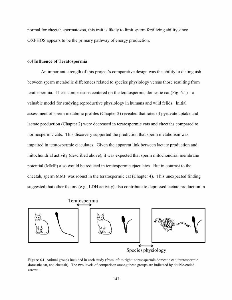
143
normal for cheetah spermatozoa, this trait is likely to limit sperm fertilizing ability since
OXPHOS appears to be the primary pathway of energy production.
6.4 Influence of Teratospermia
An important strength of this project’s comparative design was the ability to distinguish
between sperm metabolic differences related to species physiology versus those resulting from
teratospermia. These comparisons centered on the teratospermic domestic cat (Fig. 6.1) – a
valuable model for studying reproductive physiology in humans and wild felids. Initial
assessment of sperm metabolic profiles (Chapter 2) revealed that rates of pyruvate uptake and
lactate production (Chapter 2) were decreased in teratospermic cats and cheetahs compared to
normospermic cats. This discovery supported the prediction that sperm metabolism was
impaired in teratospermic ejaculates. Given the apparent link between lactate production and
mitochondrial activity (described above), it was expected that sperm mitochondrial membrane
potential (MMP) also would be reduced in teratospermic ejaculates. But in contrast to the
cheetah, sperm MMP was robust in the teratospermic cat (Chapter 4). This unexpected finding
suggested that other factors (e.g., LDH activity) also contribute to depressed lactate production in
Teratospermia
Species physiology
Figure 6.1 Animal groups included in each study (from left to right: normospermic domestic cat, teratospermic domestic cat, and cheetah). The two levels of comparison among these groups are indicated by double-ended arrows.
144
felid spermatozoa. Such an interpretation is supported by the observation that sperm lactate
production was more severely depressed in the cheetah than in the teratospermic cat. Thus,
teratospermic felids may share a common mechanism of impaired lactate production, while the
low MMP of cheetah spermatozoa exacerbates this defect. Although requiring further study,
these findings illustrate the importance of the domestic cat model for identifying physiological
consequences of teratospermia in felids, particularly in species such as the cheetah that lack a
normospermic ‘control’ population.
6.5 Influence of Sperm Cryopreservation
Consistent with previous studies [7-9], cryopreservation of cat and cheetah spermatozoa
resulted in reduced post-thaw motility and severe losses in acrosomal integrity (Chapter 5).
These cells also exhibited decreased rates of lactate production, providing the first evidence of
metabolic cryo-damage in felid spermatozoa. The same pattern was observed with respect to
pyruvate uptake, but differences were obscured by the large variation in those data. Although
lactate production was markedly reduced following cryopreservation, it was unclear whether
metabolic machinery was impaired directly (e.g., mitochondrial damage) or indirectly (e.g.,
increased plasma membrane permeability) – an intriguing question for further study.
Evaluation of post-thaw processing methods revealed that a metabolically-robust sperm
subpopulation could be recovered in the domestic cat by selectively isolating highly motile
spermatozoa. In contrast, metabolic function remained compromised in spermatozoa isolated on
the basis of morphology, suggesting that structurally normal cells are as susceptible to metabolic
cryo-damage as their abnormal counterparts. Results of post-thaw processing were similar
between normospermic and teratospermic domestic cats (data were presented as a single group to
145
increase sample size). In the cheetah, sperm lactate production remained compromised after
thawing, regardless of processing method. This key finding indicated that, in contrast to the
domestic cat, a metabolically ‘normal’ subpopulation of cheetah spermatozoa did not exist after
cryopreservation. Therefore, efforts to improve the fertilizing potential of cryopreserved cheetah
ejaculates should focus on preventing, rather than mitigating metabolic cryo-damage. Finally,
because metabolically-robust cells were recovered from the teratospermic cat, but not the
cheetah, the mechanism allowing certain felid spermatozoa to retain metabolic function post-
thaw appears to be species-specific.
6.5 Conclusions and Recommendations
Preserving and/or enhancing ejaculate quality in felids requires understanding the sperm
metabolic pathways that support, directly or indirectly, every individual process required for
fertilization. Both glycolysis and oxidative phosphorylation were essential for sperm motility
and viability in the cat and cheetah, but the role of each pathway in other cellular processes (e.g.,
capacitation) should be investigated. Felid spermatozoa actively metabolized endogenous
substrates, but this energy source may be limited, especially in poor quality ejaculates. Various
forms of lipid (e.g., phospholipid) should be evaluated as supplemental energy sources to
optimize sperm culture conditions. Furthermore, differences in the amount or composition of
substrates stored intracellularly may contribute to the significant variation in ejaculate quality
observed within felid species. Therefore, the characterization of sperm lipid profiles in felid
ejaculates is a high research priority. Mitochondrial activity was particularly important for
sperm energy production, but unidentified compounds present in the cryopreservation medium
interfered with mitochondrial probes, presenting a challenge to post-thaw evaluation of MMP. A
146
chemically-defined sperm cryopreservation medium is needed to overcome this challenge and
elucidate the mechanisms of metabolic cryo-damage. Cheetah spermatozoa were unable to
survive cryopreservation with metabolic function intact, indicating a need to further optimize
freezing/thawing protocols. However, certain domestic cat spermatozoa (even those from
teratospermic ejaculates) somehow were able to escape metabolic cryo-damage. Identifying the
factors that cause these cells to respond to freezing/thawing differently from other sperm
populations could generate innovative strategies for improving cryopreservation success.
Overall, this project emphasizes the importance of domestic models and comparative
research for understanding complex biological phenomena in rare animal species. Similarities in
sperm metabolic profiles between the cat and cheetah generated reliable conclusions about
energy production in felid spermatozoa. Yet even more exciting were the differences revealed
by these comparative studies. Teratospermia and species physiology influenced sperm
metabolism and/or cryosensitivity in unexpected, but important ways. These intriguing
differences provide an excellent starting point for future studies of felid sperm physiology that
can ultimately benefit both wildlife conservation and human health.
6.7 References
1. Pukazhenthi BS, Neubauer K, Jewgenow K, Howard JG, Wildt DE. The impact and
potential etiology of teratospermia in the domestic cat and its wild relatives.
Theriogenology 2006; 66: 112-121.
2. Wildt DE, Bush M, Howard JG, O'Brien SJ, Meltzer D, van Dyk A, Ebedes H, Brand DJ.
Unique seminal quality in the South African cheetah and a comparative evaluation in the
domestic cat. Biol Reprod 1983; 29: 1019-1025.
147
3. Wildt DE, O'Brien SJ, Howard JG, Caro TM, Roelke ME, Brown JL, Bush M. Similarity
in ejaculate-endocrine characteristics in captive versus free-ranging cheetahs of two
subspecies. Biol Reprod 1987; 36: 351-360.
4. Crosier AE, Marker L, Howard JG, Pukazhenthi BS, Henghali JN, Wildt DE. Ejaculate
traits in the Namibian cheetah (Acinonyx jubatus): influence of age, season, and captivity.
Reprod Fertil Dev 2007; 19: 370-382.
5. Rigau T, Piedrafita J, Reverter A, Canal M, Rodríguez-Gil JE. The rate of L-lactate
production: a feasible parameter for the fresh diluted boar semen quality analysis. Anim
Reprod Sci 1996; 43: 161-172.
6. Miró J, Lobo V, Quintero-Moreno A, Medrano A, Peňa A, Rigau T. Sperm motility
patterns and metabolism in Catalonian donkey semen. Theriogenology 2005; 63: 1706-
1716.
7. Crosier AE, Pukazhenthi BS, Henghali JN, Howard JG, Dickman AJ, Marker L, Wildt
DE. Cryopreservation of spermatozoa from wild-born Namibian cheetahs (Acinonyx
jubatus) and influence of glycerol on cryosurvival. Cryobiology 2006; 52: 169-181.
8. Crosier A, Henghali J, Howard JG, Pukazhenthi BS, Terrell KA, Marker LL, Wildt DE.
Improved quality of cryopreserved cheetah (Acinonyx jubatus) spermatozoa after
centrifugation through Accudenz. J Androl 2009; 30: 298-308.
9. Pukazhenthi B, Pelican K, Wildt D, Howard JG. Sensitivity of domestic cat (Felis catus)
sperm from normospermic versus teratospermic donors to cold-induced acrosome
damage. Biol Reprod 1999; 61: 135-141.
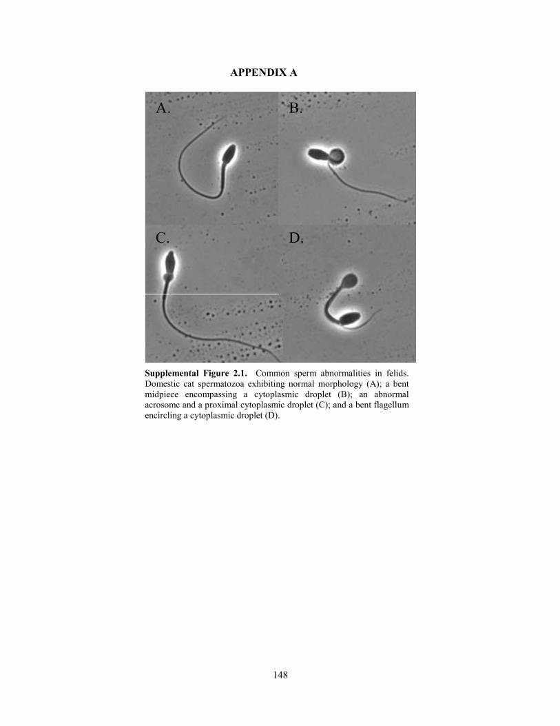
148
APPENDIX A
A.
C.
B.
D.
Supplemental Figure 2.1. Common sperm abnormalities in felids. Domestic cat spermatozoa exhibiting normal morphology (A); a bent midpiece encompassing a cytoplasmic droplet (B); an abnormal acrosome and a proximal cytoplasmic droplet (C); and a bent flagellum encircling a cytoplasmic droplet (D).
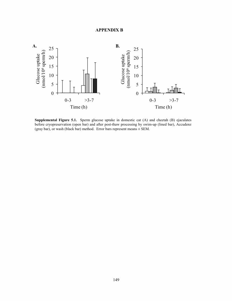
149
APPENDIX B
0
5
10
15
20
25
0-3 >3-7
Glu
cose
upt
ake
(nm
ol/1
06sp
erm
/h)
Time (h)
0
5
10
15
20
25
0-3 >3-7
Glu
cose
upt
ake
(nm
ol/1
06sp
erm
/h)
Time (h)
B. A.
Supplemental Figure 5.1. Sperm glucose uptake in domestic cat (A) and cheetah (B) ejaculates before cryopreservation (open bar) and after post-thaw processing by swim-up (lined bar), Accudenz (gray bar), or wash (black bar) method. Error bars represent means ± SEM.
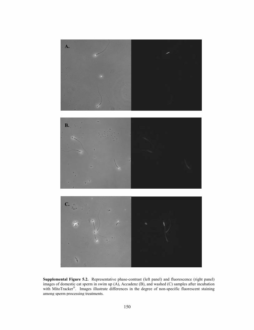
150
Supplemental Figure 5.2. Representative phase-contrast (left panel) and fluorescence (right panel) images of domestic cat sperm in swim up (A), Accudenz (B), and washed (C) samples after incubation with MitoTracker®. Images illustrate differences in the degree of non-specific fluorescent staining among sperm processing treatments.
A.
B.
C.
151
VITA
The author was born in Park Ridge, Illinois. She earned a B.S. in Cell and Molecular
Biology and a B.A. in Political Science from Tulane University in 2005. The following year, she
joined the University of New Orleans Department of Biological Sciences to pursue a Ph.D. in
Conservation Biology. She was the first student of the ZUNO program, a research partnership
between UNO and the Smithsonian’s National Zoological Park (NZP) established by her mentor,
Dr. Barry Bavister. In 2007 she joined Dr. Nicola Anthony’s laboratory, and the following year
relocated to Virginia to complete her dissertation research at the NZP’s Front Royal campus
(now the Smithsonian Conservation Biology Institute, SCBI). Dr. David Wildt (head of SCBI’s
Department of Reproductive Sciences) and Dr. Nicola Anthony (UNO) served as her dissertation
co-advisors. Drs. Barry Bavister, Adrienne Crosier, Stanley Leibo, and Bernard Rees were
members of her dissertation committee.