Embed Size (px)
Citation preview

DMD # 61747
1
Metabolism of MRX-I, a Novel Antibacterial Oxazolidinone, in
Humans: the Oxidative Ring-opening of 2,3-Dihydropyridin-4-one
Catalyzed by Non-P450 Enzymes
Jian Meng, Dafang Zhong, Liang Li, Zhengyu Yuan, Hong Yuan, Cen Xie, Jialan
Zhou, Chen Li, Mikhail Fedorovich Gordeev, Jinqian Liu, Xiaoyan Chen
Shanghai Institute of Materia Medica, Chinese Academy of Sciences, Shanghai,
China (J.M., D.Z., L.L., C.X., J.Z., C.L., X.C.); MicuRx Pharmaceuticals, Inc.,
Shanghai, China (Z.Y., H.Y.); and MicuRx Pharmaceuticals, Inc., Hayward, United
states (M.G., J.L.)
This article has not been copyedited and formatted. The final version may differ from this version.DMD Fast Forward. Published on February 20, 2015 as DOI: 10.1124/dmd.114.061747
at ASPE
T Journals on M
ay 24, 2018dm
d.aspetjournals.orgD
ownloaded from

DMD # 61747
2
Running Title
Metabolism of Antibacterial Oxazolidinone MRX-I in Humans
Corresponding author: Xiaoyan Chen
Shanghai Institute of Materia Medica, Chinese Academy of Sciences, 501 Haike Road,
Shanghai, China
Phone: +86-21-50800738; Fax: +86-21-50800738;
Email: [email protected]
Number of text pages: 25
Number of tables: 4
Number of figures: 11
Number of references: 19
Number of words in the Abstract: 236
Number of words in the Introduction: 348
Number of words in the Discussion: 1359
ABBREVIATIONS: FMO, flavin-containing monooxygenase; UPLC,
ultraperformance liquid chromatography; Triple TOF MS, triple time-of-flight mass
spectrometry; ABT, 1-aminobenzotriazole; 4-MP, 4-methylpyrazole; P450,
cytochrome P450; ADH, alcohol dehydrogenase; AKR, aldo-keto reductase; SDR,
short-chain dehydrogenase/reductase; ALDH, aldehyde dehydrogenase.
This article has not been copyedited and formatted. The final version may differ from this version.DMD Fast Forward. Published on February 20, 2015 as DOI: 10.1124/dmd.114.061747
at ASPE
T Journals on M
ay 24, 2018dm
d.aspetjournals.orgD
ownloaded from

DMD # 61747
3
ABSTRACT
MRX-I is an analogue of linezolid, containing a 2,3-dihydropyridin-4-one (DHPO)
ring rather than a morpholine ring. Our objectives were to characterize the major
metabolic pathways of MRX-I in humans and to clarify the mechanism underlying the
oxidative ring-opening of the DHPO. After an oral dose of MRX-I (600 mg), nine
metabolites were identified in humans. The principal metabolic pathway proposed
involved DHPO ring-opening, generating the main metabolites in the plasma and
urine: the hydroxyethyl amino propionic acid metabolite MRX445-1 and the
carboxymethyl amino propionic acid metabolite MRX459. An in vitro phenotyping
study demonstrated that multiple non-P450 enzymes are involved in the formation of
MRX445-1 and MRX459, including flavin-containing monooxygenase 5 (FMO5),
short-chain dehydrogenase/reductase (SDR), aldehyde ketone reductase (AKR), and
aldehyde dehydrogenase (ALDH). H218O experiments revealed that two 18O atoms are
incorporated into MRX445-1, one in the carboxyethyl group and the other in the
hydroxyl group, and three 18O atoms are incorporated into MRX459, two in the
carboxymethyl group and one in the hydroxyl group. Based on these results, the
mechanism proposed for DHPO ring opening involves the metabolism of MRX-I via
FMO5-mediated Baeyer–Villiger oxidation to an enol lactone, hydrolysis to an enol,
and enol–aldehyde tautomerism to an aldehyde. The aldehyde is reduced by SDR,
AKR, ALDH to MRX445-1 or oxidized by ALDH to MRX459. Our study suggests
that few clinical adverse drug–drug interactions should be anticipated between MRX-I
and P450 inhibitors or inducers.
This article has not been copyedited and formatted. The final version may differ from this version.DMD Fast Forward. Published on February 20, 2015 as DOI: 10.1124/dmd.114.061747
at ASPE
T Journals on M
ay 24, 2018dm
d.aspetjournals.orgD
ownloaded from
DMD # 61747
4
Introduction
Today, antibiotic resistance is still threatening the health of many people worldwide.
Therefore, it is of great urgency for the pharmaceutical industry to develop new
antibiotics with better safety profiles for drug-resistant bacteria (Talbot et al., 2006).
Oxazolidinone antibacterial agents bind to a distinct region of the 23S RNA adjacent
to the peptidyl transferase center of the 50S ribosomal subunit and show activity
against major Gram-positive pathogens (Dean, 1999). Its distinct binding site reduces
the cross-resistance between oxazolidinone and other antibacterial drugs. To date,
linezolid is the only oxazolidinone antibacterial agent approved by the US Food and
Drug Administration for the treatment of serious Gram-positive infections, including
those caused by antibiotic-resistant organisms. However, linezolid is subject to
serious safety limitations. The primary safety concerns are myelosuppression and
monoamine oxidase inhibition, which limit extended linezolid therapy (Vinh and
Rubinstein, 2009).
MRX-I, (5S)-5-[(isoxazol-3-ylamino)methyl]-3-[2,3,5-trifluoro-4-(4-oxo-2,3-
dihydropyridin-1-yl)phenyl]oxazolidin-2-one, is a novel oxazolidinone antibiotic
currently in clinical trials in China. One of the important changes in the structure of
the compound is the substitution of the morpholine heterocycle in linezolid with the
more planar 2,3-dihydropyridin-4-one (DHPO) ring, which contributes to the
increased antibacterial activity of MRX-I compared with that of linezolid (Gordeev
and Yuan, 2014). In nonclinical studies, MRX-I demonstrated the same or better
efficacy than linezolid in mouse infection models (Li et al., 2014). In preclinical
toxicology studies and phase I clinical trials, MRX-I displayed less propensity to
induce myelosuppression than did linezolid (Gordeev and Yuan, 2014).
Drug metabolism study is an integral and critical part of drug development.
This article has not been copyedited and formatted. The final version may differ from this version.DMD Fast Forward. Published on February 20, 2015 as DOI: 10.1124/dmd.114.061747
at ASPE
T Journals on M
ay 24, 2018dm
d.aspetjournals.orgD
ownloaded from
DMD # 61747
5
Information, such as the major circulating metabolites and the enzymes responsible
for their formation is vital for evaluating drug safety and drug–drug interactions. A
preliminary metabolism study in humans demonstrated that MRX-I is mainly
metabolized by an unusual metabolic pathway involving the oxidative DHPO
ring-opening. Therefore, the objectives of the current study were: 1) to characterize
the metabolic pathways of MRX-I in humans following an oral dose of MRX-I (600
mg); 2) to identify the enzymes involved in the DHPO ring-opening of MRX-I; and 3)
to understand the DHPO ring-opening mechanism with H218O experiments.
This article has not been copyedited and formatted. The final version may differ from this version.DMD Fast Forward. Published on February 20, 2015 as DOI: 10.1124/dmd.114.061747
at ASPE
T Journals on M
ay 24, 2018dm
d.aspetjournals.orgD
ownloaded from

DMD # 61747
6
Materials and Methods
Materials
MRX-I, HM5013199, HM5012569, HM5013876, HM1383, MRX-1314, and
MRX-1315 were provided by MicuRx Pharmaceuticals Inc. (Shanghai, China).
Methimazole, 1-aminobenzotriazole (ABT), 4-methylpyrazole (4-MP), flufenamic
acid, raloxifene, allopurinol, and disulfiram were purchased form Sigma-Aldrich (St.
Louis, MO, USA). Menadione was bought from Sinapharm Chemical Reagent Co.,
Ltd (Shanghai, China). Methotrexate was acquired from the National Institute for the
Control of Pharmaceutical and Biological Products (Beijing, China). H218O (97 atom %
18O) was obtained from the Shanghai Research Institute of Chemical Industry
(Shanghai, China).
Cryopreserved human hepatocytes, pooled human liver S9 fractions, liver cytosolic
fractions, liver microsomes, and recombinant FMO Supersomes, including FMO1,
FMO3, and FMO5, were purchased from BD Gentest (Woburn, CA, USA). NADPH,
NADP+ and NAD+ were purchased from Sigma-Aldrich.
All solvents for the ultraperformance liquid chromatography/triple time-of-flight
mass spectrometry (UPLC/Triple TOF MS) analyses were of high-performance liquid
chromatography (HPLC) grade (Merck, Darmstadt, Germany). Ultrapure water was
generated using a Milli-Q Gradient system (Millipore Corporation, Molsheim,
France). Other reagents were of analytical grade (Shanghai Chemical Plant, Shanghai,
China).
Study Protocol and Sample Collection
The human blood, urine and faces samples were collected from four healthy male
subjects during the course of a Phase-I clinical trial of MRX-I conducted at the
Clinical Trial Center of Shanghai Huashan Hospital. The study was conducted in
This article has not been copyedited and formatted. The final version may differ from this version.DMD Fast Forward. Published on February 20, 2015 as DOI: 10.1124/dmd.114.061747
at ASPE
T Journals on M
ay 24, 2018dm
d.aspetjournals.orgD
ownloaded from

DMD # 61747
7
accordance with the Declaration of Helsinki, and was approved by the Ethics
Committee of Huashan Hospital affiliated to Fudan University. All subjects gave their
voluntary informed consent and received multiple dose of 600 mg of MRX-I (tablet)
through q12h oral administration for 14 days. Blood, urine and faces samples were
collected within 24 hours before receiving the first dose of MRX-I,and were collected
at predetermined time points after receiving the last dose. Following the collection of
blood samples, the plasma was isolated by centrifugation, and stored at –20 °C until
analysis. To each fecal sample was added five volumes of methanol (1 g/5 ml),
followed by homogenization. The urine and feces samples were stored frozen at
–20 °C until analysis.
Metabolite Profiling and Identification
Preparation of Samples Plasma, urine, and feces samples were pooled and
prepared for metabolite profiling. Plasma samples were segregated by sample
collection time (predose, and 1.5 hours and 6 hours postdose), and equal volumes (50
μl) from each subject were pooled. The plasma proteins were precipitated by the
addition of three volumes of acetonitrile, vortexed for 1 min, and centrifuged for 5
min at 11,000 × g. The supernatant was transferred to glass tubes and evaporated to
dryness under N2 at 40 °C. The residues were reconstituted in 20% aqueous methanol
(400 μl), and a 10 μl aliquot of the resulting solution was injected into the UPLC
apparatus for analysis.
Urine or feces samples (predose and 0–24 hours postdose) were pooled by
combining volumes proportional to the total weight excreted from each subject at
each urine or feces collection interval. The pooled urine samples were centrifuged for
5 min at 11,000 × g and a 10 μl aliquot of the resulting supernatant was injected into
the UPLC apparatus for analysis. The pooled feces samples (500 μl) were transferred
This article has not been copyedited and formatted. The final version may differ from this version.DMD Fast Forward. Published on February 20, 2015 as DOI: 10.1124/dmd.114.061747
at ASPE
T Journals on M
ay 24, 2018dm
d.aspetjournals.orgD
ownloaded from

DMD # 61747
8
into glass tubes for evaporation to dryness under N2 at 40 °C. The residues were
reconstituted in 20% aqueous methanol (400 μl) and a 10 μl aliquot of the resulting
solution was injected into the UPLC apparatus for analysis.
UPLC-UV/Triple-TOF MS Analysis Chromatographic separation for the
metabolite profiling and identification was conducted on an Acquity UPLC HSS T3
column (100 mm × 2.1 mm i.d., 1.8 μm) using an Acquity UPLC system (Waters,
Milford, MA, USA). The mobile phase consisted of 0.05% formic acid in water
(solvent A) and methanol (solvent B). The flow rate was maintained at 0.45 ml/min.
MRX-I and its metabolites were eluted with a linear gradient of the mobile phase
composition, as follows: 5% solvent B for 5 min, a linear gradient to 40% solvent B at
17 min, held at 40% solvent B until 20 min, a linear gradient to 100% solvent B at 22
min, and reequilibration of the column to 5% solvent B for 4 min. The eluted fractions
were monitored by UV detection at 254 nm.
MS detection was achieved using a Triple TOF 5600+ MS/MS system (AB Sciex,
Concord, Ontario, Canada) in the positive ESI mode. The mass range was set at m/z
100–1000. The following parameter settings were used: ion spray voltage, 5500 V;
declustering potential, 60 V; ion source heater, 550 °C; curtain gas, 40 psi; ion source
gas 1, 55 psi; and ion source gas 2, 50 psi. For the TOF MS scans, the collision energy
(CE) was 10 eV; for product ion scans, CE was 35 eV with a spread of 15 eV in the
MS/MS experiment. Information-dependent acquisition (IDA) was used to trigger the
acquisition of MS/MS spectra for ions matching the IDA criteria. A real-time multiple
mass defect filter was used for the IDA criteria.
Metabolite Isolation and NMR Spectral Characterization
Metabolites MRX445-1 and MRX445-2 were isolated from human urine. Briefly, a
pooled urine sample (approximately 800 ml) from one subject was concentrated to
This article has not been copyedited and formatted. The final version may differ from this version.DMD Fast Forward. Published on February 20, 2015 as DOI: 10.1124/dmd.114.061747
at ASPE
T Journals on M
ay 24, 2018dm
d.aspetjournals.orgD
ownloaded from

DMD # 61747
9
approximately 100 under vacuum, and then fractionated using YMC*GEL
ODS-A-HG column chromatography (0 – 100% anhydrous methanol), yielding seven
fractions (A–G). Fraction E, eluted with water:methanol (3:7, v/v), was further
purified on a Shimadzu LC-6AD semipreparative HPLC apparatus using a YMC-Pack
ODS-A column (10 × 250 mm, 5 mm i.d.; YMC Company Ltd., Kyoto, Japan)
detected at 254 nm with an SPD-20A UV detector. The mobile phase was a mixture
of 5 mM ammonium acetate and acetonitrile (87:13, v/v), with a follow rate of 2.0
ml/min. Fraction E produced MRX445-1 (22 mg) and MRX445-2 (2.1 mg). Using a
similar method to those used to isolate MRX445-1 and MRX445-2, metabolite
MRX413 (5 mg) was isolated from the feces homogenate.
The structures of MRX445-1, MRX445-2, and MRX413 were characterized with
1H and 13C NMR. These compounds were dissolved into dimethylsulfoxide-d6 and the
NMR measurements were made on a Bruker AVANCE III-400 (Newark, DE, USA)
using tetramethylsilane as the internal standard. The NMR data for MRX-I were
acquired under the same conditions.
Metabolism of MRX-I In Vitro
Incubation of MRX-I in Human Liver Microsomes, Cytosol, or S9 Fractions
The MRX-I in methanol stock solution was added to 200 μl of potassium phosphate
buffer (0.1 M, pH 7.4), at a final concentration of 10 μM (final methanol 0.5%). A
stock solution of NADPH was also added to a final concentration of 2.0 mM. The
mixture was preincubated at 37 °C for 3 min. The reactions were initiated by the
addition of ice-cold human liver microsomes at a final protein concentration of 0.5
mg/ml, human liver cytosol at a final protein concentration of 2.0 mg/ml, or human
liver S9 fractions at a final protein concentration of 2.5 mg/ml. The mixtures were
incubated at 37 °C for 120 min. Each reaction was quenched by the addition of an
This article has not been copyedited and formatted. The final version may differ from this version.DMD Fast Forward. Published on February 20, 2015 as DOI: 10.1124/dmd.114.061747
at ASPE
T Journals on M
ay 24, 2018dm
d.aspetjournals.orgD
ownloaded from

DMD # 61747
10
equal volume of acetonitrile with subsequent centrifugation (11,000 × g, 5 min, 4 °C).
The supernatant was dried under a stream of nitrogen at 40 °C, reconstituted in 20%
aqueous methanol (200 μl) and then analyzed by UPLC/Triple TOF MS, as described
above for metabolite profiling.
Enzymes Phenotyping Involved in DHPO Ring-opening To determine the
enzymes responsible for the formation of metabolites related to MRX-I DHPO
ring-opening, various specific phenotypic inhibitors were added to the S9 fraction
incubation system: ABT (1000 μM final concentration, P450 inhibitor), methimazole
(100 μM final concentration, P450 and FMO inhibitor, except FMO5), menadione (1
– 100 μM final concentration, aldehyde oxidase [AO] and short-chain dehydrogenase
reductase [SDR] inhibitor) (Morrison et al., 2012; Rosemond and Walsh, 2004),
raloxifene (0.001 – 100 μM final concentration, AO inhibitor), methotrexate (50 μM
final concentration, xanthine oxidase [XO] inhibitor), allopurinol (100 μM final
concentration, XO inhibitor) (Morrison et al., 2012), 4-MP (100 μM final
concentration, alcohol dehydrogenase [ADH] inhibitor), flufenamic acid (100 μM
final concentration, aldo-keto reductase [AKR] inhibitor) (Rosemond and Walsh,
2004) and disulfiram (50 μM final concentration, aldehyde dehydrogenase [ALDH]
inhibitor) (Stagos et al., 2010). After these inhibitors were incubated with human liver
S9 fractions in the presence of NADPH for 15 min, the MRX-I in methanol stock
solution was added to the incubation system. NAD+ was also supplemented when
4-MP was used as an ADH inhibitor and NADP+ for disulfiram as an ALDH inhibitor.
To test the involvement of the thermally unstable FMOs in the oxidation reaction,
human liver S9 fractions were preincubated at 50 °C for 5 min in the absence of
NADPH and then added to the incubation system. The samples were incubated at
37 °C for 120 min and then processed and analyzed by UPLC/Triple TOF MS as
This article has not been copyedited and formatted. The final version may differ from this version.DMD Fast Forward. Published on February 20, 2015 as DOI: 10.1124/dmd.114.061747
at ASPE
T Journals on M
ay 24, 2018dm
d.aspetjournals.orgD
ownloaded from

DMD # 61747
11
described above.
Incubation of MRX-I in Liver Cytosol-fortified Recombinant FMOs The
MRX-I in methanol stock solution was added at a final concentration of 10 μM (final
methanol 0.5%) to a final volume of 200 μL of potassium phosphate buffer (0.1 M,
pH 7.4). A stock solution of NADPH and a stock solution of human liver cytosol were
also added at final concentrations of 2.0 mM and 1 mg/ml, respectively. The mixtures
were preincubated at 37 °C for 3 min, and the reactions were initiated by the addition
of ice-cold recombinant FMO1, FMO3, or FMO5 Supersomes at a final protein
concentration of 0.5 mg/ml. The mixtures were incubated at 37 °C for 120 min,
quenched by the addition of an equal volume of acetonitrile, and then centrifuged
(11,000 × g, 5 min, 4 °C). The resulting supernatant was dried under a stream of
nitrogen at 40°C, reconstituted in 20% aqueous methanol (200 μl), and then analyzed
by UPLC/Triple TOF MS as described above.
H218O Incorporation into the DHPO ring-opened MetabolitesTo probe the
presence of the aldehyde intermediate, the incorporation of oxygen from the medium
into the metabolites was assessed by incubation of MRX-I (10 μM) with liver
cytosol-fortified FMO5 supplemented with NADPH (2.0 mM) containing H218O (90%
v/v). The reactions were incubated at 37 °C for 120 min and quenched by the addition
of an equal volume of acetonitrile. For comparison, the DHPO ring-hydrogenated
MRX-I, MRX-1314, was also subjected to the same in vitro experiment as MRX-I.
The samples were processed and analyzed by UPLC/Triple TOF MS, as described
above, to assess the incorporation of H218O into the metabolites, including MRX445-1,
MRX445-2, and MRX459.
Incubation of MRX-I and MRX-1314 with Human Hepatocytes. The
metabolism of MRX-I and MRX-1314 was evaluated in human hepatocytes. The
This article has not been copyedited and formatted. The final version may differ from this version.DMD Fast Forward. Published on February 20, 2015 as DOI: 10.1124/dmd.114.061747
at ASPE
T Journals on M
ay 24, 2018dm
d.aspetjournals.orgD
ownloaded from

DMD # 61747
12
reactions were incubated in Williams’ E medium at a hepatocyte density of 1 × 10⁶
cells/ml. The final MRX-I or MRX-1314 concentration in the cell suspension was 10
μM in a volume of 200 μl. The reactions were incubated at 37 °C for 120 min,
quenched by the addition of an equal volume of acetonitrile, and then centrifuged
(11,000 × g, 5 min, 4 °C). The resulting supernatant was dried under a stream of
nitrogen at 40 °C, reconstituted in 20% aqueous methanol (200 μl), and then analyzed
by UPLC/Triple TOF MS as described above.
This article has not been copyedited and formatted. The final version may differ from this version.DMD Fast Forward. Published on February 20, 2015 as DOI: 10.1124/dmd.114.061747
at ASPE
T Journals on M
ay 24, 2018dm
d.aspetjournals.orgD
ownloaded from

DMD # 61747
13
Results
UPLC/Triple TOF MS Analysis of MRX-I
The chromatographic and MS fragmentation behaviors of the parent drug, MRX-I,
was first investigated to identify MRX-I metabolites. MRX-I was eluted at 17.2 min
under the chromatographic conditions employed. In ESI (+) mode, MRX-I gave a
protonated molecular ion at m/z 409.1111. The main characteristic product ions of m/z
409.1111 were observed at m/z 365.1219, 281.0899, 255.0749, 251.0790, 243.0736,
and 123.0549. The fragment ion at m/z 365.1219 was proposed to be the result of the
loss of CO2 from MRX-I, the fragment ions at m/z 281.0899, 255.0749, 251.0790, and
243.0736 formed by the cleavage of the oxazolidinone ring, whereas the fragment ion
at m/z 123.0549 from the part of the isoxazole amine of MRX-I, based on the
high-resolution mass spectral information (Table 1, Supplemental Figure 1).
MRX-I Metabolite Profiling and Identification
MRX-I and its metabolites in humans were determined by UPLC-UV/Triple TOF
MS. In addition to MRX-I, seven metabolites were identified in the pooled human
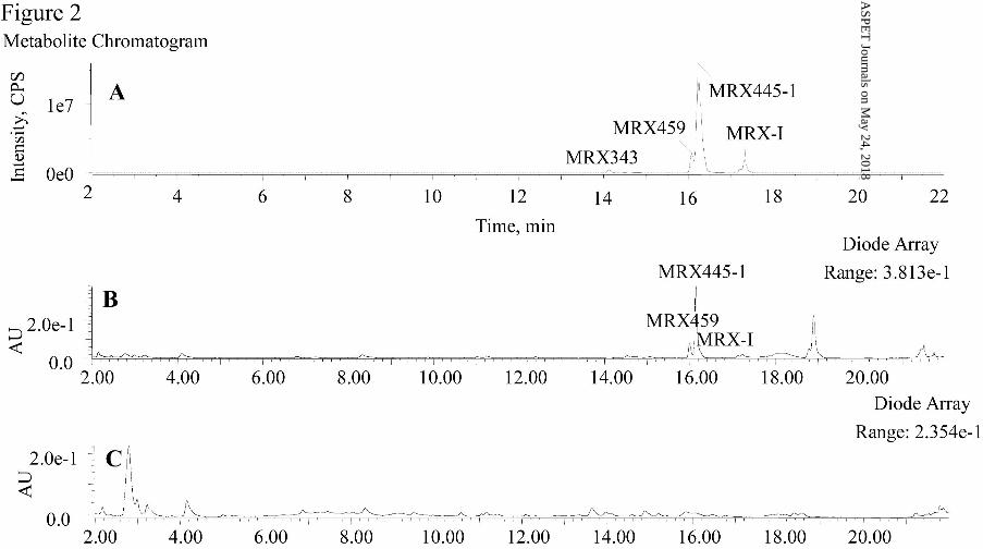
plasma (Fig. 1) and urine (Fig. 2), whereas nine metabolites were detected in human
feces (Fig. 3). The metabolite profile of human plasma at 6 hours postdose was not
shown, for it was similar to that at 1.5 hours. Although metabolic modifications of the
parent drug usually result in different ionization efficiency and UV absorption
coefficient, the MS response and UV absorbance roughly reflect the relative quantity
of the metabolites. As shown in Fig. 1, the most abundant circulating drug-related
component was the parent drug. The predominant metabolites in the plasma were
MRX445-1and MRX459, eluted at 16.2 min and 16.1 min, respectively. The plasma
concentrations of MRX-I,MRX445-1 and MRX459 were determined by a
LC-MS/MS method and the plasma exposures of MRX445-1 and MRX459 were 26.4%
This article has not been copyedited and formatted. The final version may differ from this version.DMD Fast Forward. Published on February 20, 2015 as DOI: 10.1124/dmd.114.061747
at ASPE
T Journals on M
ay 24, 2018dm
d.aspetjournals.orgD
ownloaded from

DMD # 61747
14
and 24.0% of the parent drug exposure (data not shown). Furthermore, MRX445-1
and MRX459 were also the main drug-related materials detected in the urine (Fig. 2).
A large amount of unchanged parent drug was detected in feces, and the major
metabolite detected was MRX413, eluted at 9.3 min (Fig. 3).
Table 1 lists detailed information about these metabolites, including the proposed
structures, protonated molecular ions, retention times, and the characteristic fragment
ions. The metabolites were named in accordance with their protonated molecular
weights, and metabolites with the same molecular weight were named in the
sequential order of their retention times. The metabolites were identified as follows.
Parent Drug M0. A chromatographic peak was detected at 17.2 min in human
plasma, urine, and feces. It gave a protonated molecular ion at m/z 409.1111,
indicating that its elemental composition was C18H15N4O4F3. The retention time and
mass spectral fragmentation patterns were identical to the parent drug, MRX-I,
indicating that this component was unmetabolized MRX-I, designated M0.
MRX343. Metabolite MRX343, found in plasma, urine and feces, was eluted at
14.1 min, with a protonated molecular mass of 343.0895. The elemental composition
of MRX343 was C15H13N2O4F3. The fragment ions at 255.0737 and 251.0788 were
the same as those of MRX-I. The fragment ion at m/z 123.0549 was not detected,
indicating that the isoxazole amine was lost from the parent drug. The fragment ion at
m/z 325.0795 was proposed to be the result of the neutral loss of H2O from MRX343.
The chromatographic retention time and mass spectral fragmentation patterns of
MRX343 were identical to those of HM5013199, so MRX343 was accordingly
confirmed as the deaminated metabolite of MRX-I.
MRX357. Metabolite MRX357, found in plasma, urine, and feces, was eluted at
15.1 min and had a protonated molecular mass of 357.0693, with an elemental
This article has not been copyedited and formatted. The final version may differ from this version.DMD Fast Forward. Published on February 20, 2015 as DOI: 10.1124/dmd.114.061747
at ASPE
T Journals on M
ay 24, 2018dm
d.aspetjournals.orgD
ownloaded from

DMD # 61747
15
composition of C15H11N2O5F3. The fragment ion at 251.0787 was the same as that of
MRX-I. The fragment ion at m/z 123.0549 was not detected, indicating that the
isoxazole amine was lost from the parent drug. The fragment ion at m/z 339.0587 was
proposed to be the result of the neutral loss of H2O from MRX357. The
chromatographic retention time and mass spectral fragmentation pattern of MRX357
were identical to those of HM5012596, the authentic standard. MRX357 was
therefore confirmed as the carboxylic acid metabolite of MRX-I, formed by oxidative
deamination.
MRX407. Metabolite MRX407 was found in plasma, urine, and feces, and was
eluted at 14.2 min. It had a protonated molecular mass of 407.0962 with an elemental
composition of C18H13N4O4F3, indicating that MRX407 was the dehydrogenated
metabolite of MRX-I. The fragment ion at m/z 123.0553 was the same as that of
MRX-I. The fragment ions at 363.1061, 279.0737, 253.0585, and 241.0579 were 2 Da
smaller than those of the parent drug, indicating that dehydrogenation occurred on the
DHPO ring. The chromatographic retention time and mass spectral fragmentation
pattern of MRX407 were identical to those of HM5013876. MRX407 was
accordingly confirmed as DHPO ring-dehydrogenated MRX-I.
MRX413. Metabolite MRX413 was only found in feces and was eluted at 9.3 min.
It had a protonated molecular mass of 413.1428, with an elemental composition of
C18H19N4O4F3. The fragment ion at m/z 255.0733 was the same as that of MRX-I,
indicating that the DHPO ring and trifluorobenzene moieties were not modified. The
fragment ion at m/z 369.1528 was a result of the neutral loss of CO2 from MRX413
and the fragment ion at m/z 339.1426 was proposed to result from the further loss of
CH2O from the fragment ion at m/z 369.1528. The fragment ion at m/z 123.0549 was
not present, indicating that the isoxazole ring was modified. The structure of MRX413
This article has not been copyedited and formatted. The final version may differ from this version.DMD Fast Forward. Published on February 20, 2015 as DOI: 10.1124/dmd.114.061747
at ASPE
T Journals on M
ay 24, 2018dm
d.aspetjournals.orgD
ownloaded from

DMD # 61747
16
was further characterized by NMR after the metabolite was isolated from human feces.
The 1H and 13C NMR data are listed in Table 2 and 3, respectively. Comparison of the
chemical shifts of MRX413 with those of MRX-I, it was shown that the DHPO ring
and trifluorobenzene moieties were unchanged. However, the sp2-hybridized methine
signals of isoxazole disappeared and new signals from a methylene group attached to
an oxygen atom (i.e., δC 58.2, δH 3.88, CH2-18) and a methylene group (i.e., δC 35.9,
δH 2.72, CH2-17) appeared. The structure of MRX413 was further confirmed as the
reduced isoxazole ring-opened metabolite with 1H-1H COSY, HMBC, and HSQC
(Table 1, Supplemental Figure 5).
MRX445-1. Metabolite MRX445-1 was the most prominent metabolite in plasma
and urine, and was also found in feces. MRX445-1 was eluted at 16.2 min and had a
protonated molecular mass of 445.1322 with an elemental composition of
C18H19N4O6F3, indicating the dihydration (2H2O) of MRX-I. The mass spectrum of
m/z 445.1322 gave major fragment ions at m/z 427.1224, 385.1109, 341.0863, and
123.0555. The fragment ion at m/z 427.1224 was the result of the loss of H2O from
MRX445-1. The fragment ion at m/z 123.0555 was the same as that of the parent drug,
indicating that the isoxazole ring was not modified. However, the major fragment ions
at m/z 365.1219, 281.0899, 255.0749, and 243.0736 of MRX-I were not found in
MRX445-1, indicating that the DHPO ring might be modified. The structure of
MRX445-1 was further characterized by NMR after the metabolite was isolated from
human urine. The 1H and 13C NMR data are listed in Table 2 and 3, respectively.
Comparison of the chemical shifts of MRX445-1 with those of MRX-I showed that
the trifluorobenzene, oxazolidinone, and isoxazole amine moieties were unchanged.
However, the characteristic NMR signals of the MRX-I DHPO ring had disappeared
in MRX445-1. Signals appeared indicating a new hydroxylethyl group (δC 59.8, δH
This article has not been copyedited and formatted. The final version may differ from this version.DMD Fast Forward. Published on February 20, 2015 as DOI: 10.1124/dmd.114.061747
at ASPE
T Journals on M
ay 24, 2018dm
d.aspetjournals.orgD
ownloaded from

DMD # 61747
17
3.14, CH2-1; δC 55.5, δH 3.20-3.50, CH2-2) and carboxyethyl group (δC 49.4, δH
3.20-3.50, CH2-3; δC 33.7, δH 2.33, CH2-4; δC 173.4, C-5). Therefore, MRX445-1 was
identified with 1H-1H COSY, HMBC, and HSQC NMR (Supplemental Figure 3) as
the hydroxyethyl amino propionic acid metabolite formed by the oxidative opening of
the DHPO ring (Table 1). This structure was confirmed with its synthesized standard.
The fragment pathways of MRX445-1 are shown in Table 1. The fragment ions at m/z
385.1109 and 341.0863 were proposed to be the result of the neutral loss of C2H4O2
and C2H4O2C2H4O from MRX445-1, respectively.
MRX401. Metabolite MRX401 was found in plasma, urine and feces, and was
eluted at 17.1 min. It had a protonated molecular mass of 401.1070, with an elemental
composition of C16H15N4O5F3. The fragment ions at m/z 341.0860 and 123.0555 were
the same as those of MRX445-1, indicating that the trifluorobenzene, oxazolidinone,
and isoxazole amine moieties were not modified. However, the fragment ion at m/z
385.1109 was not detected, indicating the loss of the hydroxyethyl group from
MRX445-1. MRX401 was accordingly identified as N-dehydroxyethyl MRX445-1.
MRX459. Metabolite MRX459 was eluted at 16.1 min and was a major metabolite
in plasma and urine, also found in feces. It had a protonated molecular mass of
459.1114, with an elemental composition of C18H15N4O7F3, indicating the
introduction of an oxygen atom accompanied by dehydrogenation relative to
MRX445-1. The fragment ions at m/z 341.0860, 187.0473, and 123.0555 were the
same as those of MRX445-1, suggesting that the trifluorobenzene, oxazolidinone, and
isoxazole amine moieties were not modified. The fragment ion at m/z 385.1109 was
not observed but a fragment ion at m/z 399.0902 (+14 Da) appeared. A fragment ion
derived from decarboxylation was also observed at m/z 413.1059. The
chromatographic retention time and mass spectral fragmentation pattern of MRX459
This article has not been copyedited and formatted. The final version may differ from this version.DMD Fast Forward. Published on February 20, 2015 as DOI: 10.1124/dmd.114.061747
at ASPE
T Journals on M
ay 24, 2018dm
d.aspetjournals.orgD
ownloaded from

DMD # 61747
18
were identical to those of HM1383. Therefore, MRX459 was identified as oxidized
MRX445-1, where the hydroxyethyl group was converted to a carboxylic acid (Table
1).
MRX445-2. Metabolite MRX445-2, found in plasma, urine, and feces, was eluted
at 16.7 min and had a protonated molecular mass of 445.1316, the same as that of
MRX445-1. Although MRX445-2 had the same elemental composition as MRX445-1,
it produced different fragment ions. The structure of MRX445-2 was characterized by
NMR after the metabolite was isolated from human urine. The 1H and 13C NMR data
are listed in Table 2 and 3. Comparison of the chemical shifts of MRX445-2 with
those of MRX-I and MRX445-1 showed that the trifluorobenzene, oxazolidinone, and
isoxazole amine moieties were unchanged. Like MRX445-1, the characteristic NMR
signals of the DHPO ring of MRX-I had disappeared in MRX445-2. New signals
from a carboxyl group (δC 173.5, C-2), methylene group (δC 37.8, δH 2.34, and 2.27,
C-1), and a methine attached to an oxygen atom (δC 65.7, δH 3.92, CH-5) had
appeared. The 1H NMR spectrum of MRX445-2 revealed the presence of two active
NH protons (δH 6.59 and 5.61), indicating the opening of the DHPO ring at the C–N
bond. The introduction of NH shifted the C-3 and C-11 signals upfield from δC 49.6 to
43.1 and from δC 121.8 to 113.5, respectively. The reduction of the carbonyl group on
C-5 shifted the C-4 signals downfield from δC 36.5 to 42.3 (Supplemental Figure 4).
MRX445-2 was accordingly identified as the 5-amino-3-hydroxypentanoic acid
metabolite of MRX-I formed by the opening of the DHPO ring (Table 1).
MRX427. Metabolite MRX427, found only in feces, was eluted at 13.6 min. It had
a protonated molecular mass of 427.1227 with an elemental composition of
C18H17N4O5F3. The mass spectrum of m/z 427.1 showed major fragment ions at m/z
409.1119, 368.0851, 281.0889, 243.0727, and 123.0544. In the product ion spectrum
This article has not been copyedited and formatted. The final version may differ from this version.DMD Fast Forward. Published on February 20, 2015 as DOI: 10.1124/dmd.114.061747
at ASPE
T Journals on M
ay 24, 2018dm
d.aspetjournals.orgD
ownloaded from

DMD # 61747
19
of m/z 427.1, the fragment ion at m/z 409.1119 was a result of the neutral loss of H2O
(-18.0108 Da) from MRX427, indicating that MRX427 might contain an alcoholic
hydroxyl group. The fragment ions at m/z 281.0889, 243.0727 and 123.0544 were the
same as those of MRX-I. The exact structure of MRX427 needs to be further
characterized.
Formation of MRX445-1 and MRX459 Cocatalyzed by Enzymes in Human
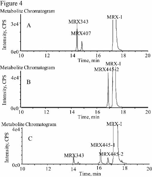
Liver Microsomes and Cytosolic Fractions. After MRX-I was incubated with
human liver microsomes in the presence of NADPH for 120 min, traces of oxidatively
deaminated metabolites of MRX-I, such as MRX343, were detected by UPLC/Triple
TOF MS. The formation of DHPO ring-opened metabolites, including MRX445-1,
MRX445-2, and MRX459, was not observed in the human liver microsomes (Fig.
4A). After MRX-I was incubated with human liver cytosolic fractions in the presence
of NADPH for 120 min, only MRX445-2 was produced, without the major
metabolites in humans, MRX445-1 and MRX459 (Fig. 4B). However, all the DHPO
ring-opened metabolites detected in humans, including MRX445-1, MRX445-2, and
MRX459, were observed after MRX-I was incubated with human liver S9 fractions
for 120 min (Fig. 4C). These results indicate that liver microsomal or cytosolic
enzymes alone do not allow the biotransformation of MRX-I to MRX445-1 and
MRX459, whereas S9 fractions provided the necessary enzyme systems to complete
the metabolic pathways. The enzymes in liver microsome and cytosolic fractions
cocatalyzed the opening of the DHPO ring of MRX-I.
Chemical Inhibition of the Formation of MRX445-1, MRX445-2, and MRX459
in S9 Fractions. MRX-I was incubated with the NADPH-supplemented human liver
S9 fractions in the presence of various specific chemical inhibitors to investigate the
enzymes involved in the formation of MRX445-1, MRX445-2, and MRX459. As
This article has not been copyedited and formatted. The final version may differ from this version.DMD Fast Forward. Published on February 20, 2015 as DOI: 10.1124/dmd.114.061747
at ASPE
T Journals on M
ay 24, 2018dm
d.aspetjournals.orgD
ownloaded from

DMD # 61747
20
shown in Table 4, ABT and methimazole did not significantly inhibit the formation of
MRX445-1, MRX445-2, and MRX459, revealing that the oxidative opening of the
DHPO ring was not catalyzed by P450, FMO1, FMO2, FMO3, and FMO4. However,
the formation of MRX445-1 and MRX459 was reduced to 7.22% and 0%,
respectively, after brief preincubation of liver S9 fractions at 50 °C in the absence of
the cofactor. This indicates FMOs, other than FMO1, FMO2, FMO3, and FMO4, are
involved in the formation of MRX445-1 and MRX459. Menadione, flufenamic acid
and disulfiram reduced the formation of MRX445-1 to 44.6%, 69.4 and 80.0%,
respectively in liver S9 fractions, whereas 4-MP had no effect on the formation of
MRX445-1. This indicates that the reduction of the carbonyl group could be a step in
the formation of MRX445-1 from MRX-I, catalyzed by multiple enzymes including
SDR, AKR and ALDH. Furthermore, disulfiram also decreased the formation of
MRX459 to 10.7%, suggesting that the oxidation of the aldehyde intermediate could
be a step of the formation of MRX459, catalyzed by ALDH. The XO inhibitors,
methotrexate and allopurinol, did not affect the formation of MRX445-2 from MRX-I.
However, the formation of MRX445-2 was concentration-dependently inhibited by
the AO inhibitors, Menadione (IC50 = 8.0 µM) and raloxifene (IC50 = 0.66 µM),
respectively, as shown in Supplemental Figure S6. These indicated that AO is
involved in the formation of MRX445-2.
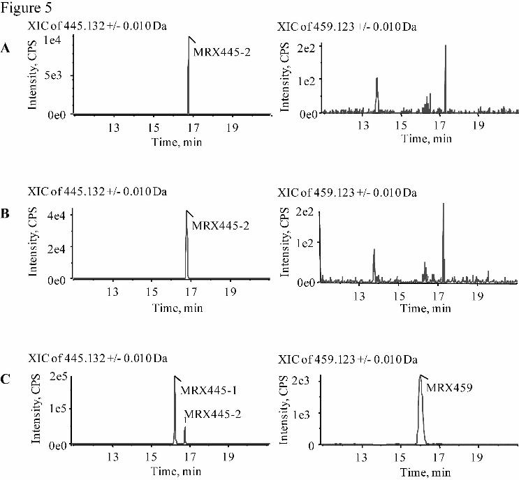
Formation of MRX445-1, MRX445-2, and MRX459 by Incubation of MRX-I
with Liver Cytosol-ortified FMO5. After MRX-I was incubated with liver
cytosol-fortified FMO1, FMO3, or FMO5 in the presence of NADPH for 120 min,
MRX445-2 was produced from MRX-I (Fig. 5). This is consistent with the results of
incubating MRX-I with human liver cytosolic fractions and the chemical inhibition of
MRX445-2 formation, which demonstrated that the formation of MRX445-2 was
This article has not been copyedited and formatted. The final version may differ from this version.DMD Fast Forward. Published on February 20, 2015 as DOI: 10.1124/dmd.114.061747
at ASPE
T Journals on M
ay 24, 2018dm
d.aspetjournals.orgD
ownloaded from
DMD # 61747
21
catalyzed by AO and XO in the liver cytosolic fractions, not by oxidative enzymes in
the liver microsomes. The formation of MRX445-1 and MRX459 was not observed
during the incubation of MRX-I with liver cytosol-fortified FMO1 or FMO3, but with
liver cytosol-fortified FMO5. This confirms that FMO5 is involved in the formation
of MRX445-1 and MRX459, as shown in the inhibition experiments for MRX445-1
and MRX459 formation in the human liver S9 fractions.
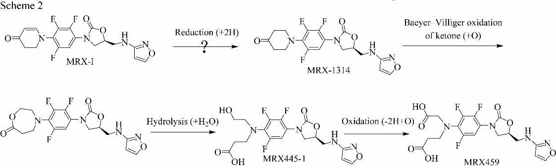
Water is the Source of Oxygen Atoms in DHPO Ring-opened Metabolites. To
explore the mechanism of oxidative DHPO ring-opening, MRX-I and DHPO
ring-hydrogenated MRX-I (MRX-1314) were incubated with liver cytosol-fortified
FMO5 in the presence of H218O. UPLC/Triple TOF MS analysis clearly showed that
MRX445-1 derived from MRX-I had a protonated molecular weight of 449.1303 (Fig.
6B), suggesting that two atoms of 18O from H218O were incorporated into MRX445-1
by comparing the protonated molecular weight of MRX445-1 (445.1322) in the
absence of H218O (Fig. 6A). MS/MS analysis of the protonated molecular ion at m/z
449.1 showed major fragment ions at m/z 387.1065 and 341.0760. The fragment ion
at m/z 387.1065 resulted from the loss of CH3CO18OH and the fragment ion at m/z
341.0760 arose from the further loss of CHCH218OH. This reveals that one 18O atom
was incorporated into the hydroxylethyl group and the other 18O atom was
incorporated into the carboxylethyl group of MRX445-1 obtained from MRX-I (Fig.
6B). The UPLC/Triple TOF MS analysis also showed that MRX445-1 obtained from
MRX-1314 had a protonated molecular weight of 449.1307 (Fig. 6C), suggesting that
two 18O atoms from H218O were incorporated into MRX445-1. MS/MS analysis of the
protonated molecular ion at m/z 449.1 showed the major fragment ions at m/z
385.1017 and 341.0762. The fragment ion at m/z 385.1017 was a result of the loss of
CH3C18O18OH. This indicates that two 18O atoms from H2
18O were incorporated into
This article has not been copyedited and formatted. The final version may differ from this version.DMD Fast Forward. Published on February 20, 2015 as DOI: 10.1124/dmd.114.061747
at ASPE
T Journals on M
ay 24, 2018dm
d.aspetjournals.orgD
ownloaded from

DMD # 61747
22
the carboxylethyl group of MRX445-1 obtained from MRX-1314 (Fig. 6C). These
results demonstrate that the ring opening of MRX-I and MRX-1314 occurred via
different routes.
The UPLC/Triple TOF MS analysis showed that MRX445-2 from MRX-I had a
protonated molecular weight of 449.1296 (Fig. 6E). Comparison of the protonated
molecular weight of MRX445-2 in the absence of H218O (445.1316) suggests that two
18O atoms from H218O were incorporated into MRX445-2 (Fig. 6D). MS/MS analysis
of the protonated molecular ion at m/z 449.1 showed major fragment ions at m/z
429.1146, 411.1043, and 341.0765. The fragment ion at m/z 429.1146 was the result
of the loss of H218O; the fragment ion at m/z 411.1048 was the result of the further
loss of H2O; and the fragment ion at m/z 341.0767 was the result of the loss of
CH3CH2CHOHCH2C18O18OH. This indicates that two 18O atoms from water were
incorporated into the carboxyl group of MRX445-2 (Fig. 6E).
The UPLC/Triple TOF MS analysis showed that MRX459 from MRX-I had a
protonated molecular weight of 465.1118 (Fig. 6G). Comparison of the protonated
molecular weight of MRX459 in the absence of H218O (459.1114) (Fig. 6F) suggests
that three 18O atoms from H218O were incorporated into MRX459. MS/MS analysis of
the protonated molecular ion at m/z 465.1 showed major fragment ions at m/z
415.0993, 403.0888, and 341.0768. The fragment ion at m/z 415.0993 resulted from
the loss of CH18O2H; the fragment ion at m/z 403.0888 resulted from the loss of
CH3C18OOH; and the fragment ion at m/z 341.0758 was formed by the neutral loss of
CH2CO18OHCH2C18O18OH. This shows that one 18O atom was incorporated into the
carboxylethyl group and that the other two atoms were incorporated into the
carboxylmethyl group of MRX459 (Fig. 6G). Incubating MRX-1314 with human
liver cytosol-fortified FMO5 did not produce MRX459 or MRX445-2.
This article has not been copyedited and formatted. The final version may differ from this version.DMD Fast Forward. Published on February 20, 2015 as DOI: 10.1124/dmd.114.061747
at ASPE
T Journals on M
ay 24, 2018dm
d.aspetjournals.orgD
ownloaded from
DMD # 61747
23
Marked Metabolic Differences between MRX-I and MRX-1314 in Human
Hepatocytes. The formation of MRX445-1 in the H218O experiments showed that the
opening of the DHPO ring in MRX-I and MRX-1314 occurs via different routes. To
clarify the possible mechanisms further, MRX-I and MRX-1314 were incubated with
human hepatocytes, which contain all the liver enzymes in the liver and provide a
more representative system than other in vitro liver models. After the incubation of
MRX-I with human hepatocytes, all the DHPO ring-opened metabolites detected in
humans were observed, including MRX445-1, MRX445-2, MRX459, and MRX401
(Fig. 7A). Furthermore, MRX445-1 was identified as the principle metabolite of
MRX-I, as was observed in vivo. MRX445-1 was also present in the MRX-1314
incubation system at a similar level to that in the MRX-I incubation system (Fig. 7B).
However, most of the MRX-1314 in the human hepatocyte incubation system was
reduced to MRX-1315, which was not observed in the MRX-I incubation system.
MRX459 was also not detected in the MRX-1314 incubation system. This
demonstrates further significant metabolic differences between MRX-I and
MRX-1314.
This article has not been copyedited and formatted. The final version may differ from this version.DMD Fast Forward. Published on February 20, 2015 as DOI: 10.1124/dmd.114.061747
at ASPE
T Journals on M
ay 24, 2018dm
d.aspetjournals.orgD
ownloaded from

DMD # 61747
24
Discussion
The metabolism of MRX-I, a novel antibiotic oxazolidinone for treatment of
Gram-positive infections, was studied by UPLC-UV/Triple TOF MS analysis after an
oral dose of 600 mg of MRX-I to healthy male volunteers during phase I clinical trials.
Nine metabolites were detected and six were confirmed with synthesized or isolated
reference standards. The proposed metabolic processes are shown in Scheme 1. The
oxidative ring-opening of the DHPO is the main metabolic pathway of MRX-I in
humans, via which the major metabolites MRX445-1, MRX445-2, and MRX459 were
formed. To identify the enzymes involved in the DHPO ring-opening, MRX-I was
incubated with various in vitro human liver models, including liver microsomes,
cytosol, S9 fractions, and primary hepatocytes. By coincubating MRX-I with various
specific chemical inhibitors in the human liver S9 fractions, the enzymes involved
were identified as FMOs, AKR, SDR, ALDH, and AO. The involvement of FMO5 in
the formation of MRX445-1 and MRX459 was further confirmed by incubating
MRX-I with liver cytosol-fortified FMOs, including FMO1, FMO3, and FMO5.
As a group of important drug-metabolizing enzymes, FMOs catalyze oxidative
reactions that are complementary to P450-mediated biotransformations (Benedetti et
al., 2006). Among the isoforms identified, FMO3 and FMO5 are recognized as the
major isoforms in adult human liver (Zhang and Cashman, 2006; Overby et al., 1997).
Typical FMO-catalyzed reactions have long been recognized, including the
monooxygenation of heteroatoms such as nitrogen, sulfur, and phosphorus (Benedetti
et al., 2006; Cashman 2008). However, recent studies have shown that FMO5
specifically catalyzes the Baeyer–Villiger oxidation of piperidine-4-one (Lai et al.,
2011) and cyclic α,β-unsaturated ketone moieties (Verhoeven et al., 1998). Verhoeven
et al. also proposed that the ring-opening mechanism of cyclic α,β-unsaturated
This article has not been copyedited and formatted. The final version may differ from this version.DMD Fast Forward. Published on February 20, 2015 as DOI: 10.1124/dmd.114.061747
at ASPE
T Journals on M
ay 24, 2018dm
d.aspetjournals.orgD
ownloaded from

DMD # 61747
25
ketones involved the reduction of the double bond of the unsaturated ketone followed
by Baeyer–Villiger oxidation catalyzed by FMOs. By analogy with previous reports,
we proposed that the mechanism for DHPO ring-opening of MRX-I was as follows.
MRX-I is first reduced to MRX-1314 by liver cytosolic reductases, followed by
ring-expanding oxidation to a lactone via the Baeyer–Villiger reaction in liver
microsomes. The lactone was then hydrolyzed to produce MRX445-1, which can be
further oxidized to MRX459 (Scheme 2).
To test the proposed mechanism, the DHPO ring-hydrogenated metabolic
intermediate, MRX-1314, was synthesized and incubated with human hepatocytes in
vitro and compared with MRX-I. Although similar levels of MRX445-1 were
produced independently when MRX-I and MRX-1314 were used as substrates,
marked differences between MRX-I and MRX-1314 metabolism were observed. In
the hepatocyte system, most of the MRX-1314 was converted to its carbonyl-reduced
metabolite, MRX-1315, which indicates that the reduction of ketone is the principal
metabolic pathway of MRX-1314. However, MRX-1315 was not detected in the
MRX-I incubation systems. This suggests that MRX-1314 is not the metabolic
intermediate in the formation of MRX445-1 from MRX-I. Furthermore, during its
incubation with human hepatocytes, some MRX-I was transformed into MRX459,
which could be formed by the further oxidation of the hydroxylethyl group on
MRX445-1, because the oxidation of primary alcohols to carboxylic acids is one of
the most common metabolic pathways of drugs in humans. However, no MRX459
was detected in the MRX-1314 incubation system, although the level of MRX445-1
produced in the MRX-1314 system was similar to that produced in the MRX-I system.
This can be explained by the presence of an aldehyde intermediate in the MRX-I
incubation system, from which MRX445-1 is formed by reduction and MRX459 is
This article has not been copyedited and formatted. The final version may differ from this version.DMD Fast Forward. Published on February 20, 2015 as DOI: 10.1124/dmd.114.061747
at ASPE
T Journals on M
ay 24, 2018dm
d.aspetjournals.orgD
ownloaded from

DMD # 61747
26
formed by oxidation. This is plausible because menadione (SDR inhibitor),
flufenamic acid (AKR inhibitor), and disulfiram (ALDH) inhibited the formation of
MRX445-1 and disulfiram (ALDH) inhibited the formation of MRX 459 from MRX-I
(Table 4). Therefore, we propose a novel metabolic mechanism (Scheme 3) for the
formation of MRX445-1 and MRX459. MRX-I is metabolized to an enol lactone via
Baeyer–Villiger oxidation, hydrolyzed to an enol, and transformed to an aldehyde by
enol–aldehyde tautomerism, after which the aldehyde intermediate is reduced to
MRX445-1 or oxidized to MRX459.
In the Baeyer–Villiger oxidation of α,β-unsaturated ketones, vinyl migration is
generally favored over alkyl migration to produce ring-expanded enol lactones (Shono
et al., 1974; Krafft and Katzenellenbogen, 1981), which supports the mechanism
proposed in Scheme 3. Although the regiochemical selectivity of Baeyer–Villiger
oxidation is recognized in chemistry, this is the first observation of the metabolism of
xenobiotics to an aldehyde intermediate by direct Baeyer–Villiger oxidation of an
α,β-unsaturated ketone and hydrolysis of the enol lactone. To confirm the existence of
the aldehyde intermediate, MRX-I or MRX-1314 was incubated in liver
cytosol-fortified recombinant FMO5 in the presence of H218O. The isotope
experiment revealed that the MRX445-1 produced from MRX-I had incorporated two
18O atoms derived from water, one in the carboxyethyl group and the other in the
hydroxyethyl group. Because Baeyer–Villiger oxidation is catalyzed specifically by
FMO5, an oxygenase, the hydroxyethyl oxygen atom of MRX445-1 should be
derived from O2 rather than from H2O. Therefore, the 18O atom of the hydroxyethyl
group of MRX445-1 indicates the existence of an aldehyde intermediate, which
incorporated an 18O atom from H218O during nonenzymatic equilibrium with its
geminal diol (Scheme 3). It is clear that the hydroxyl 18O atom of the carboxylic acid
This article has not been copyedited and formatted. The final version may differ from this version.DMD Fast Forward. Published on February 20, 2015 as DOI: 10.1124/dmd.114.061747
at ASPE
T Journals on M
ay 24, 2018dm
d.aspetjournals.orgD
ownloaded from
DMD # 61747
27
was derived from H218O during the hydrolysis of the enol lactone. The isotope
experiments also demonstrated that the MRX445-1 produced from MRX-1314
incorporated two 18O atoms derived from H218O. However, both atoms were
incorporated in the carboxyethyl group, which differed from the results for the
MRX445-1 produced from MRX-I. The carbonyl oxygen of the carboxyethyl group
incorporated an 18O atom because the ketone 16O atom was exchanged with the H218O
18O atom during ketone–geminal diol equilibrium (Scheme 4). Furthermore,
MRX-1315 with an 18O-labeled hydroxyl group was the main metabolite of
MRX-1314 in the presence of H218O, which also indicates that ketone–geminal diol
equilibrium of MRX-1314 occurred (data not shown). MRX445-1 produced from
MRX-1314 incorporated an 18O atom in the hydroxyl group of the carboxyethyl group
during lactone hydrolysis, which was identical to the results for MRX445-1 produced
from MRX-I.
MRX459 incorporated three 18O atoms from H218O was produced when MRX-I
was incubated with liver cytosol fortified-FMO5 in the presence of H218O. Two 18O
atoms were incorporated in the carboxyl hydroxyl groups of MRX459. The other 18O
atom was on the carboxymethyl carbonyl group, which could be incorporated during
the oxidation of the aldehyde intermediate to the carboxylic acid (Scheme 3). This
indicates that not oxygenases but oxidases, such as AO, OX and ALDH (Uetrecht and
Trager, 2007), are involved in the formation of M459, because the incorporated
oxygen atom into substrates by these enzymes are from H2O rather than from O2. The
involvement of ALDH in the formation of MRX459 was confirmed that disulfiram
reduced the formation of MRX459 from MRX-I to 10.7%. The reaction phenotyping
experiments showed that menadione and raloxifene reduced the formation of
MRX445-2 with the IC50 values of 8.0 µM and 0.66 µM, respectively. This suggests
This article has not been copyedited and formatted. The final version may differ from this version.DMD Fast Forward. Published on February 20, 2015 as DOI: 10.1124/dmd.114.061747
at ASPE
T Journals on M
ay 24, 2018dm
d.aspetjournals.orgD
ownloaded from

DMD # 61747
28
that AO catalyzed the formation of MRX445-2. The involvement of AO in the
formation of MRX445-2 was also supported by the H218O experiments. After MRX-I
was incubated with human liver cytosol fortified-FMO5 in the presence of H218O, two
18O atoms were incorporated into MRX445-2, one in the carbonyl group of carboxyl
acid, which indicates that the carbon was oxidized by AO (Scheme 3).
Finally, we have characterized the metabolism of MRX-I in humans. The major
metabolic pathway of MRX-I involves the oxidative opening of the DHPO ring. The
mechanism underlying the directed opening of the DHPO ring proposed is that
MRX-I is first oxidized to an enol lactone via the Baeyer–Villiger ring-expanding
reaction, specifically catalyzed by FMO5, followed by its hydrolysis to an enol, and
then transformation to an aldehyde intermediate by enol–aldehyde tautomerism. The
aldehyde is reduced by SDR, AKR and ALDH to MRX445-1 or oxidized to MRX459
by ALDH. Our study demonstrated that the main metabolic pathway of MRX-I, via
the opening of the DHPO ring, is not catalyzed by P450s, the enzymes primarily
responsible for metabolizing drugs and xenobiotics, but by multiple other enzymes,
including FMO5, SDR, AKR, ALDH and AO. Therefore, few clinical adverse
drug–drug interactions should be anticipated between MRX-I and P450 inhibitors or
inducers.
Acknowledgments
We would like thank the staff of Clinical Trial Center of Shanghai Huashan
Hospital and the volunteers in the clinical study
This article has not been copyedited and formatted. The final version may differ from this version.DMD Fast Forward. Published on February 20, 2015 as DOI: 10.1124/dmd.114.061747
at ASPE
T Journals on M
ay 24, 2018dm
d.aspetjournals.orgD
ownloaded from

DMD # 61747
29
Authorship Contributions
Participated in research design: Jian Meng, Dafang Zhong, Zhengyu Yuan, Xiaoyan Chen
Conducted experiments: Jian Meng, Liang Li, Hong Yuan, Jialan Zhou, Chen Li
Performed data analysis: Jian Meng, Liang Li, Cen Xie, Mikhail Fedorovich Gordeev, Jinqian Liu,
Xiaoyan Chen
Wrote or contributed to the writing of the manuscript: Jian Meng, Liang Li, Xiaoyan Chen
This article has not been copyedited and formatted. The final version may differ from this version.DMD Fast Forward. Published on February 20, 2015 as DOI: 10.1124/dmd.114.061747
at ASPE
T Journals on M
ay 24, 2018dm
d.aspetjournals.orgD
ownloaded from

DMD # 61747
30
References
Benedetti MS, Whomsley R, and Baltes E (2006) Involvement of enzymes other than
CYPs in the oxidative metabolism of xenobiotics. Expert Opin Drug Metab
Toxicol 2:895–921.
Brickner SJ, Barbachyn MR, Hutchinson DK, and Manninen PR (2008) Linezolid
(ZYVOX), the first member of a completely new class of antibacterial agents for
treatment of serious Gram-positive infections. J Med Chem 51:1981–1990.
Cashman JR (2008) Role of flavin-containing monooxgenase in drug development.
Expert Opin Drug Metab Toxicol 4:1507–1521.
Dean S (1999) Mechanism of action of the oxazolidinone antibacterial agents. Expert
Opin Investig Drugs 8:1195−1202.
Gordeev MF, and Yuan ZY (2014) New potent antibacterial oxazolidinone (MRX-I)
with an improved class safety profile. J Med Chem 57:4487–4497.
Krafft GA and Katzenellenbogen JA (1981) Synthesis of haloenol lactones,
mechanism-based inactivators of serine proteases. J Am Chem Soc
103:5459–5466.
Lai WG, Farah N, Moniz GA, and Wong YN (2011) A Baeyer-Villiger oxidation
specifically catalyzed by human flavin-containing monooxygenase 5. Drug
Metab Dispos 39:61–70.
Li CR, Zhai QQ, Wang XK, Hu XX, Li GQ, Zhang WX, Pang J, Lu X, Yuan H,
Gordeev MF, Chen LT, Yang XY, You XF (2014) In vivo antibacterial activity of
MRX-I, a new oxazolidinone. Antimicrob Agents Chemother 58:2418–2421.
This article has not been copyedited and formatted. The final version may differ from this version.DMD Fast Forward. Published on February 20, 2015 as DOI: 10.1124/dmd.114.061747
at ASPE
T Journals on M
ay 24, 2018dm
d.aspetjournals.orgD
ownloaded from

DMD # 61747
31
Morrison RD, Blobaum AL, Byers FW, Santomango TS, Bridges TM, Stec D, Brewer
KA, Sanchez-Ponce R, Corlew MM, Rush R, Felts AS, Manka J, Bates BS,
Venable DF, Rodriguez AL, Jones CK, Niswender CM, Conn PJ, Lindsley CW,
Emmitte KA, and Daniels JS (2010) The role of aldehyde oxidase and xanthine
oxidase in the biotransformation of a novel negative allosteric modulator of
metabotropic glutamate receptor subtype 5. Drug Metabo Dispos 38:667–678.
Nagiec EE, Wu L, Swaney SM, Chosay JG, Ross DE, Brieland JK, and Leach KL
(2005) Oxazolidinones inhibit cellular proliferation via inhibition of
mitochondrial protein synthesis. Antimicrob Agents Chemother 49:3896–3902.
Overby LH, Carver GC, and Philpot RM (1997) Quantitation and kinetic properties of
hepatic microsomal and recombinant flavin-containing monooxygenases 3 and 5
from humans. Chem Biol Interact 106:29–45.
Rosemond MJC, and Walsh JS (2004) Human carbonyl reduction pathways and a
strategy for their study in vitro. Drug Metab Rev 36:335–361.
Shono T, Matsumura Y, Hibino K, and Miyawaki S (1974) A novel synthesis of
1-citronellol from 1-menthone. Tetrahedron Let 1295:1295–1298.
Stagos D, Chen Y, Brocker C, Donald E, Jackson BC, Orlicky DJ, Thompson DC, and
Vasiliou V (2010) Aldehyde dehydrogenase 1B1: molecular cloning and
characterization of a novel mitochondrial acetaldehyde-metabolizing enzyme.
Drug Metabo Dispos 38:1679–1687.
Talbot GH, Bradley J, Edwards JE, Gilbert D, Scheld M, and Bartlett JG (2006) Bad
bugs need drugs: an update on the development pipeline from the antimicrobial
This article has not been copyedited and formatted. The final version may differ from this version.DMD Fast Forward. Published on February 20, 2015 as DOI: 10.1124/dmd.114.061747
at ASPE
T Journals on M
ay 24, 2018dm
d.aspetjournals.orgD
ownloaded from

DMD # 61747
32
availability task force of the infectious diseases society of america. Clin Infect
Dis 42:657–668.
Uetrecht JP and Targer W (2007) Drug Metabolism: Chemical and Enzymetic Aspects,
pp 61, Informa Healthcare, New York
Vinh DC, and Rubinstein E (2009) Linezolid: a review of safety and tolerability. J
Infect 59:S59–S74.
Vos RME (1998) In vitro and in vivo metabolism of the progestagen ORG 30659 in
several species. Drug Metab Dispos 26:1102–1112.
Zhang J, and Cashman JR (2006) Quantitative analysis of FMO gene mRNA levels in
human tissues. Drug Metab Dispos 34:19–26
This article has not been copyedited and formatted. The final version may differ from this version.DMD Fast Forward. Published on February 20, 2015 as DOI: 10.1124/dmd.114.061747
at ASPE
T Journals on M
ay 24, 2018dm
d.aspetjournals.orgD
ownloaded from

DMD # 61747
33
Scheme Legends
Scheme 1. Proposed metabolic pathway of MRX-I in humans.
Scheme 2. Proposed metabolic pathway for the formation of MRX445 from MRX-I
by analogy with previous studies (Lai et al., 2011; Verhoeven et al., 1998).
Scheme 3. Proposed mechanism for the formation of MRX445-1, MRX445-2, and
MRX459 from MRX-I in the presence of H218O.
Scheme 4. Proposed mechanism for the formation of MRX445-1 and MRX-1315
from MRX-1314 in the presence of H218O.
Figure Legends
Fig. 1. Metabolite profile of human plasma 1.5 hours after the oral administration of
600 mg MRX-I, detected by UPLC/Triple TOF MS (A) and UPLC-UV
chromatograms of pooled plasma (B) and blank plasma (C). AU, arbitrary units. The
inset is the expanded chromatogram in the region of 13–18 minutes.
Fig. 2. Metabolite profile of human urine 0–24 hours after the oral administration of
600 mg MRX-I, detected by UPLC/Triple TOF MS (A) and UPLC-UV
chromatograms of pooled urine (B) and blank urine (C). AU, arbitrary units.
Fig. 3. Metabolite profile of human feces 0–24 hours after the oral administration of
600 mg MRX-I, detected by UPLC/Triple TOF MS (A) and UPLC-UV
chromatograms of pooled feces (B) and blank feces (C). AU, arbitrary units.
Fig. 4. Metabolite profiles of MRX-I incubated with human liver microsomes (A),
cytosol (B), and S9 fractions (C) for 120 minutes detected by UPLC/ Triple TOF MS.
Fig. 5. UPLC/Triple TOF MS extracted ion chromatograms (EIC) of MRX445-1,
This article has not been copyedited and formatted. The final version may differ from this version.DMD Fast Forward. Published on February 20, 2015 as DOI: 10.1124/dmd.114.061747
at ASPE
T Journals on M
ay 24, 2018dm
d.aspetjournals.orgD
ownloaded from

DMD # 61747
34
MRX445-2, and MRX459 in samples after MRX-I was incubated for 120 minutes
with human liver cytosol-fortified FMO1 (A), FMO3 (B), and FMO5 (C). Data were
filtered to show only MRX445-1, MRX445-2 (m/z 445.133 ± 0.010 Da), and
MRX459 (m/z 459.123 ± 0.010 Da).
Fig. 6. MS/MS spectra of MRX445-1 (A, formed from MRX-I in H216O; B, formed
from MRX-I in H218O; C, formed from MRX-1314 in H2
18O), MRX445-2 (D, formed
from MRX-I in H216O; E, formed from MRX-I in H2
18O), and MRX459 (F, formed
from MRX-I in H216O; G, formed from MRX-I in H2
18O) in samples of MRX-I or
MRX-1314 was incubated for 120 minutes with liver cytosol-fortified FMO5.
Fig. 7. Metabolite profiles of MRX-I (A) or MRX-1314 (B) incubated with human
hepatocytes for 120 minutes detected, by UPLC/Q-TOF MS.
This article has not been copyedited and formatted. The final version may differ from this version.DMD Fast Forward. Published on February 20, 2015 as DOI: 10.1124/dmd.114.061747
at ASPE
T Journals on M
ay 24, 2018dm
d.aspetjournals.orgD
ownloaded from

DMD # 61747
35
TABLE 1 Mass spectral fragmentation and structures of MRX-I and its proposed
metabolites (Mass spectra of MRX-I and metabolites shown in Supplemental Figure
S1).
Structure tR (min) [M + H]+ Characteristic Fragment Ions
M0
(MRX-I)
17.2 409.1111 365.1219 (-CO2), 281.0899,
255.0749, 243.0736, 123.0549
MRX343 14.1 343.0895 325.0795 (-H2O), 255.0737,
251.0788
MRX357 15.1 357.0693 339.0587 (-H2O), 251.0787
MRX401 17.1 401.1070 341.0860, 187.0471, 123.0547
MRX407 14.2 407.0962 363.1061 (-CO2), 279.0737,
253.0585. 241.0579, 123.0553
MRX413 9.30 413.1428, 369.1528 (-CO2), 339.1426,
255.0733
MRX427 13.6 427.1227 409.1119 (-H2O), 368.0851
(-C2H5NO), 281.0889, 243.0727,
123.0538
MRX445-1 16.2 445.1322 427.1224 (-H2O), 385.1109,
341.0863, 123.0555
MRX445-2 16.7 445.1316 427.1212 (-H2O), 409.1095 (-2H2O),
341.0855, 187.0465, 123.0544
MRX459
16.1 459.1114 413.1059, 399.0902, 341.0848,
187.0473, 123.0551
N NO
HN
F
F
F O
ON
HO
OHO
m/z 341
m/z 385
m/z 123
N NO
OHO
F
F
F O
-H2Om/z 251
O
N NO
OHO
F
F
F O
m/z 255
-H2Om/z 251
N NO
HNO
F
F
F O
ON
m/z 123
m/z 281m/z 255m/z 243
HN NO
HN
F
F
F O
ON
OHO
m/z 341
m/z 123m/z 187
N NO
HNO
F
F
F O
ON
m/z 123
m/z 279m/z 253m/z 241
N NO
HNO
F
F
F O
OHNH
-CO2m/z 339
m/z 255
HN NO
HN
F
F
F O
ON
OH
HO
Om/z 123
m/z 341
m/z 187
N NO
HN
F
F
F O
ON
HO
OH
m/z 341
m/z399
m/z 123
O
m/z 413
O
N NO
HNO
F
F
F O
ON
m/z 123
m/z 281m/z 243
-H2O
This article has not been copyedited and formatted. The final version may differ from this version.DMD Fast Forward. Published on February 20, 2015 as DOI: 10.1124/dmd.114.061747
at ASPE
T Journals on M
ay 24, 2018dm
d.aspetjournals.orgD
ownloaded from

DMD # 61747
36
TABLE 2 1H NMR chemical shift assignments for MRX-I, MRX445-1, MRX445-2, and MRX413 (NMR spectra shown in Supplemental
Figure S2 – S5).
Chemical Shift (ppm) for
Protons
MRX-I
MRX445-1
MRX445-2
MRX413
1 5.07, d (J = 7.8 Hz) 3.14, t (J = 5.7 Hz, 2H) 2.34, dd J = 15.2, 5.4 Hz; 2.27, dd, J = 15.2, 7.8 Hz 5.26, d, J = 7.7 Hz
2 7.48, d (J = 7.8 Hz) 3.20-3.50, m (6H) 7.44, d, J = 7.7 Hz
3 3.88, m 3.20-3.50, m (6H) 3.70, m 3.97, t, J = 7.5 Hz
4 2.51, t (J = 7.4 Hz, 2H) 2.33, t (J = 6.4 Hz, 2H) 1.67, m; 1.55, m 2.45, t, J = 7.6 Hz, 2H
5 3.92, m
9 7.56, m 7.26, m 7.18, m 7.51, m
13 4.95, m 4.88, brs 4.88, m 5.02, m
14 3.88, m; 4.18, t (J = 8.6 Hz) 4.08, t (J = 8.5 Hz); 3.77, dd (J = 7.8, 6.7 Hz) 3.92, m; 4.02, t (J = 8.6 Hz) 4.27, t, J = 8.9 Hz; 3.93, t, J = 6.9 Hz
15 3.48, t (J = 5.4 Hz, 2H) 3.20-3.50, m (6H) 3.44, m 3.81, dd, J = 14.9, 3.4 Hz; 3.72, dd, J = 14.9, 6.9 Hz
17 6.02, d (J = 1.8 Hz) 5.99, m 6.02, d (J = 1.2 Hz) 2.72, t, J = 5.9 Hz
18 8.41, d (J = 1.8 Hz) 8.37, brs 8.41, d (J = 1.2 Hz) 3.88, dd, J = 5.8, 1.5 Hz, 2H
NH 6.59, t (J = 6.0 Hz) 6.54, brt (J = 5.5 Hz) 6.59, t (J = 5.9 Hz); 5.61, brs
d, doublet; q, quartet; m, multiplet; s, singlet; t, triplet
N NO
HNO
F
F
F O
ON
1 2
345
67
8
91011
12
1314 15
16
1718
N NO
HN
F
F
F O
ON
12
34
5
67
8
91011
12
1314 15
16
1718OH
HO
O
HN NO
HNHO
F
F
F O
ON
12
345
67
8
91011
12
1314 15
16
1718
OHO
N NO
HNO
F
F
F O
OHNH
1 2
345
67
8
91011
12
1314 1516
1718
This article has not been copyedited and form
atted. The final version m
ay differ from this version.
DM
D Fast Forw
ard. Published on February 20, 2015 as DO
I: 10.1124/dmd.114.061747
at ASPET Journals on May 24, 2018 dmd.aspetjournals.org Downloaded from

DMD # 61747
37
TABLE 3 13C NMR chemical shift assignments for MRX-I, MRX445-1, MRX445-2, and MRX413 (NMR spectra shown in Supplemental Figure S2 – S5).
Chemical Shift (ppm)
Carbon
MRX-I
MRX445-1
MRX445-2
MRX413
1 102.5, d 59.8, t 37.8, t 101.4, d
2 152.5, d 55.4, t 173.5, s 153.5, d
3 49.6, t 49.3, t 43.1, t 49.2, t
4 36.5, t 33.6, t 42.3, t 35.4, t
5 191.1, s 173.4, s 65.7, d 193.6, s
6 145.5-148.0, d 147.1-149.1, d 146.2-148.0, d
7 141.2-143.6, d 141.9-143.9, d 139.8-141.7, d
8 125.5, s 126.8, s 127.2, s 125.1, s
9 108.5, d 108.6, d 109.5, d 108.1, d
10 151.3-153.7, d 153.3-155.2, d 153.3-155.1, d
11 121.8, s 121.9, s 113.5, s 121.5, s
12 154.9, s 155.2, s 155.7, s 154.9, s
13 73.3, d 73.0, d 72.8, d 72.2, d
14 49.3, t 49.1, t 49.7, t 48.6, t
15 46.4, t 46.3, t 46.5, t 48.5, t
16 164.3, s 164.2, s 164.2, s 168.1, s
17 96.9, d 96.9, d 97.0, d 35.9, t
18 159.0, d 159.0, d 159.1, d 58.2, t
d, CH or CF; q, CH3; s, C; t, CH2
N NO
HNO
F
F
F O
ON
1 2
345
67
8
91011
12
1314 15
16
1718
N NO
HN
F
F
F O
ON
12
34
5
67
8
91011
12
1314 15
16
1718OH
HO
O
HN NO
HNHO
F
F
F O
ON
12
345
67
8
91011
12
1314 15
16
1718
OHO
N NO
HNO
F
F
F O
OHNH
1 2
345
67
8
91011
12
1314 15
16
1718
This article has not been copyedited and form
atted. The final version m
ay differ from this version.
DM
D Fast Forw
ard. Published on February 20, 2015 as DO
I: 10.1124/dmd.114.061747
at ASPET Journals on May 24, 2018 dmd.aspetjournals.org Downloaded from

DMD # 61747
38
TABLE 4 Effects of enzyme inhibition on the formation of MRX445-1, MRX445-2,
and MRX459 from MRX-I (10 μM) in human liver S9 fractions (protein
concentration, 2.5 mg/ml).
Inhibitor
or Process
Inhibitor
Concentration (μM)
Target Enzyme
Metabolite Formation (%)
MRX445-1 MRX445-2 MRX459
No inhibitor (Control)
100 100 100
ABTa 1000 P450s 91.2 91.6 88.7
Methimazolea 100 P450s, FMO1, FMO2,FMO3 and FMO4 92.0 94.8 97.8
Heateda – FMO1, FMO3, FMO4, FMO5 7.22 90.0 n. d.
Menadionea 10 AO and SDR 44.6 42.9 107
Raloxifenea 10 AO 83.1 19.9 98.0
Methotrexatea 50 XO 90.2 88.3 106
Allopurinola 100 XO 110 103 94.2
Flufenamic acida 100 AKR 69.4 75.2 88.1
4-MPb 100 ADH 103 88.5 93.3
Disulfiramc 50 ALDH 80.0 57.1 10.7
n.d., Not detected.
aNADPH (2.0 mM) was supplemented as coenzyme.
bNADPH (2.0 mM) and NAD+ (2.0 mM) were supplemented as coenzyme.
cNADPH (2.0 mM) and NADP+ (2.0 mM) were supplemented as coenzyme.
This article has not been copyedited and formatted. The final version may differ from this version.DMD Fast Forward. Published on February 20, 2015 as DOI: 10.1124/dmd.114.061747
at ASPE
T Journals on M
ay 24, 2018dm
d.aspetjournals.orgD
ownloaded from

This article has not been copyedited and formatted. The final version may differ from this version.DMD Fast Forward. Published on February 20, 2015 as DOI: 10.1124/dmd.114.061747
at ASPE
T Journals on M
ay 24, 2018dm
d.aspetjournals.orgD
ownloaded from
This article has not been copyedited and formatted. The final version may differ from this version.DMD Fast Forward. Published on February 20, 2015 as DOI: 10.1124/dmd.114.061747
at ASPE
T Journals on M
ay 24, 2018dm
d.aspetjournals.orgD
ownloaded from

This article has not been copyedited and formatted. The final version may differ from this version.DMD Fast Forward. Published on February 20, 2015 as DOI: 10.1124/dmd.114.061747
at ASPE
T Journals on M
ay 24, 2018dm
d.aspetjournals.orgD
ownloaded from

This article has not been copyedited and formatted. The final version may differ from this version.DMD Fast Forward. Published on February 20, 2015 as DOI: 10.1124/dmd.114.061747
at ASPE
T Journals on M
ay 24, 2018dm
d.aspetjournals.orgD
ownloaded from

This article has not been copyedited and formatted. The final version may differ from this version.DMD Fast Forward. Published on February 20, 2015 as DOI: 10.1124/dmd.114.061747
at ASPE
T Journals on M
ay 24, 2018dm
d.aspetjournals.orgD
ownloaded from
This article has not been copyedited and formatted. The final version may differ from this version.DMD Fast Forward. Published on February 20, 2015 as DOI: 10.1124/dmd.114.061747
at ASPE
T Journals on M
ay 24, 2018dm
d.aspetjournals.orgD
ownloaded from

This article has not been copyedited and formatted. The final version may differ from this version.DMD Fast Forward. Published on February 20, 2015 as DOI: 10.1124/dmd.114.061747
at ASPE
T Journals on M
ay 24, 2018dm
d.aspetjournals.orgD
ownloaded from
This article has not been copyedited and formatted. The final version may differ from this version.DMD Fast Forward. Published on February 20, 2015 as DOI: 10.1124/dmd.114.061747
at ASPE
T Journals on M
ay 24, 2018dm
d.aspetjournals.orgD
ownloaded from
This article has not been copyedited and formatted. The final version may differ from this version.DMD Fast Forward. Published on February 20, 2015 as DOI: 10.1124/dmd.114.061747
at ASPE
T Journals on M
ay 24, 2018dm
d.aspetjournals.orgD
ownloaded from

This article has not been copyedited and formatted. The final version may differ from this version.DMD Fast Forward. Published on February 20, 2015 as DOI: 10.1124/dmd.114.061747
at ASPE
T Journals on M
ay 24, 2018dm
d.aspetjournals.orgD
ownloaded from

This article has not been copyedited and formatted. The final version may differ from this version.DMD Fast Forward. Published on February 20, 2015 as DOI: 10.1124/dmd.114.061747
at ASPE
T Journals on M
ay 24, 2018dm
d.aspetjournals.orgD
ownloaded from