Embed Size (px)
Citation preview
Published in: Biochemical Pharmacology (2007), vol. 74 ,iss. 12, pp. 1686-1701.
Status: Postprint (Author’s version)
Metallo-β-lactamases (classification, activity, genetic organization,
structure, zinc coordination) and their superfamily
Carine Bebrone
Center for Protein Engineering/Biological Macromolecules, University of Liège, Allée du 6 Août B6, Sart-Tilman 4000 Liège, Belgium
Abstract
One strategy employed by bacterial strains to resist β-lactam antibiotics is the expression of metallo-β-
lactamases requiring Zn2+
for activity. In the last few years, many new zinc β-lactamases have been described
and several pathogens are now known to synthesize members of this class. Metallo-β-lactamases are especially
worrisome due to: (1) their broad activity profiles that encompass most β-lactam antibiotics, including the
carbapenems; (2) potential for horizontal transference; and (3) the absence of clinically useful inhibitors. On the
basis of the known sequences, three different lineages, identified as subclasses Bl, B2, and B3 have been
characterized. The three-dimensional structure of at least one metallo-β-lactamase of each subclass has been
solved. These very similar 3D structures are characterized by the presence of an αββα-fold.
In addition to metallo-β-lactamases which cleave the amide bond of the β-lactam ring, the metallo-β-lactamase
superfamily includes enzymes which hydrolyze thiol-ester, phos-phodiester and sulfuric ester bonds as well as
oxydoreductases. Most of the 6000 members of this superfamily share five conserved motifs, the most
characteristic being the His116-X-His118-X-Asp120-His121 signature. They all exhibit an αββα-fold, similar to
that found in the structure of zinc β-lactamases. Many members of this superfamily are involved in mRNA
maturation and DNA reparation. This fact suggests the hypothesis that metallo-β-lactamases may be the result of
divergent evolution starting from an ancestral protein which did not have a β-lactamase activity.
Keywords: Bacterial resistance ; class B β-lactamases ; zinc binding sites ; αββα-fold ; conserved motifs ;
metallo-β-lactamase superfamily
Abbreviations: MBL, metallo-β-lactamase; BBL, class B β-lactamase; ISCR, insertion sequences common
regions
1. β-Lactams
The β-lactam antibiotics comprise six different structural subtypes, including penams, cephems, monobactams,
cla-vams, penems, and carbapenems. The penams include benzylpenicillin and ampicillin. The cephems include
classical cephalosporins such as cephaloridine, nitrocefm, and cefotaxime, as well as cephamycins, which are
7-α-methoxy-cephalosporins. The monobactams are monocyclic β-lactams and include aztreonam. The penems
have a 2, 3-double bond in the fused thiazolidine ring (hence dihydrothiazole), similar to the carbapenems (e.g.
imipenem, biapenem), which also have an unsaturated fused five membered ring, with carbon in place of sulfur
at the 1-position.
2. Metallo-β-lactamases
β-Lactamases are the major cause of resistance of bacteria to β-lactam antibiotics. These enzymes cleave the
amide bond of the β-lactam ring thus inactivating the antibiotic, β-Lactamases fall into four classes. Classes A, C
and D are serine-β-lactamases which employ an active-site serine to catalyze hydrolysis, while metallo-β-
lactamases or class B β-lactamases are metallo-enzymes requiring one or two zinc ions for their activity.
Metallo-β-lactamases can degrade all classes of β-lactams except monobactams and are special for their constant
and efficient carbapenemase activity. This is a most worrisome characteristic because carbapenems, which are
stable against the vast majority of serine-β-lactamases produced by resistant pathogens, are the antibiotics with
the broadest spectrum of activity. Moreover, metallo-β-lactamases are not susceptible to therapeutic
β-lactamase inhibitors. Thefirst of these enzymes, the Bacillus cereus metallo-β-lactamase, was identified in
1966 when Sabbath and Abraham showed that the cephalosporinase activity produced by a B. cereus isolate was
inhibited by EDTA [1]. During the two following decades, it was the only known example of metallo-β-
lactamase and was merely considered as a biochemical curiosity. The situation changed after 1980. Indeed, zinc
β-lactamases have been discovered in an increasing number of nosocomial strains such as Bacteroides fragilis
[2], Pseudomonas aeruginosa [3], Aeromonas hydrophila [4], Serratia marcescens [5], Elizabethkingia
meningoseptica (formely Chryseobacterium meningosepticum) [6] and a silent gene coding for a metallo-β-
lactamase was discovered in Bacillus anthracis [7]. The emergence and dissemination of acquired metallo-β-
Published in: Biochemical Pharmacology (2007), vol. 74 ,iss. 12, pp. 1686-1701.
Status: Postprint (Author’s version)
lactamases, encoded by genes carried on mobile DNA elements, among major Gram-negative pathogens,
including members of the family Enterobacteriaceae, P. aeruginosa, and Acinetobacter species made the
situation even more worrying [8]. Moreover, many metallo-β-lactamase genes are present in environmental
species which thus constitute reservoirs of β-lactam resistance genes [9-12]. Metallo-β-lactamases are now
regarded as a therapeutic challenge and the comprehension of their frightening properties is necessary in the fight
to overcome them.
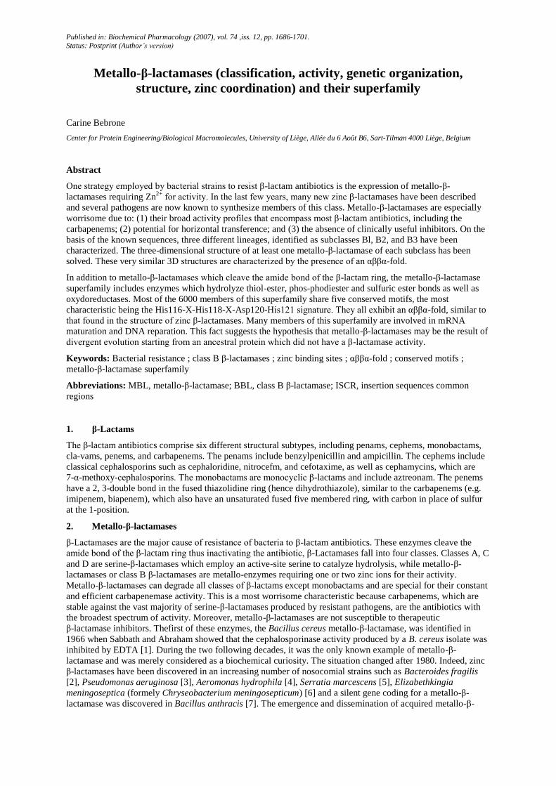
Table 1 - Metallo-β-lactamases
Subclass Enzyme Strain Discovery
in
GenBank
accession no.
Accession no.
(protein)
Structure
Bl BcII B. cereus 1966 M11189 AAA22276 Mono-zinc [48],
Di-zinc [50]
Apo-form [50]
CcrA B. fragilis 1990 M63556 AAA22904 Di-zinc [49]
BlaB E. meningoseptica 1998 AF189298 008498 Di-zinc [52]
IND-1 Chryseobacterium
indologenes
1999 EF394437 AB021411
EBR-1 E. breυis 2002 AF416700 AAN32638
SFB-1 Shewαnellα frigidimarina 2005 AY590119 AAT90847
SLB-1 Shewαnellα
livingstonensis
2005 AY590118 AAT90846
Acquired-
B1
IMP-1 S. marcescens, P.
aeruginosa
1994 S71932,
AY168635
AAB30289,
AAN87168
Di-zinc[51]
VIM-1 P. aeruginosa, A.
baumannii
1999 Y18050 CAE46717
VIM-2 P. aeruginosa, A.
baumannii
2000 AF191564 AAK26253 Mono-zinc 1K02,
Di-zinc 1K03
IMP-2 A. baumannii, S.
marcescens
2000 AB182996 BAD26594
SPM-1 P. aeruginosa 2002 AY341249 AAR15341 Mono-zinc [53]
VIM-4 P. aeruginosa, K.
pneumoniae,
Enterobacter cloacae
2003 AY135661 AAR22402
GIM-1 P. aeruginosa 2004 AJ620678 CAF05908
SIM-1 A. baumannii 2005 AY887066 AAX76774
B2 CphA A. hydrophila 1991 X57102 P26918 Mono-zinc [36]
ImiS Aeromonas veronii 1996 Y01415 CAA71411
Sfh-1 S. fonticola 2003 AF197943 AAF09244
B3 L1 Stenotrophomonas
maltophilia
1991 AB294542 CAB75346 Di-zinc [22],
Mono-zinc
2H6A,
Apo-form 2FU6
GOB-1 E. meningoseptica 2000 AF090141 AB021417
FEZ-1 L. gormanii 2000 Y17896 CAB96921 Di-zinc [54]
THIN-B J. lividum 2001 AJ250876 CAC33832
Mbl1b C. crescentus 2001 AJ315850 CAC48262
CAU-1 Caulobacter vibrioides 2002 AJ308331 CAC87665
BJP-1 B. japonicum 2006 NP_772870 Di-zinc 2GMN
They are separated into the three subclasses and classified by year of discovery. All the variants are not included in this table.

Published in: Biochemical Pharmacology (2007), vol. 74 ,iss. 12, pp. 1686-1701.
Status: Postprint (Author’s version)
Table 2 - Minimal Inhibitory Concentrations (µg/ml) for E.coli strains producing some metallo-β-lactamases
E. coli
DH10B
(reference)
E. coli
DH10B
(pBlaB-1)
(B1)
E. coli
DH10B
(pEBR-1)
(B1)
E. coli
DH10B
(pSLB-1) (B1)
E. coli
DH10B
(pSFB-1)
(B1)
E. coli
DH10B
(pVIM-2)
(acquired-B1)
E. coli
DH10B
(pGOB-1)
(B3)
E. coli
DH5α
(Ref.)
E. coli
DH5α
(pGIM-1)
(acquired-
B1)
E. coli
DH5α
(pFEZ-1)
(B3)
E. coli
XL-1
(Ref.)
E. coli
XL-1 (pSIM-1)
(acquired-B1)
Amoxicillin 2 128 >512 512 16 >512 64 - - - - -
Amoxicillin-
CLA
2 - >512 512 16 512 - - - - - -
Ticarcillin 2 256 >512 512 16 > 512 64 - - - 4 >128
Piperacillin 1 4 8 4 1 4 2 1 16 1 1 1
Cephalothin 2 16 32 8 4 256 32 4 - 32 4 >128
Cefuroxime 4 - 128 32 2 - - 4 - 32 4 >128
Ceftazidime 0.06 - 0.5 32 0.12 16 - 0.12 256 0.5 0.12 32
Cefotaxime 0.06 0.12 0.5 0.5 0.12 8 0.25 0.06 - 4 <0.06 32
Cefepime 0.06 0.03 0.12 0.06 0.06 0.06 0.06 0.06 4 - <0.06 2
Cefpirome 0.06 0.06 0.12 - - - 0.5 - - - - -
Ceftriaxone 0.06 - 0.25 - - - - 0.125 128 - <0.06 32
Cefoxitine 4 2 8 4 4 128 16 2 - 32 2 >128
Moxalactam 0.12 0.12 1 0.06 0.06 - 1 - - - - -
Aztreonam 0.12 0.25 0.12 0.06 0.12 0.12 0.25 0.12 0.125 0.12 0.25 0.25
Imipenem 0.06 0.5 0.5 0.5 0.25 1 0.5 0.12 0.5 0.25 0.12 1
Meropenem 0.06 0.12 0.25 0.12 0.25 0.25 0.12 0.03 0.125 0.25 <0.06 2
[15] [20] [15] [25] [25] [40] [20] [21] [41] [21] [42] [42]
-, not determined.
Published in: Biochemical Pharmacology (2007), vol. 74 ,iss. 12, pp. 1686-1701.
Status: Postprint (Author’s version)
3. Classification
The class B β-lactamases are classified into three subclasses Bl, B2 and B3 on the basis of sequence alignments
[13]. The classification task was complicated by the generally low degree of similarity between subclass
sequences but facilitated by the availability of X-ray structures (see below), which allowed the identification of
corresponding secondary structures elements, even when the sequence similarity was not obvious. The standard
BBL numbering of class B β-lactamases allows easy comparisons of the sequences of these enzymes [13].
Subclass B1 enzymes share more than 23% identity. These enzymes include the prototypical BcII from B. cereus
[14], CcrA from B. fragilis [2], BlaB from E. meningoseptica [6] and EBR-1 from Empedobacter breυis [15].
The acquired IMP-type MBLs found in some clinical isolates of P. aeruginosa [16], S. marcescens [5],
Klebsiella pneumoniae (GenBank EMBL accession no D29636) and Acinetobacter baumannii [8], as the VIM-
type or SPM-1 enzymes produced by clinical isolates of P. aeruginosa [8,17], belong also to this subclass (Table
1).
The members of subclass B2 show only 11% of identity with subclass Bl members. This subclass includes the
enzymes produced by different species of Aeromonas (the most studied are CphA produced by A. hydrophila [4]
and ImiS produced by A. veronii [18]) as well as the Sfh-I enzyme produced by Serratia fonticola [9] (Table 1).
Subclass B3 metallo-β-lactamases have only nine conserved residues when compared with the other metallo-β-
lactamases. Subclass B3 includes the L1 and GOB-1 metallo-β-lactamases produced by clinical strains of
S. maltophilia [19] and E. meningoseptica (formely C. meningosepticum) [20] and FEZ-1 isolated from
Legionnella gormanii [21]. To date, 18 variants of GOB-1 have been reported, but none outside the E.
meningoseptica species. In GOB enzymes, His116 is replaced by a glutamine. THIN-B [10], Mbl1b [11], and
BJP-1 [12] produced by environmental bacteria (Janthinobacter lividum, Caulobacter crescentus and
Bradyrhizobium japonicum, respectively) belong also to this subclass (Table 1). With the exception of L1 which
is a homo-tetramer [22], all these enzymes are monomeric as are the B1 and B2 enzymes.
4. MICs
Compared to the reference Escherichia coli strains, strains producing a metallo-β-lactamase exhibit decreased
susceptibility to a broad array of β-lactam antibiotics, including penicillins, narrow- and expanded-spectrum
cephalospor-ins, and carbapenems (Table 2), indicating that metallo-β-lactamases have a broad overall substrate
specificity profile. Only the MICs of aztreonam are unchanged, suggesting that theses enzymes are not active
against this compound. It has also been observed that E. coli expressing the cphA gene is resistant only to
carbapenems and only when high inocula (108 CFU) are used for MIC determinations [23]. However, a rational
approach of the correlation between the MICs and the kinetic parameters of the β-lactamase is made difficult by
the absence of knowledge of outer membrane permeability for many of the usually studied β-lactam antibiotics
and by the fact that most authors do not estimate the amount of enzyme produced.
5. Activity profiles
The metallo-β-lactamases belonging to subclasses B1 and B3 are broad spectrum enzymes and hydrolyze most
β-lactam antibiotics including carbapenems [24] (Table 3). Only the monobactams are neither hydrolyzed nor
recognized by these enzymes which are not inhibited by classical inhibitors of serine-active β-lactamases such as
clavulanic acid and tazobactam. These compounds in fact behave as poor substrates. SFB-1 from the
psychrophilic Shewanella frigidi-maria is an unusual subclass Bl β-lactamase which does not significantly
hydrolyze benzylpenicillin, ticarcillin, third generation cephalosporins and meropenem [25].
The subclass B2 enzymes are strict carbapenemases [18,23,24]. They only hydrolyze carbapenems efficiently
and show a very weak activity, if any, towards penicillins and cephalosporins (Table 3). In B2 enzymes, the
conserved His116 of B1 and B3 enzymes, is replaced by an asparagine. The presence of this asparagine in
position 116 is far from being the only reason which explains the narrow spectrum of B2 metallo-β-lactamases,
since, although increased, the activity of the N116H CphA mutant towards penicillins and cephalosporins
remains low [26]. A mutant of CphA with a considerably broadened activity spectrum, N116H-N220G, was
recently obtained by site-directed mutagenesis [27]. In contrast to the wild-type enzyme, it is able to efficiently
hydrolyze penicillins and cephalosporins in addition to carbapenems although with a reduced efficiency towards
the latter (Table 3).

Published in: Biochemical Pharmacology (2007), vol. 74 ,iss. 12, pp. 1686-1701.
Status: Postprint (Author’s version)
Table 3 - Kinetics parameters of some metallo-β-lactamases
Benzylpenicillin Nitrocefin Imipenem
kcat
( s-1
)
Km
(µM)
kcat/Km
(M-1
s-1
)
kcat
( s-1
)
Km
(µM)
kcat/Km
(M-1
s-1
)
kcat
( s-1
)
Km
(µM)
kcat/Km
(M-1
s-1
)
Bl
Bell 680 1500 4.5 × 10⁵ 45 70 6.4 × 10
5 >100 >1000 1 × 10
5 [24]
CcrA 190 40 4.8 × 10⁶ 200 20 1 × 10
7 200 270 7.4 × 10
5 [24]
EBR-1 120 50 2.4 × 10⁶ - - - 190 780 2.4 × 10
5 [15]
BlaB 280 30 9 × 10⁶ 20 70 2.8 × 10
5 350 370 9.5 × 10
5 [6]
Acquired-Bl
IMP-1 320 520 6.2 × 10⁵ 60 30 2 × 10
6 50 40 1.2 × 10
6 [16]
VIM-1 30 840 3.5 × 10⁴ 100 20 5 × 10
6 2 2 1 × 10
6 [8]
VIM-2 60 50 1.2 × 10⁶ - - - 10 10 1 × 10
6 [8]
SPM-1 110 40 2.8 × 10⁶ 0.50 4 1.2 × 10
5 30 40 7.5 × 10
5 [53]
GIM-1 7 50 1.4 × 10⁵ 6 10 6 × 10
5 30 290 1 × 10
5 [41]
B2
CphA 0.03 870 35 0.003 1200 2.5 1200 340 3.5 × 10⁶ [26]
CphA N116H 0.4 910 440 0.09 30 3 × 10³ 150 1400 1.1 × 10
5 [26]
CphA N116H-
N220G
3 150 2 × 10⁴ 0.7 4 1.8 × 10
5 20 190 1 × 10
5 [27]
ImiS 0.50 250 2 × 10³ 0.06 20 3 × 10
3 160 180 9 × 10
5 [18]
B3
LI 1100 50 2 × 10⁷ 20 10 2 × 10
6 65 90 7.2 × 10
5 [24]
FEZ-1 70 590 1.2 × 10⁵ 90 100 9 × 10
5 >200 >1000 2 × 10
5 [88]
GOB-1 200 110 1.8 × 10⁶ - - - 40 60 6.6 × 10
5 [20]
Mbllb 770 20 3.8 × 10⁷ 1800 100 1.8 × 10
7 940 200 4.7 × 10
6 [11]
-, not determined.
6. Phylogeny
Subclasses B1 and B2 are descended from a common ancestor and really form a single group within which the
sequences exhibit significant similarities, with subclasses Bl and B2 forming two distinct clades within the
group. Subclass B3 shares structural but not sequence similarities with the B1/B2 group [28]. A phylogenetic
tree with representative members of all the three subclass is given in the previous Ref. [28]. Hall and Barlow
have proposed, on this basis, to revise the classification of metallo-β-lactamases [29]. However, the presently
accepted classification reflects other properties of these enzymes. Functional and mechanistic factors clearly
distinguish the B1, B2 and B3 enzymes from each other and, on this basis, B2 is not more related to Bl than B3
(see activity profile, zinc coordination points). Thus it seems unnecessary to modify the broadly accepted
nomenclature [30].
7. Catalytic mechanism
Bounaga et al. (1998) [31] have proposed a mechanism for the monozinc form of the subclass B1 BcII enzyme.
In this mechanism, the zinc ion interacts with His116, His118, His196 and a water molecule. The zinc ion
behaves as a Lewis acid and decreases the pK of the water molecule so that it exists as a hydroxide ion at neutral
pH. This ion performs a nucleophilic attack on the carbon of the carboxyl group of the β-lactam, leading to the
formation of a tetrahedral intermediate stabilized by its interaction with the zinc ion. Asp120, acting as a general
base, deprotonates the -OH group to generate a dianionic tetrahedral second intermediate which is also stabilized
by the zinc ion. In a subsequent step, Asp120 gives the proton to the nitrogen of the β-lactam ring and takes part
in cycle opening.
A mechanism based on the study of the hydrolysis of nitrocefin (a chromogenic cephalosporin) by the CcrA and
L1 enzymes has been proposed for the dizinc form of metallo-β-lactamases. A ring-opened intermediate
containing an anionic nitrogen was identified, the protonation of which was rate-limiting [32-34]. But the
mechanism observed for nitrocefin seems to be atypical, due to its highly conjugated bis-nitro-substituted styryl
substituent which is ideally suited to stabilize the negatively charged nitrogen. When other substrates are used,
the cleavage of the tetrahedral intermediate, which occurs concomitantly to the protonation of the nitrogen of the
β-lactam ring, appears to be the rate-limiting step [35].
Published in: Biochemical Pharmacology (2007), vol. 74 ,iss. 12, pp. 1686-1701.
Status: Postprint (Author’s version)
A different reaction mechanism has been proposed for the monozinc subclass B2 enzymes [36,37]. The
nucleophilic hydroxyde is generated by the Aspl20 and His118 residues. This nucleophile attacks the β-lactam
carbonyl, possibly activated by His196, to generate a tetrahedral intermediate. As observed for Bl and B3
enzymes, the rate-limiting step in the reaction is the C-N bond cleavage. The crystal structure of CphA shows the
presence of a bicyclic product of biapenem in the active site of the enzyme [36]. Garau et al. proposed that, after
β-lactam bond cleavage, a substantial bond rotation occurs that allows the nucleophilic attack of C3 by the
oxygen of the 6-hydroxyethyl substituent with concomitant transfer of its proton to C2. This cyclization occurs
after C-N bond cleavage. Some results indicate that the final product of imipenem hydrolysis by ImiS is different
from that by L1 but the NMR spectra of both products are the same [37].
A very recent study of D120N, D120E, D120Q and D120S BcII mutants indicates that Asp120 is not the proton
donor, nor does it play an essential role in nucleophilic activation. It is proposed that the role of Asp120 is to act
as a strong Zn2 ligand, locating this ion optimally for substrate binding, stabilization of the development of a
partial negative charge in the β-lactam nitrogen, and protonation of this atom by a zinc-bound water molecule
[38].
8. Genetic organization
Some members of subclass B1 are encoded by the chromosome such as BcII from B. cereus [14] and BlaB from
E. meningoseptica [6]. The gene coding for CcrA (B. fragilis) is often quiescent and requires a surrogate
sequence to provide an adequate promoter. Insertion elements, such as IS942, IS1186 and IS4351, have been
shown to lie immediately upstream of the ribosome-binding site, providing enhanced transcriptional capabilities
for the CcrA gene [39].
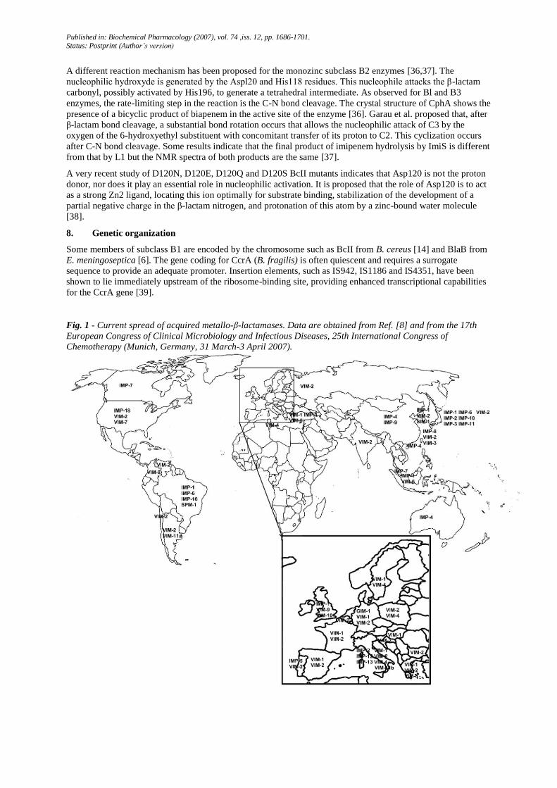
Fig. 1 - Current spread of acquired metallo-β-lactamases. Data are obtained from Ref. [8] and from the 17th
European Congress of Clinical Microbiology and Infectious Diseases, 25th International Congress of
Chemotherapy (Munich, Germany, 31 March-3 April 2007).

Published in: Biochemical Pharmacology (2007), vol. 74 ,iss. 12, pp. 1686-1701.
Status: Postprint (Author’s version)
Five plasmid-borne sets of transferable B1 metallo-β-lactamase genes have presently been identified of which
the IMP- and VIM-types occur most frequently (Table 1). The IMP-type enzymes, produced by P. aeruginosa
[16], S. marcescens [5], K. pneumoniae (GenBank accession no. D29636), have now spread worldwide [8] (Fig.
1). The VIM-type enzymes, produced by P. aeruginosa and Acinetobacter, appear as the most prevalent in
Europe with more than 12 allelic variants [8] (Fig. 1). The other foci for the VIM-enzymes appear to be East
Asia and Americas [8] (Fig. 1). VIM-2, first discovered in France (Marseille) in 2000 [40], is currently the most
widespread acquired MBL. It was found repeatedly in Western (Belgium, Germany, Greece, Italy, Portugal,
Spain) and Eastern Europe (Croatia, Poland, Russia), United States, Latin America and Asia (China, India,
Japan, Korea, Taiwan). Three other types of acquired metallo-β-lactamases, SPM-1, GIM-1 and SIM-1, have
been found in South America [17], Germany [41] and Korea [42] (Fig. 1) which suggests that the capture by
bacteria of similar resistance genes in the clinical setting could be an ongoing, relatively common, and rapidly
increasing phenomenon. These enzymes are produced by P. aeruginosa (SPM-1, GIM-1) and A. baumannii
(SIM-1).
The transferable metallo-β-lactamases are commonly encoded by genes carried by type 1 or type 3 integrons [8].
These integrons could be carried by large plasmids or be located on the chromosome. The MBL blaSPM-1 is not
part of a gene cassette, nor is it found in the vicinity of a class 1 integron as found for other metallo-β-lactamase
genes, but it is located besides the ISCR variant ISCR4 [8-43]. ISCR (IS common regions), a new type of genetic
element, was recently identified as being closely associated with the spread of many antibiotic resistance genes.
They can be divided into two groups: ISCRs1 are those that form complex class 1 integrons and ISCRs-2 to -13
are those associated with non-class 1 integrons. ISCRs are also associated with Salmonella enterica serovar
Typhimurium pathogenecity islands. ISCR elements are very unusual in that they can mobilize large sections of
adjacent DNA via a rolling circle replication. Normally, termination of replication occurs at a defined site;
misreading of this site leads to replication of large sections of DNA to the left-hand end of the element including
antibiotic resistance genes. Using a PCR strategy employing degenerate primers designed against the aligned
DNA sequences of ISCRsl to -5, Toleman has detected ISCR elements in several strains of P. aeruginosa and A.
baumannii that harbor metallo-β-lactamase genes e.g. ISCR2 was discovered in a P. aeruginosa isolate (Brazil)
that harbored blaIMP-1 and ISCR3 was discovered in two strains of P. aeruginosa (Italy) that have blaVIM-1 [43].
The genes coding for subclass B2 enzymes are located on the chromosome and their expression can be regulated
by the presence of β-lactam antibiotics in the culture medium via a two-components BlrAB system [44].
Subclass B3 metallo-β-lactamases are generally chromosome encoded. The gene coding for L1 could be located
on the chromosome or on a large plasmid [45]. The production of this β-lactamase is induced by the presence of
β-lactam antibiotics in the medium.
Most natural producers of class B enzymes also produce class A β-lactamases, as do B. cereus [46], S.
maltophilia [45], or class D enzymes as does L. gormanii [47]. Several Aeromonas species produce a class C and
a class D β-lactamases in a co-regulated way with the metallo-β-lactamase [44].
9. Structures
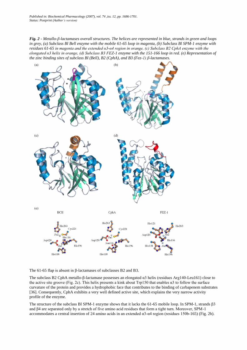
The three-dimensional structure of the mono-zinc form of BcII was the first to be solved [48]. Thereafter, the
structures of other subclass B1 enzymes, CcrA [49], the di-zinc form of BcII [50] (Fig. 2a), IMP-1 [51], BlaB
[52], VIM-2 (PDB accession no. 1K02 and 1K03), SPM-1 [53] (Fig. 2b), and subclass B3 enzymes, LI [22]
(PDB accession no. 2H6A), FEZ-1 [54] (Fig. 2d) and BJP-1 (PDB accession no. 2GMN), were determined.
More recently, the structure of a subclass B2 enzyme, CphA, was solved [36] (Fig. 2c). Despite of the low level
of identity between the amino acid sequences, the general fold of these enzymes is similar and consists of an
αββα structure, composed by two central β-sheets with five solvent-exposed α-helices. The N-terminal and C-
terminal parts of the molecule, each of them comprising a β-strand and two α helices, can be superposed by a
180° rotation around a central axis, suggesting that the complete structure might have arisen from the duplication
of a gene [48]. In all known structures, the active site is located at the external edge of the ββ sandwich.
The N-terminal domain of B1 metallo-β-lactamases incorporates a loop (residues 61-65) that can interact with
substrate or inhibitor molecules which possess hydrophobic side-chains (Fig. 2a). This loop is very flexible in
the native form of the enzyme. When the substrate or the inhibitor diffuses into the active site, the loop moves to
block the molecule in the active site. This movement of the loop is caused by the interaction of the side chain of
the W64 residue with the hydrophobic side chain of the substrate [55]. The loop is also stabilized upon binding
of inhibitors [51,52] and transforms the active site groove into a tunnel-shaped cavity. The deletion of this loop
(named flap) seriously affects the enzymatic activity by weakening the binding of substrate to the enzyme, with
the exception of imipenem [55]. Actually, the kinetic parameters of this carbapenem compound are barely
affected by the absence of the active site loop. The main difference between the carbapenems and other β-
lactams is that both penicillins and cephalosporins usually have bulky aromatic ring substitutions which are
absent in the former.
Published in: Biochemical Pharmacology (2007), vol. 74 ,iss. 12, pp. 1686-1701.
Status: Postprint (Author’s version)
Fig. 2 - Metallo-β-lactamases overall structures. The helices are represented in blue, strands in green and loops
in grey, (a) Subclass Bl Bell enzyme with the mobile 61-65 loop in magenta, (b) Subclass Bl SPM-1 enzyme with
residues 61-65 in magenta and the extended α3-α4 region in orange, (c) Subclass B2 CphA enzyme with the
elongated α3 helix in orange, (d) Subclass B3 FEZ-1 enzyme with the 151-166 loop in red. (e) Representation of
the zinc binding sites of subclass Bl (Bell), B2 (CphA), and B3 (Fez-1) β-lactamases.
The 61-65 flap is absent in β-lactamases of subclasses B2 and B3.
The subclass B2 CphA metallo-β-lactamase possesses an elongated α3 helix (residues Arg140-Leu161) close to
the active site groove (Fig. 2c). This helix presents a kink about Trp150 that enables α3 to follow the surface
curvature of the protein and provides a hydrophobic face that contributes to the binding of carbapenem substrates
[36]. Consequently, CphA exhibits a very well defined active site, which explains the very narrow activity
profile of the enzyme.
The structure of the subclass Bl SPM-1 enzyme shows that it lacks the 61-65 mobile loop. In SPM-1, strands β3
and β4 are separated only by a stretch of five amino acid residues that form a tight turn. Moreover, SPM-1
accommodates a central insertion of 24 amino acids in an extended α3-α4 region (residues 150b-165) (Fig. 2b).
Published in: Biochemical Pharmacology (2007), vol. 74 ,iss. 12, pp. 1686-1701.
Status: Postprint (Author’s version)
Deleting this insertion has only marginal effects upon binding and hydrolysis of a range of β-lactam antibiotics
[53]. Both structural features of SPM-1 are reminiscent of that found in the subclass B2 CphA enzyme. By these
aspects, SPM-1 thus represents a structural hybrid between subclasses Bl and B2. Moreover, when the sequence
of SPM-1 is compared to that of other metallo-β-lactamases, the highest identities are seen with IMP-1 (35.5%)
and then the subclass B2 enzymes ImiS (32.2%) and CphA (32.1%).
In subclass B3 enzymes, a loop between α3 and β7 formed by residues 151-166 is mobile and close to the active
site [22,54] (Fig. 2d). The I166A mutant of L1 exhibited increased Km values only when cefoxitin was the
substrate, suggesting an interaction of the isoleucine side chain with the cefoxitin methoxy group. The F160A
mutant of L1 exhibited higher Km values when using the cephalosporins as substrates, suggesting an interaction
of the cephalosporins' substituents with the phenylalanine on the loop that extends over the active site [56]. L1 is
a tetramer, a characteristic unique among β-lactamases. One main interaction between two subunits involves the
extended N-terminal region [22]. L1 also contains a disulphide bridge in its monomer and a similar one can be
seen in the structure of FEZ-1 [22,54], whereas this is no intramolecular disulphide bridge in the enzymes of
subclasses B1 or B2.
10. Zinc coordination
Metallo-β-lactamases possess two potential Zn2+
binding sites (Fig. 2e). In the case of Bl enzymes, one zinc ion
possesses a tetrahedral coordination sphere and is coordinated by His116, His118, His196 and a water molecule
or OFT ion. The other metal ion has a trigonal-pyramidal coordination sphere which involves Asp120, Cys221,
His263 and two water molecules (Table 4). One water/hydroxide molecule serves as a ligand for both metal ions.
The two binding sites are named the "histidine" and "cysteine" sites, respectively. In the mono-nuclear BcII,
VIM-2 and SPM-1 enzymes, the sole metal ion was shown to be located in the "histidine" site [48,PDB
accession no. 1KO2, 53].
Table 4 - Composition of the two metal binding sites in the MBL superfamily
Daiyasu's groups Enzymes Site 1 Site 2
Group 1 B1 MβLs His116-His118-His196 Asp120-Cys221-His263
Group 1 B2 MβLs Asp120-Cys221-His263 His118?-Met146? or His196?
Group 1 B3 MβLs His116-His118-His196 Asp120-His121-His263
Group 1 B3 MβL GOB Asp120-His121-His263 ?
Group 2 Glyoxalase II His116-His118-His196-Asp221 Asp120-His121-Asp221-His263
Group 3 ROO His116-Glu118-His196-Asp221 Asp120-Asp221-His263
Group 3 FprA His116-Glu118-His196-Asp221 Asp120-His121-Asp221-His263
Group 4 tRNaseZ, ZiPD His116-Glu118-His196-Asp221 Asp120-His121-Asp221-His263
Groups 6 and 7 CPSF-73 His116-Glu118-His196-Asp221 Asp120-His121-Asp221-His(C)
Group 9 Pce His116-His118-Asn196-Asp221 Asp120-His121-Asp221-His263
Group 12 AiiA lactonase His116-Glu118-His196-Asp221 Asp120-His121-Asp221-His263
Group 13 SdsA1 His116-His118-Glu196 Asp120-His121-Glu221-His263
Group 15 MPH His116-His118-His196-Asp221 Asp120-His121-Asp221-His263
In the B3 enzymes, the "histidine" site is the same as that found in Bl enzymes. Cys221 is replaced by a serine
and the second zinc ion is ligated by Asp120, His121, His263 and the nucleophilic water molecule which forms
a bridge between the two metal ions (Table 4). Ser221 does not directly interact with the zinc ion but with a
second water molecule located in the active site and which could serve as proton donor in the catalytic process
[22]. In contrast to all other related metallo-β-lactamases, the subclass B3 GOB-18 enzyme is fully active against
a broad range of β-lactam substrates as a mono-zinc species. Different spectroscopic techniques, 3D modeling,
and mutagenesis experiments, reveal that the zinc ion is bound to Asp120, His121, His263, and a solvent
molecule, i.e., in the canonical site 2 of dinuclear B3 metallo-β-lactamases [57] (Table 4). However, L. Horsfall
found two zinc ions per GOB-1 molecule after a purification procedure during which no Zn2+
was added to the
buffers [89]. These two GOB enzymes differ by a few point mutations, apparently far from the active site
(Leu94Phe, Ala137Val and Asp282Asn).
While the B1 and B3 enzymes exhibit maximum activity as di-zinc species, the B2 β-lactamases are inhibited in
a non-competitive manner upon binding of a second Zn2+
ion (for CphA, the dissociation constant Ki is 46 µM at
pH 6.5 [58]). In agreement with EXAFS spectroscopy studies [59] and site-directed mutagenesis [26], the
crystallographic structure of CphA shows that the first zinc ion is in the "cysteine" site [36]. Currently, the
identification of the second binding site in subclass B2 enzymes remains unsettled since even in the presence of
Published in: Biochemical Pharmacology (2007), vol. 74 ,iss. 12, pp. 1686-1701.
Status: Postprint (Author’s version)
a large excess of zinc (10 mM) and in spite of the low value of the dissociation constant, Garau and his
coworkers did not succeed in obtaining crystals of the di-zinc form. On the basis of spectroscopic studies with
the cobalt-substituted ImiS enzyme, Crawford et al. postulate that in subclass B2 the second metal binding site is
not the traditional "histidine" site [60]. Costello et al. propose that in ImiS the inhibitory zinc ion binds to both
His118 and Met146 [61]. Indeed, their EXAFS data show that the inhibitory zinc is bound by a sulfur. The sulfur
ligand in the inhibitory site should come from a methionine residue, since the only cysteine residue (Cys221) is a
ligand of Zn1. The mutation of Met146 to lie abolishes inhibition by zinc. However, mutations of other CphA
residues, which have not been implicated as Zn2 ligands, can have similar effects (lower inhibition or even
activation by excess of zinc) [26,27]. Moreover, a careful examination of the CphA 3D structure indicates that
His118 and Met146 are poorly positioned to bind the same zinc ion. The distance between their Cα's is 7.57 Å,
while the distances between the sulphur of Metl46 and the nitrogens of the imidazole group of His118 are 10.40
and 10.91 Å, respectively. Also, the space between these residues is occupied and does not allow the
accommodation of a zinc ion. The Met146 residue is not conserved in the subclass B2 Sfh-I enzyme where it is
replaced by a glutamine. Maybe, as proposed by Vanhove et al., the Cys221 side-chain is displaced upon binding
of the second metal ion [26]. The role of His118 residue in the catalytic mechanism [36,37] could explain the
inhibition by the binding of the second zinc ion, this residue being prevented from playing this role when it is
immobilized as ligand of this second zinc ion in the di-zinc form of the enzyme.
11. Zinc affinities
The subclass B2 CphA enzyme exists mainly in the mono-zinc form as the values of the dissociation constants
are 20 nM and 46 µM, respectively for the binding of the first and the second zinc ion [58]. Subclass Bl metallo-
β-lactamases show distinct stereochemical similarities in their active sites, with identical residues contributing to
metal coordination. However, the corresponding zinc affinities appear to be different, so that CcrA and IMP-1
show two high-affinity metal-binding sites, whereas BcII binds Znl and Zn2 with very different affinities.
Fluorescence spectroscopy studies with a chromophoric chelator show that BcII has very different dissociation
constants for the two metal-binding sites. For the loss of metal-ion from the mononuclear enzyme, the
dissociation constant, Kmono, is 6.2 x 10⁻¹⁰
M and that for the loss of one metal-ion from the dinuclear MBL, Kdi,
is 1.5 x 10⁻⁶
M [62]. Kmono decreases significantly, from nanomolar to picomolar values, in the presence of
substrate, whereas Kdi decreases only twofold [63]. This suggests that the mono-zinc enzyme is responsible for
the catalytic activity under physiological conditions, where the concentration of free zinc ions is in the picomolar
range [63]. A single Zn2+
ion is found by crystallographic methods in the "histidine" site, at pH 5.6, in presence
of 100 µM zinc [48]. At this pH, the activity is only three- to fourfold lower than that at pH 7.5 [31]. But the
existence of the mononuclear form, where the sole metal ion would be shared between the two binding sites is
supported by several kinetic results, PAC (Cd) and spectroscopic (Co) data [62]. However, one cannot
completely reject the hypothesis of a strong positive cooperativity between the two zinc ions with coexistence of
only the apo- and di-zinc forms. At least, Damblon and his coworkers observe that the inhibitor thiomandelic
acid induces positive cooperativity in metal binding [64].
Conversely, and despite the very close similarity with BcII, the class B1 enzyme CcrA binds both zinc ions very
tightly. Kinetic studies of CcrA with nitrocefin have shown that only the di-nuclear species is active and that a
previously observed mono-zinc CcrA enzyme [65] was a mixture of the di-zinc and the apo-enzyme [33].
BcII is active in the presence of either one or two zinc ions, although full activity is observed when two zinc ions
are bound. It has been proposed that the arginine 121 present in BcII, Vim-2, BlaB and in subclass B2 enzymes
is responsible for the lower affinities. The CcrA and IMP-1 enzymes, showing high affinity for both zinc ions,
have cysteine and serine, respectively, in the corresponding position. The replacement of the positive Arg121
side chain in CphA by a neutral one enhances the affinity for Zn2+
[27], as was already suggested for BcII
[48,49]. Rasia and Vila [66] have shown that the R121H mutation increases the affinity for the second Zn2+
ion
in BcII and that the His121 side chain replaces that of His263 as a ligand of the second Zn2+
. The C121R mutant
of CcrA was isolated in the di-zinc form, although the removal of the second zinc appeared to be facilitated [33].
All these data indicate that the replacement of Arg121 by a neutral residue (a Cys, Ser or His, respectively) in
CcrA, IMP-1, and the subclass B3 enzymes increases the affinity for the second zinc. Until recently, no mono-
zinc form of subclass B3 enzymes has ever been obtained. However, the subclass B3 GOB-18 was shown to be a
mononuclear enzyme [57]. In the subclass B1 SPM-1 enzyme, a neutral Gly121 is present and, however, SPM-1
possesses two metal sites of very different affinities [53], thus the presence of Argl21 in BcII is surely not the
only reason explaining the weaker affinity for Zn2.
As shown by the data described above, the activity and even the existence of mono-zinc forms of B1 and B3
enzymes remain controversial. As yet, it seems impossible to supply a clear answer to these questions and it
remains possible that minor sequence differences might result in different behaviours. For B2 enzymes, the
mono-zinc form is clearly the active one, but here the location of the inhibitory second zinc remains mysterious
(although this is probably biologically unrelevant).
Published in: Biochemical Pharmacology (2007), vol. 74 ,iss. 12, pp. 1686-1701.
Status: Postprint (Author’s version)
12. Inhibitors of metallo-β-lactamases
All known inhibitors or inactivators of serine β-lactamases are inefficient towards metallo-β-lactamases.
Reported inactivators of all metallo-β-lactamases are the metal chelators EDTA, o-1, 10-phenanthroline and
dipicolinic acid, however these have no clinical significance.
The propagation of metallo-β-lactamases among nosocomial bacterial strains justifies the search for compounds
which can thwart the activity of these enzymes. Unfortunately, the discovery of a clinically useful, specific
inhibitor of metallo-β-lactamases is made difficult by the fact that this compound must remain inactive towards
the human proteins which are members of the metallo-β-lactamase superfamily, or other metallo-enzymes such
as the angiotensin converting enzyme. Another difficulty is to find a compound active on all the three subclasses
of metallo-β-lactamases and even on all the enzymes within a same subclass. Currently, the known inhibitors
efficiently inhibit one or two metallo-β-lactamases and are much less active against the others. Moreover, they
remain impractical to use as therapeutic agents. In 2004, Toney and Moloughney reviewed the literature on
inhibitors of MBLs [67], and more recently Walsh et al. devoted a paragraph on experimental inhibitors in their
review on transferable MBLs [8]. A few examples are thioesters derivatives, trifluoromethyl alcohols and
ketones, sulfonyl hydra-zones, tricyclic natural products, succinic acid derivatives, biphenyl tetrazoles, cysteinyl
peptides, carbapenem and penicillin derivatives among which an interesting thiol-substituted penicillin inhibitor
[68], degradation products of cephalosporins, simple thiol compounds such as mercaptoa-cetic acid and
thioesters, thiomandelic acid [64], captopril [52,59], derivatives of benzohydroxamic acid [69], pyridine
carboxylates [70].
13. The metallo-β-lactamases (MBL) superfamily
The MBL superfamily was defined by Neuwald et al. in 1997 [71]. In addition to metallo-β-lactamases, it
includes enzymes which hydrolyze thiol-ester, phosphodiester and sulfuric ester bonds as well as
oxydoreductases.
Most of the 6000 members of this superfamily share five conserved motifs [72]: Asp84, His116-X-His118-X-
Asp120-His121, His196, Asp221 and His263 (in the order to facilitate comparison, the BBL numbering [13] is
applied to the MBL superfamily and used throughout this chapter). The fold of these proteins or of one of their
domains is of the αββα type, similar to the structure of zinc β-lactamases. Almost all these proteins exhibit a
binuclear metal center similar to that of metallo-β-lactamases. These centers occupy topologically similar
locations. Among the conserved motifs, Asp84 exhibits a strained secondary conformation and may play a role
in maintaining the protein fold. The other conserved residues are involved in metal coordination. For the
majority of these enzymes, the first metal ion binding site is composed of His116, His118 and His 196, while the
second binding site is composed of Asp120, His121 and His263. A major difference with true metallo-β-
lactamases consists in the presence of an aspartate (Asp221) in the position where a cysteine (B1) or a serine
(B3) is found in β-lactamases. Asp221 and a water molecule bridge the two metallic ions.
A large number of hypothetic enzymes found in GenBank could also share this fold and bind one or two metal
ions, but the majority of these proteins have not been functionally or structurally characterized yet. Daiyasu has
classified these proteins in 17 families on the basis of their biological functions [72]. Until recently, only three of
the 17 protein groups proposed to contain this fold had been characterized structurally, true metallo-β-lactamases
(group 1-see above), glyoxylases II (group 2), and rubredoxin oxidoreductases (group 3). However, more recent
crystal structures of other family members include acid phosphorylcholine esterase Pce (group 9), the methyl
parathion hydrolase (group 15), the N-acyl homoserine lactone hydrolase (group 12), the alkylsulfa-tase from P.
aeruginosa SdsA1 (group 13), the tRNA3'-processing endoribonuclease tRNaseZ (group 4) and proteins
members of the β-CASP family (groups 6 and 7) (Fig. 3). The composition of both metal binding sites of these
MBL superfamily enzymes of known structures is described in Table 4.
13.1. Group 2
Glyoxalase II belongs to the glyoxalase system (formed by glyoxalases I and II) which catalyzes the conversion
of toxic 2-oxoaldehydes to 2-hydroxycarboxylic acids using glutathione as coenzyme. Glutathione thiolesters
formed in the reactions catalyzed by glyoxalase I are substrates for glyoxalase II and are hydrolyzed by the latter
enzyme to the free 2-hydroxyacids and gluthathione. Glyoxalase II has been isolated from numerous mammalian
tissues and from higher plants. The structure of human glyoxalase II is composed of two domains: a metallo-β-
lactamase domain and a smaller α-helical domain (Fig. 3a). It binds 1.5 moles of zinc per mole of enzyme and
0.7 mole of iron per mole of enzyme [73]. The cytosolic glyoxalase II from Arabidopsis thaliana possesses a
binuclear center able to bind zinc, iron or manganese and some results indicate that glyoxalase II exhibits
positive cooperativity in metal binding [74]. The two metal-binding sites present the classical structural features
of the MBL superfamily (Table 4).

Published in: Biochemical Pharmacology (2007), vol. 74 ,iss. 12, pp. 1686-1701.
Status: Postprint (Author’s version)
Park et al. succeeded in introducing metallo-β-lactamase activity in the αββα scaffold of glyoxalase II by
simultaneous incorporation and adjustement of functional elements in conjunction with directed evolution [75].
However, the very low activity versus cefotaxime of the resulting enzyme (kcat/ Km = 180 M-1
s-1
) does not
explain the increased MICs of the producing cells which served as a selection tool, unless unrealistic periplasmic
concentrations of enzyme are assumed. Moreover, the engineered protein only hydrolyzes cefotaxime and no
other cephalosporins or penicillins, a very unlikely property for a β-lactamase.
Fig. 3 - Metallo-β-lactamases superfamily overall structures. The metallo-β-lactamase like domains are
represented in blue (helices), green (strands) and grey (loops and disordered regions). Zinc ions are represented
as grey spheres and iron ions as beige spheres. (a) Glyoxalase II with the additional α-helical domain in violet.
(b) ROO with the additional flavodoxin-like domain in violet. (c) phosphorylcholine esterase (Pce) domain of the
virulence factor choline-binding protein E with the elongated loop lying on the top of the active site represented
in yellow. (d) N-acyl homoserine lactone hydrolase. (e) MPH monomer. (f) SdsA1 monomer with the additional
central dimerization domain in pink and C-terminal domain in violet. (g) ZiPD monomer with the 50 amino
acids exosite in salmon. (h) TTHA0252 with the β-CASP domain in hotpink.
Published in: Biochemical Pharmacology (2007), vol. 74 ,iss. 12, pp. 1686-1701.
Status: Postprint (Author’s version)
13.2. Group 3
The metallo-β-lactamase fold is also found in a distinct group of cytosolic redox proteins. Rubredoxin:oxygen
oxydoreduc-tase (ROO) from Desulfovibrio gigas consists of two domains: a β-lactamase domain and a
flavodoxin-like domain (Fig. 3b). ROO is not a zinc-enzyme but contains a di-iron center for dioxygen reduction
[76]. The second histidine in the usual H-X-H-X-DH motif is replaced by a glutamate (Glu118). One iron ion is
ligated by two histidines (His116, His196), one glutamate (Glu118) and one aspartate (Asp221) while the second
iron ion is coordinated by one histidine (His263) and two aspartates (Asp120, Asp221). His121, although
present, does not bind Fe2 [76] (Table 4). A comparison of the ROO structure with that of true metallo-β-
lactamases shows that the L1 β-lactamase is more similar to the first domain of ROO than to other β-lactamases
[28], and FEZ-1 is closer to glyoxalase II and ROO than to BcII or CphA [13].
The structure of FprA from Moorella thermoacetica, which likely functions as a scavenging nitric oxide
reductase, has also been solved [77]. As in the case of D. gigas ROO, FprA contains a solvent-bridged non-
heme, non-sulfur di-iron site with five-coordinate iron centers bridged by an aspartate, and terminal glutamate,
aspartate, and histidine ligands. However, in contrast to ROO, the FprA di-iron site exhibits four His ligands,
two for each iron (Table 4).
13.3. Group 9
The crystal structure of a virulence factor, the phosphorylcho-line esterase (Pce) domain of the choline-binding
protein E from Streptococcus pneumoniae was solved in 2005 [78]. This domain catalyzes the hydrolysis of
choline-phosphoester bonds, releasing phosphorylcholine molecules from cell wall-associated teichoic and
lipoteichoic acids. A striking feature of Pce, unique among the reported structures of the metallo-β-lactamase
superfamily proteins, is the presence of an elongated loop, which lies on the top of the active site (Fig. 3c). This
loop would play a role in determining the accessibility of the catalytic cavity for the substrate. Iron ions are
essential for catalysis. Site 1 comprises His116, His118, Asn196 and Asp221 residues and site 2 Asp120,
His121, Asp221 and His263 (Table 4). A water/hydroxide molecule bridge completes the coordination spheres
of the two metals.
13.4. Group 15
The methyl parathion hydrolase (MPH) from Pseudomonas sp. WBC-3 catalyzes the degradation of the
organophosphate pesticide methyl parathion. MPH is a homodimer, each monomer presenting a typical metallo-
β-lactamase fold [79] (Fig. 3e). MPH possesses a binuclear di-zinc center. The coordination sphere of the zinc
ions is the same as that found in glyoxalase II (Table 4).
13.5. Group 12
The N-acyl homoserine lactone hydrolase from Bacillus thuringensis is a quorum-quenching lactonase. More
than 50 species of Gram-negative bacteria, P. aeruginosa for example, are known to use N-acyl-L-homoserine
lactones (AHLs) as signal-molecules for intercellular "quorum sensing" communication pathways necessary to
their pathogenicity. Because of their importance in pathogenicity, these pathways are possible targets in the
development of new therapeutic molecules. Nature has already developed several methods to counter these
pathways among which the production of quorum-quenching enzymes which hydrolyze the lactone residue of
AHLs. Their amino acid sequences exhibit the conserved MBL motifs. The quorum-quenching lactonase AiiA
from B. thuringensis binds two zinc ions and its activity is Zn dependent. The recently solved 3D structure of
this enzyme [80] highlights the typical αββα-fold (Fig. 3d). The coordination sphere of the zinc ions is the same
as that found in glyoxalase II (Table 4).
13.6. Group 13
P. aeruginosa is able to degrade and metabolize SDS which is usually a biocide. SdsA1 of P. aeruginosa is a
secreted SDS hydrolase that allows the bacterium to use primary sulfates such as SDS as sole carbon or sulfur
sources. The crystal structure of SdsA1 reveals three distinct domains. The N-terminal catalytic domain with a
binuclear Zn2+
cluster is a distinct member of the metallo-β-lactamase fold superfamily, the central dimerization
domain ensures resistance to high concentrations of SDS, whereas the C-terminal domain provides a
hydrophobic groove, presumably to recruit long aliphatic substrates (Fig. 3f) [81]. Zn1 is coordinated by His116,
His118, and Glu896. Asp221 is replaced by a glutamic acid residue. Zn2 is coordinated by His821, His263,
Asp820 and Glu221. A water molecule bridges the two zinc ions (Table 4).
13.7. Group 4
The crystal structures of Thermotoga maritima tRNaseZ [82], Bacillus subtilis tRNA maturase tRNaseZ [83] and
E. coli tRNase Z (ZiPD) [84] (Fig. 3g) have been determined. tRNaseZ belongs to the ELAC1/ELAC2 family
within the MBL superfamily. It is found in the vast majority of, if not all, eukaryotes and archea, and in about

Published in: Biochemical Pharmacology (2007), vol. 74 ,iss. 12, pp. 1686-1701.
Status: Postprint (Author’s version)
half of the sequenced bacterial genomes. Two ElaC variants were recently associated with prostate cancer in
man. The structure of B. subtilis tRNaseZ is characterized by a long flexible arm extending from the core β-
lactamase domain, this arm permits RNA recognition and hydrolysis of phosphodiester bonds. The E.coli
homolog of tRNaseZ, ZiPD, also possesses a specific sequence insertion module of ~50 amino acids, referred to
by the authors as "ZiPD exosite", located between the zinc ligands His196 and Asp221. This exosite is not
required for phosphodiesterase activity but is essential for pre-tRNA processing and tRNA binding. ZiPD is
capable of binding two zinc or two iron ions. However, it displays phosphodiesterase activity only in the zinc
form. The coordination sphere of the zinc ions is the same as that found in glyoxalase II (Table 4).
13.8. Groups 6 and 7
A separate family of enzymes within those exhibiting the metallo-β-lactamase fold, the β-CASP family,
comprises several important proteins acting on nucleic acid substrates, involved in DNA repair (Artemis, Apollo,
SNM1 and PS02) and RNA processing (cleavage and polyadenylation specificity factor subunits CPSF-73 and
CPSF-100) [85]. Two regions can be defined within the protein sequence: the MBL domain and a region specific
to members of the MBL superfamily acting on nucleic acids. This region is named the β-CASP motif (metallo-β-
lactamases-associated CPSF Artemis SNM1 PSO2). β-CASP family proteins are characterized by three
conserved sequence motifs [A (Asp or Glu), B (His) and C (His)], following the four typical metallo-β-lactamase
motifs (1-4), whereas other members of the MBL superfamily have motif 5 (His263) just after motif 4 (Asp221).
The crystal structure of TTHA0252 from Thermus thermo-philus HB8, a RNA degradation protein, was the first
report of the tertiary structure of a β-CASP family protein [86]. TTHA0252 comprises two separate domains: a
metallo-β-lactamase domain and a "clamp" domain (β-CASP domain) (Fig. 3h). The active site of the enzyme is
located in a cleft between the two domains, which includes two zinc ions coordinated by seven conserved
residues. Although this geometry is similar to those of members of the MBL superfamily, TTHA0252 has one
conserved His residue characteristic of the β-CASP family as a ligand.
Most eukaryotic messenger RNA precursors (pre-mRNAs) undergo extensive maturational processing, including
cleavage and polyadenylation at the 3'-end. Recent analyses and the resolution of the 3D structure [87] indicated
that the 73-kDa subunit of cleavage and polyadenylation specificity factor (CPSF-73) is the endonuclease for this
and related reactions. CPSF-73 contains two domains, a metallo-β-lactamase domain and a β-CASP domain.
This second domain can be considered as a cassette inserted into the metallo-β-lactamase domain. The active site
of CPSF-73, with two zinc ions, is located at the interface of the two domains. The two zinc ions in the active
site are each bound in an octahedral environment. Zn1 is coordinated by Hisll6, His118 and His196. Zn2 is
coordinated by Asp120, His121 and motif C (His) of the β-CASP domain. A hydroxide ion and Asp221 are
bridging ligands (Table 4). CPSF-73 possesses RNA endonuclease activity. Mutations, that disrupt zinc binding
in the active site, abolish this activity. CPSF-100 shares sequence conservation and a similar domain architecture
with CPSF-73 but lacks the zinc-binding residues in the metallo-β-lactamase domain. Therefore, CPSF-100
cannot bind zinc and is unlikely to possess nuclease activity.
14. Conclusion
The metallo-β-lactamase fold clearly represents a stable scaffold that has repeatedly been used during evolution
to catalyze a diverse range of chemical reactions. Structurally, this diversity is achieved by varying the sequence
and length of loops near the active site to accommodate various substrates. By combining the metallo-β-
lactamase domain with auxiliary substrate- , product- or cofactor-binding domains, the range of functions has
been extended yet further.
The catalytic diversity of this superfamily explains how certain members might have easily evolved into metallo-
β-lactamases in response to environmental antibiotics. The most recent therapeutic utilization of carbapenems
has subsequently favoured the emergence of preexisting enzymes in pathogenic strains. These data support the
hypothesis that metallo-β-lactamases may be the result of divergent evolution starting from an ancestral protein
which did not have a β-lactamase activity.
Acknowledgements
Grateful thanks to Professor Jean-Marie Frère (Center for Protein Engineering, University of Liège) and
Professor Moreno Galleni (Biological Macromolecules, University of Liège) for helpful discussions, scientific
advices and for reviewing this manuscript. Thanks to P. Lassaux. Thanks to S. Baurin and S. Bebrone for the
help in the design of illustrations and Dr. F. Kerf for the examination of the CphA 3D structure. The work in
Liège was supported by FRFC grants 2.4511.06 and 2.4524.03 (FRS-FNRS, Brussels) and PAI P5/33 (Politique
Scientifique Fédérale, Brussels). Carine Bebrone is a postdoctoral researcher of the F.R.S.-FNRS (Belgium).

Published in: Biochemical Pharmacology (2007), vol. 74 ,iss. 12, pp. 1686-1701.
Status: Postprint (Author’s version)
References
[1] Abraham EP, Chain E. An enzyme from bacteria able to destroy penicillin. Nature 1966;146:837.
[2] Rasmussen BA, Gluzman Y, Tally FP. Cloning and sequencing of the class B beta-lactamase gene (ccrA) from Bacteroides fragilis
TAL3636. Antimicrob Agents Chemother 1990;34:1590-2.
[3] Watanabe M, Iyobe S, Inoue M, Mitsuhashi S. Transferable imipenem resistance in Pseudomonas aeruginosa. Antimicrob Agents
Chemother 1991;35:147-51.
[4] Massidda O, Rossolini GM, Satta G. The Aeromonas hydrophila CphA gene: molecular heterogeneity among class B metallo-beta-lactamases. J Bacteriol 1991;173: 4611-7.
[5] Osano E, Arakawa Y, Wacharotayankun R, Ohta M, Horii T, Ito H, et al. Molecular characterization of an enterobacterial metallo-beta-
lactamase found in a clinical isolate of Serratia marcescens that shows imipenem resistance. Antimicrob Agents Chemother 1994;38:71-8.
[6] Rossolini GM, Franceschini N, Riccio ML, Mercuri PS, Perilli M, Galleni M, et al. Characterization and sequence of the
Chryseobacterium (Flavobacterium) meningosepticum carbapenemase: a new molecular class B beta-lactamase showing a broad substrate
profile. Biochem J 1998;332: 145-52.
[7] Chen Y, Succi J, Tenover FC, Koehler TM. Beta-lactamase genes of the penicillin-susceptible Bacillus anthracis Sterne strain. J
Bacteriol 2003;185:823-30.
[8] Walsh TR, Toleman MA, Poirel L, Nordmann P. Metallo-β-lactamases: the quiet before the storm. Clin Microbiol Rev 2005;18:306-25.
[9] Saavedra MJ, Peixe L, Sousa JC, Henriques I, Alves A, Correia A. Sfh-I, a subclass B2 metallo-β-lactamase from a Serratia fonticola
environmental isolate. Antimicrob Agents Chemother 2003;47:2330-3.
[10] Rossolini GM, Condemi MA, Pantanella F, Docquier JD, Amicosante G, Thaller MC. Metallo-beta-lactamase producers in
environmental microbiota: new molecular class B enzyme in Janthindbacterium lividum. Antimicrob Agents Chemother 2001;45:837-44.
[11] Simm AM, Higgins CS, Pullan ST, Avison MB, Niumsup P, Erdozain O, et al. A novel metallo-beta-lactamase, Mbl1b, produced by the environmental bacterium Caulobacter crescentus. FEBS Lett 2001;509:350-4.
[12] Stoczko M, Frère JM, Rossolini GM, Docquier JD. Postgenomic scan of metallo-beta-lactamase homologues in rhizobacteria:
identification and characterization of BJP-1, a subclass B3 ortholog from Bradyrhizobacterium japonicum. Antimicrob Agents Chemother 2006;50: 1973-81.
[13] Garau G, Garcia-Saez I, Bebrone C, Anne C, Mercuri P, Galleni M, et al. Update of the standard numbering scheme for class B beta-
lactamases. Antimicrob Agents Chemother 2004;48:2347-9.
[14] Hussain M, Carlino A, Madonna MJ, Lampen JO. Cloning and sequencing of the metallothioprotein beta-lactamase II gene of Bacillus
cereus 569/H in Escherichia coli. J Bacteriol 1985;164:223-9.
[15] Bellais S, Girlich D, Karim A, Nordmann P. EBR-la novel Ambler subclass B1 beta-lactamase from Empedobacter brevis. Antimicrob Agents Chemother 2005;46:3233-7.
[16] Laraki N, Franceschini N, Rossolini GM, Santucci P, Meunier C, de Pauw E, et al. Biochemical characterization of the Pseudomonas
aeruginosa 101/1477 metallo-beta-lactamase IMP-1 produced by Escherichia coli. Antimicrob Agents Chemother 1999;43:902-6.
[17] Toleman MA, Simm AM, Murphy TA, Gales AC, Biedenbach DJ, Jones RN, et al. Molecular characterization of SPM-1, a novel
metallo-β-lactamase isolated in Latin America: report from the SENTRY antimicrobial surveillance programme. J Antimicrob Chemother
2002;50:673-9.
[18] Walsh TR, Gamblin S, Emery DC, MacGowan AP, Bennett PM. Enzyme kinetics and biochemical analysis of ImiS, the metallo-beta-
lactamase from Aeromonas υeronii 163a. J Antimicrob Chemother 1996;37:423-31.
[19] Walsh TR, Hall L, Assinder SJ, Nichols WW, Cartwright SJ, MacGowan AP, et al. Sequence analysis of the LI metallo-beta-lactamase
from Xanthomonas maltophilia. Biochim Biophys Acta 1994;1218:199-201.
[20] Bellais S, Aubert D, Naas T, Nordmann P. Molecular and biochemical heterogeneity of class B carbapenem-hydrolyzing metallo-beta-
lactamases in Chryseobacterium meningosepticum. Antimicrob Agents Chemother 2000;44:1878-86.
[21] Boschi L, Mercuri PS, Riccio ML, Amicosante G, Galleni M, Frère JM, et al. The Legionella (Fluoribacter) gormanii metallo-beta-
lactamase: a new member of the highly divergent lineage of molecular-subclass B3 beta-lactamases. Antimicrob Agents Chemother
2000;44:1538-43.
[22] Ullah JH, Walsh TR, Taylor IA, Emery DC, Verma CS, Gamblin SJ, et al. The crystal structure of the LI metallo-beta-lactamase from
Stenotrophomonas maltophilia at 1.7 A resolution. J Mol Biol 1998;284:125-36.
[23] Segatore B, Massidda O, Satta G, Setacci D, Amicosante G. High specificity of cphA-encoded metallo-beta-lactamase from Aeromonas hydrophila AE036 for carbapenems and its contribution to beta-lactam resistance. Antimicrob Agents Chemother 1993;37:1324-8.
[24] Felici A, Amicosante G, Oratore A, Strom R, Ledent P, Joris B, et al. An overview of the kinetic parameters of class B beta-lactamases.
Biochem J 1993;291:151-5.
[25] Poirel L, Héritier C, Nordmann P. Genetic and biochemical characterization of the chromosome-encoded class B beta-lactamases from
Shewanella livingstonensis (SLB-1) and Shewanella frigidimarina (SFB-1). J Antimicrob Chermother 2005;55:680-5.
[26] Vanhove M, Zakhem M, Devreese B, Franceschini N, Anne C, Bebrone C, et al. Role of Cys221 and Asnll6 in the zinc-binding sites of the Aeromonas hydrophila metallo-beta-lactamase. Cell Mol Life Sci 2003;60:2501-9.
[27] Bebrone C, Anne C, De Vriendt K, Devreese B, Rossolini GM, Van Beeumen J, et al. Dramatic broadening of the substrate profile of
the Aeromonas hydrophila CphA metallo-beta- lactamase by site-directed mutagenesis. J Biol Chem 2005;280:28195-202.
Published in: Biochemical Pharmacology (2007), vol. 74 ,iss. 12, pp. 1686-1701.
Status: Postprint (Author’s version)
[28] Garau G, Di Guilmi AM, Hall BG. Structure-based phylogeny of the metallo-beta-lactamases. Antimicrob Agents Chemother
2005;49:2778-84.
[29] Hall BG, Barlow M. Revised Ambler classification of beta-lactamases. J Antimicrob Chemother 2005;95:1050-1.
[30] Frère JM, Galleni M, Bush K, Dideberg O. Is it necessary to change the classification of beta-lactamases? J Antimicrob Chemother
2005;95:1051-3.
[31] Bounaga S, Laws AP, Galleni M, Page MI. The mechanism of catalysis and the inhibition of the Bacillus cereus zinc-dependent beta-lactamase. BiochemJ 1998;331:703-11.
[32] Wang Z, Fast W, Benkovic SJ. On the mechanism of the metallo-beta-lactamase from Bacteroides fragilis. Biochemistry
1999;38:10013-2.
[33] Fast W, Wang Z, Benkovic SJ. Familial mutations and zinc stoichiometry determine the rate-limiting step of nitrocefin hydrolysis by
metallo-beta-lactamase from Bacteroides fragilis. Biochemistry 2001;40:1640-50.
[34] McManus-Munoz S, Crowder MW. Kinetic mechanism of metallo-beta-lactamase L1 from Stenotrophomonas maltophilia. Biochemistry 1999;38:1547-53.
[35] Spencer J, Clark AR, Walsh TR. Novel mechanism of hydrolysis of therapeutic beta-lactams by Stenotrophomonas maltophilia L1
metallo-beta-lactamase. J Biol Chem 2001;276:33638-44.
[36] Garau G, Bebrone C, Anne C, Galleni M, Frère JM, Dideberg O. A Metallo-beta-lactamase enzyme in action: crystal structures of the
monozinc carbapenemase CphA and its complex with biapenem. J Mol Biol 2005;345:785-95.
[37] Sharma NP, Hajdin C, Chandrasekar S, Bennett B, Yang KW, Crowder MW. Mechanistic studies on the mononuclear Znll-containing metallo-beta-lactamase ImiS from Aeromonas sobria. Biochemistry 2006;45:10729-38.
[38] Llarrull LI, Fabiane SM, Kowalski JM, Bennett B, Sutton BJ, Vila AJ. Aspl20 locates Zn2 for optimal metallo-beta-lactamase activity. J
Biol Chem 2007;282:18276-85.
[39] Rasmussen BA, Kovacs E. Identification and DNA sequence of a new Bacteroides fragilis insertion sequence-like element. Plasmid
1991;25:141-4.
[40] Poirel L, Naas T, Nicolas D, Collet L, Bellais S, Cavallo JD, et al. Characterization of VIM-2, a carbapenem-hydrolyzing metallo-beta-lactamase and its plasmid- and integron-borne gene from a Pseudomonas aeruginosa clinical isolate in France. Antimicrob Agents
Chemother 2000;44:891-7.
[41] Castanheira M, Toleman MA, Jones RN, Schmidt FJ, Walsh TR. Molecular characterization of a beta-lactamase gene, blaGIM-1, encoding a new subclass of metallo-beta-lactamase. Antimicrob Agents Chemother 2004;48:4654-61.
[42] Lee K, Yum JH, Yong D, Lee HM, Kim HD, Docquier JD, et al. Novel acquired metallo-beta-lactamase gene, bla(SIM-l), in a class 1
integron from Acinetobacter baumannii clinical isolates from Korea. Antimicrob Agents Chemother 2005;49:4485-91.
[43] Toleman MA, Bennett PM, Walsh TR. ISCR elements: novel gene-capturing systems of the 21st century? Microbiol Mol Biol Rev
2006;70:296-316.
[44] Alksne LE, Rasmussen BA. Expression of the AsbAl, OXA-12, and AsbM1 beta-lactamases in Aeromonas jandaei AER 14 is coordinated by a two-component regulon. J Bacteriol 1997;179:2006-13.
[45] Avison MB, Higgins CS, von Heldreich CJ, Bennett PM, Walsh TR. Plasmid location and molecular heterogeneity of the LI and L2
beta-lactamase genes of Stenotrophomonas maltophilia. Antimicrob Agents Chemother 2001;45:413-9.
[46] Davies RB, Abraham EP. Separation, purification and properties of beta-lactamase I and beta-lactamase II from Bacillus cereus
569/H/9. Biochem J 1974;143:115-27.
[47] Franceschini N, Boschi L, Pollini S, Herman R, Perilli M, Galleni M, et al. Characterization of OXA-29 from Legionella (Fluoribacter)
gormanii: molecular class D beta-lactamase with unusual properties. Antimicrob Agents Chemother 2001;45:3509-16.
[48] Carfi A, Pares S, Duee E, Galleni M, Duez C, Frère JM, et al. The 3-D structure of a zinc metallo-beta-lactamase from Bacillus cereus reveals a new type of protein fold. Embo J 1995;14:4914-21.
[49] Concha NO, Rasmussen BA, Bush K, Herzberg O. Crystal structure of the wide-spectrum binuclear zinc beta-lactamase from
Bacteroides fragilis. Structure 1996;4:823-36.
[50] Carfi A, Duee E, Galleni M, Frère JM, Dideberg O. 1.85 Å resolution structure of the zinc (II) beta-lactamase from Bacillus cereus.
Acta Crystallogr D Biol Crystallogr 1998;54:313-23.
[51] Concha NO, Janson CA, Rowling P, Pearson S, Cheever CA, Clarke BP, et al. Crystal structure of the IMP-1 metallo beta-lactamase from Pseudomonas aeruginosa and its complex with a mercaptocarboxylate inhibitor: binding determinants of a potent, broad-spectrum
inhibitor. Biochemistry 2000;39:4288-98.
[52] Garcia-Saez I, Hopkins J, Papamicael C, Franceschini N, Amicosante G, Rossolini GM, et al. The 1.5 A structure of Chryseobacterium meningosepticum zinc beta-lactamase in complex with the inhibitor, D-captopril. J Biol Chem 2003;278:23868-73.
[53] Murphy TA, Catto LE, Halford SE, Haldfield AT, Minor W, Walsh TR, et al. Crystal structure of Pseudomonas aeruginosa SPM-1
provides insights into variable zinc affinity of metallo-beta-lactamases. J Mol Biol 2006;357:890-903.
[54] Garcia-Saez I, Mercuri PS, Papamicael C, Kahn R, Frere JM, Galleni M, et al. Three-dimensional structure of FEZ-1, a monomeric
subclass B3 metallo-beta-lactamase from Fluoribacter gormanii, in native form and in complex with D-captopril. J Mol Biol 2003;325:651-
60.
[55] Moali C, Anne C, Lamotte-Brasseur J, Groslambert S, Devreese B, Van Beeumen J, et al. Analysis of the importance of the metallo-β-
lactamase active site loop in substrate binding and catalysis. Chem Biol 2003;10: 319-29.
Published in: Biochemical Pharmacology (2007), vol. 74 ,iss. 12, pp. 1686-1701.
Status: Postprint (Author’s version)
[56] Carenbauer AL, Garrity JD, Perriyannan G, Yates RB, Crowder MW. Probing substrate binding to metallo-beta-lactamase L1 from
Stenotrophomonas maltophilia by using site-directed mutagenesis. BMC Biochem 2002;3:4.
[57] Morán-Barrio J, González JM, Lisa MN, Costello AL, Dal Peraro M, Carloni P, et al. The metallo-β-lactamase GOB is a mono-Zn(II) enzyme with a novel active site. J Biol Chem 2007;282:18286-93.
[58] Hernandez Valladares M, Felici A, Weber G, Adolph HW, Zeppezauer M, Rossolini GM, et al. Zn(II) dependence of the Aeromonas
hydrophila AE036 metallo-beta-lactamase activity and stability. Biochemistry 2007;36:11534-41.
[59] Heinz U, Bauer R, Wommer S, Meyer-Klaucke W, Papamichaels C, Bateson J, et al. Coordination geometries of metal ions in D- or L-
captopril-inhibited metallo-beta-lactamases. J Biol Chem 2003;278:20659-66.
[60] Crawford PA, Yang KW, Sharma N, Bennett B, Crowder MW. Spectrocopic studies on cobalt(II)-substituted metallo-beta-lactamase ImiS from Aeromonas sobria bv. sobria. Biochemistry 2005;44:5168-76.
[61] Costello AL, Sharma NP, Yang KW, Crowder MW, Tierney DL. X-ray absorption spectroscopy of the zinc-binding sites in the class B2
metallo-beta-lactamase ImiS from Aeromonas veronii bv. sobria. Biochemistry 2006;45:13650-8.
[62] de Seny D, Heinz U, Wommer S, Kiefer M, Meyer-Klaucke W, Galleni M, et al. Metal ion binding and coordination geometry for wild
type and mutants of metallo-beta-lactamase from Bacillus cereus 569/H/9 (Bell): a combined thermodynamic, kinetic and spectroscopic
approach. J Biol Chem 2001;276:45065-78.
[63] Wommer S, Rival S, Heinz U, Galleni M, Frère JM, Franceschini N, et al. Substrate-activated zinc binding of metallo-beta-lactamases:
physiological importance of mononuclear enzymes. J Biol Chem 2002;277:24142-7.
[64] Damblon C, Jensen M, Ababou A, Barsukov I, Papamicael C, Schofield CJ, et al. The inhibitor thiomandelic acid binds to both metal ions in metallo-β-lactamase and induces positive cooperativity in metal binding. J Biol Chem 2003;278:29240-51.
[65] Paul-Soto R, Hernandez-Valladares M, Galleni M, Bauer R, Zeppezauer M, Frère JM, et al. Mono- and binuclear Zn-beta-lactamase
from Bacteroides fragilis: catalytic and structural roles of the zinc ions. FEBS Lett 1998;438: 137-40.
[66] Rasia RM, Ceolin M, Vila AJ. Grafting a new metal ligand in the cocatalytic site of B. cereus metallo-beta-lactamase: structural
flexibility without loss of activity. Protein Sci 2003;12:1538-46.
[67] Toney JH, Moloughney JG. Metallo-beta-lactamase inhibitors: promise for the future? Curr Opin Invest Drugs 2004;5:823-6.
[68] Buynak JD, Chen H, Vogeti L, Gadhachanda VR, Buchanan CA, Palzkill T, et al. Penicillin-derived inhibitors that simultaneously target
both metallo- and serine-beta-lactamases. Bioorg Med Chem Lett 2004;14:1299-304.
[69] Lienard BM, Horsfall LE, Galleni M, Frère JM, Schofield CJ. Inhibitors of the FEZ-1 metallo-beta-lactamase. Bioorg Med Chem Lett 2006;17:964-8.
[70] Horsfall LE, Garau G, Lienard BM, Dideberg O, Schofield CJ, Frère JM, et al. Competitive inhibitors of the CphA metallo-beta-
lactamase from Aeromonas hydrophila. Antimicrob Agents Chemother 2007;51:2136-42.
[71] Neuwald AF, Liu JS, Lipman DJ, Lawrence CE. Extracting protein alignment models from the sequence database. Nucleic Acids Res
1997;25:1665-77.
[72] Daiyasu H, Osaka K, Ishino Y, Toh H. Expansion of the zinc metallo-hydrolase family of the beta-lactamase fold. FEBS Lett 2001;503:1-6.
[73] Cameron AD, Ridderstrom M, Olin B, Mannervik B. Crystal structure of human glyoxalase II and its complex with a glutathione
thiolester substrate analogue. Struct Fold Des 1999;7:1067-78.
[74] Wenzel M, Carenbauer AL, Pfiester MP, Schilling O, Meyer-Klaucke W, Makaroff CA, et al. The binding of iron and zinc to glyoxalase
II occurs exclusively as di-metal centers and is unique within the metallo-beta-lactamase family. J Biol Inorg Chem 2004;9:429-38.
[75] Park HS, Nam SH, Lee JK, Yoon CN, Mannervik B, Benkovic SJ, et al. Design and evolution of new catalytic activity with an existing
protein scaffold. Science 2006;311:535-8.
[76] Frazao C, Silva G, Gomes CM, Matias P, Coelho R, Sieker L, et al. Structure of a dioxygen reduction enzyme from Desulfovibrio gigas. Nat Struct Biol 2000;7:1041-5.
[77] Silaghi-Dumitrescu R, Kurtz Jr DM, Ljungdahl LG, Lanzilotta WN. X-ray crystal structures of Moorella thermoacetica FprA Novel
diiron site structure and mechanistic insights into a scavenging nitric oxide reductase. Biochemistry 2005;44:6492-501.
[78] Garau G, Lemaire D, Vernet T, Dideberg O, Di AM, Guilmi. Crystal structure of phosphorylcholine esterase domain of the virulence
factor choline-binding protein e from Streptococcus pneumoniae: new structural features among the metallo-beta-lactamase superfamily. J
Biol Chem 2005;280:28591-600.
[79] Dong YJ, Bartlam M, Sun L, Zhou YF, Zhang ZP, Zhang CG, et al. Crystal structure of methyl parathion hydrolase from Pseudomonas
sp WBC-3. J Mol Biol 2005;353:655-63.
[80] Liu D, Lepore BW, Petsko GA, Thomas PW, Stone EM, Fast W, et al. Three-dimensional structure of the quorum-quenching N-acyl homoserine lactone hydrolase from Bacillus thuringiensis. Proc Natl Acad Sci USA 2005;102:11882-7.
[81] Hagelueken G, Adams TM, Wiehlmann L, Widow U, Kolmar H, Tummler B, et al. The crystal structure of SdsAl, an alkylsulfatase
from Pseudomonas aeruginosa, defines a third class of sulfatases. Proc Natl Acad Sci USA 2006;103:7631-6.
[82] Ishii R, Minagawa A, Takaku H, Takagi M, Nashimoto M, Yokoyama S. Crystal structure of the tRNA 3' processing endoribonuclease
tRNase Z from Thermotoga maritima. J Biol Chem 2005;280:14138-414.
[83] de la Sierria-Gallay IL, Pellegrini O, Condon C. Structural basis for substrate binding, cleavage and allostery in the tRNA maturase RNase Z. Nature 2005;433:657-61.

Published in: Biochemical Pharmacology (2007), vol. 74 ,iss. 12, pp. 1686-1701.
Status: Postprint (Author’s version)
[84] Kostelecky B, Pohl E, Vogel A, Schilling O, Meyer-Klaucke W. Structural basis for substrate binding, cleavage and allostery in the
tRNA maturase RNase Z. Nature 2005;433:657-61.
[85] Callebaut I, Moshous D, Mornon JP, de VillartayJP. Metallo-beta-lactamase fold within nucleic acids processing enzymes: the beta-CASP family. Nucleic Acids Res 2002;30:3592-601.
[86] Ishikawa H, Nakagawa N, Kuramitsu S, Masui R. Crystal structure of TTHA0252 from Thermus thermophilus HB8, a RNA degradation
protein of the metallo-beta-lactamase superfamily. J Biochem (Tokyo) 2006;140:535-42.
[87] Mandel CR, Kaneko S, Zhang H, Gebauer D, Vethantham V, Manley JL, et al. Polyadenylation factor CPSF-73 is the pre-mRNA 3'-end
processing endonuclease. Nature 2006;444:953-6.
[88] Mercuri PS, Bouillenne F, Boschi L, Lamotte-Brasseur J, Amicosante G, Devreese B, et al. Biochemical characterization of the FEZ-1 metallo-beta-lactamase of Legionella gormanii ATCC 33297T produced in Escherichia coli. Antimicrob Agents Chemother 2001;45:1254-
62.
[89] Horsfall L, Study of the subclass B3 and inhibitors of the metallo-β-lactamases, Ph.D. Thesis, University of Liège, Belgium, 2007.


![Metallo-β-Lactamases: A Major Threat to Human Healthfile.scirp.org/pdf/AJMB_2014071615272629.pdf · tobacter baumannii [9]-[11]is facile transfer of genetic inform). ... ased on](https://img.pdfslide.net/doc/110x75/5b7a3ec67f8b9ae7368b5881/metallo-lactamases-a-major-threat-to-human-tobacter-baumannii-9-11is.jpg)