Embed Size (px)
Citation preview
Watson et al. BMC Neuroscience 2013, 14:59http://www.biomedcentral.com/1471-2202/14/59
METHODOLOGY ARTICLE Open Access
Modelling the endothelial blood-CNS barriers:a method for the production of robust in vitromodels of the rat blood-brain barrier andblood-spinal cord barrierP Marc D Watson1*, Judy C Paterson1, George Thom1, Ulrika Ginman2, Stefan Lundquist2 and Carl I Webster1
Abstract
Background: Modelling the blood-CNS barriers of the brain and spinal cord in vitro continues to provide aconsiderable challenge for research studying the passage of large and small molecules in and out of the centralnervous system, both within the context of basic biology and for pharmaceutical drug discovery. Although therehas been considerable success over the previous two decades in establishing useful in vitro primary endothelial cellcultures from the blood-CNS barriers, no model fully mimics the high electrical resistance, low paracellularpermeability and selective influx/efflux characteristics of the in vivo situation. Furthermore, such primary-derivedcultures are typically labour-intensive and generate low yields of cells, limiting scope for experimental work. Wethus aimed to establish protocols for the high yield isolation and culture of endothelial cells from both rat brainand spinal cord. Our aim was to optimise in vitro conditions for inducing phenotypic characteristics in these cellsthat were reminiscent of the in vivo situation, such that they developed into tight endothelial barriers suitable forperforming investigative biology and permeability studies.
Methods: Brain and spinal cord tissue was taken from the same rats and used to specifically isolate endothelialcells to reconstitute as in vitro blood-CNS barrier models. Isolated endothelial cells were cultured to expand thecellular yield and then passaged onto cell culture inserts for further investigation. Cell culture conditions wereoptimised using commercially available reagents and the resulting barrier-forming endothelial monolayers werecharacterised by functional permeability experiments and in vitro phenotyping by immunocytochemistry andwestern blotting.
Results: Using a combination of modified handling techniques and cell culture conditions, we have establishedand optimised a protocol for the in vitro culture of brain and, for the first time in rat, spinal cord endothelial cells.High yields of both CNS endothelial cell types can be obtained, and these can be passaged onto large numbers ofcell culture inserts for in vitro permeability studies. The passaged brain and spinal cord endothelial cells are pureand express endothelial markers, tight junction proteins and intracellular transport machinery. Further, both modelsexhibit tight, functional barrier characteristics that are discriminating against large and small molecules inpermeability assays and show functional expression of the pharmaceutically important P-gp efflux transporter.
Conclusions: Our techniques allow the provision of high yields of robust sister cultures of endothelial cells thataccurately model the blood-CNS barriers in vitro. These models are ideally suited for use in studying the biology ofthe blood-brain barrier and blood-spinal cord barrier in vitro and for pre-clinical drug discovery.
Keywords: Blood-brain barrier, Blood-spinal cord barrier, in vitro, TEER, Drug discovery, Permeability coefficient,FITC-dextran, Lucifer yellow, Hydrodynamic radius
* Correspondence: [email protected] Ltd, Granta Park, Cambridgeshire CB21 6HG, UKFull list of author information is available at the end of the article
© 2013 Watson et al.; licensee BioMed CentraCommons Attribution License (http://creativecreproduction in any medium, provided the or
l Ltd. This is an Open Access article distributed under the terms of the Creativeommons.org/licenses/by/2.0), which permits unrestricted use, distribution, andiginal work is properly cited.
Watson et al. BMC Neuroscience 2013, 14:59 Page 2 of 21http://www.biomedcentral.com/1471-2202/14/59
BackgroundThe endothelial blood-CNS barriers, located at themicrovascular cells of the brain and spinal cord, repre-sent the crucial interface between the maelstrom of theperipheral circulation and the tightly regulated environ-ment of the central nervous system (CNS). Here, theblood–brain barrier (BBB) and blood-spinal cord barrier(BSCB) present a formidable structural and metabolicbarrier that partitions the CNS parenchyma. Far frombeing impenetrable blockades, the blood-CNS barriersare highly dynamic regulatory interfaces that apply strictcontrol over the passage of blood-borne substances intothe CNS, and oversee regulated transport of large andsmall molecules back into the periphery. The blood-CNS barriers are of great relevance to pharmaceuticaldrug discovery, as the BBB and BSCB present obstaclesto the delivery of compounds aimed at the treatment ofCNS disorders affecting the brain and spinal cord. Afuller understanding of each of these barriers will aid thedevelopment of CNS-targeted small and large moleculetherapies to treat wide-ranging and devastating neuro-logical diseases, from neurodegeneration to chronic pain[1-4]. To facilitate basic research and drug discovery, itis therefore highly desirable to have robust and conveni-ent in vitro models of the BBB and BSCB, from speciesrelevant for pre-clinical investigations [1,5]. Such modelsmust aim to faithfully recreate the exquisite in vivo tis-sue microenvironment that induces a blood-barrierphenotype. For the BBB, as well as the more poorlyunderstood BSCB, this has posed a considerable tech-nical challenge. The goal for in vitro BBB and BSCBmodel development is to obtain convenient primary cellcultures that can be easily and inexpensively establishedand possess robust barrier phenotypes similar to thoseseen in vivo. Good in vitro barriers will possess proper-ties such as high transendothelial electrical resistance(TEER) across the endothelial monolayer and low pas-sive, non-specific paracellular permeability to small andlarge molecules such as Lucifer yellow (LY), hydropho-bic compounds and FITC-labelled dextrans. For a trulyrepresentative model, other features such as expressionof receptors and transporters on the endothelial cellsurface and intracellular transcytosis machinery mustbe maintained to allow transcellular transport pathwaysfor ions, small molecules, peptides and proteins tobe reconstituted in vitro. An additional problem forestablishing robust in vitro blood-CNS barrier modelsis the provision of sufficient numbers of cells to allowfor rigorous characterisation of the models and investi-gative biology or drug screening. The typically lowyields of endothelial cells can severely limit research ef-forts, particularly for tissues such as the spinal cordwhere the amount of tissue recovered per animal isespecially low.
The fundamental features of the blood-CNS barriersin vivo are well known but difficult to fully replicatein vitro. These barrier-forming elements include highlydeveloped endothelial tight junctions that lead to highTEER, lack of endothelial fenestrae, low non-specificpinocytosis and the expression of receptors and trans-porters that facilitate small and large molecule influxand efflux [6]. One of the greatest hurdles to translatingthese in vivo features into robust in vitro models isthat the development of the in vivo CNS-blood barrierphenotype is exquisitely regulated by the cellular micro-environment of the brain and spinal cord endothelialcells. Astrocytes have long been demonstrated to inducebarrier function at the BBB in vitro and in vivo [7] andincreasing evidence is pointing to a similarly importantrole for pericytes in barrier development and mainten-ance [8-12]. In spite of these challenges, in vitro model-ling of the BBB, and to a lesser extent the BSCB, hasprogressed significantly over the previous two decades.BBB primary endothelial cell culture models have beenestablished with cells isolated from human [13-19],mouse [20-26], rat [16,27-35], bovine [36-43] and pig[44-54] brain tissues. BSCB endothelial models have, incontrast, currently only been described in vitro for a sin-gle species, namely mouse [55]. BBB in vitro primary cellculture barrier models have progressed from simplesolo-cultures of brain endothelial cells to more complexco-culture models in which endothelial cells are grownon porous cell culture inserts and co-cultured with post-natal rodent astrocytes [7]. Astrocytes may be plated ei-ther into the bottom of a multi-well dish into which theinsert is placed or grown on the underside of the insert it-self in so-called back-to-back contact co-culture models.Recently, increasingly complex co-culture models, such astriple cultures of endothelial cells with astrocytes andpericytes [10-12] have been developed. However, althoughthese models display good barrier phenotypes in vitro,they are particularly labour-intensive and expensive to es-tablish. It has also been demonstrated that neural stemcells have the ability to induce barrier properties in vitroin a manner which may be representative of BBB develop-ment in vivo [56,57]. Further improvements to barrierphenotype have been demonstrated through the manipu-lation of cell culture conditions. It has been knownfor several years that factors such as modulators ofintracellular cAMP signalling [58,59], glucocortocoids[22,26,53,60,61] and growth factors such as bFGF [62,63]can induce improvements in barrier phenotype in culturedprimary brain endothelial cells. Other manipulations, suchas modulating the buffering capacity of cell culturemedium [64] and optimising endothelial cell seeding dens-ity [23,31] can influence and improve barrier function. Inrecent years, the inclusion of puromycin as a method forremoving contaminating non-endothelial cells has become
Watson et al. BMC Neuroscience 2013, 14:59 Page 3 of 21http://www.biomedcentral.com/1471-2202/14/59
established as a key element of in vitro BBB cell cultureprotocols [27,31,51,61,65].There continues to be a need, however, to evolve blood-
CNS barrier modelling techniques to achieve increasinglyrepresentative in vitro phenotypes that faithfully recapitu-late the tight, discriminative situation found in brain andspinal cord capillaries in vivo. The reproducibility of BBBcell culture models can be inconsistent from week-to-week or lab-to-lab, and thus for routine use in academicand pharmaceutical studies it is highly desirable to haveprotocols that produce robust and reliable in vitro barriers.Additionally, it is also highly useful to have such in vitroblood-CNS barrier models from commonly used pre-clinical species, such as the rat, so that in vitro data is rele-vant to the in vivo models employed during early CNSdrug discovery efforts. Such representative in vitro modelsmay then be employed to characterise drug toxicity andpermeability early in pharmaceutical development andthus have great potential for contributing to a reduction inthe high attrition rate of drugs in early development forCNS diseases.We set out to investigate whether an easy and highly
robust protocol could be established that allowed theproduction of large numbers of brain and, for the firsttime in rat, spinal cord endothelial cells from a minimalamount of starting tissue. The aim was to obtain highyields of cells that could be passaged onto cell cultureinserts and induced to form tight monolayer barriers forpermeability studies. By optimizing culture conditionsusing specific handling techniques and commerciallyavailable reagents, we have demonstrated the isolationand culture of large numbers of both types of endothe-lial cell, from the same animals. These barrier culturesare pure endothelial in nature, show correct localisationof tight junction proteins, have discriminating barriercharacteristics and restrict the paracellular permeabilityof large and small molecules. We thus present a furtherevolution in the techniques for establishing in vitro blood-CNS barriers in a relevant pre-clinical species. Thesemodels have utility for investigation of the basic biology ofthe BBB and BSCB in vitro and in CNS-focused pharma-ceutical drug development and toxicity studies.
MethodsMaterialsAll tissue culture media, supplements and reagents arefrom Gibco, Life Technologies UK, unless otherwisestated. All compounds and reagents are from Sigma-Aldrich, UK unless otherwise stated.
Isolation of rat brain microvascular endothelial cellsAll procedures were carried out in accordance with theAnimals (Scientific Procedures) Act, 1986. Four male
Wister rats (200–250 grams, Charles River, UK) wereeuthanized humanely and whole brains removed andstored in HBSS buffer (calcium/magnesium-free, plus10 mM HEPES, penicillin/streptomycin) on ice. Underaseptic conditions, the brain stem and cerebellum wasdissected and each brain was cut in half sagitally. Themid-brain white matter and the choroid plexus wereremoved and the remaining cortical tissue rolled on dryWhatmann paper to remove the meninges. The meninges-free cortical tissue was transferred into ice-cold isolationbuffer (HBSS plus calcium and magnesium, 10 mMHEPES, 0.1% BSA) and homogenised on ice using a 15 mLDounce homogeniser with 20 strokes of the loose pestlefollowed by 10 strokes of the tight pestle. Following eachhomogenisation, the pestle was washed with isolationbuffer to recover as much brain tissue as possible. Thebrain homogenate was pelleted by centrifugation at 240 × gfor five minutes at 4°C. The supernatant was aspirated andthe pellet resuspended in pre-warmed digestion mix,containing 1 mg/mL collagenase/dispase (Roche, UK),10 μg/mL DNAse I (Roche, UK) and 0.147 μg/mL tosyl-lysine-chloromethylketone (TLCK). The tissue/digestionmix was incubated at 37°C for 30 minutes with gentleshaking. Digested tissue was pelleted by centrifugation at240 × g for five minutes at 4°C and the pellet wasresuspended in 22% (w/v) bovine serum albumin (First-Link, UK) by shaking vigorously. Centrifugation at 1500 ×g for 15 minutes at 4°C resulted in a pellet containingmicrovessels, with a buoyant layer of myelin floating at thetop. The myelin/BSA fraction was poured off, re-mixedand centrifuged again. The microvessel pellet wasresuspended in isolation buffer and stored on ice. Theprocess was repeated for a total of four centrifugations andthe resuspended microvessels were pooled and pelleted bycentrifugation at 240 × g for five minutes at 4°C. Thesupernatant was aspirated and the microvessel pellet wasresuspended in pre-warmed digestion mix, followed byincubation at 37°C for 15 minutes with gentle shaking.After digestion, the microvessel fragments were pelleted bycentrifugation at 240 × g for five minutes at 4°C and washedonce in serum-containing cell culture medium to removetraces of enzyme. The resulting microvessel fragments wereresuspended in DMEM (phenol red-free, supplementedwith 15% plasma-derived serum [PDS, First-Link, UK], glu-tamine, BME vitamins (Sigma), antibiotic/antimycotic sup-plement, 200 μM ascorbic acid, 3 μM puromycin and 1×microvascular growth supplement [MVGS, Life Technolo-gies]), and plated in eight wells over two 6-well plates pre-coated with 10 μg/cm2 collagen I (BD Biosciences) and5 μg/cm2 fibronectin. The commercial MVGS supplementcontains foetal bovine serum, hydrocortisone, human FGF,heparin, human EGF and dibutyryl cyclic AMP. Culturemedium was replaced after 2–3 days in vitro (DIV) foridentical medium, without ascorbic acid. Puromycin was
Watson et al. BMC Neuroscience 2013, 14:59 Page 4 of 21http://www.biomedcentral.com/1471-2202/14/59
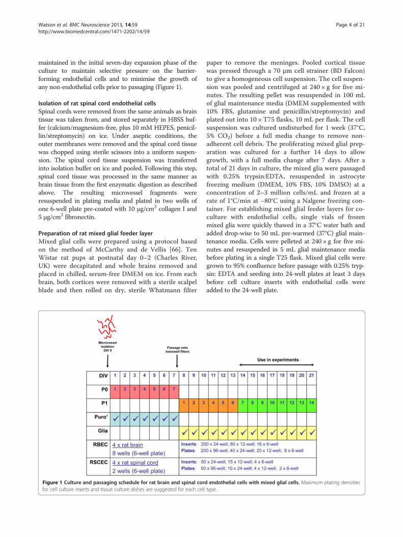
maintained in the initial seven-day expansion phase of theculture to maintain selective pressure on the barrier-forming endothelial cells and to minimise the growth ofany non-endothelial cells prior to passaging (Figure 1).
Isolation of rat spinal cord endothelial cellsSpinal cords were removed from the same animals as braintissue was taken from, and stored separately in HBSS buf-fer (calcium/magnesium-free, plus 10 mM HEPES, penicil-lin/streptomycin) on ice. Under aseptic conditions, theouter membranes were removed and the spinal cord tissuewas chopped using sterile scissors into a uniform suspen-sion. The spinal cord tissue suspension was transferredinto isolation buffer on ice and pooled. Following this step,spinal cord tissue was processed in the same manner asbrain tissue from the first enzymatic digestion as describedabove. The resulting microvessel fragments wereresuspended in plating media and plated in two wells ofone 6-well plate pre-coated with 10 μg/cm2 collagen I and5 μg/cm2 fibronectin.
Preparation of rat mixed glial feeder layerMixed glial cells were prepared using a protocol basedon the method of McCarthy and de Vellis [66]. TenWistar rat pups at postnatal day 0–2 (Charles River,UK) were decapitated and whole brains removed andplaced in chilled, serum-free DMEM on ice. From eachbrain, both cortices were removed with a sterile scalpelblade and then rolled on dry, sterile Whatmann filter
Figure 1 Culture and passaging schedule for rat brain and spinal cordfor cell culture inserts and tissue culture dishes are suggested for each cell
paper to remove the meninges. Pooled cortical tissuewas pressed through a 70 μm cell strainer (BD Falcon)to give a homogeneous cell suspension. The cell suspen-sion was pooled and centrifuged at 240 × g for five mi-nutes. The resulting pellet was resuspended in 100 mLof glial maintenance media (DMEM supplemented with10% FBS, glutamine and penicillin/streptomycin) andplated out into 10 × T75 flasks, 10 mL per flask. The cellsuspension was cultured undisturbed for 1 week (37°C,5% CO2) before a full media change to remove non-adherent cell debris. The proliferating mixed glial prep-aration was cultured for a further 14 days to allowgrowth, with a full media change after 7 days. After atotal of 21 days in culture, the mixed glia were passagedwith 0.25% trypsin:EDTA, resuspended in astrocytefreezing medium (DMEM, 10% FBS, 10% DMSO) at aconcentration of 2–3 million cells/mL and frozen at arate of 1°C/min at −80°C using a Nalgene freezing con-tainer. For establishing mixed glial feeder layers for co-culture with endothelial cells, single vials of frozenmixed glia were quickly thawed in a 37°C water bath andadded drop-wise to 50 mL pre-warmed (37°C) glial main-tenance media. Cells were pelleted at 240 × g for five mi-nutes and resuspended in 5 mL glial maintenance mediabefore plating in a single T25 flask. Mixed glial cells weregrown to 95% confluence before passage with 0.25% tryp-sin: EDTA and seeding into 24-well plates at least 3 daysbefore cell culture inserts with endothelial cells wereadded to the 24-well plate.
endothelial cells with mixed glial cells. Maximum plating densitiestype.
Watson et al. BMC Neuroscience 2013, 14:59 Page 5 of 21http://www.biomedcentral.com/1471-2202/14/59
Passage of primary rat brain and spinal cord endothelialcells onto cell culture inserts and tissue culture platesRat brain or spinal cord endothelial cells grown on col-lagen I/fibronectin-coated plates were passaged at ~95%confluence. Cells were washed twice with pre-warmedPBS and 400 μL of pre-warmed 0.25% trypsin was addedto each well. The plates were immediately returned to a37°C incubator for four minutes. The trypsinisationwas stopped by adding 1 mL of cell culture mediumcontaining 15% PDS to each well. The endothelial cellswere washed off and resuspended by gently pipetting upand down. Cells were split at a ~1:1 ratio, on a surfacearea basis. For example, the 9.5 cm2 of a single well on a6-well plate could cover the equivalent of 28 × 24-wellcell culture inserts each with a surface area of 0.33 cm2
(Figure 1). Cells were resuspended in either DMEM/MVGS (phenol red-free DMEM supplemented with 20%PDS, glutamine, BME vitamins, antibiotic/antimycoticsupplement and 1× MVGS) or EBM-2/EGM-2 (EBM-2media plus 15% PDS, glutamine, BME vitamins, BulletKitSingleQuots minus the human recombinant VEGF sup-plement [recombinant human FGF, recombinant humanEGF, recombinant human IGF, hydrocortisone, GA-1000,ascorbic acid], all Lonza, UK) media formulations. TheMVGS supplement does not contain VEGF, a factorknown to increase permeability across brain endothelialcell monolayers [52], and so this factor was not addedfrom the EGM-2 BulletKit. The resuspended cells at theadjusted concentration were plated in the upper chambersof cell culture inserts in the 24-well format at 200 μL/well(Millipore, PET, 1.0 μm pore size). Pre-seeded mixed glialcells were switched from astrocyte maintenance mediuminto 1 mL DMEM/MVGS or EBM-2/EGM-2 and the in-serts with endothelial cells were added. Brain and spinalcord endothelial cells and astrocytes were cultured for afurther 7–14 days, with media changes every 2–3 days.
Measurement of transendothelial electrical resistanceCells cultured on inserts in 24-well plates were removedfrom the tissue culture incubator (37°C, 5% CO2), andallowed to equilibrate to room temperature for 20 mi-nutes. TEER values were measured using an EVOM2voltometer with STX-2 electrodes (World Precision In-struments). To calculate TEER (Ω (Ohms) × cm2), elec-trical resistance across a collagen I/fibronectin-coatedinsert without cells was subtracted from the readingsobtained on inserts with cells and this value was multi-plied by the surface area of the insert (0.33 cm2).
Monolayer permeability to Lucifer yellow/FITC-labelleddextrans and calculation of permeability coefficients.Lucifer yellow (LY) and FITC-labelled dextran stocksolutions were prepared in Ringers-HEPES buffer
(150 mM NaCl, 3.4 mM CaCl2, 1.2 mM MgCl2, 5.2 mMKCl, 0.5 mM NaHCO3, 2.8 mM glucose, and 10 mMHEPES) and frozen at −20°C. RBECs were passagedonto cell culture inserts as described and cultured for afurther 7–14 days in vitro. For transport experiments,all media was removed from the upper chamber of theinsert and replaced with 75 μL pre-warmed Ringers-HEPES buffer plus 0.1% BSA followed by equilibrationto 37°C for 10–15 minutes. Solutions of LY and FITC-labelled dextran were diluted to 2X working concen-trations and pre-warmed to 37°C. At time-point 0 mi-nutes, 75 μL of LY/FITC-labelled dextran solution wasadded to the upper chamber of the inserts, which werethen transferred to new 24-well plates containing 1 mLof pre-warmed Ringers-HEPES buffer plus 0.1% BSA.For each compound, inserts with endothelial cells wereused in triplicate and cell-free, collagen I/fibronectin-coated inserts were used in duplicate. The plates wereincubated in an orbital shaking incubator (VWR) at37°C, 25 rpm. At each time-point, the inserts weremoved into a fresh 24-well plate containing 1 mL ofpre-warmed Ringers-HEPES buffer plus 0.1% BSA, toprevent back-diffusion of the compounds into the topchamber. Samples were collected at 30, 60 and 90 mi-nutes. At the end of each experiment, the concentra-tion of the fluorescent compounds accumulated in thebottom chamber was calculated by transferring 50 μLof each sample to a black walled-96 well plate (Nunc)and measuring with an Envision fluorescence platereader (Perkin Elmer). Concentrations were calculatedusing standard curves generated from the stock solu-tions of each compound. Permeability coefficients (Pe),that take into account the barrier to transport fromboth the endothelial monolayer and the cell cultureinsert, were calculated as described by others [11,31,67,68].Briefly, the volume cleared across cell-free and cell-containing inserts was calculated for each compound usingthe following equation:
Cleared volume μLð Þ ¼ Concentrationabluminal � Volumeabluminal
Concentrationluminal
The average cleared volumes were plotted versus timein minutes for each 90-minute experiment. Clearanceslopes for the empty filters (PSfilter) and the filters withendothelial cells (PScells + filter) were calculated using lin-ear regression analysis and used to obtain a permeabilityproduct value for endothelial monolayer alone (PScells):
1PScells
¼ 1PScellsþfilter
−1
PSfilter
Permeability coefficients (Pe) for each compoundacross the cell monolayer were finally derived by divid-ing the PScells value by the surface area of the cell culture

Watson et al. BMC Neuroscience 2013, 14:59 Page 6 of 21http://www.biomedcentral.com/1471-2202/14/59
insert (0.33 cm2 for 24-well format). Data are presentedwith units of × 10-6 cm/sec.
Assessment of claudin-5 protein levels by WesternblottingRBECs were passaged into two 35 mm dishes, onewith RBEC/MVGS formulation media and one withEBM-2/EGM-2 formulation media. The cells were cul-tured to confluence and then lysed on ice by theaddition of RIPA buffer (Sigma) with protease inhibi-tors followed by scraping. The levels of claudin-5 pro-tein present in 10 μg total soluble protein wereassessed by SDS-PAGE and Western blotting using themouse anti-claudin-5 antibody (at 1:500; 1 μg/mL) alsoused for immunocytochemistry (Table 1). The mem-branes were re-probed using a mouse monoclonal anti-body (ACTN05 (C4), Abcam, 1:2000). Western blotswere imaged using a Li-Cor Odyssey CLx and quantifi-cation of band intensity was carried out using theLi-Cor software.
ImmunocytochemistryImmunocytochemistry was performed on RBECs andRSCECs cultured on collagen I/fibronectin coated 96-well plates. Cells were cultured to confluence andmaintained for a further two days. Cultures were fixed ineither ice-cold methanol for two minutes (antibodies fortight junction protein staining) or in 3.7% formaldehydefor 20 minutes at room temperature (all other anti-bodies). Formaldehyde-fixed cells were permeabilisedwith 0.2% Triton X-100 in PBS for five minutes. Afterrinsing once in PBS, cells were blocked in 5% BSA inPBS for 30 minutes. All antibodies were diluted to work-ing concentration in 1% BSA in PBS (Table 1). Cellswere incubated with primary antibody for 1 hour atroom temperature or overnight at 4°C, followed by three5 minute washes in PBS. Secondary antibodies (AlexaFluor 488 donkey anti-goat IgG, Alexa Fluor 488 goatanti-mouse IgG, Alexa Fluor 546 goat anti-mouse IgG,Alexa Fluor 488 goat anti-rabbit IgG, Alexa Fluor 546
Table 1 Antibodies used for immunocytochemical characteris
Antigen Species
Caveolin 1 Rabbit
Clathrin heavy chain Rabbit
Claudin-5 Mouse
Occludin Mouse
P-gp Mouse
Smooth muscle actin Mouse
Von Willebrand factor Rabbit
ZO-1 Rabbit
donkey anti-rabbit IgG; all from Life Technologies, Mo-lecular Probes) were used at a final concentration of2 μg/mL. Cells were incubated with secondary antibodyfor 1 hour at room temperature followed by three5 minute washes in PBS. Cells were finally counter-stained with Hoechst (Life Technologies, MolecularProbes), diluted to 1 μg/mL in 1% BSA/PBS, for oneminute and rinsed a further three times in PBS. Sam-ples were imaged using an Olympus IX81 fluorescencemicroscope.
Analysis of small molecule permeability using liquidchromatography/mass spectrometrySmall molecule compounds were dissolved in DMSO toa concentration of 1 mM and further diluted in Ringers-HEPES buffer (without BSA) to give a final concentra-tion of 4 μM. RBECs and RSCECs were passaged ontocell culture inserts as described and cultured for a fur-ther 7–14 days in vitro. Cell culture medium (EBM-2/EGM-2 formulation) was removed from the upper andlower compartments of RBECs and RSCECs cultured intriplicate on cell culture inserts and duplicate cell-freeinserts and replaced with Ringer-HEPES buffer(without BSA). The small molecules were added to eachupper compartment to yield a final concentration of2 μM. Cultures were incubated at 37°C with shakingand transferred to a new well with fresh buffer in thelower compartment after 30, 60 and 90 minutes. Sam-ples were collected from the lower compartments andanalysed by liquid chromatography mass spectrometry(LC-MS/MS). Small molecules were analysed on anAcquity™ UPLC system with an Acquity UPLC® BEH C18,1.7 μm column (Waters Corp., Milford, MA, USA). 10 μLof each sample was injected onto the column and elutedby gradients. The flow rate was 0.6 mL/min and therun time was 1.1 min. The Acquity™ UPLC-system wasconnected to a triple quadrupole tandem mass spectrom-eter (Quattro Premier XE, Waters Corp., Milford, MA,USA) operating in the positive ion electrospray ionisationmode, with MassLynx 4.1 running in the MRM mode
ation of cultured RBECs and RSCECs
Manufacturer Concentration
Abcam ab2910 20 μg/ml
Abcam ab21679 20 μg/ml
Life Technologies 35-2500 10 μg/ml
Life Technologies 33-1500 10 μg/ml
Abcam ab3366 3.35 μg/ml
R & D Systems MAB1420 4 μg/ml
Abcam ab6994 156 μg/ml
Abcam ab59720 10 μg/ml
Watson et al. BMC Neuroscience 2013, 14:59 Page 7 of 21http://www.biomedcentral.com/1471-2202/14/59
(MS/MS). Permeability coefficients were calculated as de-scribed above.
P-gp functional efflux assayRBECs and RSCECs were passaged onto cell cultureinserts as described and cultured for a further 7–14 daysin vitro. Cell culture medium (EBM-2/EGM-2 formula-tion) was removed from the upper and lower compart-ments of RBECs and RSCECs cultured on cell cultureinserts in 24-well plates and was replaced with Ringers-HEPES buffer with 0.1% BSA, containing either100 μM verapamil or vehicle (0.5% DMSO), followedby incubation at 37°C for 30 minutes. Cells were dye-loaded by removing buffer from upper compartmentsand replacing with fresh buffer containing 200 ng/mlrhodamine 123. Triplicate cell culture inserts with cellswere used for each condition. The inserts were incu-bated at 37°C for 30 minutes. The dye-loaded insertswere transferred to a fresh plate and the buffer was re-moved from the upper compartments. The cells werewashed three times in Ringer-HEPES buffer (with 0.1%BSA). Fresh assay buffer was added and the insertswere incubated at 37°C for 1 hour to allow dye efflux.At the end of the incubation, the inserts were trans-ferred to a fresh plate, the cells were washed threetimes in PBS and lysed for 20 minutes in RIPA buffer.Fluorescence values were measured for each sampleusing an Envision multi-well fluorescence plate-reader(Perkin Elmer) with excitation at 485 nm and emissioncollected at 535 nm. Standard curves were generatedusing stock rhodamine 123 and then used to calculatecellular uptake of the dye.
Determination of FITC-dextran hydrodynamic radii bydynamic light scatteringFITC-labelled dextrans (Sigma) were prepared at a con-centration of 0.8 mg/mL in Ringers-HEPES buffer with-out BSA. Samples were filtered through a 0.22 μm filterprior to loading. Hydrodynamic radii were determinedusing a Zetasizer Nano (Malvern). The backscatter oflight at 173° was measured with an equilibration time offive minutes and measurements were performed in trip-licate with no delay between them. Laser attenuationand measurement duration were determined automatic-ally by the software with data processing performed atnormal resolution.
Analysis and statisticsStandard curves were generated and sample concentra-tions interpolated by linear regression using MicrosoftExcel. Statistical analysis, using the appropriate mathem-atical functions as outlined in the text, was carried outusing GraphPad Prism. Values in figures are expressedas mean ± SEM.
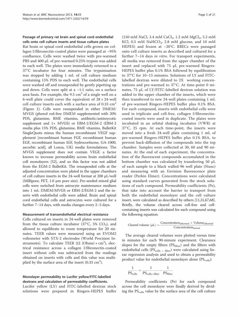
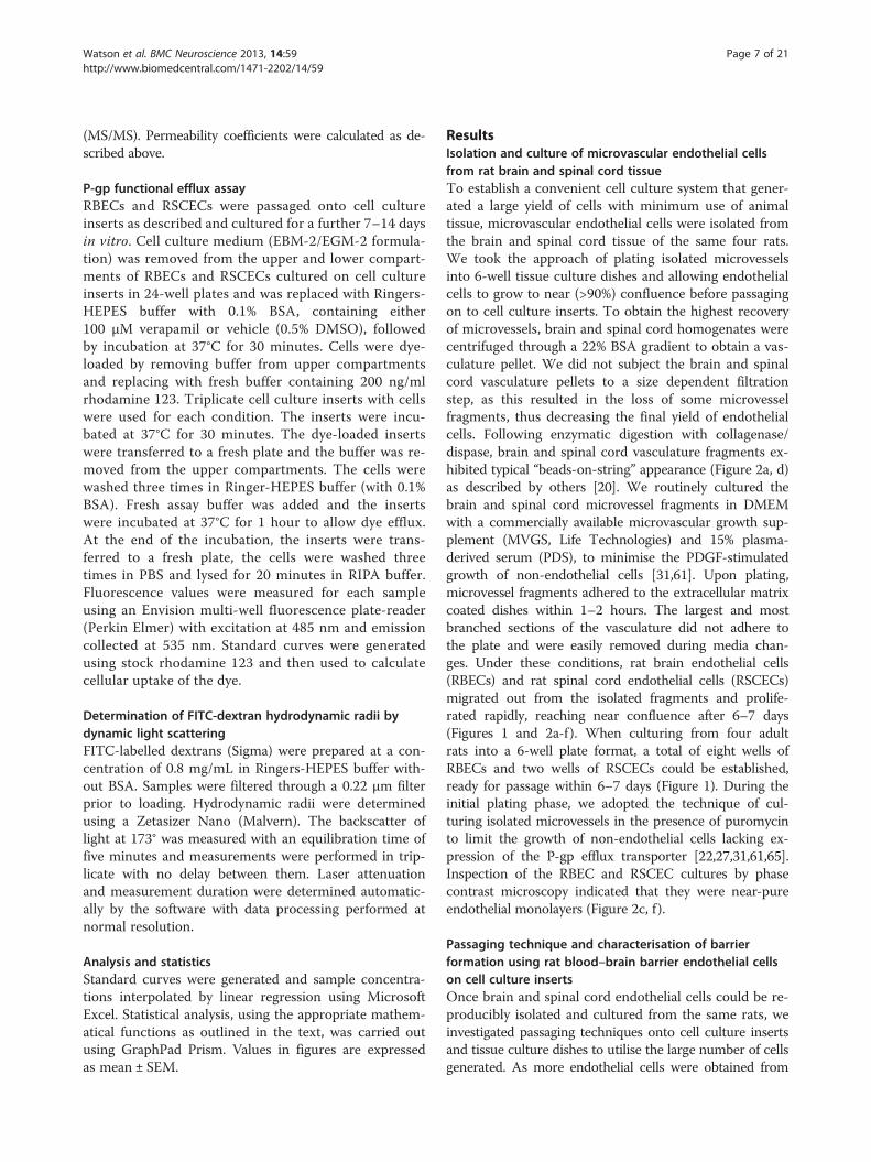
ResultsIsolation and culture of microvascular endothelial cellsfrom rat brain and spinal cord tissueTo establish a convenient cell culture system that gener-ated a large yield of cells with minimum use of animaltissue, microvascular endothelial cells were isolated fromthe brain and spinal cord tissue of the same four rats.We took the approach of plating isolated microvesselsinto 6-well tissue culture dishes and allowing endothelialcells to grow to near (>90%) confluence before passagingon to cell culture inserts. To obtain the highest recoveryof microvessels, brain and spinal cord homogenates werecentrifuged through a 22% BSA gradient to obtain a vas-culature pellet. We did not subject the brain and spinalcord vasculature pellets to a size dependent filtrationstep, as this resulted in the loss of some microvesselfragments, thus decreasing the final yield of endothelialcells. Following enzymatic digestion with collagenase/dispase, brain and spinal cord vasculature fragments ex-hibited typical “beads-on-string” appearance (Figure 2a, d)as described by others [20]. We routinely cultured thebrain and spinal cord microvessel fragments in DMEMwith a commercially available microvascular growth sup-plement (MVGS, Life Technologies) and 15% plasma-derived serum (PDS), to minimise the PDGF-stimulatedgrowth of non-endothelial cells [31,61]. Upon plating,microvessel fragments adhered to the extracellular matrixcoated dishes within 1–2 hours. The largest and mostbranched sections of the vasculature did not adhere tothe plate and were easily removed during media chan-ges. Under these conditions, rat brain endothelial cells(RBECs) and rat spinal cord endothelial cells (RSCECs)migrated out from the isolated fragments and prolife-rated rapidly, reaching near confluence after 6–7 days(Figures 1 and 2a-f). When culturing from four adultrats into a 6-well plate format, a total of eight wells ofRBECs and two wells of RSCECs could be established,ready for passage within 6–7 days (Figure 1). During theinitial plating phase, we adopted the technique of cul-turing isolated microvessels in the presence of puromycinto limit the growth of non-endothelial cells lacking ex-pression of the P-gp efflux transporter [22,27,31,61,65].Inspection of the RBEC and RSCEC cultures by phasecontrast microscopy indicated that they were near-pureendothelial monolayers (Figure 2c, f ).
Passaging technique and characterisation of barrierformation using rat blood–brain barrier endothelial cellson cell culture insertsOnce brain and spinal cord endothelial cells could be re-producibly isolated and cultured from the same rats, weinvestigated passaging techniques onto cell culture insertsand tissue culture dishes to utilise the large number of cellsgenerated. As more endothelial cells were obtained from
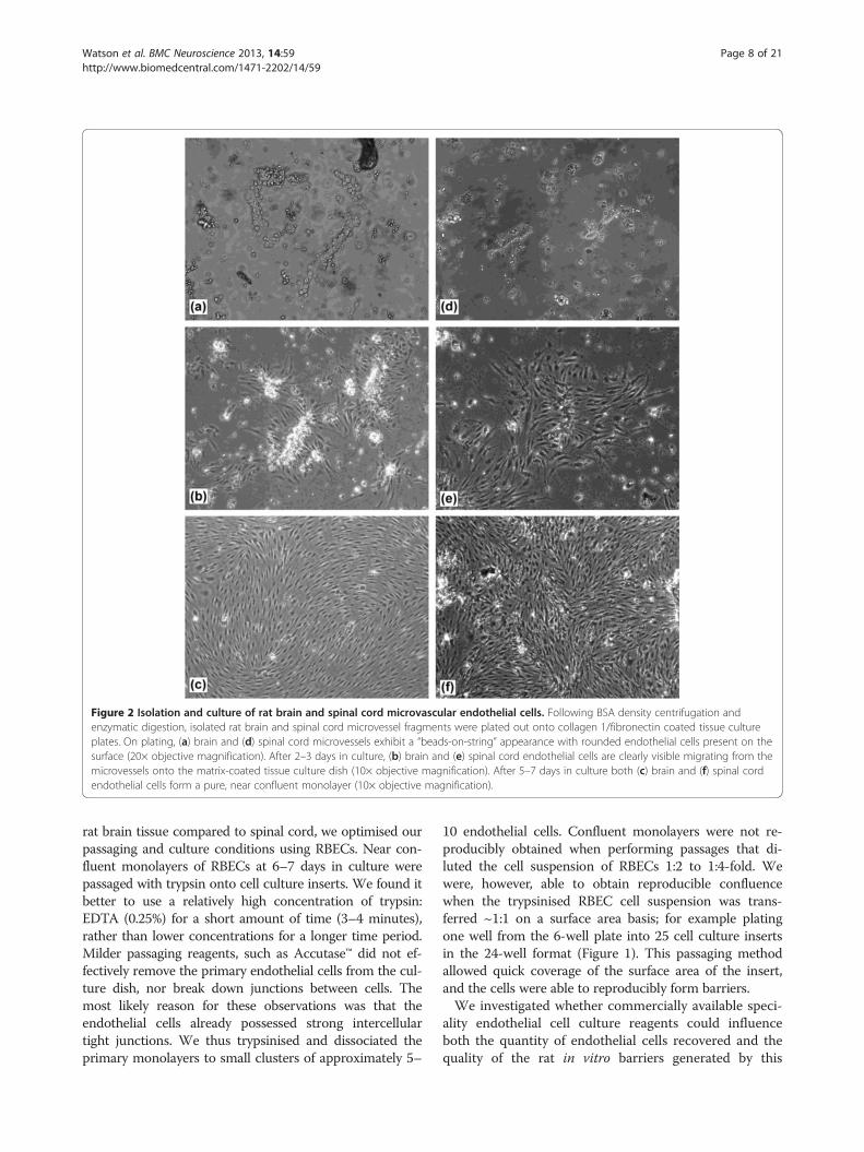
Figure 2 Isolation and culture of rat brain and spinal cord microvascular endothelial cells. Following BSA density centrifugation andenzymatic digestion, isolated rat brain and spinal cord microvessel fragments were plated out onto collagen 1/fibronectin coated tissue cultureplates. On plating, (a) brain and (d) spinal cord microvessels exhibit a “beads-on-string” appearance with rounded endothelial cells present on thesurface (20× objective magnification). After 2–3 days in culture, (b) brain and (e) spinal cord endothelial cells are clearly visible migrating from themicrovessels onto the matrix-coated tissue culture dish (10× objective magnification). After 5–7 days in culture both (c) brain and (f) spinal cordendothelial cells form a pure, near confluent monolayer (10× objective magnification).
Watson et al. BMC Neuroscience 2013, 14:59 Page 8 of 21http://www.biomedcentral.com/1471-2202/14/59
rat brain tissue compared to spinal cord, we optimised ourpassaging and culture conditions using RBECs. Near con-fluent monolayers of RBECs at 6–7 days in culture werepassaged with trypsin onto cell culture inserts. We found itbetter to use a relatively high concentration of trypsin:EDTA (0.25%) for a short amount of time (3–4 minutes),rather than lower concentrations for a longer time period.Milder passaging reagents, such as Accutase™ did not ef-fectively remove the primary endothelial cells from the cul-ture dish, nor break down junctions between cells. Themost likely reason for these observations was that theendothelial cells already possessed strong intercellulartight junctions. We thus trypsinised and dissociated theprimary monolayers to small clusters of approximately 5–
10 endothelial cells. Confluent monolayers were not re-producibly obtained when performing passages that di-luted the cell suspension of RBECs 1:2 to 1:4-fold. Wewere, however, able to obtain reproducible confluencewhen the trypsinised RBEC cell suspension was trans-ferred ~1:1 on a surface area basis; for example platingone well from the 6-well plate into 25 cell culture insertsin the 24-well format (Figure 1). This passaging methodallowed quick coverage of the surface area of the insert,and the cells were able to reproducibly form barriers.We investigated whether commercially available speci-
ality endothelial cell culture reagents could influenceboth the quantity of endothelial cells recovered and thequality of the rat in vitro barriers generated by this
Watson et al. BMC Neuroscience 2013, 14:59 Page 9 of 21http://www.biomedcentral.com/1471-2202/14/59
passaging technique. Following initial plating in DMEMwith MVGS supplement, we passaged the RBECs ontocollagen I/fibronectin-coated cell culture inserts andcompared two media formulations in both the top welland bottom well: (a) DMEM with MVGS, and (b) EBM-2 microvascular endothelial cell media with the EGM-2
Figure 3 Effect of media composition on RBEC barrier formation andEBM-2/EGM-2 media formulations on the TEER of RBECs grown for 14 daysanalysed using an unpaired, two-tailed students t-test, ***P < 0.0001; n = 5per experiment, equivalent to 15 inserts total . (b) Calculated permeabilityyellow over a 90 minute period at 37°C across RBEC monolayers on cell cuformulations. Data is presented as mean ± SEM and was analysed using ancell culture experiments, with 3 inserts per experiment, equivalent to 15 insantibody raised against the tight junction protein claudin-5 following cultuindicate regions of discontinuous claudin-5 staining. Images are representaindividual preparation of cells using the 20× objective on an Olympus IX81levels in RBECs cultured in DMEM/MVGS and EBM-2/EGM-2. Blots were replysates. (f) Densitometry analysis of claudin-5 band intensity, normalised topresented as mean ± SEM and was analysed using an unpaired, two-tailed
BulletKit without VEGF (Lonza). The endothelial cellswere co-cultured with mixed glia plated into the bottomchamber of the dish, as the role of astrocytes in inducingbarrier phenotype in primary brain endothelial cellsin vitro is well validated [7,42,69,70]. We quantified barrierphenotype by two standard methods; TEER, measured at
characteristics. (a) Comparison of the effects of the DMEM/MVGS andon cell culture inserts. Data is presented as mean ± SEM and wasindependent cell culture experiments in 24-well plates, with 3 insertscoefficients for the paracellular passage of 100 μM (50 μg/mL) Luciferlture inserts cultured in DMEM/MVGS and EBM-2/EGM-2 mediaunpaired, two-tailed students t-test, ***P < 0.0001; n = 5 independenterts total. Fluorescence microscope images of RBECs stained with anre in (c) DMEM/MVGS supplement, or (d) EBM-2/EGM-2. White arrowstive of 3 independent cultures, with five fields of view taken from eachmicroscope. (e) Western blot analysis of claudin-5 protein expression
robed with anti-actin antibodies as a control for equal loading of cellactin levels, for RBECs grown in DMEM/MVGS vs. EBM-2/EGM-2. Data isstudents t-test, *P < 0.01; n = 3 independent experiments.
Watson et al. BMC Neuroscience 2013, 14:59 Page 10 of 21http://www.biomedcentral.com/1471-2202/14/59
room temperature, and paracellular permeability to Luciferyellow over 90 minutes. When measuring TEER, we tookthe approach of removing the cells from the incubator andallowing them to equilibrate to room temperature. Thistechnique allowed greater consistency in TEER readingswhen measuring with the commonly-used STX2 chop-stick electrodes. Measuring large numbers of inserts dir-ectly after removal from the incubator resulted in errone-ous measurements due to media buffering when movingfrom the regulated temperature and CO2 of a tissue cultureincubator. When removing inserts and measuring resist-ance immediately, we found that TEER rose steadily until astable level was reached after approximately 20 minutes.Allowing TEER values to stabilise at room temperature in-creased the accuracy and consistency of the measurementswhen measuring a large number of inserts.Both the DMEM/MVGS and EBM-2/EGM-2 media
formulations lead to the development of reproduciblyrobust barriers after 14 days in culture (Figure 3a, b).Average pre-experimental TEER values were significantlyhigher for the RBECs cultured in the EBM-2/EGM-2media when compared to the DMEM/MVGS formula-tion, with average TEER values at room temperature of529 ± 14 Ω × cm2 versus 90 ± 3.6 Ω × cm2 (Figure 3a).Peak TEER values measured at room temperaturein this experiment were as high as 802 Ω × cm2 in EBM-2/EGM-2, versus 252 Ω × cm2 for the DMEM/MVGSformulation. In agreement with the TEER data, smallmolecule permeability for the same cell cultures was sig-nificantly decreased for the passaged RBECs cultured inEBM-2/EGM-2 media, with permeability coefficientsaveraging 2.9 ± 0.26 × 10⁻⁶ cm/sec compared to 8.6 ±0.76 × 10⁻⁶ cm/sec for DMEM/MVGS (Figure 3b). Toexplore the effect of the two media conditions on tightjunction formation we immunostained cells grown oncell culture inserts with an antibody raised againstclaudin-5, a protein whose role in establishing restrictivebarrier phenotype in brain endothelial cells is well docu-mented in vivo and in vitro [71-73]. The RBECs grown inDMEM/MVGS showed localisation of claudin-5 aroundthe periphery of the cells, indicating intercellular tightjunction formation (Figure 3c). Under these conditionshowever, several areas of discontinuous staining were alsoobserved, indicating potentially “leaky” gaps in the endo-thelial tight junctions (Figure 3c). RBECs grown in theEBM-2/EGM-2 media formulation however, showed in-creased cell density and continuous claudin-5 staining atthe cell periphery, suggesting the formation of highlyorganised, continuous tight junctions (Figure 3d). Westernblot analysis of cell lysates prepared from RBECs culturedin the two different conditions, demonstrated that theoverall expression of claudin-5 was significantly increasedin the EBM-2/EGM-2 conditions, with a 2.4-fold increasein protein levels (Figure 3e, f), The difference in claudin-5
expression and localisation at tight junctions observed be-tween cells cultured in the two media formulations maycontribute to the higher TEER and lower Pe to LY ob-served when culturing RBECs in EBM-2/EGM-2. Culturingpassaged primary RBECs in the endothelial EBM-2/EGM-2media combination thus significantly improved the qualityof the barrier phenotype developed by these high yield cellcultures.We further characterised RBEC barrier function in the
optimal EBM-2/EGM-2 culture conditions. TEER values inthe optimised media reached a maximum at 10 days in cul-ture and remained at this level for several days, indicatingthe persistent formation of continuous tight junctions(Figure 4a). Furthermore, for RBECs passaged onto cellculture inserts in the optimised conditions, a strong cor-relative relationship was observed between the pre-ex-perimental TEER values and subsequent permeability toLucifer yellow (Figure 4b). This relationship fitted an expo-nential decay curve (R2 = 0.78), indicating that as TEER de-creased, Pe to LY markedly increased. The exponentialdecay curve reached plateau at an equivalent Pe to LY of2.7 × 10-6 cm/sec. Such an exponential relationship inendothelial permeability is in accordance with previouslydescribed data in primary brain endothelial cells fromother species [74] and in brain endothelial cell lines [75].We next characterised RBEC barrier permeability to
larger molecules that non-specifically cross the mono-layer by paracellular diffusion. RBECs cultured on cellculture inserts in EBM-2/EGM-2 media were used tomeasure the permeability coefficients for FITC-labelleddextrans of increasing size (Figure 4c). The observed Pevalue for transport of each FITC-labelled dextran mol-ecule was plotted versus its hydrodynamic radius (HR)(Figure 4c). When performing such experiments it ismore accurate to use the HR of a molecule rather thanits molecular weight. Molecules of the same weight canhave different HR and diffusion profiles in solution dueto their shape (e.g. rod-like FITC- labelled dextrans ver-sus spherical globular proteins). To obtain accuratehydrodynamic radii for the FITC-labelled dextrans used,we analysed each molecule using dynamic light scatter-ing (DLS, Table 2). When permeability was plotted ver-sus HR, a strong relationship between the two wasobserved, which fitted to an exponential decay curve(R2 = 0.86, Figure 4c). The smaller molecules showed thehighest non-specific paracellular permeability, and per-meability reached plateau at Pe = 0.09 × 10⁻⁶ cm/sec, cor-responding to molecules with a hydrodynamic radius of4.5 nm (i.e. 40 kDa FITC-dextran) and above.
Characterisation of barrier-related protein expression inrat brain endothelial cells cultured in EBM-2/EGM-2 mediaHaving established that the most reproducible and robustRBEC barrier phenotypes were induced by co-culture in
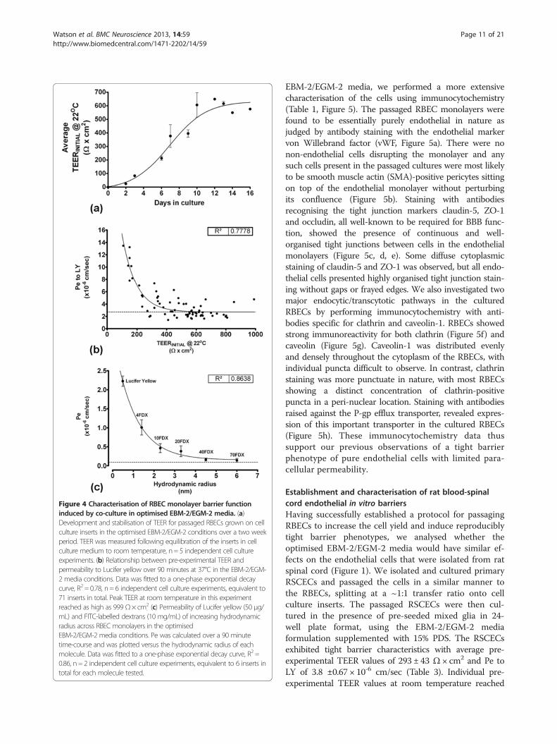
Figure 4 Characterisation of RBEC monolayer barrier functioninduced by co-culture in optimised EBM-2/EGM-2 media. (a)Development and stabilisation of TEER for passaged RBECs grown on cellculture inserts in the optimised EBM-2/EGM-2 conditions over a two weekperiod. TEER was measured following equilibration of the inserts in cellculture medium to room temperature, n = 5 independent cell cultureexperiments. (b) Relationship between pre-experimental TEER andpermeability to Lucifer yellow over 90 minutes at 37°C in the EBM-2/EGM-2 media conditions. Data was fitted to a one-phase exponential decaycurve, R2 = 0.78, n = 6 independent cell culture experiments, equivalent to71 inserts in total. Peak TEER at room temperature in this experimentreached as high as 999 Ω× cm2
. (c) Permeability of Lucifer yellow (50 μg/mL) and FITC-labelled dextrans (10 mg/mL) of increasing hydrodynamicradius across RBEC monolayers in the optimisedEBM-2/EGM-2 media conditions. Pe was calculated over a 90 minutetime-course and was plotted versus the hydrodynamic radius of eachmolecule. Data was fitted to a one-phase exponential decay curve, R2 =0.86, n = 2 independent cell culture experiments, equivalent to 6 inserts intotal for each molecule tested.
Watson et al. BMC Neuroscience 2013, 14:59 Page 11 of 21http://www.biomedcentral.com/1471-2202/14/59
EBM-2/EGM-2 media, we performed a more extensivecharacterisation of the cells using immunocytochemistry(Table 1, Figure 5). The passaged RBEC monolayers werefound to be essentially purely endothelial in nature asjudged by antibody staining with the endothelial markervon Willebrand factor (vWF, Figure 5a). There were nonon-endothelial cells disrupting the monolayer and anysuch cells present in the passaged cultures were most likelyto be smooth muscle actin (SMA)-positive pericytes sittingon top of the endothelial monolayer without perturbingits confluence (Figure 5b). Staining with antibodiesrecognising the tight junction markers claudin-5, ZO-1and occludin, all well-known to be required for BBB func-tion, showed the presence of continuous and well-organised tight junctions between cells in the endothelialmonolayers (Figure 5c, d, e). Some diffuse cytoplasmicstaining of claudin-5 and ZO-1 was observed, but all endo-thelial cells presented highly organised tight junction stain-ing without gaps or frayed edges. We also investigated twomajor endocytic/transcytotic pathways in the culturedRBECs by performing immunocytochemistry with anti-bodies specific for clathrin and caveolin-1. RBECs showedstrong immunoreactivity for both clathrin (Figure 5f) andcaveolin (Figure 5g). Caveolin-1 was distributed evenlyand densely throughout the cytoplasm of the RBECs, withindividual puncta difficult to observe. In contrast, clathrinstaining was more punctuate in nature, with most RBECsshowing a distinct concentration of clathrin-positivepuncta in a peri-nuclear location. Staining with antibodiesraised against the P-gp efflux transporter, revealed expres-sion of this important transporter in the cultured RBECs(Figure 5h). These immunocytochemistry data thussupport our previous observations of a tight barrierphenotype of pure endothelial cells with limited para-cellular permeability.
Establishment and characterisation of rat blood-spinalcord endothelial in vitro barriersHaving successfully established a protocol for passagingRBECs to increase the cell yield and induce reproduciblytight barrier phenotypes, we analysed whether theoptimised EBM-2/EGM-2 media would have similar ef-fects on the endothelial cells that were isolated from ratspinal cord (Figure 1). We isolated and cultured primaryRSCECs and passaged the cells in a similar manner tothe RBECs, splitting at a ~1:1 transfer ratio onto cellculture inserts. The passaged RSCECs were then cul-tured in the presence of pre-seeded mixed glia in 24-well plate format, using the EBM-2/EGM-2 mediaformulation supplemented with 15% PDS. The RSCECsexhibited tight barrier characteristics with average pre-experimental TEER values of 293 ± 43 Ω × cm2 and Pe toLY of 3.8 ±0.67 × 10⁻⁶ cm/sec (Table 3). Individual pre-experimental TEER values at room temperature reached
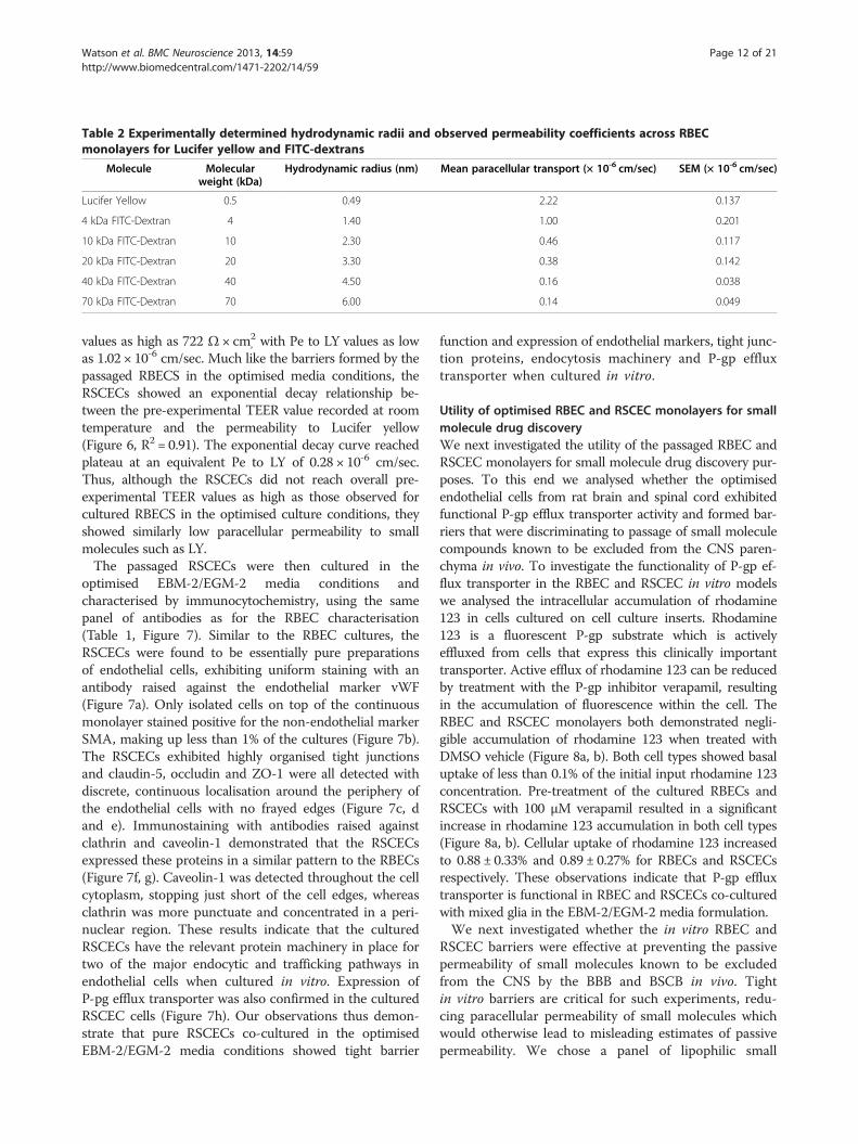
Table 2 Experimentally determined hydrodynamic radii and observed permeability coefficients across RBECmonolayers for Lucifer yellow and FITC-dextrans
Molecule Molecularweight (kDa)
Hydrodynamic radius (nm) Mean paracellular transport (× 10-6 cm/sec) SEM (× 10-6 cm/sec)
Lucifer Yellow 0.5 0.49 2.22 0.137
4 kDa FITC-Dextran 4 1.40 1.00 0.201
10 kDa FITC-Dextran 10 2.30 0.46 0.117
20 kDa FITC-Dextran 20 3.30 0.38 0.142
40 kDa FITC-Dextran 40 4.50 0.16 0.038
70 kDa FITC-Dextran 70 6.00 0.14 0.049
Watson et al. BMC Neuroscience 2013, 14:59 Page 12 of 21http://www.biomedcentral.com/1471-2202/14/59
values as high as 722 Ω × cm2, with Pe to LY values as low
as 1.02 × 10⁻⁶ cm/sec. Much like the barriers formed by thepassaged RBECS in the optimised media conditions, theRSCECs showed an exponential decay relationship be-tween the pre-experimental TEER value recorded at roomtemperature and the permeability to Lucifer yellow(Figure 6, R2 = 0.91). The exponential decay curve reachedplateau at an equivalent Pe to LY of 0.28 × 10⁻⁶ cm/sec.Thus, although the RSCECs did not reach overall pre-experimental TEER values as high as those observed forcultured RBECS in the optimised culture conditions, theyshowed similarly low paracellular permeability to smallmolecules such as LY.The passaged RSCECs were then cultured in the
optimised EBM-2/EGM-2 media conditions andcharacterised by immunocytochemistry, using the samepanel of antibodies as for the RBEC characterisation(Table 1, Figure 7). Similar to the RBEC cultures, theRSCECs were found to be essentially pure preparationsof endothelial cells, exhibiting uniform staining with anantibody raised against the endothelial marker vWF(Figure 7a). Only isolated cells on top of the continuousmonolayer stained positive for the non-endothelial markerSMA, making up less than 1% of the cultures (Figure 7b).The RSCECs exhibited highly organised tight junctionsand claudin-5, occludin and ZO-1 were all detected withdiscrete, continuous localisation around the periphery ofthe endothelial cells with no frayed edges (Figure 7c, dand e). Immunostaining with antibodies raised againstclathrin and caveolin-1 demonstrated that the RSCECsexpressed these proteins in a similar pattern to the RBECs(Figure 7f, g). Caveolin-1 was detected throughout the cellcytoplasm, stopping just short of the cell edges, whereasclathrin was more punctuate and concentrated in a peri-nuclear region. These results indicate that the culturedRSCECs have the relevant protein machinery in place fortwo of the major endocytic and trafficking pathways inendothelial cells when cultured in vitro. Expression ofP-pg efflux transporter was also confirmed in the culturedRSCEC cells (Figure 7h). Our observations thus demon-strate that pure RSCECs co-cultured in the optimisedEBM-2/EGM-2 media conditions showed tight barrier
function and expression of endothelial markers, tight junc-tion proteins, endocytosis machinery and P-gp effluxtransporter when cultured in vitro.
Utility of optimised RBEC and RSCEC monolayers for smallmolecule drug discoveryWe next investigated the utility of the passaged RBEC andRSCEC monolayers for small molecule drug discovery pur-poses. To this end we analysed whether the optimisedendothelial cells from rat brain and spinal cord exhibitedfunctional P-gp efflux transporter activity and formed bar-riers that were discriminating to passage of small moleculecompounds known to be excluded from the CNS paren-chyma in vivo. To investigate the functionality of P-gp ef-flux transporter in the RBEC and RSCEC in vitro modelswe analysed the intracellular accumulation of rhodamine123 in cells cultured on cell culture inserts. Rhodamine123 is a fluorescent P-gp substrate which is activelyeffluxed from cells that express this clinically importanttransporter. Active efflux of rhodamine 123 can be reducedby treatment with the P-gp inhibitor verapamil, resultingin the accumulation of fluorescence within the cell. TheRBEC and RSCEC monolayers both demonstrated negli-gible accumulation of rhodamine 123 when treated withDMSO vehicle (Figure 8a, b). Both cell types showed basaluptake of less than 0.1% of the initial input rhodamine 123concentration. Pre-treatment of the cultured RBECs andRSCECs with 100 μM verapamil resulted in a significantincrease in rhodamine 123 accumulation in both cell types(Figure 8a, b). Cellular uptake of rhodamine 123 increasedto 0.88 ± 0.33% and 0.89 ± 0.27% for RBECs and RSCECsrespectively. These observations indicate that P-gp effluxtransporter is functional in RBEC and RSCECs co-culturedwith mixed glia in the EBM-2/EGM-2 media formulation.We next investigated whether the in vitro RBEC and
RSCEC barriers were effective at preventing the passivepermeability of small molecules known to be excludedfrom the CNS by the BBB and BSCB in vivo. Tightin vitro barriers are critical for such experiments, redu-cing paracellular permeability of small molecules whichwould otherwise lead to misleading estimates of passivepermeability. We chose a panel of lipophilic small
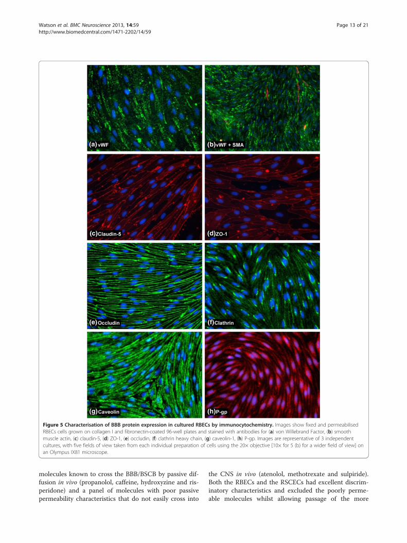
Figure 5 Characterisation of BBB protein expression in cultured RBECs by immunocytochemistry. Images show fixed and permeabilisedRBECs cells grown on collagen I and fibronectin-coated 96-well plates and stained with antibodies for (a) von Willebrand Factor, (b) smoothmuscle actin, (c) claudin-5, (d) ZO-1, (e) occludin, (f) clathrin heavy chain, (g) caveolin-1, (h) P-gp. Images are representative of 3 independentcultures, with five fields of view taken from each individual preparation of cells using the 20× objective [10× for 5 (b) for a wider field of view] onan Olympus IX81 microscope.
Watson et al. BMC Neuroscience 2013, 14:59 Page 13 of 21http://www.biomedcentral.com/1471-2202/14/59
molecules known to cross the BBB/BSCB by passive dif-fusion in vivo (propanolol, caffeine, hydroxyzine and ris-peridone) and a panel of molecules with poor passivepermeability characteristics that do not easily cross into
the CNS in vivo (atenolol, methotrexate and sulpiride).Both the RBECs and the RSCECs had excellent discrim-inatory characteristics and excluded the poorly perme-able molecules whilst allowing passage of the more
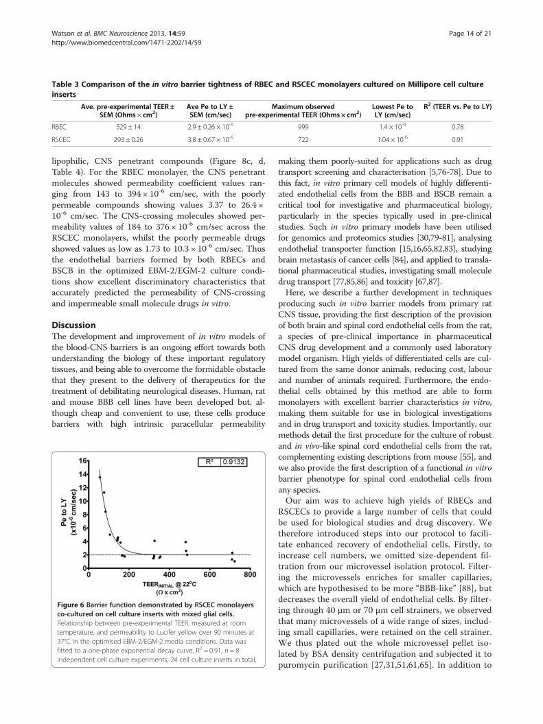
Table 3 Comparison of the in vitro barrier tightness of RBEC and RSCEC monolayers cultured on Millipore cell cultureinserts
Ave. pre-experimental TEER ±SEM (Ohms × cm2)
Ave Pe to LY ±SEM (cm/sec)
Maximum observedpre-experimental TEER (Ohms × cm2)
Lowest Pe toLY (cm/sec)
R2 (TEER vs. Pe to LY)
RBEC 529 ± 14 2.9 ± 0.26 × 10⁻⁶ 999 1.4 × 10⁻⁶ 0.78
RSCEC 293 ± 0.26 3.8 ± 0.67 × 10⁻⁶ 722 1.04 × 10⁻⁶ 0.91
Watson et al. BMC Neuroscience 2013, 14:59 Page 14 of 21http://www.biomedcentral.com/1471-2202/14/59
lipophilic, CNS penetrant compounds (Figure 8c, d,Table 4). For the RBEC monolayer, the CNS penetrantmolecules showed permeability coefficient values ran-ging from 143 to 394 × 10-6 cm/sec, with the poorlypermeable compounds showing values 3.37 to 26.4 ×10⁻⁶ cm/sec. The CNS-crossing molecules showed per-meability values of 184 to 376 × 10-6 cm/sec across theRSCEC monolayers, whilst the poorly permeable drugsshowed values as low as 1.73 to 10.3 × 10⁻⁶ cm/sec. Thusthe endothelial barriers formed by both RBECs andBSCB in the optimized EBM-2/EGM-2 culture condi-tions show excellent discriminatory characteristics thataccurately predicted the permeability of CNS-crossingand impermeable small molecule drugs in vitro.
DiscussionThe development and improvement of in vitro models ofthe blood-CNS barriers is an ongoing effort towards bothunderstanding the biology of these important regulatorytissues, and being able to overcome the formidable obstaclethat they present to the delivery of therapeutics for thetreatment of debilitating neurological diseases. Human, ratand mouse BBB cell lines have been developed but, al-though cheap and convenient to use, these cells producebarriers with high intrinsic paracellular permeability
Figure 6 Barrier function demonstrated by RSCEC monolayersco-cultured on cell culture inserts with mixed glial cells.Relationship between pre-experimental TEER, measured at roomtemperature, and permeability to Lucifer yellow over 90 minutes at37°C in the optimised EBM-2/EGM-2 media conditions. Data wasfitted to a one-phase exponential decay curve, R2 = 0.91, n = 8independent cell culture experiments, 24 cell culture inserts in total.
making them poorly-suited for applications such as drugtransport screening and characterisation [5,76-78]. Due tothis fact, in vitro primary cell models of highly differenti-ated endothelial cells from the BBB and BSCB remain acritical tool for investigative and pharmaceutical biology,particularly in the species typically used in pre-clinicalstudies. Such in vitro primary models have been utilisedfor genomics and proteomics studies [30,79-81], analysingendothelial transporter function [15,16,65,82,83], studyingbrain metastasis of cancer cells [84], and applied to transla-tional pharmaceutical studies, investigating small moleculedrug transport [77,85,86] and toxicity [67,87].Here, we describe a further development in techniques
producing such in vitro barrier models from primary ratCNS tissue, providing the first description of the provisionof both brain and spinal cord endothelial cells from the rat,a species of pre-clinical importance in pharmaceuticalCNS drug development and a commonly used laboratorymodel organism. High yields of differentiated cells are cul-tured from the same donor animals, reducing cost, labourand number of animals required. Furthermore, the endo-thelial cells obtained by this method are able to formmonolayers with excellent barrier characteristics in vitro,making them suitable for use in biological investigationsand in drug transport and toxicity studies. Importantly, ourmethods detail the first procedure for the culture of robustand in vivo-like spinal cord endothelial cells from the rat,complementing existing descriptions from mouse [55], andwe also provide the first description of a functional in vitrobarrier phenotype for spinal cord endothelial cells fromany species.Our aim was to achieve high yields of RBECs and
RSCECs to provide a large number of cells that couldbe used for biological studies and drug discovery. Wetherefore introduced steps into our protocol to facili-tate enhanced recovery of endothelial cells. Firstly, toincrease cell numbers, we omitted size-dependent fil-tration from our microvessel isolation protocol. Filter-ing the microvessels enriches for smaller capillaries,which are hypothesised to be more “BBB-like” [88], butdecreases the overall yield of endothelial cells. By filter-ing through 40 μm or 70 μm cell strainers, we observedthat many microvessels of a wide range of sizes, includ-ing small capillaries, were retained on the cell strainer.We thus plated out the whole microvessel pellet iso-lated by BSA density centrifugation and subjected it topuromycin purification [27,31,51,61,65]. In addition to
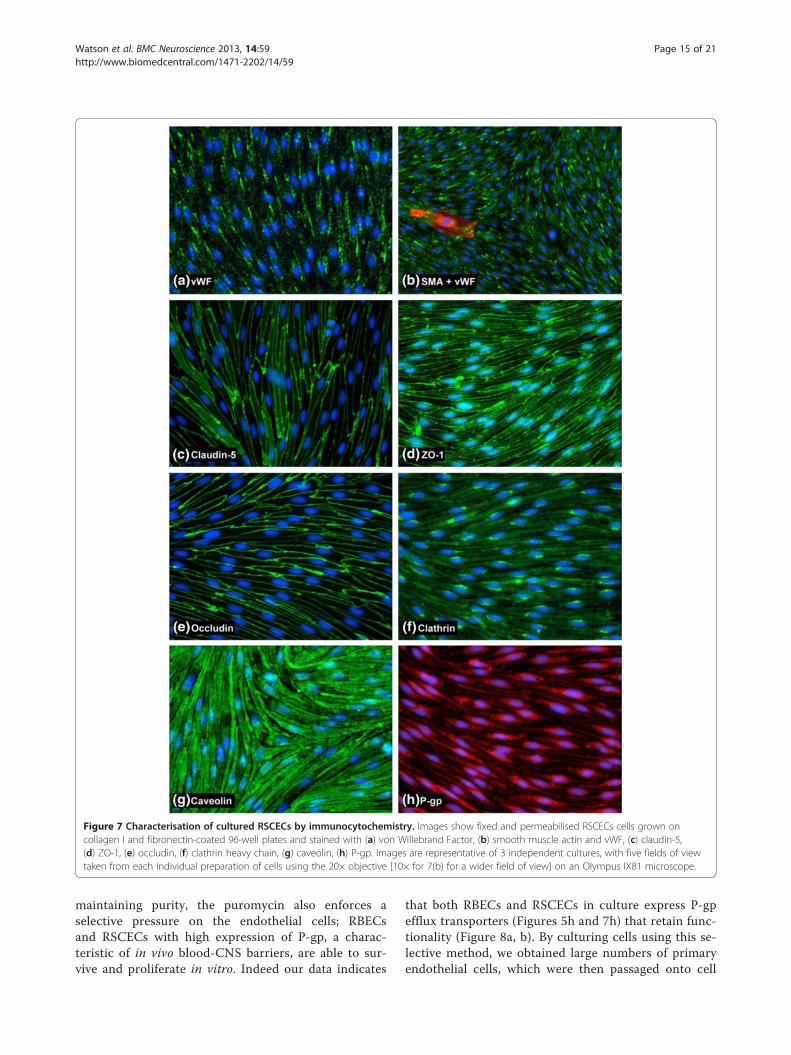
Figure 7 Characterisation of cultured RSCECs by immunocytochemistry. Images show fixed and permeabilised RSCECs cells grown oncollagen I and fibronectin-coated 96-well plates and stained with (a) von Willebrand Factor, (b) smooth muscle actin and vWF, (c) claudin-5,(d) ZO-1, (e) occludin, (f) clathrin heavy chain, (g) caveolin, (h) P-gp. Images are representative of 3 independent cultures, with five fields of viewtaken from each individual preparation of cells using the 20× objective [10× for 7(b) for a wider field of view] on an Olympus IX81 microscope.
Watson et al. BMC Neuroscience 2013, 14:59 Page 15 of 21http://www.biomedcentral.com/1471-2202/14/59
maintaining purity, the puromycin also enforces aselective pressure on the endothelial cells; RBECsand RSCECs with high expression of P-gp, a charac-teristic of in vivo blood-CNS barriers, are able to sur-vive and proliferate in vitro. Indeed our data indicates
that both RBECs and RSCECs in culture express P-gpefflux transporters (Figures 5h and 7h) that retain func-tionality (Figure 8a, b). By culturing cells using this se-lective method, we obtained large numbers of primaryendothelial cells, which were then passaged onto cell
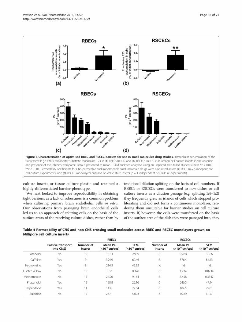
Figure 8 Characterisation of optimised RBEC and RSCEC barriers for use in small molecules drug studies. Intracellular accumulation of thefluorescent P-gp efflux transporter substrate rhodamine 123 in (a) RBECs (n = 4) and (b) RSCECs (n = 3) cultured on cell culture inserts in the absenceand presence of the inhibitor verapamil. Data is presented as mean ± SEM and was analysed using an unpaired, two-tailed students t-test, *P < 0.01,**P < 0.001. Permeability coefficients for CNS-permeable and impermeable small molecule drugs were calculated across (c) RBEC (n = 5 independentcell culture experiments) and (d) RSCEC monolayers cultured on cell culture inserts (n = 3 independent cell culture experiments).
Watson et al. BMC Neuroscience 2013, 14:59 Page 16 of 21http://www.biomedcentral.com/1471-2202/14/59
culture inserts or tissue culture plastic and retained ahighly-differentiated barrier phenotype.We next looked to improve reproducibility in obtaining
tight barriers, as a lack of robustness is a common problemwhen culturing primary brain endothelial cells in vitro.Our observations from passaging brain endothelial cellsled us to an approach of splitting cells on the basis of thesurface areas of the receiving culture dishes, rather than by
Table 4 Permeability of CNS and non-CNS crossing small molMillipore cell culture inserts
RBECs
Passive transportinto CNS?
Number ofinserts
Mean Pe(×10-6 cm/sec)
Atenolol No 15 16.53
Caffeine Yes 9 394.9
Hydroxyzine Yes 8 234.3
Lucifer yellow No 15 3.37
Methotrexate No 15 24.26
Propanolol Yes 15 198.8
Risperidone Yes 11 143.1
Sulpiride No 15 26.41
traditional dilution splitting on the basis of cell numbers. IfRBECs or RSCECs were transferred to new dishes or cellculture inserts as a dilution passage (e.g. splitting 1:4–1:2)they frequently grew as islands of cells which stopped pro-liferating and did not form a continuous monolayer, ren-dering them unsuitable for barrier studies on cell cultureinserts. If, however, the cells were transferred on the basisof the surface area of the dish they were passaged into, they
ecules across RBEC and RSCEC monolayers grown on
RSCECs
SEM(×10-6 cm/sec)
Number ofinserts
Mean Pe(×10-6 cm/sec)
SEM(×10-6 cm/sec)
2.939 6 9.788 3.166
60.46 6 376.4 81.13
42.92 nd nd nd
0.328 6 1.734 0.0734
9.164 6 3.458 0.3547
22.16 6 246.5 47.94
22.54 6 184.5 29.01
5.003 6 10.29 1.157
Watson et al. BMC Neuroscience 2013, 14:59 Page 17 of 21http://www.biomedcentral.com/1471-2202/14/59
quickly reached confluence and formed functional barriers(Figure 1). Thus, a key element of our protocol is the con-cept of the ~1:1 passage of endothelial cells. This transfermethod facilitated excellent barrier phenotypes for bothRBECS and RSCECs (Figures 3, 4 and 6), with no obviousendothelial de-differentiation as judged by immunocyto-chemistry which demonstrated well organised, maturetight junctions and expression of endocytic transport ma-chinery (Figures 5 and 7). Furthermore, the technique reli-ably resulted in the provision of useable barriers in almostevery insert. We observed very low losses of individual in-serts where barriers did not form, as often happened withdilution passaging. Individual inserts where the barrierfailed were usually found to be due to handling techniqueand mechanical damage to the monolayer.A major finding of our study was that barriers with
high TEER and low Pe to small molecules such as LYwere reproducibly obtained when culturing RBECs andRSCECs in Lonza’s EBM-2 basal medium with theEGM-2 BulletKit minus VEGF. The Lonza BulletKit con-tains supplements, such as hydrocortisone and FGF, thatare well validated to improve endothelial barrier func-tion in vitro [22,61]. This EBM-2/EGM-2 media combin-ation outperformed DMEM supplemented with anothercommercial supplement, MVGS. The optimised condi-tions also included the addition of 15% plasma-derivedserum and the monolayers did not display the sensitivityto serum-derived factors that has been observed in somein vitro BBB cell culture models [52].Historically, the BBB has been more highly studied
than the BSCB both in vivo and in vitro. An emergingconsensus is that the two are broadly similar with somesubtle differences, for example in their permeability andtheir vulnerability to certain insults and diseases [4,89].The BSCB has an almost identical physical structure tothat of the BBB, with tight junction-containing endothe-lial cells surrounded by and interacting with astrocytesand pericytes [89]. The BSCB also appears to be morepermeable than the BBB in certain sub-regions but isstill a tight and highly regulated barrier that protects thespinal cord parenchyma. Studies in vivo have indicatedthat the BSCB is more permeable than the BBB to smalltracers, cytokines and neurotrophins, with lumbar regionsof the spinal cord in particular being more permeable[90-94]. Some cytokines and growth factors however, suchas IL-1α and granulocyte-macrophage colony-stimulatingfactor (GMCSF), have been shown to have similar trans-port across the BSCB as the BBB in vivo [95,96]. Other evi-dence suggests that the BBB and BSCB have similarly lowpermeability to large plasma proteins such as IgGs and al-bumin [95,97]. Similarities between the BBB and BSCBhave also been noted in the expression and functionality ofABC transporters, which are hypothesised to play key rolesin disease and drug resistance. Isolated capillaries from
mouse brain and spinal cord show similar expression andfunctionality of P-gp, MDR2 and BRCP [82,83]. ABC-transporters at the BBB and BSCB also share similar in-creased expression and functionality following exposure todioxins, and in mouse models of amyotropic lateral scler-osis (ALS) in vivo [82,83]. Interestingly, our models showthat RSCEC monolayers are generally slightly more perme-able than with RBECs, in spite of the fact that both are cul-tured in the presence of glial cells derived from braintissue. Although data is lacking attesting to differences be-tween brain and spinal cord astrocytes in their ability to in-duce barrier phenotypes, our observations may indicatethat some aspects of the permeability properties ofRSCECs are cell-intrinsic.The only previously published in vitro study compar-
ing BBB and BSCB cells was carried out using endothe-lial cells derived from mouse [55]. In that study, cultureconditions for both endothelial cell types wereestablished and expression levels of proteins associatedwith barrier function were characterised, although nofunctional barrier data was presented. Ge and Pachter(2006) found that cultured endothelial cells from bothtype of CNS tissue were indistinguishable under themicroscope and showed identical expression of theendothelial markers vWF and PECAM-1, as well assimilar uptake of LDL [55]. Our extensive characterisa-tion data for endothelial markers, tight junction pro-teins, endocytic machinery and the P-gp effluxtransporter, suggests a similar situation to be true forbrain and spinal cord endothelial cells from rat(Figures 5 and 7). Additionally, Ge and Pachter provi-ded a highly useful comparison of several genes impor-tant for barrier function in these cultured endothelialcells [55]. Gene expression of claudin-1, claudin-5,P-gp and transferrin receptor were unchanged betweenboth types of endothelial cell in mouse, but expressionlevels of ZO-1, occludin, β-catenin and VE-cadherinwere lower in spinal cord endothelial cells compared tothose from brain tissue [55]. This observation is inagreement with in vivo descriptions of the BSCB beingmore permeable than the BBB.Our observations further support and extend these ob-
servations on the structure and function of the BBB andthe BSCB. We have shown that cells from both the ratBBB and BSCB can be cultured on cell culture insertsin vitro to form functionally restrictive cell monolayers,with the endothelial cells of the brain forming slightlytighter barriers than those of the spinal cord. In this re-gard our in vitro models apparently mimic the in vivosituation for the BBB and BSCB. We observed an excel-lent relationship between pre-experimental TEER valuesand Pe to LY in permeability assays for both models(R2 = 0.78 for RBECs and 0.91 for RSCECs). Importantly,this indicates that the pre-experimental TEER value is
Watson et al. BMC Neuroscience 2013, 14:59 Page 18 of 21http://www.biomedcentral.com/1471-2202/14/59
predictive of the Pe to LY, allowing consistent and repro-ducible experiments to be performed. Cell culture in-serts with high TEER values can be selected from theoutset and matched with inserts of similar barrier tight-ness, allowing robust experiments to be performed onprimary-derived cells that have similar intrinsic perme-ability properties.Our BBB and BSCB models exhibited excellent dis-
crimination characteristics for limiting the passage ofsmall and large molecules that cross the barrier by para-cellular diffusion, such as LY and FITC-dextrans, andalso for small molecules that enter the CNS poorly onthe basis of their low lipophilicity. This makes ourmodels ideally suited for in vitro permeability studies,particularly for small molecule drugs where tight in vitrobarriers are critically required to minimise non-specificparacellular transport that would mask true permeabilitycharacteristics and kinetics. Small molecule permeabilityacross an in vitro barrier has been demonstrated by sev-eral groups using different species, including human,porcine, mouse and rat, but only across endothelialmonolayers derived from cells of the BBB [16,25,54,65].Our methods show that we are able generate a largenumber of tight in vitro barriers representing the ratBBB, but we also demonstrate for the first time anin vitro model of rat spinal cord endothelial cells thatshows similar restrictive properties to small molecules.These data suggest that our models would be suitablefor a broad range of CNS drug discovery studies, par-ticularly for instances where a drug target is locatedwithin the spinal cord as well as, or instead of, in thebrain. The RBEC and RSCEC barriers also show expres-sion and functionality of the clinically important effluxtransport P-gp (Figures 5h, 7h and 8a, b). These BBBand BSCB models could thus be used for in vitro studiesof barrier function involving this transporter, such as de-termining the efflux of chemotherapeutic small moleculedrugs which are often also P-gp substrates.Since differences between the BBB and BSCB exist, it
is therefore essential that in vitro models for bothbarriers are available for research purposes. An in vitromodel for one barrier may not necessarily be an appro-priate substitute for the other. This may be of particu-lar relevance when studying diseases which affect oneCNS compartment more than the other [4,89]. Ourin vitro models of both types of blood-CNS barrier arethus of great potential value for the investigationof such disease processes. Since these models areoptimised for rat tissues, a species for which relevantand well-characterised in vivo models of CNS-diseaseexist, they have great potential utility in translationalstudies. Our novel in vitro RSCEC model of the ratBSCB may also contribute to the furthering of know-ledge about this poorly-understood blood-CNS barrier
and could be applied to genomics and proteomics stud-ies in the future.
ConclusionIn conclusion, we have demonstrated an easy and ro-bust method to prepare large yields of endothelial cellsfrom rat brain and spinal cord tissue. Our method hasthe advantages of ease-of-use and reproducibility andprovides culture conditions suitable for the isolationand culture of both RBECs and RSCECs as coincidentsister cultures. The high yields of cells obtained gosome way to overcoming the often limiting amount ofmaterial available for experiments, a common problemthat is often encountered when performing studies withprimary cell models of the BBB and BSCB. Theoptimised RBEC and RSCEC cultures show expressionof typical markers representative of the blood-CNS bar-riers in vivo and form functional barriers in vitro thatare discriminating in preventing the passage of largemolecules and poorly lipophilic small molecule drugs.The tight barrier phenotype obtained for both modelsallows predictive drug permeability studies to beperformed, due to the low intrinsic non-specific para-cellular permeability of the pure endothelial mono-layers. We hope that these models will prove to be avaluable addition to the tools available to academic andindustrial researchers for both drug discovery andstudying the biology of BBB and BSCB in an in vitrosetting.
AbbreviationsBBB: Blood–brain barrier; BSCB: Blood-spinal cord barrier; RBEC: Rat brainendothelial cells; DIV: Days in vitro; RSCEC: Rat spinal cord endothelial cells;TEER: Transendothelial electrical resistance; MVGS: Microvascular growthsupplement; LY: Lucifer yellow; DLS: Dynamic light scattering;Pe: Permeability coefficient; vWF: Von Willebrand factor; SMA: Smoothmuscle actin; PDGF: Platelet-derived growth factor; PDS: Plasma-derivedbovine serum; PECAM-1: Platelet/endothelial cell adhesion molecule-1;LDL: Low density lipoprotein; TLCK: Tosyl-lysine-chloromethylketone; LC-MS/MS: Liquid chromatography mass spectrometry; P-gp: P-glycoprotein;FGF: Fibroblast growth factor; FITC: Fluorescein isothiocyanate; CNS: Centralnervous system.
Competing interestsPMDW, JP, GT and CW are employees of MedImmune Ltd, an AstraZenecaPLC owned company. At the time of the study SL and UG were employeesof AstraZeneca PLC.
Authors’ contributionsPMDW and JP designed and performed all experimental work (with theexception of LC-MS/MS) and wrote the manuscript. SL and UG assisted withthe design of the small molecule permeability study and UG performed LC/MS experiments. PMDW, JP, GT, SL, UG and CIW analysed and interpretedexperimental data. All authors have approved the final version of themanuscript.
AcknowledgementsWe extend many thanks to Maxime Culot for his expertise and kind helpthroughout our research project. Thanks to Ian Wilkinson for his help andexpertise in determining the hydrodynamic radii of FITC-dextrans used inpermeability studies. Many thanks also to Ann Traher, Tracey Myers, Karen

Watson et al. BMC Neuroscience 2013, 14:59 Page 19 of 21http://www.biomedcentral.com/1471-2202/14/59
Balch, Helen Brant, and Sarah Welsted for their critical assistance in preparingand providing tissue samples.
Author details1MedImmune Ltd, Granta Park, Cambridgeshire CB21 6HG, UK. 2AstraZeneca,Södertälje SE 15185, Sweden.
Received: 5 October 2012 Accepted: 5 June 2013Published: 18 June 2013
References1. Cecchelli R, Berezowski V, Lundquist S, Culot M, Renftel M, Dehouck MP,
Fenart L: Modelling of the blood–brain barrier in drug discovery anddevelopment. Nat Rev Drug Discov 2007, 6(8):650–661.
2. Toth A, Veszelka S, Nakagawa S, Niwa M, Deli MA: Patented in vitro blood–brain barrier models in CNS drug discovery. Recent Pat CNS Drug Discov2011, 6(2):107–118.
3. Zlokovic BV: The blood–brain barrier in health and chronicneurodegenerative disorders. Neuron 2008, 57(2):178–201.
4. Palmer AM: The role of the blood-CNS barrier in CNS disorders and theirtreatment. Neurobiol Dis 2010, 37(1):3–12.
5. Deli MA, Abraham CS, Kataoka Y, Niwa M: Permeability studies on in vitroblood–brain barrier models: physiology, pathology, and pharmacology.Cell Mol Neurobiol 2005, 25(1):59–127.
6. Abbott NJ, Patabendige AA, Dolman DE, Yusof SR, Begley DJ: Structure andfunction of the blood–brain barrier. Neurobiol Dis 2010, 37(1):13–25.
7. Abbott NJ, Ronnback L, Hansson E: Astrocyte-endothelial interactions atthe blood–brain barrier. Nature reviews 2006, 7(1):41–53.
8. Daneman R, Zhou L, Kebede AA, Barres BA: Pericytes are required forblood–brain barrier integrity during embryogenesis. Nature 2010,468(7323):562–566.
9. Vandenhaute E, Dehouck L, Boucau MC, Sevin E, Uzbekov R, Tardivel M,Gosselet F, Fenart L, Cecchelli R, Dehouck MP: Modelling the neurovascularunit and the blood–brain barrier with the unique function of pericytes.Curr Neurovasc Res 2011, 8(4):258–269.
10. Nakagawa S, Deli MA, Nakao S, Honda M, Hayashi K, Nakaoke R, Kataoka Y,Niwa M: Pericytes from brain microvessels strengthen the barrierintegrity in primary cultures of rat brain endothelial cells. Cell MolNeurobiol 2007, 27(6):687–694.
11. Nakagawa S, Deli MA, Kawaguchi H, Shimizudani T, Shimono T, Kittel A,Tanaka K, Niwa M: A new blood–brain barrier model using primary ratbrain endothelial cells, pericytes and astrocytes. Neurochem Int 2009,54(3–4):253–263.
12. Thanabalasundaram G, Schneidewind J, Pieper C, Galla HJ: The impact ofpericytes on the blood–brain barrier integrity depends critically on thepericyte differentiation stage. Int J Biochem Cell Biol 2011, 43(9):1284–1293.
13. Biegel D, Spencer DD, Pachter JS: Isolation and culture of human brainmicrovessel endothelial cells for the study of blood–brain barrierproperties in vitro. Brain Res 1995, 692(1–2):183–189.
14. Bernas MJ, Cardoso FL, Daley SK, Weinand ME, Campos AR, Ferreira AJ,Hoying JB, Witte MH, Brites D, Persidsky Y, et al: Establishment of primarycultures of human brain microvascular endothelial cells to provide anin vitro cellular model of the blood–brain barrier. Nat Protoc 2010,5(7):1265–1272.
15. Cioni C, Turlizzi E, Zanelli U, Oliveri G, Annunziata P: Expression of tightjunction and drug efflux transporter proteins in an in vitro model ofhuman blood–brain barrier. Front Psychiatry 2012, 3:47.
16. Lacombe O, Videau O, Chevillon D, Guyot AC, Contreras C, Blondel S,Nicolas L, Ghettas A, Benech H, Thevenot E, et al: In vitro primary humanand animal cell-based blood–brain barrier models as a screening tool indrug discovery. Mol Pharm 2011, 8(3):651–663.
17. Mabondzo A, Bottlaender M, Guyot AC, Tsaouin K, Deverre JR, Balimane PV:Validation of in vitro cell-based human blood–brain barrier model usingclinical positron emission tomography radioligands to predict in vivohuman brain penetration. Mol Pharm 2010, 7(5):1805–1815.
18. Megard I, Garrigues A, Orlowski S, Jorajuria S, Clayette P, Ezan E, MabondzoA: A co-culture-based model of human blood–brain barrier: applicationto active transport of indinavir and in vivo-in vitro correlation. Brain Res2002, 927(2):153–167.
19. Dorovini-Zis K, Prameya R, Bowman PD: Culture and characterization ofmicrovascular endothelial cells derived from human brain. Lab Invest1991, 64(3):425–436.
20. Song L, Pachter JS: Culture of murine brain microvascular endothelialcells that maintain expression and cytoskeletal association of tightjunction-associated proteins. In Vitro Cell Dev Biol 2003, 39(7):313–320.
21. Schrot S, Weidenfeller C, Schaffer TE, Robenek H, Galla HJ: Influence ofhydrocortisone on the mechanical properties of the cerebralendothelium in vitro. Biophys J 2005, 89(6):3904–3910.
22. Weidenfeller C, Schrot S, Zozulya A, Galla HJ: Murine brain capillaryendothelial cells exhibit improved barrier properties under the influenceof hydrocortisone. Brain Res 2005, 1053(1–2):162–174.
23. Wuest DM, Lee KH: Optimization of endothelial cell growth in amurine in vitro blood–brain barrier model. Biotechnol J 2012,7(3):409–417.
24. Coisne C, Dehouck L, Faveeuw C, Delplace Y, Miller F, Landry C, MorissetteC, Fenart L, Cecchelli R, Tremblay P, et al: Mouse syngenic in vitro blood–brain barrier model: a new tool to examine inflammatory events incerebral endothelium. Lab Invest 2005, 85(6):734–746.
25. Shayan G, Choi YS, Shusta EV, Shuler ML, Lee KH: Murine in vitro model ofthe blood–brain barrier for evaluating drug transport. Eur J Pharm Sci2011, 42(1–2):148–155.
26. Forster C, Silwedel C, Golenhofen N, Burek M, Kietz S, Mankertz J,Drenckhahn D: Occludin as direct target for glucocorticoid-induced improvement of blood–brain barrier properties in amurine in vitro system. J Physiol 2005, 565(Pt 2):475–486.
27. Abbott NJ, Dolman DE, Drndarski S, Fredriksson SM: An improved in vitroblood–brain barrier model: rat brain endothelial cells co-cultured withastrocytes. Methods Mol Biol 2012, 814:415–430.
28. Abbott NJ, Hughes CC, Revest PA, Greenwood J: Development andcharacterisation of a rat brain capillary endothelial culture:towards an in vitro blood–brain barrier. J Cell Sci 1992,103(Pt 1):23–37.
29. Bowman PD, Betz AL, Ar D, Wolinsky JS, Penney JB, Shivers RR, GoldsteinGW: Primary culture of capillary endothelium from rat brain. In Vitro 1981,17(4):353–362.
30. Calabria AR, Shusta EV: Blood–brain barrier genomics andproteomics: elucidating phenotype, identifying disease targetsand enabling brain drug delivery. Drug Discov Today 2006,11(17–18):792–799.
31. Perriere N, Demeuse P, Garcia E, Regina A, Debray M, Andreux JP, CouvreurP, Scherrmann JM, Temsamani J, Couraud PO, et al: Puromycin-basedpurification of rat brain capillary endothelial cell cultures. Effect on theexpression of blood–brain barrier-specific properties. J Neurochem 2005,93(2):279–289.
32. Szabo CA, Deli MA, Ngo TK, Joo F: Production of pure primary rat cerebralendothelial cell culture: a comparison of different methods.Neurobiology (Bp) 1997, 5(1):1–16.
33. Kis B, Kaiya H, Nishi R, Deli MA, Abraham CS, Yanagita T, Isse T, Gotoh S,Kobayashi H, Wada A, et al: Cerebral endothelial cells are a major sourceof adrenomedullin. J Neuroendocrinol 2002, 14(4):283–293.
34. Diglio CA, Grammas P, Giacomelli F, Wiener J: Primary culture of ratcerebral microvascular endothelial cells. Isolation, growth, andcharacterization. Lab Invest 1982, 46(6):554–563.
35. Garcia-Garcia E, Gil S, Andrieux K, Desmaele D, Nicolas V, Taran F, Georgin D,Andreux JP, Roux F, Couvreur P: A relevant in vitro rat model for theevaluation of blood–brain barrier translocation of nanoparticles. Cell MolLife Sci 2005, 62(12):1400–1408.
36. Audus KL, Borchardt RT: Characteristics of the large neutral amino acidtransport system of bovine brain microvessel endothelial cellmonolayers. J Neurochem 1986, 47(2):484–488.
37. Audus KL, Borchardt RT: Bovine brain microvessel endothelial cellmonolayers as a model system for the blood–brain barrier. Ann N Y AcadSci 1987, 507:9–18.
38. Dorovini-Zis K, Bowman PD, Betz AL, Goldstein GW: Formation of a barrierby brain microvessel endothelial cells in culture. Fed Proc 1987,46(8):2521–2522.
39. Guillot FL, Audus KL, Raub TJ: Fluid-phase endocytosis by primary culturesof bovine brain microvessel endothelial cell monolayers. Microvasc Res1990, 39(1):1–14.
Watson et al. BMC Neuroscience 2013, 14:59 Page 20 of 21http://www.biomedcentral.com/1471-2202/14/59
40. van Bree JB, de Boer AG, Danhof M, Ginsel LA, Breimer DD:Characterization of an "in vitro" blood–brain barrier: effects of molecularsize and lipophilicity on cerebrovascular endothelial transport rates ofdrugs. J Pharmacol Exp Ther 1988, 247(3):1233–1239.
41. Gaillard PJ, Voorwinden LH, Nielsen JL, Ivanov A, Atsumi R, Engman H,Ringbom C, de Boer AG, Breimer DD: Establishment and functionalcharacterization of an in vitro model of the blood–brain barrier,comprising a co-culture of brain capillary endothelial cells andastrocytes. Eur J Pharm Sci 2001, 12(3):215–222.
42. Dehouck MP, Meresse S, Delorme P, Fruchart JC, Cecchelli R: An easier,reproducible, and mass-production method to study the blood–brainbarrier in vitro. J Neurochem 1990, 54(5):1798–1801.
43. Vandenhaute E, Sevin E, Hallier-Vanuxeem D, Dehouck MP, Cecchelli R: Casestudy: adapting in vitro blood–brain barrier models for use in early-stagedrug discovery. Drug Discov Today 2012, 17(7–8):285–290.
44. Skinner RA, Gibson RM, Rothwell NJ, Pinteaux E, Penny JI: Transport ofinterleukin-1 across cerebromicrovascular endothelial cells. Br JPharmacol 2009, 156(7):1115–1123.
45. Franke H, Galla H, Beuckmann CT: Primary cultures of brain microvesselendothelial cells: a valid and flexible model to study drug transportthrough the blood–brain barrier in vitro. Brain Res Brain Res Protoc 2000,5(3):248–256.
46. Franke H, Galla HJ, Beuckmann CT: An improved low-permeability in vitro-model of the blood–brain barrier: transport studies on retinoids, sucrose,haloperidol, caffeine and mannitol. Brain Res 1999, 818(1):65–71.
47. Rauh J, Meyer J, Beuckmann C, Galla HJ: Development of an in vitro cellculture system to mimic the blood–brain barrier. Prog Brain Res 1992,91:117–121.
48. Smith M, Omidi Y, Gumbleton M: Primary porcine brain microvascularendothelial cells: biochemical and functional characterisation as a modelfor drug transport and targeting. J Drug Target 2007, 15(4):253–268.
49. Torok M, Huwyler J, Gutmann H, Fricker G, Drewe J: Modulation oftransendothelial permeability and expression of ATP-binding cassettetransporters in cultured brain capillary endothelial cells by astrocyticfactors and cell-culture conditions. Exp Brain Res 2003, 153(3):356–365.
50. Cohen-Kashi Malina K, Cooper I, Teichberg VI: Closing the gap between thein-vivo and in-vitro blood–brain barrier tightness. Brain Res 2009, 1284:12–21.
51. Patabendige A, Skinner RA, Abbott NJ: Establishment of a simplifiedin vitro porcine blood–brain barrier model with high transendothelialelectrical resistance. Brain Res 2012.
52. Nitz T, Eisenblatter T, Psathaki K, Galla HJ: Serum-derived factors weakenthe barrier properties of cultured porcine brain capillary endothelial cellsin vitro. Brain Res 2003, 981(1–2):30–40.
53. Hoheisel D, Nitz T, Franke H, Wegener J, Hakvoort A, Tilling T, Galla HJ:Hydrocortisone reinforces the blood–brain barrier properties in a serum freecell culture system. Biochem Biophys Res Commun 1998, 244(1):312–316.
54. Zhang Y, Li CS, Ye Y, Johnson K, Poe J, Johnson S, Bobrowski W, Garrido R,Madhu C: Porcine brain microvessel endothelial cells as an in vitro modelto predict in vivo blood–brain barrier permeability. Drug Metab Dispos2006, 34(11):1935–1943.
55. Ge S, Pachter JS: Isolation and culture of microvascular endothelial cellsfrom murine spinal cord. J Neuroimmunol 2006, 177(1–2):209–214.
56. Lippmann ES, Weidenfeller C, Svendsen CN, Shusta EV: Blood–brain barriermodeling with co-cultured neural progenitor cell-derived astrocytes andneurons. J Neurochem 2011, 119(3):507–520.
57. Weidenfeller C, Svendsen CN, Shusta EV: Differentiating embryonic neuralprogenitor cells induce blood–brain barrier properties. J Neurochem 2007,101(2):555–565.
58. Rubin LL, Hall DE, Porter S, Barbu K, Cannon C, Horner HC, Janatpour M,Liaw CW, Manning K, Morales J, et al: A cell culture model of the blood–brain barrier. J Cell Biol 1991, 115(6):1725–1735.
59. Beuckmann C, Hellwig S, Galla HJ: Induction of the blood/brain-barrier-associated enzyme alkaline phosphatase in endothelial cells from cerebralcapillaries is mediated via cAMP. Eur J Biochem 1995, 229(3):641–644.
60. Forster C, Waschke J, Burek M, Leers J, Drenckhahn D: Glucocorticoideffects on mouse microvascular endothelial barrier permeability arebrain specific. J Physiol 2006, 573(Pt 2):413–425.
61. Calabria AR, Weidenfeller C, Jones AR, de Vries HE, Shusta EV: Puromycin-purified rat brain microvascular endothelial cell cultures exhibitimproved barrier properties in response to glucocorticoid induction.J Neurochem 2006, 97(4):922–933.
62. Sobue K, Yamamoto N, Yoneda K, Hodgson ME, Yamashiro K, Tsuruoka N,Tsuda T, Katsuya H, Miura Y, Asai K, et al: Induction of blood–brain barrierproperties in immortalized bovine brain endothelial cells by astrocyticfactors. Neurosci Res 1999, 35(2):155–164.
63. Bendfeldt K, Radojevic V, Kapfhammer J, Nitsch C: Basic fibroblast growthfactor modulates density of blood vessels and preserves tight junctionsin organotypic cortical cultures of mice: a new in vitro model of theblood–brain barrier. J Neurosci 2007, 27(12):3260–3267.
64. Helms HC, Waagepetersen HS, Nielsen CU, Brodin B: Paracellular tightnessand claudin-5 expression is increased in the BCEC/astrocyte blood–brainbarrier model by increasing media buffer capacity during growth. AAPS J2010, 12(4):759–770.
65. Perriere N, Yousif S, Cazaubon S, Chaverot N, Bourasset F, Cisternino S,Decleves X, Hori S, Terasaki T, Deli M, et al: A functional in vitro model ofrat blood–brain barrier for molecular analysis of efflux transporters.Brain Res 2007, 1150:1–13.
66. McCarthy KD, de Vellis J: Preparation of separate astroglial andoligodendroglial cell cultures from rat cerebral tissue. J Cell Biol 1980,85(3):890–902.
67. Culot M, Lundquist S, Vanuxeem D, Nion S, Landry C, Delplace Y, DehouckMP, Berezowski V, Fenart L, Cecchelli R: An in vitro blood–brain barriermodel for high throughput (HTS) toxicological screening. Toxicol In Vitro2008, 22(3):799–811.
68. Siflinger-Birnboim A, Del Vecchio PJ, Cooper JA, Blumenstock FA, ShepardJM, Malik AB: Molecular sieving characteristics of the cultured endothelialmonolayer. J Cell Physiol 1987, 132(1):111–117.
69. Hayashi Y, Nomura M, Yamagishi S, Harada S, Yamashita J, Yamamoto H:Induction of various blood–brain barrier properties in non-neuralendothelial cells by close apposition to co-cultured astrocytes. Glia 1997,19(1):13–26.
70. Gaillard PJ, van der Sandt IC, Voorwinden LH, Vu D, Nielsen JL, de Boer AG,Breimer DD: Astrocytes increase the functional expression of P-glycoprotein in an in vitro model of the blood–brain barrier. Pharm Res2000, 17(10):1198–1205.
71. Ohtsuki S, Sato S, Yamaguchi H, Kamoi M, Asashima T, Terasaki T:Exogenous expression of claudin-5 induces barrier properties in culturedrat brain capillary endothelial cells. J Cell Physiol 2007, 210(1):81–86.
72. Nitta T, Hata M, Gotoh S, Seo Y, Sasaki H, Hashimoto N, Furuse M, Tsukita S:Size-selective loosening of the blood–brain barrier in claudin-5-deficientmice. J Cell Biol 2003, 161(3):653–660.
73. Cooper I, Cohen-Kashi-Malina K, Teichberg VI: Claudin-5 expression inin vitro models of the blood–brain barrier. Methods Mol Biol 2011,762:347–354.
74. Gaillard PJ, de Boer AG: Relationship between permeability status of theblood–brain barrier and in vitro permeability coefficient of a drug. Eur JPharm Sci 2000, 12(2):95–102.
75. Neuhaus W, Plattner VE, Wirth M, Germann B, Lachmann B, Gabor F, NoeCR: Validation of in vitro cell culture models of the blood–brain barrier:tightness characterization of two promising cell lines. J Pharm Sci 2008,97(12):5158–5175.
76. Roux F, Couraud PO: Rat brain endothelial cell lines for the study ofblood–brain barrier permeability and transport functions. Cell MolNeurobiol 2005, 25(1):41–58.
77. Garberg P, Ball M, Borg N, Cecchelli R, Fenart L, Hurst RD, Lindmark T,Mabondzo A, Nilsson JE, Raub TJ, et al: In vitro models for the blood–brainbarrier. Toxicol In Vitro 2005, 19(3):299–334.
78. Wilhelm I, Fazakas C, Krizbai IA: In vitro models of the blood–brain barrier.Acta Neurobiol Exp 2011, 71(1):113–128.
79. Shusta EV: Blood–brain barrier genomics, proteomics, and newtransporter discovery. NeuroRx 2005, 2(1):151–161.
80. Murugesan N, Macdonald JA, Lu Q, Wu SL, Hancock WS, Pachter JS: Analysis ofmouse brain microvascular endothelium using laser capture microdissectioncoupled with proteomics. Methods Mol Biol 2011, 686:297–311.
81. Pottiez G, Flahaut C, Cecchelli R, Karamanos Y: Understanding the blood–brain barrier using gene and protein expression profiling technologies.Brain Res Rev 2009, 62(1):83–98.
82. Campos CR, Schroter C, Wang X, Miller DS: ABC transporter function andregulation at the blood-spinal cord barrier. J Cereb Blood Flow Metab2012, 32(8):1559–1566.
83. Jablonski MR, Jacob DA, Campos C, Miller DS, Maragakis NJ, Pasinelli P,Trotti D: Selective increase of two ABC drug efflux transporters at the
Watson et al. BMC Neuroscience 2013, 14:59 Page 21 of 21http://www.biomedcentral.com/1471-2202/14/59
blood-spinal cord barrier suggests induced pharmacoresistance in ALS.Neurobiol Dis 2012, 47(2):194–200.
84. Fazakas C, Wilhelm I, Nagyoszi P, Farkas AE, Hasko J, Molnar J, Bauer H,Bauer HC, Ayaydin F, Dung NT, et al: Transmigration of melanoma cellsthrough the blood–brain barrier: role of endothelial tight junctions andmelanoma-released serine proteases. PLoS One 2012, 6(6):e20758.
85. Bickel U: How to measure drug transport across the blood–brain barrier.NeuroRx 2005, 2(1):15–26.
86. Berezowski V, Landry C, Lundquist S, Dehouck L, Cecchelli R, Dehouck MP,Fenart L: Transport screening of drug cocktails through an in vitroblood–brain barrier: is it a good strategy for increasing the throughputof the discovery pipeline? Pharm Res 2004, 21(5):756–760.
87. Wager TT, Liras JL, Mente S, Trapa P: Strategies to minimize CNS toxicity:in vitro high-throughput assays and computational modeling. Expert OpinDrug Metab Toxicol 2012, 8(5):531–542.
88. Ge S, Song L, Pachter JS: Where is the blood–brain barrier … really? JNeurosci Res 2005, 79(4):421–427.
89. Bartanusz V, Jezova D, Alajajian B, Digicaylioglu M: The blood-spinal cordbarrier: morphology and clinical implications. Ann Neurol 2011,70(2):194–206.
90. Pan W, Banks WA, Fasold MB, Bluth J, Kastin AJ: Transport of brain-derivedneurotrophic factor across the blood–brain barrier. Neuropharmacology1998, 37(12):1553–1561.
91. Pan W, Banks WA, Kastin AJ: Permeability of the blood–brain and blood-spinal cord barriers to interferons. J Neuroimmunol 1997, 76(1–2):105–111.
92. Pan W, Banks WA, Kastin AJ: Permeability of the blood–brain barrier toneurotrophins. Brain Res 1998, 788(1–2):87–94.
93. Pan W, Kastin AJ: Penetration of neurotrophins and cytokines across theblood–brain/blood-spinal cord barrier. Adv Drug Deliv Rev 1999,36(2–3):291–298.
94. Prockop LD, Naidu KA, Binard JE, Ransohoff J: Selective permeability of[3H]-D-mannitol and [14C]-carboxyl-inulin across the blood–brain barrierand blood-spinal cord barrier in the rabbit. J Spinal Cord Med 1995,18(4):221–226.
95. Banks WA, Kastin AJ, Ehrensing CA: Blood-borne interleukin-1 alpha istransported across the endothelial blood-spinal cord barrier of mice.J Physiol 1994, 479(Pt 2):257–264.
96. McLay RN, Kimura M, Banks WA, Kastin AJ: Granulocyte-macrophagecolony-stimulating factor crosses the blood–brain and blood–spinal cordbarriers. Brain 1997, 120(Pt 11):2083–2091.
97. Seitz RJ, Heininger K, Schwendemann G, Toyka KV, Wechsler W: The mouseblood–brain barrier and blood-nerve barrier for IgG: a tracer study byuse of the avidin-biotin system. Acta Neuropathol 1985, 68(1):15–21.
doi:10.1186/1471-2202-14-59Cite this article as: Watson et al.: Modelling the endothelial blood-CNSbarriers: a method for the production of robust in vitro models of therat blood-brain barrier and blood-spinal cord barrier. BMC Neuroscience2013 14:59.
Submit your next manuscript to BioMed Centraland take full advantage of:
• Convenient online submission
• Thorough peer review
• No space constraints or color figure charges
• Immediate publication on acceptance
• Inclusion in PubMed, CAS, Scopus and Google Scholar
• Research which is freely available for redistribution
Submit your manuscript at www.biomedcentral.com/submit