Embed Size (px)
Citation preview
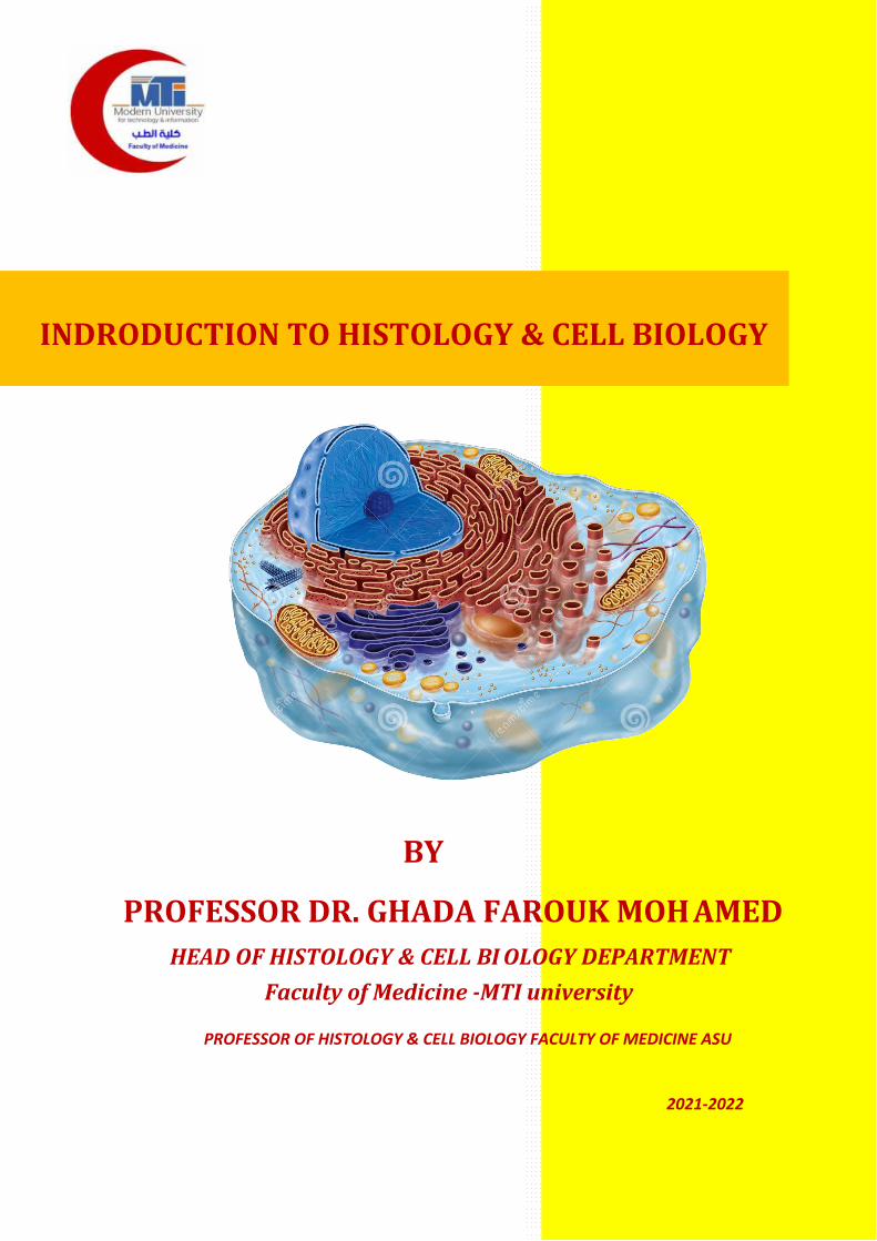
2021-2022
INDRODUCTION TO HISTOLOGY & CELL BIOLOGY
BY
PROFESSOR DR. GHADA FAROUK MOH AMED HEAD OF HISTOLOGY & CELL BI OLOGY DEPARTMENT
Faculty of Medicine -MTI university
PROFESSOR OF HISTOLOGY & CELL BIOLOGY FACULTY OF MEDICINE ASU
CONTENTS
CHAPTER PAGE
METHODS OF STUDY 8
THE CELL 18
EPITHELIAL TISSUE 78
2
Medical Education Department Faculty of Medicine MTI University
Adopted from: National Authority for Quality Assurance and Accreditation of
Education(NAQAAE)
MMTTII UUnniivveerrssiittyy FFaaccuullttyy ooff MMeeddiicciinnee DDeeppaarrttmmeenntt ooff HHiissttoollooggyy
CCoouurrssee SSppeecciiffiiccaattiioonn
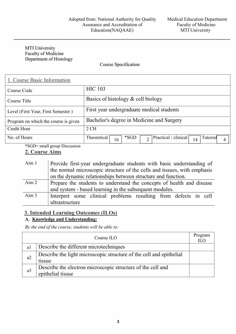
1. Course Basic Information
Course Code HIC 103
Course Title Basics of histology & cell biology
Level (First Year, First Semester ) First year undergraduate medical students
Program on which the course is given Bachelor's degree in Medicine and Surgery Credit Hour 2 CH
No. of Hours Theoretical *SGD Practical / clinical Tutorial
*SGD= small group Discussion2. Course Aims
Aim 1 Provide first-year undergraduate students with basic understanding of the normal microscopic structure of the cells and tissues, with emphasis on the dynamic relationships between structure and function.
Aim 2 Prepare the students to understand the concepts of health and disease and system - based learning in the subsequent modules.
Aim 3 Interpret some clinical problems resulting from defects in cell ultrastructure
3. Intended Learning Outcomes (ILOs)A. Knowledge and Understanding: By the end of the course, students will be able to:
Course ILO Program ILO
a1 Describe the different microtechniques
a2 Describe the light microscopic structure of the cell and epithelial tissue
a3 Describe the electron microscopic structure of the cell and epithelial tissue
16 2 14 4
3
Medical Education Department Faculty of Medicine MTI University
Adopted from: National Authority for Quality
Assurance and Accreditation of Education(NAQAAE)
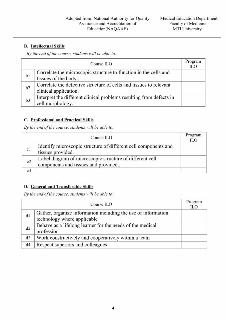
B. Intellectual Skills
By the end of the course, students will be able to:
Course ILO Program ILO
b1 Correlate the microscopic structure to function in the cells and tissues of the body..
b2 Correlate the defective structure of cells and tissues to relevant clinical application.
b3 Interpret the different clinical problems resulting from defects in cell morphology.
C. Professional and Practical Skills By the end of the course, students will be able to:
Course ILO Program ILO
c1 Identify microscopic structure of different cell components and tissues provided.
c2 Label diagram of microscopic structure of different cell components and tissues and provided..
c3
D. General and Transferable Skills By the end of the course, students will be able to:
Course ILO Program ILO
d1 Gather, organize information including the use of information technology where applicable
d2 Behave as a lifelong learner for the needs of the medical profession
d3 Work constructively and cooperatively within a team d4 Respect superiors and colleagues
4
Medical Education Department Faculty of Medicine MTI University
Adopted from: National Authority for Quality
Assurance and Accreditation of Education(NAQAAE)
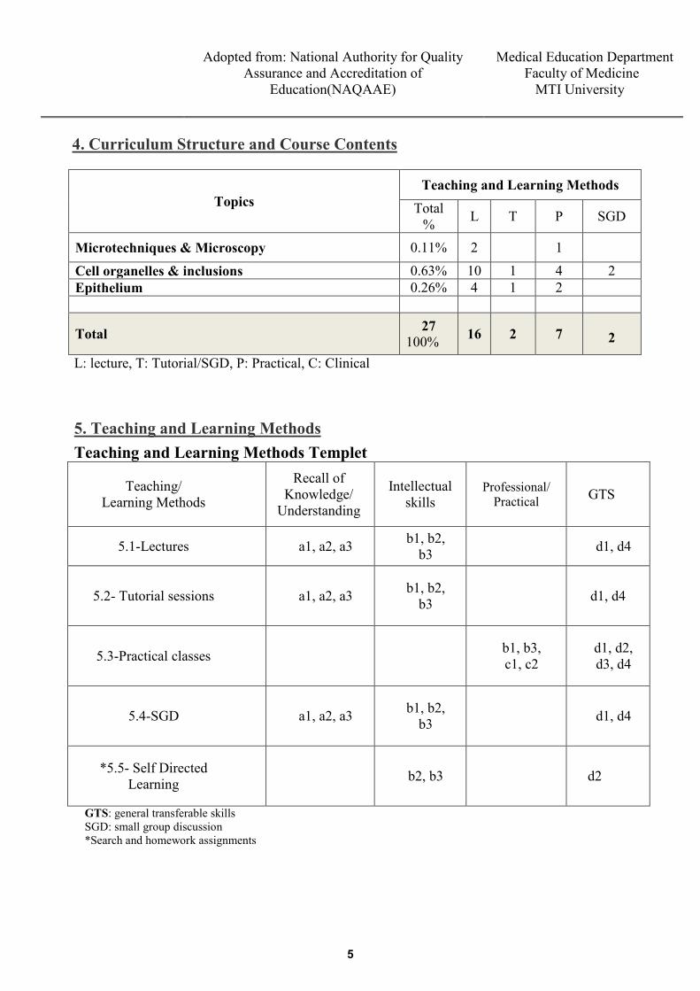
4. Curriculum Structure and Course Contents
Topics Teaching and Learning Methods
Total % L T P SGD
Microtechniques & Microscopy 0.11% 2 1
Cell organelles & inclusions 0.63% 10 1 4 2 Epithelium 0.26% 4 1 2
Total 27 100% 16 2 7
2
L: lecture, T: Tutorial/SGD, P: Practical, C: Clinical
Teaching and Learning Methods Templet
Teaching/ Learning Methods
Recall of Knowledge/
Understanding
Intellectual skills
Professional/ Practical GTS
5.1-Lectures a1, a2, a3 b1, b2, b3 d1, d4
5.2- Tutorial sessions a1, a2, a3 b1, b2, b3 d1, d4
5.3-Practical classes b1, b3, c1, c2
d1, d2, d3, d4
5.4-SGD a1, a2, a3 b1, b2, b3 d1, d4
*5.5- Self Directed Learning b2, b3 d2
GTS: general transferable skills SGD: small group discussion *Search and homework assignments
5. Teaching and Learning Methods
5
Medical Education Department Faculty of Medicine MTI University
Adopted from: National Authority for Quality
Assurance and Accreditation of Education(NAQAAE)
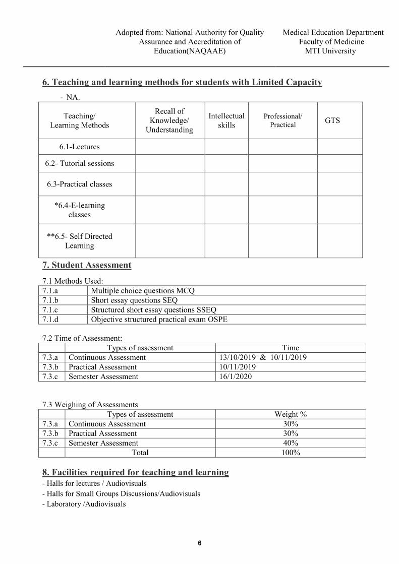
6. Teaching and learning methods for students with Limited Capacity
- NA.
Teaching/ Learning Methods
Recall of Knowledge/
Understanding
Intellectual skills
Professional/ Practical GTS
6.1-Lectures
6.2- Tutorial sessions
6.3-Practical classes
*6.4-E-learning classes
**6.5- Self Directed Learning
7. Student Assessment
7.1 Methods Used: 7.1.a Multiple choice questions MCQ 7.1.b Short essay questions SEQ 7.1.c Structured short essay questions SSEQ 7.1.d Objective structured practical exam OSPE 7.2 Time of Assessment: Types of assessment Time 7.3.a Continuous Assessment 13/10/2019 & 10/11/2019 7.3.b Practical Assessment 10/11/2019 7.3.c Semester Assessment 16/1/2020 7.3 Weighing of Assessments Types of assessment Weight % 7.3.a Continuous Assessment 30% 7.3.b Practical Assessment 30% 7.3.c Semester Assessment 40% Total 100% 8. Facilities required for teaching and learning - Halls for lectures / Audiovisuals - Halls for Small Groups Discussions/Audiovisuals - Laboratory /Audiovisuals
6
Medical Education Department Faculty of Medicine MTI University
Adopted from: National Authority for Quality Assurance and Accreditation of
Education(NAQAAE)
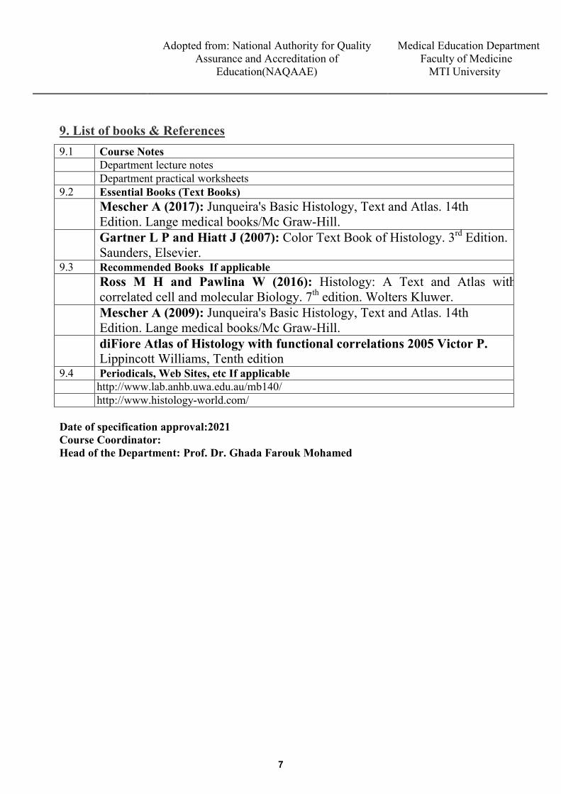
9. List of books & References9.1 Course Notes
Department lecture notes Department practical worksheets
9.2 Essential Books (Text Books) Mescher A (2017): Junqueira's Basic Histology, Text and Atlas. 14th Edition. Lange medical books/Mc Graw-Hill. Gartner L P and Hiatt J (2007): Color Text Book of Histology. 3rd Edition. Saunders, Elsevier.
9.3 Recommended Books If applicable Ross M H and Pawlina W (2016): Histology: A Text and Atlas with correlated cell and molecular Biology. 7th edition. Wolters Kluwer. Mescher A (2009): Junqueira's Basic Histology, Text and Atlas. 14th Edition. Lange medical books/Mc Graw-Hill. diFiore Atlas of Histology with functional correlations 2005 Victor P. Lippincott Williams, Tenth edition
9.4 Periodicals, Web Sites, etc If applicable http://www.lab.anhb.uwa.edu.au/mb140/ http://www.histology-world.com/
Date of specification approval:2021 Course Coordinator: Head of the Department: Prof. Dr. Ghada Farouk Mohamed
7
METHODS OF STUDY Histology is the science that deals with the detailed
microscopic structure of normal cells and tissues and the functions that they perform.
The basic techniques used for microscopic examination of tissue samples are: 1. Microtechniques. 2. Tissue culture: • Special technique for examination of living cells by
incubating them in growing media. • It is used in studying chromosomes and microorganisms.
3. Tissue smear or spreading: useful for studying blood film and bone marrow.
MICROTECHNIQUES Histological microtechniques are the methods by which the microscopic samples are made ready for examination. The importance of microtechniques:
• Hardening of soft tissue to be sectioned by knife of microtome into thin sections.
• Staining of these sections to be examined by microscope.
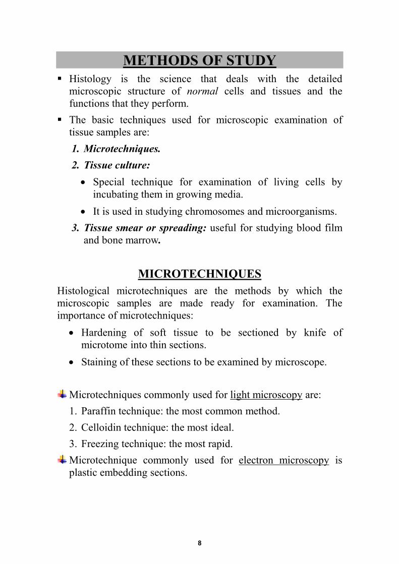
Microtechniques commonly used for light microscopy are: 1. Paraffin technique: the most common method. 2. Celloidin technique: the most ideal. 3. Freezing technique: the most rapid. Microtechnique commonly used for electron microscopy is plastic embedding sections.
8
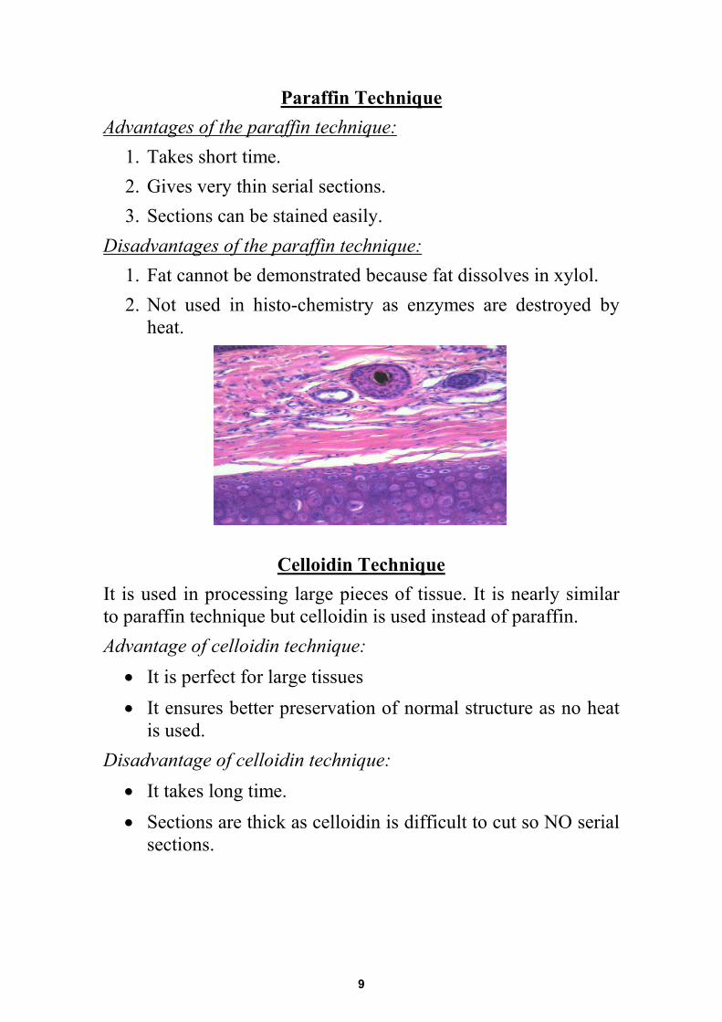
Paraffin Technique Advantages of the paraffin technique:
1. Takes short time. 2. Gives very thin serial sections. 3. Sections can be stained easily.
Disadvantages of the paraffin technique: 1. Fat cannot be demonstrated because fat dissolves in xylol. 2. Not used in histo-chemistry as enzymes are destroyed by
heat.
Celloidin Technique It is used in processing large pieces of tissue. It is nearly similar to paraffin technique but celloidin is used instead of paraffin. Advantage of celloidin technique:
• It is perfect for large tissues • It ensures better preservation of normal structure as no heat
is used. Disadvantage of celloidin technique:
• It takes long time. • Sections are thick as celloidin is difficult to cut so NO serial
sections.
9
Freezing Techniqsue The fresh tissue is rapidly frozen. Sectioning is done in a Cryostat at a subzero temperature and then staining is performed. Advantages of freezing technique:
• Very rapid so it is used in urgent conditions e.g. duringsurgery.
• It is used in histochemistry as no heat is used.Disadvantages of freezing technique:
• No thin serial sections as thin sections are fragmented.• Can not be kept for long time.
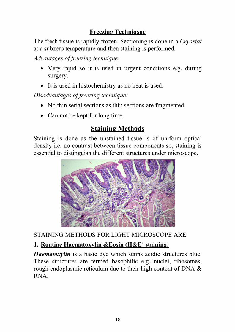
Staining Methods Staining is done as the unstained tissue is of uniform optical density i.e. no contrast between tissue components so, staining is essential to distinguish the different structures under microscope.
STAINING METHODS FOR LIGHT MICROSCOPE ARE: 1. Routine Haematoxylin &Eosin (H&E) staining:Haematoxylin is a basic dye which stains acidic structures blue. These structures are termed basophilic e.g. nuclei, ribosomes, rough endoplasmic reticulum due to their high content of DNA & RNA.
10
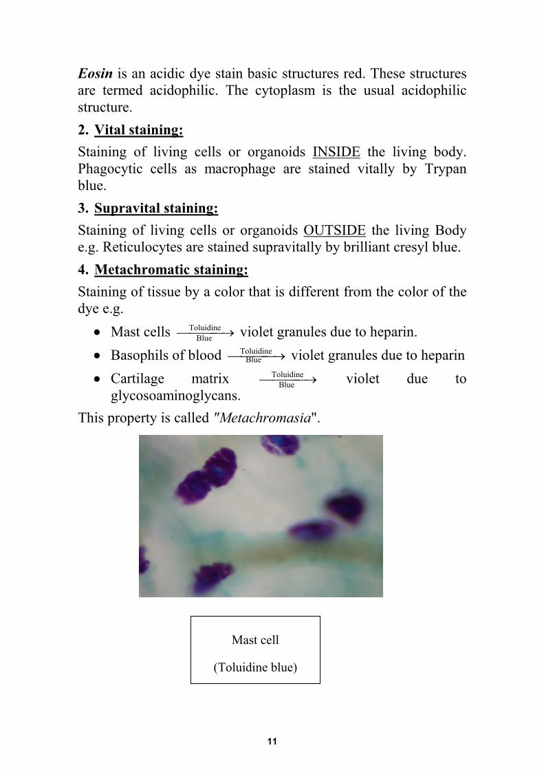
Eosin is an acidic dye stain basic structures red. These structures are termed acidophilic. The cytoplasm is the usual acidophilic structure. 2. Vital staining: Staining of living cells or organoids INSIDE the living body. Phagocytic cells as macrophage are stained vitally by Trypan blue. 3. Supravital staining: Staining of living cells or organoids OUTSIDE the living Body e.g. Reticulocytes are stained supravitally by brilliant cresyl blue. 4. Metachromatic staining: Staining of tissue by a color that is different from the color of the dye e.g.
• Mast cells →Toluidine violet granules due to heparin. • Basophils of blood →Toluidine violet granules due to heparin • Cartilage matrix →Toluidine violet due to
glycosoaminoglycans. This property is called "Metachromasia".
Blue
Blue
Blue
Mast cell
(Toluidine blue)
11
5. Histochemical staining: Is staining of chemical substances in cells and tissues e.g.
• Staining of lipids orange by Sudan III or black by osmic acid or Sudan black.
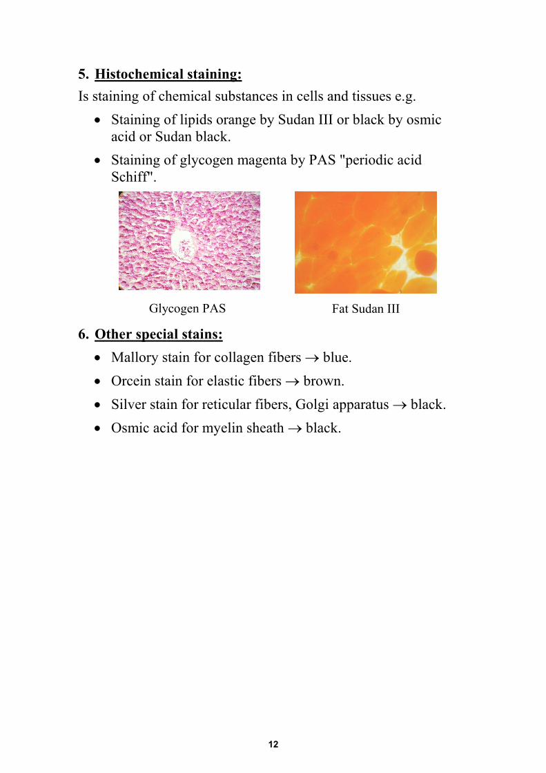
• Staining of glycogen magenta by PAS "periodic acid Schiff".
6. Other special stains: • Mallory stain for collagen fibers → blue. • Orcein stain for elastic fibers → brown. • Silver stain for reticular fibers, Golgi apparatus → black. • Osmic acid for myelin sheath → black.
Glycogen PAS Fat Sudan III
12
Microscopy Is to use an instrument "microscope" to magnify and view the detailed structures of cells and tissues.
LIGHT MICROSCOPE
Therefore any microscope has two powers: • Magnification power. • Resolution power: is the ability of the microscope to
differentiate clearly between too close structures. It is calculated by the smallest distance between two particles at which they could be seen as two separate particles.
Several types of microscopes are used to study the stained specimen. The most important types are: 1. Light microscope (LM) 2. Phase contrast microscope: used to examine living cells. 3. Polarizing microscope. 4. Fluorescence microscope. 5. Electron microscope (EM). →
The magnification power of the LM = power of objective lens multiplied by the power of eye piece. The resolution power of the LM = 0.2 µm.
They use the visible light
It use the invisible light
13
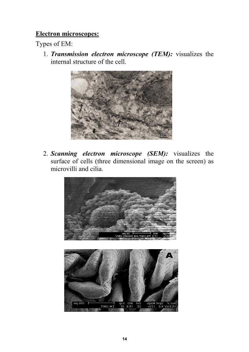
Electron microscopes: Types of EM:
1. Transmission electron microscope (TEM): visualizes the internal structure of the cell.
2. Scanning electron microscope (SEM): visualizes the surface of cells (three dimensional image on the screen) as microvilli and cilia.
14
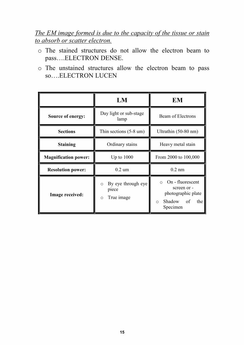
The EM image formed is due to the capacity of the tissue or stain to absorb or scatter electron. o The stained structures do not allow the electron beam to
pass….ELECTRON DENSE.o The unstained structures allow the electron beam to pass
so….ELECTRON LUCEN
LM EM
Source of energy: Day light or sub-stage lamp Beam of Electrons
Sections Thin sections (5-8 um) Ultrathin (50-80 nm)
Staining Ordinary stains Heavy metal stain
Magnification power: Up to 1000 From 2000 to 100,000
Resolution power: 0.2 um 0.2 nm
Image received:
o By eye through eyepiece
o True image
o On - fluorescentscreen or -
photographic plate
o Shadow of theSpecimen
15
THE CELL
The cell is the basic structural unit of any living organism. Though various cells of the body differ in their structures, most of them have common structural components. This chapter will deals with the common structure of the cell. The cell: • The cell is the unit of protoplasm that is bounded by a cell
membrane.• It is subdivided into:
a) Cytoplasm.b) Nucleus.
• We shall study the structure of the cell under the followingthree headings:
1. Cell membrane.2. Cytoplasm.3. Nucleus.
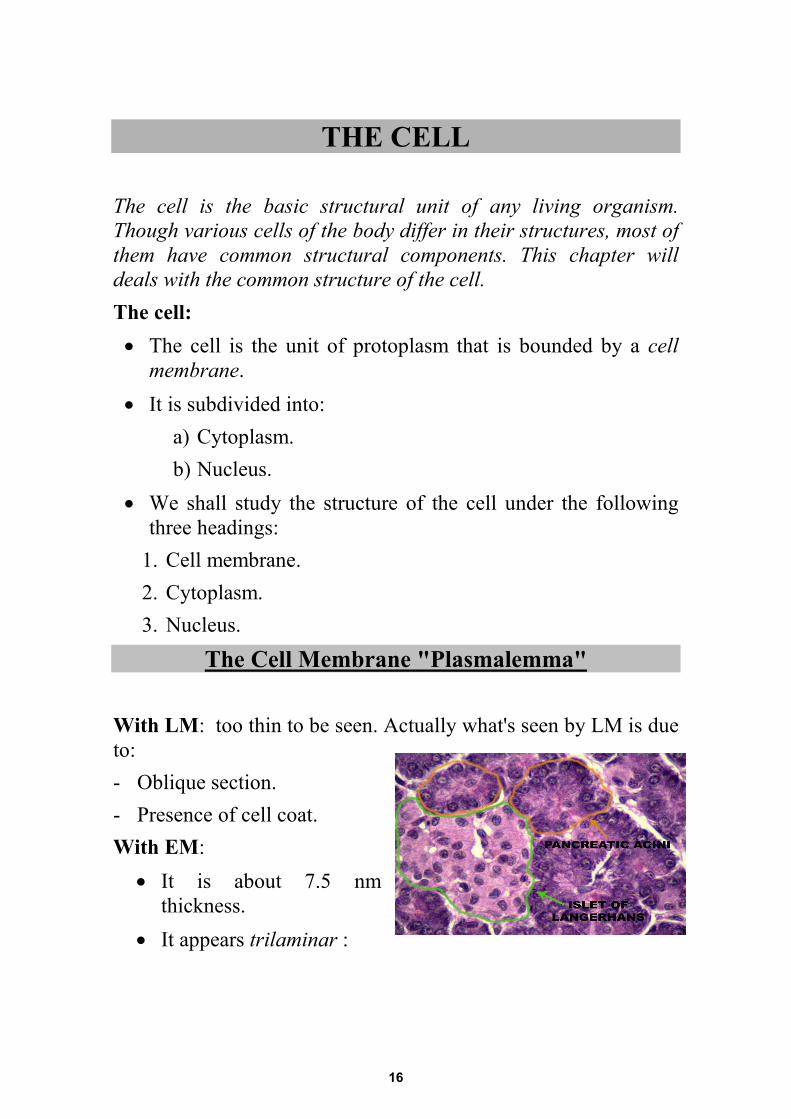
The Cell Membrane "Plasmalemma"
With LM: too thin to be seen. Actually what's seen by LM is due to: - Oblique section. - Presence of cell coat. With EM:
• It is about 7.5 nmthickness.
• It appears trilaminar :
16
- Outer & inner dark layers "2.5 nm each". - Middle light layer "2.5 nm".
• This trilaminar appearance is due to fixation by osmic acid Because of the molecular composition of the cell membrane.
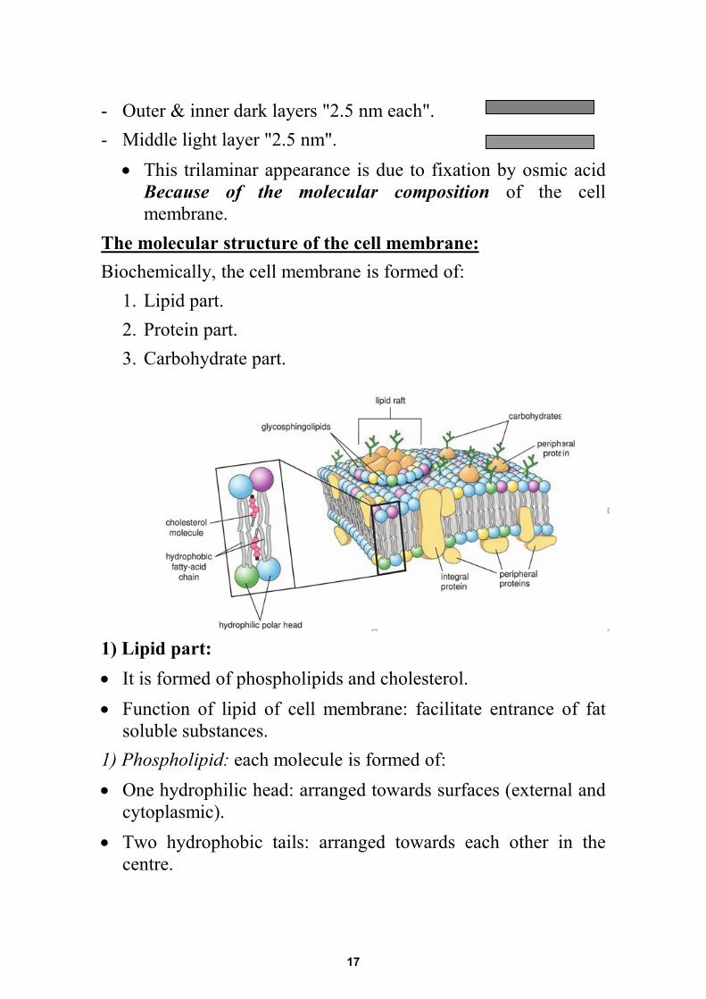
The molecular structure of the cell membrane: Biochemically, the cell membrane is formed of:
1. Lipid part. 2. Protein part. 3. Carbohydrate part.
1) Lipid part: • It is formed of phospholipids and cholesterol. • Function of lipid of cell membrane: facilitate entrance of fat
soluble substances. 1) Phospholipid: each molecule is formed of: • One hydrophilic head: arranged towards surfaces (external and
cytoplasmic). • Two hydrophobic tails: arranged towards each other in the
centre.
17
b) Cholesterol: • Present toward both aspects of cell membrane. • It provides stability and rigidity of cell membrane. 2) Protein part:
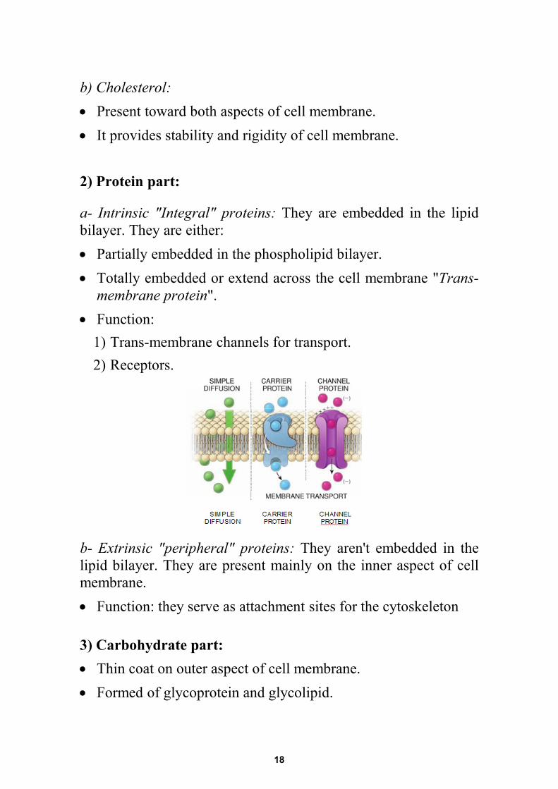
a- Intrinsic "Integral" proteins: They are embedded in the lipid bilayer. They are either: • Partially embedded in the phospholipid bilayer. • Totally embedded or extend across the cell membrane "Trans-
membrane protein". • Function:
1) Trans-membrane channels for transport. 2) Receptors.
b- Extrinsic "peripheral" proteins: They aren't embedded in the lipid bilayer. They are present mainly on the inner aspect of cell membrane. • Function: they serve as attachment sites for the cytoskeleton
3) Carbohydrate part: • Thin coat on outer aspect of cell membrane. • Formed of glycoprotein and glycolipid.
18
• It forms the glycocalyx or the cell coat. Glycocalyx or the cell coat:
- It is the sugar coat on the outer surface of the cell membrane. - It is either glycoprotein or glycolipid. - By LM: it is PAS+ve. - By EM: it shows fuzzy appearance. Function:
a- Cell adhesion. b- Cell receptors. c- Cell recognition. d- Formation of basement membrane.
Medical application: Defective membrane receptors can leads to many diseases e.g. dwarfism. It is due to defective growth hormone receptors.
Function of cell membrane: 1) Maintain the shape of the cell 2) Exchange: by:
- Diffusion. - Active transport: require energy. - Passive transport: by channels, carrier.
19
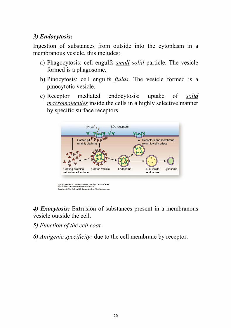
3) Endocytosis: Ingestion of substances from outside into the cytoplasm in a membranous vesicle, this includes:
a) Phagocytosis: cell engulfs small solid particle. The vesicle formed is a phagosome.
b) Pinocytosis: cell engulfs fluids. The vesicle formed is a pinocytotic vesicle.
c) Receptor mediated endocytosis: uptake of solid macromolecules inside the cells in a highly selective manner by specific surface receptors.
4) Exocytosis: Extrusion of substances present in a membranous vesicle outside the cell. 5) Function of the cell coat.
6) Antigenic specificity: due to the cell membrane by receptor.
20
CYTOPLASM The cytoplasm is formed of:
1. Organelles: either membranous or non membranous. 2. Inclusions. 3. Cytosol which is the fluid component of the
cytoplasm.
ORGANELLES • They are minute living structures that perform specific
functions. • They are either membranous or non membranous according
whether or not surrounded by the unit membrane.
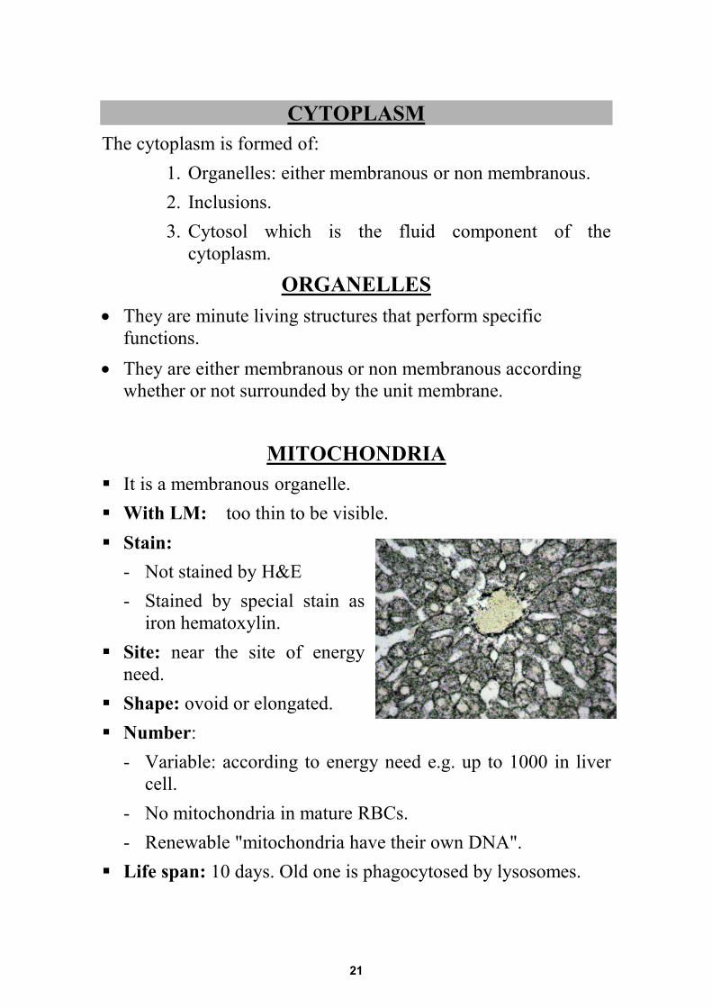
MITOCHONDRIA
It is a membranous organelle. With LM: too thin to be visible. Stain:
- Not stained by H&E - Stained by special stain as
iron hematoxylin. Site: near the site of energy
need. Shape: ovoid or elongated. Number:
- Variable: according to energy need e.g. up to 1000 in liver cell.
- No mitochondria in mature RBCs. - Renewable "mitochondria have their own DNA".
Life span: 10 days. Old one is phagocytosed by lysosomes.
21
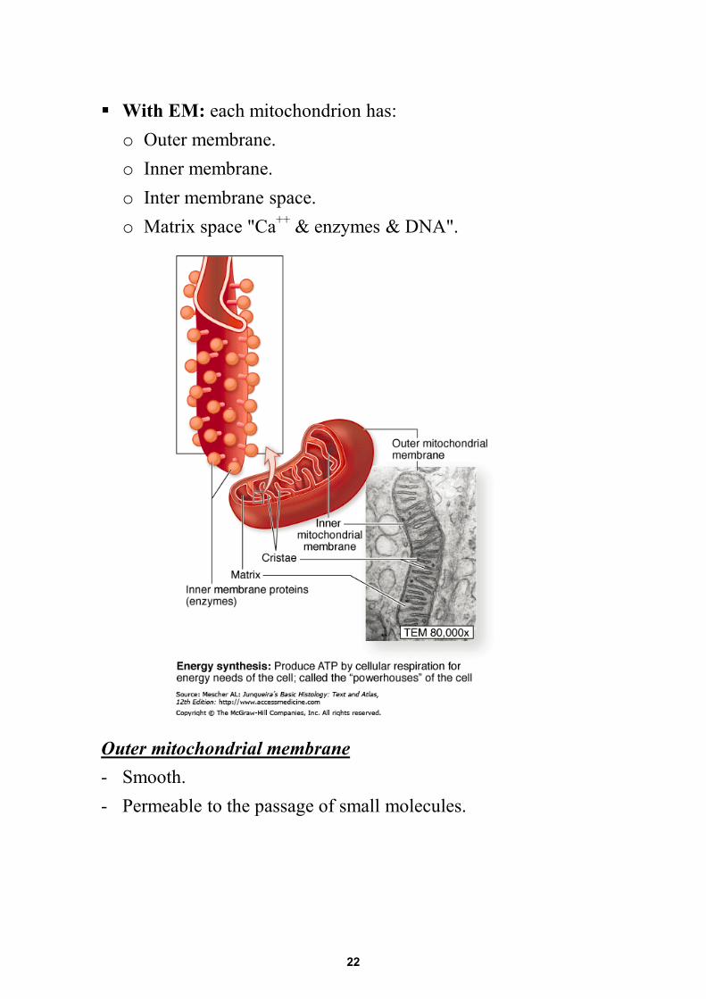
With EM: each mitochondrion has:o Outer membrane.o Inner membrane.o Inter membrane space.o Matrix space "Ca++ & enzymes & DNA".
Outer mitochondrial membrane - Smooth. - Permeable to the passage of small molecules.
22
Inner mitochondrial membrane: - Has cristae or folds "shelf like projection" (tubular or flat). - Highly selective to the passage of small molecules. - Contain the respiratory chain enzymes. - Has drum stick structures which has ATP synthatase activity
called F1 segment or lollypop structure. Matrix: - Fills the interior of mitochondria. - Has granules containing DNA & Ca++.
Function of Mitochondria: - It is the power house of the cell "production of ATP". - In Brown fat, mitochondria are concerned with heat production
not ATP production. - They have role in initiating cell death by release of cytochrome
c enzyme
Medical application: Mitochondrial deficiency of cytochrome c enzyme leads to organ dysfunction such as developmental delay, decreased muscle tone and deafness.
23
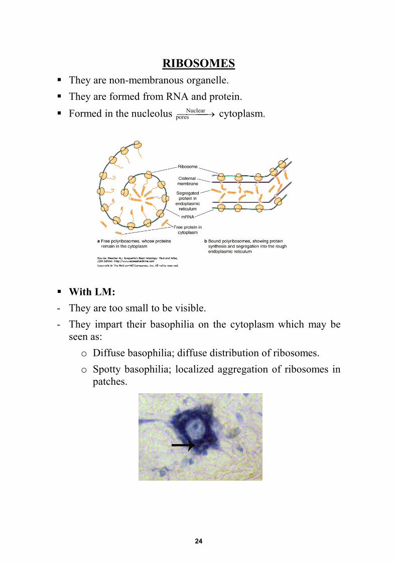
RIBOSOMES They are non-membranous organelle. They are formed from RNA and protein. Formed in the nucleolus →Nuclear cytoplasm.
With LM: - They are too small to be visible. - They impart their basophilia on the cytoplasm which may be
seen as: o Diffuse basophilia; diffuse distribution of ribosomes. o Spotty basophilia; localized aggregation of ribosomes in
patches.
pores
24
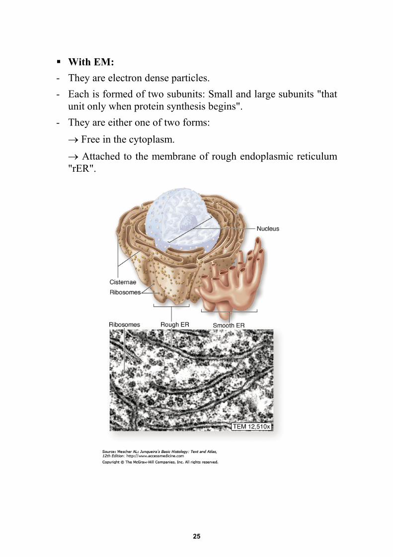
With EM: - They are electron dense particles. - Each is formed of two subunits: Small and large subunits "that
unit only when protein synthesis begins". - They are either one of two forms:
→ Free in the cytoplasm. → Attached to the membrane of rough endoplasmic reticulum "rER".
25
Polysomes: group of ribosomes linked by mRNA. Function: Synthesis of proteins. - Free ribosomes: synthesis of proteins for intracellular use "cell
existence". - Attached ribosomes: synthesis of proteins for extracellular use
or export → outside the cell. The proper formed proteins are folded and guided by protein chaperonin The denaturated or unfolded ones are conjugated to the protein ubiquitin for breakdown by proteasomes.
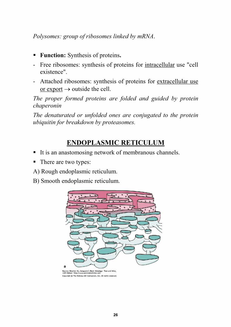
ENDOPLASMIC RETICULUM
It is an anastomosing network of membranous channels. There are two types: A) Rough endoplasmic reticulum. B) Smooth endoplasmic reticulum.
26
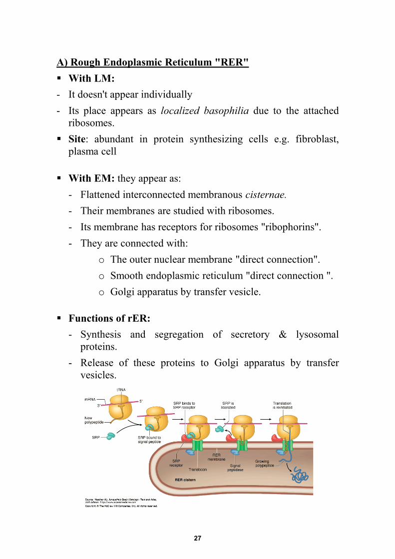
A) Rough Endoplasmic Reticulum "RER" With LM: - It doesn't appear individually - Its place appears as localized basophilia due to the attached
ribosomes. Site: abundant in protein synthesizing cells e.g. fibroblast,
plasma cell
With EM: they appear as: - Flattened interconnected membranous cisternae. - Their membranes are studied with ribosomes. - Its membrane has receptors for ribosomes "ribophorins". - They are connected with:
o The outer nuclear membrane "direct connection". o Smooth endoplasmic reticulum "direct connection ". o Golgi apparatus by transfer vesicle.
Functions of rER: - Synthesis and segregation of secretory & lysosomal
proteins. - Release of these proteins to Golgi apparatus by transfer
vesicles.
27
B) Smooth Endoplasmic Reticulum "SER": With LM: Not seen by LM Site: they are abundant in steroid synthesizing cells. With EM: they appear as: - Branching & anastomosing membranous tubules. - They have smooth membrane surface. - They have no ribosomes. - They have no ribosomes receptors. Function of SER
1- Lipid synthesis. 2- Glycogen synthesis. 3- Detoxification of drugs. 4- Regulation of Ca++ in muscle. 5- Conjugation of bilirubin in the liver
Medical application:
Jaundice means yellowish coloration of the skin. It occurs in newborns due to underdevelopment of the SER of the liver cells.
GOLGI APPARATUS
It is a membranous organelle. With LM: not seen by LM. Size: variable; large Golgi leave an unstained area in the
cytoplasm called -ve Golgi image Stain: silver stain.
28
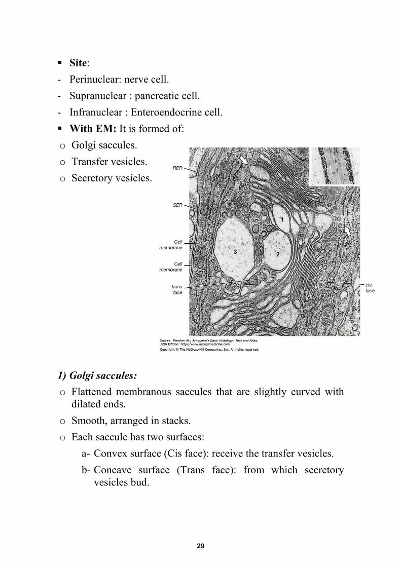
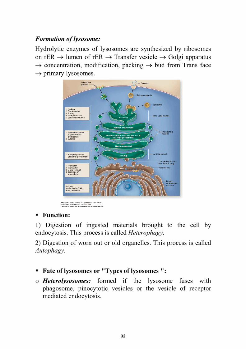
Site: - Perinuclear: nerve cell. - Supranuclear : pancreatic cell. - Infranuclear : Enteroendocrine cell. With EM: It is formed of: o Golgi saccules. o Transfer vesicles. o Secretory vesicles. 1) Golgi saccules: o Flattened membranous saccules that are slightly curved with
dilated ends. o Smooth, arranged in stacks. o Each saccule has two surfaces:
a- Convex surface (Cis face): receive the transfer vesicles. b- Concave surface (Trans face): from which secretory
vesicles bud.
29
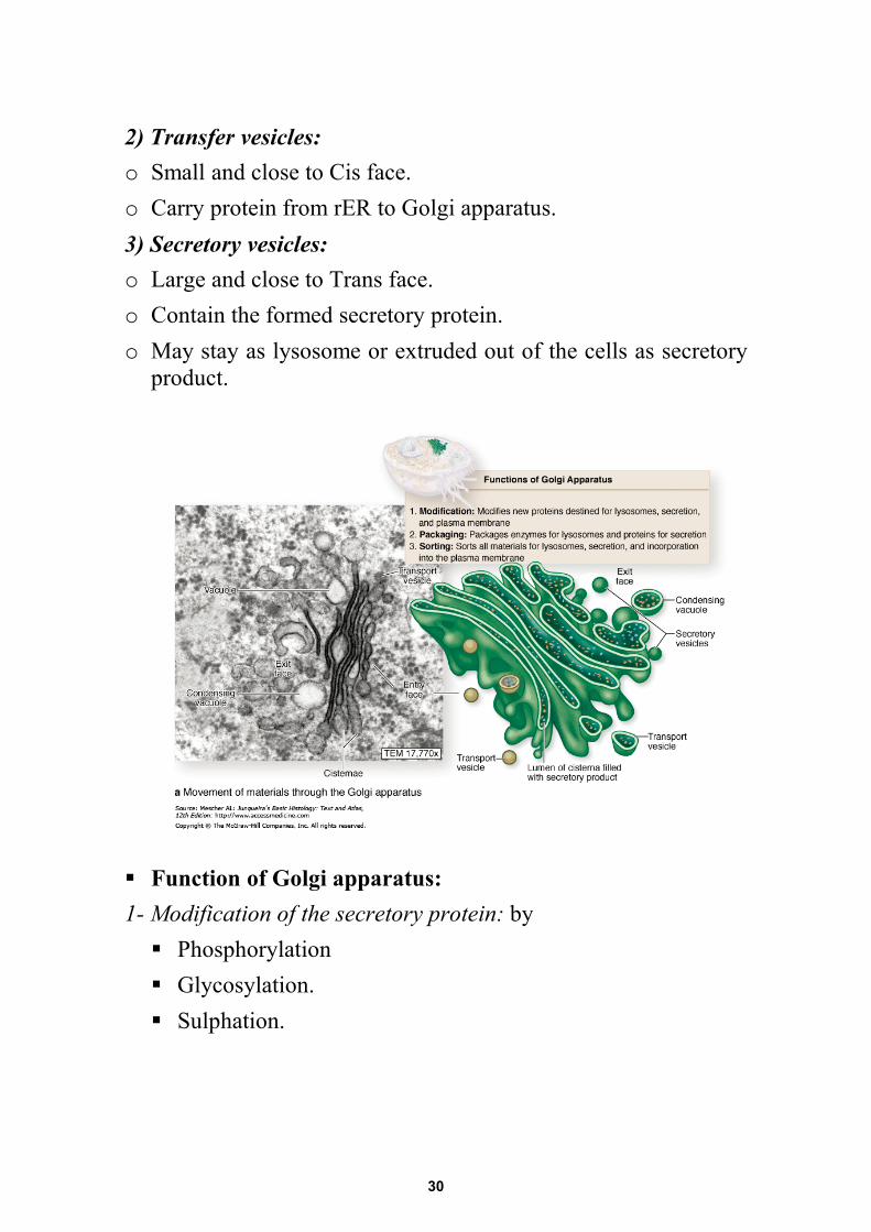
2) Transfer vesicles:o Small and close to Cis face.o Carry protein from rER to Golgi apparatus.3) Secretory vesicles:o Large and close to Trans face.o Contain the formed secretory protein.o May stay as lysosome or extruded out of the cells as secretory
product.
Function of Golgi apparatus:1- Modification of the secretory protein: by Phosphorylation Glycosylation. Sulphation.
30
2- Sorting of the secretory protein: By specific receptors on the Trans face to address the protein which to: Stay as lysosomes. Go out as secretion.
3- Packaging of the sorted protein: Acid hydrolases in lysosomes. Secretory proteins in secretory vesicles.
4- Recycling of cell Membrane.
Medical application: I cell disease: it is due to defective Golgi phosphorylation that results in defective formation of lysosomal enzymes leading to accumulation of undigested substances within the cells.
LYSOSOME It is a membranous organelle. It is formed by Golgi apparatus. It contains hydrolytic enzymes in an acidic pH "5" (its
membrane has proton pump). With LM: It is not seen by H&E. Stain: Needs histochemical techniques for its enzymes. Number: abundant in phagocytic cells as macrophages and
neutrophils. With EM: - Round electron dense bodies. - Formed by budding from the Trans face of Golgi.
31
Formation of lysosome: Hydrolytic enzymes of lysosomes are synthesized by ribosomes on rER → lumen of rER → Transfer vesicle → Golgi apparatus → concentration, modification, packing → bud from Trans face → primary lysosomes. Function: 1) Digestion of ingested materials brought to the cell by endocytosis. This process is called Heterophagy. 2) Digestion of worn out or old organelles. This process is called Autophagy. Fate of lysosomes or "Types of lysosomes ": o Heterolysosomes: formed if the lysosome fuses with
phagosome, pinocytotic vesicles or the vesicle of receptor mediated endocytosis.
32
o Autolysosomes: formed if the lysosome fuses with an old organelle.
Following digestion of the contents:
- Nutrients diffuse to the cytoplasm across the lysosomal membrane.
- Indigestible compounds are remained within the vacuole forming Residual bodies.
Fate of residual bodies: - Extruded from the cell by exocytosis as in macrphages. - Accumulate in the cytoplasm as in long lived cells as in
heart muscle and nerve cells and are called Lipofuscin granules or age pigment which appear golden brown by LM.
Medical application:
o Hypoxia leads release of lysosomal enzymes causing cell digestion
o After cell death, it is responsible for postmortem autolysis
o Lysosomal dysfunctions leads to accumulated undigested materials causing disturbed cell functions "lysosomal storage diseases"
PEROXISOMES "MICROBODIES"
They are membranous organelles. They contain enzymes oxidases and catalases. With LM: not seen. With EM: small vesicles that has electron dense content Site: in close relation to SER. Number: abundant in liver cells.
33
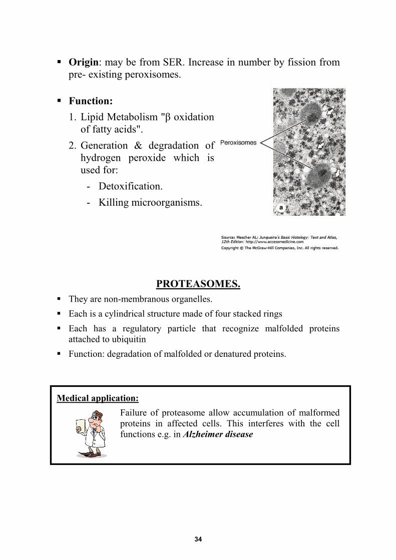
Origin: may be from SER. Increase in number by fission from pre- existing peroxisomes.
Function: 1. Lipid Metabolism "β oxidation
of fatty acids". 2. Generation & degradation of
hydrogen peroxide which is used for:
- Detoxification. - Killing microorganisms.
PROTEASOMES. They are non-membranous organelles. Each is a cylindrical structure made of four stacked rings Each has a regulatory particle that recognize malfolded proteins
attached to ubiquitin Function: degradation of malfolded or denatured proteins. Medical application:
Failure of proteasome allow accumulation of malformed proteins in affected cells. This interferes with the cell functions e.g. in Alzheimer disease
34
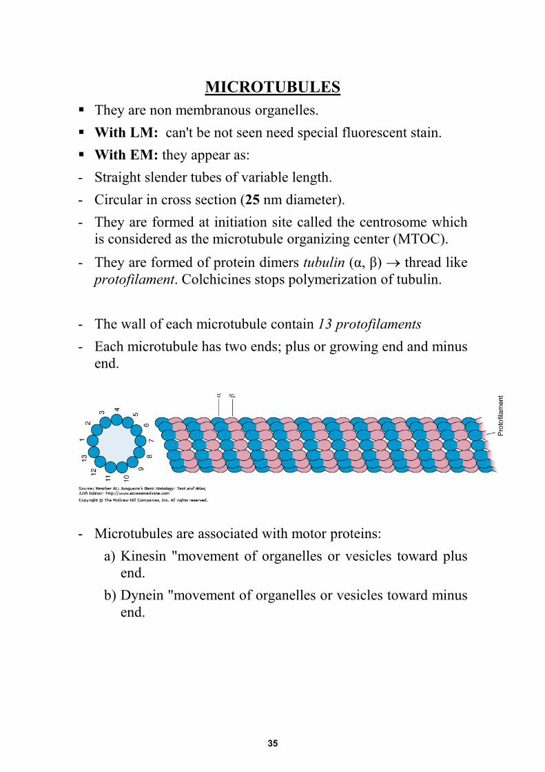
MICROTUBULES They are non membranous organelles. With LM: can't be not seen need special fluorescent stain. With EM: they appear as: - Straight slender tubes of variable length. - Circular in cross section (25 nm diameter). - They are formed at initiation site called the centrosome which
is considered as the microtubule organizing center (MTOC). - They are formed of protein dimers tubulin (α, β) → thread like
protofilament. Colchicines stops polymerization of tubulin. - The wall of each microtubule contain 13 protofilaments - Each microtubule has two ends; plus or growing end and minus
end.
- Microtubules are associated with motor proteins:
a) Kinesin "movement of organelles or vesicles toward plus end.
b) Dynein "movement of organelles or vesicles toward minus end.
35
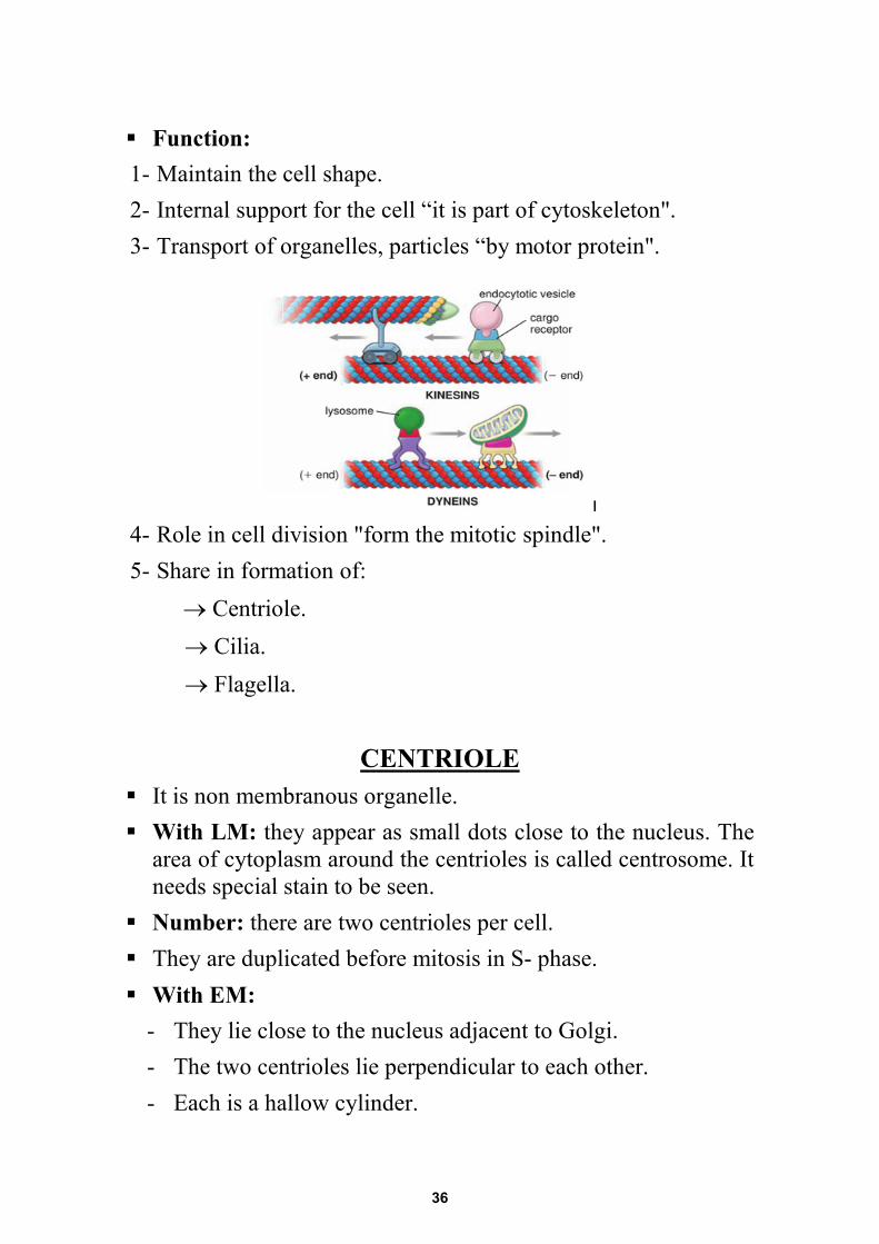
Function: 1- Maintain the cell shape. 2- Internal support for the cell “it is part of cytoskeleton". 3- Transport of organelles, particles “by motor protein". 4- Role in cell division "form the mitotic spindle". 5- Share in formation of: → Centriole. → Cilia. → Flagella.
CENTRIOLE It is non membranous organelle. With LM: they appear as small dots close to the nucleus. The
area of cytoplasm around the centrioles is called centrosome. It needs special stain to be seen.
Number: there are two centrioles per cell. They are duplicated before mitosis in S- phase. With EM:
- They lie close to the nucleus adjacent to Golgi. - The two centrioles lie perpendicular to each other. - Each is a hallow cylinder.
36
- Each made up of 27 microtubules. - Microtubules are arranged in 9 bundles. - Each bundle is formed of 3 microtubules "Triplets" [9
triplets + 0]
Function: - Centriole and centrosome have role in cell division "MTOC". - Form the basal body of cilia and flagella.
CILIA
They are non membranous
organelles. With LM: they appear as
hair like processes Number: several hundred
per cell.
37
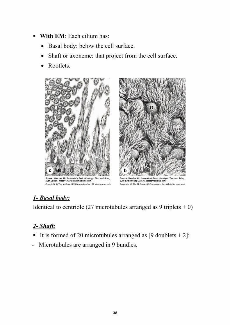
With EM: Each cilium has: • Basal body: below the cell surface. • Shaft or axoneme: that project from the cell surface. • Rootlets.
1- Basal body: Identical to centriole (27 microtubules arranged as 9 triplets + 0) 2- Shaft: It is formed of 20 microtubules arranged as [9 doublets + 2]: - Microtubules are arranged in 9 bundles.
38
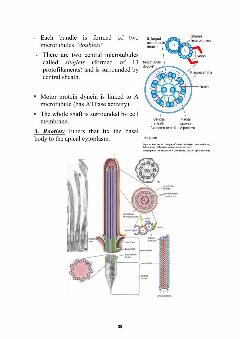
- Each bundle is formed of two microtubules "doublets"
- There are two central microtubules called singlets (formed of 13 protofilaments) and is surrounded by central sheath.
Motor protein dynein is linked to Amicrotubule (has ATPase activity)
The whole shaft is surrounded by cellmembrane.
3. Rootles: Fibers that fix the basalbody to the apical cytoplasm.
39
Function of cilia: - Pushing fluid by rhythmic movement over cell surface. - Some cell has single cilium for perception of stimuli e.g. rod
and cones.
FLAGELLA The structure of the flagellum is the same as cilium, But: - It is single per cell. - It is longer than cilia. It is present in mammals only in spermatozoa. Function: movement of cell as in sperms.
Medical application:
- Mutation in the genes of cilia and flagella leads to Immotile Cilia Syndrome “Kartagener syndrome”
- The patient suffers from chronic respiratory infections and infertility
-
FILAMENTS They are non membranous organelles. They are thread like. There are three types: a) Thick filament : o 12 nm. o Formed of protein Myosin. o Found in striated muscles for contraction.
40
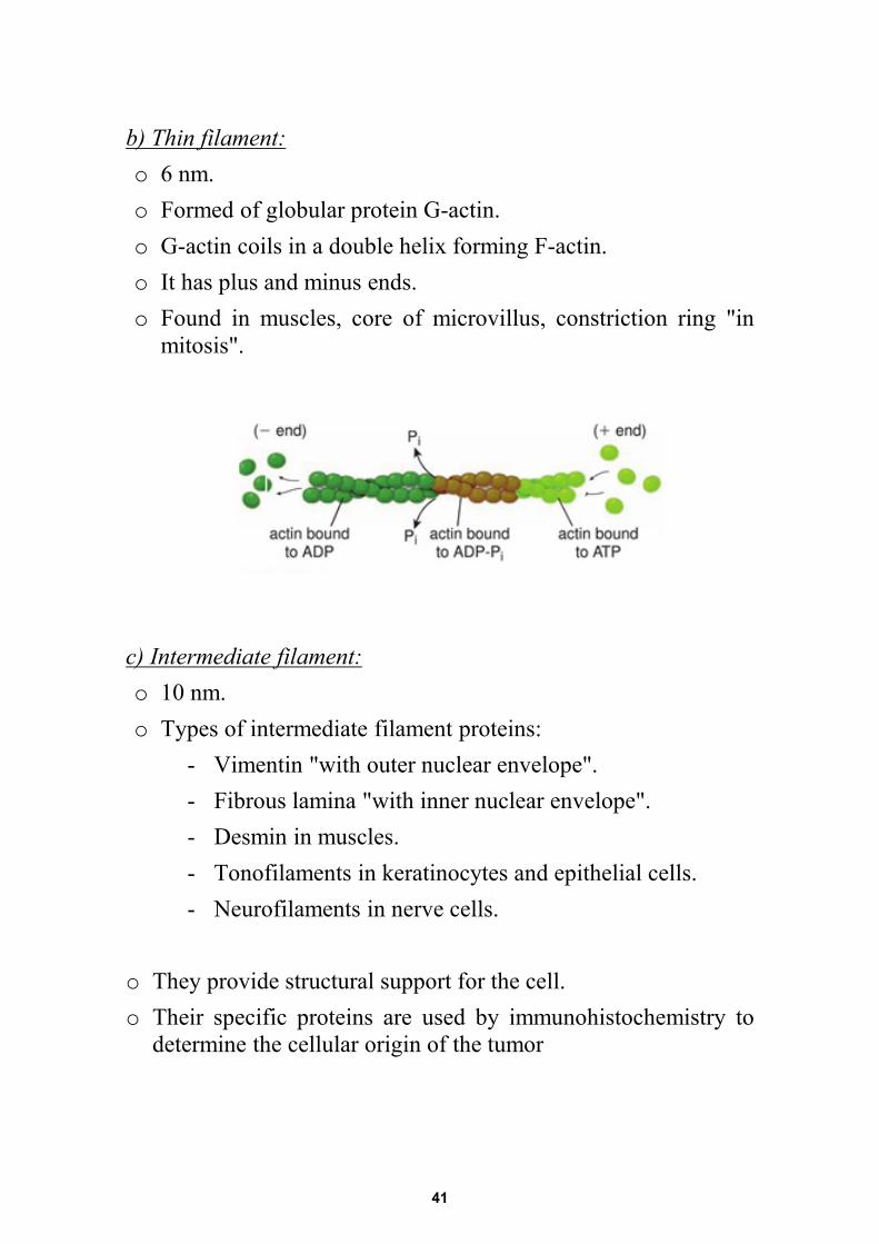
b) Thin filament:o 6 nm.o Formed of globular protein G-actin.o G-actin coils in a double helix forming F-actin.o It has plus and minus ends.o Found in muscles, core of microvillus, constriction ring "in
mitosis".
c) Intermediate filament:o 10 nm.o Types of intermediate filament proteins:
- Vimentin "with outer nuclear envelope". - Fibrous lamina "with inner nuclear envelope". - Desmin in muscles. - Tonofilaments in keratinocytes and epithelial cells. - Neurofilaments in nerve cells.
o They provide structural support for the cell.o Their specific proteins are used by immunohistochemistry to
determine the cellular origin of the tumor
41
CYTOSKELETON o It is a network throughout the cytoplasm of filaments and
tubules.o This network provides the shape for the cell and organizes the
cellular contents.o The cytoskeleton is formed of:
a) Microtubules.b) Microfilaments.c) Intermediate filaments.
INCLUSIONS They non living non functioning components of the cell. They are non-membranous except lipofuscin pigment. They are either by-product of metabolism or taken from
outside. They include:
a) Stored food.b) Pigments.c) Crystals.
A) Stored food: only carbohydrates and fat are stored.1) Glycogen
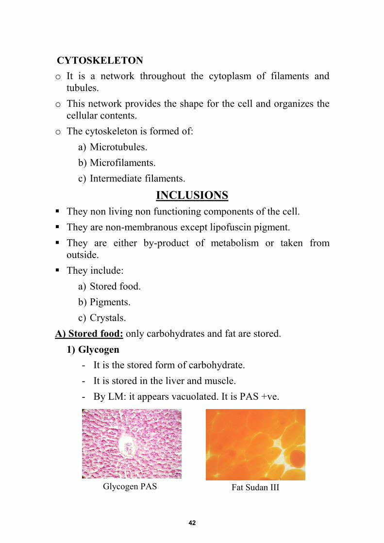
- It is the stored form of carbohydrate. - It is stored in the liver and muscle. - By LM: it appears vacuolated. It is PAS +ve.
Glycogen PAS Fat Sudan III
42
2) Lipid (fat):- It is stored if fat cells and liver cells.- By LM, fat appears empty spaces as it dissolved.- It is specially stained orange by Sudan III.
B) Pigments: are classified as:1) Exogenous: are taken to the body from outside:
- Lipochrome: carotene gives yellow color of skin. - Dust & carbon particles: black particles in lung
macrophages, - Minerals: e.g. lead, silver and tattooing of the skin,
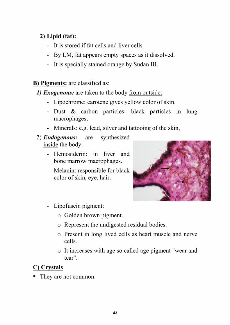
2) Endogenous: are synthesizedinside the body:
- Hemosiderin: in liver and bone marrow macrophages.
- Melanin: responsible for black color of skin, eye, hair.
- Lipofuscin pigment: o Golden brown pigment.o Represent the undigested residual bodies.o Present in long lived cells as heart muscle and nerve
cells.o It increases with age so called age pigment "wear and
tear".C) Crystals They are not common.
43

44
MICROTECHIQUES
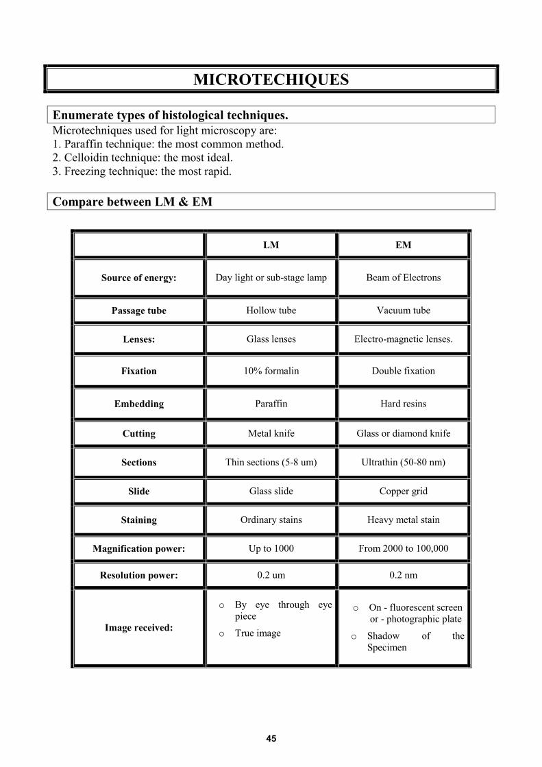
Enumerate types of histological techniques. Microtechniques used for light microscopy are: 1. Paraffin technique: the most common method.2. Celloidin technique: the most ideal.3. Freezing technique: the most rapid.
Compare between LM & EM
LM EM
Source of energy: Day light or sub-stage lamp Beam of Electrons
Passage tube Hollow tube Vacuum tube
Lenses: Glass lenses Electro-magnetic lenses.
Fixation 10% formalin Double fixation
Embedding Paraffin Hard resins
Cutting Metal knife Glass or diamond knife
Sections Thin sections (5-8 um) Ultrathin (50-80 nm)
Slide Glass slide Copper grid
Staining Ordinary stains Heavy metal stain
Magnification power: Up to 1000 From 2000 to 100,000
Resolution power: 0.2 um 0.2 nm
Image received:
o By eye through eyepiece
o True image
o On - fluorescent screenor - photographic plate
o Shadow of the Specimen
45
Give reasons for: 1. Paraffin technique is the most common: as it gives thin serial sections so it
insures better studying of the tissues 2. Freezing technique is used for histochemistry: as it is rapid so it insures
better demonstration for enzyme activity.
3. Fat appears as empty spaces Fat is dissolved during paraffin preparation due to: The heat used The chemicals used
4. Fixation is an important step in tissue processing: as fixation aims to:
• Hardening the tissue by coagulation of proteins. • Preventing autolysis by stopping lysosomal enzymes. • Preventing putrefaction by killing bacteria. • Enhancing staining “mordanting effect”.
5. EM picture is displayed in black and white: because:
• The electron dense stained structures scatter the electron beam and so it can not pass giving black shadow
• While the electron lucent non stained structures allow the electron beam to pass giving white areas
46
CYTOLOGY Enumerate the molecules constituting the cell membrane. Describe the microscopic structure of the part which accounts for the largest percentage of its structure U
The molecules constituting the structure of the cell membrane are: 1. Lipid part. 2. Protein part. 3. Carbohydrate part.
The major constituent of the cell membrane is the protein part Protein part of cell membrane: a- Intrinsic "Integral" proteins: They are embedded in the lipid bilayer. They are either:
• Partially embedded in the phospholipid bilayer. • Totally embedded or extend across the cell membrane "Trans-membrane protein". • Function:
1) Trans-membrane channels for transport. 2) Receptors.
b- Extrinsic "peripheral" proteins: They aren't embedded in the lipid bilayer. They are present mainly on the inner aspect of cell membrane. • Function: they serve as attachment sites for the cytoskeleton Describe the microscopic structure of the lipid part of cell membrane Lipid part of cell membrane:
• It is formed of phospholipids and cholesterol. • Function of lipid of cell membrane: facilitate entrance of fat soluble substances.
1) Phospholipid: each molecule is formed of:
• One hydrophilic head: arranged towards surfaces (external and cytoplasmic). • Two hydrophobic tails: arranged towards each other in the center.
b) Cholesterol:
• Present toward both aspects of cell membrane. • It provides stability and rigidity of cell membrane.
47
Describe the microscopic structure of the cell coat with special reference to its importance
- It is the sugar coat on the outer surface of the cell membrane. - It is either glycoprotein or glycolipid. - By LM: it is PAS+ve. - By EM: it shows fuzzy appearance. - Function:
• Cell adhesion. • Cell receptors. • Cell recognition. • Formation of basement membrane.
UMedical application: U defective structure of the cell coat in absorptive cells of the small intestine leads to UMalabsorption syndrome
Enumerate the difference between outer and inner aspects of cell membrane The outer aspect of the cell membrane: Faces the extracellular compartment Has cell coat Has receptors
The inner aspect of the cell membrane: Faces the cytoplasmic compartment Has no cell coat Has no receptors Its peripheral protein act as receptors for parts of cytoskeleton
Enumerate double walled organelles inside the cell. Describe the electron microscopic structure of the ATP producing one The double walled organelles in the cell are:
• The nucleus • The mitochondria
UMITOCHONDRIA With EM: each mitochondrion has: Outer membrane. Inner membrane. Inter membrane space. Matrix space
U
48
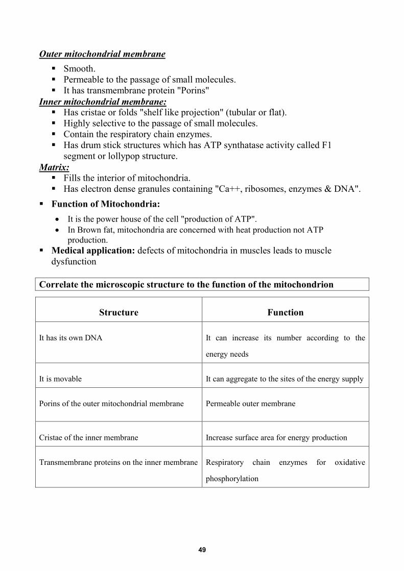
Outer mitochondrial membrane Smooth. Permeable to the passage of small molecules. It has transmembrane protein "Porins"
UInner mitochondrial membrane: Has cristae or folds "shelf like projection" (tubular or flat). Highly selective to the passage of small molecules. Contain the respiratory chain enzymes. Has drum stick structures which has ATP synthatase activity called F1
segment or lollypop structure. UMatrix:U Fills the interior of mitochondria. Has electron dense granules containing "Ca++, ribosomes, enzymes & DNA".
Function of Mitochondria: • It is the power house of the cell "production of ATP". • In Brown fat, mitochondria are concerned with heat production not ATP
production. Medical application: defects of mitochondria in muscles leads to muscle
dysfunction Correlate the microscopic structure to the function of the mitochondrion
Structure Function
It has its own DNA It can increase its number according to the
energy needs
It is movable It can aggregate to the sites of the energy supply
Porins of the outer mitochondrial membrane Permeable outer membrane
Cristae of the inner membrane Increase surface area for energy production
Transmembrane proteins on the inner membrane Respiratory chain enzymes for oxidative
phosphorylation
49
Compare between the outer and inner mitochondrial membranes
Outer membrane Inner membrane
Surface area Small "smooth no folds" Large "folded in the form of cristae"
Porins Present Absent
Oxidative enzymes Absent Present
Lolly pop "drum stick" structure Absent Present
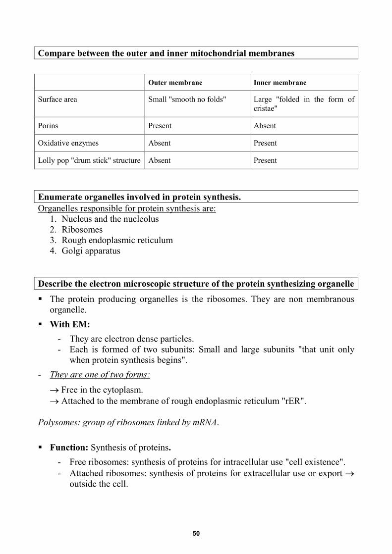
Enumerate organelles involved in protein synthesis. UOrganelles responsible for protein synthesis are:
1. Nucleus and the nucleolus 2. Ribosomes 3. Rough endoplasmic reticulum 4. Golgi apparatus
Describe the electron microscopic structure of the protein synthesizing organelle
The protein producing organelles is the ribosomes. They are non membranous organelle.
With EM: - They are electron dense particles. - Each is formed of two subunits: Small and large subunits "that unit only
when protein synthesis begins". - UThey are one of two forms:
→ Free in the cytoplasm. → Attached to the membrane of rough endoplasmic reticulum "rER".
Polysomes: group of ribosomes linked by mRNA. Function: Synthesis of proteins.
- Free ribosomes: synthesis of proteins for intracellular use "cell existence". - Attached ribosomes: synthesis of proteins for extracellular use or export →
outside the cell.
50
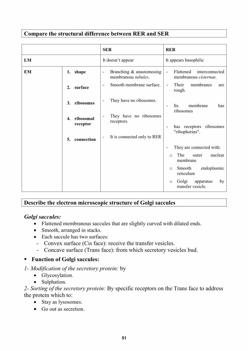
Compare the structural difference between RER and SER SER RER
LM It doesn’t appear It appears basophilic
EM
1. shape
2. surface
3. ribosomes
4. ribosomal receptor
5. connection
- Branching & anastomosing membranous tubules.
- Smooth membrane surface.
- They have no ribosomes.
- They have no ribosomes receptors.
- It is connected only to RER
- Flattened interconnected membranous cisternae.
- Their membranes are rough.
- Its membrane has ribosomes
- has receptors ribosomes "ribophorins".
- They are connected with:
o The outer nuclear membrane
o Smooth endoplasmic reticulum
o Golgi apparatus by transfer vesicle.
Describe the electron microscopic structure of Golgi saccules Golgi saccules:
• Flattened membranous saccules that are slightly curved with dilated ends. • Smooth, arranged in stacks. • Each saccule has two surfaces: - Convex surface (Cis face): receive the transfer vesicles. - Concave surface (Trans face): from which secretory vesicles bud.
Function of Golgi saccules: 1- Modification of the secretory protein: by
• Glycosylation. • Sulphation.
2- Sorting of the secretory protein: By specific receptors on the Trans face to address the protein which to:
• Stay as lysosomes. • Go out as secretion.
51
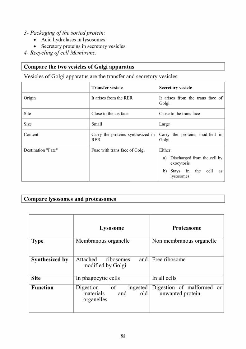
3- Packaging of the sorted protein: • Acid hydrolases in lysosomes. • Secretory proteins in secretory vesicles.
4- Recycling of cell Membrane. Compare the two vesicles of Golgi apparatus
Vesicles of Golgi apparatus are the transfer and secretory vesicles
Transfer vesicle Secretory vesicle
Origin It arises from the RER It arises from the trans face of Golgi
Site Close to the cis face Close to the trans face
Size Small Large
Content Carry the proteins synthesized in RER
Carry the proteins modified in Golgi
Destination "Fate" Fuse with trans face of Golgi Either:
a) Discharged from the cell by exocytosis
b) Stays in the cell as lysosomes
Compare lysosomes and proteasomes
Lysosome Proteasome
Type Membranous organelle Non membranous organelle
Synthesized by Attached ribosomes and modified by Golgi
Free ribosome
Site In phagocytic cells In all cells Function Digestion of ingested
materials and old organelles
Digestion of malformed or unwanted protein
52
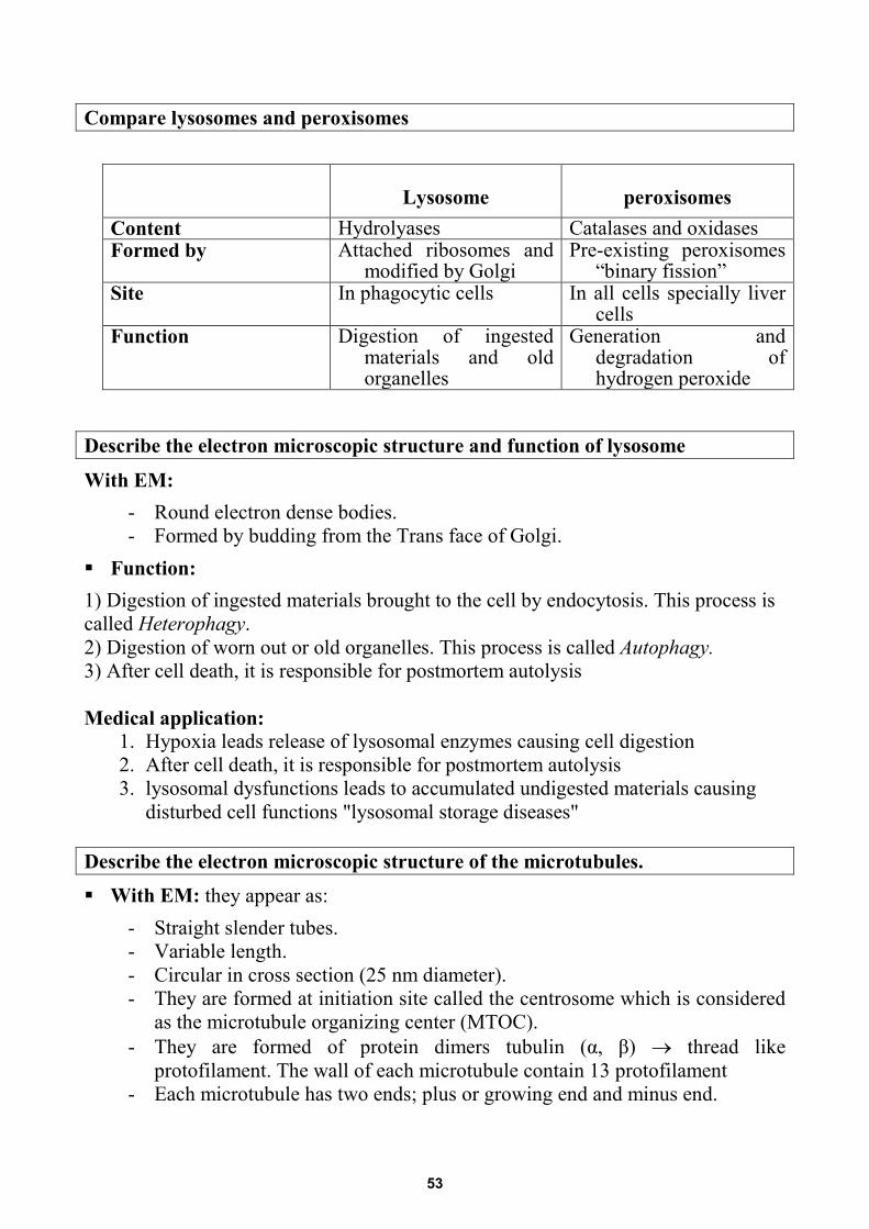
Compare lysosomes and peroxisomes
Lysosome peroxisomes Content Hydrolyases Catalases and oxidases Formed by Attached ribosomes and
modified by Golgi Pre-existing peroxisomes
“binary fission” Site In phagocytic cells In all cells specially liver
cells Function Digestion of ingested
materials and old organelles
Generation and degradation of hydrogen peroxide
Describe the electron microscopic structure and function of lysosome
With EM: - Round electron dense bodies. - Formed by budding from the Trans face of Golgi.
Function: 1) Digestion of ingested materials brought to the cell by endocytosis. This process is called Heterophagy. 2) Digestion of worn out or old organelles. This process is called Autophagy. 3) After cell death, it is responsible for postmortem autolysis Medical application:
1. Hypoxia leads release of lysosomal enzymes causing cell digestion 2. After cell death, it is responsible for postmortem autolysis 3. lysosomal dysfunctions leads to accumulated undigested materials causing
disturbed cell functions "lysosomal storage diseases" Describe the electron microscopic structure of the microtubules.
With EM: they appear as: - Straight slender tubes. - Variable length. - Circular in cross section (25 nm diameter). - They are formed at initiation site called the centrosome which is considered
as the microtubule organizing center (MTOC). - They are formed of protein dimers tubulin (α, β) → thread like
protofilament. The wall of each microtubule contain 13 protofilament - Each microtubule has two ends; plus or growing end and minus end.
53
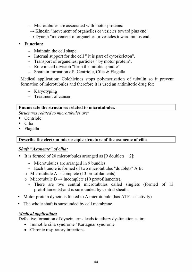
- Microtubules are associated with motor proteins: → Kinesin "movement of organelles or vesicles toward plus end. → Dynein "movement of organelles or vesicles toward minus end. Function:
- Maintain the cell shape. - Internal support for the cell " it is part of cytoskeleton". - Transport of organelles, particles " by motor protein". - Role in cell division "form the mitotic spindle". - Share in formation of: Centriole, Cilia & Flagella.
UMedical application U: Colchicines stops polymerization of tubulin so it prevent formation of microtubules and therefore it is used an antimitotic drug for:
- Karyotyping - Treatment of cancer
Enumerate the structures related to microtubules. Structures related to microtubules are: Centriole Cilia Flagella Describe the electron microscopic structure of the axoneme of cilia U
Shaft "Axoneme" of cilia: It is formed of 20 microtubules arranged as [9 doublets + 2]:
- Microtubules are arranged in 9 bundles. - Each bundle is formed of two microtubules "doublets" A,B:
o Microtubule A is complete (13 protofilaments). o Microtubule B → incomplete (10 protofilaments).
- There are two central microtubules called singlets (formed of 13 protofilaments) and is surrounded by central sheath.
Motor protein dynein is linked to A microtubule (has ATPase activity) The whole shaft is surrounded by cell membrane.
U
Medical application: Defective formation of dynein arms leads to ciliary dysfunction as in:
• Immotile cilia syndrome "Kartagnar syndrome" • Chronic respiratory infections
54
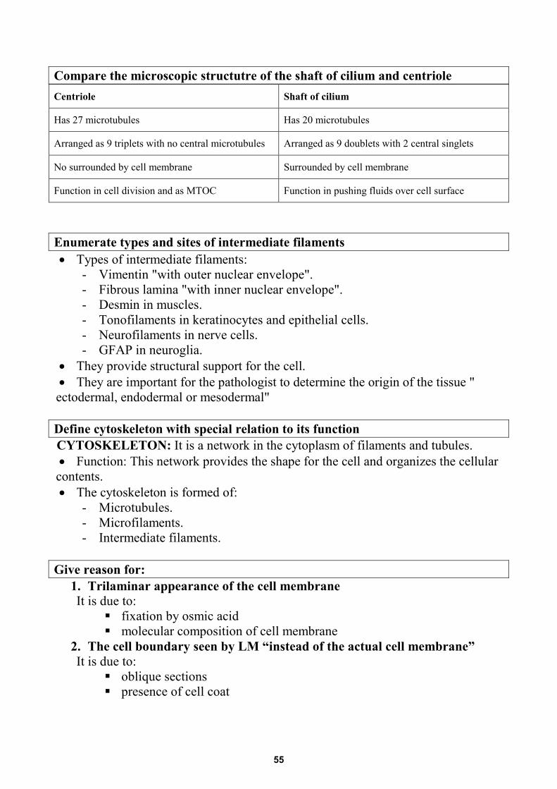
Compare the microscopic structutre of the shaft of cilium and centriole Centriole Shaft of cilium
Has 27 microtubules Has 20 microtubules
Arranged as 9 triplets with no central microtubules Arranged as 9 doublets with 2 central singlets
No surrounded by cell membrane Surrounded by cell membrane
Function in cell division and as MTOC Function in pushing fluids over cell surface
Enumerate types and sites of intermediate filaments • Types of intermediate filaments:
- Vimentin "with outer nuclear envelope". - Fibrous lamina "with inner nuclear envelope". - Desmin in muscles. - Tonofilaments in keratinocytes and epithelial cells. - Neurofilaments in nerve cells. - GFAP in neuroglia.
• They provide structural support for the cell. • They are important for the pathologist to determine the origin of the tissue "
ectodermal, endodermal or mesodermal" Define cytoskeleton with special relation to its function CYTOSKELETON: It is a network in the cytoplasm of filaments and tubules. • Function: This network provides the shape for the cell and organizes the cellular
contents. • The cytoskeleton is formed of:
- Microtubules. - Microfilaments. - Intermediate filaments.
Give reason for:
1. Trilaminar appearance of the cell membrane It is due to:
fixation by osmic acid molecular composition of cell membrane
2. The cell boundary seen by LM “instead of the actual cell membrane” It is due to:
oblique sections presence of cell coat
55
3. Lysosomal enzymes do not injury the cytoplasm of the cells The cytoplasm is protected from the effect of lysosomal enzymes as:
the lysosomal enzymes are secreted first inactive the pH of the cytoplasm is not suitable for the action of them
4. Ribosomes are not attached to the membrane of SER: this is due to absence
of ribosomal receptor "ribophorins" from the membrane of SER
5. The outer mitochondrial membrane is permeable: this is due to presence of porins transmembrane protein in it
6. The inner membrane of mitochondria is concerned with production of
ATP: this is due to presence of these transmembrane proteins: o oxidative enzymes o lolly pop structure "drum stick structure" which has ATP synthase activity
7. Colchicine is used as an antimitotic drug as it prevents polymerization of
tubulin into microtubules so, o It prevents formation of the mitotic spindle o It stops mitosis at metaphase
8. The cells of our body have different shapes: this is due to presence of
cytoskeletal components which have different distribution in each cell to maintain its specific shape
56
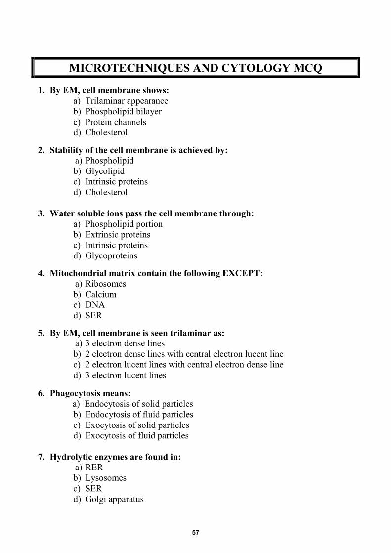
MICROTECHNIQUES AND CYTOLOGY MCQ
1. By EM, cell membrane shows:a) Trilaminar appearanceb) Phospholipid bilayerc) Protein channelsd) Cholesterol
2. Stability of the cell membrane is achieved by:a) Phospholipidb) Glycolipidc) Intrinsic proteinsd) Cholesterol
3. Water soluble ions pass the cell membrane through:a) Phospholipid portionb) Extrinsic proteinsc) Intrinsic proteinsd) Glycoproteins
4. Mitochondrial matrix contain the following EXCEPT:a) Ribosomesb) Calciumc) DNAd) SER
5. By EM, cell membrane is seen trilaminar as:a) 3 electron dense linesb) 2 electron dense lines with central electron lucent linec) 2 electron lucent lines with central electron dense lined) 3 electron lucent lines
6. Phagocytosis means:a) Endocytosis of solid particlesb) Endocytosis of fluid particlesc) Exocytosis of solid particlesd) Exocytosis of fluid particles
7. Hydrolytic enzymes are found in:a) RERb) Lysosomesc) SERd) Golgi apparatus
57
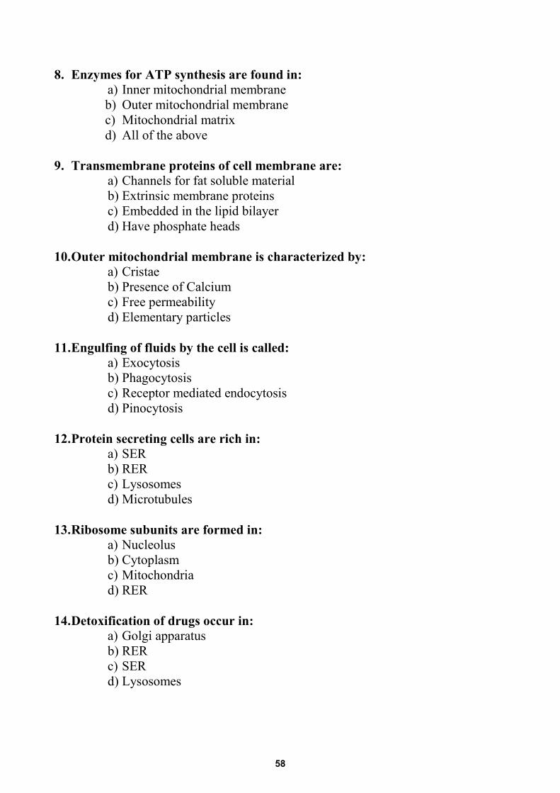
8. Enzymes for ATP synthesis are found in: a) Inner mitochondrial membrane b) Outer mitochondrial membrane c) Mitochondrial matrix d) All of the above
9. Transmembrane proteins of cell membrane are:
a) Channels for fat soluble material b) Extrinsic membrane proteins c) Embedded in the lipid bilayer d) Have phosphate heads
10. Outer mitochondrial membrane is characterized by:
a) Cristae b) Presence of Calcium c) Free permeability d) Elementary particles
11. Engulfing of fluids by the cell is called:
a) Exocytosis b) Phagocytosis c) Receptor mediated endocytosis d) Pinocytosis
12. Protein secreting cells are rich in:
a) SER b) RER c) Lysosomes d) Microtubules
13. Ribosome subunits are formed in:
a) Nucleolus b) Cytoplasm c) Mitochondria d) RER
14. Detoxification of drugs occur in:
a) Golgi apparatus b) RER c) SER d) Lysosomes
58
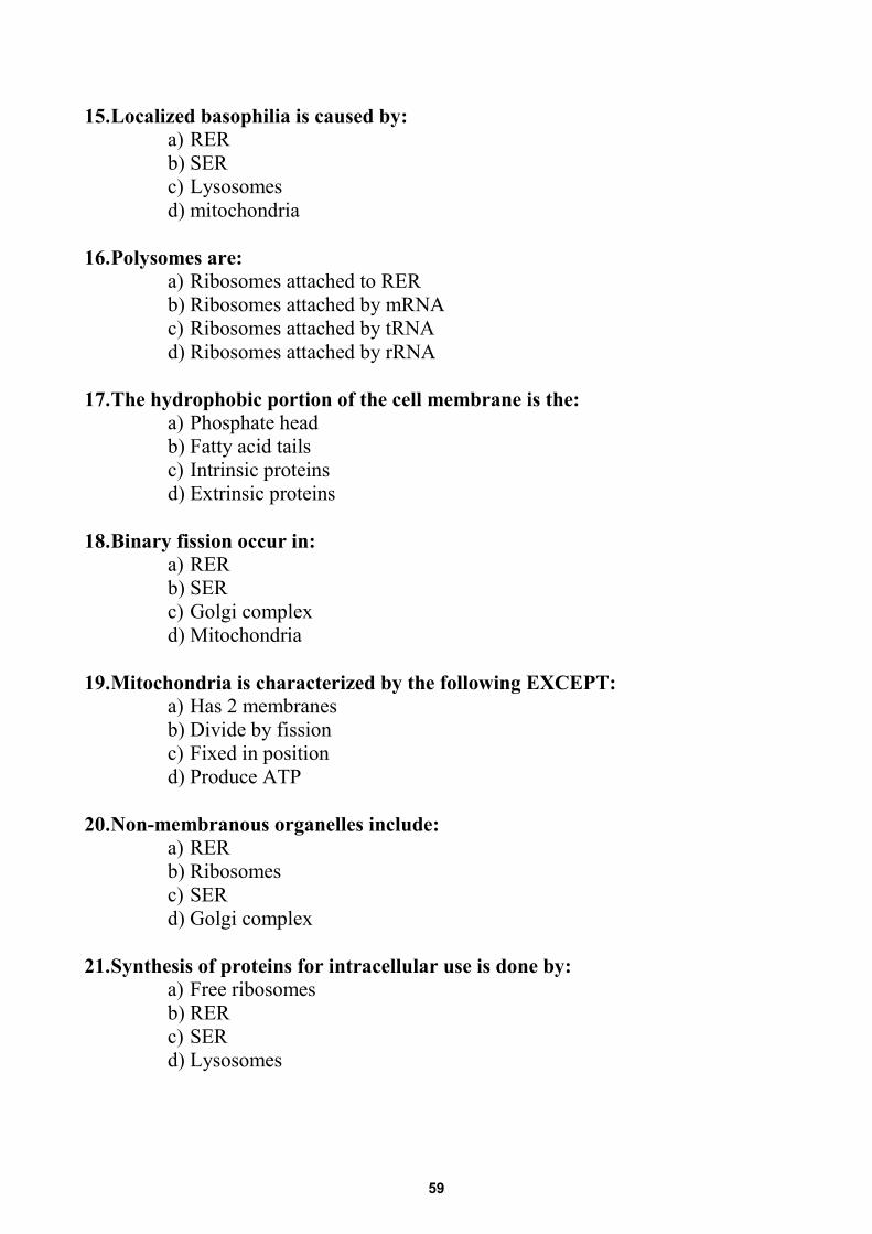
15. Localized basophilia is caused by: a) RER b) SER c) Lysosomes d) mitochondria
16. Polysomes are:
a) Ribosomes attached to RER b) Ribosomes attached by mRNA c) Ribosomes attached by tRNA d) Ribosomes attached by rRNA
17. The hydrophobic portion of the cell membrane is the:
a) Phosphate head b) Fatty acid tails c) Intrinsic proteins d) Extrinsic proteins
18. Binary fission occur in:
a) RER b) SER c) Golgi complex d) Mitochondria
19. Mitochondria is characterized by the following EXCEPT:
a) Has 2 membranes b) Divide by fission c) Fixed in position d) Produce ATP
20. Non-membranous organelles include:
a) RER b) Ribosomes c) SER d) Golgi complex
21. Synthesis of proteins for intracellular use is done by:
a) Free ribosomes b) RER c) SER d) Lysosomes
59

22. Ribosomes are: a) Membranous organelles b) Synthesized in nucleolus c) Acidophilic d) Undergoing binary fission
23. By EM, rough endoplasmic reticulum appear as:
a) Branching tubules b) Parallel cisternae c) Cisternae with bulbous ends d) 3 subunits
24. RER are characterized by having:
a) Attached lysosomes b) Continuation with the perinuclear space c) Bulbous ends d) All of the above
25. Lipid synthesis occur in the:
a) SER b) RER c) Free ribosomes d) Mitochondria
26. Smooth endoplasmic reticulum are:
a) Basophilic b) Parallel cisterna c) Continuous with the lysosomes d) Continuous with RER
27. Cis face of Golgi apparatus are:
a) Related to transfer vesicles b) The mature face c) Facing RER d) Facing SER
28. Transfer vesicles bud from:
a) SER b) RER c) Golgi complex d) All of the above
60
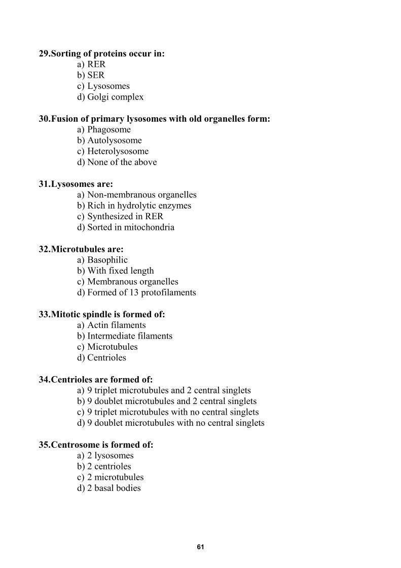
29. Sorting of proteins occur in: a) RER b) SER c) Lysosomes d) Golgi complex
30. Fusion of primary lysosomes with old organelles form:
a) Phagosome b) Autolysosome c) Heterolysosome d) None of the above
31. Lysosomes are:
a) Non-membranous organelles b) Rich in hydrolytic enzymes c) Synthesized in RER d) Sorted in mitochondria
32. Microtubules are:
a) Basophilic b) With fixed length c) Membranous organelles d) Formed of 13 protofilaments
33. Mitotic spindle is formed of:
a) Actin filaments b) Intermediate filaments c) Microtubules d) Centrioles
34. Centrioles are formed of:
a) 9 triplet microtubules and 2 central singlets b) 9 doublet microtubules and 2 central singlets c) 9 triplet microtubules with no central singlets d) 9 doublet microtubules with no central singlets
35. Centrosome is formed of:
a) 2 lysosomes b) 2 centrioles c) 2 microtubules d) 2 basal bodies
61
36. The basal body of cilia is formed of: a) 9 triplet microtubules and 2 central singlets b) 9 doublet microtubules and 2 central singlets c) 9 triplet microtubules with no central singlets d) 9 doublet microtubules with no central singlets
37. Cilia shaft is formed of: a) 9 triplet microtubules and 2 central singlets b) 9 doublet microtubules and 2 central singlets c) 9 triplet microtubules with no central singlets d) 9 doublet microtubules with no central singlets
38. Cytoskeleton is formed of the following EXCEPT: a) Microtubules b) Actin filaments c) Intermediate filaments d) Myosin filaments
39. Hemoglobin is:
a) Exogenous pigment b) Endogenous pigment c) An organelle d) A crystal
40. Vinblastine is a chemotherapeutic drug. It stops mitosis in cancer cells by inhibiting the formation of:
a) Actin filaments. b) Protofilaments. c) Intermediate filaments. d) Microtubules
41. When cells are infected by a virus, abnormal proteins are formed. These proteins are degraded by:
a) Lysosomes b) Peroxisomes c) Proteasomes d) Endosomes
42. A 3-day old baby was presented to the clinic with yellowish discoloration of his
eyes from the second day after delivery. The defective organelle responsible for such condition is the:
a) RER b) SER c) Lysosome d) Golgi apparatus
62
43. Which of the following organelles can help to identify the cellular origin of the tumor:
a) Microfilaments b) Thick filaments c) Microtubules d) Intermediate filaments
44. An infertile male patient complains of repeated chronic respiratory tract
infections with inability to cough mucous. The defective structure is: a) Kinesin b) Dynein c) Tubulin d) Nexin
45. The most stable component of the of cytoskeleton is:
a) Microtubules b) Microfilaments c) Intermediate filaments d) Thick filaments
46. The microscope which can provide three dimensional image is:
a) Light microscope b) Transmission electron microscope c) Scanning electron microscope d) Fluorescent microscope
47. 4- Which of the following is INCORRECT about the smooth endoplasmic
reticulum? a) Involved in detoxification of drugs. b) It is abundant in steroid producing cells. c) It forms the sarcoplasmic reticulum in skeletal muscle fibers. d) It is abundant in cells producing protein.
48. The stain of choice to demonstrate glycogen in the cell is:
a) Eosin. b) Osmic acid. c) Hematoxylin. d) Periodic acid Schiff (PAS).
49. Lipofuscin is associated with which of the following?
a) Golgi apparatus. b) Residual bodies. c) Primary lysosomes. d) Peroxisomes.
63
50. All of the following are considered intermediate filaments EXCEPT: a) Vimentin. b) Desmin. c) Actin. d) Neurofilament.
51. All of the following regarding centrioles are correct EXCEPT:
a) Duplicate prior to mitosis. b) Located in centrosome. c) Comprised of microfilaments. d) Function in the formation of cilia.
52. All of the following regarding mitochondria are correct EXCEPT:
a) Possess matrix granules. b) Divide to produce new mitochondria. c) Outer membrane folded to form cristae. d) Contain DNA.
53. Primary lysosomes:
a) Are also termed residual bodies. b) Are also termed phagosomes. c) Possess a low pH environment. d) Are not membrane bound.
54. All of the following regarding the Golgi apparatus are correct EXCEPT:
a) Well developed in cells secreting protein by exocytosis. b) Consists, in part, of flattened curved cisternae. c) Stains intensely with hematoxytlin. d) Function in lysosome formation.
55. Which one of the following is endogenous pigment? a) Tattooing. b) Lipofuscin. c) Dust particles d) Melatonin.
56. A 23-year-old woman complains of bone pain and enlarged abdomen. The
patient is diagnosed to have mutations in the genes of one hydrolytic enzyme. Undigested materials accumulate within which of the following cellular organelle:
a) Proteasome. b) Endoplasmic reticulum. c) Lysosome. d) Peroxisome.
64
57. Mitochondria are present in apical “upper” part of:a) Muscle cells.b) Fat cells.c) Ciliated cells.d) Nerve cells.
58. Dynamic cytoskeletal components include:a) Microfilaments and microtubules.b) Myosin filaments and intermediate filaments.c) Intermediate filaments and microtubules.d) Myosin filaments and microfilaments.
59. Hematoxylin stains:a) Lysosomes.b) RER.c) SER.d) Golgi apparatus.
60. Lipid content of the cell membrane:a) Allows diffusion of hydrophilic substances.b) Is firmly bound to all membrane proteins.c) Is arranged into bilayer.d) Is formed of phospholipids and fatty acids.
61. In Alzheimer’s disease, accumulation of abnormal proteins in brain tissues isdue to defective:
a) Delivery of acid hydrolases to lysosomes.b) Fusion of secretory vesicles with the plasmalemma.c) Generation of hydrogen peroxide inside peroxisomes.d) Activation of the ubiquitin–proteasome pathway.
62. The transport of neurotransmitter to the axon terminal at site of synapse ismediated by:
a) Ubiquitin.b) Kinesin.c) Myosin.d) Actin
63. A 3-day old baby was presented to the clinic with yellowish discoloration of hiseyes from the second day after delivery. The defective organelle responsible forsuch condition is the:
a) RER.b) SER.c) Lysosome.d) Golgi apparatus.
65
64. Infertile 45 year-old male complaining of repeated chest infections since childhood. The defective structure in this case is:
a) Axonemal dynein. b) Protofilament tubulin. c) Filamentous actin. d) Motor kinesin.
65. A membrane bound vesicle best describes:
a) RER. b) Mitochondria. c) Lysosome. d) Golgi complex.
66. Diffuse cytoplasmic basophilia indicates the presence of:
a) RER. b) Free ribosomes. c) Golgi apparatus. d) Heterolysosomes.
67. Tubulin protein of the microtubules is synthesized by:
a) Free ribosomes. b) RER. c) Microtubules organizing center “MTOC”. d) Proteasomes.
68. Lysosomes are formed by Budding from the:
a) RER. b) Golgi complex. c) SER d) Cell membrane.
69. Transfer vesicle arise from:
a) Cis face of Golgi saccules. b) Trans face of Golgi saccules. c) SER d) RER
70. Concerning the glycocalyx, it:
a) Is firmly embedded in the lipid bilayer. b) Extends across the whole thickness of the cell membrane. c) Has fuzzy appearance by EM. d) Is present on outer and inner surfaces of cell membrane.
66
71. Branching and anastomosing membranous tubules best describes: a) Lysosomes. b) Mitochondria. c) SER. d) Golgi complex.
72. Basophilic structures in the cell:
a) Are basic in nature. b) Have affinity for staining by eosin. c) Are PAS positive. d) Appear bluish purple by LM
73. Proteins of the secretory vesicles are synthesized by:
a) Free ribosomes. b) RER. c) Golgi apparatus. d) Proteasomes.
74. A 75 year old male presented with loss of recent memory. He was diagnosed as
Alzheimer disease and the doctor told him that this is due to accumulation of abnormal malfolded proteins. These proteins should be degraded by
a) Lysosomes. b) Endosomes. c) Peroxisomes d) Proteasomes.
75. Heterolysosome arise from:
a) Trans face of Golgi complex. b) Early endosomes. c) Fusion with phagosome. d) Fusion with clathrin coated vesicles.
76. PAS stains which of the followings:
a) Cell coat b) Mitochondria c) Lysosomes d) RER
77. Dynamic cytoskeleton includes
a) Intermediate filaments b) Myosin filaments c) Microtubules d) Integral membrane proteins
67
78. Enzymes associated with drug detoxification are located in: a) SER. b) RER. c) Golgi apparatus. d) Inner mitochondrial membrane.
79. Disturbed growth in dwarfism could be attributed to defective:
a) Phospholipids. b) Receptors. c) Endocytosis. d) Exocytosis.
80. A self-replicating organelle is:
a) RER. b) Mitochondria. c) Lysosome. d) Golgi complex.
81. Accumulated residual bodies filled with indigestible molecules is due to defect
in: a) Lysosomal enzymes b) Peroxisomal enzymes c) Endosomal enzymes d) Proteasomal enzymes
82. Lysosomes are:
a) Cytoplasmic inclusions b) Membrane-bound structures c) Produced by smooth endoplasmic reticulum d) Produced by free ribosomes
83. Secretory granules release its contents at the cell surface by:
a) Diffusion b) Exocytosis. c) Phagocytosis. d) Pinocytosis.
84. Extrinsic or peripheral membrane proteins:
a) Are firmly embedded in the lipid bilayer. b) Extend across the whole thickness of the cell membrane. c) Contain transmembrane channels. d) Loosely bound to outer and inner surfaces of cell membrane.
68
85. By histochemical techniques, detection of catalase enzyme indicates the presence of:
a) Mitochondria b) Lysosomes c) Peroxisomes d) Ribosomes
86. Primary function of intermediate filaments is to:
a) Generate movements. b) Provide mechanical stability. c) Transport organelles within the cell. d) Stabilize microtubules against disassembly.
87. One of the following regarding saccules of Golgi apparatus is true:
a) Their trans face fuses with transfer vesicle b) They are branching and anastomosing tubules c) They stain intensely with hematoxytlin. d) They modify the segregated proteins
88. The outer mitochondrial membrane is characterized by the presence of:
a) Porins b) Respiratory chain enzymes c) Elementary particle d) Cristae
89. Regarding ribosomes, they:
a) Are formed of t-RNA and proteins b) Are synthesized in the nucleolus c) Impart intense acidophilia on the cytoplasm d) Attach to ribophorins on the membrane of SER
90. Cell membrane carbohydrates are :
a) Embedded in the lipid bilayer. b) Important for cell adhesion. c) Trans membrane channels. d) Attached to the cytoskeleton.
91. Which of the following is a part of the cytoskeleton?
a) Thick filament. b) Ribosome. c) Microtubule. d) Lysosome.
69
92. Degradation of old worn out organelles is mediated by :a) Lysosomes.b) Peroxisomes.c) Proteasomes.d) Endosomes.
93. Attached ribosomes synthesize proteins of:a) Microtubules.b) Proteasomes.c) Microfilaments.d) Lysosomes.
94. The cell organelles that contain DNA are:a) Lysosomesb) RERc) Mitochondriad) Golgi complex
95. One of the following is a function of microtubules:a) They form the contractile ring during cell divisionb) They facilitates intracellular transport of substancesc) They forms the main structural components of muscle tissued) They form the core of microvilli
96. One of the following is the main function of RER:a) Steroid hormone synthesisb) Drug detoxificationc) Regulation of muscle contractiond) Protein synthesis
97. The basal body of cilium is:a) Surrounded by cell membraneb) Formed of nine triplets of microtubulesc) Has dynein arms connecting the tubulesd) Has two central singlets
98. Concerning peroxisomes:a) They are formed by Golgi apparatusb) They are increased by simple binary fissionc) They synthesize proteinsd) They are involved in phagocytosis
70
99. Which of the following is exogenous pigment: a) Melanin pigment b) Haemoglobin c) Lipochrome pigment d) Lipofuscin pigment
100. The organelles concerned with detoxification of alcohol are:
a) Proteasomes. b) Peroxisomes. c) Lysosomes. d) Ribosomes.
101. Which of the following organelles contain calcium ions?
a) Mitochondria and Golgi complex. b) Mitochondria and sER. c) RER and sER. d) RER and Golgi complex.
102. Which of the following organelles can help identify the cellular origin of the
tumor? a) Microfilament. b) Thick filament. c) Intermediate filaments. d) Protofilaments.
103. In long lived cells, which of the following increase with age?
a) Fat. b) Glycogen. c) Lipofuscin pigment. d) Hemosiderin granules.
104. Phagocytosis is a process of:
a) Engulfing solid particles. b) Engulfing small amounts of fluid. c) Passive diffusion of water. d) Active transport of Na+ and K+.
105. By histochemical techniques, detection of catalase enzyme indicates the
presence of: a) Mitochondria b) Lysosomes c) Peroxisomes d) Ribosomes
71
106. The most suitable method for studying living cells is: a) Tissue impregnation b) Tissue culture c) Tissue preservation d) Tissue smear
107. Free ribosomes are responsible for synthesis of :
a) Mucous secretion. b) Serous secretion. c) Lipid droplet. d) Filaments of cytoskeleton.
108. Which of the following divide by fission:
a) Centrioles. b) Smooth endoplasmic reticulum. c) Golgi apparatus. d) Peroxisomes
109. Lipofuscin pigment is seen in the cytoplasm of cells that are likely taken
from: a) Brain b) Blood c) Skin d) Lung
110. Which of the following proteins forms the core of cilia found along the
apical membrane domain of the columnar epithelial cells? a) Myosin b) Keratin c) Tubulin d) Actin
111. Concerning mitochondria: a) They are the only double walled organelle inside the cell b) Respiratory chain enzymes are transmembrane proteins along the inner
membrane c) The outer membrane is folded and permeable d) The matrix stores ATP molecules
112. Attached ribosomes synthesize: a) Glycolipids b) Tubulin c) Acid hydrolases d) Triglycerides
72
113. A-56-year old farmer presented with an ulcerated tumor in his right cheek. On examination, the draining lymph nodes were enlarged. To determine the origin of his tumor a biopsy was taken and stained immune-histochemically for demonstration of:
a) Myofilaments b) Actin filaments c) Intermediate filaments d) Thick filaments
114. A 2-year-old female child had been presented with heart failure of three
months duration. Echocardiography showed left ventricular hypertrophy. The assay for alpha-glucosidase was consistent with the diagnosis of Pompe's disease which is explained by the defective structure of the:
a) Lysosomes b) Proteasomes c) Peroxisomes d) Nucleosomes
115. A 55-year-old male presented with obesity and on physical examination, mild hepatomegaly was noticed. Abdominal ultrasound revealed confirmed features of non-alcoholic fatty liver disease (NAFLD). A liver biopsy was taken and specifically stained by:
a) Masson trichrome b) Periodic acid Schiff c) Orcein d) Sudan III
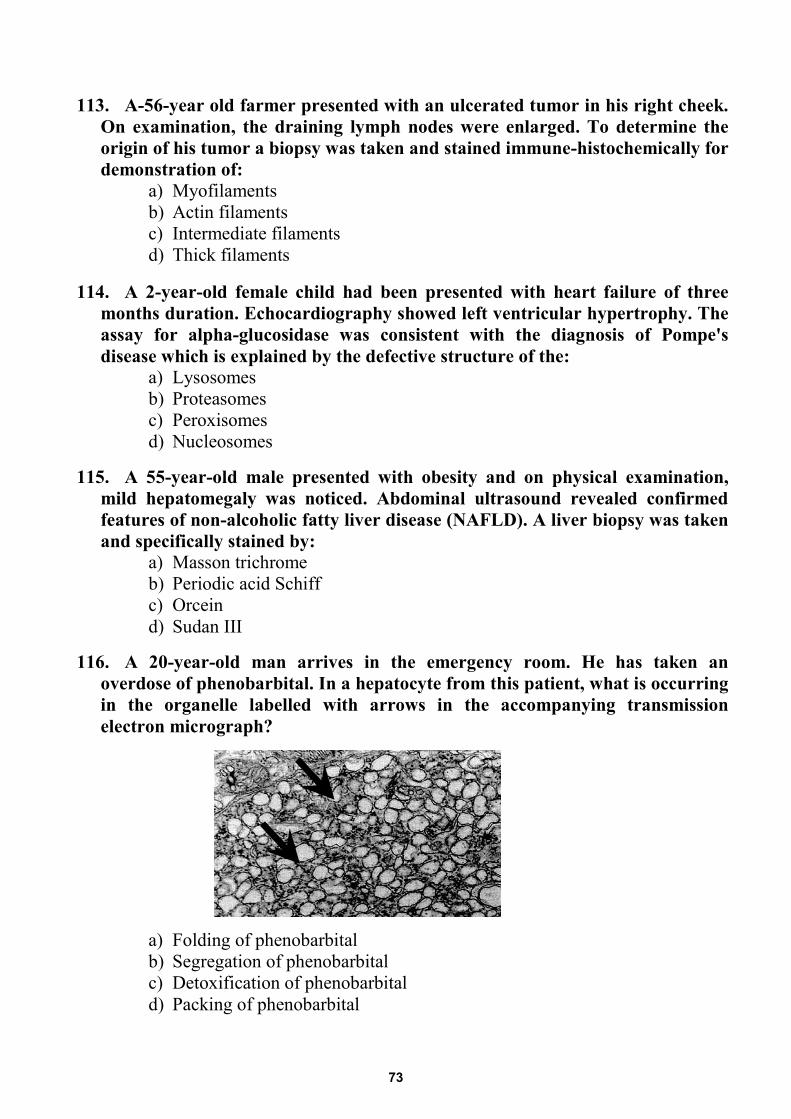
116. A 20-year-old man arrives in the emergency room. He has taken an
overdose of phenobarbital. In a hepatocyte from this patient, what is occurring in the organelle labelled with arrows in the accompanying transmission electron micrograph?
a) Folding of phenobarbital b) Segregation of phenobarbital c) Detoxification of phenobarbital d) Packing of phenobarbital
73
117. A boy is born with multiple congenital malformations as ventricular septal defect, glaucoma, cataracts and hepatomegaly. A liver biopsy is prepared for electron microscopy. It shows the presence of empty peroxisomes that were described as peroxisome “ghosts”. Which one of the following cellular activities would be decreased in the hepatocytes from this patient?
a) Production of plasma proteins b) Synthesis of glycogen c) Beta oxidation of fatty acids d) Conjugation of bilirubin
118. A 65-year-old man presents to the neurology clinic with a several year history in which he has less energy, memory loss and mood swings. This disease is believed to be caused by defective protein folding which is carried out inside the cell by:
a) Laminin b) Ubiquitin c) Chaperonin d) Fibronectin
119. A 6-month-old infant is brought to the neurology clinic with developmental delay, and repeated attacks of vomiting. Examination reveals decreased muscle tone and deafness. Muscle biopsy shows normal histology, but laboratory tests reveal a deficiency in mitochondrial cytochrome c oxidase. In electron micrograph, which of the following best describe that organelle?
a) It has flattened interconnecting membranous cisternae b) Its outer membrane fuses with the outer nuclear membrane c) Its inner membranes has numerous shelf like projections d) It consists of curved saccules and membranous vesicles
120. A 72-year-old woman is presented to the physician with mood swings and uncharacteristic moments of anger and aggressiveness. She also "gets lost" on travel to routine destinations. The patient was diagnosed as Alzheimer s disease which is attributed to defective function of the:
a) Peroxisomes b) Proteasomes c) Lysosomes d) Nucleosomes
121. An infertile 45 year-old male with Kartagnar “Immotile cilia” syndrome came to the clinic complaining of repeated chest infections since childhood. The defective structure in this case is:
a) Axonemal dynein. b) Protofilament tubulin. c) Filamentous actin. d) Motor kinesin.
74
122. A 73-year-old man complaining of abdominal pain was asked to do CT scan. A suspicious lesion was seen in the right lobe of the liver. A biopsy taken one hour after a carbohydrate revealed strongly PAS positive cells due to high accumulation of:
a) Phospholipidsb) Carbohydratesc) Proteinsd) Triglycerides
123. A 22-year-old woman presented to the ophthalmology clinic with inability to drive at night because. After investigations, there was rod degeneration with pigment deposits in the peripheral retina. This was explained by failure of rod pigment and other protein vesicle transport along the:
a) Microfilamentsb) Thick filamentsc) Microtubulesd) Intermediate filaments
75
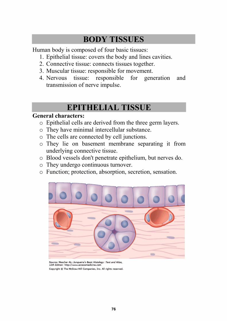
BODY TISSUES Human body is composed of four basic tissues:
1. Epithelial tissue: covers the body and lines cavities.2. Connective tissue: connects tissues together.3. Muscular tissue: responsible for movement.4. Nervous tissue: responsible for generation and
transmission of nerve impulse.
EPITHELIAL TISSUE General characters: o Epithelial cells are derived from the three germ layers.o They have minimal intercellular substance.o The cells are connected by cell junctions.o They lie on basement membrane separating it from
underlying connective tissue.o Blood vessels don't penetrate epithelium, but nerves do.o They undergo continuous turnover.o Function; protection, absorption, secretion, sensation.
76
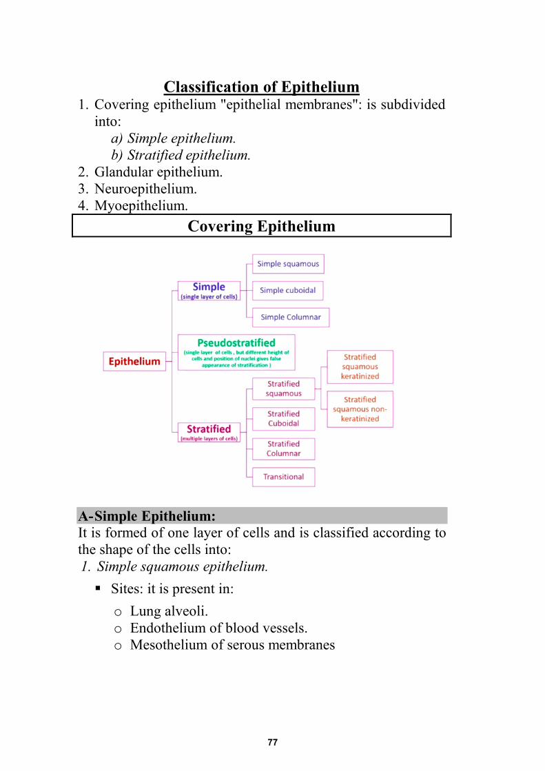
Classification of Epithelium 1. Covering epithelium "epithelial membranes": is subdivided
into: a) Simple epithelium. b) Stratified epithelium.
2. Glandular epithelium. 3. Neuroepithelium. 4. Myoepithelium.
Covering Epithelium
A- Simple Epithelium: It is formed of one layer of cells and is classified according to the shape of the cells into: 1. Simple squamous epithelium. Sites: it is present in:
o Lung alveoli. o Endothelium of blood vessels. o Mesothelium of serous membranes
77
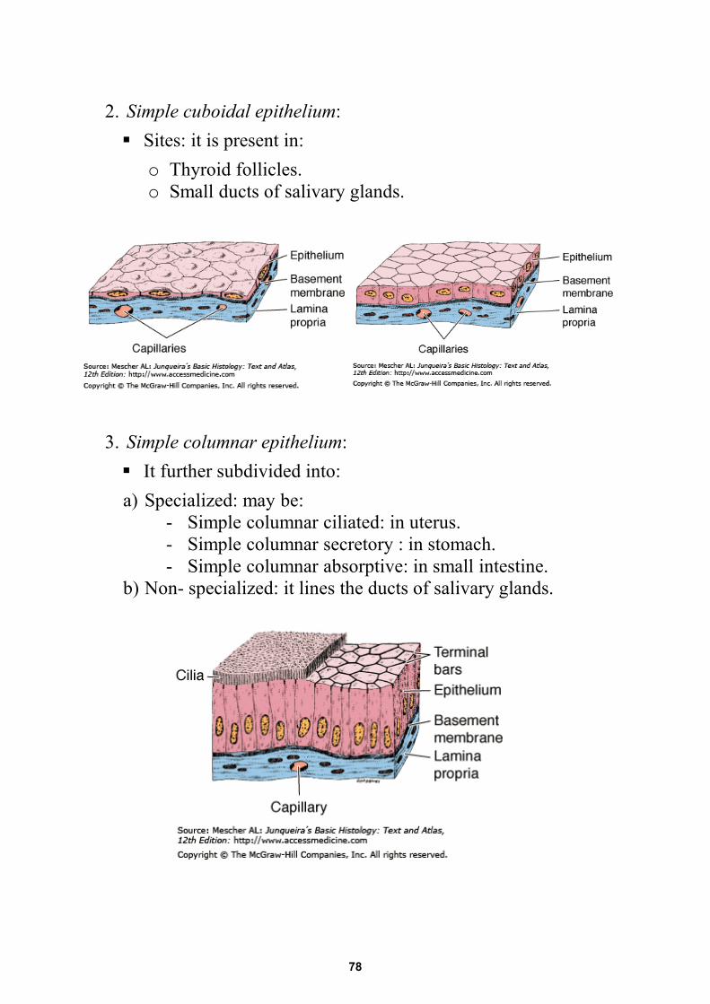
2. Simple cuboidal epithelium: Sites: it is present in:
o Thyroid follicles. o Small ducts of salivary glands.
3. Simple columnar epithelium: It further subdivided into: a) Specialized: may be:
- Simple columnar ciliated: in uterus. - Simple columnar secretory : in stomach. - Simple columnar absorptive: in small intestine.
b) Non- specialized: it lines the ducts of salivary glands.
78
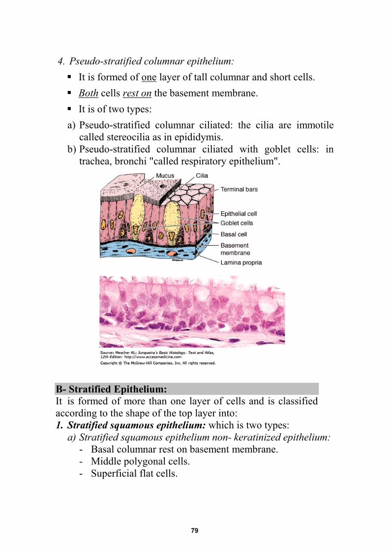
4. Pseudo-stratified columnar epithelium: It is formed of one layer of tall columnar and short cells. Both cells rest on the basement membrane. It is of two types: a) Pseudo-stratified columnar ciliated: the cilia are immotile
called stereocilia as in epididymis. b) Pseudo-stratified columnar ciliated with goblet cells: in
trachea, bronchi "called respiratory epithelium".
B- Stratified Epithelium: It is formed of more than one layer of cells and is classified according to the shape of the top layer into: 1. Stratified squamous epithelium: which is two types:
a) Stratified squamous epithelium non- keratinized epithelium: - Basal columnar rest on basement membrane. - Middle polygonal cells. - Superficial flat cells.
79
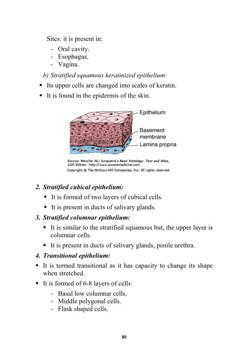
Sites: it is present in: - Oral cavity. - Esophagus. - Vagina.
b) Stratified squamous keratinized epithelium: Its upper cells are changed into scales of keratin. It is found in the epidermis of the skin.
2. Stratified cubical epithelium: It is formed of two layers of cubical cells. It is present in ducts of salivary glands.
3. Stratified columnar epithelium: It is similar to the stratified squamous but, the upper layer is
columnar cells. It is present in ducts of salivary glands, penile urethra.
4. Transitional epithelium: It is termed transitional as it has capacity to change its shape
when stretched. It is formed of 6-8 layers of cells:
- Basal low columnar cells. - Middle polygonal cells. - Flask shaped cells.
80
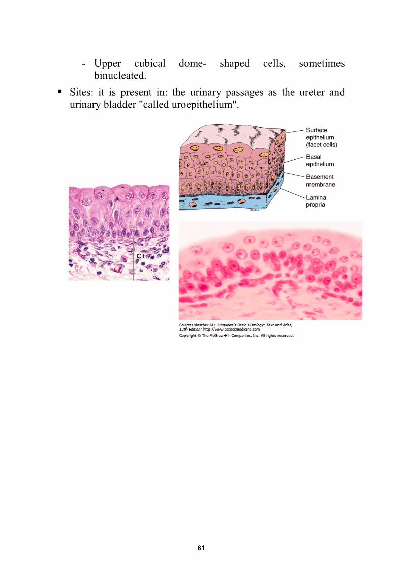
- Upper cubical dome- shaped cells, sometimes binucleated.
Sites: it is present in: the urinary passages as the ureter and urinary bladder "called uroepithelium".
81
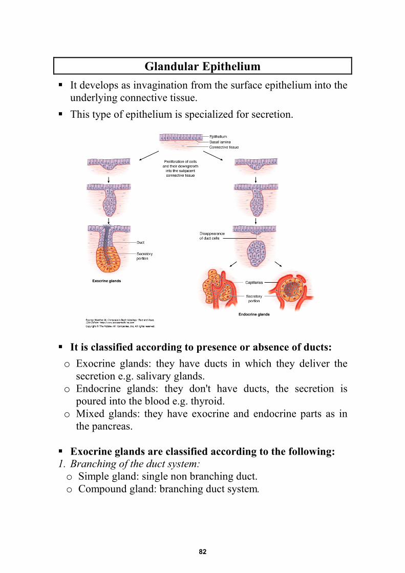
Glandular Epithelium It develops as invagination from the surface epithelium into the
underlying connective tissue. This type of epithelium is specialized for secretion. It is classified according to presence or absence of ducts: o Exocrine glands: they have ducts in which they deliver the
secretion e.g. salivary glands. o Endocrine glands: they don't have ducts, the secretion is
poured into the blood e.g. thyroid. o Mixed glands: they have exocrine and endocrine parts as in
the pancreas.
Exocrine glands are classified according to the following: 1. Branching of the duct system: o Simple gland: single non branching duct. o Compound gland: branching duct system.
82
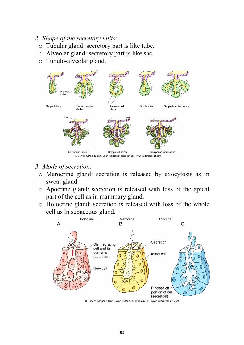
2. Shape of the secretory units: o Tubular gland: secretory part is like tube. o Alveolar gland: secretory part is like sac. o Tubulo-alveolar gland.
3. Mode of secretion: o Merocrine gland: secretion is released by exocytosis as in
sweat gland. o Apocrine gland: secretion is released with loss of the apical
part of the cell as in mammary gland. o Holocrine gland: secretion is released with loss of the whole
cell as in sebaceous gland.
Holocrine Merocrine Apocrine
83
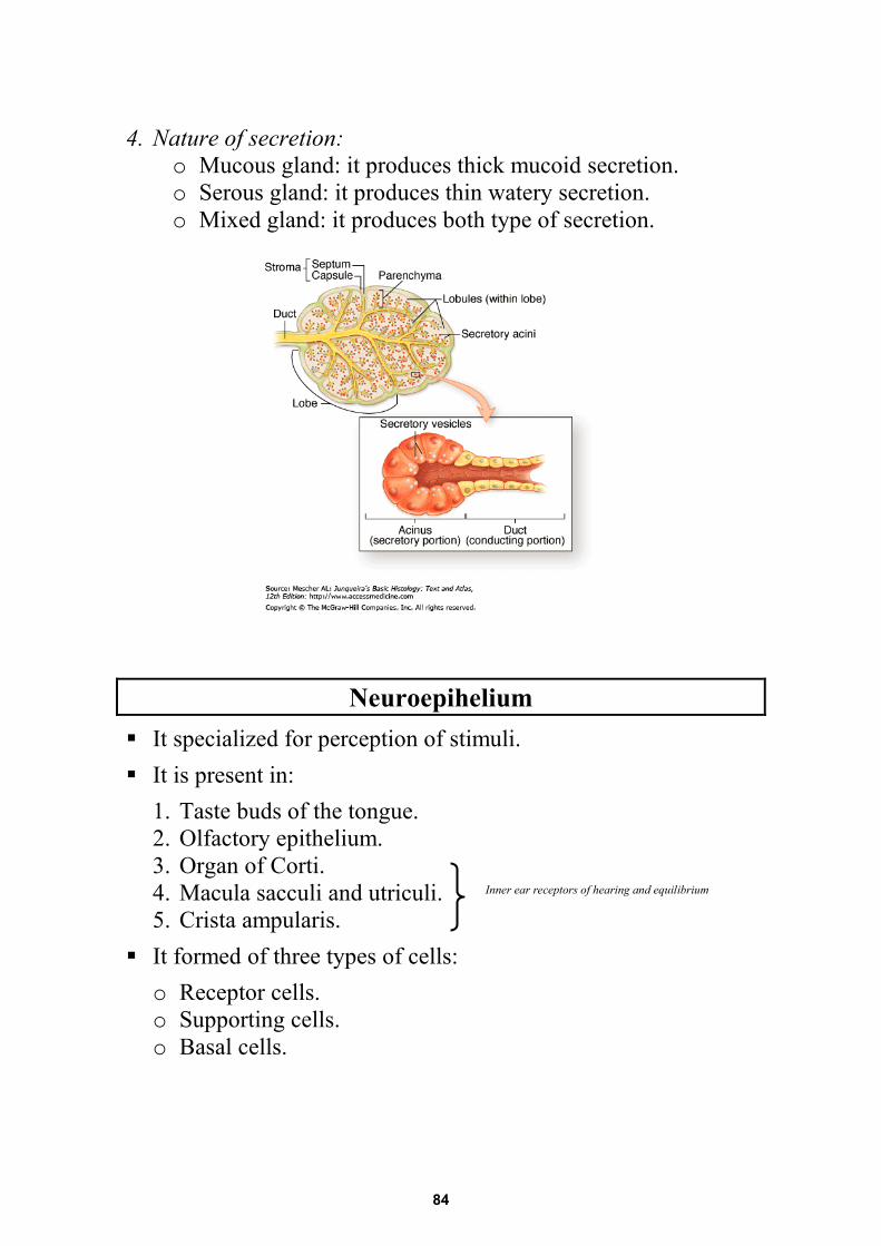
4. Nature of secretion: o Mucous gland: it produces thick mucoid secretion. o Serous gland: it produces thin watery secretion. o Mixed gland: it produces both type of secretion.
Neuroepihelium
It specialized for perception of stimuli. It is present in:
1. Taste buds of the tongue. 2. Olfactory epithelium. 3. Organ of Corti. 4. Macula sacculi and utriculi. 5. Crista ampularis.
It formed of three types of cells: o Receptor cells. o Supporting cells. o Basal cells.
Inner ear receptors of hearing and equilibrium
84
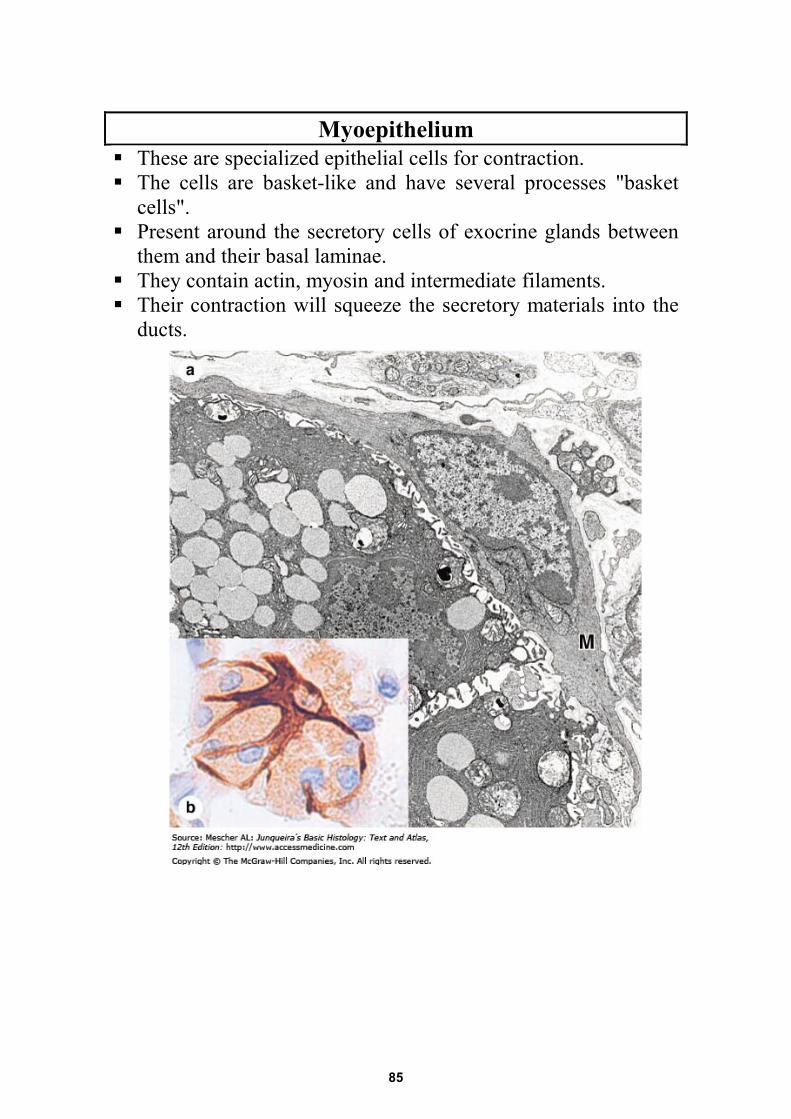
Myoepithelium These are specialized epithelial cells for contraction. The cells are basket-like and have several processes "basket
cells". Present around the secretory cells of exocrine glands between
them and their basal laminae. They contain actin, myosin and intermediate filaments. Their contraction will squeeze the secretory materials into the
ducts.
85
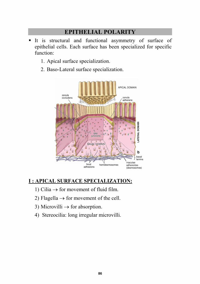
EPITHELIAL POLARITY It is structural and functional asymmetry of surface of
epithelial cells. Each surface has been specialized for specific function:
1. Apical surface specialization. 2. Baso-Lateral surface specialization.
I : APICAL SURFACE SPECIALIZATION: 1) Cilia → for movement of fluid film. 2) Flagella → for movement of the cell. 3) Microvilli → for absorption. 4) Stereocilia: long irregular microvilli.
86
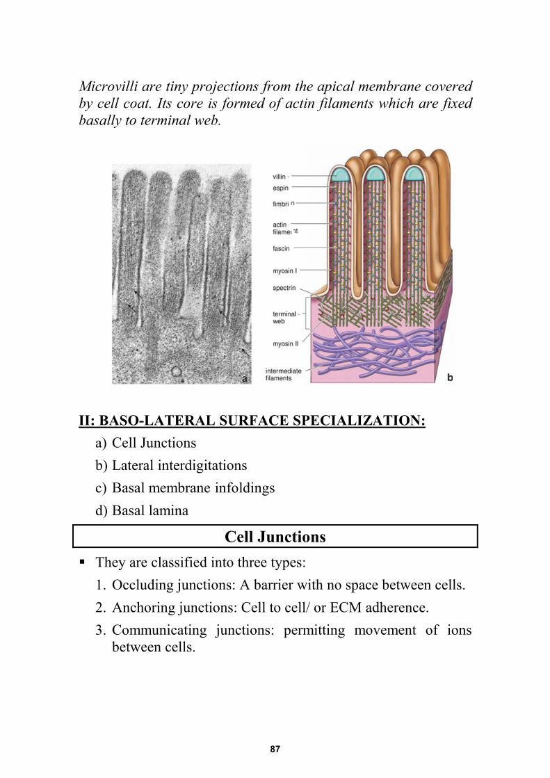
Microvilli are tiny projections from the apical membrane covered by cell coat. Its core is formed of actin filaments which are fixed basally to terminal web.
II: BASO-LATERAL SURFACE SPECIALIZATION:
a) Cell Junctions b) Lateral interdigitations c) Basal membrane infoldings d) Basal lamina
Cell Junctions They are classified into three types:
1. Occluding junctions: A barrier with no space between cells. 2. Anchoring junctions: Cell to cell/ or ECM adherence. 3. Communicating junctions: permitting movement of ions
between cells.
87
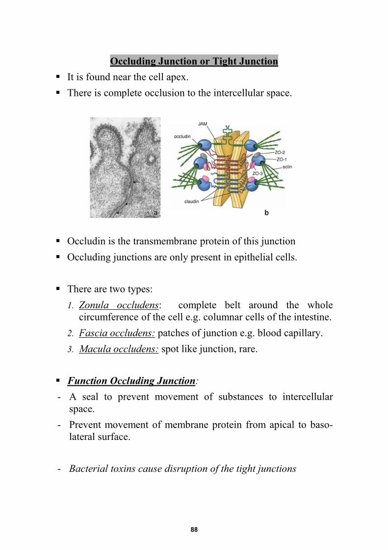
Occluding Junction or Tight Junction It is found near the cell apex. There is complete occlusion to the intercellular space.
Occludin is the transmembrane protein of this junction Occluding junctions are only present in epithelial cells. There are two types:
1. Zonula occludens: complete belt around the whole circumference of the cell e.g. columnar cells of the intestine.
2. Fascia occludens: patches of junction e.g. blood capillary. 3. Macula occludens: spot like junction, rare.
Function Occluding Junction: - A seal to prevent movement of substances to intercellular
space. - Prevent movement of membrane protein from apical to baso-
lateral surface. - Bacterial toxins cause disruption of the tight junctions
88
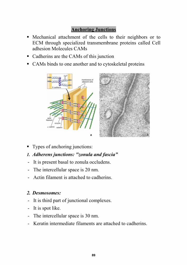
Anchoring Junctions Mechanical attachment of the cells to their neighbors or to
ECM through specialized transmembrane proteins called Cell adhesion Molecules CAMs
Cadherins are the CAMs of this junction CAMs binds to one another and to cytoskeletal proteins
Types of anchoring junctions: 1. Adherens junctions: "zonula and fascia” - It is present basal to zonula occludens. - The intercellular space is 20 nm. - Actin filament is attached to cadherins.
2. Desmosomes: - It is third part of junctional complexes. - It is spot like. - The intercellular space is 30 nm. - Keratin intermediate filaments are attached to cadherins.
89
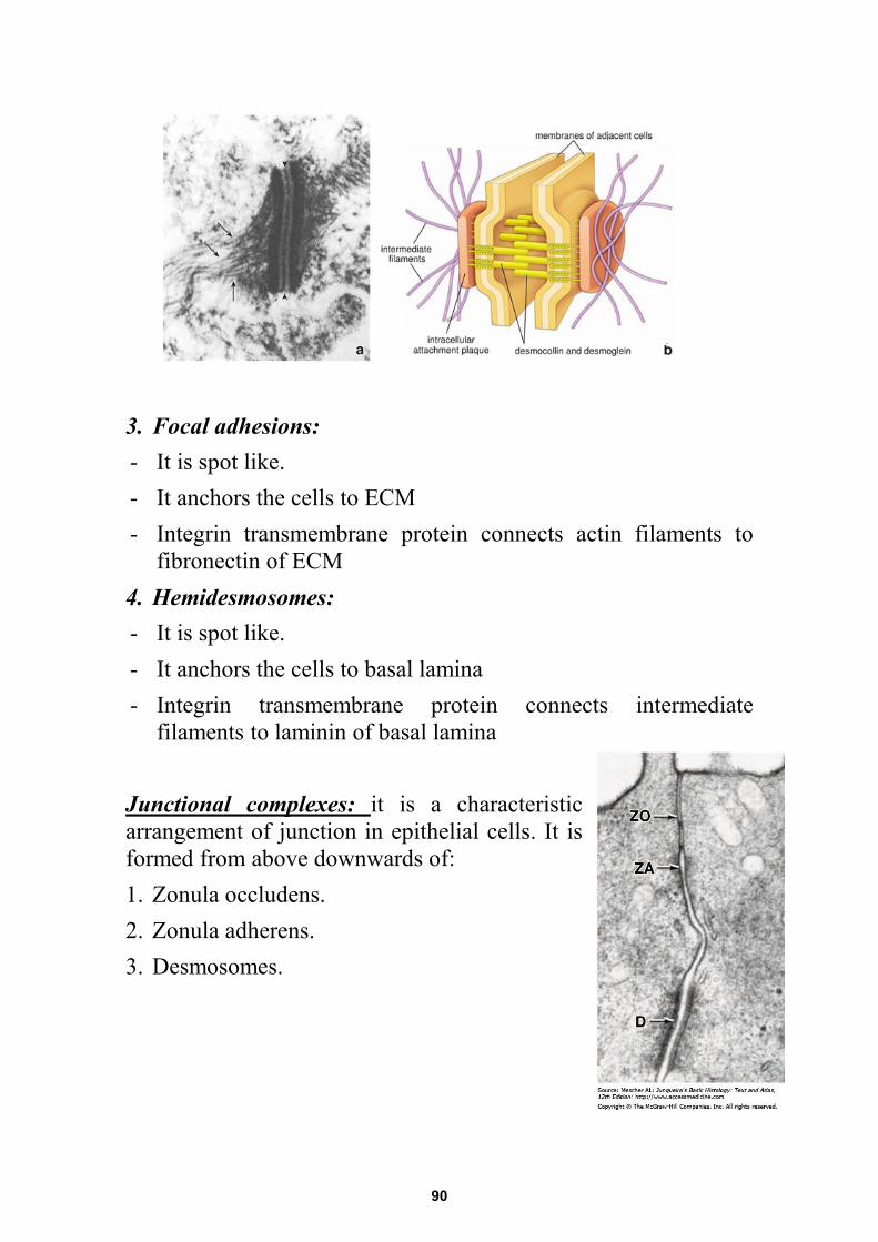
3. Focal adhesions: - It is spot like. - It anchors the cells to ECM - Integrin transmembrane protein connects actin filaments to
fibronectin of ECM 4. Hemidesmosomes: - It is spot like. - It anchors the cells to basal lamina - Integrin transmembrane protein connects intermediate
filaments to laminin of basal lamina Junctional complexes: it is a characteristic arrangement of junction in epithelial cells. It is formed from above downwards of: 1. Zonula occludens. 2. Zonula adherens. 3. Desmosomes.
90
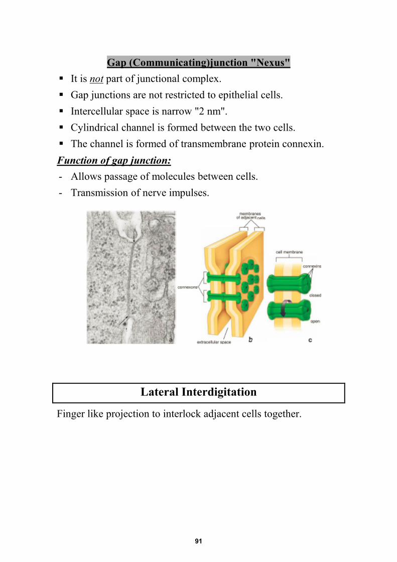
Gap (Communicating)junction "Nexus" It is not part of junctional complex. Gap junctions are not restricted to epithelial cells. Intercellular space is narrow "2 nm". Cylindrical channel is formed between the two cells. The channel is formed of transmembrane protein connexin. Function of gap junction: - Allows passage of molecules between cells. - Transmission of nerve impulses.
Lateral Interdigitation
Finger like projection to interlock adjacent cells together.
91
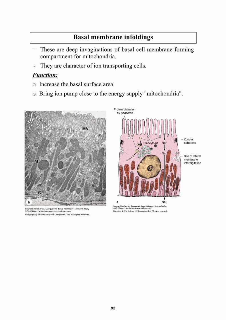
Basal membrane infoldings
- These are deep invaginations of basal cell membrane forming compartment for mitochondria.
- They are character of ion transporting cells. Function: o Increase the basal surface area. o Bring ion pump close to the energy supply "mitochondria".
92
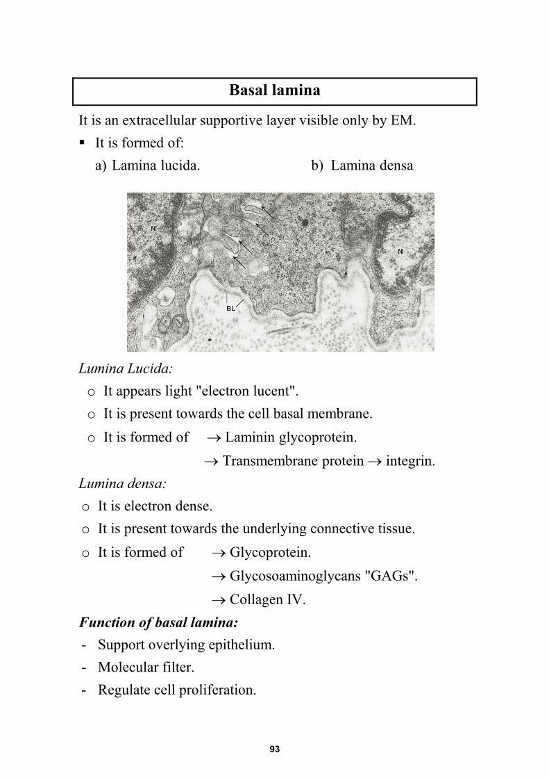
Basal lamina
It is an extracellular supportive layer visible only by EM. It is formed of:
a) Lamina lucida. b) Lamina densa Lumina Lucida: o It appears light "electron lucent". o It is present towards the cell basal membrane. o It is formed of → Laminin glycoprotein.
→ Transmembrane protein → integrin. Lumina densa: o It is electron dense. o It is present towards the underlying connective tissue. o It is formed of → Glycoprotein.
→ Glycosoaminoglycans "GAGs". → Collagen IV.
Function of basal lamina: - Support overlying epithelium. - Molecular filter. - Regulate cell proliferation.
93
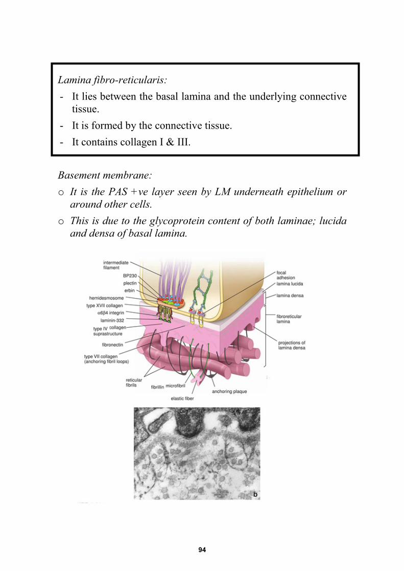
Lamina fibro-reticularis: - It lies between the basal lamina and the underlying connective
tissue. - It is formed by the connective tissue. - It contains collagen I & III. Basement membrane: o It is the PAS +ve layer seen by LM underneath epithelium or
around other cells. o This is due to the glycoprotein content of both laminae; lucida
and densa of basal lamina.
94
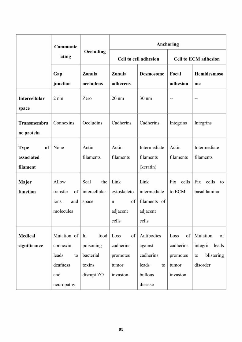
Communic
ating Occluding
Anchoring
Cell to cell adhesion Cell to ECM adhesion
Gap
junction
Zonula
occludens
Zonula
adherens
Desmosome Focal
adhesion
Hemidesmoso
me
Intercellular
space
2 nm Zero 20 nm 30 nm -- --
Transmembra
ne protein
Connexins Occludins Cadherins Cadherins Integrins Integrins
Type of
associated
filament
None Actin
filaments
Actin
filaments
Intermediate
filaments
(keratin)
Actin
filaments
Intermediate
filaments
Major
function
Allow
transfer of
ions and
molecules
Seal the
intercellular
space
Link
cytoskeleto
n of
adjacent
cells
Link
intermediate
filaments of
adjacent
cells
Fix cells
to ECM
Fix cells to
basal lamina
Medical
significance
Mutation of
connexin
leads to
deafness
and
neuropathy
In food
poisoning
bacterial
toxins
disrupt ZO
Loss of
cadherins
promotes
tumor
invasion
Antibodies
against
cadherins
leads to
bullous
disease
Loss of
cadherins
promotes
tumor
invasion
Mutation of
integrin leads
to blistering
disorder
95
EPITHELIUM Classify glands according to the mode of secretion of secretion. Give examples Glands are classified according to the mode of secretion into: o Merocrine gland: secretion is released by exocytosis as in sweat gland. o Apocrine gland: secretion is released with loss of the apical part of the cell as in
mammary gland. o Holocrine gland: secretion is released with loss of the whole cell as in sebaceous
gland. Compare the structural difference between serous and mucous acinus Enumerate specialization of the upper surface of epithelial cells. UApical surface specialization: a- Cilia → for movement of fluid film b- Flagella → movement of cell c- Microvilli → Absorption d- Stereocilia: long irregular M.V. List the ways by which the epithelial cells are held together. The cells are held together by means of: a) UCell Junctions • Adhering junctions • Gap junctions • Occluding junction
b) ULateral interdigitations
MUCOUS SEROUS
Large Small Size Wide Narrow Lumen
Cubical Pyramidal Cell Distinct Indistinct Cell boundaries
Flattened peripheral Round - central Nucleus
Foamy vacuolated basophilic
Apical acidophilic granules and basal basophilia Cytoplasm: LM
rER- Golgi- mitochondria-
Granules- no lateral interdigitation
rER- Golgi- mitochondria-Granules- lateral
interdigitation Cytoplasm: EM
More numerous Less Myo-epithelial cell
96
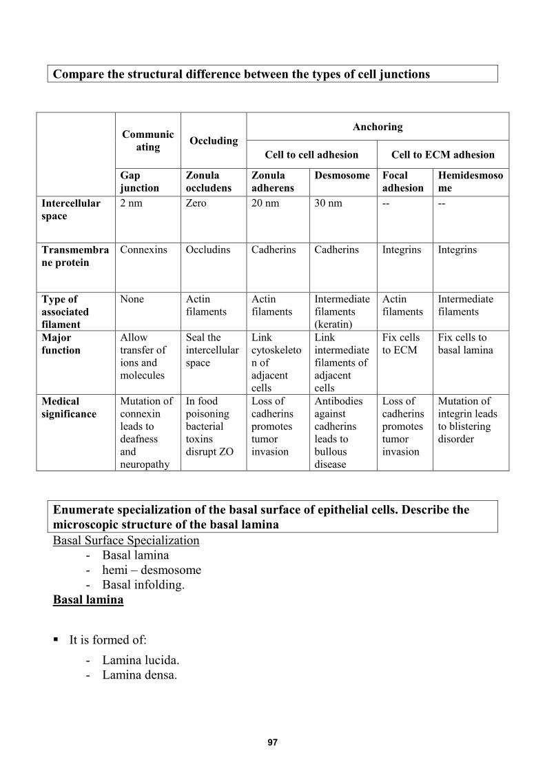
Compare the structural difference between the types of cell junctions
Enumerate specialization of the basal surface of epithelial cells. Describe the microscopic structure of the basal lamina UBasal Surface Specialization
- Basal lamina - hemi – desmosome - Basal infolding.
UBasal lamina
It is formed of: - Lamina lucida. - Lamina densa.
Communic
ating Occluding Anchoring
Cell to cell adhesion Cell to ECM adhesion
Gap junction
Zonula occludens
Zonula adherens
Desmosome Focal adhesion
Hemidesmosome
Intercellular space
2 nm Zero 20 nm 30 nm -- --
Transmembrane protein
Connexins Occludins Cadherins Cadherins Integrins Integrins
Type of associated filament
None Actin filaments
Actin filaments
Intermediate filaments (keratin)
Actin filaments
Intermediate filaments
Major function
Allow transfer of ions and molecules
Seal the intercellular space
Link cytoskeleton of adjacent cells
Link intermediate filaments of adjacent cells
Fix cells to ECM
Fix cells to basal lamina
Medical significance
Mutation of connexin leads to deafness and neuropathy
In food poisoning bacterial toxins disrupt ZO
Loss of cadherins promotes tumor invasion
Antibodies against cadherins leads to bullous disease
Loss of cadherins promotes tumor invasion
Mutation of integrin leads to blistering disorder
97
Lumina Lucida: o It appears light "electron lucent". o It is present towards the cell basal membrane. o It is formed of → glycoprotein.
→ Transmembrane protein → integrin. Lumina densa:
o It is electron dense. o It is present towards the underlying connective tissue. o It is formed of → Glycoprotein.
→ Glycosoaminoglycans "GAGs". → Collagen IV.
Describe the microscopic structure of the basement membrane Basement membrane:
o It is the UPAS +ve layer seen by LMU underneath epithelium or around other cells.
o It is formed of: - Basal lamina - Lamina fibroreticularis
Lamina fibro-reticularis:
- It lies between the basal lamina and the underlying connective tissue. - Formed by the connective tissue. - Contain collagen I & III. - It is connected to the basal lamina by anchoring fibers « collagen VII » - It binds C.T. to the basal lamina
Gibe reasons for:
• The epithelial cells are joined to the underlying C.T. Epithelial cell are held to the underlying C.T.by: 1. hemidesmosomes 2. basal lamina • The basement membrane appeared PAS +ve by LM This is due to the glycoprotein content of both lamina lucida and lamina densa of basal lamina.
• Myoepithelial cells are present around the acini To contract to squeeze the secretion to the duct as they contain contractile filaments in their cytoplasm
98
• The epithelium forms a protective covering sheets over our body surfacesThis because epithelium has the following characters:
• It is formed by large number of cells with minimal intercellular substance• The cells are held together by cell junctions• The cells rest on a protective barrier which is the basement membrane
99
EPITHELIUM MCQ
1. Basal layer of stratified squamous epithelium is rich in:a) RERb) SERc) Free ribosomesd) lysosomes
2. Oral cavity is lined by:a) Transitional epitheliumb) Stratified squamous epitheliumc) Pseudostratified epitheliumd) Simple columnar epithelium
3. Absorptive columnar cells show:a) Apical ciliab) Apical microvillic) Basal infoldingsd) All of the above
4. Motile specialization of apical cell domain is:a) Ciliab) Stereociliac) Flagellad) Microvilli
5. Microvilli contain:a) 9 triplet microtubulesb) 9 doublet microtubulesc) Actin filamentsd) Intermediate filaments
6. The distensible epithelium is:a) Transitional epitheliumb) Stratified squamous epitheliumc) Pseudostratified epitheliumd) Simple columnar epithlium
7. Top layer of transition epithelium is:a) Keratinizedb) Squamous cellsc) Cuboidal cellsd) Columnar cells
100
8. Epithelium is penetrated by: a) Lymph vessels b) Blood vessels c) Nerves d) All of the above
9. Specialization of lateral domain of epithelial cells:
a) Cilial b) Villi c) Cell junctions d) Stereocilia
10. Channel between cells occur in:
a) Tight junction b) Gap junction c) Adherent junction d) Desmosomes
11. Trachea is lined by:
a) Transitional epithelium b) Stratified squamous epithelium c) Pseudostratified ciliated epithelium d) Simple columnar ciliated epithelium
12. In stratified squamous epithelium:
a) Basal cells are flat b) Basal cell are polygonal c) Top cell layers are cuboidal d) Top cell layers are flat
13. Mesothelium lines:
a) Lung alveoli b) Blood vessels c) Bowman's capsule d) Serous membranes
14. Cells characterized by basal membrane infoldings are :
a) Ion transporting b) Protein secreting c) Steroid secreting d) Contractile
101
15. Endothelium lines: a) Lung alveoli b) Blood vessels c) Bowman's capsule d) Serous membranes
16. The type of epithelium which allows transcellular transport is:
a) Stratified squamous non keratinized b) Transitional c) Simple squamous d) Pseudostratified columnar
17. The type of epithelium which resists friction is:
a) Stratified squamous keratinized b) Transitional c) Simple squamous d) Pseudostratified columnar
18. A 20 year-old male presented to the dermatology clinic complaining of
extensive blisters on the skin (bullous). This case was diagnosed as an autoimmune disease affecting:
a) Zonula occludens. b) Zonula adherens. c) Gap junction. d) Desmosomes.
19. Connexons are characteristic features of
a) The trans-Golgi network. b) The sER. c) Gap junction. d) Lysosomes.
20. Which one of the following statements about the desmosome is true? a) It permits the passage of large proteins between cells b) It has a plaque made up of many connexons. c) It facilitates metabolic coupling between adjacent cells. d) It is a disk-shaped adhesion site between epithelial cells.
21. Which of the following is most directly an adaptation of epithelial cells that, as
part of their function, absorb large amounts of small nutrient molecules through their apical plasmalemmas?
a) Adhesive intercellular junctions. b) Extensive rER. c) Extensive sER. d) Microvilli.
102
22. During the process of secretion, the cells of the holocrine glands show: a) Damage of the whole cells. b) Loss of the apical cytoplasm. c) Intact secretory cells. d) Direct secretion into blood.
23. A male child presented with diarrhea was chronically dependent on
intravenous nutrition. He was diagnosed to have malabsorption syndrome. The most likely affected structure of his intestinal epithelium is the:
a) Apical microvilli b) Junctional complexes c) Lateral interdigitation d) Basal membrane infoldings
24. The structure responsible for the linkage of the intermediate filaments of
epithelial cells to the basal lamina is: a) Macula adherens b) Zonula adherens c) Hemidesmosomes d) Zonula occludens
25. Which of the following transmembrane proteins forms seal between adjacent
epithelial cells in occluding junctions? a) Occludins b) Connexins c) Integrins d) Cadherins
26. Which of the following proteins forms the core of microvilli found along the
apical membrane domain of the columnar absorptive epithelial cells? a) Myosin b) Keratin c) Tubulin d) Actin
27. Which of the following types of epithelium describes the epidermis of the skin?
a) Keratinized stratified squamous b) Non-keratinized stratified squamous c) Pseudostratified columnar d) Stratified cuboidal
103
28. Transmembrane proteins involved in the formation of adhering junctions between adjacent epithelial cells are?
a) Occludins b) Connexins c) Desmoplakins d) Cadherins
29. During the process of secretion, the cells of the apocrine glands show:
a) Damage of the whole cells. b) Loss of the apical cytoplasm. c) Intact secretory cells. d) Direct secretion into blood.
30. The type of junction that forms a channel between adjacent cells is:
a) Desmosomes. b) Adherent junction. c) Gap junction. d) Tight junction.
31. The intercellular space in occluding junction is:
a) Zero nm. b) 20 nm. c) 30 nm. d) 0.2 nm.
32. The blood vessels are lined by: a) Endothelium b) Mesothelium c) Neuroepithelium d) Myoepithelium
33. Taste buds are example of:
a) Glandular epithelium b) Stratified epithelium c) Neuroepithelium d) Myoepithelium
34. Junctional complex is formed of:
a) Zonula occludens, desmosome and fascia adherens b) Zonula occludens, zonula adherens and desmosomes c) Zonula adherens, macula adherens and gap junction d) Zonula occludens, desmosomes and connexons
104
35. Gap junctions are present in a) Endothelial cells of blood capillaries b) Intestinal columnar cells c) Prickle cell layer of the epidermis d) Cardiac muscle fibers
36. The type of epithelium lining the pelvis of kidney is:
a) Transitional b) Stratified squamous c) Psuedo-stratified columnar d) Stratified columnar
37. Stereocilia are:
a) Long non motile cilia b) Short non motile microvilli c) Long motile cilia d) Long non motile microvilli
38. Epithelial tissue is characterized by one of the following:
a) Polarity b) High vascularity c) Low regenerative capacity d) Abundant intercellular space
39. Actin filaments are involved in the structure of::
a) Macula adherens b) Zonula occludens c) Stereocilia d) Cilia
40. The type of epithelium that can accommodate during stretch is the:
a) Stratified squamous keratinized b) Stratified squamous non-keratinized c) Transitional d) Pseudostratified columnar
41. Which of the following proteins regulates the motility of cilia found along the
apical membrane domain of the columnar epithelial cells lining the trachea? a) Actin b) Desmin c) Keratin d) Dynein
105
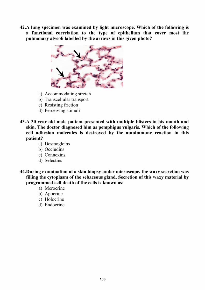
42. A lung specimen was examined by light microscope. Which of the following isa functional correlation to the type of epithelium that cover most thepulmonary alveoli labelled by the arrows in this given photo?
a) Accommodating stretchb) Transcellular transportc) Resisting frictiond) Perceiving stimuli
43. A-30-year old male patient presented with multiple blisters in his mouth andskin. The doctor diagnosed him as pemphigus vulgaris. Which of the followingcell adhesion molecules is destroyed by the autoimmune reaction in thispatient?
a) Desmogleinsb) Occludinsc) Connexinsd) Selectins
44. During examination of a skin biopsy under microscope, the waxy secretion wasfilling the cytoplasm of the sebaceous gland. Secretion of this waxy material byprogrammed cell death of the cells is known as:
a) Merocrineb) Apocrinec) Holocrined) Endocrine
106