Embed Size (px)
Citation preview

@MGH, Boston, October 19, 2007
PET protoytpe development and experimental results for "in vivo"
hadrontherapy dosimetry
Alberto Del GuerraProfessor of Medical Physics Head and DirectorSpecialty School in Medical PhysicsHead, Functional Imaging andInstrumentation Group
Department of Physics “E.Fermi”, University of Pisa
and INFN, Pisa (Italy)
Department of Physics “E.Fermi”
University of Pisa
INFN - Pisa
Functional imaging and Instrumentation Group – Univ. Pisa
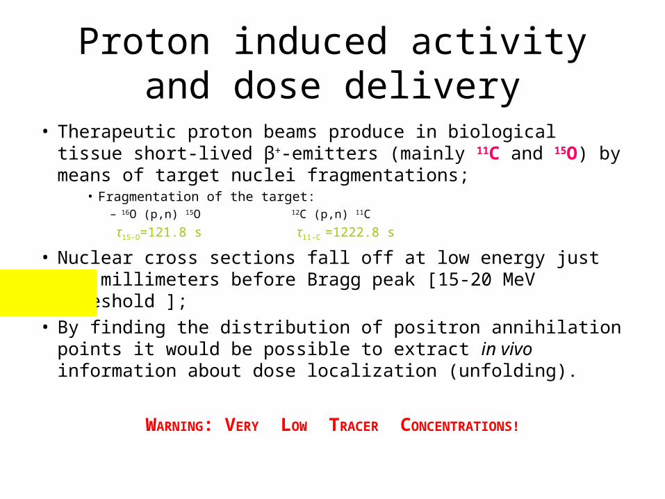
Proton induced activity and dose delivery
• Therapeutic proton beams produce in biological tissue short-lived β+-emitters (mainly 11C and 15O) by means of target nuclei fragmentations;
• Fragmentation of the target:
– 16O (p,n) 15O 12C (p,n) 11C
τ15-O=121.8 s τ11-C =1222.8 s
• Nuclear cross sections fall off at low energy just few millimeters before Bragg peak [15-20 MeV threshold ];
• By finding the distribution of positron annihilation points it would be possible to extract in vivo information about dose localization (unfolding).
WARNING: VERY LOW TRACER CONCENTRATIONS!

How to gather information on proton range from reconstructed activity?
FILTERING APPROACH TO UNFOLDING• Parodi & Bortfeld * proposed a fast method based on the description
of the PET image as a convolution of the dose distribution with a filter function, also showing the uniqueness of the filter for proton energy ranging from 70 to 220 MeV.
• Reconstructing expected PET signal from local planned dose distribution and comparing it with measured one it is possible to verify if dose has been delivered correctly.
AN INVERSION OF THE FILTER WOULD ALLOW
DIRECT DOSE INFORMATION• We have studied the possibility to adopt the same approach in the
energy range of interest for CATANA (40–70 MeV), as a starting point for the inverse process.
• K Parodi and T.Bortfeld. A filtering approach based on Gaussian-power law convolutions for local PET verification of proton radiotherapy (Phys. Med. Biol. 51 (2006) 1991 - 2009).

Theoretical research of filter functions
• Predicted 3D depth dose and activity profiles were obtained by means of a code based on a semi-analytical model of the Ziegler-Vavilov theory:– Fokker-Planck scheme for evolving energy distribution
is combined with nuclear cross sections interpolated from EXFOR.
• Monoenergetic Bragg peaks and activity profiles have been simulated for several initial energies;
• Two isotope distribution filters have been computed and weighted according to PET acquisition time in order to give the total activity filter.
THE UNIQUENESS OF THE FILTER FUNCTIONS (F
and F-1) ENSURES THAT THE METHOD CAN BE APPLIED TO EXTENDED DOSE DISTRIBUTIONS

• Reconstructed activity profile in comparison with the filtered dose (SOBP of 20 mm modulation);
• TIrr: [0, 173] s;• TScan: [173, 2000]
s;• Transverse
domain of integration: 19 mm Ø;
• Reconstructed activity profile in comparison with the filtered dose (SOBP of 12 mm modulation);
• TIrr: [0, 189] s;• TScan: [189, 2000]
s;• Transverse
domain of integration: 19 mm Ø;
Testing the theoretical filters on experimental data
• Reconstructed activity profile in comparison with the filtered dose (monoenergetic Bragg curve);
• TIrr: [0, 138] s;• TScan: [138, 1938]
s;• Transverse
domain of integration: 19 mm Ø;

Separation of Isotope Contributions
• Individual main isotope components (11C and 15O) has been separated by time analysis on the true count rate from measured list mode file;
• It has been thus possible to quantify quite accurately the relative abundance and the spatial distribution of each isotope specie produced at the end of irradiation;
• This way, in spite of the low statistics, we obtained at once both a CROSS SECTION VALIDATION and a more cogent TEST FOR FILTERING APPROACH suitability.12 mm SOBP isotope production
FE isotope production

β+- emitter fragments with Protons and with Carbon ions:
– Proton as a projectile:• Fragmentation of the target:
– 16O (p,n) 15O 12C (p,n) 11C
τ15-O=121.8 s τ11-C =1222.8 s[15-20 MeV threshold for p-induced nuclear reactions that cause poor spatial
correlation between β+-activity and dose depth profile]
– Carbon as a projectile; • Fragmentation of the target:
– X (12C, 11C+n) X X (12C, 10C+2n) X τ11-C =1222.8 s τ10-C =19.3 s
• Fragmentation of the projectile:
– 16O (12C, X) 15O+n 12C (12C, X) 11C +n τ15-O=121.8 s τ11-C =1222.8 s
Hadron-driven PET with Ions

The PET-tomograph prototype
•Two planar heads, each with an active area of 45 mm x 45 mm •Distance between the heads: 5+5 cm, 7+7 cm, 10+10 cm (for phantoms study) and 5+15 cm (for eye monitoring)
• LYSO crystal matrix, 21 x 21 pixels 2.152 mm x 2.152 mm each (Hilger Crystals)•Crystal thickness: 18 mm •64-anode PMT (Hamamatsu)•“multiplexed” read-out electronics: 64-inputs/4 outputs

A cylindrical PMMA phantom ( 7cm diameter, 7cm length) was irradiated with 3 mono-energetic 12C beams ( 108.53 112.60 116.57 AMeV).A square section beam of 28mm in side was adopted and a total dose of 60Gy was delivered for each energy. The acquisition time was set at ~30 minutes for both PET systems.
The experimental configuration of the two PET systems is reported on the left.

The linear profiles from the central slice of the Dopet reconstructed activity at 3 12C energies are reported below on the left. On the right, for comparison, the activities acquired, at 116.57 AMeV, with Dopet( ) and Bastei (---).

YB1
SCD + 2D chainScheme + sum
SCD + 2D chainScheme + sum
YB2
YB3 YB4
YA1 YA2
YA3 YA4
XB1 XB2
XB3 XB4
XA1 XA2
XA3 XA4
XA2
XA1
XA4
XA3
XB2
XB1
XB4
XB3
YA2
YA1
YA4
YA3
YB2
YB1
YB4
YB3
XA
XB
YA
YB
Resistors in the 2D chain are weighted according to the formula:
RNA = R0/N; RNB=R0/(16-N+1)
Where N ranges between 1 and 16

STATUS OF MAROC-SPIROC measurement
Michel Bouchel, Stéphane Callier, Frédéric Dulucq, Julien Fleury, Gisèle Martin-Chassard,
Christophe de La Taille, Ludovic Raux

MAROC : 64 ch MAPMT chip for ATLAS lumi
• Complete front-end chip for 64 channels multi-anode photomultipliers– Auto-trigger on 1/3 p.e. at 10 MHz, 12 bit charge output– SiGe 0.35 µm, 12 mm2, Pd = 350mW
PMF
Hold signal
Photomultiplier
64 channels
Photons Variable
GainPreamp
.
VariableSlow
Shaper20-100 ns
S&H
BipolarFast
Shaper
Unipolar Fast
Shaper
Gain correction64*6bits
3 discri thresholds (3*12 bits)
MultiplexedAnalog charge
output
LUCID
S&H
3 DACs12 bits
80 MHz encoder
64 Wilkinson
12 bit ADC
64 trigger outputs (to FPGA)
MultiplexedDigital charge
output
64 inputs

Active board pictures
MAROC side Lattice side
64 ch PMTMAROC2 chip bounded at CERN