Embed Size (px)
DESCRIPTION
BIOCHEMISTRY - PowerPoint PPT Presentation
Citation preview
M.Prasad NaiduMSc Medical Biochemistry,
Ph.D.,Research scholar
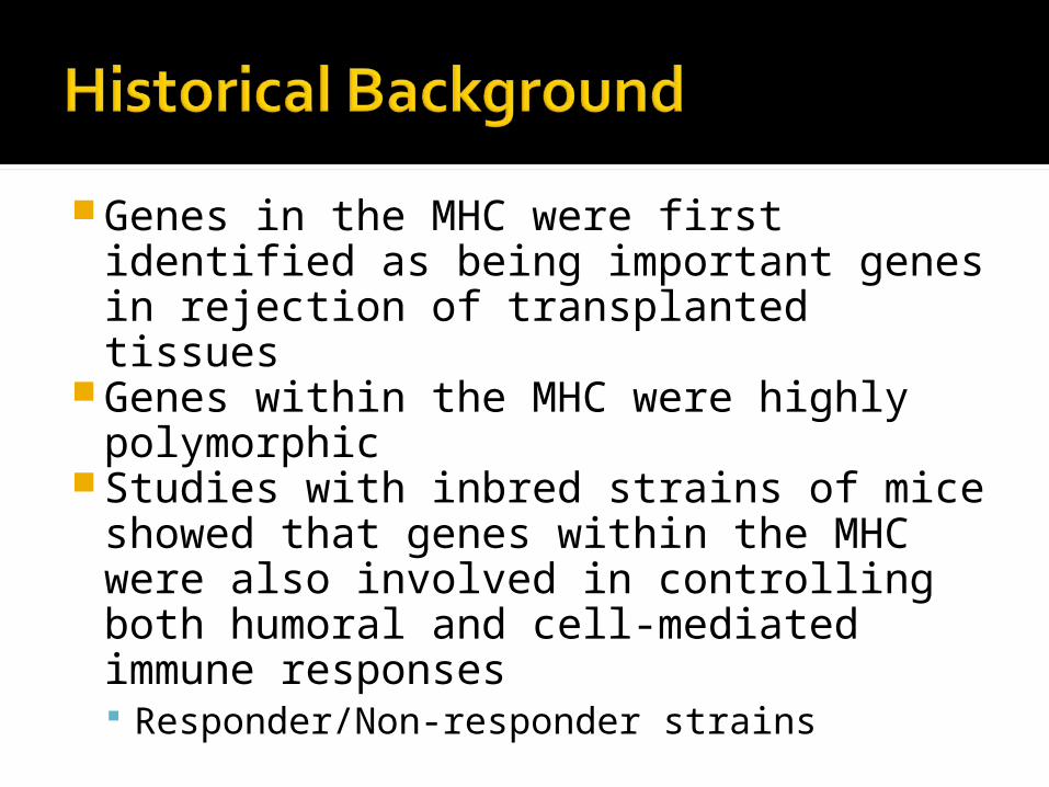
Genes in the MHC were first identified as being important genes in rejection of transplanted tissues
Genes within the MHC were highly polymorphic
Studies with inbred strains of mice showed that genes within the MHC were also involved in controlling both humoral and cell-mediated immune responses Responder/Non-responder strains
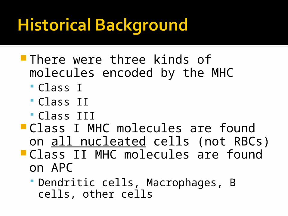
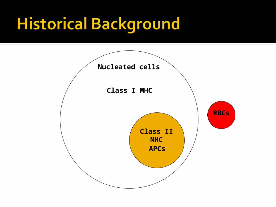
There were three kinds of molecules encoded by the MHC Class I Class II Class III
Class I MHC molecules are found on all nucleated cells (not RBCs)
Class II MHC molecules are found on APC Dendritic cells, Macrophages, B cells,
other cells
Class I MHC
Class II MHC
RBCs
APCs
Nucleated cells
Class III MHC molecules Some complement components Transporter proteins
It was not until the discovery of how the TCR recognizes antigen that the role of MHC genes in immune responses was understood TCR recognizes antigenic peptides in
association with MHC molecules T cells recognize portions of protein
antigens that are bound non-covalently to MHC gene products Tc cells recognize peptides bound to class I
MHC molecules Th cells recognize peptides bound to class II
MHC molecules
Three dimensional structures of MHC molecules and the TCR have been determined by X-ray crystallography
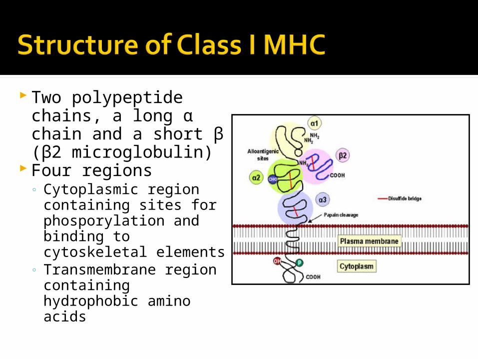
Two polypeptide chains, a long α chain and a short β (β2 microglobulin)
Four regions◦ Cytoplasmic region
containing sites for phosporylation and binding to cytoskeletal elements
◦ Transmembrane region containing hydrophobic amino acids
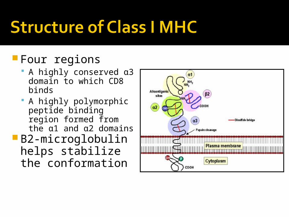
Four regions A highly conserved
α3 domain to which CD8 binds
A highly polymorphic peptide binding region formed from the α1 and α2 domains
Β2-microglobulin helps stabilize the conformation
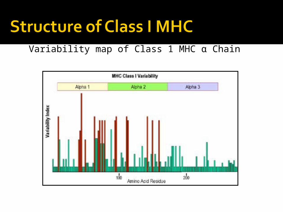
Variability map of Class 1 MHC α Chain
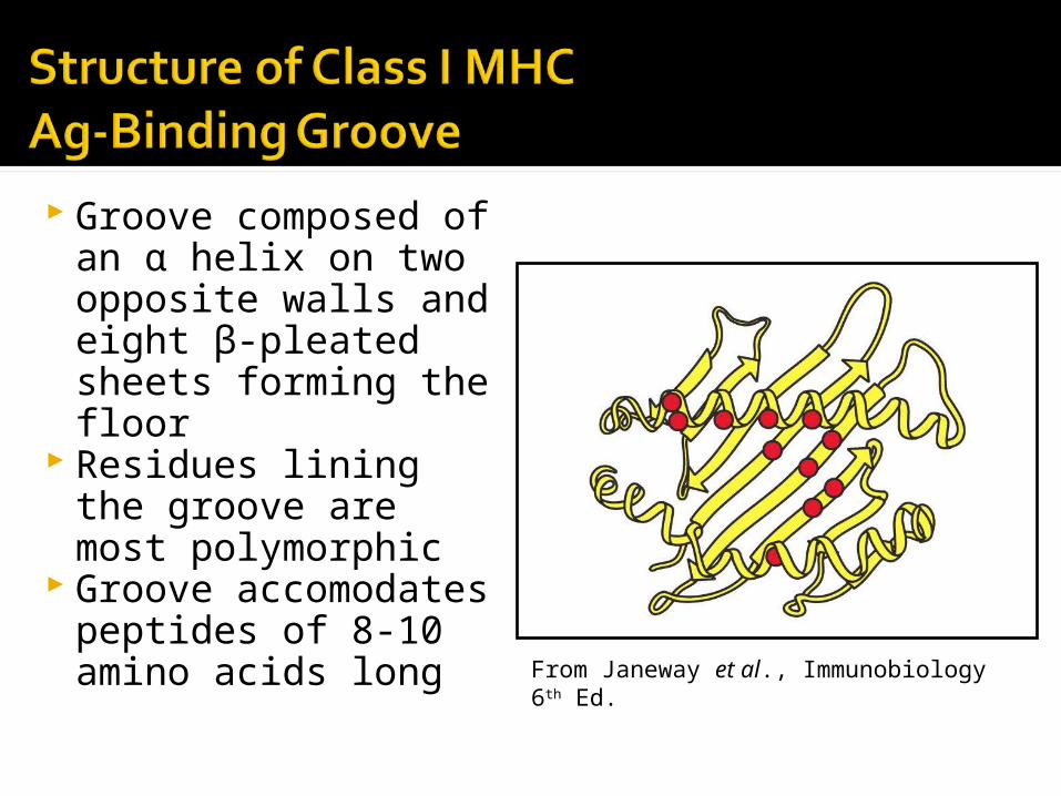
Groove composed of an α helix on two opposite walls and eight β-pleated sheets forming the floor
Residues lining the groove are most polymorphic
Groove accomodates peptides of 8-10 amino acids long From Janeway et al., Immunobiology 6th Ed.
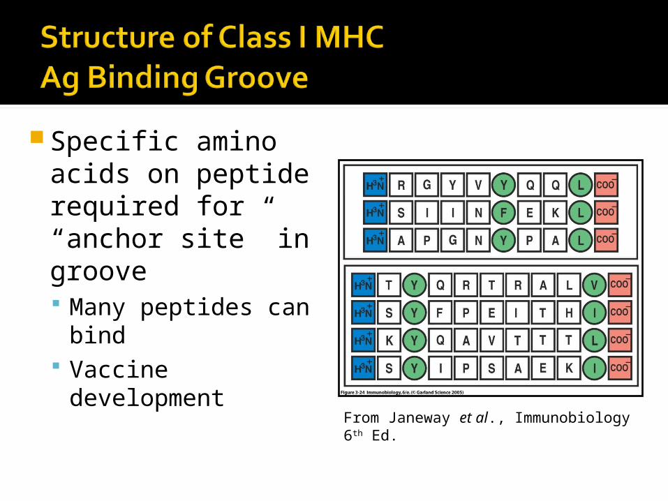
Specific amino acids on peptide required for “anchor site” in groove Many peptides
can bind Vaccine
development From Janeway et al., Immunobiology 6th Ed.
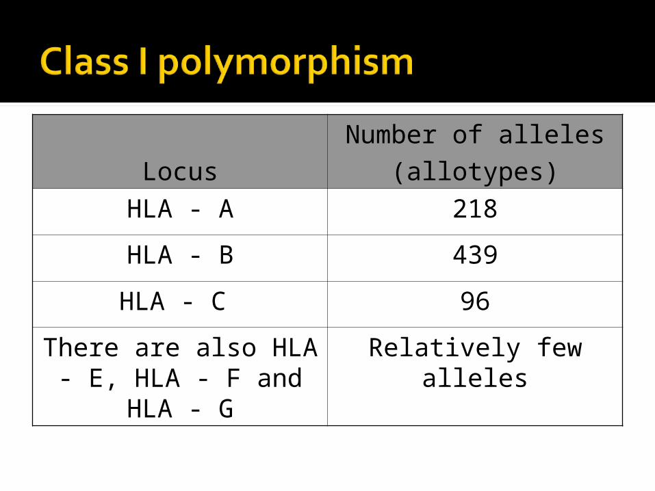
Locus
Number of alleles
(allotypes)
HLA - A 218
HLA - B 439
HLA - C 96
There are also HLA - E, HLA - F and HLA - G
Relatively few alleles
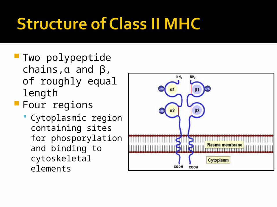
Two polypeptide chains,α and β, of roughly equal length
Four regions Cytoplasmic region
containing sites for phosporylation and binding to cytoskeletal elements
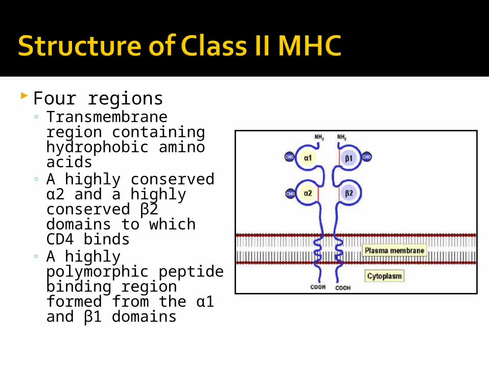
Four regions◦ Transmembrane
region containing hydrophobic amino acids
◦ A highly conserved α2 and a highly conserved β2 domains to which CD4 binds
◦ A highly polymorphic peptide binding region formed from the α1 and β1 domains
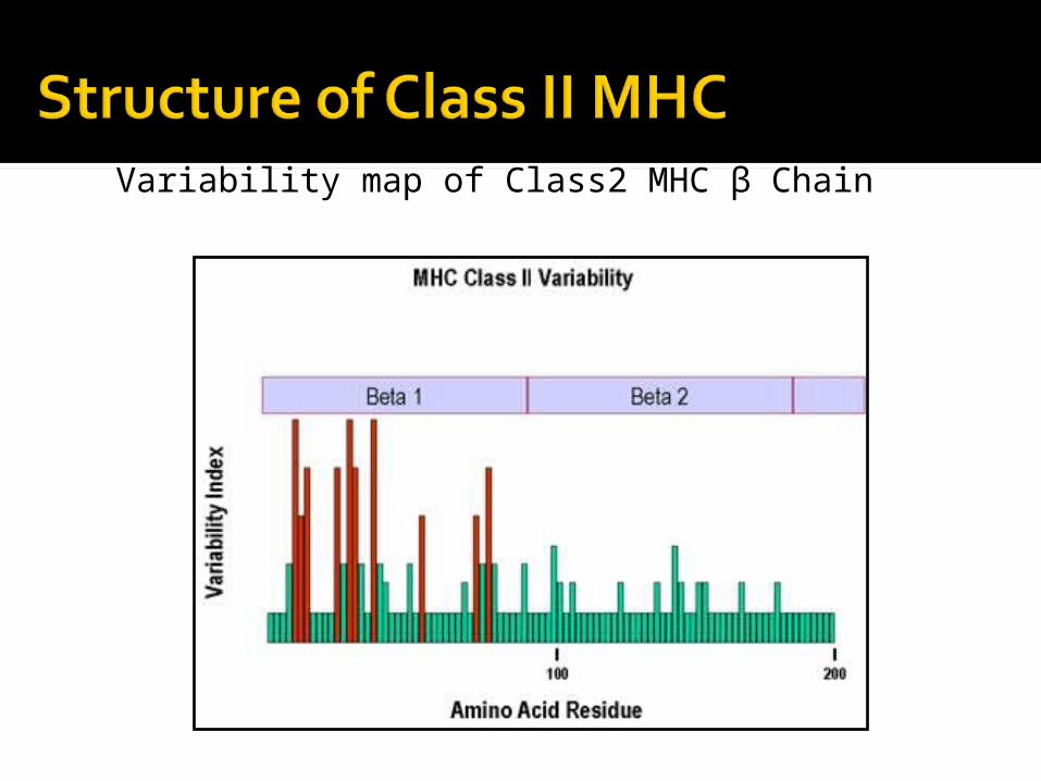
Variability map of Class2 MHC β Chain

Groove composed of an α helix on two opposite walls and eight β-pleated sheets forming the floor
Both the α1 and β1 domains make up the groove
Residues lining the groove are most polymorphic
From Janeway et al., Immunobiology 6th Ed.
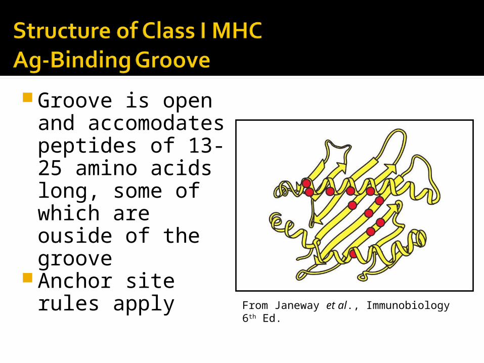
Groove is open and accomodates peptides of 13-25 amino acids long, some of which are ouside of the groove
Anchor site rules apply From Janeway et al., Immunobiology 6th Ed.
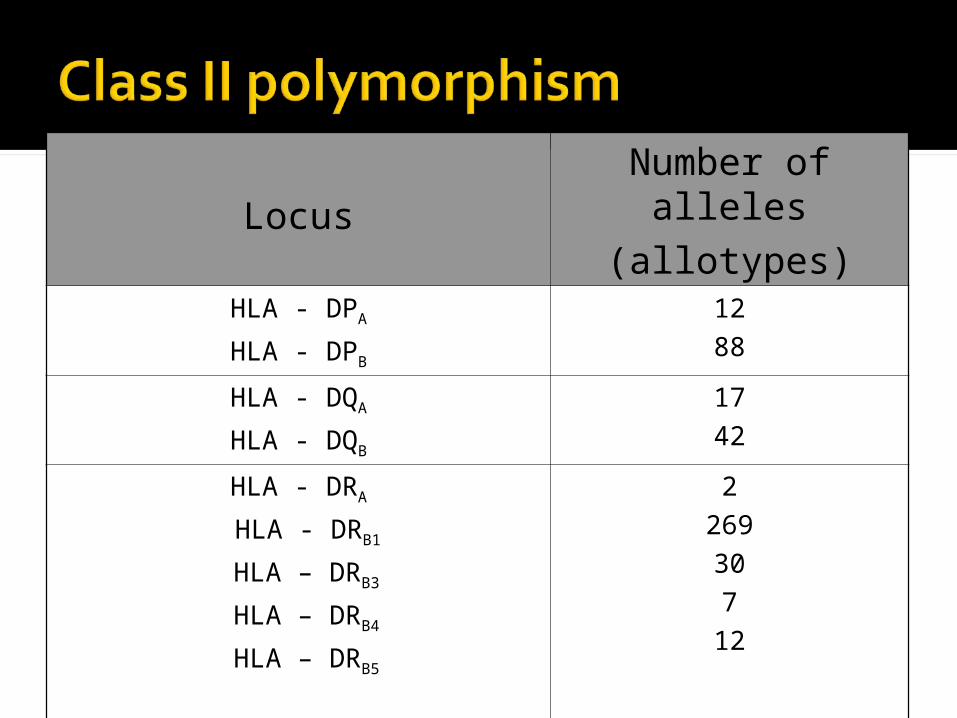
Locus
Number of alleles
(allotypes)HLA - DPA
HLA - DPB
12
88
HLA - DQA
HLA - DQB
17
42
HLA - DRA
HLA - DRB1
HLA – DRB3
HLA – DRB4
HLA – DRB5
2
269
30
7
12
There are also HLA - DM and HLA - DO Relatively few alleles
Although there is a high degree of polymorphism for a species, an individual has maximum of six different class I MHC products and only slightly more class II MHC products (considering only the major loci).
Each MHC molecule has only one binding site. The different peptides a given MHC molecule can bind all bind to the same site, but only one at a time.
Because each MHC molecule can bind many different peptides, binding is termed degenerate.
MHC polymorphism is determined only in the germline. There are no recombinational mechanisms for generating diversity.
MHC molecules are membrane-bound; recognition by T cells requires cell-cell contact.
Alleles for MHC genes are co-dominant. Each MHC gene product is expressed on the cell surface of an individual nucleated cell.
A peptide must associate with a given MHC of that individual, otherwise no immune response can occur. That is one level of control.
Mature T cells must have a T cell receptor that recognizes the peptide associated with MHC. This is the second level of control.
Cytokines (especially interferon-γ) increase level of expression of MHC.
Peptides from the cytosol associate with class I MHC and are recognized by Tc cells . Peptides from within vesicles associate with class II MHC and are recognized by Th cells.
Why so much polymorphism? Survival of the species
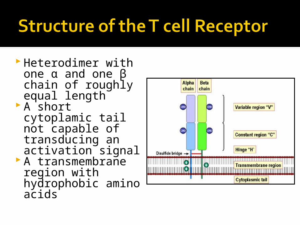
Heterodimer with one α and one β chain of roughly equal length
A short cytoplamic tail not capable of transducing an activation signal
A transmembrane region with hydrophobic amino acids
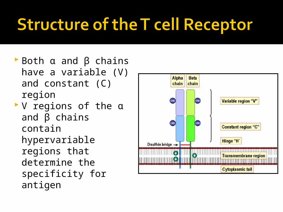
Both α and β chains have a variable (V) and constant (C) region
V regions of the α and β chains contain hypervariable regions that determine the specificity for antigen
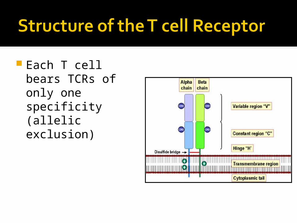
Each T cell bears TCRs of only one specificity (allelic exclusion)
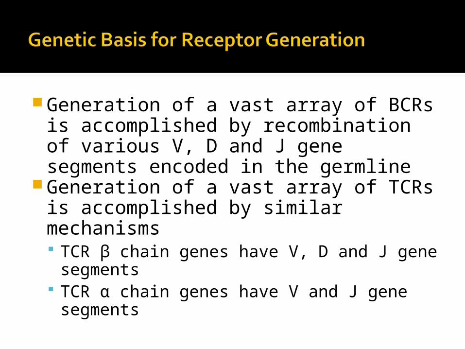
Generation of a vast array of BCRs is accomplished by recombination of various V, D and J gene segments encoded in the germline
Generation of a vast array of TCRs is accomplished by similar mechanisms TCR β chain genes have V, D and J gene
segments TCR α chain genes have V and J gene
segments
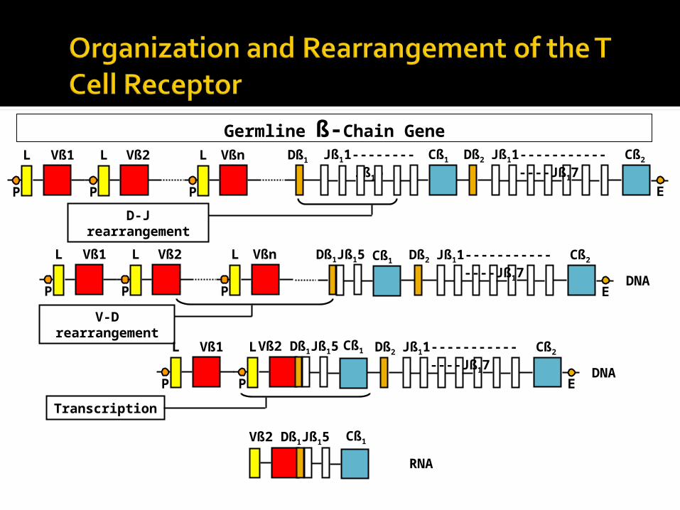
V-D rearrangement
D-J rearrangement
Transcription
Germline ß-Chain Gene Vß1L
P
Vßn Vß2L
P
L
P
Dß1 Jß11--------Jß16 Dß2Cß1 Jß11---------------Jß17 Cß2
E
DNA
Vß1L
P
Vßn Vß2L
P
L
P
Dß1Jß15 Cß1 Dß2 Jß11---------------Jß17 Cß2
E
Vß1L
P
L
PDNA
Vß2 Dß1Jß15 Cß1 Dß2 Jß11---------------Jß17 Cß2
E
RNA
Vß2 Dß1Jß15 Cß1
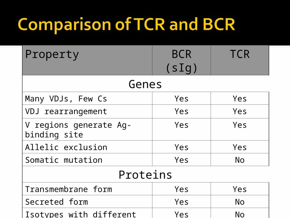
Property BCR (sIg) TCR
GenesMany VDJs, Few Cs Yes Yes
VDJ rearrangement Yes Yes
V regions generate Ag-binding site Yes Yes
Allelic exclusion Yes Yes
Somatic mutation Yes No
ProteinsTransmembrane form Yes Yes
Secreted form Yes No
Isotypes with different functions Yes No
Valence 2 1
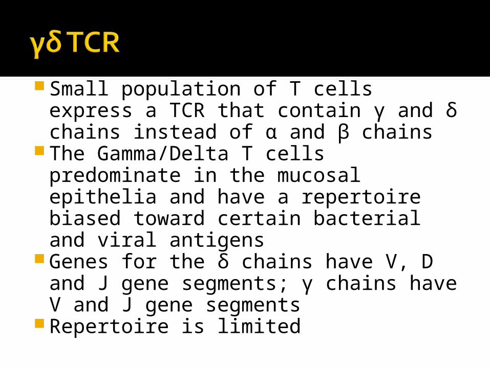
Small population of T cells express a TCR that contain γ and δ chains instead of α and β chains
The Gamma/Delta T cells predominate in the mucosal epithelia and have a repertoire biased toward certain bacterial and viral antigens
Genes for the δ chains have V, D and J gene segments; γ chains have V and J gene segments
Repertoire is limited
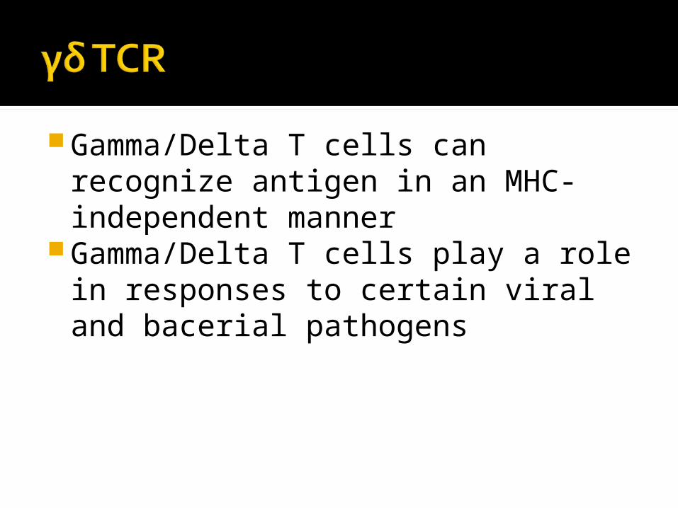
Gamma/Delta T cells can recognize antigen in an MHC-independent manner
Gamma/Delta T cells play a role in responses to certain viral and bacerial pathogens
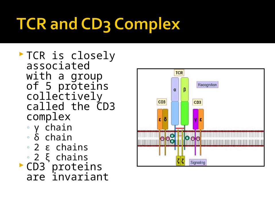
TCR is closely associated with a group of 5 proteins collectively called the CD3 complex◦ γ chain◦ δ chain◦ 2 ε chains◦ 2 ξ chains
CD3 proteins are invariant
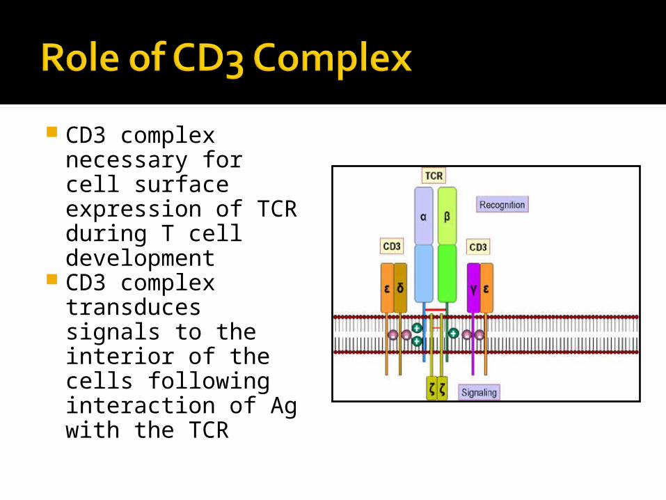
CD3 complex necessary for cell surface expression of TCR during T cell development
CD3 complex transduces signals to the interior of the cells following interaction of Ag with the TCR
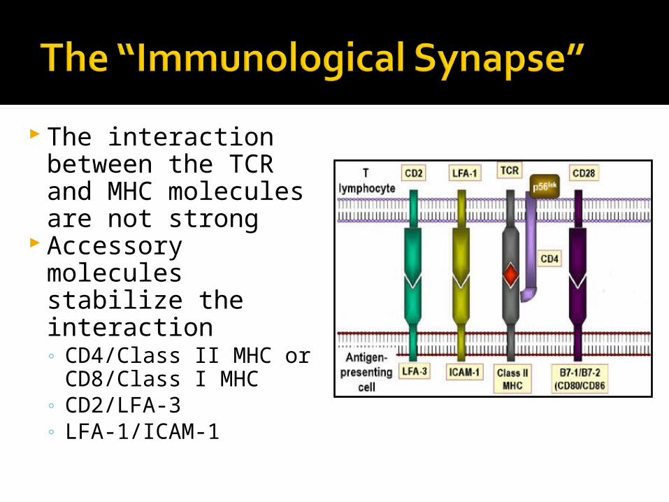
The interaction between the TCR and MHC molecules are not strong
Accessory molecules stabilize the interaction◦ CD4/Class II MHC or
CD8/Class I MHC◦ CD2/LFA-3◦ LFA-1/ICAM-1
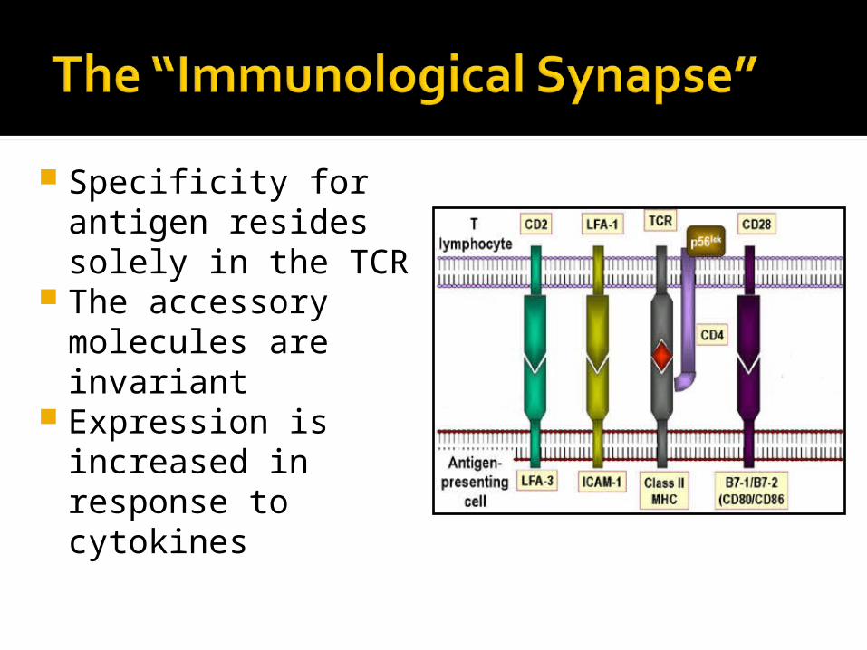
Specificity for antigen resides solely in the TCR
The accessory molecules are invariant
Expression is increased in response to cytokines
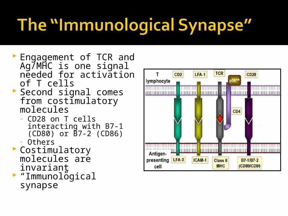
Engagement of TCR and Ag/MHC is one signal needed for activation of T cells
Second signal comes from costimulatory molecules◦ CD28 on T cells
interacting with B7-1 (CD80) or B7-2 (CD86)
◦ Others Costimulatory
molecules are invariant “Immunological
synapse”
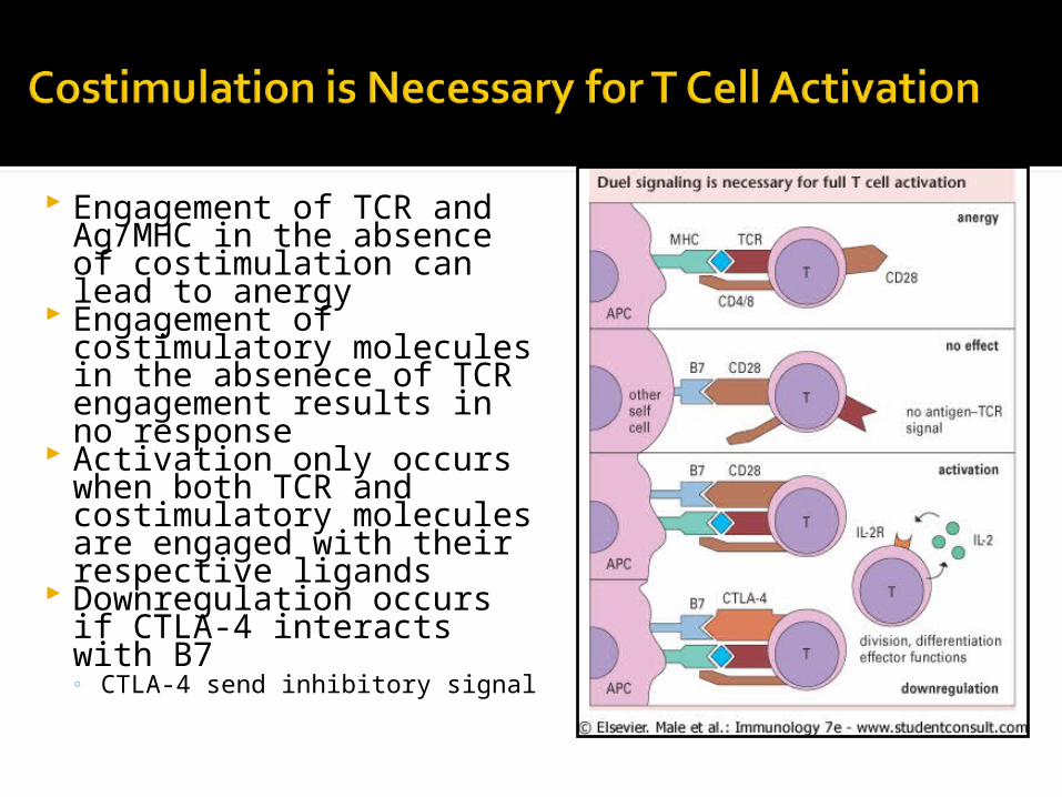
Engagement of TCR and Ag/MHC in the absence of costimulation can lead to anergy
Engagement of costimulatory molecules in the absenece of TCR engagement results in no response
Activation only occurs when both TCR and costimulatory molecules are engaged with their respective ligands
Downregulation occurs if CTLA-4 interacts with B7◦ CTLA-4 send inhibitory signal
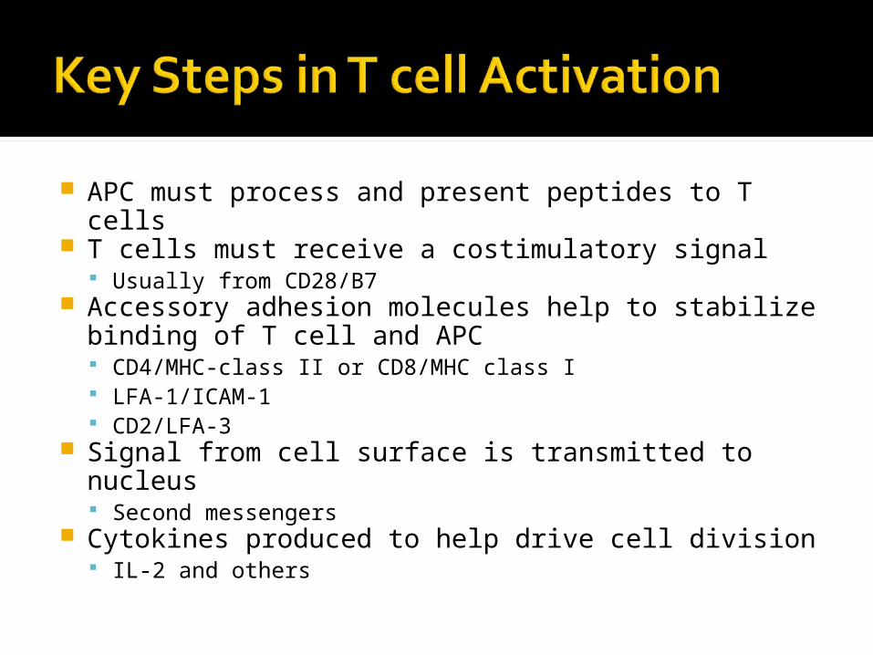
APC must process and present peptides to T cells T cells must receive a costimulatory signal
Usually from CD28/B7 Accessory adhesion molecules help to stabilize
binding of T cell and APC CD4/MHC-class II or CD8/MHC class I LFA-1/ICAM-1 CD2/LFA-3
Signal from cell surface is transmitted to nucleus Second messengers
Cytokines produced to help drive cell division IL-2 and others


![TCR-like antibodies in cancer immunotherapytide/MHC complex by TCR-like antibodies, however, can trigger much broader pharmacological pathways than that of the TCRs in T cells [7]](https://img.pdfslide.net/doc/110x75/60be7e0e102e870c562b6e46/tcr-like-antibodies-in-cancer-immunotherapy-tidemhc-complex-by-tcr-like-antibodies.jpg)