Embed Size (px)
Citation preview
MICHAEL HOOKER MICROSCOPY
• Core Facility—open to all UNC investigators
• Confocal – fluorescence/reflection/frap/fret• Atomic Force Microscope• Laser Micro-Dissection• Light Microscopy –
tans./fluor./DIC/phase/multimode/time-lapse
http://mhmicroscopy.med.unc.edu
Mike Chua 843-3268
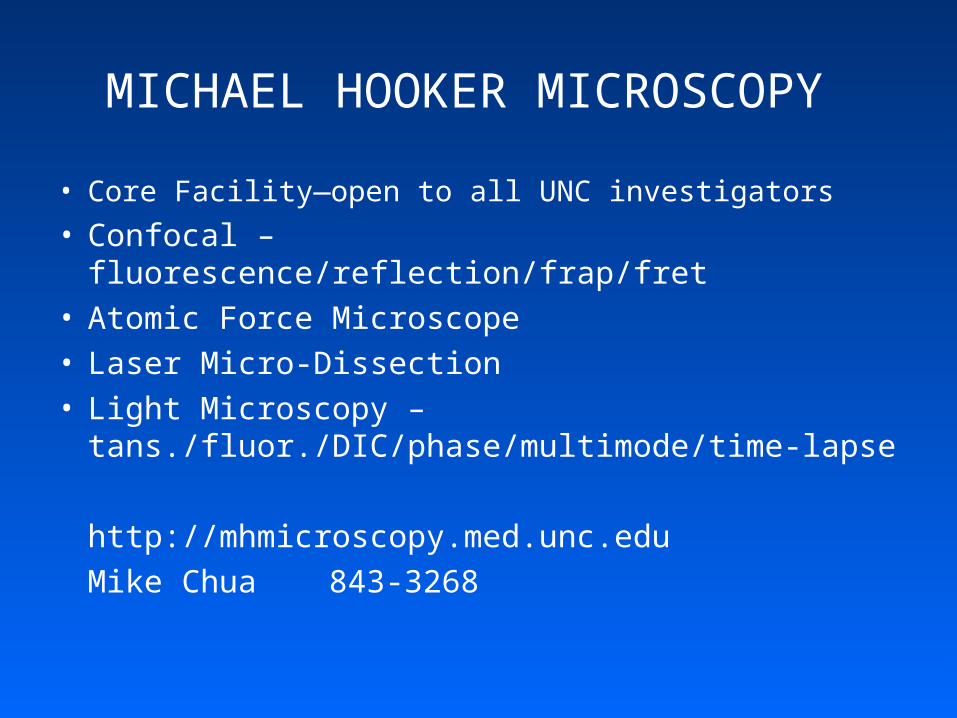
MICROSCOPY SERVICES
• UNC Core facility• Confocal• Laser Capture Microscopy• Electron Microscopy• Light Microscopy
http://www.med.unc.edu/microscopy/Bob Bagnell 966-2413
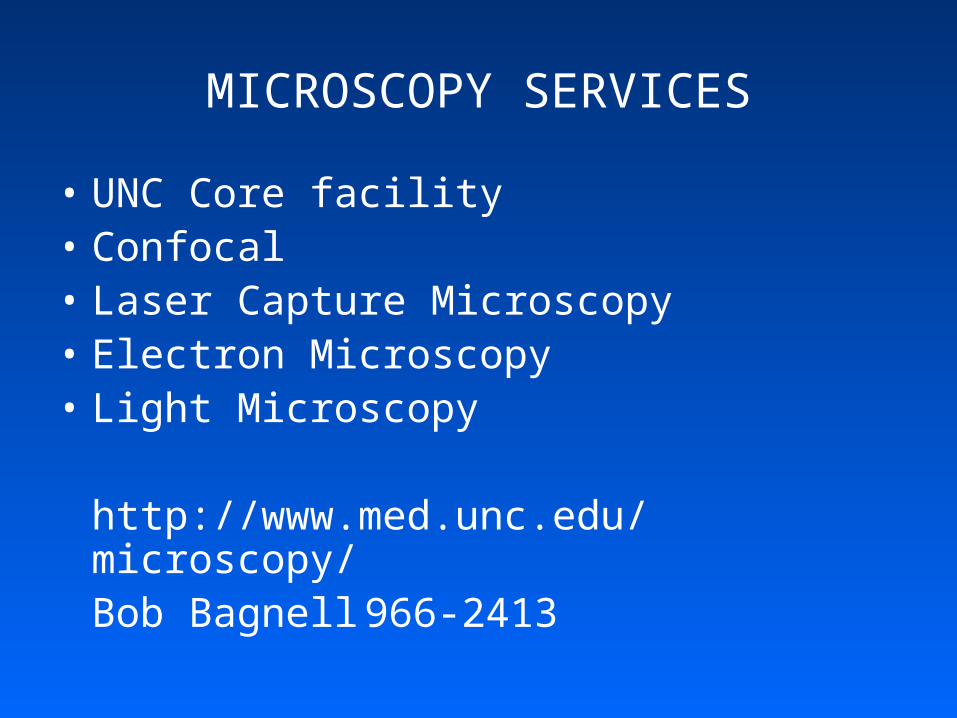
FLOW CYTOMETRY CORE
• UNC Core Facility
• Deconvolution System (3D)
• Laser Scanning Cytometer
• FACS
http://flowcytometry.med.unc.edu/Larry Arnold 966-1530
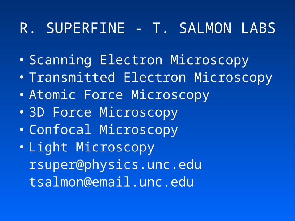
R. SUPERFINE - T. SALMON LABS
• Scanning Electron Microscopy• Transmitted Electron Microscopy• Atomic Force Microscopy• 3D Force Microscopy• Confocal Microscopy• Light Microscopy
[email protected]@email.unc.edu
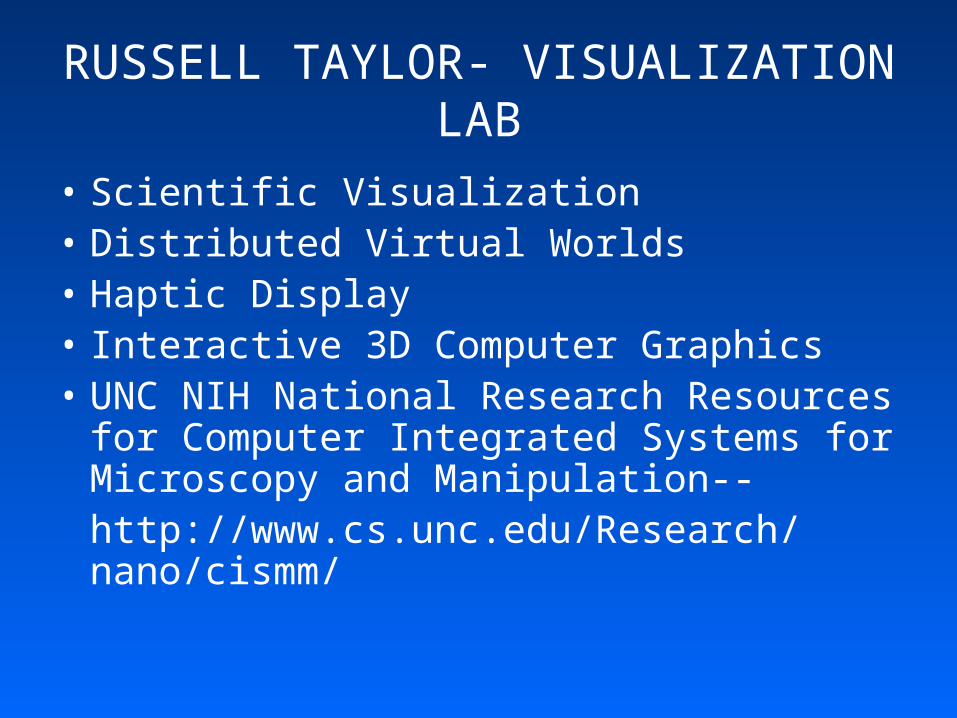
RUSSELL TAYLOR- VISUALIZATION LAB
• Scientific Visualization• Distributed Virtual Worlds• Haptic Display• Interactive 3D Computer Graphics• UNC NIH National Research Resources
for Computer Integrated Systems for Microscopy and Manipulation--http://www.cs.unc.edu/Research/nano/cismm/
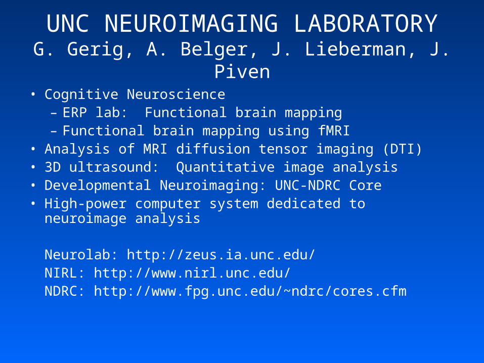
UNC NEUROIMAGING LABORATORYG. Gerig, A. Belger, J. Lieberman, J. Piven
• Cognitive Neuroscience– ERP lab: Functional brain mapping– Functional brain mapping using fMRI
• Analysis of MRI diffusion tensor imaging (DTI)• 3D ultrasound: Quantitative image analysis• Developmental Neuroimaging: UNC-NDRC Core• High-power computer system dedicated to neuroimage analysis
Neurolab: http://zeus.ia.unc.edu/NIRL: http://www.nirl.unc.edu/NDRC: http://www.fpg.unc.edu/~ndrc/cores.cfm
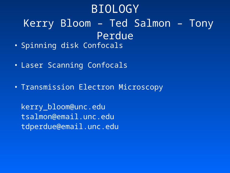
BIOLOGY Kerry Bloom – Ted Salmon – Tony Perdue
• Spinning disk Confocals
• Laser Scanning Confocals
• Transmission Electron Microscopy
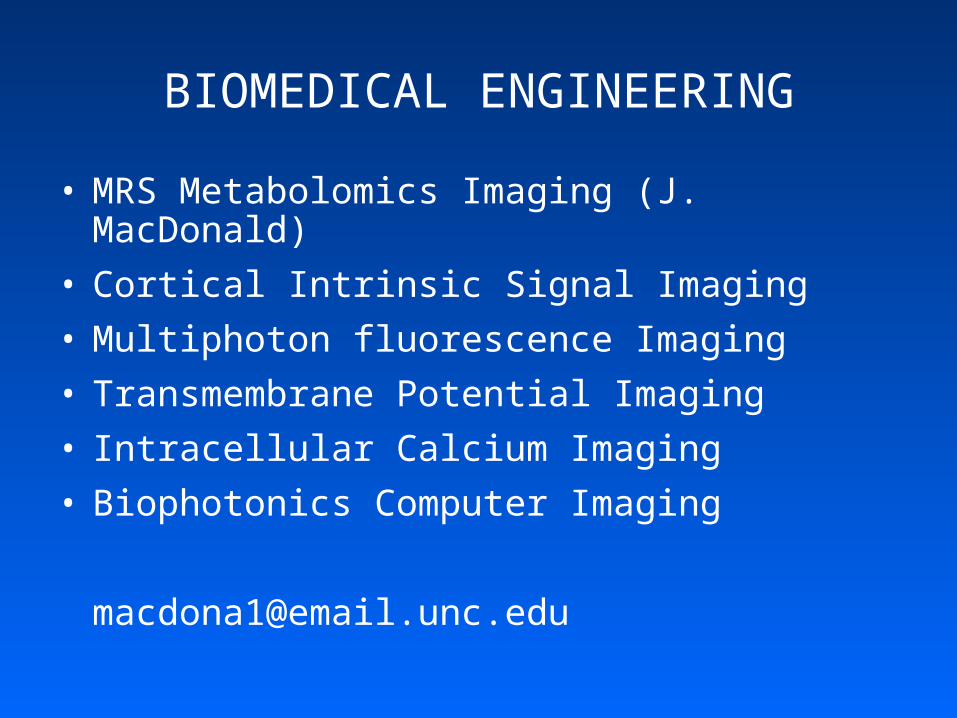
BIOMEDICAL ENGINEERING
• MRS Metabolomics Imaging (J. MacDonald)
• Cortical Intrinsic Signal Imaging
• Multiphoton fluorescence Imaging
• Transmembrane Potential Imaging
• Intracellular Calcium Imaging
• Biophotonics Computer Imaging
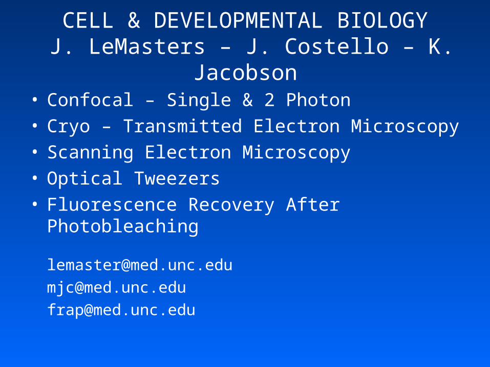
CELL & DEVELOPMENTAL BIOLOGY J. LeMasters – J. Costello – K. Jacobson
• Confocal – Single & 2 Photon• Cryo – Transmitted Electron Microscopy• Scanning Electron Microscopy• Optical Tweezers• Fluorescence Recovery After Photobleaching
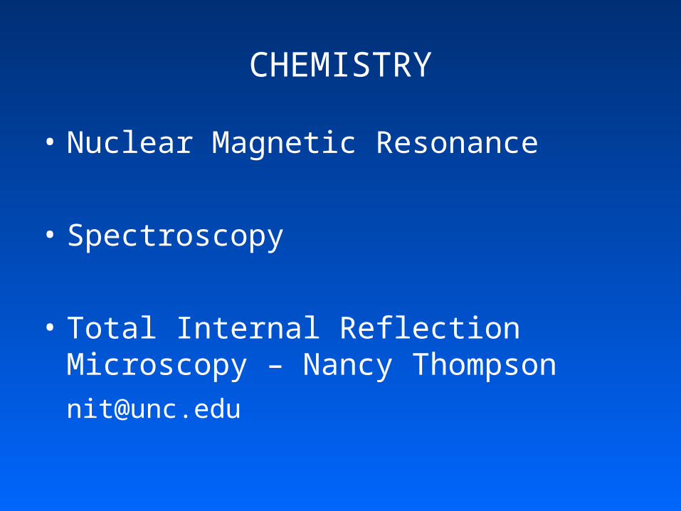
CHEMISTRY
• Nuclear Magnetic Resonance
• Spectroscopy
• Total Internal Reflection Microscopy – Nancy Thompson
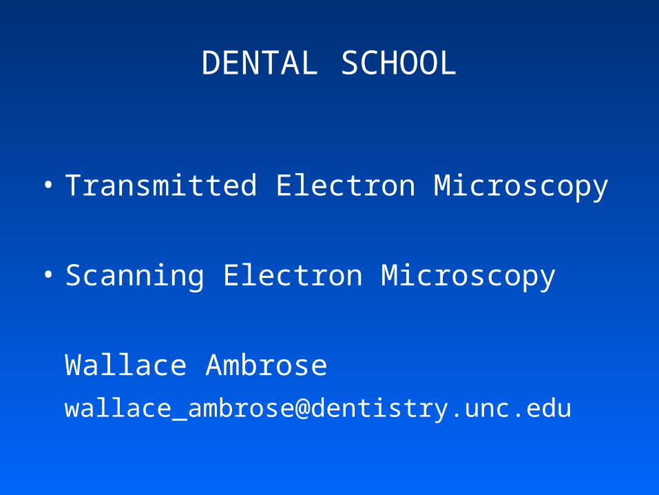
DENTAL SCHOOL
• Transmitted Electron Microscopy
• Scanning Electron Microscopy
Wallace Ambrose
PEDIATRICS/EPA—J. CARSON
• Freeze Fracture
• Transmitted Electron Microscopy
• Scanning Electron Microscopy
• Light Microscopy
RADIATION ONCOLOGYEdward Chaney
• CT
• Segmentation
• Registration
• 3D & 4D Display
RADIOLOGY – RESEARCHWeili Lin– Etta Pisano – Stephen Aylward
• 3 Tesla Magnetic Resonance Imaging (Lin)• Micro Single-Photon Computed Tomography
(Lin)• Diffraction Enhanced Imaging(Pisano)• Computer Aided Diagnosis and Display Lab
(CADDLab: Aylward)-- 2D and 3D image segmentation and registration--Multi-modal image resgistration
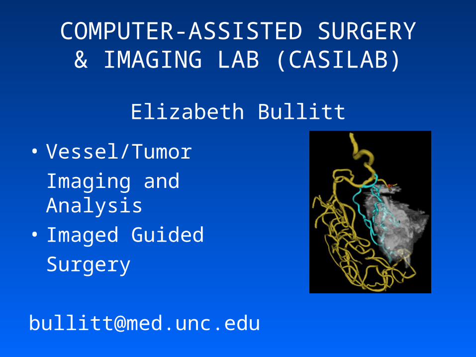
COMPUTER-ASSISTED SURGERY& IMAGING LAB (CASILAB)
Elizabeth Bullitt
• Vessel/Tumor
Imaging and Analysis
• Imaged Guided
Surgery
GENE THERAPY CENTER
• Optical Imaging (Cryocooled, luminescence and fluorescence)
Allison Hawke
LINEBERGER COMPREHENSIVE CANCER CENTER
• Light Microscopy
• Fluorescence Microscopy
Steve Oglesbee
NEUROSCIENCES & NEURODEVELOPMENTAL DISORDERS RESEARCH CENTERS
Eva Anton - Robert Sealock
• Confocals – Single & 2-Photon
• Light Microscopy
[email protected]@med.unc.edu
MEDICAL IMAGE DISPLAY & ANALYSIS GROUP (MIDAG)
• At Comp Sci, Psychiatry, Rad Onc, Radiology, Surgery
• Analysis Capabilities• Image Analysis• Image Display• Augmented Reality (Computer Science)• Segmentation• Registration
http://midag.cs.unc.edu
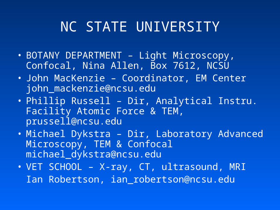
NC STATE UNIVERSITY
• BOTANY DEPARTMENT – Light Microscopy, Confocal, Nina Allen, Box 7612, NCSU
• John MacKenzie – Coordinator, EM Center [email protected]
• Phillip Russell – Dir, Analytical Instru. Facility Atomic Force & TEM, [email protected]
• Michael Dykstra – Dir, Laboratory Advanced Microscopy, TEM & Confocal [email protected]
• VET SCHOOL – X-ray, CT, ultrasound, MRIIan Robertson, [email protected]
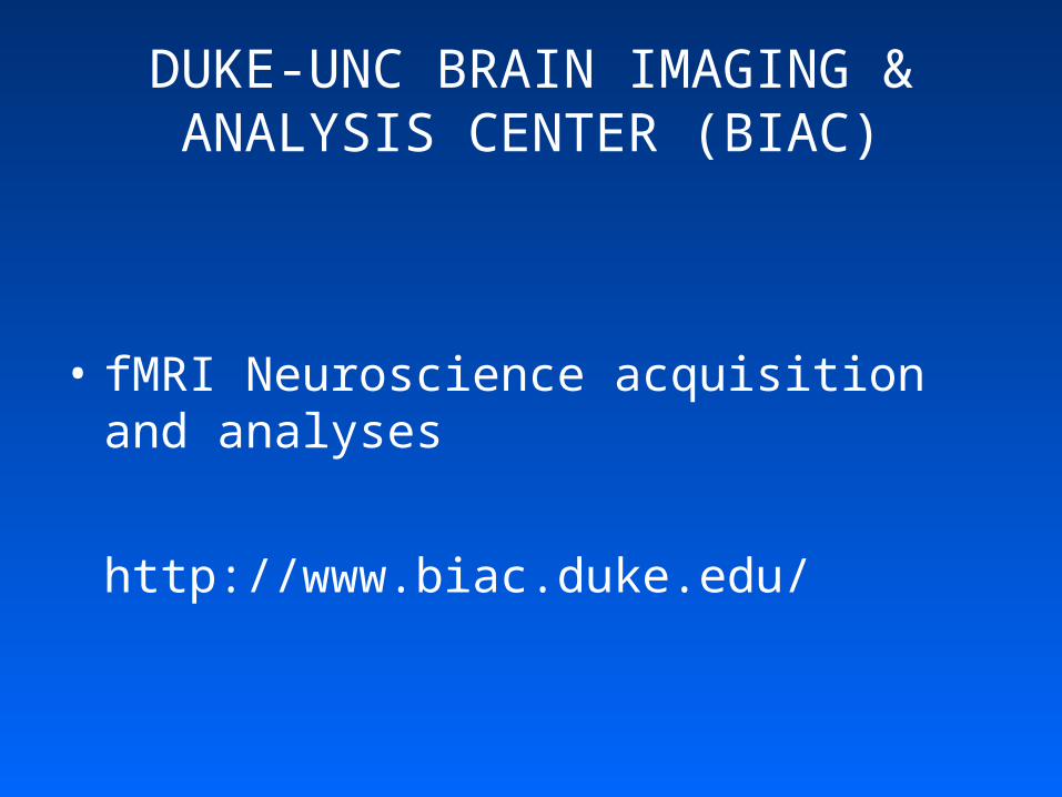
DUKE-UNC BRAIN IMAGING & ANALYSIS CENTER (BIAC)
• fMRI Neuroscience acquisition and analyses
http://www.biac.duke.edu/
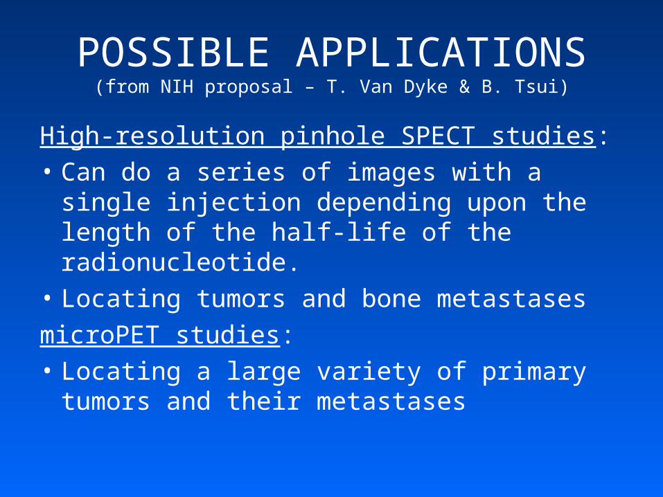
POSSIBLE APPLICATIONS(from NIH proposal – T. Van Dyke & B. Tsui)
High-resolution pinhole SPECT studies:
• Can do a series of images with a single injection depending upon the length of the half-life of the radionucleotide.
• Locating tumors and bone metastases
microPET studies:
• Locating a large variety of primary tumors and their metastases
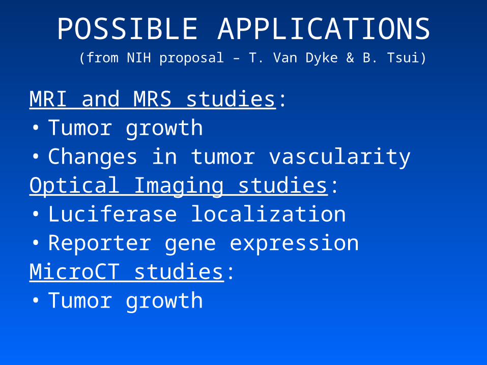
POSSIBLE APPLICATIONS (from NIH proposal – T. Van Dyke & B. Tsui)
MRI and MRS studies:• Tumor growth • Changes in tumor vascularityOptical Imaging studies:• Luciferase localization• Reporter gene expressionMicroCT studies:• Tumor growth
FUTURE DEVELOPMENTS(from NIH proposal – T. Van Dyke & B. Tsui)
• Use Animal Model systems for refining specific imaging technologies—i.e. technologies to measure blood flow and microvascular permeability within tumors.
• Imaging results compared to histologic analyses can help develop technologies to measure disease progression and then be used to screen development of disease in backgrounds with additional genetic alterations.