Embed Size (px)
Citation preview

REVIEW Open Access
Micro-CT – a digital 3D microstructuralvoyage into scaffolds: a systematic reviewof the reported methods and resultsIbrahim Fatih Cengiz1,2*, Joaquim Miguel Oliveira1,2,3 and Rui L. Reis1,2,3
Abstract
Background: Cell behavior is the key to tissue regeneration. Given the fact that most of the cells used in tissueengineering are anchorage-dependent, their behavior including adhesion, growth, migration, matrix synthesis, anddifferentiation is related to the design of the scaffolds. Thus, characterization of the scaffolds is highly required.Micro-computed tomography (micro-CT) provides a powerful platform to analyze, visualize, and explore any portionof interest in the scaffold in a 3D fashion without cutting or destroying it with the benefit of almost no samplepreparation need.
Main body: This review highlights the relationship between the scaffold microstructure and cell behavior, andprovides the basics of the micro-CT method. In this work, we also analyzed the original papers that were publishedin 2016 through a systematic search to address the need for specific improvements in the methods section of thepapers including the amount of provided information from the obtained results.
Conclusion: Micro-CT offers a unique microstructural analysis of biomaterials, notwithstanding the associatedchallenges and limitations. Future studies that will include micro-CT characterization of scaffolds should report theimportant details of the method, and the derived quantitative and qualitative information can be maximized.
Keywords: Systematic review, Microstructure, Mineral density, Scaffolds, Tissue engineering
BackgroundIn nature, structural materials including animal and hu-man tissues have complex hierarchical architectures atmultiple scales from nano to macro [1]. To achieve theregeneration of functional tissues, as in nature, completeunderstanding and biomimicry of those 3D architecturesare necessary. Differences in the design of 3D scaffolds[2] such as composition, surface chemistry, architecture,and mechanical properties, can yield to almost countlessdifferent scaffolds. Scaffolds host and interact with cells,and the design of a scaffold affects the entire behavior ofthe cells including adhesion, growth, migration, differenti-ation, and matrix synthesis [3–8]. Certainly, the functional
performance of a scaffold in vivo not only depends on itsmicrostructure but also on all other factors involved in tis-sue engineering which are very complex and probably notyet completely known [9].Tomography is defined as a method by which an object’s
3D image that corresponds to its internal structure is ob-tained. Micro-computed tomography (micro-CT) [10, 11]is a high-resolution CT that has a pixel size typicallybetween 1 μm and 50 μm, and allows to investigate themicrostructure of samples using X-rays. Conventionally,the samples can be analyzed almost without any samplepreparation process generally in a non-destructive way.Today, a search on “micro-CT” in PubMed yields morethan 10,000 items by being used in many fields. Withinthe field of tissue engineering it has a place in many ap-plication domains including (i) scaffold characterization[12–15], (ii) in vivo small laboratory animal tissuecharacterization [16–19] including assessment of boneturnover using 4D micro-CT data [20], and tumor detec-tion [19], (iii) ex vivo characterization of human tissues
* Correspondence: [email protected]’s Research Group, I3Bs – Research Institute on Biomaterials,Biodegradables and Biomimetics, University of Minho, Headquarters of theEuropean Institute of Excellence on Tissue Engineering and RegenerativeMedicine, AvePark, Parque de Ciência e Tecnologia, Zona Industrial daGandra, Barco, 4805-017 Guimarães, Portugal2ICVS/3B’s – PT Government Associate Laboratory, Braga/Guimarães, PortugalFull list of author information is available at the end of the article
© The Author(s). 2018 Open Access This article is distributed under the terms of the Creative Commons Attribution 4.0International License (http://creativecommons.org/licenses/by/4.0/), which permits unrestricted use, distribution, andreproduction in any medium, provided you give appropriate credit to the original author(s) and the source, provide a link tothe Creative Commons license, and indicate if changes were made. The Creative Commons Public Domain Dedication waiver(http://creativecommons.org/publicdomain/zero/1.0/) applies to the data made available in this article, unless otherwise stated.
Cengiz et al. Biomaterials Research (2018) 22:26 https://doi.org/10.1186/s40824-018-0136-8
[21, 22] and animal tissues [23–27]. Micro-CT has fre-quently been used in bone studies, and typically, the in-vestigated parameters include volume, microstructuralfeatures, and mineral density. The investigation of softtissues is relatively challenging due to their low contrastin conventional micro-CT imaging; thus it may requirean extra effort such as employing high-atomic-numberelement probes [28] or contrast agents [29–32].
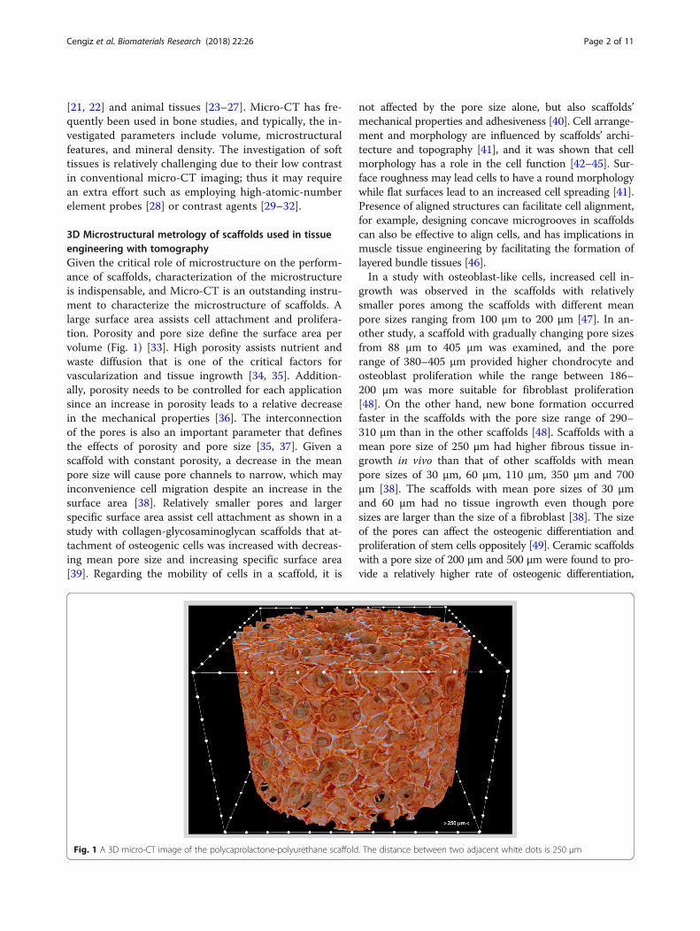
3D Microstructural metrology of scaffolds used in tissueengineering with tomographyGiven the critical role of microstructure on the perform-ance of scaffolds, characterization of the microstructureis indispensable, and Micro-CT is an outstanding instru-ment to characterize the microstructure of scaffolds. Alarge surface area assists cell attachment and prolifera-tion. Porosity and pore size define the surface area pervolume (Fig. 1) [33]. High porosity assists nutrient andwaste diffusion that is one of the critical factors forvascularization and tissue ingrowth [34, 35]. Addition-ally, porosity needs to be controlled for each applicationsince an increase in porosity leads to a relative decreasein the mechanical properties [36]. The interconnectionof the pores is also an important parameter that definesthe effects of porosity and pore size [35, 37]. Given ascaffold with constant porosity, a decrease in the meanpore size will cause pore channels to narrow, which mayinconvenience cell migration despite an increase in thesurface area [38]. Relatively smaller pores and largerspecific surface area assist cell attachment as shown in astudy with collagen-glycosaminoglycan scaffolds that at-tachment of osteogenic cells was increased with decreas-ing mean pore size and increasing specific surface area[39]. Regarding the mobility of cells in a scaffold, it is
not affected by the pore size alone, but also scaffolds’mechanical properties and adhesiveness [40]. Cell arrange-ment and morphology are influenced by scaffolds’ archi-tecture and topography [41], and it was shown that cellmorphology has a role in the cell function [42–45]. Sur-face roughness may lead cells to have a round morphologywhile flat surfaces lead to an increased cell spreading [41].Presence of aligned structures can facilitate cell alignment,for example, designing concave microgrooves in scaffoldscan also be effective to align cells, and has implications inmuscle tissue engineering by facilitating the formation oflayered bundle tissues [46].In a study with osteoblast-like cells, increased cell in-
growth was observed in the scaffolds with relativelysmaller pores among the scaffolds with different meanpore sizes ranging from 100 μm to 200 μm [47]. In an-other study, a scaffold with gradually changing pore sizesfrom 88 μm to 405 μm was examined, and the porerange of 380–405 μm provided higher chondrocyte andosteoblast proliferation while the range between 186–200 μm was more suitable for fibroblast proliferation[48]. On the other hand, new bone formation occurredfaster in the scaffolds with the pore size range of 290–310 μm than in the other scaffolds [48]. Scaffolds with amean pore size of 250 μm had higher fibrous tissue in-growth in vivo than that of other scaffolds with meanpore sizes of 30 μm, 60 μm, 110 μm, 350 μm and 700μm [38]. The scaffolds with mean pore sizes of 30 μmand 60 μm had no tissue ingrowth even though poresizes are larger than the size of a fibroblast [38]. The sizeof the pores can affect the osteogenic differentiation andproliferation of stem cells oppositely [49]. Ceramic scaffoldswith a pore size of 200 μm and 500 μm were found to pro-vide a relatively higher rate of osteogenic differentiation,
Fig. 1 A 3D micro-CT image of the polycaprolactone-polyurethane scaffold. The distance between two adjacent white dots is 250 μm
Cengiz et al. Biomaterials Research (2018) 22:26 Page 2 of 11

and proliferation, respectively, when compared to eachother [49]. Both surface chemistry and pore size can signifi-cantly affect the lineage-specific differentiation of stem cellswhile the effect of surface chemistry found to be relativelylarger if the pore size is smaller than 300 μm [50].Micro-CT characterization comprises three major sequen-
tial processes: acquisition, reconstruction, and analysis [13].The X-rays are generated by the source and emitted to thetarget sample. When passing through the sample, the X-raysare attenuated based on the properties of a sample that isbeing scanned (e.g., its density, thickness, and composition).The acquisition is completed by collecting 2D projection im-ages (radiographs) from many viewing angles. In the conven-tional micro-CT instruments, the X-ray system is fixed, andthe sample stage rotates, while in the instruments for livesmall animals the stage is fixed, and the X-ray system rotatesto scan the animal. The projection images are reconstructedby a computer using algorithms [51–53] to obtain thecross-sectional 2D images in the transverse plane (Fig. 2).Binarization is the process by which the pixels that belong tosample are discriminated in the reconstructed images thatare in a contrast scale of 0-255, and the images are madeblack and white (Fig. 3). Typically, white indicates the mater-ial while black indicates empty space. Volume, porosity, poresize, and suchlike results can be quantified using the binaryimage dataset. Commercial micro-CT instruments comewith the manufacturers’ software while an external softwaresuch as ImageJ (https://imagej.nih.gov/ij/index.html)could also be used.
Systematic search of the papers that were published in 2016A systematic search was performed on the papers thatwere published in a predetermined year, 2016, to answerthese questions: (i) what type of data was reported in the
papers, i.e., quantitative or qualitative, or both, (ii) whatkind of quantitative results were reported (such as micro-structure (e.g., porosity, pore size, wall thickness), volume,and mineral density if relevant), and (iii) whether thereporting of the methods were adequate including the in-formation on pixel size, number of replicates, rotation stepor number of obtained projections, voltage and currentvalues, and use of any filter). A search was performed onthe electronic databases of Scopus, Web of Science, andPubMed using the term “micro-CT” with “scaffold” toidentify the relevant original research papers in Englishthat were published in 2016. The year 2016 was selectedin the current work because it was presumed as a repre-sentative period. The papers that involve micro-CTcharacterization of scaffolds were considered as eligible.The papers were screened, selected as shown in the flow-chart that is presented in Fig. 4, and analyzed to answerthe aforementioned questions.
Findings of the systematic search and discussionA total of 105 papers [54–158] were included in thesystematic analysis. The data indicated that micro-CTwas used only qualitatively in around 15% of the papers[63, 74, 75, 79, 95, 102, 106, 108, 112, 116, 122, 129,133, 146, 149, 158], and only quantitatively in 9.5% ofthe papers [55, 78, 83, 85, 98, 111, 125, 131, 137, 155].Micro-CT was used both qualitatively and quantita-tively in the rest of the papers [54, 56–62, 64–73, 76,77, 80–82, 84, 86–94, 96, 97, 99–101, 103–105, 107,109, 110, 113–115, 117–121, 123, 124, 126–128, 130,132, 134–136, 138–145, 147, 148, 150–154, 156, 157].Only around 29% of the papers reported the number ofreplicates (i.e., “n”) they analyzed, and around 77% ofthe papers reported the used pixel size. Among the
Fig. 2 Schematic illustration showing the basics of micro-CT. Cone-beam X-rays travel from the source to the detector through thesample with attenuation, and a gray-scale projection image is acquired at each rotation angle. Projection images are then reconstructed,and the reconstructed image dataset is used for analysis. The red dashed line indicates the vertical position of the reconstructed image,i.e., the cross-sectional image
Cengiz et al. Biomaterials Research (2018) 22:26 Page 3 of 11

papers that reported n, the mean size of n was 4.4 with astandard deviation (SD) of 2.8. The mean used pixel sizewas 16.8 μm with an SD of 13.3. The analysis also showedthat less than 3% of the papers used the term “spatial reso-lution”, and around 14% of the papers used the term “reso-lution” which might get confused with “pixel size” as it wasdiscussed below. As Fig. 5 illustrates, volume measure-ments, microstructural characterization, and mineral dens-ity determination are the three most frequent quantitative
results. The results categorized as “other” include histo-grams, area/distance measurements such as tissue thick-ness, bone contact area or graft diameter, callus index andintensity signals. The rotation step, or the number of ob-tained projections, is not mentioned in over 68% of the pa-pers. The information on the used voltage and current wasnot reported in around 26% of the papers, while around73% of the papers did not report whether they used a filteror not. One paper was identified that the term “in vivoimaging” was used when only explants were characterized.The internal architecture of a scaffold has major roles in
the cell behavior and thus on the overall performance ofthe construct, the characterization of its macro (> 100 μm)and micro (0.1 μm - 100 μm) structure is inherently ofinterest. With the demonstrated use and advantages ofmicro-CT, it has been a valuable instrument in researchlaboratories with its challenges that are summarized inTable 1.Micro-CT has been considered as the gold standard
for bone explants’ microstructure and morphology study[25]. In the case of scanning live animals, excessive ex-posure to radiation [159–161] can be a problem for thesurvival of the animal, tumorigenesis, and ethics. Using apixel size less than 50 μm may be fine to prevent exces-sive exposure to radiation [11].From the results of this systematic analysis, two major
points can be emphasized to be resolved in future studiesthat will use micro-CT: (i) methods section from acquisi-tion to analysis should be complete, and (ii) informationobtained should be maximized. The methods part shouldcover the used pixel size, voltage and current, number ofreplicates, scanning medium, use of any filter, rotation step/number of projections, and use of any specific image pro-cessing. Various results can be derived from a Micro-CTcharacterization. These results can be quantitative or quali-tative, and obtained in a 2D or 3D fashion. Qualitativeresults include X-ray and reconstructed image of the sam-ple, mineral density color map, pores size color map, andstructure thickness color map, and thus, provide valuableinformation. Quantitative results include quantification/proportion of volumes, microstructural features (mean poresize, mean wall thickness, and their distribution and changevertically across the sample), mineral density, histograms,quantification of surface area, and surface:volume ratio. Itshould be noted that some of these parameters are relevantonly for certain studies. For instance, bone mineral densityis related only with scaffolds for bone tissue engineering.Therefore, this is a factor affecting the outcome of thesystematic analysis.CT experts may accept that resolution is a quite sensi-
tive topic among authors by being a measure of imagequality. As the outcome of the systematic analysis shows,some authors including some of the CT experts preferto use terms “resolution” and “spatial resolution” instead
Fig. 3 Binarization of reconstructed micro-CT images. Gray-scalereconstructed 2D image of a silk-based tissue engineering scaffoldwith a circular region of interest (ø 3 mm) (a), half-tone views(b, d, and f) and the corresponding binary images (c, e, and g)respectively if no gray-scale value is included that yields to completeblack, if entire gray-scale values are included that yields to completewhite, and if the right gray-scale values (that is in this case 38-255) areobtained by global thresholding that yields to a binary image showingthe microstructure of the scaffold
Cengiz et al. Biomaterials Research (2018) 22:26 Page 4 of 11

of “pixel size” or “voxel size”, while some micro-CT sys-tem manufacturers prefer to communicate the size ofthe pixels or voxel of the images. Additionally, there isno standardized approach for spatial resolution reportingaccepted by the micro-CT system manufacturers [25].The definitions of these terms are presented in Table 2.
The studies that report a value for resolution need toshow how they quantify the resolution whether it is thepixel size given by the scanner software or they quanti-fied it with calibrated thickness wires [162]. Given thedefinitions of the terms, the images that have the samenumber of pixels would contain different information iftheir pixel sizes are not identical. It is noteworthy toknow that a smaller pixel size does not necessarily pro-vide a better image, for instance, the image does not im-prove when the pixel size is smaller than the X-ray spotsize [162, 163]. Some other factors [25, 162, 164, 165]are needed to be considered as well, regarding X-ray(e.g., energy, geometry, and focal-spot size), sensitivity ofdetector, use of filter, integration time of X-ray with thesample per projection, characteristics of the sample (e.g.,composition and size), scanning medium, noise and arti-facts. The attenuation of the X-rays depends on the char-acteristics of the sample such as its density and thickness.The level of the X-ray energy, i.e., the source voltage andcurrent, should be tuned for the sample (denser samplesrequire higher energy) since the obtained the images are ingray-scale and associated with the X-ray absorption that islinked with the sample’s electron density [163]. Therefore,the reconstructed images are quantitatively densitometric.The pixel size and rotation step can have significant influ-ences on the quantitative and qualitative results of theμ-CT characterization of scaffolds, as well as the size of
Fig. 4 The flowchart of the systematic search
Fig. 5 Venn diagram showing the number of the papers thatreported quantitative results regarding the volume, microstructure,mineral density and other measures. Two of the seven papers thatreported volume, microstructure, and mineral density also reportedadditional results. The sizes of the circles in the diagrams are directlyproportional to the number of the associated papers
Cengiz et al. Biomaterials Research (2018) 22:26 Page 5 of 11

the generated data and the duration of the characterization[13]. Integration time is the duration of each projection. Alonger integration time provides more photos to be de-tected and affect the micro-CT image [166]. The use of afilter can improve the characterization of thick/dense sam-ples by attenuating the low-energy X-rays and minimizingthe beam hardening effect [25]. Spatial resolution dependson the geometry of the X-ray, and detector [10]. Conebeam X-rays and flat panel detectors provide significantlyshorter scanning times because the data for multiple slicesis obtained in each rotation step, while fan beam X-raysand linear detectors provide less scatter effects forthick samples [167] and higher accuracy [164]. Re-construction method also depends on the geometryof the source. The scanning medium (e.g., air, water,phosphate-buffered saline, or ethanol) influences thecharacterization results since different media affectthe X-ray attenuation differently [25, 168].
ConclusionThe microstructure of tissue engineering scaffoldsgreatly influence the behavior of cells, and the perform-ance of the tissue engineering construct; therefore, thecharacterization of the scaffolds’ microstructure is of
Table 1 Challenges in the conventional micro-CT characterization
Challenge Potential solution Reference
Artifacts and noise. Detailed in Table 1 of Ref. [162] [162, 164, 169, 170]
Soft tissue characterization is not as easy as a dry-stateporous scaffold.
Using contrast agents or high-atomic-numberelement probes can help.
[28–32]
Operator-determined acquisition parameters may affectthe results.
Parameters should be optimized during thepreliminary study.
[13]
Harsh acquisitions may damage/alter the sample/tissue(for example, discoloring of a biomaterial or tumorigenesisin animals); and radiation exposure of animals in liveanimal studies, lethal dose 50/30 (both ethical and scientificconsiderations).
Long scans (either due to very low rotation step,frame averaging, or long exposure time) should beavoided, and/or X-ray energy could be decreased.
[11, 159–161]
Comparing micro-CT results of different studies is not easyif the used parameters are not identical.
There is a need to establish a protocol withdetermined values for parameters.
[13]
Issues with very dense/thick samples resulting in a datasetof almost only black images.
This is because there will be no contrast sinceno X-ray can pass; however, use of a filter mayresolve the problem, but it may greatly increasethe acquisition time. The sample can be cut toits smaller representative volume. If it is notpossible, then another instrument could be usedsuch as a scanning electron microscope.
Issues with very thin/light samples or a hydrogel, then nocontrast will be obtained (this gives a dataset of imageswith very low contrast).
Contrast agents can be used. [29–32]
A limited volume of sample that can be analyzed at once(the images have a certain number of pixels with a certainsize of a pixel).
Display matrix size and/or pixel size can beadapted. The representative portion of thesample can be pre-determined.
Overlaps in gray-scale values in multi-material samples(i.e., scaffold-bone explants or composites).
Advanced segmentation protocol can be used. [171]
Considerations on the maintenance and sharing of micro-CT data. During the preliminary study, the duration of themicro-CT characterization and the disc spacerequirements can be estimated.
[13]
Table 2 Terms that are associated with the micro-CT images,and their definitions
Term Definition
Pixel size Size of the 2D discrete parts that make up a 2Dmicro-CT image. Usually, expressed as a singlevalue, e.g., 10 μm, indicating the size of eachpixel is 10 μm × 10 μm.
Voxel size The 3D equivalent of pixel size indicates the sizeof each voxel, Typically, micro-CT images isotropic(identical size in all dimensions), e.g., 10 μm × 10 μm× 10 μm, in which case pixel size and voxel sizeprovide identical information.
Resolution ▪ Smallest perceptible detail (complete and exactdefinition can be found in Ref. [164])
▪ Resolution of an image can indicate the numberof pixels in an image (such as 800 × 600). Thesecond definition is referred to the display matrixsize in the standard guide of ASTM International:E1441-11.
Spatial resolution ▪ Smallest displacement that can be measured inthe measurement direction [164]
▪ Smallest separation where two points can beidentified as separate parts (complete and exactdefinition can be found in the standard guide ofASTM International: E1441-11)
Cengiz et al. Biomaterials Research (2018) 22:26 Page 6 of 11

keen interest. Micro-CT is an outstanding instrument forthe quantitative 2D and 3D analysis and visualization ofscaffolds. The analysis results showed that the methodssections of the majority of the analyzed papers are in needof improvement in reporting the details of micro-CTcharacterization. Moreover, the amount of quantitative andqualitative information from micro-CT characterizationcan be maximized. Given the fact that the obtained resultsfrom micro-CT characterization significantly depend onseveral parameters, the important acquisition related de-tails should be clearly provided in the papers. It is recom-mended that the parameters include the used pixel size,rotation step, X-ray energy, scanning medium, use of filter,as well as the number of samples analyzed since the omit-ted data complicate the reproducibility of the experiments.
AcknowledgmentsThis work is a result of the project FROnTHERA (NORTE-01-0145-FEDER-000023)supported by the Norte Portugal Regional Operational Programme (NORTE2020) under the Portugal 2020 Partnership Agreement, through EuropeanRegional Development Fund (ERDF). IFC thanks the Portuguese Foundation forScience and Technology (FCT) for the Ph.D. scholarship (SFRH/BD/99555/2014).JMO also thanks the FCT for the funds provided under the programInvestigador FCT 2012 and 2015 (IF/00423/2012 and IF/01285/2015).
FundingFROnTHERA Project (NORTE-01-0145-FEDER-000023) within the Norte PortugalRegional Operational Programme (NORTE 2020) under the Portugal 2020Partnership Agreement, through ERDF, and Portuguese Foundation for Scienceand Technology (SFRH/BD/99555/2014, IF/00423/2012, and IF/01285/2015).
Author’s contributionsAll authors wrote and revised the manuscript. All authors read and approvedthe final manuscript.
Ethics approval and consent to participateNot applicable.
Consent for publicationNot applicable.
Competing interestsThe authors declare that they have no competing interests.
Publisher’s NoteSpringer Nature remains neutral with regard to jurisdictional claims inpublished maps and institutional affiliations.
Author details13B’s Research Group, I3Bs – Research Institute on Biomaterials,Biodegradables and Biomimetics, University of Minho, Headquarters of theEuropean Institute of Excellence on Tissue Engineering and RegenerativeMedicine, AvePark, Parque de Ciência e Tecnologia, Zona Industrial daGandra, Barco, 4805-017 Guimarães, Portugal. 2ICVS/3B’s – PT GovernmentAssociate Laboratory, Braga/Guimarães, Portugal. 3The Discoveries Centre forRegenerative and Precision Medicine, Headquarters at University of Minho,AvePark, Parque de Ciência e Tecnologia, Zona Industrial da Gandra, Barco,4805-017 Guimarães, Portugal.
Received: 12 June 2018 Accepted: 3 September 2018
References1. Wegst UG, Bai H, Saiz E, Tomsia AP, Ritchie RO. Bioinspired structural
materials. Nat Mater. 2015;14(1):23–36.
2. Lee J, Cuddihy MJ, Kotov NA. Three-dimensional cell culture matrices: Stateof the art. Tissue Eng Part B-Re. 2008;14(1):61–86.
3. Baker BM, Chen CS. Deconstructing the third dimension - How 3D culturemicroenvironments alter cellular cues. J Cell Sci. 2012;125(13):3015–24.
4. Cengiz IF, Pereira H, de Girolamo L, Cucchiarini M, Espregueira-Mendes J,Reis RL. Oliveira JM Orthopaedic regenerative tissue engineering en routeto the holy grail: disequilibrium between the demand and the supply in theoperating room. J Exp Orthop. 2018;5(1):14.
5. Chew SY, Low WC. Scaffold-based approach to direct stem cell neural andcardiovascular differentiation: An analysis of physical and biochemicaleffects. J Biomed Mater Res A. 2011;97(3):355–74.
6. Kim K, Yeatts A, Dean D, Fisher JP. Stereolithographic bone scaffold designparameters: Osteogenic differentiation and signal expression. Tissue EngPart B-Re. 2010;16(5):523–39.
7. Yamamoto M, Rafii S, Rabbany SY. Scaffold biomaterials for nano-pathophysiology. Adv Drug Del Rev. 2014;74:104–14.
8. Zhang B, Xiao Y, Hsieh A, Thavandiran N, Radisic M. Micro-andnanotechnology in cardiovascular tissue engineering. Nanotechnology.2011;22(49):494003.
9. Cengiz IF, Pereira H, Espregueira-Mendes J, Oliveira JM, Reis RL. Treatmentsof meniscus lesions of the knee: Current concepts and future perspectives.Regen Eng Transl Med. 2017;3(1):1–19.
10. Landis EN. Keane DT X-ray microtomography. Mater Charact. 2010;61(12):1305–16.
11. Ritman EL. Micro-computed tomography - Current status anddevelopments. Annu Rev Biomed Eng. 2004;6:185–208.
12. Cengiz IF, Pitikakis M, Cesario L, Parascandolo P, Vosilla L, Viano G,Oliveira J, Reis R. Building the basis for patient-specific meniscalscaffolds: From human knee MRI to fabrication of 3D printed scaffolds.Bioprinting. 2016;1:1–10.
13. Cengiz IF, Oliveira JM, Reis RL. Micro-computed tomographycharacterization of tissue engineering scaffolds: Effects of pixel size androtation step. J Mater Sci Mater Med. 2017;28(8):129.
14. Ho ST, Hutmacher DW. A comparison of micro-CT with other techniquesused in the characterization of scaffolds. Biomaterials. 2006;27(8):1362–76.
15. Jones AC, Arns CH, Sheppard AP, Hutmacher DW, Milthorpe BK, KnackstedtMA. Assessment of bone ingrowth into porous biomaterials using Micro-CT.Biomaterials. 2007;28(15):2491–504.
16. Lienemann PS, Metzger S, Kiveliö A-S, Blanc A, Papageorgiou P, AstolfoA, Pinzer BR, Cinelli P, Weber FE, Schibli R. Longitudinal in vivoevaluation of bone regeneration by combined measurement of multi-pinhole SPECT and micro-CT for tissue engineering. Scientific Reports.2015;5:10238.
17. Luu Y, Lublinsky S, Ozcivici E, Capilla E, Pessin J, Rubin C, Judex S. In vivoquantification of subcutaneous and visceral adiposity by micro-computedtomography in a small animal model. Med Eng Phys. 2009;31(1):34–41.
18. Paulus MJ, Gleason SS, Kennel SJ, Hunsicker PR, Johnson DK. High resolutionX-ray computed tomography: An emerging tool for small animal cancerresearch. Neoplasia. 2000;2(1-2):62–70.
19. Waarsing J, Day J, Van der Linden J, Ederveen A, Spanjers C, De Clerck N,Sasov A, Verhaar J, Weinans H. Detecting and tracking local changes in thetibiae of individual rats: A novel method to analyse longitudinal in vivomicro-CT data. Bone. 2004;34(1):163–9.
20. Birkhold AI, Razi H, Weinkamer R, Duda GN, Checa S, Willie BM. Monitoringin vivo (re) modeling: A computational approach using 4D microCT data toquantify bone surface movements. Bone. 2015;75:210–21.
21. Hildebrand T, Laib A, Müller R, Dequeker J, Rüegsegger P. Direct three-dimensional morphometric analysis of human cancellous bone:Microstructural data from spine, femur, iliac crest, and calcaneus. J BoneMiner Res. 1999;14(7):1167–74.
22. Pereira H, Caridade S, Frias A, Silva-Correia J, Pereira D, Cengiz IF, Mano J,Oliveira JM, Espregueira-Mendes J, Reis R. Biomechanical and cellularsegmental characterization of human meniscus: Building the basis for TissueEngineering therapies. Osteoarthritis Cartilage. 2014;22(9):1271–81.
23. Batiste DL, Kirkley A, Laverty S, Thain LM, Spouge AR, Holdsworth DW. Exvivo characterization of articular cartilage and bone lesions in a rabbit ACLtransection model of osteoarthritis using MRI and micro-CT. OsteoarthritisCartilage. 2004;12(12):986–96.
24. Bentley MD, Ortiz MC, Ritman EL, Romero JC. The use of microcomputedtomography to study microvasculature in small rodents. Am J Physiol RegulIntegr Comp Physiol. 2002;282(5):R1267–R79.
Cengiz et al. Biomaterials Research (2018) 22:26 Page 7 of 11

25. Bouxsein ML, Boyd SK, Christiansen BA, Guldberg RE, Jepsen KJ, Müller R.Guidelines for assessment of bone microstructure in rodents using micro-computed tomography. J Bone Miner Res. 2010;25(7):1468–86.
26. Cardeira J, Gavaia PJ, Fernández I, Cengiz IF, Moreira-Silva J, Oliveira JM, ReisRL, Cancela ML, Laizé V. Quantitative assessment of the regenerative andmineralogenic performances of the zebrafish caudal fin. Scientific Reports.2016;6:39191.
27. Layton MW, Goldstein SA, Goulet RW, Feldkamp LA, Kubinski DJ, Bole GG.Examination of subchondral bone architecture in experimental osteoarthritisby microscopic computed axial tomography. Arthritis Rheumatol. 1988;31(11):1400–5.
28. Mizutani R, Suzuki Y. X-ray microtomography in biology. Micron. 2012;43(2):104–15.
29. Lusic H, Grinstaff MW. X-ray-computed tomography contrast agents. ChemRev. 2012;113(3):1641–66.
30. Pauwels E, Van Loo D, Cornillie P, Brabant L, Van Hoorebeke L. Anexploratory study of contrast agents for soft tissue visualization by means ofhigh resolution X-ray computed tomography imaging. J Microsc. 2013;250(1):21–31.
31. Xing R, Wilde D, McCann G, Ridwan Y, Schrauwen JT, Steen AF, Gijsen FJ,Heiden K. Contrast-enhanced micro-CT imaging in murine carotid arteries: Anew protocol for computing wall shear stress. Biomed Eng Online. 2016;15(2):156.
32. Young S, Kretlow JD, Nguyen C, Bashoura AG, Baggett LS, Jansen JA, WongM, Mikos AG. Microcomputed tomography characterization ofneovascularization in bone tissue engineering applications. Tissue Eng PartB-Re. 2008;14(3):295–306.
33. Yang S, Leong K-F, Du Z, Chua C-K. The design of scaffolds for use in tissueengineering. Part I. Traditional factors. Tissue Eng. 2001;7(6):679–89.
34. Karande TS, Ong JL, Agrawal CM. Diffusion in musculoskeletal tissueengineering scaffolds: Design issues related to porosity, permeability,architecture, and nutrient mixing. Ann Biomed Eng. 2004;32(12):1728–43.
35. Mastrogiacomo M, Scaglione S, Martinetti R, Dolcini L, Beltrame F,Cancedda R, Quarto R. Role of scaffold internal structure on in vivobone formation in macroporous calcium phosphate bioceramics.Biomaterials. 2006;27(17):3230–7.
36. Karageorgiou V, Kaplan D. Porosity of 3D biomaterial scaffolds andosteogenesis. Biomaterials. 2005;26(27):5474–91.
37. Freyman T, Yannas I, Gibson L. Cellular materials as porous scaffolds fortissue engineering. Prog Mater Sci. 2001;46(3):273–82.
38. Yamamoto M, Tabata Y, Kawasaki H, Ikada Y. Promotion of fibrovasculartissue ingrowth into porous sponges by basic fibroblast growth factor.J Mater Sci Mater Med. 2000;11(4):213–8.
39. O’Brien FJ, Harley B, Yannas IV, Gibson LJ. The effect of pore size on celladhesion in collagen-GAG scaffolds. Biomaterials. 2005;26(4):433–41.
40. Peyton SR, Kalcioglu ZI, Cohen JC, Runkle AP, Van Vliet KJ, LauffenburgerDA, Griffith LG. Marrow-derived stem cell motility in 3D synthetic scaffold isgoverned by geometry along with adhesivity and stiffness. BiotechnolBioeng. 2011;108(5):1181–93.
41. Clegg JR, Wechsler ME, Peppas NA. Vision for functionally decorated andmolecularly imprinted polymers in regenerative engineering. Regen EngTransl Med. 2017;3(3):166–75.
42. Folkman J, Moscona A. Role of cell shape in growth control. Nature. 1978;273(5661):345.
43. Kumar G, Tison CK, Chatterjee K, Pine PS, McDaniel JH, Salit ML, YoungMF, Simon CG Jr. The determination of stem cell fate by 3D scaffoldstructures through the control of cell shape. Biomaterials. 2011;32(35):9188–96.
44. Kumar G, Waters MS, Farooque TM, Young MF, Simon CG Jr. Freeformfabricated scaffolds with roughened struts that enhance both stem cellproliferation and differentiation by controlling cell shape. Biomaterials. 2012;33(16):4022–30.
45. McBeath R, Pirone DM, Nelson CM, Bhadriraju K, Chen CS. Cell shape,cytoskeletal tension, and RhoA regulate stem cell lineage commitment. DevCell. 2004;6(4):483–95.
46. Chen S, Nakamoto T, Kawazoe N, Chen G. Engineering multi-layered skeletalmuscle tissue by using 3D microgrooved collagen scaffolds. Biomaterials.2015;73:23–31.
47. Chen J, Paetzell E, Zhou J, Lyons L, Soboyejo W. Osteoblast-like cellingrowth, adhesion and proliferation on porous Ti-6Al-4V with particulateand fiber scaffolds. Mater Sci Eng, C. 2010;30(5):647–56.
48. Oh SH, Park IK, Kim JM, Lee JH. In vitro and in vivo characteristics of PCLscaffolds with pore size gradient fabricated by a centrifugation method.Biomaterials. 2007;28(9):1664–71.
49. Mygind T, Stiehler M, Baatrup A, Li H, Zou X, Flyvbjerg A, Kassem M, BüngerC. Mesenchymal stem cell ingrowth and differentiation on corallinehydroxyapatite scaffolds. Biomaterials. 2007;28(6):1036–47.
50. Zhao Y, Tan K, Zhou Y, Ye Z, Tan W-S. A combinatorial variation in surfacechemistry and pore size of three-dimensional porous poly (ε-caprolactone)scaffolds modulates the behaviors of mesenchymal stem cells. Mater SciEng, C. 2016;59:193–202.
51. Faridani A. Results, old and new, in computed tomography. In: InverseProblems in Wave Propagation. New York: Springer; 1997. p. 167–93.
52. Feldkamp L, Davis L. Kress J Practical cone-beam algorithm. JOSA A. 1984;1(6):612–9.
53. Gordon R, Herman G. Three-dimensional reconstruction from projections: Areview of algorithms. Int Rev Cytol. 1974;38:111–51.
54. Adamzyk C, Kachel P, Hoss M, Gremse F, Modabber A, Hölzle F, Tolba R,Neuss S, Lethaus B. Bone tissue engineering using polyetherketoneketonescaffolds combined with autologous mesenchymal stem cells in a sheepcalvarial defect model. J Craniomaxillofac Surg. 2016;44(8):985–94.
55. Almela T, Brook IM, Moharamzadeh K. Development of three-dimensionaltissue engineered bone-oral mucosal composite models. J Mater Sci MaterMed. 2016;27(4):65.
56. Altamura D, Pastore SG, Raucci MG, Siliqi D, De Pascalis F, Nacucchi M,Ambrosio L, Giannini C. Scanning small-and wide-angle X-ray scatteringmicroscopy selectively probes ha content in gelatin/hydroxyapatitescaffolds for osteochondral defect repair. ACS Appl Mater Inter. 2016;8(13):8728–36.
57. Amini AR, Xu TO, Chidambaram RM, Nukavarapu SP. Oxygen tension-controlled matrices with osteogenic and vasculogenic cells for vascularizedbone regeneration in vivo. Tissue Eng Pt A. 2016;22(7-8):610–20.
58. Arabnejad S, Johnston RB, Pura JA, Singh B, Tanzer M, Pasini D. High-strength porous biomaterials for bone replacement: A strategy to assess theinterplay between cell morphology, mechanical properties, bone ingrowthand manufacturing constraints. Acta Biomater. 2016;30:345–56.
59. Araujo-Pires AC, Mendes VC, Ferreira-Junior O, Carvalho PSP, Guan L, DaviesJE. Investigation of a novel PLGA/CaP scaffold in the healing of toothextraction sockets to alveolar bone preservation in humans. Clin ImplantDent R. 2016;18(3):559–70.
60. Arifvianto B, Leeflang M, Zhou J. Characterization of the porous structures ofthe green body and sintered biomedical titanium scaffolds with micro-computed tomography. Mater Charact. 2016;121:48–60.
61. Barui S, Chatterjee S, Mandal S, Kumar A, Basu B. Microstructure andcompression properties of 3D powder printed Ti-6Al-4V scaffolds withdesigned porosity: Experimental and computational analysis. Mater Sci Eng,C. 2017;70:812–23.
62. Beck A, Murphy DJ, Carey-Smith R, Wood DJ, Zheng MH. Treatment ofarticular cartilage defects with microfracture and autologous matrix-inducedchondrogenesis leads to extensive subchondral bone cyst formation in asheep model. Am J Sport Med. 2016;44(10):2629–43.
63. Bolaños RV, Cokelaere S, McDermott JE, Benders K, Gbureck U, Plomp S,Weinans H, Groll J, van Weeren P, Malda J. The use of a cartilagedecellularized matrix scaffold for the repair of osteochondral defects: Theimportance of long-term studies in a large animal model. Osteoarthr Cartil.2017;25(3):413–20.
64. Carlisle PL, Guda T, Silliman DT, Lien W, Hale RG, Brown Baer PR.Investigation of a pre-clinical mandibular bone notch defect model inminiature pigs: Clinical computed tomography, micro-computedtomography, and histological evaluation. J Korean Assoc Oral MaxillofacSurg. 2016;42(1):20–30.
65. Chamieh F, Collignon A-M, Coyac BR, Lesieur J, Ribes S, Sadoine J, Llorens A,Nicoletti A, Letourneur D, Colombier M-L. Accelerated craniofacial boneregeneration through dense collagen gel scaffolds seeded with dental pulpstem cells. Sci Rep. 2016;6:38814.
66. Chandran S, Babu SS, Varma H, John A. Osteogenic efficacy of strontiumhydroxyapatite micro-granules in osteoporotic rat model. J Biomater Appl.2016;31(4):499–509.
67. Chang B, Song W, Han T, Yan J, Li F, Zhao L, Kou H, Zhang Y. Influence ofpore size of porous titanium fabricated by vacuum diffusion bonding oftitanium meshes on cell penetration and bone ingrowth. Acta Biomater.2016;33:311–21.
Cengiz et al. Biomaterials Research (2018) 22:26 Page 8 of 11

68. Chang Y-L, Lo Y-J, Feng S-W, Huang Y-C, Tsai H-Y, Lin C-T, Fan K-H, HuangH-M. Bone healing improvements using hyaluronic acid andhydroxyapatite/beta-tricalcium phosphate in combination: an animal study.BioMed Res Int. 2016;2016:8301624.
69. Chen G, Yang L, Lv Y. Cell-free scaffolds with different stiffness but samemicrostructure promote bone regeneration in rabbit large bone defectmodel. J Biomed Mater Res A. 2016;104(4):833–41.
70. Costantini M, Colosi C, Mozetic P, Jaroszewicz J, Tosato A, Rainer A,Trombetta M, Święszkowski W, Dentini M, Barbetta A. Correlation betweenporous texture and cell seeding efficiency of gas foaming and microfluidicfoaming scaffolds. Mater Sci Eng, C. 2016;62:668–77.
71. Crica LE, Wengenroth J, Tiainen H, Ionita M, Haugen HJ. Enhanced X-rayabsorption for micro-CT analysis of low density polymers. J Biomater SciPolym Ed. 2016;27(9):805–23.
72. Cui W, Sun G, Qu Y, Xiong Y, Sun T, Ji Y, Yang L, Shao Z, Ma J, Zhang S.Repair of rat calvarial defects using Si-doped hydroxyapatite scaffoldsloaded with a bone morphogenetic protein-2-related peptide. J Orth Res.2016;34(11):1874–82.
73. Ding M, Henriksen SS, Theilgaard N, Overgaard S. Assessment of activatedporous granules on implant fixation and early bone formation in sheep. JOrthop Transl. 2016;5:38–47.
74. Dumont VC, Mansur AA, Carvalho SM, Borsagli FGM, Pereira MM, Mansur HS.Chitosan and carboxymethyl-chitosan capping ligands: Effects on thenucleation and growth of hydroxyapatite nanoparticles for producingbiocomposite membranes. Mater Sci Eng, C. 2016;59:265–77.
75. Dumont VC, Mansur HS, Mansur AA, Carvalho SM, Capanema NS,Barrioni BR. Glycol chitosan/nanohydroxyapatite biocomposites forpotential bone tissue engineering and regenerative medicine. Int J BiolMacromol. 2016;93:1465–78.
76. Erdogan O, Supachawaroj N, Soontornvipart K, Kheolamai P. Treatment ofperi-implant defects in the rabbit’s tibia with adipose or bone marrow-derived mesenchymal stems cells. Clin Implant Dent R. 2016;18(5):1003–14.
77. Fang X, Xie J, Zhong L, Li J, Rong D, Li X, Ouyang J. Biomimetic gelatinmethacrylamide hydrogel scaffolds for bone tissue engineering. J MaterChem B. 2016;4(6):1070–80.
78. Fortier LA, Chapman HS, Pownder SL, Roller BL, Cross JA, Cook JL, ColeBJ. BioCartilage improves cartilage repair compared with microfracturealone in an equine model of full-thickness cartilage loss. Am J SportMed. 2016;44(9):2366–74.
79. Frohbergh ME, Guevara JM, Grelsamer RP, Barbe MF, He X, Simonaro CM,Schuchman EH. Acid ceramidase treatment enhances the outcome ofautologous chondrocyte implantation in a rat osteochondral defect model.Osteoarthr Cartil. 2016;24(4):752–62.
80. Geng H, Todd NM, Devlin-Mullin A, Poologasundarampillai G, Kim TB, MadiK, Cartmell S, Mitchell CA, Jones JR, Lee PD. A correlative imaging basedmethodology for accurate quantitative assessment of bone formation inadditive manufactured implants. J Mater Sci Mater Med. 2016;27(6):112.
81. Giuliani A, Manescu A, Mohammadi S, Mazzoni S, Piattelli A, Mangano F,Iezzi G, Mangano C. Quantitative kinetics evaluation of blocks versusgranules of biphasic calcium phosphate scaffolds (HA/β-TCP 30/70) bysynchrotron radiation X-ray microtomography: A human study. ImplantDent. 2016;25(1):6–15.
82. Gong W, Lei D, Li S, Huang P, Qi Q, Sun Y, Zhang Y, Wang Z, You Z, Ye X.Hybrid small-diameter vascular grafts: Anti-expansion effect of electrospunpoly ε-caprolactone on heparin-coated decellularized matrices. Biomaterials.2016;76:359–70.
83. Goodrich LR, Chen AC, Werpy NM, Williams AA, Kisiday JD, Su AW, Cory E,Morley PS, McIlwraith CW, Sah RL. Addition of mesenchymal stem cells toautologous platelet-enhanced fibrin scaffolds in chondral defects: Does itenhance repair? J Bone Joint Surg Am. 2016;98(1):23.
84. Hassana OB, Guessasma S, Belhabib S, Nouri H. Explaining the differencebetween real part and virtual design of 3D printed porous polymer at themicrostructural level. Macromol Mater Eng. 2016;301(5):566–76.
85. He S, Lin KF, Sun Z, Song Y, Zhao YN, Wang Z, Bi L, Liu J. Effects of Nano-hydroxyapatite/poly (dl-lactic-co-glycolic acid) Microsphere-basedComposite Scaffolds on Repair of Bone Defects: Evaluating the Role ofNano-hydroxyapatite Content. Artif Organs. 2016;40(7):128–35.
86. Hirota M, Shima T, Sato I, Ozawa T, Iwai T, Ametani A, Sato M, Noishiki Y,Ogawa T, Hayakawa T. Development of a biointegrated mandibularreconstruction device consisting of bone compatible titanium fiber meshscaffold. Biomaterials. 2016;75:223–36.
87. Huang X, Li Y, Xu J, Liu K, Yu X, Cheng X, Xu D, Li Z. Restoration of murinefemoral segmental defect using CTGF-overexpressing MC3T3-E1 cells. Am JTransl Res. 2016;8(3):1530.
88. Huynh NC-N, Everts V, Nifuji A, Pavasant P, Ampornaramveth RS. Histonedeacetylase inhibition enhances in-vivo bone regeneration induced byhuman periodontal ligament cells. Bone. 2017;95:76–84.
89. Hwang PT, Lim D-J, Fee T, Alexander GC, Tambralli A, Andukuri A, Tian L,Cui W, Berry J, Gilbert SR. A bio-inspired hybrid nanosack for graftvascularization at the omentum. Acta Biomater. 2016;41:224–34.
90. Jang JY, Park SH, Park JH, Lee BK, Yun J-H, Lee B, Kim JH, Min BH, KimMS. In vivo osteogenic differentiation of human dental pulp stem cellsembedded in an injectable in vivo-forming hydrogel. Macromol Biosci.2016;16(8):1158–69.
91. Jaroszewicz J, Kosowska A, Hutmacher D, Swieszkowski W, Moskalewski S.Insight into characteristic features of cartilage growth plate as aphysiological template for bone formation. J Biomed Mater Res A. 2016;104(2):357–66.
92. Jia P, Chen H, Kang H, Qi J, Zhao P, Jiang M, Guo L, Zhou Q, Qian ND, ZhouHB. Deferoxamine released from poly (lactic-co-glycolic acid) promoteshealing of osteoporotic bone defect via enhanced angiogenesis andosteogenesis. J Biomed Mater Res A. 2016;104(10):2515–27.
93. John L, Janeta M, Rajczakowska M, Ejfler J, Lydzba D, Szafert S. Synthesis andmicrostructural properties of the scaffold based on a 3-(trimethoxysilyl)propyl methacrylate-POSS hybrid towards potential tissue engineeringapplications. RSC Advances. 2016;6(70):66037–47.
94. Kerckhofs G, Chai Y, Luyten F, Geris L. Combining microCT-basedcharacterization with empirical modelling as a robust screening approachfor the design of optimized CaP-containing scaffolds for progenitor cell-mediated bone formation. Acta Biomater. 2016;35:330–40.
95. Khan PK, Mahato A, Kundu B, Nandi SK, Mukherjee P, Datta S, Sarkar S,Mukherjee J, Nath S, Balla VK. Influence of single and binary doping ofstrontium and lithium on in vivo biological properties of bioactive glassscaffolds. Sci Rep. 2016;6:32964.
96. Kim HJ, Lee S, Yun H-W, Yin XY, Kim SH, Choi BH, Kim YJ, Kim MS, Min B-H.In vivo degradation profile of porcine cartilage-derived extracellular matrixpowder scaffolds using a non-invasive fluorescence imaging method. JBiomater Sci Polym Ed. 2016;27(2):177–90.
97. Kim J-A, Lim J, Naren R, H-s Y, Park EK. Effect of the biodegradation ratecontrolled by pore structures in magnesium phosphate ceramic scaffoldson bone tissue regeneration in vivo. Acta Biomater. 2016;44:155–67.
98. Krueger E, Chang AN, Brown D, Eixenberger J, Brown R, Rastegar S, YochamKM, Cantley KD, Estrada D. Graphene foam as a three-dimensional platformfor myotube growth. ACS Biomater Sci Eng. 2016;2(8):1234–41.
99. Kumpová I, Vavřík D, Fíla T, Koudelka P, Jandejsek I, Jakůbek J, Kytýř D,Zlámal P, Vopálenský M, Gantar A. High resolution micro-CT of lowattenuating organic materials using large area photon-counting detector. JInstrum. 2016;11(02):C02003.
100. Lappalainen O-P, Karhula S, Haapea M, Kyllönen L, Haimi S, Miettinen S,Saarakkala S, Korpi J, Ylikontiola LP, Serlo WS. Bone healing in rabbit calvarialcritical-sized defects filled with stem cells and growth factors combinedwith granular or solid scaffolds. Childs Nerv Syst. 2016;32(4):681–8.
101. Lappalainen O-P, Karhula SS, Haapea M, Kauppinen S, Finnilä M, SaarakkalaS, Serlo W, Sándor GK. Micro-CT analysis of bone healing in rabbit calvarialcritical-sized defects with solid bioactive glass, tricalcium phosphategranules or autogenous bone. J Oral Maxillofac Res. 2016;7(2):e4.
102. Lee DY, Park SA, Lee SJ, Kim TH, Oh SH, Lee JH, Kwon SK. Segmentaltracheal reconstruction by 3D-printed scaffold: Pivotal role of asymmetricallyporous membrane. Laryngoscope. 2016;126(9):e304.
103. Lee JY, Son SJ, Son JS, Kang SS, Choi SH. Bone-healing capacity of PCL/PLGA/duck beak scaffold in critical bone defects in a rabbit model. BioMedRes Int. 2016;2016:2136215.
104. Levingstone TJ, Thompson E, Matsiko A, Schepens A, Gleeson JP, O’Brien FJ.Multi-layered collagen-based scaffolds for osteochondral defect repair inrabbits. Acta Biomater. 2016;32:149–60.
105. Li H, Fan J, Sun L, Liu X, Cheng P, Fan H. Functional regeneration ofligament-bone interface using a triphasic silk-based graft. Biomaterials. 2016;106:180–92.
106. Li W, Kang J, Yuan Y, Xiao F, Yao H, Liu S, Lu J, Wang Y, Wang Z, RenL. Preparation and characterization of PVA-PEEK/PVA-β-TCP bilayeredhydrogels for articular cartilage tissue repair. Compos Sci Technol. 2016;128:58–64.
Cengiz et al. Biomaterials Research (2018) 22:26 Page 9 of 11

107. Liang Y, Wen L, Shang F, Wu J, Sui K, Ding Y. Endothelial progenitorsenhanced the osteogenic capacities of mesenchymal stem cells in vitro andin a rat alveolar bone defect model. Arch Oral Biol. 2016;68:123–30.
108. Lin W-J, Zhang D-Y, Zhang G, Sun H-T, Qi H-P, Chen L-P, Liu Z-Q, Gao R-L,Zheng W. Design and characterization of a novel biocorrodible iron-baseddrug-eluting coronary scaffold. Mater Des. 2016;91:72–9.
109. Liu X, Bao C, Xu HH, Pan J, Hu J, Wang P, Luo E. Osteoprotegerin gene-modified BMSCs with hydroxyapatite scaffold for treating critical-sizedmandibular defects in ovariectomized osteoporotic rats. Acta Biomater.2016;42:378–88.
110. Lovati A, Lopa S, Recordati C, Talò G, Turrisi C, Bottagisio M, Losa M, ScanzianiE, Moretti M. In vivo bone formation within engineered hydroxyapatitescaffolds in a sheep model. Calcif Tissue Int. 2016;99(2):209–23.
111. Lv X, Li Z, Chen S, Xie M, Huang J, Peng X, Yang R, Wang H, Xu Y, Feng C.Structural and functional evaluation of oxygenating keratin/silk fibroinscaffold and initial assessment of their potential for urethral tissueengineering. Biomaterials. 2016;84:99–110.
112. Maiti SK, Ninu AR, Sangeetha P, Mathew DD, Tamilmahan P, Kritaniya D,Kumar N, Hescheler J. Mesenchymal stem cells-seeded bio-ceramicconstruct for bone regeneration in large critical-size bone defect in rabbit. JStem Cells Regen Med. 2016;12(2):87.
113. Majumdar S, Pothirajan P, Dorcemus D, Nukavarapu S, Kotecha M. High fieldsodium MRI assessment of stem cell chondrogenesis in a tissue-engineeredmatrix. Ann Biomed Eng. 2016;44(4):1120–7.
114. Manescu A, Giuliani A, Mohammadi S, Tromba G, Mazzoni S, Diomede F,Zini N, Piattelli A, Trubiani O. Osteogenic potential of dualblocks culturedwith human periodontal ligament stem cells: In vitro and synchrotronmicrotomography study. J Periodontal Res. 2016;51(1):112–24.
115. Meininger S, Mandal S, Kumar A, Groll J, Basu B, Gbureck U. Strengthreliability and in vitro degradation of three-dimensional powder printedstrontium-substituted magnesium phosphate scaffolds. Acta Biomater. 2016;31:401–11.
116. Namli H, Erdogan Ö, Gönlüsen G, Kahraman OE, Aydin HM, Karabag S, TatliU. Vertical bone augmentation using bone marrow–derived stem cells: Anin vivo study in the rabbit calvaria. Implant Dent. 2016;25(1):54–62.
117. Nau C, Henrich D, Seebach C, Schröder K, Fitzsimmons S-J, Hankel S, BarkerJH, Marzi I, Frank J. Treatment of large bone defects with a vascularizedperiosteal flap in combination with biodegradable scaffold seeded withbone marrow-derived mononuclear cells: An experimental study in rats.Tissue Eng Pt A. 2015;22(1-2):133–41.
118. Neves N, Campos BB, Almeida IF, Costa PC, Cabral AT, Barbosa MA, RibeiroCC. Strontium-rich injectable hybrid system for bone regeneration. Mater SciEng, C. 2016;59:818–27.
119. Novajra G, Boetti NG, Lousteau J, Fiorilli S, Milanese D, Vitale-Brovarone C.Phosphate glass fibre scaffolds: Tailoring of the properties andenhancement of the bioactivity through mesoporous glass particles. MaterSci Eng, C. 2016;67:570–80.
120. Offeddu G, Ashworth J, Cameron R, Oyen M. Structural determinants ofhydration, mechanics and fluid flow in freeze-dried collagen scaffolds. ActaBiomater. 2016;41:193–203.
121. Olubamiji AD, Izadifar Z, Si JL, Cooper DM, Eames BF, Chen DX. Modulatingmechanical behaviour of 3D-printed cartilage-mimetic PCL scaffolds:Influence of molecular weight and pore geometry. Biofabrication. 2016;8(2):025020.
122. Park HJ, Min KD, Lee MC, Kim SH, Lee OJ, Ju HW, Moon BM, Lee JM, ParkYR, Kim DW. Fabrication of 3D porous SF/β-TCP hybrid scaffolds for bonetissue reconstruction. J Biomed Mater Res A. 2016;104(7):1779–87.
123. Pilipchuk SP, Monje A, Jiao Y, Hao J, Kruger L, Flanagan CL, Hollister SJ,Giannobile WV. Integration of 3D printed and micropatternedpolycaprolactone scaffolds for guidance of oriented collagenous tissueformation in vivo. Adv Healthc Mater. 2016;5(6):676–87.
124. Poudel SB, Bhattarai G, Kim J-H, Kook S-H, Seo Y-K, Jeon Y-M, Lee J-C. Localdelivery of recombinant human FGF7 enhances bone formation in ratmandible defects. J Bone Miner Metab. 2017;35(5):485–96.
125. Pripatnanont P, Praserttham P, Suttapreyasri S, Leepong N, MonmaturapojN. Bone regeneration potential of biphasic nanocalcium phosphate withhigh hydroxyapatite/tricalcium phosphate ratios in rabbit calvarial defects.Int J Oral Maxillofac Implants. 2016;31(2):294–303.
126. Przekora A, Palka K, Ginalska G. Biomedical potential of chitosan/HA andchitosan/β-1, 3-glucan/HA biomaterials as scaffolds for bone regeneration -A comparative study. Mater Sci Eng, C. 2016;58:891–9.
127. Qi X, Huang Y, Han D, Zhang J, Cao J, Jin X, Huang J, Li X, Wang T. Three-dimensional poly (ε-caprolactone)/hydroxyapatite/collagen scaffoldsincorporating bone marrow mesenchymal stem cells for the repair of bonedefects. Biomed Mater. 2016;11(2):025005.
128. Qi X, Zhang J, Yuan H, Xu Z, Li Q, Niu X, Hu B, Wang Y, Li X. Exosomessecreted by human-induced pluripotent stem cell-derived mesenchymalstem cells repair critical-sized bone defects through enhanced angiogenesisand osteogenesis in osteoporotic rats. Int J Biol Sci. 2016;12(7):836.
129. Saito K, Anada T, Shiwaku Y, Chiba S, Miyatake N, Suzuki K, Tsuchiya K,Suzuki O. Dose-dependent enhancement of octacalcium phosphatebiodegradation with a gelatin matrix during bone regeneration in a rabbittibial defect model. RSC Advances. 2016;6(69):64165–74.
130. Schouman T, Schmitt M, Adam C, Dubois G, Rouch P. Influence of theoverall stiffness of a load-bearing porous titanium implant on boneingrowth in critical-size mandibular bone defects in sheep. J Mech BehavBiomed. 2016;59:484–96.
131. Shahgholi M, Oliviero S, Baino F, Vitale-Brovarone C, Gastaldi D, Vena P.Mechanical characterization of glass-ceramic scaffolds at multiple characteristiclengths through nanoindentation. J Eur Ceram Soc. 2016;36(9):2403–9.
132. Sharma S, Sapkota D, Xue Y, Sun Y, Finne-Wistrand A, Bruland O, Mustafa K.Adenoviral mediated expression of BMP2 by bone marrow stromal cellscultured in 3D copolymer scaffolds enhances bone formation. PloS one.2016;11(1):e0147507.
133. Sheikh FA, Ju HW, Moon BM, Lee OJ, Kim J-H, Park HJ, Kim DW, Kim D-K,Jang JE, Khang G. Hybrid scaffolds based on PLGA and silk for bone tissueengineering. Tissue Eng Regen M. 2016;10(3):209–21.
134. Shi J, Sun J, Zhang W, Liang H, Shi Q, Li X, Chen Y, Zhuang Y, Dai J.Demineralized bone matrix scaffolds modified by CBD-SDF-1α promotebone regeneration via recruiting endogenous stem cells. ACS Appl MaterInter. 2016;8(41):27511–22.
135. Suliman S, Sun Y, Pedersen TO, Xue Y, Nickel J, Waag T, Finne-Wistrand A, Steinmüller-Nethl D, Krueger A, Costea DE. In vivo hostresponse and degradation of copolymer scaffolds functionalized withnanodiamonds and bone morphogenetic protein 2. Adv HealthcMater. 2016;5(6):730–42.
136. Sun Y, Liu S, Fu Y, Kou X-X, He D-Q, Wang G-N, Fu C-C, Liu Y, Zhou Y-H.Mineralized Collagen Regulates Macrophage Polarization During BoneRegeneration. J Biomed Nanotechnol. 2016;12(11):2029–40.
137. Tagliabue S, Rossi E, Baino F, Vitale-Brovarone C, Gastaldi D, Vena P. Micro-CT based finite element models for elastic properties of glass-ceramicscaffolds. J Mech Behav Biomed. 2017;65:248–55.
138. Tan H, Yang S, Liu W, Dai P, Tang T, Li W. Calcium sulfate cement combinedwith silica-based mesoporous material for bone regeneration: in vitro and invivo study. Int J Clin Exp Med. 2016;9(11):20907–18.
139. Tang W, Lin D, Yu Y, Niu H, Guo H, Yuan Y, Liu C. Bioinspiredtrimodal macro/micro/nano-porous scaffolds loading rhBMP-2 forcomplete regeneration of critical size bone defect. Acta Biomater.2016;32:309–23.
140. Tansik G, Kilic E, Beter M, Demiralp B, Sendur GK, Can N, Ozkan H, Ergul E,Guler MO, Tekinay AB. A glycosaminoglycan mimetic peptide nanofiber gelas an osteoinductive scaffold. Biomater Sci. 2016;4(9):1328–39.
141. Tao Z-S, K-k T, Huang Z-L, Zhou Q, Sun T, Xu H-M, Zhou Y-L, Lv Y-X, Cui W,Yang L. Combined treatment with parathyroid hormone (1–34) and beta-tricalcium phosphate had an additive effect on local bone formation in arat defect model. Med Biol Eng Comput. 2016;54(9):1353–62.
142. Tu S, Hu F, Cai W, Xiao L, Zhang L, Zheng H, Jiang Q, Chen L. Visualizingpolymeric bioresorbable scaffolds with three-dimensional imagereconstruction using contrast-enhanced micro-computed tomography. Int JCardiovas Imag. 2017;33(5):731–7.
143. Vuornos K, Björninen M, Talvitie E, Paakinaho K, Kellomäki M, Huhtala H,Miettinen S, Seppänen-Kaijansinkko R, Haimi S. Human adipose stem cellsdifferentiated on braided polylactide scaffolds is a potential approach fortendon tissue engineering. Tissue Eng Pt A. 2016;22(5-6):513–23.
144. Wang H, He X-Q, Jin T, Li Y, Fan X-Y, Wang Y, Xu Y-Q. Wnt11 plays animportant role in the osteogenesis of human mesenchymal stem cells in aPHA/FN/ALG composite scaffold: possible treatment for infected bonedefect. Stem Cell Res Ther. 2016;7(1):18.
145. Wang X-F, Lu P-J, Song Y, Sun Y-C, Wang Y-G, Wang Y. Nano hydroxyapatiteparticles promote osteogenesis in a three-dimensional bio-printingconstruct consisting of alginate/gelatin/hASCs. RSC Advances. 2016;6(8):6832–42.
Cengiz et al. Biomaterials Research (2018) 22:26 Page 10 of 11

146. Xia Y, Zhou P, Wang F, Qiu C, Wang P, Zhang Y, Zhao L, Xu S. Degradability,biocompatibility, and osteogenesis of biocomposite scaffolds containingnano magnesium phosphate and wheat protein both in vitro and in vivofor bone regeneration. Int J Nanomed. 2016;11:3435.
147. Xie Q, Wang Z, Zhou H, Yu Z, Huang Y, Sun H, Bi X, Wang Y, Shi W, Gu P.The role of miR-135-modified adipose-derived mesenchymal stem cells inbone regeneration. Biomaterials. 2016;75:279–94.
148. Yang L, Lu W, Pang Y, Huang X, Wang Z, Qin A, Hu Q. Fabrication of anovel chitosan scaffold with asymmetric structure for guided tissueregeneration. RSC Advances. 2016;6(75):71567–73.
149. Yi S, Yu M, Yang S, Miron RJ, Zhang Y. Tcf12, a member of basic helix-loop-helix transcription factors, mediates bone marrow mesenchymal stem cellosteogenic differentiation in vitro and in vivo. Stem Cells. 2017;35(2):386–97.
150. Yu W, Zhao H, Ding Z, Zhang Z, Sun B, Shen J, Chen S, Zhang B, Yang K, LiuM. In vitro and in vivo evaluation of MgF2 coated AZ31 magnesium alloyporous scaffolds for bone regeneration. Colloids Surf B Biointerfaces. 2017;149:330–40.
151. Yuan X, Smith RJ Jr, Guan H, Ionita CN, Khobragade P, Dziak R, Liu Z, PangM, Wang C, Guan G. Hybrid biomaterial with conjugated growth factors andmesenchymal stem cells for ectopic bone formation. Tissue Eng Pt A. 2016;22(13-14):928–39.
152. Zhang J, Wang H, Shi J, Wang Y, Lai K, Yang X, Chen X, Yang G.Combination of simvastatin, calcium silicate/gypsum, and gelatin and boneregeneration in rabbit calvarial defects. Scientific Reports. 2016;6:23422.
153. Zhang J, Yang Y, Chen Y, Liu X, Guo S, Zhu L, Wang Y. An in situphototriggered-imine-crosslink composite hydrogel for bone defect repair.J Mater Chem B. 2016;4(5):973–81.
154. Zhang Y, Yang S, Zhou W, Fu H, Qian L, Miron RJ. Addition of a syntheticallyfabricated osteoinductive biphasic calcium phosphate bone graft to BMP2improves new bone formation. Clin Implant Dent R. 2016;18(6):1238–47.
155. Zhou Q, Yu B-H, Liu W-C, Wang Z-L. BM-MSCs and Bio-Oss complexesenhanced new bone formation during maxillary sinus floor augmentationby promoting differentiation of BM-MSCs. In Vitro Cell Dev Biol Anim. 2016;52(7):757–71.
156. Zidek J, Vojtova L, Abdel-Mohsen A, Chmelik J, Zikmund T, Brtnikova J,Jakubicek R, Zubal L, Jan J, Kaiser J. Accurate micro-computed tomographyimaging of pore spaces in collagen-based scaffold. J Mater Sci Mater Med.2016;27(6):110.
157. Zimmerer R, Jehn P, Kokemüller H, Abedian R, Lalk M, Tavassol F,Gellrich N-C, Spalthoff S. In vivo tissue engineered bone versusautologous bone: Stability and structure. Int J Oral Maxillofac Surg.2017;46(3):385–93.
158. Zo SM, Singh D, Singh D, Han SS. Altering kinetics of polymerization canmodulate mesenchymal stem cells interaction with 3D matrix. Sci AdvMater. 2016;8(8):1688–95.
159. Boone JM, Velazquez O, Cherry SR. Small-animal X-ray dose from micro-CT.Mol Imaging. 2004;3(3):15353500200404118.
160. Figueroa SD, Winkelmann CT, Miller WH, Volkert WA, Hoffman TJ. TLDassessment of mouse dosimetry during microCT imaging. Med Phys. 2008;35(9):3866–74.
161. Willekens I, Buls N, Lahoutte T, Baeyens L, Vanhove C, Caveliers V,Deklerck R, Bossuyt A, de Mey J. Evaluation of the radiation dose inmicro-CT with optimization of the scan protocol. Contrast Media Mol I.2010;5(4):201–7.
162. Du Plessis A, Broeckhoven C, Guelpa A, Le Roux SG. Laboratory X-ray micro-computed tomography: A user guideline for biological samples.GigaScience. 2017;6(6):1–11.
163. Ritman EL. Current status of developments and applications of micro-CT.Annu Rev Biomed Eng. 2011;13:531–52.
164. Kruth JP, Bartscher M, Carmignato S, Schmitt R, De Chiffre L, WeckenmannA. Computed tomography for dimensional metrology. CIRP Ann-ManufTechn. 2011;60(2):821–42.
165. Stock SR. Microcomputed tomography: Methodology and applications. BocaRaton, FL: CRC press, 9; 2008.
166. Morris DE, Mather ML, Simon CG Jr, Crowe JA. Time-optimized X-ray micro-CT imaging of polymer based scaffolds. J Biomed Mater Res B ApplBiomater. 2012;100((2):360–7.
167. De Chiffre L, Carmignato S, Kruth J-P, Schmitt R, Weckenmann A.Industrial applications of computed tomography. CIRP Ann-ManufTechn. 2014;63(2):655–77.
168. Nazarian A, Snyder BD, Zurakowski D, Müller R. Quantitative micro-computed tomography: a non-invasive method to assess equivalent bonemineral density. Bone. 2008;43(2):302–11.
169. Barrett JF, Keat N. Artifacts in CT: Recognition and avoidance. Radiographics.2004;24(6):1679–91.
170. Boas FE, Fleischmann D. CT artifacts: Causes and reduction techniques.Imaging Med. 2012;4(2):229–40.
171. Sweedy A, Bohner M, van Lenthe GH, Baroud G. A novel method forsegmenting and aligning the pre-and post-implantation scaffolds ofresorbable calcium-phosphate bone substitutes. Acta Biomater. 2017;54:441–53.
Cengiz et al. Biomaterials Research (2018) 22:26 Page 11 of 11