Embed Size (px)
Citation preview
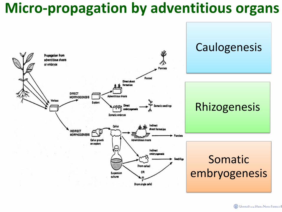
Micro-propagation by adventitious organs
Caulogenesis
Rhizogenesis
Somatic embryogenesis
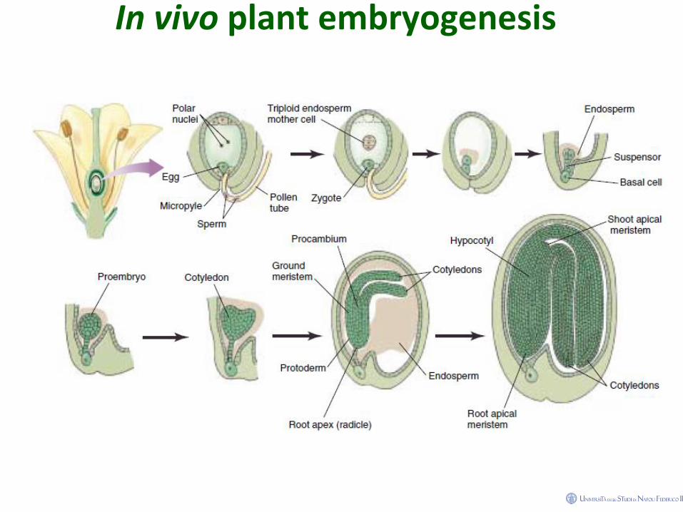
In vivo plant embryogenesis
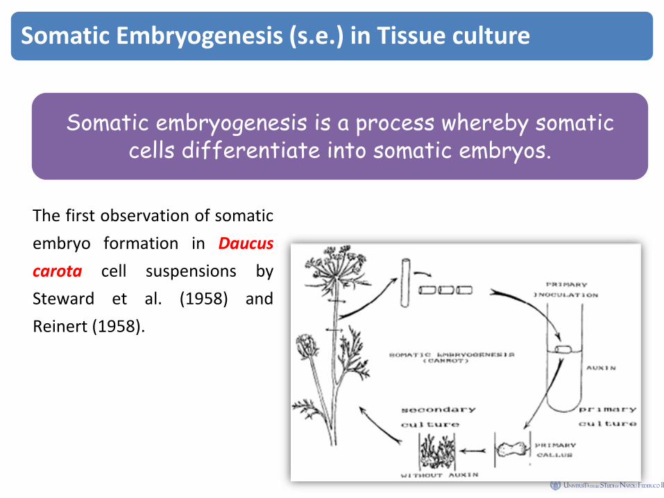
Somatic Embryogenesis (s.e.) in Tissue culture
Somatic embryogenesis is a process whereby somatic cells differentiate into somatic embryos.
The first observation of somatic
embryo formation in Daucus
carota cell suspensions by
Steward et al. (1958) and
Reinert (1958).
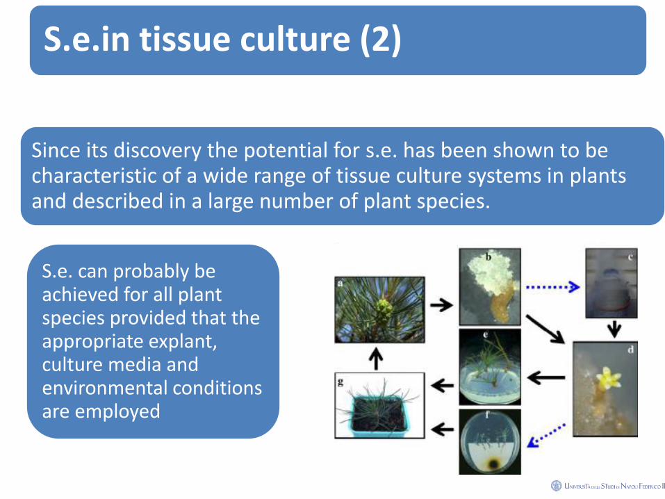
S.e.in tissue culture (2)
Since its discovery the potential for s.e. has been shown to be characteristic of a wide range of tissue culture systems in plants and described in a large number of plant species.
S.e. can probably be achieved for all plant species provided that the appropriate explant, culture media and environmental conditions are employed
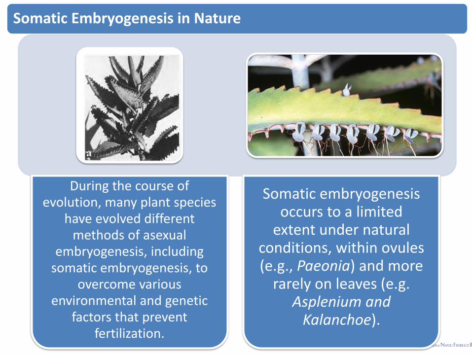
Somatic Embryogenesis in Nature
During the course of evolution, many plant species
have evolved different methods of asexual
embryogenesis, including somatic embryogenesis, to
overcome various environmental and genetic
factors that prevent fertilization.
Somatic embryogenesis occurs to a limited
extent under natural conditions, within ovules (e.g., Paeonia) and more
rarely on leaves (e.g. Asplenium and
Kalanchoe).
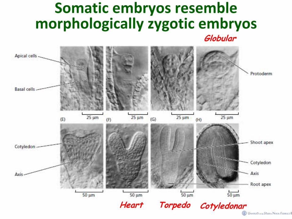
Somatic embryos resemble morphologically zygotic embryos
Globular
Cotyledonar Heart Torpedo

S.e. stages in eggplant
Globular
Hearth
Torpedo
Germinated s.embryos
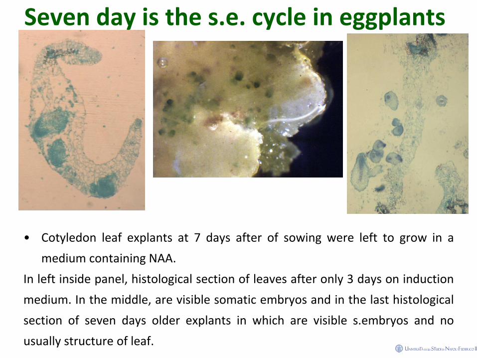
Seven day is the s.e. cycle in eggplants
• Cotyledon leaf explants at 7 days after of sowing were left to grow in a
medium containing NAA.
In left inside panel, histological section of leaves after only 3 days on induction
medium. In the middle, are visible somatic embryos and in the last histological
section of seven days older explants in which are visible s.embryos and no
usually structure of leaf.
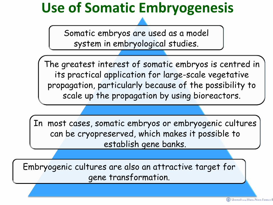
Use of Somatic Embryogenesis
Somatic embryos are used as a model system in embryological studies.
The greatest interest of somatic embryos is centred in its practical application for large-scale vegetative
propagation, particularly because of the possibility to scale up the propagation by using bioreactors.
In most cases, somatic embryos or embryogenic cultures can be cryopreserved, which makes it possible to
establish gene banks.
Embryogenic cultures are also an attractive target for gene transformation.
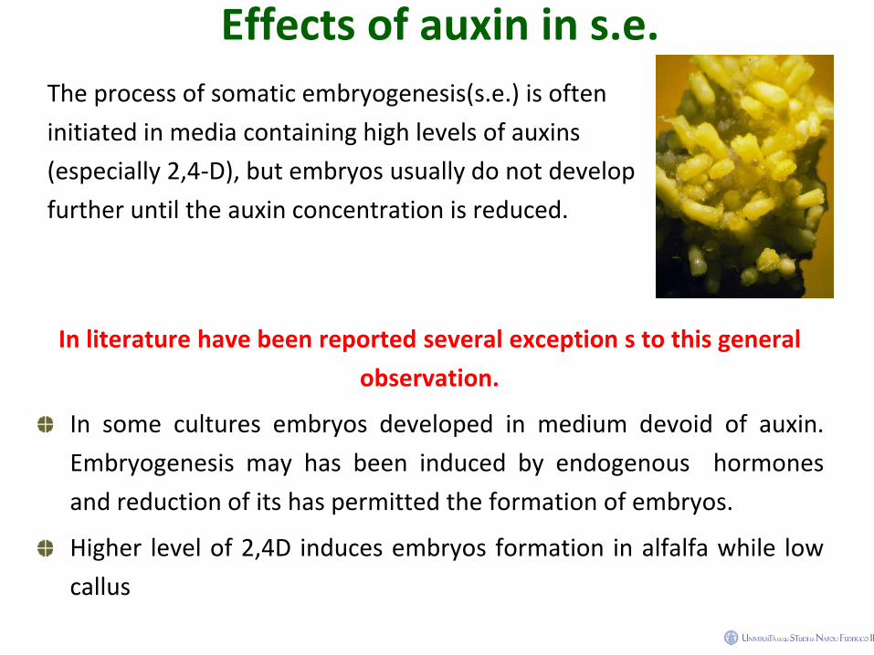
Effects of auxin in s.e.
The process of somatic embryogenesis(s.e.) is often
initiated in media containing high levels of auxins
(especially 2,4-D), but embryos usually do not develop
further until the auxin concentration is reduced.
In literature have been reported several exception s to this general
observation.
In some cultures embryos developed in medium devoid of auxin.
Embryogenesis may has been induced by endogenous hormones
and reduction of its has permitted the formation of embryos.
Higher level of 2,4D induces embryos formation in alfalfa while low
callus
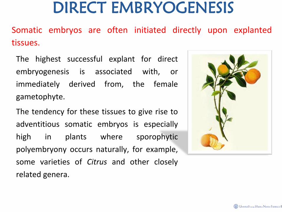
DIRECT EMBRYOGENESIS
Somatic embryos are often initiated directly upon explanted
tissues.
The highest successful explant for direct
embryogenesis is associated with, or
immediately derived from, the female
gametophyte.
The tendency for these tissues to give rise to
adventitious somatic embryos is especially
high in plants where sporophytic
polyembryony occurs naturally, for example,
some varieties of Citrus and other closely
related genera.
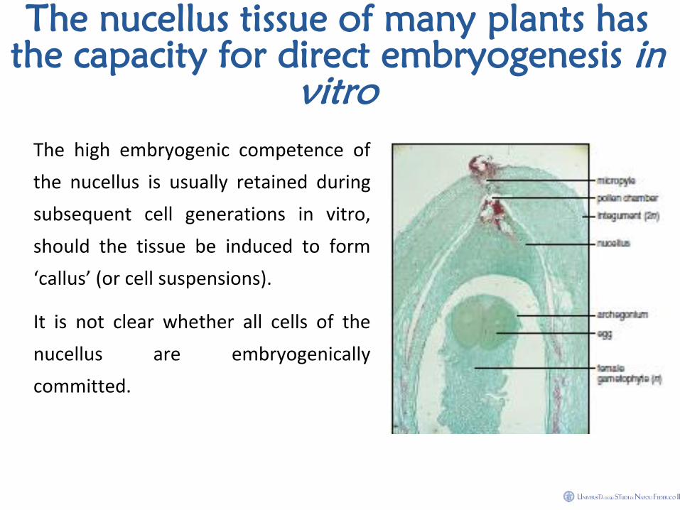
The nucellus tissue of many plants has
the capacity for direct embryogenesis in
vitro
The high embryogenic competence of
the nucellus is usually retained during
subsequent cell generations in vitro,
should the tissue be induced to form
‘callus’ (or cell suspensions).
It is not clear whether all cells of the
nucellus are embryogenically
committed.
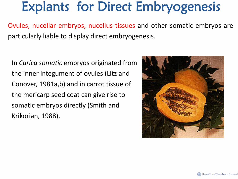
Explants for Direct Embryogenesis
Ovules, nucellar embryos, nucellus tissues and other somatic embryos are
particularly liable to display direct embryogenesis.
In Carica somatic embryos originated from
the inner integument of ovules (Litz and
Conover, 1981a,b) and in carrot tissue of
the mericarp seed coat can give rise to
somatic embryos directly (Smith and
Krikorian, 1988).
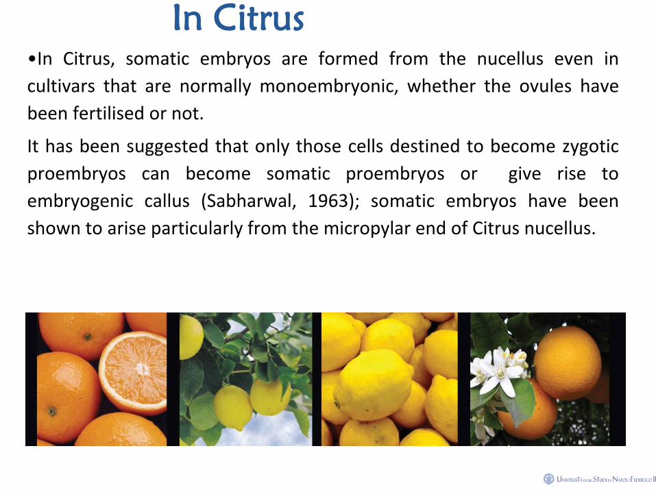
In Citrus
•In Citrus, somatic embryos are formed from the nucellus even in
cultivars that are normally monoembryonic, whether the ovules have
been fertilised or not.
It has been suggested that only those cells destined to become zygotic
proembryos can become somatic proembryos or give rise to
embryogenic callus (Sabharwal, 1963); somatic embryos have been
shown to arise particularly from the micropylar end of Citrus nucellus.
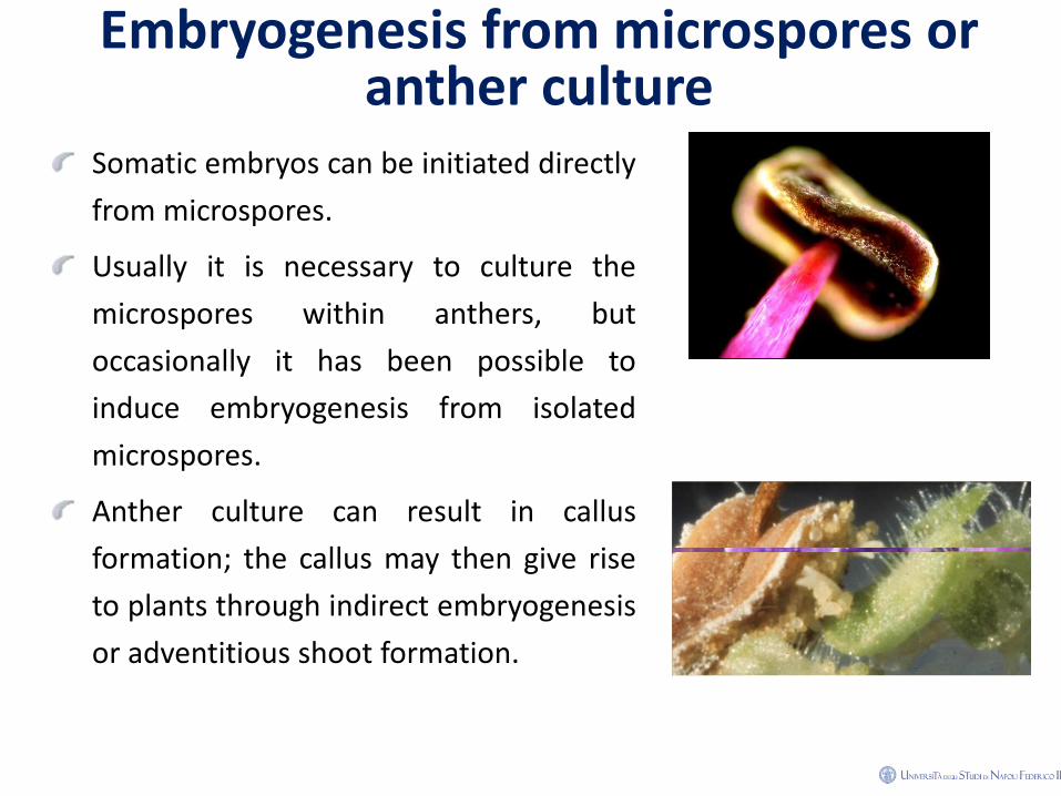
Embryogenesis from microspores or anther culture
Somatic embryos can be initiated directly
from microspores.
Usually it is necessary to culture the
microspores within anthers, but
occasionally it has been possible to
induce embryogenesis from isolated
microspores.
Anther culture can result in callus
formation; the callus may then give rise
to plants through indirect embryogenesis
or adventitious shoot formation.
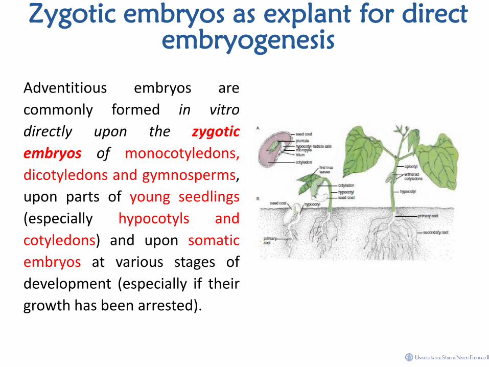
Zygotic embryos as explant for direct
embryogenesis
Adventitious embryos are
commonly formed in vitro
directly upon the zygotic
embryos of monocotyledons,
dicotyledons and gymnosperms,
upon parts of young seedlings
(especially hypocotyls and
cotyledons) and upon somatic
embryos at various stages of
development (especially if their
growth has been arrested).
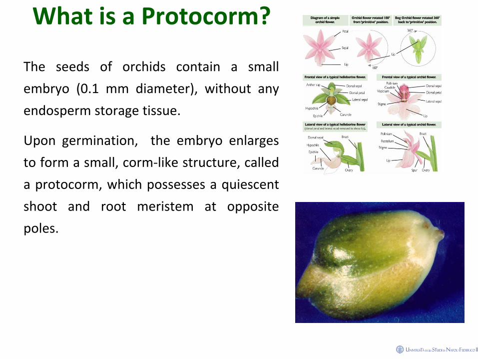
What is a Protocorm?
The seeds of orchids contain a small
embryo (0.1 mm diameter), without any
endosperm storage tissue.
Upon germination, the embryo enlarges
to form a small, corm-like structure, called
a protocorm, which possesses a quiescent
shoot and root meristem at opposite
poles.
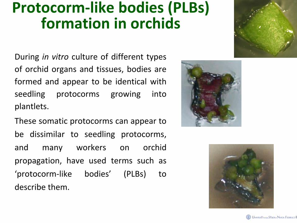
Protocorm-like bodies (PLBs) formation in orchids
During in vitro culture of different types
of orchid organs and tissues, bodies are
formed and appear to be identical with
seedling protocorms growing into
plantlets.
These somatic protocorms can appear to
be dissimilar to seedling protocorms,
and many workers on orchid
propagation, have used terms such as
‘protocorm-like bodies’ (PLBs) to
describe them.
Practical uses in propagation
From a quantitative point of view, indirect embryogenesis does
provide an efficient method of micropropagation; the same is not
true of direct embryogenesis when it is unaccompanied by the
proliferation of embryogenic tissue.
Although plants can be regenerated from embryos directly initiated
in vitro, and may be present in sufficient numbers for limited plant
production in breeding programmes, the numbers of primary
embryos per explant will usually be inadequate for large scale
cloning.
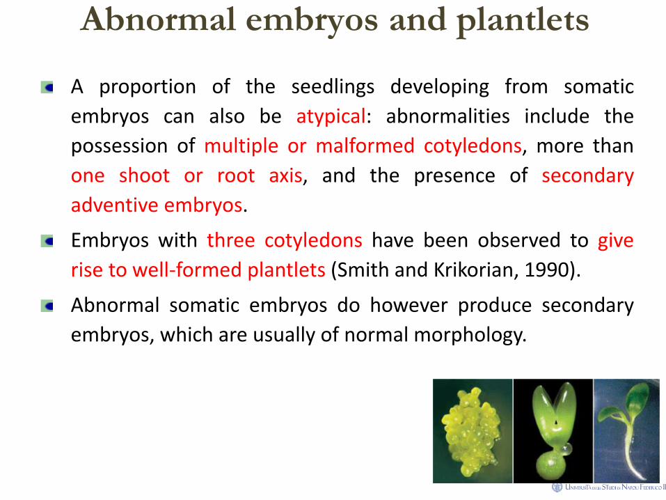
Abnormal embryos and plantlets
A proportion of the seedlings developing from somatic
embryos can also be atypical: abnormalities include the
possession of multiple or malformed cotyledons, more than
one shoot or root axis, and the presence of secondary
adventive embryos.
Embryos with three cotyledons have been observed to give
rise to well-formed plantlets (Smith and Krikorian, 1990).
Abnormal somatic embryos do however produce secondary
embryos, which are usually of normal morphology.
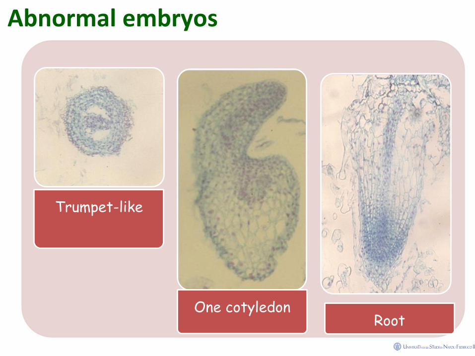
Abnormal embryos
Trumpet-like
One cotyledon Root
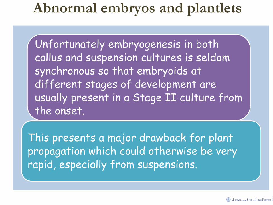
Abnormal embryos and plantlets
Unfortunately embryogenesis in both callus and suspension cultures is seldom synchronous so that embryoids at different stages of development are usually present in a Stage II culture from the onset.
This presents a major drawback for plant propagation which could otherwise be very rapid, especially from suspensions.
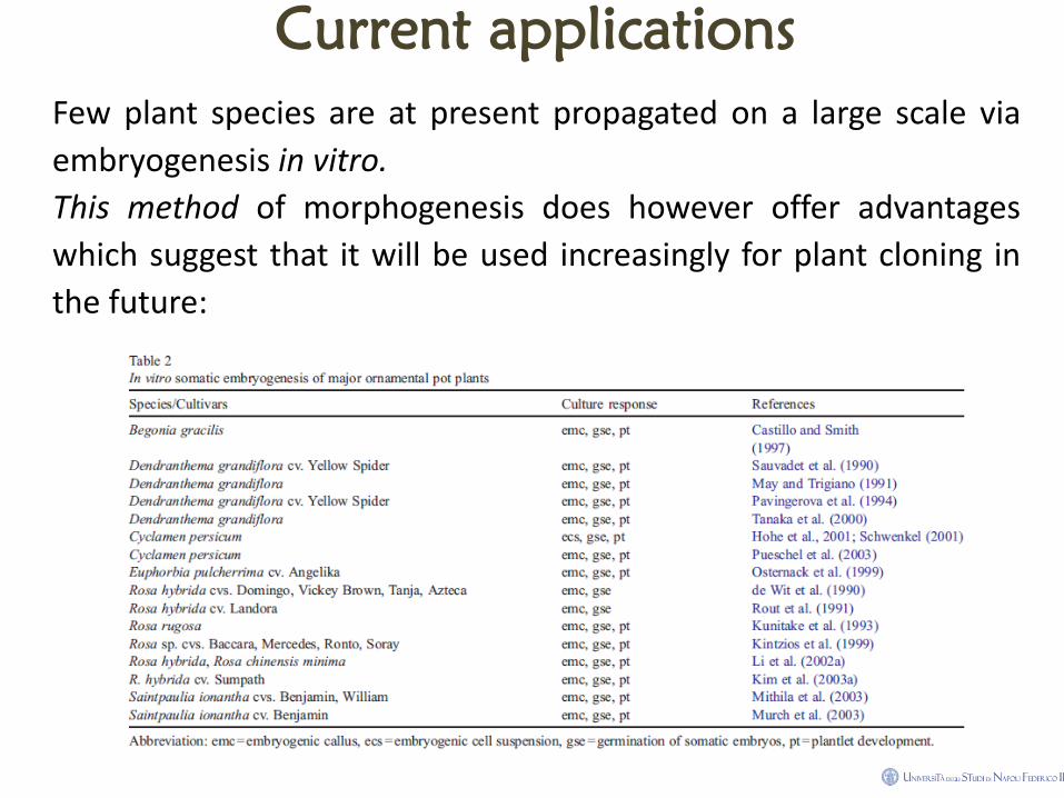
Current applications
Few plant species are at present propagated on a large scale via
embryogenesis in vitro.
This method of morphogenesis does however offer advantages
which suggest that it will be used increasingly for plant cloning in
the future:
Current applications
• In some monocotyledons (e.g. cereals, date palm and oil palm) it provides a
method of micropropagation where shoot culture has not been successful
(but note however that in some attempts to clone oil palms through
embryogenesis, the resulting plants have been very variable);
• Providing embryogenic cell suspensions can be established, plantlets can
theoretically be produced in large numbers and at much lower cost because
plantlets do not have to be handled and subcultured individually;
• Somatic embryos probably provide the only way for tissue culture methods of
plant propagation to be economically deployed on extensively planted field
crops and forest trees.
Direct somatic embryogenesis and plant
regeneration from leaf explants of
Phalaenopsis amabilis
Abstract
Leaf explants of Phalaenopsis amabilis var. formosa formed
clusters of somatic embryos directly from epidermal cells
without an intervening callus within 20 - 30 d when cultured on
1/2-strength modified Murashige and Skoog medium
supplemented with 0.1, 1 and 3 mg dm-3 TDZ.
Repetitive production of embryos involved secondary
embryogenesis could be obtained by culturing segments of
embryogenic masses on TDZ-containing media.
Plantlet conversion from embryos was successfully achieved on
regulator-free growth medium.
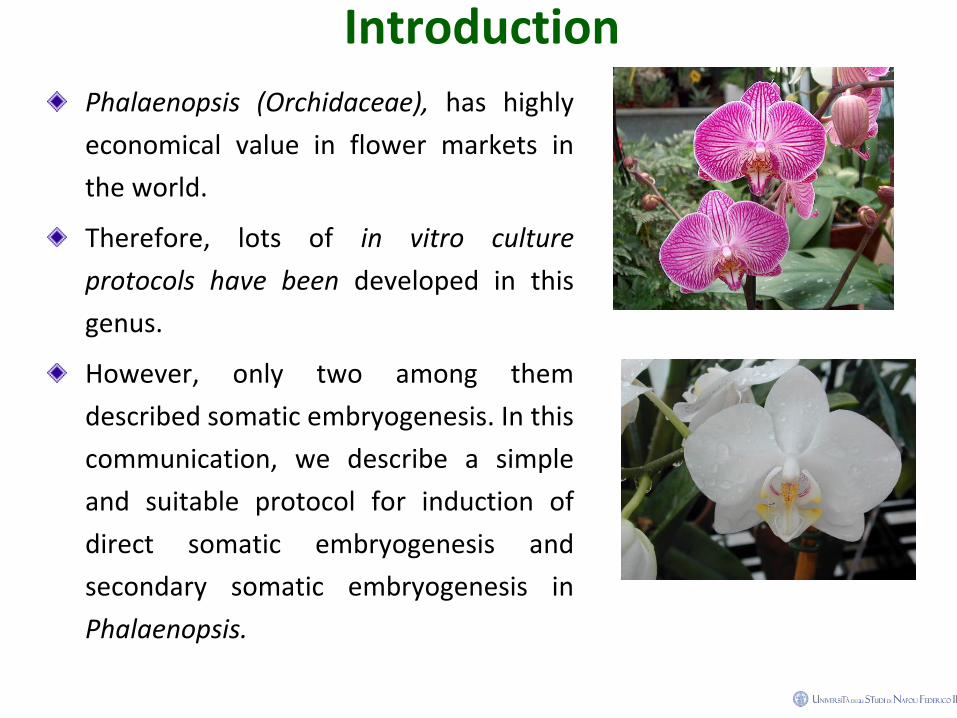
Introduction
Phalaenopsis (Orchidaceae), has highly
economical value in flower markets in
the world.
Therefore, lots of in vitro culture
protocols have been developed in this
genus.
However, only two among them
described somatic embryogenesis. In this
communication, we describe a simple
and suitable protocol for induction of
direct somatic embryogenesis and
secondary somatic embryogenesis in
Phalaenopsis.
Materials and methods Plants and culture conditions
• Green capsules were collected from pot plants of Phalaenopsis amabilis
Shimadzu var. formosa after self-pollination for three months. The capsules
were immersed in 70 % alcohol for 30 s, and followed by agitation for 15 min
in a solution of 2 % sodium hypochlorite and 0.05 % Tween (1:1 v/v).
• Seeds from these capsules were sown on modified Murashige and Skoog
(1962; MS) basal medium containing halh-strength macro- and micro-
elements supplemented with [mg dm-3]: myo-inositol (100), niacin (0.5),
pyridoxine HCl (0.5), thiamine HCl (0.1), glycine (2.0), peptone (1 000),
NaH2PO4 (170), sucrose (20 000), and Gelrite (2 200).
• Plant growth regulators were added prior to autoclaving. The pH of the
media was adjusted to 5.2 with 1 M KOH or HCl prior to autoclaving for 15
min at 121 °C.
Materials and methods • Culture growing condition: Leaf explants were incubated in 90 × 15 mm Petri
dishes under a 16-h photoperiod at irradiance of 28 - 36 μmol m-2 s-1
(daylight fluorescent tubes FL-30D/29, 40 W, China Electric Co., Taipei,
Taiwan) and temperature of 26 ± 1 °C.
• Subculture and explants : the subculture period was 2 months. After 180 d of
culture, these seeds developed into plants with 3 - 5 leaves and 2 - 4 roots.
These seedlings were used as donor plants.
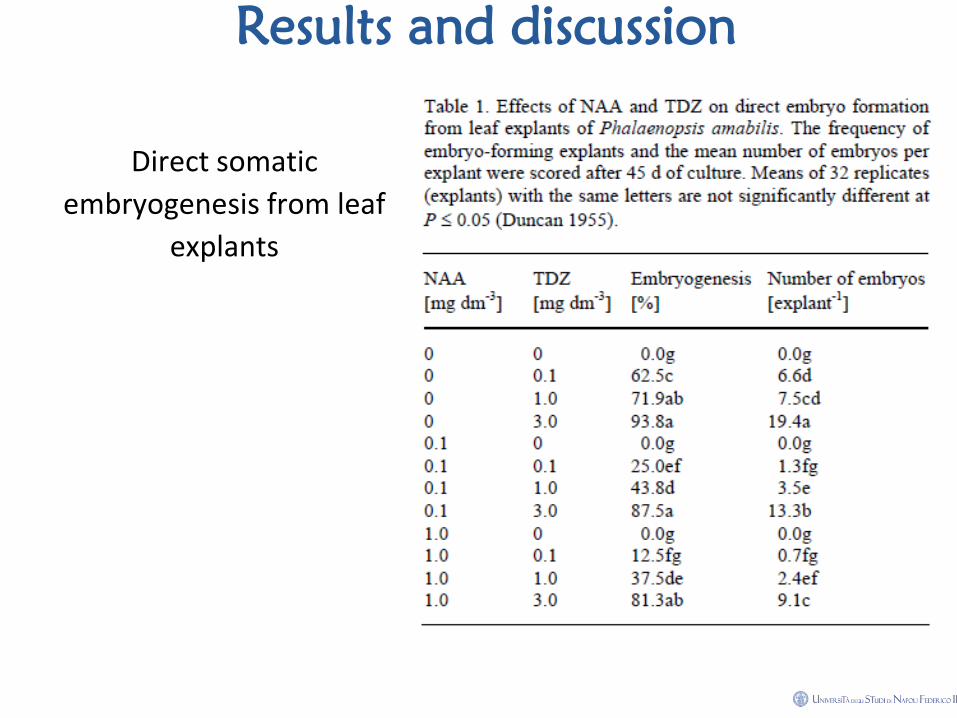
• Induction of direct embryo formation from leaf explants: leaf tip segments
(about 1 cm in length) taken from the donor plants were used to test the
effects of naphthaleneacetic acid (NAA; 0, 0.1, 1 mg dm-3) and thidiazuron
(TDZ; 0, 0.1, 1, 3 mg dm-3) on direct somatic embryogenesis. Eight replicates
(dishes) each with four leaf explants were used for each treatment.
Materials and methods • How to score data: The percentage of explants forming somatic embryos
was recorded. The number of embryos formed from each responding
explant was counted under a stereomicroscope (SZH, Olympus, Tokyo,
Japan) at the protocorm stage (45 d of culture). Treatment means were
compared by following Duncan's Multiple Range Test (Duncan 1955).
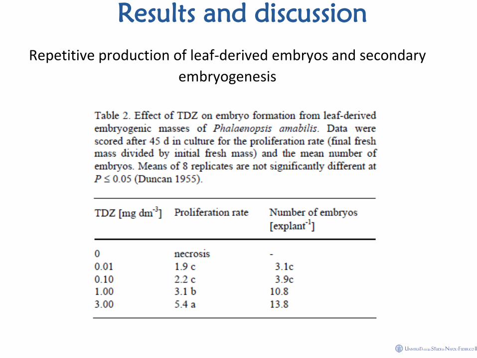
• Induction of embryo formation from leaf-derived nodular masses and
secondary embryogenesis: Pieces of nodular masses (about 0.1 g) were
used as explants to test the effect of TDZ (0, 0.01, 0.1, 1, 3 mg dm-3) on
secondary somatic embryogenesis. Eight replicates (tubes) each with one
explant were used for each treatment. The proliferation rate of nodular
masses was measured as final fresh mass divided by initial fresh mass. The
number of embryos formed from each responding explant was counted at
the protocorm stage (45 d of culture).
Materials and methods
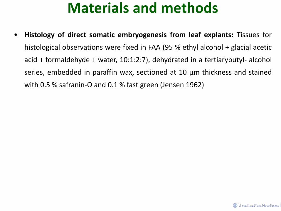
• Histology of direct somatic embryogenesis from leaf explants: Tissues for
histological observations were fixed in FAA (95 % ethyl alcohol + glacial acetic
acid + formaldehyde + water, 10:1:2:7), dehydrated in a tertiarybutyl- alcohol
series, embedded in paraffin wax, sectioned at 10 μm thickness and stained
with 0.5 % safranin-O and 0.1 % fast green (Jensen 1962)
Results and discussion
Direct somatic
embryogenesis from leaf
explants
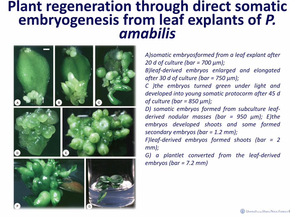
Plant regeneration through direct somatic embryogenesis from leaf explants of P.
amabilis
A)somatic embryosformed from a leaf explant after 20 d of culture (bar = 700 μm); B)leaf-derived embryos enlarged and elongated after 30 d of culture (bar = 750 μm); C )the embryos turned green under light and developed into young somatic protocorm after 45 d of culture (bar = 850 μm); D) somatic embryos formed from subculture leaf-derived nodular masses (bar = 950 μm); E)the embryos developed shoots and some formed secondary embryos (bar = 1.2 mm); F)leaf-derived embryos formed shoots (bar = 2 mm); G) a plantlet converted from the leaf-derived embryos (bar = 7.2 mm)
Results and discussion
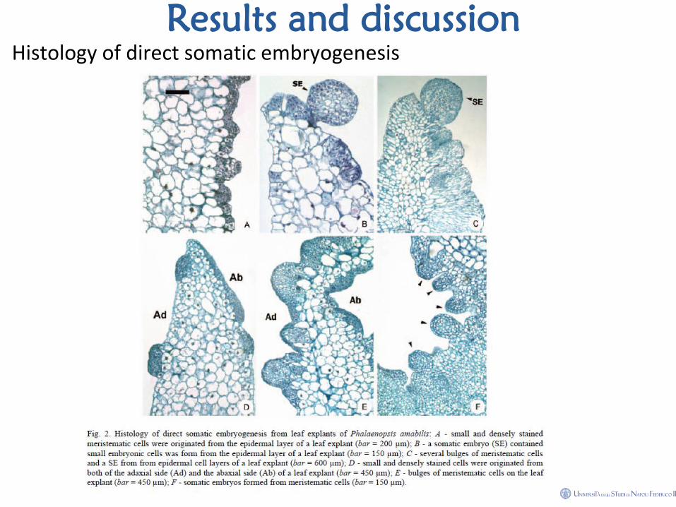
Histology of direct somatic embryogenesis
Results and discussion
Repetitive production of leaf-derived embryos and secondary
embryogenesis
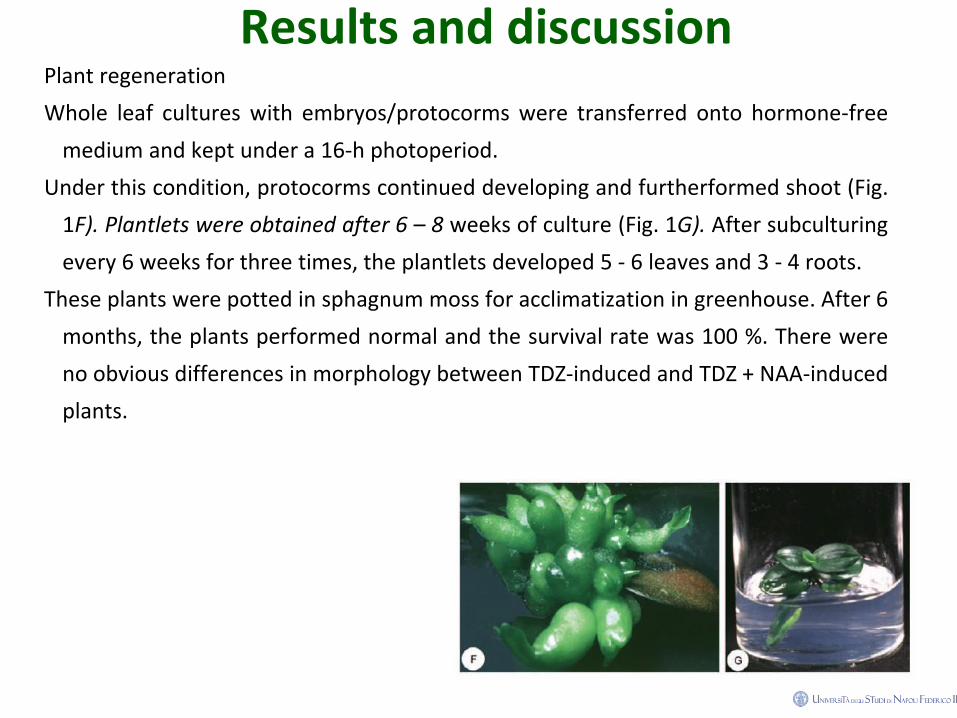
Results and discussion Plant regeneration
Whole leaf cultures with embryos/protocorms were transferred onto hormone-free
medium and kept under a 16-h photoperiod.
Under this condition, protocorms continued developing and furtherformed shoot (Fig.
1F). Plantlets were obtained after 6 – 8 weeks of culture (Fig. 1G). After subculturing
every 6 weeks for three times, the plantlets developed 5 - 6 leaves and 3 - 4 roots.
These plants were potted in sphagnum moss for acclimatization in greenhouse. After 6
months, the plants performed normal and the survival rate was 100 %. There were
no obvious differences in morphology between TDZ-induced and TDZ + NAA-induced
plants.


![A Co-Opted Hormonal Cascade Activates Dormant Adventitious ... · A Co-Opted Hormonal Cascade Activates Dormant Adventitious Root Primordia upon Flooding in Solanum dulcamara1[OPEN]](https://img.pdfslide.net/doc/110x75/5f12401a54c0792d087e2ad0/a-co-opted-hormonal-cascade-activates-dormant-adventitious-a-co-opted-hormonal.jpg)


![Biosynthesis of Diterpenoids in Tripterygium Adventitious ... · PDF fileBiosynthesis of Diterpenoids inTripterygium Adventitious Root Cultures1[OPEN] Fainmarinat S. Inabuy,a Justin](https://img.pdfslide.net/doc/110x75/5a77abaa7f8b9ad22a8e5b12/biosynthesis-of-diterpenoids-in-tripterygium-adventitious-a-biosynthesis.jpg)