Embed Size (px)
Citation preview
Microbial growth at hyperaccelerations up to403,627 × gShigeru Deguchi (出口 茂)a,1, Hirokazu Shimoshige (下重裕一)a,2, Mikiko Tsudome (津留美紀子)a,Sada-atsu Mukai (向井貞篤)a,b, Robert W. Corkeryc, Susumu Ito (伊藤 進)d,and Koki Horikoshi (掘越弘毅)a
aInstitute of Biogeosciences, Japan Agency for Marine-Earth Science and Technology, Yokosuka 237-0061, Japan; bInstitute for Advanced Study, KyushuUniversity, Higashi-ku, Fukuoka 812-8581, Japan; cInstitute for Surface Chemistry, 114 28 Stockholm, Sweden; and dDepartment of Bioscience andBiotechnology, Faculty of Agriculture, University of the Ryukyus, Nishihra, Okinawa 903-0213, Japan
Edited by Henry J. Melosh, University of Arizona, Tucson, AZ, and approved March 15, 2011 (received for review December 19, 2010)
It is well known that prokaryotic life can withstand extremes oftemperature, pH, pressure, and radiation. Little is known aboutthe proliferation of prokaryotic life under conditions of hyper-acceleration attributable to extreme gravity, however. We foundthat living organisms can be surprisingly proliferative duringhyperacceleration. In tests reported here, a variety of microorgan-isms, including Gram-negative Escherichia coli, Paracoccus denitri-ficans, and Shewanella amazonensis; Gram-positive Lactobacillusdelbrueckii; and eukaryotic Saccharomyces cerevisiae, were cul-tured while being subjected to hyperaccelerative conditions. Weobserved and quantified robust cellular growth in these culturesacross a wide range of hyperacceleration values. Most notably, theorganisms P. denitrificans and E. coli were able to proliferate evenat 403,627 × g. Analysis shows that the small size of prokaryoticcells is essential for their proliferation under conditions of hyper-acceleration. Our results indicate that microorganisms cannot onlysurvive during hyperacceleration but can display such robust pro-liferative behavior that the habitability of extraterrestrial environ-ments must not be limited by gravity.
astrobiology | extremophiles
The robustness of prokaryotic life to physical extremes oftemperature, pH, pressure, and radiation is well known (1)
and has led to their ubiquitous presence on Earth (2, 3). Resil-ience to physical extremes is also extremely likely to be requiredfor the existence of life beyond this planet (1, 4). Finding ex-traterrestrial life is a major motivation driving searches forextrasolar planets (5); thus, understanding the physical limits forknown organisms is crucial in evaluating the probability that suchplanets harbor life (1, 4). Assessing the habitability of extrater-restrial environments requires an expanded set of criteria in-volving factors that can be ignored for terrestrial environments.The effect that extremes of gravity have on organisms is one suchfactor to consider when exploring for life beyond Earth.The effect of microgravity on biological processes has been an
active area of research particularly because it is relevant to hu-man health during space flight (6, 7). Microorganisms make idealmodel life forms for microgravity research because they arelightweight, small, and relatively easy to handle in space and haveshort generation times (7). Consequently, numerous experimentshave been performed on microorganisms both in orbit and inEarth-based clinostats that simulate microgravity. The resultsdemonstrate that microgravity affects microorganisms in a widevariety of ways related to their growth, physiology, pathogenesis,stress resistance, and gene expression (7–23).The majority of these studies indicate that microgravity stim-
ulates the growth of microorganisms (e.g., Salmonella entericaserovar Typhimurium, Bacillus subtilis, Escherichia coli) com-pared with 1 × g controls (8–17). In the case of E. coli, for ex-ample, the lag phase was shortened, the duration of exponentialgrowth was increased, and the final cell population density wasapproximately doubled during space flights (13). Simulated mi-crogravity can also affect the secondary metabolism of micro-organisms. For example, production of β-lactam antibiotics by
Streptomyces clavuligerus, production of rapamycin by Strepto-myces hygroscopicus, and production of microcin B17 by E. coliwere suppressed during culturing in simulated microgravity,whereas production of gramicidin S by Bacillus brevis was un-affected (14, 18). S. enterica serovar Typhimurium showed en-hanced virulence in a murine infection model (19, 20) conductedin space flight and under modeled microgravity compared withconditions of normal gravity (19, 20). These microorganisms alsoshowed increased resistance to environmental stresses, increasedsurvival in macrophages, and significant changes in protein ex-pression levels (19). To elucidate the molecular mechanisms ofmicrobial responses to microgravity, 2D gel electrophoresis andDNA microarray analysis have been used (19–23). Recentanalysis of S. enterica serovar Typhimurium grown in spaceidentified 167 transcripts and 73 proteins that changed expres-sion compared with ground controls, and conserved RNA-binding protein Hfq was identified as a likely global regulator(20). Gene expression of eukaryotic Saccharomyces cerevisiae isalso affected by simulated microgravity (22, 23).Compared with the relatively active research on microbial
responses to microgravity, there are fewer studies that reportexperiments on microorganisms exposed to gravities greater than1 × g (11, 16, 24–28). Unlike in microgravity, experiments inhypergravity were performed exclusively in simulated environ-ments and primarily by subjecting microorganisms to centrifugalacceleration in centrifuges. Bouloc and D’Ari (11) reported thathyperaccelerations of 3 and 5 × g did not affect the growth ofE. coli, whereas Brown et al. (16) observed growth suppression at50 × g. Similar observations were reported for Paramecium tet-raurelia, which showed no effect at 10 × g but a significantly lowerproliferation rate and a lower population density at 20 × g (24).At hyperaccelerations much greater than ∼102 × g, the effect of
sedimentation on microbial cells becomes significant. In a typicalexample, cultures of bacterial cells subjected to centrifugation at3,000–5,000× g for 5–10min yielded pellets of intact bacterial cells(29). If microbial growth had occurred under these (or similar)conditions, it must have happened within or on the pellet. In starkcontrast, the effect of cellular sedimentation is not very significantat lower accelerations, where growth can occur planktonically.Studying microbial proliferation, and not simply survival, at suchhyperaccelerations addresses the fundamental biological questionof what are the physical limits of organismic viability (1) undera range of gravitational accelerations larger than those found onEarth. Understanding the gravity limits for microorganism growth
Author contributions: S.D., H.S., M.T., S.I., and K.H. designed research; S.D., H.S., M.T., andS.M. performed research; S.D., S.M., and R.W.C. analyzed data; and S.D. and R.W.C. wrotethe paper.
The authors declare no conflict of interest.
This article is a PNAS Direct Submission.1To whom correspondence should be addressed. E-mail: [email protected] leave from: Graduate School of Interdisciplinary New Science, Toyo University, 2100Kujirai, Kawagoe 350-0815, Japan.
This article contains supporting information online at www.pnas.org/lookup/suppl/doi:10.1073/pnas.1018027108/-/DCSupplemental.
www.pnas.org/cgi/doi/10.1073/pnas.1018027108 PNAS | May 10, 2011 | vol. 108 | no. 19 | 7997–8002
MICRO
BIOLO
GY
has important implications in considering the emergence, trans-port, adaptation, and evolution of life in extraterrestrial habitats(4, 30).Previous studies that dealt with microorganisms under accel-
erations much greater than ∼102 × g focused mostly on survival,however. Spores of B. subtilis tolerate accelerations exceeding10,000–15,000 × g for indefinite periods of time but were inacti-vated to a 10% survival rate when they were subjected for 65 hto 436,000 × g (25, 26). Inactivation of various microorganisms,including prokaryotic E. coli, Thiobacillus intermedius, Bacillusamyloliquefaciens, and Staphylococcus aureus as well as eukaryoticS. cerevisiae, was also studied after they were subjected to450,000 × g (27). These studies were done in phosphate-bufferedor physiological saline at 4 °C, whereinmicrobial proliferation wasnot possible even at 1 × g because of the lack of nutrients andlow temperature.To our knowledge, the only study that has dealt with the pro-
liferation of microorganisms under hyperaccelerative conditionswas that of Montgomery et al. (28). In their experiments, E. colisuspended in nutrient broth at 35 °C was subjected to centrifuga-tion at 1,000 or 110,000 × g for 24 h (28). They reported that thegrowth pattern of E. coli was not altered at 1,000 × g. They foundthat growth was disturbed at 110,000 × g, however. This wascharacterized by an increased duration of the lag phase, prolongedgeneration time, and decreased maximal cell concentration com-pared with 1 × g controls. The study of proliferation of micro-organisms under hyperaccelerations much greater than ∼100 × gstill remained largely unexplored, however (1). Here, we reportthat microorganisms can grow surprisingly well under hyper-accelerations and are able to proliferate even at 403,627 × g.
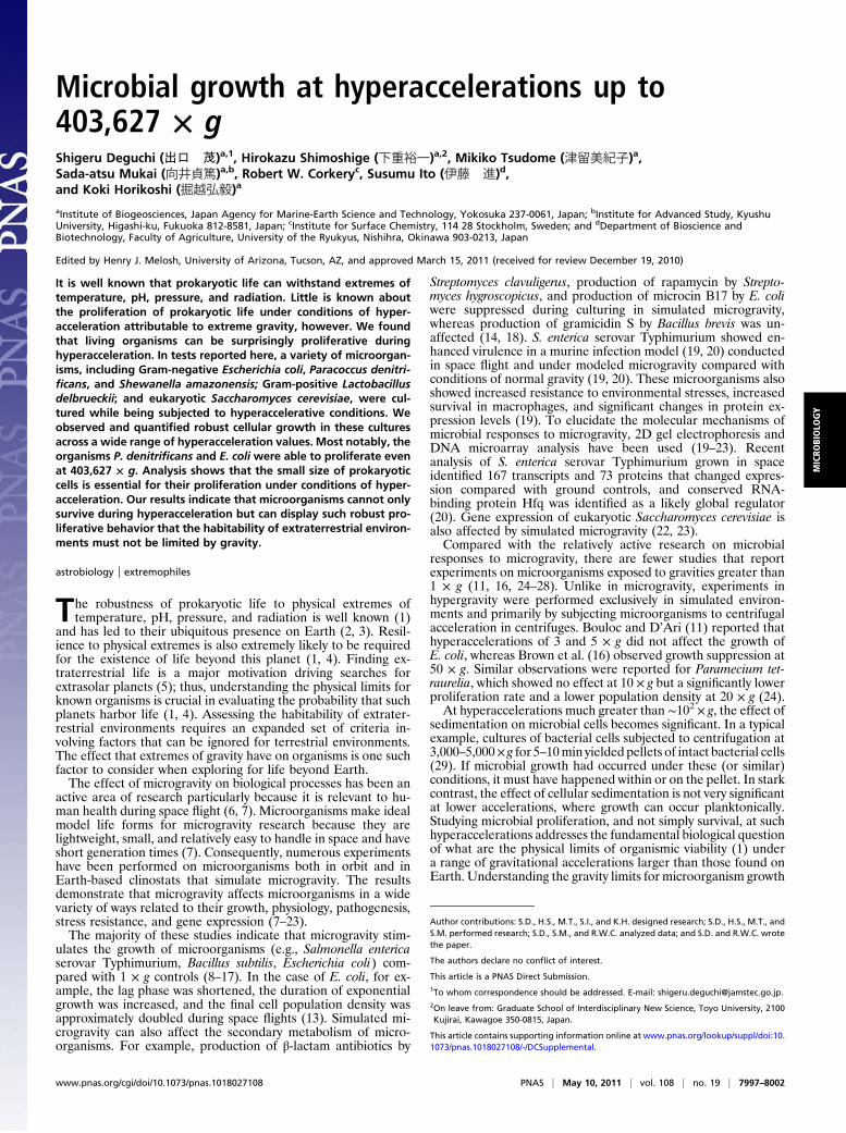
ResultsGrowth of Microorganisms Under Hyperaccelerations. A variety ofmicroorganisms were cultured in nutrient media under hyper-accelerations in centrifuges. The microorganisms studied hereinclude Gram-negative Paracoccus denitrificans, E. coli, andShewanella amazonensis; Gram-positive Lactobacillus delbrueckiisubsp. delbrueckii; and eukaryotic S. cerevisiae. Fig. 1 A–D showsphotographs of cultures of P. denitrificans in LB broth containing25 mM KNO3 after spinning in an ultracentrifuge at 403,627 × gand at 30 °C. At this acceleration, P. denitrificans cells sedimentedand formed a pellet at the bottom of a centrifuge tube soon aftercentrifugation began. Initially, a pellet was not visible (Fig. 1A)because the total number of P. denitrificans cells in the culture wassmall (∼106 cells). A pellet of a visible size formed after spinningthe culture for 6 h (Fig. 1B), however, and it increased in size with
time (Fig. 1 C and D). The observation demonstrates un-ambiguously thatP. denitrificans canproliferate evenat 403,627× g.
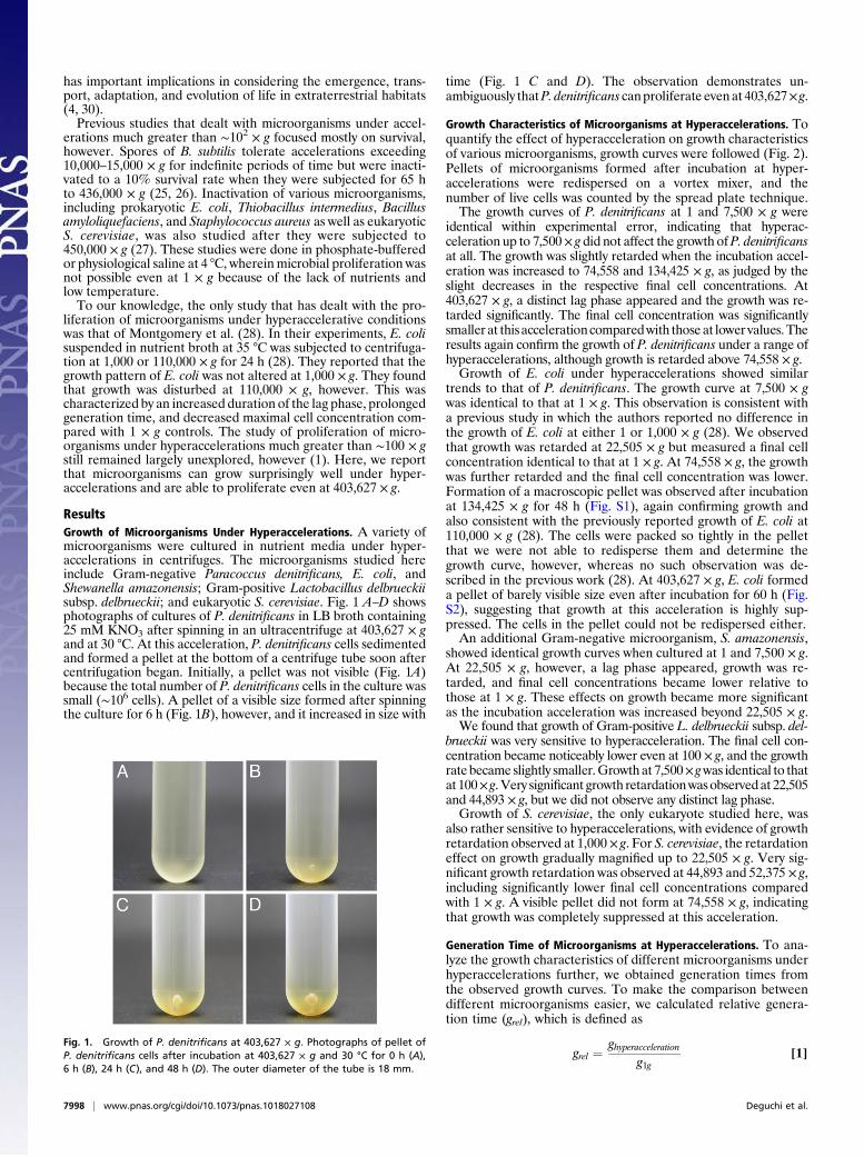
Growth Characteristics of Microorganisms at Hyperaccelerations. Toquantify the effect of hyperacceleration on growth characteristicsof various microorganisms, growth curves were followed (Fig. 2).Pellets of microorganisms formed after incubation at hyper-accelerations were redispersed on a vortex mixer, and thenumber of live cells was counted by the spread plate technique.The growth curves of P. denitrificans at 1 and 7,500 × g were
identical within experimental error, indicating that hyperac-celeration up to 7,500× g did not affect the growth ofP. denitrificansat all. The growth was slightly retarded when the incubation accel-eration was increased to 74,558 and 134,425 × g, as judged by theslight decreases in the respective final cell concentrations. At403,627 × g, a distinct lag phase appeared and the growth was re-tarded significantly. The final cell concentration was significantlysmaller at thisaccelerationcomparedwith those at lower values.Theresults again confirm the growth of P. denitrificans under a range ofhyperaccelerations, although growth is retarded above 74,558 × g.Growth of E. coli under hyperaccelerations showed similar
trends to that of P. denitrificans. The growth curve at 7,500 × gwas identical to that at 1 × g. This observation is consistent witha previous study in which the authors reported no difference inthe growth of E. coli at either 1 or 1,000 × g (28). We observedthat growth was retarded at 22,505 × g but measured a final cellconcentration identical to that at 1 × g. At 74,558 × g, the growthwas further retarded and the final cell concentration was lower.Formation of a macroscopic pellet was observed after incubationat 134,425 × g for 48 h (Fig. S1), again confirming growth andalso consistent with the previously reported growth of E. coli at110,000 × g (28). The cells were packed so tightly in the pelletthat we were not able to redisperse them and determine thegrowth curve, however, whereas no such observation was de-scribed in the previous work (28). At 403,627 × g, E. coli formeda pellet of barely visible size even after incubation for 60 h (Fig.S2), suggesting that growth at this acceleration is highly sup-pressed. The cells in the pellet could not be redispersed either.An additional Gram-negative microorganism, S. amazonensis,
showed identical growth curves when cultured at 1 and 7,500 × g.At 22,505 × g, however, a lag phase appeared, growth was re-tarded, and final cell concentrations became lower relative tothose at 1 × g. These effects on growth became more significantas the incubation acceleration was increased beyond 22,505 × g.We found that growth of Gram-positive L. delbrueckii subsp. del-
brueckii was very sensitive to hyperacceleration. The final cell con-centration became noticeably lower even at 100 × g, and the growthrate became slightly smaller.Growth at 7,500× gwas identical to thatat 100× g.Very significant growth retardationwasobservedat 22,505and 44,893 × g, but we did not observe any distinct lag phase.Growth of S. cerevisiae, the only eukaryote studied here, was
also rather sensitive to hyperaccelerations, with evidence of growthretardation observed at 1,000 × g. For S. cerevisiae, the retardationeffect on growth gradually magnified up to 22,505 × g. Very sig-nificant growth retardation was observed at 44,893 and 52,375 × g,including significantly lower final cell concentrations comparedwith 1 × g. A visible pellet did not form at 74,558 × g, indicatingthat growth was completely suppressed at this acceleration.
Generation Time of Microorganisms at Hyperaccelerations. To ana-lyze the growth characteristics of different microorganisms underhyperaccelerations further, we obtained generation times fromthe observed growth curves. To make the comparison betweendifferent microorganisms easier, we calculated relative genera-tion time (grel), which is defined as
grel ¼ ghyperaccelerationg1g
[1]Fig. 1. Growth of P. denitrificans at 403,627 × g. Photographs of pellet ofP. denitrificans cells after incubation at 403,627 × g and 30 °C for 0 h (A),6 h (B), 24 h (C), and 48 h (D). The outer diameter of the tube is 18 mm.
7998 | www.pnas.org/cgi/doi/10.1073/pnas.1018027108 Deguchi et al.
where ghyperacceleration and g1g are generation times of a given or-ganism under hyperaccelerations and at 1 × g, respectively. Theresults are summarized in Fig. 2F.For all prokaryotes tested here (i.e., both Gram-negative and
Gram-positive bacteria), hyperacceleration had either no effect ora relatively small effect on growth below 7,500× g, whereas growthwas significantly retarded above ∼2 × 10⁴ × g. Among all prokar-yotes tested here, the effect of hyperacceleration on growth waslowest forP. denitrificans. For the eukaryotic organismS. cerevisiae,the growth behavior observed here stands in contrast to that ofprokaryotes in that the generation time became progressivelylonger with increased acceleration, particularly above 44,893 × g.
Possible Effect of Hyperacceleration on Microbial Growth. To un-derstand how hyperacceleration affects microbial growth, weconsidered three aspects that differentiate the growth of micro-organisms under hyperacceleration from that at 1 × g. These aresedimentation,mechanical deformation, andhydrostatic pressure.
Effect of Sedimentation. Under conditions of hyperacceleration,microbial cells grew in sedimented congested pellets. The densityof cells within these pellets increased as gravity increased,leading to relatively smaller void volumes between cells in pelletsformed at relatively high acceleration. Therefore, diffusion ratesof small molecules in these pellets became progressively slowerbecause of an increasing obstruction effect. Consequently, nu-trient uptake and waste disposal by cells in the growing pelletswere inhibited, thereby affecting the growth rates.Conversely, differential sedimentation of subcellular moieties
and the formation of other molecular concentration gradientswithin cells may also negatively affect growth under hyper-accelerative conditions. By analogy, centrifugation at 105 × g isroutinely used for sedimentation, and thus separation of rela-
tively large particulates like ribosomes from the molecular milieuof cell lysates (31). In this study, a distinct concentration gradientof nutrient components in LB broth was observed when theculture of P. denitrificans was spun at 403,627 × g for longer than6 h (Fig. 1 B–D). Yellow-colored components formed a sedimentat the bottom of the centrifuge tube, leaving a colorless super-natant. A similar concentration gradient of cytoplasmic compo-nents may form within microbial cells at hyperaccelerations.The concentration gradient of a particle subject to gravita-
tional force at a sedimentation equilibrium is described by (32)
cðzÞ∝ e− ðm−VρmÞgz=kBT [2]
where m and V are the mass and the volume of the particle, ρm isthe density of the medium, kB is the Boltzmann constant, T is theabsolute temperature, and z is the particle position relative to thebottom. We calculated concentration gradients of model proteinsof various molecular masses (1 kDa, 10 kDa, 100 kDa, and 1 MDa)at 500,000 × g and 30 °C over a distance of 10 μm (Fig. 3A) withina model cytosol. Volumes of the respective proteins were calcu-lated using an average value of the partial specific volumes ofmost proteins (0.73 cm3·g−1) (33). A value of 1.1 g·cm−3 was usedas an average representative density of the model cytosol (34).If we consider sedimentation over 1 μm, which is a typical size
for prokaryotic cells, the calculation shows that sedimentation isnegligible for relatively small proteins having molecular massesof 1, 10, and 100 kDa. Significantly, even the 100-kDa proteinhad a concentration at the top of the cell (z = 1 μm) that was96% of the value at the bottom (z = 0 μm). In contrast, a veryprofound effect of sedimentation was calculated for the 1-MDaprotein, where the concentration at the top was 69% of that atthe bottom. These analyses indicate that intracellular sedimen-
Fig. 2. Growth of various microorganisms under hyperaccelerations. (A) Growth curves of P. denitrificans at 30 °C and hyperaccelerations up to 403,627 × g.(B) Growth of E. coli in LB broth at 37 °C and hyperaccelerations up to 74,558 × g. (C) Growth of S. amazonensis in LB broth at 37 °C and hyperaccelerations upto 74,558 × g. (D) Growth of L. delbrueckii subsp. delbrueckii in MRS broth at 37 °C and hyperaccelerations up to 30,000 × g. (E) Growth of S. cerevisiae in yeastextract-peptone-dextrose broth at 30 °C and hyperaccelerations up to 74,558 × g. (F) Change in grel of various microorganisms as a function of acceleration.
Deguchi et al. PNAS | May 10, 2011 | vol. 108 | no. 19 | 7999
MICRO
BIOLO
GY
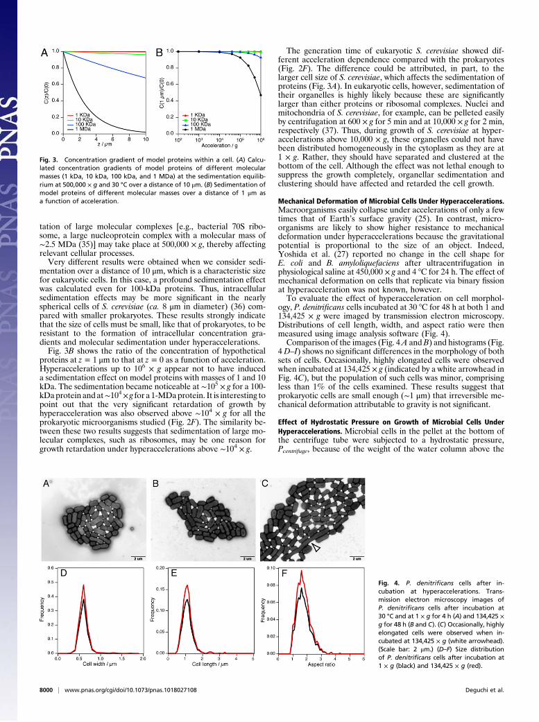
tation of large molecular complexes [e.g., bacterial 70S ribo-some, a large nucleoprotein complex with a molecular mass of∼2.5 MDa (35)] may take place at 500,000 × g, thereby affectingrelevant cellular processes.Very different results were obtained when we consider sedi-
mentation over a distance of 10 μm, which is a characteristic sizefor eukaryotic cells. In this case, a profound sedimentation effectwas calculated even for 100-kDa proteins. Thus, intracellularsedimentation effects may be more significant in the nearlyspherical cells of S. cerevisiae (ca. 8 μm in diameter) (36) com-pared with smaller prokaryotes. These results strongly indicatethat the size of cells must be small, like that of prokaryotes, to beresistant to the formation of intracellular concentration gra-dients and molecular sedimentation under hyperaccelerations.Fig. 3B shows the ratio of the concentration of hypothetical
proteins at z= 1 μm to that at z= 0 as a function of acceleration.Hyperaccelerations up to 106 × g appear not to have induceda sedimentation effect on model proteins with masses of 1 and 10kDa. The sedimentation became noticeable at ∼105 × g for a 100-kDaprotein and at∼104× g for a 1-MDaprotein. It is interesting topoint out that the very significant retardation of growth byhyperacceleration was also observed above ∼104 × g for all theprokaryotic microorganisms studied (Fig. 2F). The similarity be-tween these two results suggests that sedimentation of large mo-lecular complexes, such as ribosomes, may be one reason forgrowth retardation under hyperaccelerations above ∼104 × g.
The generation time of eukaryotic S. cerevisiae showed dif-ferent acceleration dependence compared with the prokaryotes(Fig. 2F). The difference could be attributed, in part, to thelarger cell size of S. cerevisiae, which affects the sedimentation ofproteins (Fig. 3A). In eukaryotic cells, however, sedimentation oftheir organelles is highly likely because these are significantlylarger than either proteins or ribosomal complexes. Nuclei andmitochondria of S. cerevisiae, for example, can be pelleted easilyby centrifugation at 600 × g for 5 min and at 10,000 × g for 2 min,respectively (37). Thus, during growth of S. cerevisiae at hyper-accelerations above 10,000 × g, these organelles could not havebeen distributed homogeneously in the cytoplasm as they are at1 × g. Rather, they should have separated and clustered at thebottom of the cell. Although the effect was not lethal enough tosuppress the growth completely, organellar sedimentation andclustering should have affected and retarded the cell growth.
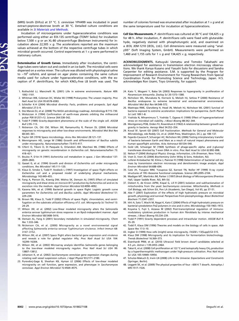
Mechanical Deformation of Microbial Cells Under Hyperaccelerations.Macroorganisms easily collapse under accelerations of only a fewtimes that of Earth’s surface gravity (25). In contrast, micro-organisms are likely to show higher resistance to mechanicaldeformation under hyperaccelerations because the gravitationalpotential is proportional to the size of an object. Indeed,Yoshida et al. (27) reported no change in the cell shape forE. coli and B. amyloliquefaciens after ultracentrifugation inphysiological saline at 450,000 × g and 4 °C for 24 h. The effect ofmechanical deformation on cells that replicate via binary fissionat hyperacceleration was not known, however.To evaluate the effect of hyperacceleration on cell morphol-
ogy, P. denitrificans cells incubated at 30 °C for 48 h at both 1 and134,425 × g were imaged by transmission electron microscopy.Distributions of cell length, width, and aspect ratio were thenmeasured using image analysis software (Fig. 4).Comparison of the images (Fig. 4A andB) and histograms (Fig.
4 D–I) shows no significant differences in the morphology of bothsets of cells. Occasionally, highly elongated cells were observedwhen incubated at 134,425 × g (indicated by a white arrowhead inFig. 4C), but the population of such cells was minor, comprisingless than 1% of the cells examined. These results suggest thatprokaryotic cells are small enough (∼1 μm) that irreversible me-chanical deformation attributable to gravity is not significant.
Effect of Hydrostatic Pressure on Growth of Microbial Cells UnderHyperaccelerations. Microbial cells in the pellet at the bottom ofthe centrifuge tube were subjected to a hydrostatic pressure,Pcentrifuge, because of the weight of the water column above the
Fig. 3. Concentration gradient of model proteins within a cell. (A) Calcu-lated concentration gradients of model proteins of different molecularmasses (1 kDa, 10 kDa, 100 kDa, and 1 MDa) at the sedimentation equilib-rium at 500,000 × g and 30 °C over a distance of 10 μm. (B) Sedimentation ofmodel proteins of different molecular masses over a distance of 1 μm asa function of acceleration.
Fig. 4. P. denitrificans cells after in-cubation at hyperaccelerations. Trans-mission electron microscopy images ofP. denitrificans cells after incubation at30 °C and at 1 × g for 4 h (A) and 134,425 ×g for 48 h (B and C). (C) Occasionally, highlyelongated cells were observed when in-cubated at 134,425 × g (white arrowhead).(Scale bar: 2 μm.) (D–F) Size distributionof P. denitrificans cells after incubation at1 × g (black) and 134,425 × g (red).
8000 | www.pnas.org/cgi/doi/10.1073/pnas.1018027108 Deguchi et al.

pellet. Pcentrifuge is negligibly small at 1 × g but increases linearlywith acceleration and becomes significant at hyperaccelerations.Pcentrifuge is estimated to be 0.1 MPa at 100 × g, 0.6 MPa at1,000 × g, 3.6 MPa at 7,500 × g, 10.3 MPa at 22,505 × g, 13.6 MPaat 29,819 × g, 20.4 MPa at 44,893 × g, 23.7 MPa at 52,375 × g,33.8 MPa at 74,558 × g, 42.2 MPa at 134,425 × g, and 126.5 MPaat 403,627 × g (SI Materials and Methods, Fig. S3, Table S1).For most mesophilic microorganisms, cell division is not af-
fected at hydrostatic pressures below ∼20MPa (38). For example,the colony-forming ability of E. coli is suppressed only when it iscultured under hydrostatic pressures above 40MPa (39). Thus, it isunlikely that hydrostatic pressure at hyperaccelerations up to52,375 × g plays a dominant role in the growth retardation underhyperaccelerations above ∼104 × g (Fig. 2F).Hydrostatic pressure may have had an impact on the growth of
P. denitrificans above 74,558 × g, where hydrostatic pressure isestimated to be higher than 33.8 MPa. Very interestingly, wefound that the growth of P. denitrificans was inhibited completelyabove 40 MPa (Fig. S4), suggesting that the observed growthhere should not have occurred above 134,425 × g. Although thereason for this discrepancy is not clear at present, one possibilityis attributable to the compositional gradient in the medium in-duced during sedimentation. As can be seen in Fig. 1 B–D, high-molecular-weight protein molecules may be advantageously ac-cumulated at the bottom of the centrifuge tube along with P.denitrificans, providing local nutrients for growth. The discrep-ancy may also be ascribed to the difference in cell density duringgrowth at elevated pressures compared with hyperaccelerations.P. denitrificans grew planktonically when it was cultured at ele-vated pressures, whereas the growth occurred in a denselypacked pellet at hyperaccelerations. The pressure effect on cel-lular processes depends on cell density in some cases (40). Hu-man dermal fibroblasts undergo a significant morphologicalchange and become rounded when they are subjected to 70 MPaat a low cell density (subconfluence). No such change is observedwhen they are pressurized at a high cell density (full confluence),however. Higher cell density at hyperacceleration may thereforeoffset the pressure effect to some extent.
DiscussionOur results clearly demonstrate that microorganisms not onlysurvive at hyperacceleration but can grow by binary fission,producing viable cells. All microorganisms studied here dis-played growth in culture at hyperaccelerations up to ∼2 × 10⁴ ×g. Because of the instrumental limitation of our ultracentrifuge,we did not determine the upper limit of acceleration for thegrowth of the most tolerant organisms. We did observe theproliferation of P. denitrificans and E. coli even at 403,627 × g,however. We argue that this latter finding is significant whentrying to understand the limits of hypergravity tolerance for ourversion of carbon-based life. Because the list of bacteria andyeast here is a short one, we anticipate that further accumulationof experimental data on various microorganisms will result indiscoveries of previously undescribed species that expand therange of habitable accelerations.In an attempt to understand our various observations of mi-
crobial growth under hyperaccelerative conditions, we consideredthree most obvious effects, namely, sedimentation of cells, me-chanical deformation of membranes, and hydrostatic pressure.Most notably, our analysis did not explain why some species aremore tolerant. For example, we do not explain why only P. deni-trificans and E. coli showed growth at 403,627 × g, whereas otherspecies did not. This likely implies that species-specific bio-chemical processes led to the differential sensitivities that wereobserved. Our experiments were aimed at gaining an improvedunderstanding of the physical effects of hyperaccelerative con-ditions on microbial cells rather than at elucidating subtle detailsof the biological processes likely affected by gravity, however(6, 41, 42).Nonetheless, we briefly consider some biological processes in
the hypergravity environment. Previous studies reported that the
physical effects of microgravity can be sensed by cell surfaces andtransformed into biochemical responses (7). Cytoskeletal proteinsand their polymerized superstructures, for example, may playimportant roles in gravity sensing of mammalian cells (43), andpossibly of microbial cells (7). It has also been suggested thatmechanical changes of cell membranes in microgravity conditionsare sensed by mechanosensitive channels (7). Under conditions ofhyperacceleration, congestive packing and jamming of cells withinmicrobial pellets would likely induce critical changes in the cur-vature and/or internal bilayer stress within the microbial cellmembranes. Consequently, changes in the conformation of vari-ous sensor proteins could then lead to direct cellular and quorumresponses (of various kinds) to the hypergravity field.Our findings compliment previous studies that primarily
focused on hypogravity and relatively mild hypergravity by ex-tending the range of studies well into the hypergravity regime.Especially relevant in this regard are our findings on the hyper-gravity tolerance ofE. coli and several othermicroorganisms. Here,the growth ofE. coliwas not affected at all by hyperaccelerations upto 7,500× g and we even observed growth at 403,627× g, although itwas significantly retarded. This range is three orders of magnitudewider than most of the previous studies (10–16).From a practical perspective, it has been suggested that al-
tering the accelerative environment could be used to manipulatebacterial fermentation processes (44) via production and locali-zation of microbial metabolites known to be affected by micro-gravity (14, 18). It is likely that hyperaccelerative conditions mayalso be used to induce unique metabolite production in growingcultures. For example, the suppressed biosynthesis of antibioticsreported under microgravity conditions compared with 1 × gcontrols (14, 18) may be enhanced at elevated gravities.Exploring the physical limits of organismic viability is crucial in
the search for life in extraterrestrial habitats, because the knowl-edge effectively helps in narrowing down possible targets to search(4). This present study expands the limits for life into the hyper-gravity regime, where this had not been seriously considered be-fore (1). We propose that this has significant implications forastrobiology. For example, microorganisms subjected to hyper-accelerations on the order of 105 × g have attracted scientificattention in terms of bacterial transport between planets (pan-spermia). The hypothesized process begins with an asteroidalimpact on a donating planet followed by consequent ejection ofbacteria-bearing rocks (30). Under impact conditions, ejectedrocks typically experience maximum accelerations of 3 × 10⁵ × gand rise times of 0.5 ms in the case of (ejection from) Mars (26).Bacteria have to survive extremes in both acceleration and rate ofchange of acceleration (25). Dormant spores of B. subtilis areinactivatedwhen theyare subjected to∼105× g (26).The inactivationfollows first-order kinetics and decreases exponentially with expo-sure time, however (26). Our results show that hyperacceleration of∼105 × g is within a habitable range for some microorganisms. Thissignificantly enhances the evidence that bacteria can remain robustlyviable after asteroidal impact-style ejection.Most significantly, our finding also extends the possibility of
life beyond planets to massive substellar objects like browndwarfs, the coldest of which has an effective surface temperatureof ∼400 K (45), which is extremely close in value to the knownupper temperature limit for life (395 K) (46). The relativelystrong gravitational fields associated with brown dwarfs are oneof several limiting factors in considering existence of life onbrown dwarfs (47). Our results unambiguously show that the∼10–102 × g gravitational fields existent on relatively cold (∼600K) brown dwarfs (48) must not be a primary limiting factor inassessing their potential for harboring life as we know it.
Materials and MethodsMicrobial Culture Under Hyperaccelerations. P. denitrificans ATCC17741T
(American Type Culture Collection) was incubated in LB broth containing25 mM KNO3 at 30 °C. E. coli W3110 and S. amazonensis ATCC 700329T
(American Type Culture Collection) were incubated in LB broth at 37 °C. L.delbrueckii subsp. delbrueckii was incubated in de Man, Rogosa, and Sharpe
Deguchi et al. PNAS | May 10, 2011 | vol. 108 | no. 19 | 8001
MICRO
BIOLO
GY
(MRS) broth (Difco) at 37 °C. S. cerevisiae YPH499 was incubated in yeastextract-peptone-dextrose broth at 30 °C. Detailed culture conditions areavailable in SI Materials and Methods.
Incubation of microorganisms under hyperaccelerative conditions wasperformed using either an EIX-135 centrifuge (TOMY Seiko) for incubationbelow 7,500 × g or an XL-80 ultracentrifuge (Beckman Instruments) for in-cubation above 22,505 × g. The accelerations reported are the maximumvalues achieved at the bottom of the respective centrifuge tubes in whichmicrobial growth occurred. Control experiments at 1 × g were performed instationary culture.
Determination of Growth Curves. Immediately after incubation, the centri-fuge tubes were taken out and cooled in an ice bath. The microbial cells weredispersed on a vortex mixer. The culture was diluted with physiological salineto ∼105 cells/mL and spread on agar plates containing the same culturemedia used for culture under hyperaccelerative conditions, with the ex-ception of P. denitrificans, for which KNO3-free LB broth was used. The
number of colonies formed was enumerated after incubation at 1 × g and atthe same temperature used for incubation at hyperaccelerations.
Cell Size Measurements. P. denitrificans was cultured at 30 °C and 134,425 × gfor 48 h. After incubation, P. denitrificans cells were fixed with glutaralde-hyde, negatively stained with phosphotungstic acid, and examined ona JEOL JEM-1210 (JEOL, Ltd.). Cell dimensions were measured using “anal-ySIS” (Soft Imaging System, GmbH). Measurements were performed on1,460 and 1,155 cells for 1 × g and 134,425 × g, respectively.
ACKNOWLEDGMENTS. Katsuyuki Uematsu and Tomoko Takahashi areacknowledged for assistance in transmission electron microscopy observa-tions. We thank Kanya Kusano and Yasushi Suto for discussions and SandraCarpenter for editing assistance. S.M. is supported by the Program forImprovement of Research Environment for Young Researchers from SpecialCoordination Funds for Promoting Science and Technology, Japan. H.S.acknowledges Ron Usami, Toyo University, for support.
1. Rothschild LJ, Mancinelli RL (2001) Life in extreme environments. Nature 409:1092–1101.
2. Whitman WB, Coleman DC, Wiebe WJ (1998) Prokaryotes: The unseen majority. ProcNatl Acad Sci USA 95:6578–6583.
3. Schleifer K-H (2004) Microbial diversity: Facts, problems and prospects. Syst ApplMicrobiol 27:3–9.
4. Des Marais DJ, et al. (2008) The NASA astrobiology roadmap. Astrobiology 8:715–730.5. Wolszczan A (1994) Confirmation of earth-mass planets orbiting the millisecond
pulsar PSR B1257+12. Science 264:538–542.6. Todd P (1989) Gravity-dependent phenomena at the scale of the single cell. ASGSB
Bull 2:95–113.7. Nickerson CA, Ott CM, Wilson JW, Ramamurthy R, Pierson DL (2004) Microbial
responses to microgravity and other low-shear environments. Microbiol Mol Biol Rev68:345–361.
8. Taylor GR (1974) Space microbiology. Annu Rev Microbiol 28:121–137.9. Mennigmann HD, Lange M (1986) Growth and differentiation of Bacillus subtilis
under microgravity. Naturwissenschaften 73:415–417.10. Ciferri O, Tiboni O, Di Pasquale G, Orlandoni AM, Marchesi ML (1986) Effects of
microgravity on genetic recombination in Escherichia coli. Naturwissenschaften 73:418–421.
11. Bouloc P, D’Ari R (1991) Escherichia coli metabolism in space. J Gen Microbiol 137:2839–2843.
12. Gasset G, et al. (1994) Growth and division of Escherichia coli under microgravityconditions. Res Microbiol 145:111–120.
13. Klaus D, Simske S, Todd P, Stodieck L (1997) Investigation of space flight effects onEscherichia coli and a proposed model of underlying physical mechanisms.Microbiology 143:449–455.
14. Fang A, Pierson DL, Koenig DW, Mishra SK, Demain AL (1997) Effect of simulatedmicrogravity and shear stress on microcin B17 production by Escherichia coli and on itsexcretion into the medium. Appl Environ Microbiol 63:4090–4092.
15. Kacena MA, et al. (1999) Bacterial growth in space flight: Logistic growth curveparameters for Escherichia coli and Bacillus subtilis. Appl Microbiol Biotechnol 51:229–234.
16. Brown RB, Klaus D, Todd P (2002) Effects of space flight, clinorotation, and centri-fugation on the substrate utilization efficiency of E. coli. Microgravity Sci Technol 13:24–29.
17. Wilson JW, et al. (2002) Low-Shear modeled microgravity alters the Salmonellaenterica serovar typhimurium stress response in an RpoS-independent manner. ApplEnviron Microbiol 68:5408–5416.
18. Demain AL, Fang A (2001) Secondary metabolism in simulated microgravity. ChemRec 1:333–346.
19. Nickerson CA, et al. (2000) Microgravity as a novel environmental signalaffecting Salmonella enterica serovar Typhimurium virulence. Infect Immun 68:3147–3152.
20. Wilson JW, et al. (2007) Space flight alters bacterial gene expression and virulenceand reveals a role for global regulator Hfq. Proc Natl Acad Sci USA 104:16299–16304.
21. Wilson JW, et al. (2002) Microarray analysis identifies Salmonella genes belongingto the low-shear modeled microgravity regulon. Proc Natl Acad Sci USA 99:13807–13812.
22. Johanson K, et al. (2002) Saccharomyces cerevisiae gene expression changes duringrotating wall vessel suspension culture. J Appl Physiol 93:2171–2180.
23. Purevdorj-Gage B, Sheehan KB, Hyman LE (2006) Effects of low-shear modeledmicrogravity on cell function, gene expression, and phenotype in Saccharomycescerevisiae. Appl Environ Microbiol 72:4569–4575.
24. Kato Y, Mogami Y, Baba SA (2003) Responses to hypergravity in proliferation ofParamecium tetraurelia. Zoolog Sci 20:1373–1380.
25. Nicholson WL, Munakata N, Horneck G, Melosh HJ, Setlow P (2000) Resistance ofBacillus endospores to extreme terrestrial and extraterrestrial environments.Microbiol Mol Biol Rev 64:548–572.
26. Mastrapa RME, Glanzberg H, Head JN, Melosh HJ, Nicholson WL (2001) Survival ofbacteria exposed to extreme acceleration: Implications for panspermia. Earth PlanetSci Lett 189:1–8.
27. Yoshida N, Minamimura T, Yoshida T, Ogawa K (1999) Effect of hypergravitationalstress on microbial cell viability. J Biosci Bioeng 88:342–344.
28. Montgomery POB, Orden FV, Rosenblum E (1963) A relationship between growth andgravity in bacteria. Aerosp Med 34:352–354.
29. Koval SF, Sprott GD (2007) Cell fractionation. Methods for General and MolecularMicrobiology, eds Reddy CA, et al. (ASM Press, Washington, DC.), pp 108–137.
30. Fajardo-Cavazos P, Schuerger AC, Nicholson WL (2006) Testing interplanetary transferof bacteria between Earth and Mars as a result of natural impact phenomena andhuman spaceflight activities. Acta Astronaut 60:534–540.
31. Gold LM, Schweiger M (1969) Synthesis of phage-specific alpha- and β-glucosyltransferases directed by T-even DNA in vitro. Proc Natl Acad Sci USA 62:892–898.
32. Nelson P (2004) Biological Physics: Energy, Information, Life (Freeman, New York).33. Voet D, Voet JG (2004) Biochemistry (John Wiley & Sons, Hoboken, NJ).34. Loferer-Krössbacher M, Klima J, Psenner R (1998) Determination of bacterial cell dry
mass by transmission electron microscopy and densitometric image analysis. ApplEnviron Microbiol 64:688–694.
35. Cate JH, Yusupov MM, Yusupova GZ, Earnest TN, Noller HF (1999) X-ray crystalstructures of 70S ribosome functional complexes. Science 285:2095–2104.
36. Madigan MT, Martinko JM, Parker J (1997) Brock Biology of Microorganisms (PrenticeHall, Upper Saddle River, NJ), 8th Ed.
37. Diekert K, de Kroon AIPM, Kispal G, Lill R (2001) Isolation and subfractionation ofmitochondria from the yeast Saccharomyces cerevisiae. Mitochondria, Methods inCell Biology, eds Schon EA, Pon LA (Academic, San Diego), Vol 65, pp 37–51.
38. Abe F (2007) Exploration of the effects of high hydrostatic pressure on microbialgrowth, physiology and survival: Perspectives from piezophysiology. Biosci BiotechnolBiochem 71:2347–2357.
39. Ishii A, Sato T, Wachi M, Nagai K, Kato C (2004) Effects of high hydrostatic pressure onbacterial cytoskeleton FtsZ polymers in vivo and in vitro.Microbiology 150:1965–1972.
40. Koyama S, Fujii S, Aizawa M (2002) Post-transcriptional regulation of immuno-modulatory cytokines production in human skin fibroblasts by intense mechanicalstresses. J Biosci Bioeng 93:234–239.
41. Todd P (1991) Gravity dependent processes and intracellular motion. ASGSB Bull 4:35–39.
42. Todd P, Klaus DM (1996) Theories and models on the biology of cells in space. AdvSpace Res 17:3–10.
43. Ingber D (1999) How cells (might) sense microgravity. FASEB J 13(Suppl):S3–S15.44. Klaus DM (1998) Microgravity and its implication for fermentation biotechnology.
Trends Biotechnol 16:369–373.45. Eisenhardt PRM, et al. (2010) Ultracool field brown dwarf candidates selected at
4.5 μm. Astron J 139:2455–2464.46. Takai K, et al. (2008) Cell proliferation at 122 °C and isotopically heavy CH4 production
by a hyperthermophilic methanogen under high-pressure cultivation. Proc Natl AcadSci USA 105:10949–10954.
47. Schulze-Makuch D, Irwin LN (2008) Life in the Universe: Expectations and Constraints(Springer, Berlin).
48. Leggett SK, et al. (2009) The physical properties of four ∼600 K T dwarfs. Astrophys J695:1517–1526.
8002 | www.pnas.org/cgi/doi/10.1073/pnas.1018027108 Deguchi et al.