Embed Size (px)
Citation preview

MicrobiologyAN INTRODUCTION
EIGHTH EDITION
TORTORA • FUNKE • CASE
Chapter 3
Observing Microorganisms Through a Microscope

Units of Measurement
Know the prefixes and be able to convert from one to another!
• 1 µm = 10-6 m = 10-3 mm
• 1 nm = 10-9 m = 10-6 mm
• 1000 nm = 1 µm
• 0.001 µm = 1 nm

• Types of microscopes:– Light -simple or
compound• Brightfield• Darkfield• Phase contrast
– Electron• Scanning• Transmission• other
• A simple microscope has only one lens.
Microscopy: The Instruments
Figure 1.2b

• In a compound microscope the image from the objective lens is magnified again by the ocular lens.
• Total magnification =objective lens ocular lens
Microscopy: The Instruments
Figure 3.1b

• Resolution is the ability of the lenses to distinguish two points.
• A microscope with a resolving power of 0.4 nm can distinguish between two points ≥ 0.4 nm.
• Shorter wavelengths of light provide greater resolution.– What color has shortest wavelength?– What is the resolution of our microscopes?
Microscopy: The Instruments

• Refractive index is the light-bending ability of a medium.
• The light may bend in air so much that it misses the small high-magnification lens.
• Immersion oil is used to keep light from bending.
Microscopy: The Instruments
Figure 3.3

• Dark objects are visible against a bright background.
• Light reflected off the specimen does not enter the objective lens.
Brightfield Illumination
Figure 3.4a, b

• Light objects are visible against a dark background.
• Light reflected off the specimen enters the objective lens.
Darkfield Illumination
Figure 3.4a, b

• Accentuates diffraction of the light that passes through a specimen.
Phase-Contrast Microscopy
Figure 3.4c

• Accentuates diffraction of the light that passes through a specimen; uses two beams of light.
Differential Interference Contrast Microscopy
Figure 3.5

• Uses UV light.• Fluorescent
substances absorb UV light and emit visible light.
• Cells may be stained with fluorescent dyes (fluorochromes).
Fluorescence Microscopy
Figure 3.6b
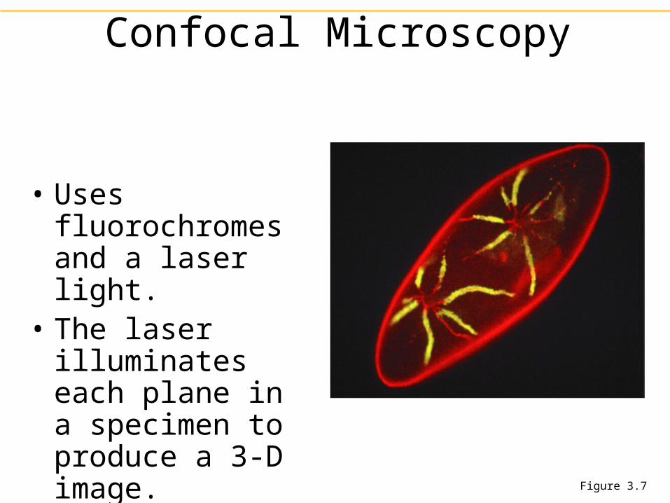
• Uses fluorochromes and a laser light.
• The laser illuminates each plane in a specimen to produce a 3-D image.
Confocal Microscopy
Figure 3.7

• Uses electrons instead of light.
• The shorter wavelength of electrons gives greater resolution.
Electron Microscopy

• Ultra thin sections of specimens.
• Light passes through specimen, then an electromagnetic lens, to a screen or film.
• Specimens may be stained with heavy metal salts.
Transmission Electron Microscopy (TEM)
Figure 3.8a

• 10,000-100,000; resolution 2.5 nm
Transmission Electron Microscopy (TEM)
Figure 3.8a

• An electron gun produces a beam of electrons that scans the surface of a whole specimen.
• Secondary electrons emitted from the specimen produce the image.
Scanning Electron Microscopy (SEM)
Figure 3.8b

• 1000-10,000; resolution 20 nm
Scanning Electron Microscopy (SEM)
Figure 3.8b

• Scanning tunneling microscopy uses a metal probe to scan a specimen.
• Resolution 1/100 of an atom.
Scanning-Probe Microscopy
Figure 3.9a

• Atomic force microscopy uses a metal and diamond probe inserted into the specimen.
• Produces 3-D images.
Scanning-Probe Microscopy
Figure 3.9b

Preparation of Specimens for
Light Microscopy• Tools:
– Loops and needles - compare uses– Stains
• basic (positive ion like crystal violet)• Acidic (negative ions like India ink)
• Hanging drop– Used for motility determination
• All other preparations require:– A thin film of a solution of microbes on a slide is a smear.
• A smear is usually fixed to attach the microbes to the slide and to kill the microbes.

Staining• Coloring microorganisms for better visibility• Steps for most preparations (except capsule
and hanging drop:– Smear– Air dry– Fix– Stain– Rinse

• Live or unstained cells have little contrast with the surrounding medium. However, researchers do make discoveries about cell behavior looking at live specimens.
Preparing Smears for Staining

• Stains consist of a positive and negative ion.
• In a basic dye, the chromophore is a cation.
• In an acidic dye, the chromophore is an anion.
• Staining the background instead of the cell is called negative staining.
Preparing Smears for Staining
• Use of a single basic dye is called a simple stain.
• A mordant may be used to hold the stain or coat the specimen to enlarge it.
Simple Stains

• Differential stains are used to differentiate between types of bacteria– Gram– Acid fast
• The Gram stain classifies bacteria into gram-positive and gram-negative.
• Gram-positive bacteria tend to be killed by penicillin and detergents.
• Gram-negative bacteria are more resistant to antibiotics.
Differential Stains: Gram Stain

Differential Stains: Gram Stain
Color of Gram + cells
Color ofGram – cells
Primary stain:Crystal violet
Purple Purple
Mordant:Iodine
Purple Purple
Decolorizing agent:Alcohol-acetone
Purple Colorless
Counterstain:Safranin
Purple Red

Differential Stains: Gram Stain
Figure 3.10b

• Cells that retain a basic stain in the presence of acid-alcohol are called acid-fast.
• Non–acid-fast cells lose the basic stain when rinsed with acid-alcohol, and are usually counterstained (with a different color basic stain) to see them.
Differential Stains: Acid-Fast Stain
Figure 3.11

• Negative staining is useful for capsules.
• Heat is required to drive a stain into endospores.
• Flagella staining requires a mordant to make the flagella wide enough to see.
Special Stains
Figure 3.12a-c