Embed Size (px)
Citation preview
The Ministry of education and science of Russian Federation
Ulyanovsk State University
Institute of Medicine, Ecology and physical culture
Department of General and Clinical Pharmacology with Microbiology course
M. N. Artamonova, N.I. Potaturkina- Nesterova, I. S. Nemova
Microbiology, virology:
guidelines for practical classes for foreign students
Part 1
Ulyanovsk 2016
2
УДК 535.317.68 (07.07)
ББК 52.64 я 73
G 36
It was recommended by the scientific-methodological council of Institute of
Medicine, Ecology and physical culture of Ulyanovsk State University
Reviewer:
A. S. Nesterov, PhD of Medicine, professor
M. N. Artamonova, N.I. Potaturkina- Nesterova, I. S. Nemova
Microbiology, virology: guidelines for practical classes for foreign students.
Part 1/ M. N. Artamonova, N.I. Potaturkina- Nesterova. - Ulyanovsk: ULSU,
2016.-85 p.
Guidelines for practical training on "Microbiology, Virology" is
recommended for students of English department and build in accordance with the
existing curriculum, the federal state educational standards in the direction of
preparation 31.05.01.- "General Medicine".
The handout is structured on topics that focused on the basic concepts of the
discipline. Each practice lesson includes a description of the learning objectives,
job occupation, questions for self-control, tasks for students' independent work,
which describe the types of performed work; basic terms of the subject; examples
of typical case studies; list of recommended literature, additional material,
comprising - instructions for students on labor protection.
© M. N. Artamonova, N.I. Potaturkina- Nesterova,
I. S. Nemova 2016
© Ulyanovsk State University
3
CONTENTS
Practical lesson 1. Subject and aims of medical microbiology. The
morphology and taxonomy microorganisms. Microscopic research method.
Sterilization and disinfection……………………………………….…………
4
Practical lesson 2. Physiology of microorganisms. Metabolism. Nutrition
and respiration of bacteria. Culturing methods of aerobic and anaerobic
bacteria ………………………………………………………………………
19
Practical lesson 3. Viruses. The discovery of viruses, its classification.
Bacteriophages. The practical importance of phages in biology and medicine.
The genetics of microorganisms. Biotechnology in microbiology. Molecular-
biological methods of diagnosis……………………………………………….
30
Practical lesson 4. Infection: the role of microorganisms in infectious
process. Biological diagnostic method. The role of host in the infectious
process. Microbiological basics of antimicrobial therapy. Methods for
determination sensitivity of bacteria to antibiotics………………………..
38
Practical lesson 5. Ecology of microorganisms. The human microflora and
its function. Dysbiosis, methods of correction. Biofilms. Sanitary
demonstration water microorganisms, soil, air, and methods of
research………………………………………………………………………..
43
Practical lesson 6. Immunity. Serological methods – agglutination test,
precipitation test, complement fixation test ………………………………..
47
Practical lesson 7. Clinical Microbiology. Etiology and pathogenesis of
nosocomial infections. The causative agents, properties and microbiological
diagnostics of nosocomial infections. Pathogenic and opportunistic gram-
positive and gram-negative cocci…………………………………………….
51
Practical lesson 8. Pathogens of escherichiosis and dysentery. The causative
agents of typhoid, salmonella poisoning and cholera………………………
64
Practical lesson 9. Pathogens of diphtheria, whooping cough. Pathogens of
legionellosis. Pathogenic mycobacteria. Concluding lesson 2. Colloquium.

4
The final lesson ………………………………………………………………. 75
The list of literature………………………………………………………….. 85
5
Practical lesson 1
Subject and aims of medical microbiology. The morphology and
taxonomy microorganisms. Microscopic research method.
Objective: formation of knowledge about the structure, aims and methods of
microbiology as a science, the history of its formation and position among other
sciences.
Tasks:
1. Learn to work with bacterial cultures.
2. Prepare fixed preparations
Questions:
1. Subject of Medical Microbiology and its importance for practical health care.
The history of microbiology.
2. The system and nomenclature of microorganisms.
3. Research microbiology methods.
4. Simple and complex staining techniques. The mechanism of staining smears.
Tinctorial properties of microorganisms.
5. The light microscope, its main characteristics. Types of light microscopy (dark-
field, phase - contrast, fluorescent). Immersion microscopy principles. Electron
microscopy, atomic force microscopy.
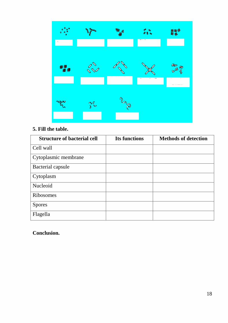
6. The forms of the bacteria.
7. The structure of the bacterial cell: genome, cytoplasm, ribosome. Its structure,
functions and methods of detection.
8. The shell of bacteria: cytoplasmic membrane, cell wall, capsule. The structure,
functions and methods of detection.
9. Fimbria and pili. Its structure, functions and methods of detection.
10.Bacterial spores. Its role and structural features. Spore formation and methods
of detection.
6
Morphology of microorganisms. Simple methods of staining preparations
Notion Definition/ explanation
Microbiology
Microbiology is the study (logy) of very small (micro)
living (bio) organisms. Often, people are scared by the
topic's name of the theme and by science itself
Bacteria Bacteria are relatively simple in structure. They are
procaryotic simple unicellular organism with no nuclear
membrane, mitochondria,
Golgi bodies, or endoplasmic reticulum that
reproduces by asexual division. Although the cell wall
encircling bacteria is itself complex, there are two basic
forms: gram-positive cell wall with a thick peptidoglycan
layer and gram-negative cell wall with a thin
peptidoglycan layer and an overlaying outer membrane.
Principle of the
microscopy with oil
immersion
Placing a drop of oil with the same refractive index as
glass between the cover slip and objective lens eliminates
two refractive surfaces, so that magnifications of 1000x or
greater can be achieved while still preserving good
resolution
Types of bacteria Bacteria have three basic shapes: spherical (round), rod-
shaped, and spiraled. A round bacterium is called a coccus
(plural, cocci). A rod-shaped organism is called a bacillus
(plural, bacilli) or simply a rod. A spiraled bacterium with
at least two or three curves in its body is called a spirillum
(plural, spirilla). Long sinuous organisms with many loose
or tight coils are called spirochaetes
Cocci The patterns formed by bacterial cells grouping
together as they multiply are often characteristic for

7
individual bacterial genera or species. Cocci may occur in
pairs (diplococci), chains (streptococci), clusters
(staphylococci), or packets of four (tetrads), and are
seldom found singly
Rod-shaped
bacteria (bacilli)
Rod-shaped bacteria (bacilli) generally occur as
individual cells, but they may appear as end-to-end pairs
(diplobacilli) or line up in chains (streptobacilli). Some
species tend to palisade, that is, line up in bundles of
parallel bacilli, others may form V, X, or Y figures as they
divide and split. Some may show great variation in their
size and length (pleomorphism)
Spiral forms of
bacteria
Vibrio, spirochaete, spirilla
Structure of the bacterial cells. Complex method of preparations staining.
Notion Definition/explanation
Cell wall Outer covering of most cells that protects the bacterial cell
and gives shape to it
Gram staining The Danish bacteriologist J. M. C. Gram (1853–1938)
devised a method of bacteria staining using a dye called
crystal (gentian) violet. Gram's method helps distinguish
different types of bacteria.
The characteristics of bacteria Gram staining are denoted
as positive or negative, depending upon whether the bacteria
take up and retain the crystal violet stain or not.
Gram-positive bacteria retain the colour of the crystal
violet stain in the Gram stain. This is characteristic of
bacteria that have a cell wall composed of a thick layer of a
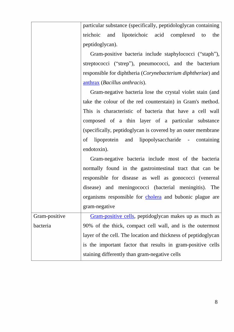
8
particular substance (specifically, peptidologlycan containing
teichoic and lipoteichoic acid complexed to the
peptidoglycan).
Gram-positive bacteria include staphylococci (“staph”),
streptococci (“strep”), pneumococci, and the bacterium
responsible for diphtheria (Corynebacterium diphtheriae) and
anthrax (Bacillus anthracis).
Gram-negative bacteria lose the crystal violet stain (and
take the colour of the red counterstain) in Gram's method.
This is characteristic of bacteria that have a cell wall
composed of a thin layer of a particular substance
(specifically, peptidoglycan is covered by an outer membrane
of lipoprotein and lipopolysaccharide - containing
endotoxin).
Gram-negative bacteria include most of the bacteria
normally found in the gastrointestinal tract that can be
responsible for disease as well as gonococci (venereal
disease) and meningococci (bacterial meningitis). The
organisms responsible for cholera and bubonic plague are
gram-negative
Gram-positive
bacteria
Gram-positive cells, peptidoglycan makes up as much as
90% of the thick, compact cell wall, and is the outermost
layer of the cell. The location and thickness of peptidoglycan
is the important factor that results in gram-positive cells
staining differently than gram-negative cells
9
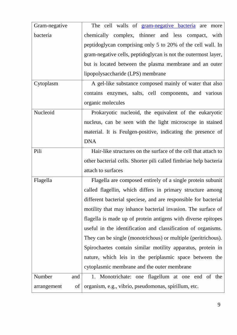
Gram-negative
bacteria
The cell walls of gram-negative bacteria are more
chemically complex, thinner and less compact, with
peptidoglycan comprising only 5 to 20% of the cell wall. In
gram-negative cells, peptidoglycan is not the outermost layer,
but is located between the plasma membrane and an outer
lipopolysaccharide (LPS) membrane
Cytoplasm A gel-like substance composed mainly of water that also
contains enzymes, salts, cell components, and various
organic molecules
Nucleoid
Prokaryotic nucleoid, the equivalent of the eukaryotic
nucleus, can be seen with the light microscope in stained
material. It is Feulgen-positive, indicating the presence of
DNA
Pili Hair-like structures on the surface of the cell that attach to
other bacterial cells. Shorter pili called fimbriae help bacteria
attach to surfaces
Flagella
Flagella are composed entirely of a single protein subunit
called flagellin, which differs in primary structure among
different bacterial speciese, and are responsible for bacterial
motility that may inhance bacterial invasion. The surface of
flagella is made up of protein antigens with diverse epitopes
useful in the identification and classification of organisms.
They can be single (monotrichous) or multiple (peritrichous).
Spirochaetes contain similar motility apparatus, protein in
nature, which leis in the periplasmic space between the
cytoplasmic membrane and the outer membrane
Number and
arrangement of
1. Monotrichate: one flagellum at one end of the
organism, e.g., vibrio, pseudomonas, spirillum, etc.
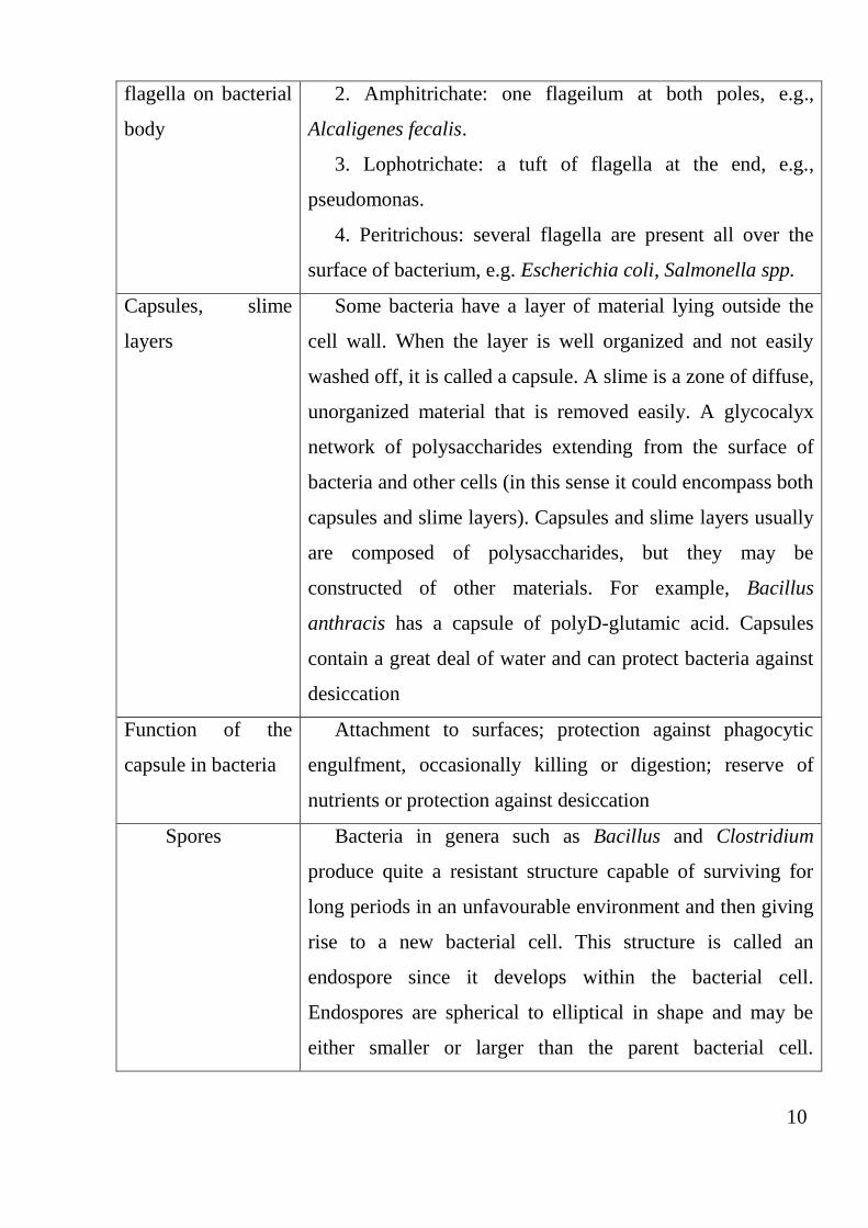
10
flagella on bacterial
body
2. Amphitrichate: one flageilum at both poles, e.g.,
Alcaligenes fecalis.
3. Lophotrichate: a tuft of flagella at the end, e.g.,
pseudomonas.
4. Peritrichous: several flagella are present all over the
surface of bacterium, e.g. Escherichia coli, Salmonella spp.
Capsules, slime
layers
Some bacteria have a layer of material lying outside the
cell wall. When the layer is well organized and not easily
washed off, it is called a capsule. A slime is a zone of diffuse,
unorganized material that is removed easily. A glycocalyx
network of polysaccharides extending from the surface of
bacteria and other cells (in this sense it could encompass both
capsules and slime layers). Capsules and slime layers usually
are composed of polysaccharides, but they may be
constructed of other materials. For example, Bacillus
anthracis has a capsule of polyD-glutamic acid. Capsules
contain a great deal of water and can protect bacteria against
desiccation
Function of the
capsule in bacteria
Attachment to surfaces; protection against phagocytic
engulfment, occasionally killing or digestion; reserve of
nutrients or protection against desiccation
Spores
Bacteria in genera such as Bacillus and Clostridium
produce quite a resistant structure capable of surviving for
long periods in an unfavourable environment and then giving
rise to a new bacterial cell. This structure is called an
endospore since it develops within the bacterial cell.
Endospores are spherical to elliptical in shape and may be
either smaller or larger than the parent bacterial cell.
11
Endospore position within the cell is characteristic and may
be central, subterminal, or terminal
Cycle of spore
formation and
germination
At the beginning of spore formation, a septum forms,
separating the nascent spore from the rest of the cell and all
of the genetic material of the cell is copied into the newly-
forming cell. The spore contents are dehydrated
Function of the
bacterial spore
Bacterial spores are the highly resistant spores. In fact the
spores indicate the resting phase of some types of
bacteria,which help to tide over the unfavourable conditions.
These spores can also be called as endospores. These
endospores contain calcium and dipicolenic acid and hence
they are very resistant. They are resistant to temperature, UV
light, disinfectants, etc. and thus they protect the bacterium.
In the favourable conditions these develop into a new
bacterium.
PROCEDURE OF PRACTICAL WORK
Task 1. Learn the safety rules in microbiology laboratory and write
them down.
GENERAL LABORATORY SAFETY RULES FOR MICROBIOLOGY
1. No smoking, eating, drinking, chewing gum, or applying cosmetics in
laboratory area, including laboratory office. No foodstuffs are to be stored in
laboratories (including cold rooms, refrigerators, and freezers).
2. Disposable laboratory gloves are not to be worn in communal areas. Door
handles, telephones, computer keyboards, and mice (except in clearly defined cir-
cumstances), lift buttons, etc. are not to be touched with gloves. If needed, wear
one glove and use the ungloved hand to open doors, operate lifts, etc.
12
3. Rubber or disposable gloves should be worn when handling/working with:
human blood or other body fluids, dangerous chemicals, infectious, or potentially
infectious materials, ultraviolet light boxes. Select an appropriate glove type.
4. Laboratory gowns or labcoats must always be worn in laboratories, but
they should be taken off before entering “clean areas”, e.g., tea room, stores,
media, toilets, library, and office rooms.
5. Clothing and footwear must be suitable for laboratory conditions. Flip-
flops, sandals or high- heeled shoes are not to be worn in the laboratory. Bare feet
are prohibited in the building. Sandals with an enclosed toes and heels are
acceptable, but for your own protection, an enclosed shoes are preferable. Long
hair must be tied back to avoid contact with microorganisms and equipment.
6. Protective glasses must be worn for all kinds of work involving corrosive
or toxic chemicals, radioactivity, and ultraviolet light.
7. The best protection from microorganisms ingestion is avoidance of their
penetration in your mouth. Labels and envelopes must not be licked. Pencils and
pens must not be laced in the mouth. Biting of fingernails, playing with hair,
applying lipstick, eating, drinking, etc., are not allowed. Wash your hands when
leaving the laboratory for lunch, etc.
8. Do not pipette by mouth. The use of pipettes with cotton plugs to reduce
contamination is preferable. Place pipettes in disinfectant solution to minimize
aerosol production. Submerge them for 18–24 hours. Residual volumes from
pipettes create aerosols; use mechanical devices that are calibrated to deliver. Fit
pipettes to a soft bulb by holding it at the plugged end to avoid the risk of cuts in
case the pipette is broken.
9. Minimize the use of “sharps”. Do not bend needles, or try to replace the
caps after use. Use syringes fitted with blunt cannulas where possible. Avoid using
syringes to mix infectious liquids (if essential, hold the tip of the needle under the
surface of the fluid and avoid excessive force). Discard used syringes and needles
into an approved sharps container.
13
10. Hazardous chemical and biological spills and blood spills on the floor,
benches or equipment should be cleaned up immediately. Special treatment is re-
quired for spills of a biohazardous nature.
11. Hands should be washed after completing each task and always before
leaving the laboratory.
12. No running or “horse play”. Report all potential hazards and problems
immediately. Try to anticipate potential problems.
13. Any faulty equipment should be removed from service for repair or
disposal.
14. When flaming wire loops, draw the loop gradually from the cooler to
the hotter part of the flame to minimize spattering, or use electric heaters. Make
sure the loop is completely closed and the loop wire is not longer than 6 cm.
15. Disposable plastic loops must be placed loop-end down in disinfectant
for 18–24 hours.
16. Petri dish cultures of fungi should be imprinted and incubated with the
lid up permost to prevent the dispersal of fungal spores. Recognize fungi as
potential pathogens and be aware of the ability of some species to produce
mycotoxins.
17. Take care when handling Petri dishes that contain condensate. This may
contain viable microorganisms that can be spread via droplets or aerosols when the
plates are opened or dropped.
18. Open and operate tissue grinders in a biological safety cabinet. Hold
glass grinders in a wad of absorbent material and wear gloves. Wait 10 minutes
before opening a blender bowl to allow aerosols to settle. Refrigerate to condense
aerosols. Use models designed to prevent leakage from rotor bearings and O-ring
gaskets or use a “stomacher”.
Task 2. Prepare the smears of mixed (staphylococcal and colibacillar)
cultures, stain them by Gram’s method, perform microscopy and draw it.
Technique of cultures smear preparation grown on the solid nutrient
14
medium
Manipulation 1. Smear preparation of the culture grown on the solid
nutrient medium
For smear preparation take a clean slide. By means of glass pencil draw a
circle 1.5–2.0 cm in diameter – a place of the material drawing. On the opposite
part of the glass prepare smear test. In the left hand take a test tube with a
physiological solution (NaCl), and in the right – a bacteriological loop. The loop is
calcinated in a flame of a spirit-lamp (sterilized) for destruction of extraneous
bacteria. By rotary movements take a wadded fuse out of the test tube, pressing it
by the 5th and 4th fingers of the right hand to a palm, and burn the edge of the test
tube. A loop is cautiously entered into a test tube, cooled about an internal surface
of the glass. Then put 1–2 drops of the sodum chloride on the glass slide, burn the
edge of the test tube again and close its fuse. Then take a test tube with the grown
up culture in the left hand, similarly open it, sterilize the edge of the test tube, enter
the loop into the test tube cautiously, grasp a material with a sliding movement,
take out a loop from the test tube, burn the edge of the test tube again and close its
fuse. The material on the bacteriological loop is brought in the drop of the sodum
chloride and prepared for a suspension, not falling outside the limits of the circle
(at correct distribution of the material in smear test at microscopy isolated bacterial
cells are visible). After preparation of smear test, bacteriological loop is carefully
burned in a flame of the spirit-lamp.
Manipulation 2. Smear test fixation
Fixe the smear by spent it three times (in 3–5 seconds) through the flame of
the spirit-lamp (smear test upwards), carrying out physical fixation.
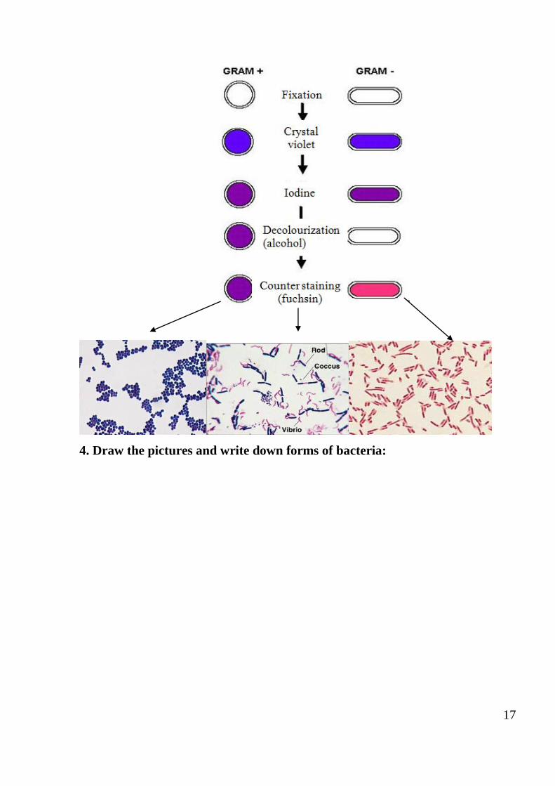
Manipulation 3. Smear staining by Gram’s method.
Gram staining procedure
1. Smear the material to be stained on a slide. Allow it to dry in air. Fix by
gentle heating, which kills
bacteria and allows the material to be attached to the slide.
15
2. Apply an appropriate solution of gentian violet. Stain it for about 1 minute.
Wash carefully.
3. Apply iodine solution (a mordant, which strengthens the bond between dye
and substrate) for 1 minute. Wash carefully.
4. Apply 95% ethyl alcohol until all but the thickest parts of the smear are
decolourized, or for not more than 10 to 15 seconds. Wash.
5. Counterstain with fuchsin for 1 minute. Wash. Dry.
6. Examine the smear using the oil-immersion lens of the light microscope.
7. Gram-positive organisms are blue-purple and gram-negative bacteria are
pink-red. If the spesimen is stained correctly, gram-positive (violet) and gram-
negative (red) bacteria will be visible on microscopy. At the wrong staining all
bacteria are the same colour (violet or red).
Manipulation 4. Performing of microscopy with immersion system .
Rules of work with immersion system:
1. Lift condenser up to the level of the objective table, open the diaphragm
of the microscope.
2. By means of eyepiece 15×, objective 8×, and flat mirror, achieve the
maximal visual field illuminance.
3. Put a preparation on the objective table, fix its plugs, by means of the
macroscrew find the contours of the image, and by means of the microscrew –
obtain precise image.
4. Turning a nosepiece of the microscope, place an immersion objective 90×
above the slide; dip it in the oil immersion.
5. Turning the microscrew of the microscope, obtain precise image of the
preparation, study microorganism morphology, determine the increase, at which
the preparation was investigated.
6. After the termination of microscopy lift a tube, take the preparation away,
wipe an objective from oil immersion, carefully turn it aside and lower a tube.
Manipulation 5. Drawing it.
16
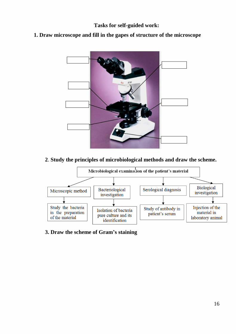
Tasks for self-guided work:
1. Draw microscope and fill in the gapes of structure of the microscope
2. Study the principles of microbiological methods and draw the scheme.
3. Draw the scheme of Gram’s staining
17
4. Draw the pictures and write down forms of bacteria:
18
5. Fill the table.
Structure of bacterial cell Its functions Methods of detection
Cell wall
Cytoplasmic membrane
Bacterial capsule
Cytoplasm
Nucleoid
Ribosomes
Spores
Flagella
Conclusion.
19
Practical lesson 2
Physiology of microorganisms. Metabolism. Nutrition and respiration of
bacteria. Culturing methods of aerobic and anaerobic bacteria
Objective: To systematize the knowledge of the metabolism of
microorganisms.
Tasks:
1. To study the mechanisms of microbial nutrition.
2. To study kinds of culture media.
3. To consider microbial seeding technique on nutrient media
Questions:
1. Anabolism and catabolism.
2. Mechanisms of nutrients transmission in the bacterial cell.
3. Autotrophs and heterotrophs, auxotrophs and prototrophs.
4.Classification of culture media.
5. Simple and complex nutrient medium.
6. Method for sowing nutritive medium.
7. Growth phases on medium.
8. Mechanism of bacterial respiration. Aerobes and anaerobes.
9. Methods of cultivation of anaerobic bacteria: culture media, equipment.
10. Isolation of pure culture of anaerobes.
11. Identification of the selectioned pure cultures of bacteria.
12. The main groups of bacteria enzymes.
13. Determination saccharolytic enzymes of bacteria.
14. Determination of proteolytic enzymes.
15. Determination peptolytic enzymes.
16. Aggression enzymes: coagulase, hyaluronidase, neuraminidase, DNA -
ase, hemolysin.
20
Physiology of bacteria. Bacteria nutrition and respiration. Nutrient
media. Isolation of pure cultures of aerobic bacteria (stage 1)
Notion Definition/explanation
Metabolism Metabolism is the total of all chemical reactions occurred
in the cell. Metabolism may be divided into major parts.
In catabolism larger and more complex molecules are
broken down into smaller, simpler molecules with the release
of energy. The bacterial cell obtains the energy for
biochemical reaction due catabolism (energy-generating or
energy-yielding process).
Anabolism is the synthesis of complex molecules from
simpler ones with the input of energy (energy-requiring
process)
Pure culture Pure culture is a population of cells arising from a single
cell.
Can be accomplished from mixtures by a variety of
procedures, including streak plates and pour plates
Culture media
Much of microbiology depends on the ability to grow and
maintain microorganisms in the laboratory.
This is possible only if suitable culture media is available.
Media can be defined (synthetic) or complex, and liquid
or solid.
Classification of
culture media
Based on the consistency:
- liquid – peptone water, nutrient broth;
- semisolid – nutrient agar stabs;
- solid – blood agar, serum agar.
Based on oxygen requirement:
- aerobic medium;
21
- anaerobic medium
Synthetic
(defined) media
Synthetic (defined) media are media in which all
components and their concentrations are known.
1. Peptones: protein hydrolysates prepared by partial
proteolytic digestion of various protein sources.
2. Extracts: aqueous extracts, usually of beef or yeast
Liquid media Liquid media: the easiest to prepare and use. Good for
growing quantities of microbes needed for analysis or
experiments. Unless inoculated with pure culture, cannot
separate different organisms
Solid media Solid media is needed for surface cultivation of
microorganisms
Selective media Inhibits the growth of some bacteria while selecting for
the growth of others. Example: brilliant green agar – dyes
inhibit the growth of gram-positive bacteria; selects for gram-
negative bacteria
Differential media Differential media distinguish between different groups of
bacteria on the basis of their biological characteristics. They
cause observable change in medium when biochemical
reaction occurs. For example: MacConkey agar has color
indicator that distinguishes presence of acid. Bacteria that
ferment a particular sugar (e.g., glucose in culture media) will
produce acid wastes on plates, turn pH indicator red. Bacteria
that cannot ferment the same sugar
will grow but not affect pH, so colonies remain white
Complex media Enriched media: blood agar (nutrient agar + 5 to 10%
sheep blood).
Melt the sterile nutrient agar by steaming, cool to 45 °C.

22
Add the blood aseptically with constant shaking.
Mix the blood with molten nutrient agar thoroughly but
gently avoiding froth formation.
Immediately pour in to the Petri dishes or tubes and allow
setting.
Use: cultivate all the fastidious organisms
Growth and reproduction of bacteria. Enzymes
Notion Definition/explanation
Isolation of microbes
from clinical
material
1. Microscopic examination: Gram staining, acid fast
stain, dark field microscopy, phases contrast.
2. Culture: use streak plate to culture sample on
appropriate media to get pure culture. Use in addition
selective media for isolation of the suspected organism.
3. Identification: use staining, differential media,
serology and or gene probe for identification of the isolated
microbe.
4. In vitro antibiotic sensitivity test.
5. Epidemiology in case of outbreak to determine the
source of infection
Clone A race of cells derived from a single ancestral cell and
sharing a single function
Colony A number of microorganisms living or multiplying
together on solid culture media in the result of
multiplication of a single cell
Pure culture Pure culture is a population of microorganism of the
same species isolated on a nutrient medium
23
Characteristic of
stroke culture
1. The degree of growth – scanty, moderate or profuse.
2. Their nature – discrete or confluent, filiform,
spreading or rhizoid
3. Their elevation, surface, edges, colour, structure,
odour, emuisifiability, consistency and cha the medium are
noted
Classification of
enzymes
1. Exoenzymes are transported extracellularly, where they
break down large food molecules or harmful chemicals;
cellulase, amylase, penicillinase.
2. Endoenzymes are retained intracellularly and function
there.
3. Constitutive enzymes are always present, they are
produced in equal amounts or at equal rates, regardless of
amount of substrate; enzymes are involved in glucose
metabolism.
4. Induced enzymes are not constantly present, they are
produced only when substrate is present; prevent cells from
resources loss
Facultative
anaerobes
Microorganisms that can live and grow with or without
molecular oxygen
Obligate anaerobes Microorganisms that can grow only in the complete
absence of molecular oxygen; some are killed by oxygen
Examples of obligate
anaerobic bacteria
Clostridium perfringens, C. tetani, C. histolyticum, C.
novyi, C. septicum, C. botulinum, Bacteroides fragilis, B.
gracilis
Facultative anaerobic
organism
A facultative anaerobic organism is an organism, usually a
bacterium, that makes ATP by aerobic respiration if oxygen
is present but is also capable of switching to fermentation.
24
In contrast, obligate anaerobes die in presence of oxygen
Examples of
facultative anaerobic
bacteria
The examples of facultative anaerobic bacteria are
Staphylococcus (gram-positive), Escherichia coli (gram-
negative), Corynebacterium (gram-positive), and Listeria
(gram-positive)
Methods of
anaerobic culture
isolation
Physical method → cultivated the anaerobic bacteria’s
in agar deep or in fluid media, but on surface presence oil,
Vinyal-Veyon’s method.
Chemical method → used in media substance for
absorption O2.
Biological method → Fortner method (subcultured to
aerobic and anaerobic)
PROCEDURE OF PRACTICAL WORK
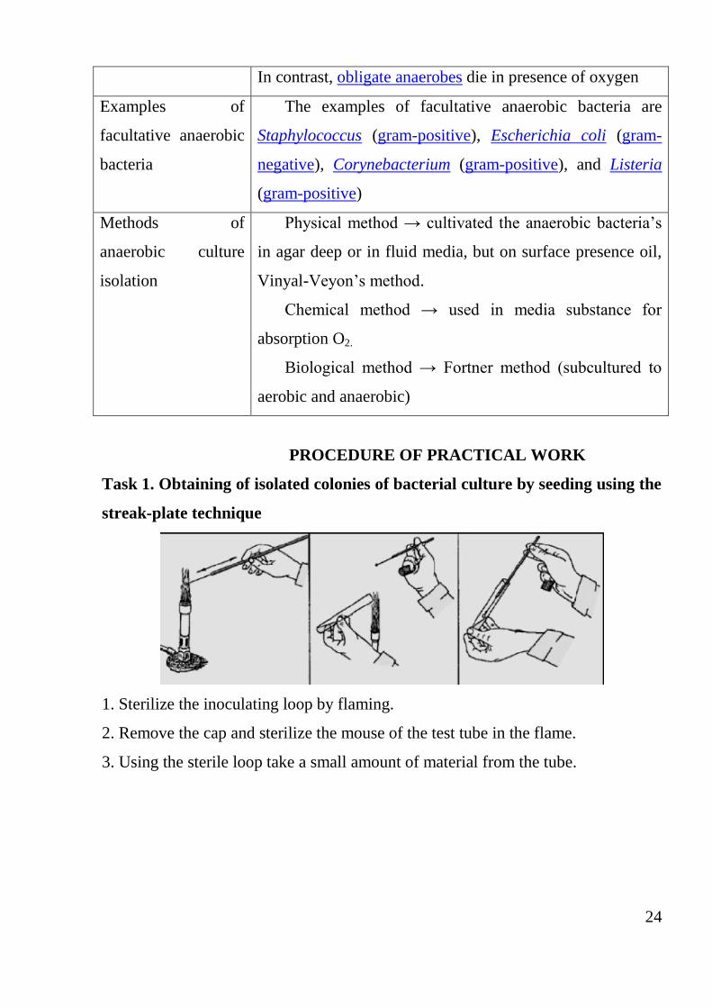
Task 1. Obtaining of isolated colonies of bacterial culture by seeding using the
streak-plate technique
1. Sterilize the inoculating loop by flaming.
2. Remove the cap and sterilize the mouse of the test tube in the flame.
3. Using the sterile loop take a small amount of material from the tube.
25
4. Sterilize the mouse of the test tube after taking the sample, recap it and place it
back. The inoculating loop is not flamed.
5. Carefully lift the top of the dish containing sterile agar just enough to insert
your inoculating loop easily and make a streak over the agar plate spreading out
the bacteria.
6. Remove the inoculating loop, close the Petri dish and rotate it counter
clockwise about 90 degrees.
7. Sterilise the inoculating loop in order to kill any remaining bacteria by flaming
them.
8. Open the agar Petri dishes again, insert the loop under the lid and cool it at the
edge of the agar. Keeping in mind where the initial streaks ended make a streak
over the agar plate spreading out bacteria starting from the previous streak.
9. Close the Petri dish and turn the Petri dish counter clockwise about 90 degrees
again.
10. It is necessary to repeat the sequence of the streaking procedures described
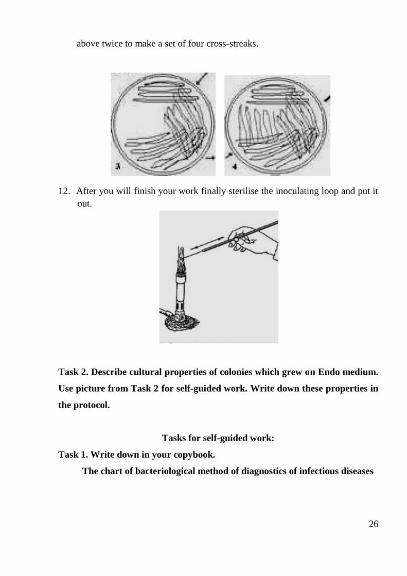
26
above twice to make a set of four cross-streaks.
12. After you will finish your work finally sterilise the inoculating loop and put it
out.
Task 2. Describe cultural properties of colonies which grew on Endo medium.
Use picture from Task 2 for self-guided work. Write down these properties in
the protocol.
Tasks for self-guided work:
Task 1. Write down in your copybook.
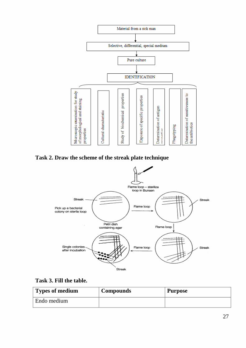
The chart of bacteriological method of diagnostics of infectious diseases
27
Task 2. Draw the scheme of the streak plate technique
Task 3. Fill the table.
Types of medium Compounds Purpose
Endo medium
28
Ploskirev’s medium
Blood agar
Salt agar
MacConkey agar
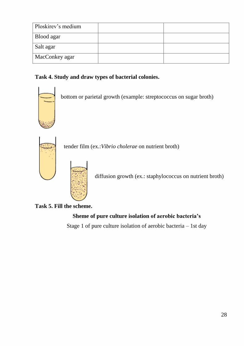
Task 4. Study and draw types of bacterial colonies.
bottom or parietal growth (example: streptococcus on sugar broth)
tender film (ex.:Vibrio cholerae on nutrient broth)
diffusion growth (ex.: staphylococcus on nutrient broth)
Task 5. Fill the scheme.
Sheme of pure culture isolation of aerobic bacteria’s
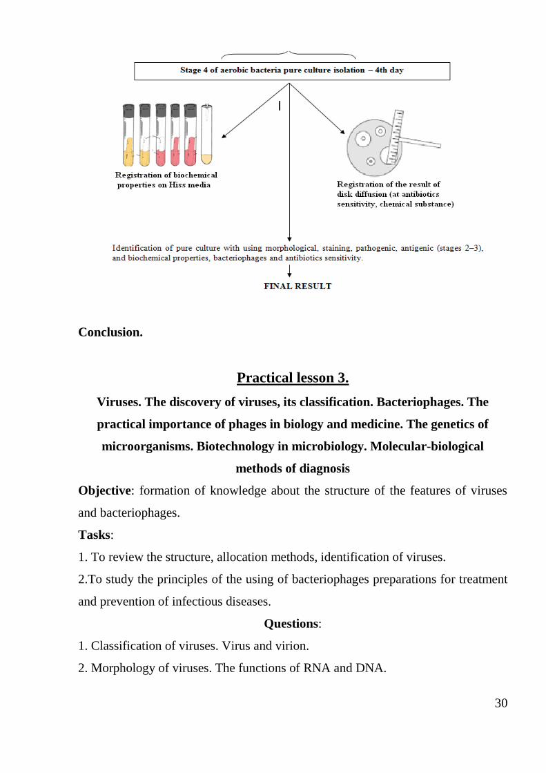
Stage 1 of pure culture isolation of aerobic bacteria – 1st day
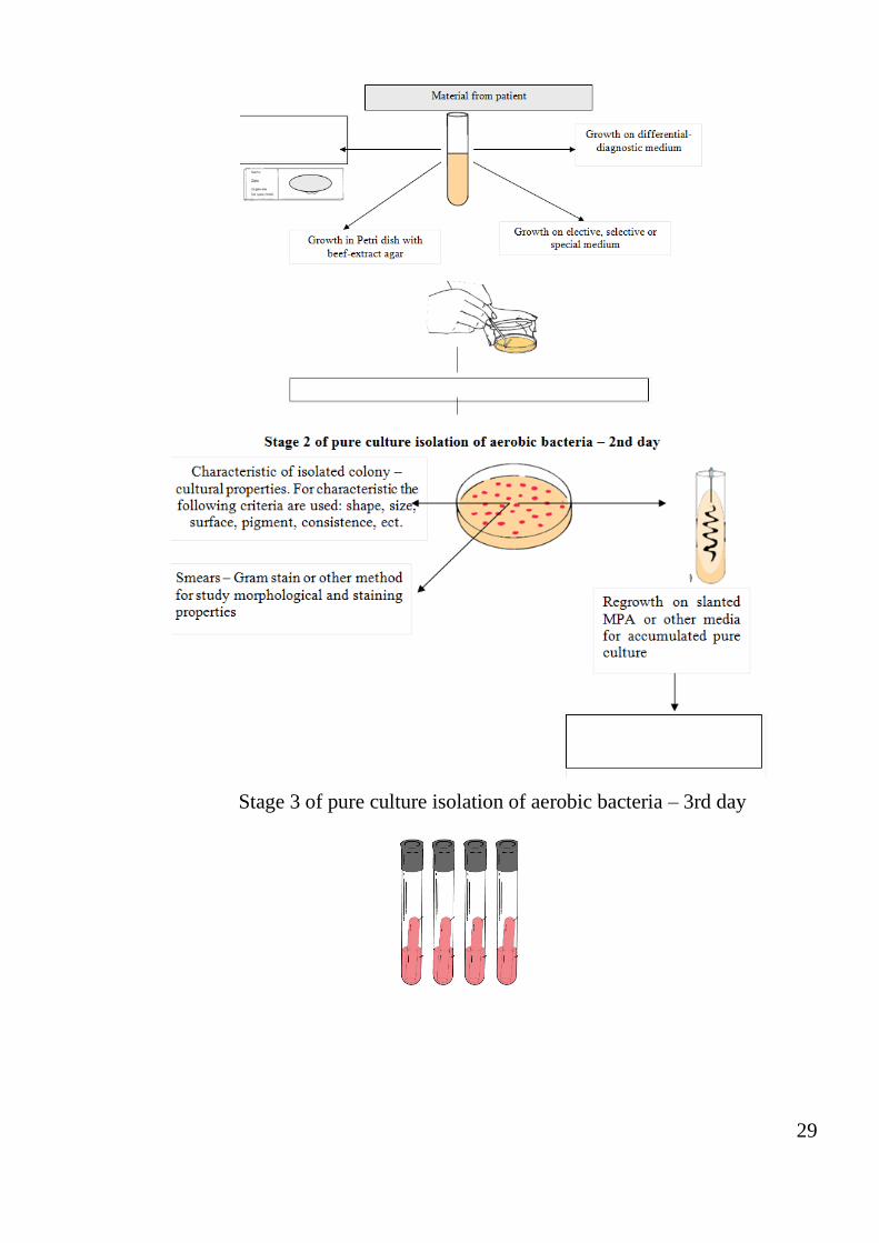
29
Stage 3 of pure culture isolation of aerobic bacteria – 3rd day
30
Conclusion.
Practical lesson 3.
Viruses. The discovery of viruses, its classification. Bacteriophages. The
practical importance of phages in biology and medicine. The genetics of
microorganisms. Biotechnology in microbiology. Molecular-biological
methods of diagnosis
Objective: formation of knowledge about the structure of the features of viruses
and bacteriophages.
Tasks:
1. To review the structure, allocation methods, identification of viruses.
2.To study the principles of the using of bacteriophages preparations for treatment
and prevention of infectious diseases.
Questions:
1. Classification of viruses. Virus and virion.
2. Morphology of viruses. The functions of RNA and DNA.
31
3. Chemical composition nukleoprotein. Enzymes.
4. Methods of viruses cultivation.
5. Interaction of viruses with cell virus. The mechanism of transcription and
replication viral genome.
6. The mechanism of DNA and RNA viral integration into the cellular genome.
7. Ways transmission of viral infections.
8. The morphology of phages.
9. The mechanism of interaction between phages and the bacterial cell.
10.Virulent and temperate phages. Lysogenesis.
11.Titr phage. Methods for its determination.
12. Obtaining of phage culture. The using of phages in medicine.
13. Genotype and phenotype of bacteria.
14. Extrachromosomal factors in bacteria – plasmids, transposons, Is – sequence;
its role.
15. Forms of variability in microorganisms.
16. Mutations, kinds of mutations in bacteria.
17. Genetic recombination in bacteria (transformation, transduction,conjugation).
18. The concept of the modifications.
19. The theoretical and practical importance of genetics: molecular-biological
methods of diagnosis The practical using of genetic engineering.
Table 4. Viruses. Bacteriophages
Notion Definition/explanation
Two types of
phages
There are two types of phages: virulent phages and
temperate phages. Virulent phages infect the bacteria,
reproduce, and then lyse and kill the bacteria. On the other
hand, temperate phages have a good temperament and do not
immediately lyse the bacteria they infect.
Types of phage Productive type: bacteriophages lyse their host bacterial
32
reproduction in
bacterial cell
cells after penetration and reproductive cycle.
Abortive type: after penetration the new phage particles
will not forming and bacteria live.
Integrative type: phage genome is integrated in bacterial
chromosome and bacterial host cell is live
Classification of
bacteriophages
Polyvalent – destroy kindred bacteria, for example,
polyvalent salmonella bacteriophages destroy all Salmonella
spp. (S. enteritidis, S. typhimurium).
Monovalent – destroy one species of bacteria, for
example, typhoid fever Vi- bacteriophages – destroy only
typhoid fever agent.
Typospecific – destroy one type of bacteria species
Prophage The integrated temperate phage genome is called a
prophage. Bacteria that have a prophage integrated into their
chromosome are called lysogenic because at some time the
repressed prophage can become activated.
Bacterial genetics.
Notion Definition/explanation
Genotype and
phenotype
The genotype of an organism is its genetic makeup, the
information that codes for all the particular characteristics
of the organism. The genotype represents potential
properties, but not the properties themselves. Phenotype
refers to actual, expressed properties, such as the
organism's ability to perform a particular chemical
reaction. Phenotype, then, is the manifestation of genotype
Transcription
Transcription is the syn thesis of a complementary
strand of RNA from a DNA template
33
Transformation Naked DNA fragments from one bacterium, released
during cell lysis,
bind to the cell wall of another bacterium. The
recipient bacterium must be competent, which means that
it has structures on its cell wall that can bind the DNA and
take it up intracellularly. Recipient competent bacteria are
usually of the same species as the donor. The DNA that
has been brought in can then incorporate itself into the
recipient's genome if there is enough homology between
strands (another reason why this transfer can only occur
between closely related bacteria)
Transduction Transduction occurs when a virus that infects bacteria,
called a bacteriophage, carries a piece of bacterial DNA
from one bacterium to another.
Types of
phage
reproduction
in bacterial
cell
Productive type: bacteriophages lyse their host
bacterial cells after penetration and reproductive cycle.
Abortive type: after penetration the new phage particles
will not forming and bacteria live.
Integrative type: phage genome is integrated in
bacterial chromosome and bacterial host cell is live
Conjugation Conjugation is bacterial sex at its best: hot and heavy.
In conjugation DNA is transferred directly by cell-to-cell
contact, resulting in an extremely efficient exchange of
genetic information. The exchange can occur between
unrelated bacteria and is the major mechanism for transfer
of antibiotic resistance
Mutation Any alteration made to the DNA sequence of an
organism is called a mutation. This may or may not have
34
an effect on the phenotype (physically manifested
properties) of the organism.
1. Morphological mutations change microorganisms
appearance, colony or cellular morphology.
2. Lethal mutations-result in death of microorganism.
3. Conditional mutations are expressed under certain
environmental conditions, e.g., not expressed at a low
temperature (permissive temp), but expressed at high
temperature (non-permissive temp).
4. Biochemical mutations alter a biosynthetic
pathway and the organisms ability to grow on minimal
media.
5. Resistant mutations – mutant that is now resistant
to an antibiotic or virus
Plasmids
Plasmids are small ds DNA molecules, usually circular
that can exisit independently of the host chromosome.
They have their own replication origin so can replicate
automonously (replicon) and
Conjugative
plasmids
Conjugative plasmids have genes for pili and can
transfer copies of themselves to other bacteria during
conjugation.
Fertility factor or
F factor
Fertility factor or F factor – these plasmids can also
intergrate into the host chromosome or be maintained as
an episome (independent of chromosome)
R factor Conjugative plasmids which have genes that code for
antibiotic resistance for the bacteria harbouring them.
These do not integrate into the host chromosome
Col Plasmids Harbour bacteriocins which are proteins that destroy
35
other bacteria (e.g., cloacins kill Enterobacter species)
Virulent plasmids Virulent plasmids – have genes which make bacteria
more pathogenic because the bacteria is better able to
resist host defences or produce toxins/invasions
Tasks for self-guided work:
Task 1. Fill the table.
Bacteriophages for treatment, prophylaxis, and diagnosis purposes.
Notion Content Purposes of its using
Polyvalent
shigellosis
bacteriophage
(liquid)
It is used for phagotyping the
selected pure culture of
shigellae (to confirm isolated
pure culture of shigellae to
Shigella genus)
Polyvalent
shigellosis
bacteriophage (in
tablet)
It is used for the emergency
prophylaxis of shigellosis and
treatment of acute shigellosis
Salmonellosis
polyvalent
А,В,С,D,Е
bacteriophage
(tabletted)
It is used for salmonella
infection urgent prophylaxis
and treatment
Cholera
bacteriophage El
Tor (liquid)
It is used for phagotyping the
selected pure culture of
cholera (for confirm isolated
36
pure culture
Task 2. Write down definition for terms.
Virulent phages – ____________________________________________________
Temperate phages – ________________________________________________
Polyvalent phages – __________________________________________________
Monovalent phages – _______________________________________________
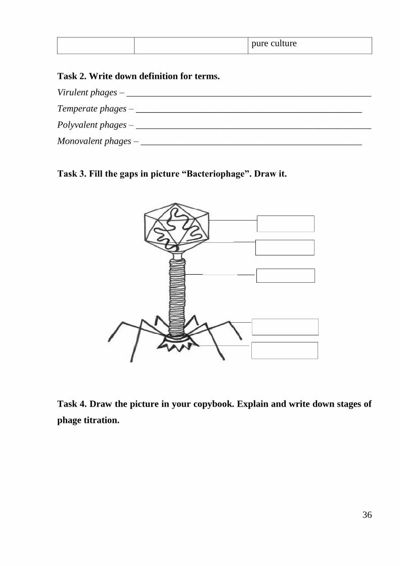
Task 3. Fill the gaps in picture “Bacteriophage”. Draw it.
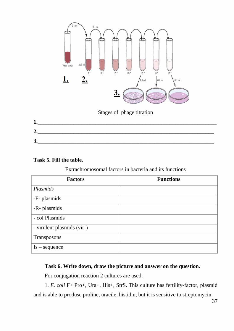
Task 4. Draw the picture in your copybook. Explain and write down stages of
phage titration.
37
Stages of phage titration
1._________________________________________________________________
2.________________________________________________________________
3.________________________________________________________________
Task 5. Fill the table.
Extrachromosomal factors in bacteria and its functions
Factors Functions
Plasmids
-F- plasmids
-R- plasmids
- col Plasmids
- virulent plasmids (vir-)
Transposons
Is – sequence
Task 6. Write down, draw the picture and answer on the question.
For conjugation reaction 2 cultures are used:
1. E. coli F+ Pro+, Ura+, His+, StrS. This culture has fertility-factor, plasmid
and is able to produse proline, uracile, histidin, but it is sensitive to streptomycin.
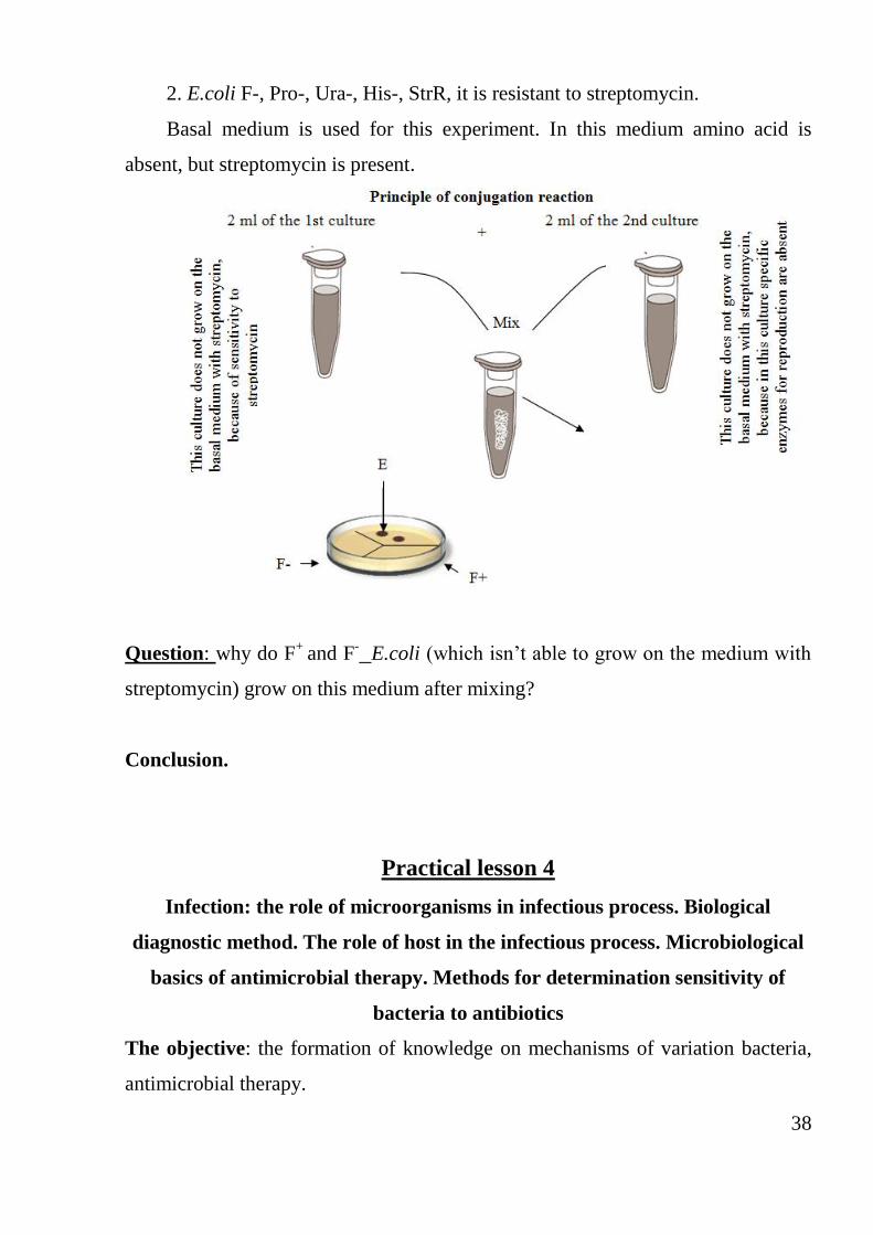
38
2. E.coli F-, Pro-, Ura-, His-, StrR, it is resistant to streptomycin.
Basal medium is used for this experiment. In this medium amino acid is
absent, but streptomycin is present.
Question: why do F+
and F- E.coli (which isn’t able to grow on the medium with
streptomycin) grow on this medium after mixing?
Conclusion.
Practical lesson 4
Infection: the role of microorganisms in infectious process. Biological
diagnostic method. The role of host in the infectious process. Microbiological
basics of antimicrobial therapy. Methods for determination sensitivity of
bacteria to antibiotics
The objective: the formation of knowledge on mechanisms of variation bacteria,
antimicrobial therapy.
39
Tasks:
1. To study with features of variability of species microorganisms.
2. To consider the modern methods of genetic research microorganisms and their
using in medical practice.
3. Determine the sensitivity of bacteria to antibiotics by Indicator disc.
Questions:
1. The notion of infection and infectious disease. Forms of infection.
2. Exogenous and endogenous infections. The term "entrance gate and infective
dose."
3. Sources, routs, and mechanisms of infections transmission.
4. The local and generalized infectious process. The spread ways of infections in
the body. Notions: bacteremia, viremia, toksinemiya, sepsis, pyosepticemia.
5. Types of infection depending on the cause, pathogenesis, methods of infection,
clinical manifestations.
6. Notions: monoinfection and mixed infection, primary and secondary infection,
reinfection, superinfection, relapse.
7. Pathogenesis of infectious diseases. Periods of infection.
8. The pathogenicity and virulence of microorganisms, measuring of virulence.
9. Virulence factors: adhesion, colonization, penetration, invasion.
10. Toxicity. Exotoxins. Classification of action mechanism.
11.Endotoxins. The chemical compound, action on the macroorganism.
12.Role microorganism in the occurrence of infection.
13. The notion of antibiotics, its discovery. Classification of antibiotics: on origin,
method of preparation, action on a microorganism, antimicrobial spectrum.
14. The mechanism of action of antibiotics on microbial cells.
15. The principle of obtaining antibiotics. Activity of antibiotics.
16. The mechanism of bacterial resistance to antibiotics and how to deal with it.
17. Methods of determining the bacterial sensitivity to antibiotics.
18. Side effects of antibiotics.
40
Infection doctrine. Antibiotics
Notion Definition/explanation
Infectious process Dynamic interaction between sensitive macroorganisms and
pathogenic or potential pathogenic microorganism in specific
environmental conditions
Pathogenicity Property (capacity) of the definite species of bacteria to cause
(excite) infectious process in sensitive people and animals
Virulence Degree of the microorganism pathogenicity
Invasiveness Ability of the microorganisms to penetrate and spread in
the body due to the presence of specific enzymes that change
the permeability of cells, and tissues. For example, presence
of the staphylococcus hyaluronidase
Toxigenicity Ability (property) of the microorganisms to produce
toxin that has large molecular mass, antigenic properties, and
affects the basic cell and body tissue
Susceptibility Ability of the macroorganism to interact with pathogens
and create conditions for its existence. Susceptibility is a
specific concept
Persistence Long causative agent keeping in the body without the
occurrence of the infection
Antibiotics Antibiotics are large group of drugs, which can inhibit
selectively growth of bacteria, fungi or inhibit growth of
tumor (cancer), without causing serious damage to the host.
PROCEDURE OF PRACTICAL WORK
Determination of antibacterial action of antibiotics with disk diffusion
technique.
41
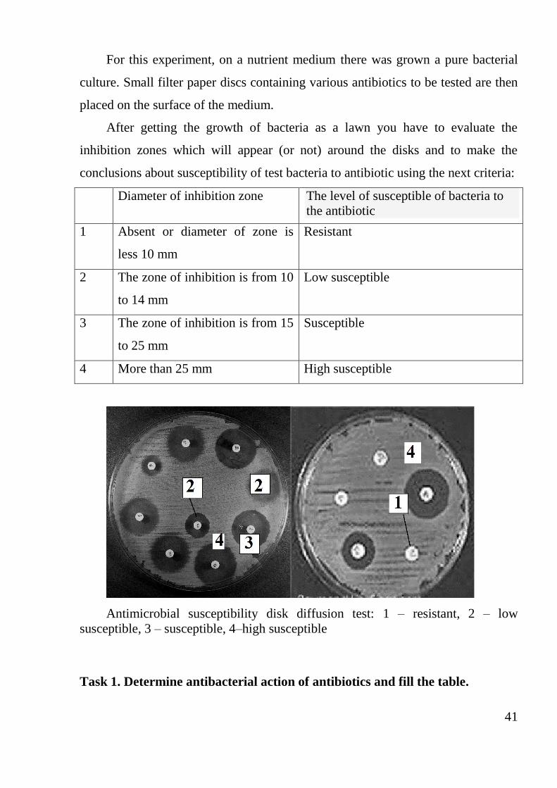
For this experiment, on a nutrient medium there was grown a pure bacterial
culture. Small filter paper discs containing various antibiotics to be tested are then
placed on the surface of the medium.
After getting the growth of bacteria as a lawn you have to evaluate the
inhibition zones which will appear (or not) around the disks and to make the
conclusions about susceptibility of test bacteria to antibiotic using the next criteria:
Diameter of inhibition zone The level of susceptible of bacteria to
the antibiotic
1 Absent or diameter of zone is
less 10 mm
Resistant
2 The zone of inhibition is from 10
to 14 mm
Low susceptible
3 The zone of inhibition is from 15
to 25 mm
Susceptible
4 More than 25 mm High susceptible
Antimicrobial susceptibility disk diffusion test: 1 – resistant, 2 – low
susceptible, 3 – susceptible, 4–high susceptible
Task 1. Determine antibacterial action of antibiotics and fill the table.
42
The level of antimicrobial activity of antibiotics.
Antibiotic Diameter of inhibition zone The level of susceptible
Tasks for self-guided work:
Task 1. Write down notions and its explanations.
Infectious process – __________________________________________________
Pathogenicity – ____________________________________________________
Virulence – ________________________________________________________
Invasiveness – ______________________________________________________
Toxigenicity – ______________________________________________________
Persistence – _______________________________________________________
Task 2. Fill the table.
Characteristic of the toxins
Properties Exotoxins Endotoxins
Chemical composition
Secreted by
The level of toxigenicity
Toxic action
Thermolabile or thermally
stable
Task 3. Examples of bacterial cultures synthesizing exotoxin and endotoxin
43
Examples of bacteria producing
exotoxin
Examples of bacteria having endotoxin
Conclusion.
Practical lesson 5
Ecology of microorganisms. The human microflora and its function.
Dysbiosis, methods of correction. Biofilms. Sanitary demonstration water
microorganisms, soil, air, and methods of research.
Objective: To systematize the knowledge of microbial ecology.
Tasks:
1. Learn how to count the microbial count of air, water.
2. Learn the composition of the normal skin microflora
Questions:
1. Ecology of microorganisms. Forms of interspecies relationships.
2. Role, value, and tasks of sanitary microbiology. Sanitary indicators of
microorganisms.
3. Microflora of water and methods of its bacteriological examination (fermenting
method, method of membrane filters). Sanitary indicators of water
microorganisms.
4. Microflora of the soil and methods of bacteriological examination. Sanitary
indicators of the soil microorganisms. Principles of the determination of microbial
number, coli titre, perfringens titre and titre of soil termophylic bacteria.
5. Microflora of air and methods of bacteriological examination: sedimentation and
aspiration methods. Sanitary indicators of air microorganisms.
44
6. General principles of sanitary and microbiological investigation of foodstuffs.
7. The normal microflora of the human body and its significance.
8. Factors that change the normal microflora of the organism. Dysbiosis, ways of
its elimination.
Sanitary and microbiological investigation of water, air, soil, foodstuffs,
and objects of external environment
Notion Definition/explanation
Sanitary indicatory
microorganisms
Permanent inhabitants of surfaces and digestive
tract of the humans and animals that are excreted from
an organism like pathogenic bacteria. The sanitary
indicatory microorganisms are indicator of pathogenic
microorganisms presence in the external environment
objects
Coli titre of water
(determination)
The least of water (ml), in which coliform bacteria
(bacteria group of Escherichia coli-BGEC) is revealed
Coli index of water
(determination)
An amount of bacteria of Escherichia coli bacteria
group, which are contained in 1 liter of the investigated
water
PROCEDURE OF PRACTICAL WORK
Task 1. Define the microbal number of the air by the sedimentation method
(by Koch)
Sedimentation method is an inoculation of microbes on the surface of solid
medium under the action of gravity. For investigation of air in apartments take
Petri dish with media, keep it opened for 60 minutes (for determination of general
semination air). Incubate Petri dish in the thermostat at 37 °C for 24 hours and then
at a room temperature (one days). Determine the amount of microbes in 1 m3 of air
45
(microbial number) and evaluate the degree of air pollution according to amount of
colonies.
After incubation count up the amount of growing colonies, calculate the
amount of microbes in 1 m3 air by using a formula:
“a” is an amount of colonies; “b is area of Petri dish (63 cm2); “t” is time of exposition.
Task 2. Inoculate the microorganisms from the skin of the hand on the
nutrient agar.
The predominant resident microorganisms of the skin are aerobic and
anaerobic diphtheroid bacilli (e.g., Corynebacterium, Propionibacterium);
nonhaemolytic aerobic and anaerobic staphylococci (Staphylococcus epidermidis,
occasionally S aureus, and peptostreptococcus species); gram-positive, aerobic,
spore-forming bacilli that are ubiquitous in air, water, and soil; alpha-
haemolytic streptococci (viridans streptococci), and enterococci (enterococcus
species); and gram-negative coliform bacilli and acinetobacter. Fungi and yeasts
are often present in skin folds; acid-fast, nonpathogenic mycobacteria occur in
areas rich in sebaceous secretions. Touch by fingers a surface of the nutrient agar
in Petri dish for inoculation of the microorganisms that inhabit the skin or got there
from environment. Incubate Petri dish in the thermostat at 37 °C for 24 hours and
then at a room temperature (one day).
Draw colonies in the protocol, make the conclusion.
Tasks for self-guided work:
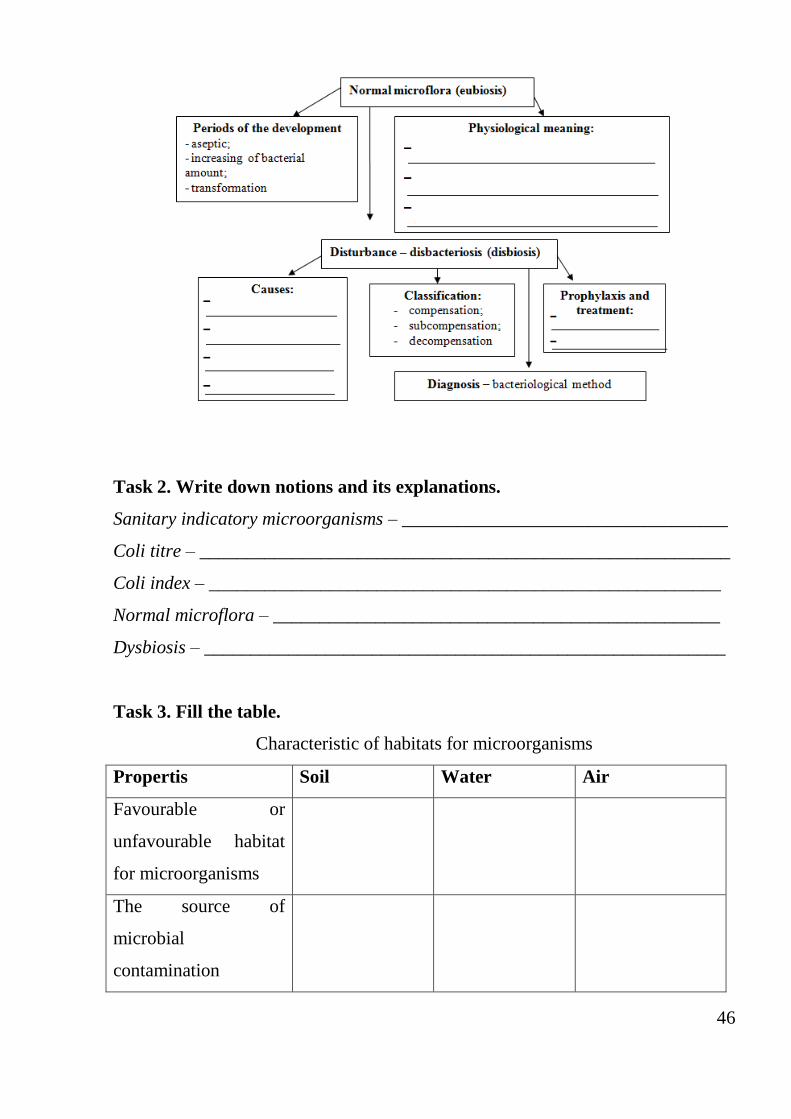
1. Draw and fill the scheme.
Normal microflora. Disbacteriosis
46
Task 2. Write down notions and its explanations.
Sanitary indicatory microorganisms – ___________________________________
Coli titre – _________________________________________________________
Coli index – _______________________________________________________
Normal microflora – ________________________________________________
Dysbiosis – ________________________________________________________
Task 3. Fill the table.
Characteristic of habitats for microorganisms
Propertis Soil Water Air
Favourable or
unfavourable habitat
for microorganisms
The source of
microbial
contamination
47
Species of sanitary
indicatory
microorganisms
Normal indexes of its
parameters (amount)
Methods for detection
of microorganisms
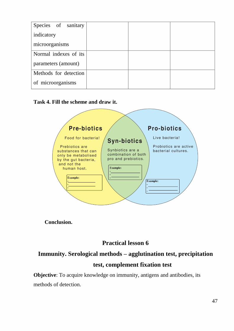
Task 4. Fill the scheme and draw it.
Conclusion.
Practical lesson 6
Immunity. Serological methods – agglutination test, precipitation
test, complement fixation test
Objective: To acquire knowledge on immunity, antigens and antibodies, its
methods of detection.
48
Tasks:
1. To consider the concept of an antigen and an antibody.
2. To study the types of immunity.
3. To learn some basic serological reactions
Questions:
1. The notion of immunity. Classification of immunity.
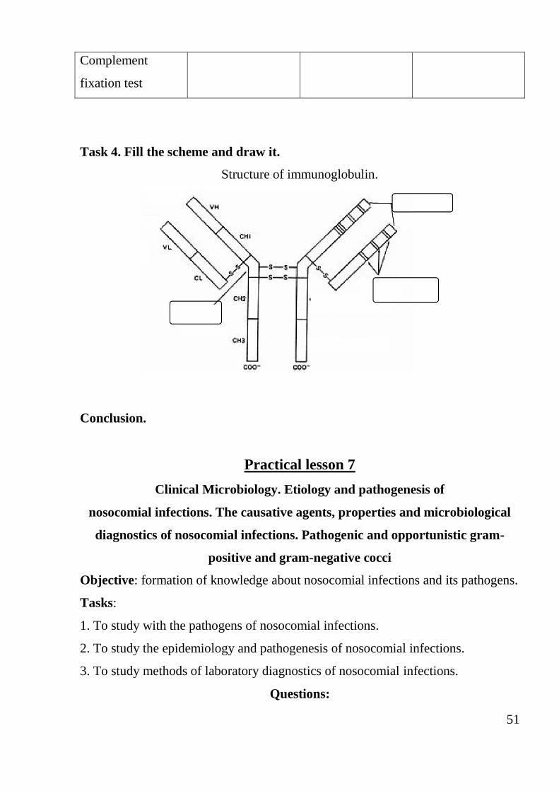
2. The notion of an antigen, a chemical composition.
3. Bacterial antigens.
4. Antigens of viruses.
5. Notion of antibody, its structure and properties.
7. Serological tests – notion and its using in microbiological diagnostics.
8. Agglutination test - definition, components, using.
9. Precipitation test - definition, components, using.
10. Complement fixation test – components, phases of reaction and using.
Immunity. Factors and mechanisms of the innate immunity
Notion Definition/explanation
Immunity Dynamic condition of the organism that consists of
the set of specific
Antigens Foreign macromolecules that when introducted into the
body cause the formation of the immune response, on
condition of their recognition by specific receptors of
lymphocytes
Antibodies Specific proteins that are synthesized in the body in
response to input (falling) of an antigen
Notion Definition/explanation
Practical use of the
agglutination test
1. In serological test for detection of the specific
antibodies formed in the serum or other body fluids by the
49
infectious diseases and other pathological conditions.
2. For identification of the bacterial antigens in the
bacteriological method
Types of the
agglutination test
1. Slide agglutination test is made on the glass or
porcelain plates with a smooth surface.
2. Tube agglutination test is made in the test tube or in
the polisterol hole in plates with consecutive dilution of
the serum. In every tube (hole) the same amount of
antigen is contributed. The titre of the antibody is detected
Precipitation test
mechanism
Interaction of a soluble antigen (precipitinogen) and
antibodies (precipitin) in the presence of electrolyte
(0.85% solution of NaCl). An aggregation of the antigen
makes opacity of transparent liquid (precipitate).
Deposition from solution complexes of the antigen-
antibody become in equivalent ratio in the range of the
interacting molecules concentrations
Practical use of
the PT
PT is used for the diagnosis of a number of infectious
diseases - anthrax, plague, tularemia, epidemic cerebrospinal
meningitis and others. Extracts from affected organs, and
other liquor are used as the antigens, and diagnostic
precipitating serum is used as antibodies (precipitins).
PT is also used to study the antigenic structure of the
bacteria. In addition, it is used to determine the species
proteins, including proteins of blood
Tasks for self-guided work:
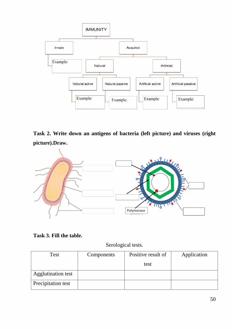
Task 1. Fill the scheme.
50
Task 2. Write down an antigens of bacteria (left picture) and viruses (right
picture).Draw.
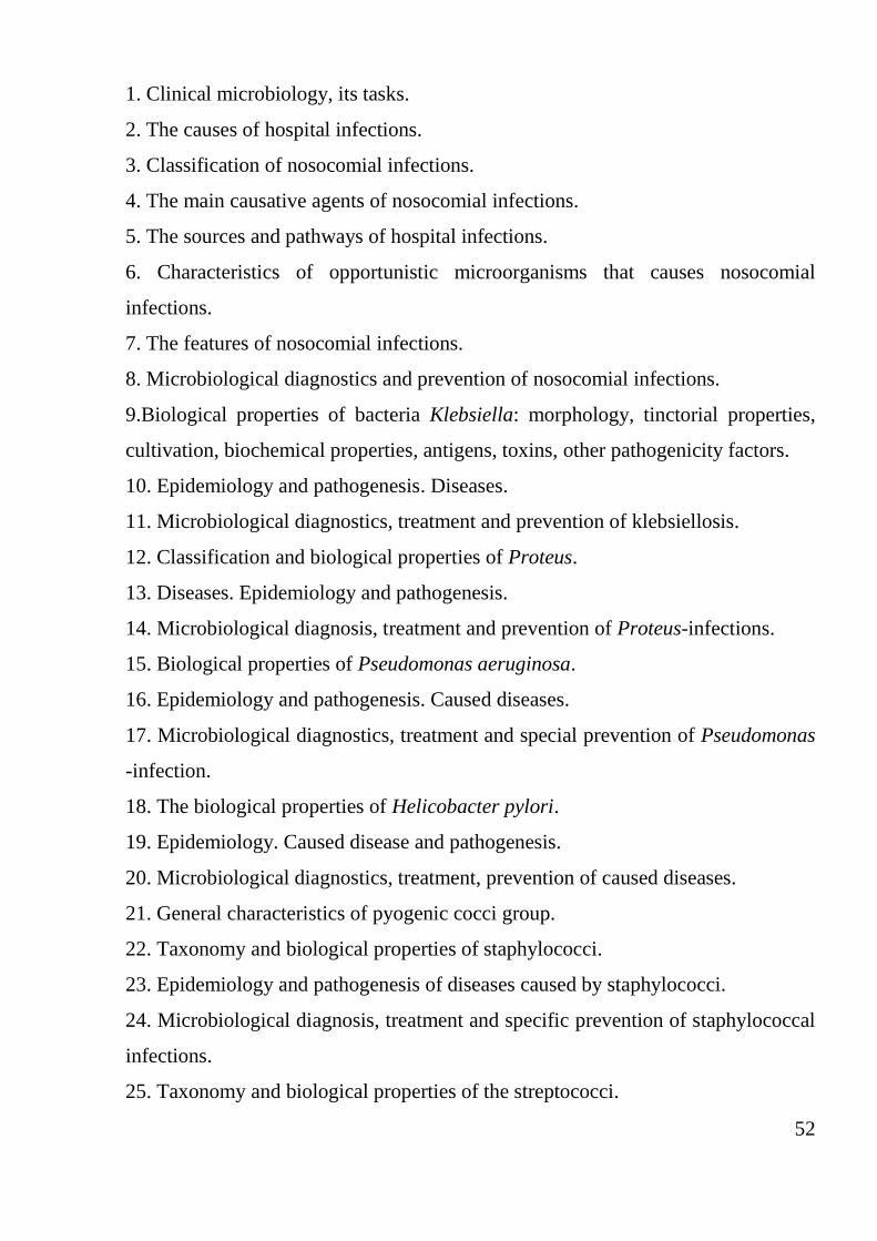
Task 3. Fill the table.
Serological tests.
Test Components Positive result of
test
Application
Agglutination test
Precipitation test
51
Complement
fixation test
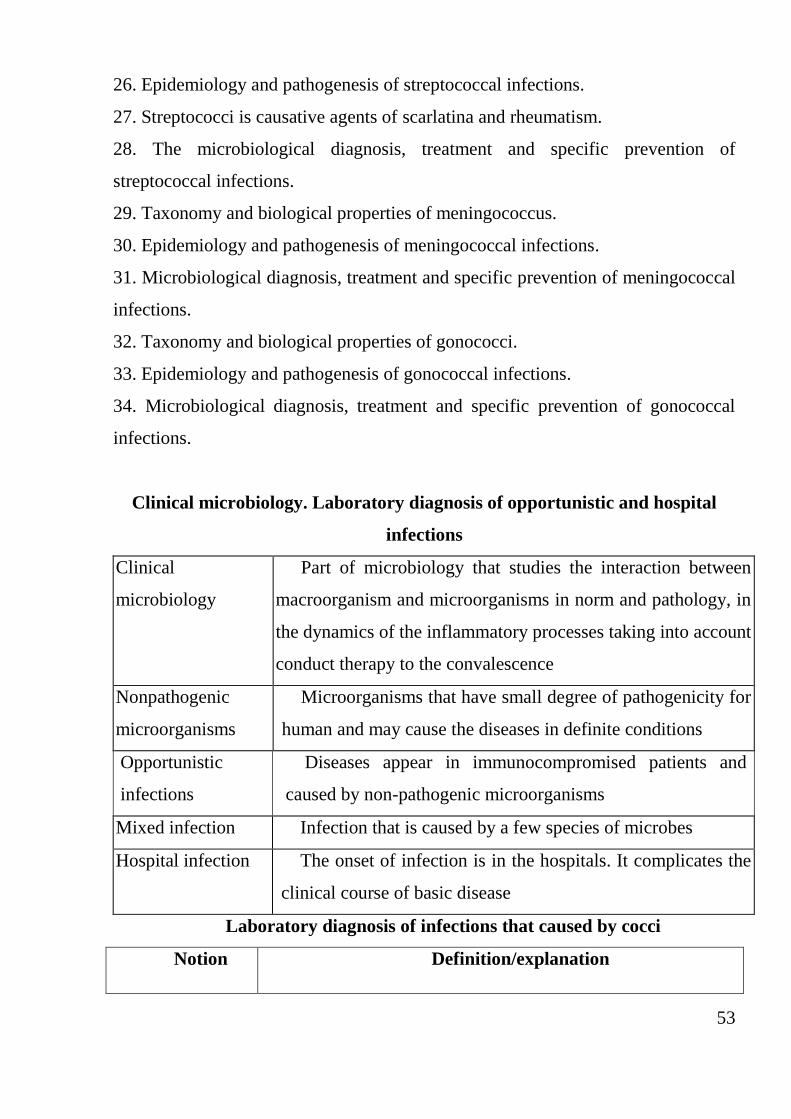
Task 4. Fill the scheme and draw it.
Structure of immunoglobulin.
Conclusion.
Practical lesson 7
Clinical Microbiology. Etiology and pathogenesis of
nosocomial infections. The causative agents, properties and microbiological
diagnostics of nosocomial infections. Pathogenic and opportunistic gram-
positive and gram-negative cocci
Objective: formation of knowledge about nosocomial infections and its pathogens.
Tasks:
1. To study with the pathogens of nosocomial infections.
2. To study the epidemiology and pathogenesis of nosocomial infections.
3. To study methods of laboratory diagnostics of nosocomial infections.
Questions:
52
1. Clinical microbiology, its tasks.
2. The causes of hospital infections.
3. Classification of nosocomial infections.
4. The main causative agents of nosocomial infections.
5. The sources and pathways of hospital infections.
6. Characteristics of opportunistic microorganisms that causes nosocomial
infections.
7. The features of nosocomial infections.
8. Microbiological diagnostics and prevention of nosocomial infections.
9.Biological properties of bacteria Klebsiella: morphology, tinctorial properties,
cultivation, biochemical properties, antigens, toxins, other pathogenicity factors.
10. Epidemiology and pathogenesis. Diseases.
11. Microbiological diagnostics, treatment and prevention of klebsiellosis.
12. Classification and biological properties of Proteus.
13. Diseases. Epidemiology and pathogenesis.
14. Microbiological diagnosis, treatment and prevention of Proteus-infections.
15. Biological properties of Pseudomonas aeruginosa.
16. Epidemiology and pathogenesis. Caused diseases.
17. Microbiological diagnostics, treatment and special prevention of Pseudomonas
-infection.
18. The biological properties of Helicobacter pylori.
19. Epidemiology. Caused disease and pathogenesis.
20. Microbiological diagnostics, treatment, prevention of caused diseases.
21. General characteristics of pyogenic cocci group.
22. Taxonomy and biological properties of staphylococci.
23. Epidemiology and pathogenesis of diseases caused by staphylococci.
24. Microbiological diagnosis, treatment and specific prevention of staphylococcal
infections.
25. Taxonomy and biological properties of the streptococci.
53
26. Epidemiology and pathogenesis of streptococcal infections.
27. Streptococci is causative agents of scarlatina and rheumatism.
28. The microbiological diagnosis, treatment and specific prevention of
streptococcal infections.
29. Taxonomy and biological properties of meningococcus.
30. Epidemiology and pathogenesis of meningococcal infections.
31. Microbiological diagnosis, treatment and specific prevention of meningococcal
infections.
32. Taxonomy and biological properties of gonococci.
33. Epidemiology and pathogenesis of gonococcal infections.
34. Microbiological diagnosis, treatment and specific prevention of gonococcal
infections.
Clinical microbiology. Laboratory diagnosis of opportunistic and hospital
infections
Clinical
microbiology
Part of microbiology that studies the interaction between
macroorganism and microorganisms in norm and pathology, in
the dynamics of the inflammatory processes taking into account
conduct therapy to the convalescence
Nonpathogenic
microorganisms
Microorganisms that have small degree of pathogenicity for
human and may cause the diseases in definite conditions
Opportunistic
infections
Diseases appear in immunocompromised patients and
caused by non-pathogenic microorganisms
Mixed infection Infection that is caused by a few species of microbes
Hospital infection The onset of infection is in the hospitals. It complicates the
clinical course of basic disease
Laboratory diagnosis of infections that caused by cocci
Notion Definition/explanation
54
Staphylococcus:
classification and
biological
properties
The term Staphylococcus is derived from the Greek term
staphyl, meaning “a bunch of grapes”. This name refers to the
fact that the cells of these gram-positive cocci grow in a
pattern resembling a cluster of grapes; however, organisms in
clinical material may also appear as single cells, pairs, or short
chains. Most staphylococci are 0.5 to 1 µm in diameter and are
nonmotile, aerobic or facultative anaerobic, and catalase-
positive and grow in a medium containing 10% sodium
chloride and at temperature ranging from 18 ºC to 40 ºC.
The organisms are present on the skin and mucous
membranes of humans and other mammals, and birds.
Staphylococcus is an important pathogen in humans, causing a
wide spectrum of life-threatening systemic diseases; infections
of the skin, soft tissues, bones, and urinary tract; and
opportunistic infections. The species most commonly
associated with human diseases are S. aureus (the most
virulent and best-known member of the genus), S. epidermidis,
S. saprophyticus, and S. haemolyticus.
Diseases of
S. aureus
1. Localized skin infections include impetigo, folliculitis,
furuncles, and carbuncles. Impetigo, a superficial infection
affecting mostly young children, occurs primarily on the face
and limbs. Initially, a small macule (flattened red spot) is seen,
and then a pus-filled vesicle (pustule) on an erythematous base
develops.
2. Toxic shock syndrome is a disease that was initially
evident in children, although it is now recognized as primarily
a disease in menstruating women.
3. Food poisoning is one of the most common food
55
poisoning in the world. The organisms are usually introduced
into food, such as processed meat, pastries, potato, salad, and
ice cream. Staphylococcal food poisoning is characterized by
severe vomiting, xudates, and abdominal pain.
4. Pneumonia is a disease that occurs among
immunosuppressed patients, the aged, children under one year
of age, and frequently in children with measles and influenza.
5. Other diseases like osteomyelitis, septicemia, and
septic arthritis
Principle of the
laboratory
diagnostics
Specimens obtained depend on the disease process and
include lesion material, pus, sputum, blood, spinal fluid, and
faeces.
İsolation and identification of S. aureus requires initial
cultivation on blood agar and/or specific medium and
overnight incubation under aerobic conditions at 37 ºC. The
organism may be identified as a gram-positive, catalase-
positive coccus exhibiting coagulase
Treatment The antibiotics of choice are oxacillin (or other
penicillinase-resistant penicillin) or vancomycin for oxacillin-
resistant strains.
The focus of infection (e.g., abscess) must be identified
and drained. Treatment is symptomatic for patients with food
poisoning
Streptococcus The genus Streptococcus is a diverse collection of gram-
positive cocci typically arranged in pairs or chains. Most
species are facultative anaerobes. Their nutritional
requirements are complex, necessitating the use of blood or
serum-enriched media for isolation. Carbohydrates are

56
fermented, resulting in the production of lactic acid, and unlike
Staphylococcus species, streptococci are catalase-negative
Group A, B
haemolytic
streptococci
Streptococcus pyogenes is the most virulent member of
this group of gram-positive cocci. This bacterium is an
important cause of a variety of suppurative and nonsuppurative
diseases
Classification of
streptococci
1. Haemolitic properties on blood agar (alfa-, betta-, and
gamma-haemolitic streptococci).
2. Serological grouping (A and B).
3. Biochemical properties
Diseases caused
by streptococci
Streptococcus pyogenes.
Pharyngitis. Group A streptococcus is the major cause of
bacterial pharyngitis, occasionally involved with group C
and G.
Scarlet fever is a complication of streptococcal pharyngitis
seen when the infecting strain is lysogenized by a temperate
bacteriophage that stimulates production of erythrogenic
toxin.
Erysipelas. This disease can affect all age groups.
Puerperal sepsis. This infection is initiated during or
following soon after the delivery of a newborn.
The major clinical manifestations are carditis, polyarthritis,
and subcutaneous nodules.
Acute haemorrhagic glomerulonephritis occurs most
commonly in children.
S. xudates is the most common cause of lobar and
lobular (broncho) pneumonia.
Treatment Adequate drainage, debridement, and antibiotic therapy
57
are essential for the treatment of localized, suppurative skin
lesions. Penicillin is the drug of choice for acute diseases.
Penicillin has no effect upon established rheumatic heart
disease and acute haemorrhagic glomerulonephritis.
Penicillin resistant strains have not been reported.
Erythromycin is the drug of choice for penicillin allergic
patients
Neisseria
gonorrhoeae
N. gonorrhoeae is a fastidious organism, requiring
complex media for growth and adversely affected by drying
and fatty acids. Soluble starch is added to the media to
neutralize the toxic effect of the fatty acids. It is highly
susceptible to environmental, physical, and chemical agents.
The optimum growth temperature is 35 ºC to 37 ºC, with poor
survival of the organism at lower temperature. A humid
atmosphere supplemented with CO2 is required or enhances
growth of N. gonorrhoeae
Diseases caused
by N.gonorrhoeae
Gonorrhoea is one of the most commonly reported
sexually transmitted diseases. A higher proportion of females
than males are generally asymptomatic; these individuals act
as the reservoir for maintaining and transmitting gonococcal
infections.
Laboratory
diagnosis
Specimens obtained depend on the disease process and
include urethral, cervical, rectal, pharyngeal, and/or
conjunctival exudates.
The direct demonstration of gram-negative intracellular
xudates ic within PMNs is diagnostic only when observed
in the urethral exudates of males with characteristic clinical
manifestations.
58
Gram stains of smears from female urethral and cervical
exudates, from rectal, pharyngeal, and conjunctival exudates
of male and female, are unreliable due to the potential
presence of nonpathogens resembling gonococcal
morphology, and presence of meningococci. All such
specimens must be cultured and the isolated organism
xudates i.
Treatment
Ceftriaxone, cefixime, ciprofloxacin, or ofloxacin can be
administered in uncomplicated cases. In vitro susceptibility
should be determined in cases unresponsive to therapy,
because antibiotic resistance is increasing.
N. meningitidis N. xudates ic causes endemic or epidemic disease
of worldwide prevalence. The most commonly recognized
form of this disease is meningitis.
N. xudates ic is an obligate parasite of humans,
harboured in the nasopharynx, and transmitted by droplet
nuclei or direct intimate contact with a sick people or bacteria
carrier.
This organism is a gram-negative, nonmotile,
encapsulated, piliated diplococcus flattened on one site to
give the appearance of a “kidney bean” or “coffee bean”.
Pathogenesis and
clinical
manifestations
The basic pathogenesis process of primary
meningococcal disease is initiated in the nasopharynx from a
sick people or carrier. Disseminated meningococcal disease
manifests most often as septicemia and meningitis.
Meningitis is the most common complication of
meningococcal septicemia. Clinical manifestations are fever,
stiff neck, vomiting, severe headache, convulsions, bulging
59
of the fontanels, and progression to coma within a few hours.
Laboratory
diagnosis
Specimens obtained depend on the disease process and
mainly include nasopharynx swabs, blood culture, and
cerebrospinal fluid. Gram stains of specimens may show
gram-negative, intracellular and extracellular xudates ic in
association with PMNs.
Primary isolations require initial blood cultures and
culture on chocolate agar plates or, if a mixed flora is
anticipated, Thyer-Martin medium, which is an enriched
chocolate agar medium, containing antibiotics to inhibit
gram-positive organisms and gram-negative rods. Incubation
for 48 hours at 35–37 ºC under aerobic conditions in the
presence of 3–10% carbon dioxide is optimal for isolation of
the organisms, which may be identified as gram-negative,
oxidase-positive, diplococcus that ferments glucose and
maltose, and agglutinates in the presence of serogroup-
specific anticapsular antibody
Treatment Early treatment with penicillin reduces the case fatality
rate in disseminated disease from 40–90% to 10–15%, but the
antibiotic is ineffective in eradicating the carrier state.
Chloramphenicol and ceftriaxone are effective in penicillin-
allergic individuals. Polysaccharide vaccines conjugated with
60
protein carriers offer protection for infants younger than 2
years
PROCEDURE OF PRACTICAL WORK
Task 1. Study the preparation of the pure cultures of Klebsiella
pneumoniae microscopically; draw it in the protocol.
Klebsiella pneumonia
The total magnification – 16x100
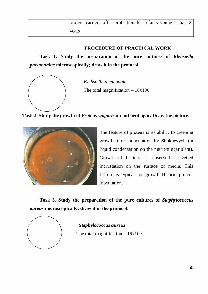
Task 2. Study the growth of Proteus vulgaris on nutrient agar. Draw the picture.
The feature of proteus is its ability to creeping
growth after innoculation by Shukhevych (in
liquid condensation on the nutrient agar slant).
Growth of bacteria is observed as veiled
incrustation on the surface of media. This
feature is typical for growth H-form proteus
inoculation.
Task 3. Study the preparation of the pure cultures of Staphylococcus
aureus microscopically; draw it in the protocol.
Staphylococcus aureus
The total magnification – 16x100
61
Task 4. Study the growth of S. aureus in nutrient agar (left picture) and
blood agar (right picture). Fill in the protocol with their cultural properties.
Task 5. Study the preparation of pure cultures of Streptococcus pyogenes
microscopically and draw it in the copy book.
Streptococcus pyogenes
The total magnification – 16x100
Task 6. Study the preparation of incomplete phagocytosis of Neisseria
gonorrhoeae microscopically and draw it in the copy book.
Neisseria gonorrhoeae
The total magnification – 16x100
Tasks for self-guided work:
62
Task 1. Study the biochemical activity of Proteus mirabilis in the Hiss
medium. Draw colour of tubes in accordance its biochemical properties.
glucose lactose maltose mannitol sucrose
Task 2. Fill the table.
Characteristics of nosocomial pathogens
Pathogens Morphology
(picture)
Nutrient
medium
A source
of disease
Transmissi
on paths
Diseases
K. pneumoniae
P. vulgaris,
P. mirabilis,
P. aeruginosa
H. pylori
Task 3. Fill the table.
Characteristics of pyogenic cocci.
Pathogens Morphology
(picture)
Nutrient
mtdium
Transmission
paths
Diseases
63
Staphylococci
Streptococci
Meningococcus
Gonococci
Task 4. Solve the case. Answer on the questions and write down it.
At the Mount Union hospital, a 5-year old white male child in good general
health and physical condition was presented at the Saturday walk-in clinic by his
mother. He was brought in because he had a fever, was cranky and had complained of
a sore throat for about 24 hours. On physical examination by the attending resident, the
patient had a fever of 39.30 C, and he had considerable swelling and drainage of the
pharynx and in the conjunctivae. His tonsils were enlarged and coated with a white
patchy xudates. He had a red throat and swollen anterior cervical lymph nodes. His
ears were clear. His chest sounded clear and he had no additional remarkable findings
on routine examination.
1. What would be your presumptive diagnosis for this child? Why?
2. What diagnostic testing would be indicated to follow this exam?
3. What is the most likely treatment for this illness? Why is it important?
4. What factors of this case allowed you to make a presumptive diagnosis?
Task 5. Fill the scheme. Draw it.
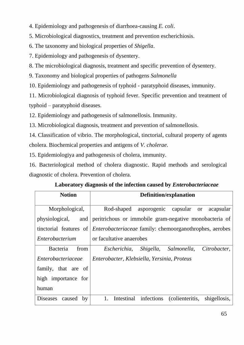
Scheme meningococcal infections and bacteria carriers
laboratory diagnosis
64
Conclusion.
Practical lesson 8
Pathogens of escherichiosis and dysentery. The causative agents of typhoid,
salmonella poisoning and cholera
Objective: formation of knowledge about the biological properties of pathogenic
enterobacteria
Tasks:
1. To study the biological properties of pathogenic enterobacteria.
2. To study the epidemiology and pathogenesis of diseases caused by its.
3. To learn the methods of microbiological diagnosis, treatment and specific
prevention of colienterit and dysentery
Questions:
1. The general characteristics of the family Enterobacteriaceae.
2. Opportunistic Escherichia: its role in the life of the human organism, caused
diseases.
3. The biological properties of Escherichia.
65
4. Epidemiology and pathogenesis of diarrhoea-causing E. coli.
5. Microbiological diagnostics, treatment and prevention escherichiosis.
6. The taxonomy and biological properties of Shigella.
7. Epidemiology and pathogenesis of dysentery.
8. The microbiological diagnosis, treatment and specific prevention of dysentery.
9. Taxonomy and biological properties of pathogens Salmonella
10. Epidemiology and pathogenesis of typhoid - paratyphoid diseases, immunity.
11. Microbiological diagnosis of typhoid fever. Specific prevention and treatment of
typhoid – paratyphoid diseases.
12. Epidemiology and pathogenesis of salmonellosis. Immunity.
13. Microbiological diagnosis, treatment and prevention of salmonellosis.
14. Classification of vibrio. The morphological, tinctorial, cultural property of agents
cholera. Biochemical properties and antigens of V. cholerae.
15. Epidemiologiya and pathogenesis of cholera, immunity.
16. Bacteriological method of cholera diagnostic. Rapid methods and serological
diagnostic of cholera. Prevention of cholera.
Laboratory diagnosis of the infection caused by Enterobacteriaceae
Notion Definition/explanation
Morphological,
physiological, and
tinctorial features of
Enterobacterium
Rod-shaped asporogenic capsular or acapsular
peritrichous or immobile gram-negative monobacteria of
Enterobacteriaceae family: chemoorganothrophes, aerobes
or facultative anaerobes
Bacteria from
Enterobacteriaceae
family, that are of
high importance for
human
Escherichia, Shigella, Salmonella, Citrobacter,
Enterobacter, Klebsiella, Yersinia, Proteus
Diseases caused by 1. Intestinal infections (colienteritis, shigellosis,
66
Enterobacteriaceae typhoid fever, food toxic infections, and yersiniosis) which
are caused by the representatives of genus of Escherichia,
Shigella, Salmonella, Yersinia. They are characterized by
the faecal-oral or oral mechanism of transmission via water,
food or contact and house routes of the infection
transmission.
2. Opportunistic (extraintestinal) infections
(bacteriemia, meningitis of newborn, traumatic, genito-
urinary, and respiratory infections) which are caused by the
opportunistic pathogenic bacteria of Escherichia,
Klebsiella, Citrobacter, Enterobacter, Proteus genus on the
background of immunodeficiency
Systematic position
of colibacillosis
causative agents
(family, genus,
Family: Enterobacteriaceae.
Genus: Escherichia.
Species: Escherichia coli
Morphological
and tinctorial features
of Escherichia
Straight asporogenic gram-negative rods with rounded
ends, which have capsule or microcapsule, peritrichous
Types of E. coli,
which cause
gastrointestinal
disease
EPEC – enteropathogenic E. coli
EIEC – enteroinvasive E. coli
ETEC – enterotoxigenic E. coli
EHEC – enterohaemorrhagic E. coli
Enterotoxigenic
E. coli and
mechanism of
pathogenic action of
ETEC on epithelium
ETEC causes cholera-like gastrointestinal disease in
children
67
Enteropathogeni
c E. coli and
mechanism of
pathogenic action of
EPEC on intestinal
cells
EPEC are agents of colienteritis (salmonella-like
infection) mainly in children of the first year of life with the
house, contact, and food routes of infection transmission.
EPEC propagates on the surface of epithelium of small
intestines, stipulating thinning, fragmentation, and tearing
away of microvilli, damage of apical surface of epithelium,
resulting in moderate inflammation and appearance of
shallow erosions
Enteroinvasive
E. coli and
mechanism of
pathogenic action of
EIEC on intestinal
cells
EIEC causes a disease closely resembling that caused
by Shigella spp. in children of senior age and adults with
the water and food routes of infection transmission. EIEC is
capable of immersion and reproduction in the epithelium of
mucous membrane of the colon, cytoplasmic damage and
death of intestinal cells, invasion to nearby epithelium,
causing focal ulcerous inflammation with surplus
infiltration and accumulation of exudation in the cavity of
colon, by erosions and acute inflammation
Enterohaemorrh
agic E. coli,
mechanism of
pathogenic action of
EHEC on epithelium
and endothelium
EHEC are agents of haemorrhagic diarrhoea
(haemorrhagic colitis) in adults and in new-born can
predetermine necrotic enterocolitis with the high
percentage of lethal consequencies with the damage of
sigmoideus, ascending and transversal large intestines;
alimentary route of infection transmission. EHEC
propagates oneself on the surface of epithelium of colon
with destruction of microvilli, damage of apical surface of
epithelium. Cytotoxins cause the death of intestinal
epithelium by interfering with protein synthesis. Some of
the toxins are identical to the Shiga toxins of Shigella

68
dysenteriae. Verocytotoxin destroys endothelium of the
blood capillaries. Formation of fibrin results in violation of
Bangui motion. About 4% of infected individuals develop
haemolytic uremic syndrome, marked by anaemia due to
lysis of red blood cells, kidney failure, and central nervous
system damage
Properties are
necessary for E. coli
identification
Antigenic structure (in slide agglutination test and
tube agglutination test with groupspecific and monovalent
escherichiosis sera – basic) and morphological, tinctorial,
cultural, and biochemical properties
Taxonomic position
of the shigellosis
causative agents
Enterobacteriaceae family.
Shigella genus
Species: S. dysenteriae, S. flexneri, S. boydii, S. sonnei
Features of
shigellosis
The heaviest course of shigellosis is caused by S.
dysenteriae, which produces Shiga toxin (exotoxin), less
difficult course of shigellosis is caused by S. sonnei
Morphological and
tinctorial features of
the shigellosis
causative agents
Short lines with rounded ends gram-negative
immobile monobacteria. Spores and capsules are not
formed
Transmission of
the infection
Through contaminated water (prevails at shigellosis
Flexneri), contaminated food: especially through milk and
milk products (S. sonnei is kept and propagated for a long
time), contact route of infection (especially for S.
dysenteriae species). Insect vectors (flies, cockroaches)
transfer causative agents on foodstuffs
Mechanism of
local immunity
Prevention of shigella adhesion on epithelium,
penetration of shigellae in a cell, prevention of
69
immunological damage of the intestine
Material
investigated at
shigellosis
Bacteriological method: faeces (basic), foodstuffs
(especially milk, cheese, sour cream) at outbreaks of
shigellosis, autopsy material (fragment of large colon,
pieces of parenchymatous organs, mesenterial lymphatic
nodes), blood and urine (at suspicion on bacteriemia).
Serological method: blood analysis
Methods of
shigellosis
microbiological
diagnosis
The basic method is bacteriological, additional –
serological (tube agglutination test, PHAT), express-
methods of antigens diagnosis: IFT (investigation of faeces
and urine), ELISA (investigation of blood serum),
molecular and biological: PCR
Media for
primary cultivation of
causative agents
Differential media: MacConkey, EMB or DCA
(desoxycholate citrate agar), selective media: Hektoen
enteric agar; enrichment medium – selenite medium
Cultural
properties of
shigellosis causative
agents
It forms colourless (lactose-negative), shallow (1–1.5
mm), convex, circular transparent smooth (S-colonies)
colonies with intact edges. S. sonnei forms also R-colonies
– flat dull with a rough surface and unequal edges
Laboratory diagnostics of typhoid fever, A and B paratyphoid and
cholera
Notion Definition/explanation
Typhoid fever
It is acute anthroponosis infection systemic disease with
faecal-oral transmission mechanism, that is caused by
Salmonella typhi, characterized by small bowel lymphatic
apparatus lesion, bacteraemia, fever, typhoid maculopapular
rash, organism intoxication and cyclic course
Taxonomic Enterobacteriaceae family.
70
classification of
typhoid fever, A and
B paratyphoid
causative agents
Salmonella genus.
Species:
1. Salmonella enterica subspecies choleraesuis serotype
typhi – causative agent of typhoid fever.
2. Salmonella enterica subspecies choleraesuis serotype
paratyphi A – causative agent of A paratyphoid.
3. Salmonella enterica subspecies choleraesuis serotype
paratyphi B (S. schottmuelleri) – causative agent of B
paratyphoid
Morphological and
tinctorial features of
the causative agent
Short straight with rounded ends gram-negative mostly
movable (peritrichaetes) monobacteria. They do not generate
spores or capsules. Various types of salmonellas do not
differ from one another morphologically
Source of typhoid
fever, A and B
paratyphoid
infection
At typhoid fever and A paratyphoid – patient and bacteria
carrier (employees of catering facilities, water supply objects
– anthroponosis infection). В paratyphoid – patients, bacteria
carriers, and animals (cattle stock, poultry) –
zooanthroponosis infection
Methods of
typhoid fever, A and
B paratyphoid
microbiological
diagnosis
1. Bacteriological (basic): diseases diagnosis, contact
persons examination, bacteria carriers identification and
infection source detection.
2. Serological method.
3. Express method
Material for
typhoid fever, A and
B paratyphoid
bacterial diagnostics
Basic material is blood (haemoculture), faeces (stool
culture), urine (urine culture), bile (bile culture), bone
marrow aspirates (myeloculture). Additional material is
roseolas content (roseola culture), autopsy material, and
drinking water
71
Typhoid fever
specific prophylaxis
Chemical adsorbed Vi-typhoid fever monovaccine
administration according to epidemiological indications
Cholera Acute extremely dangerous anthroponosis quarantine disease
caused by Vibrio cholerae O1 and O139 serogroups, which
is characterized as the spread of epidemics, profuse watery
diarrhoea, vomiting, dehydration, severe intoxication, faecal-
oral mechanism of transmission and high death-rate
Taxonomic position
of cholera germs
Vibrionaceae family.
Vibrio genus.
V. cholerae type.
Serogroup O1, serotype Ogáva (AB), Inába (AU),
Hikodzhúma (FAA), biovar: classic – V. cholerae cholerae
and El Tor – V. cholerae eltor serogroup, V. cholerae O139
(Bengál)
Morphological and
staining properties
of the cholera
causative agents
Short slightly curved or straight gram-negative rods are of
medium size (1.5–4 × 0.2–0.4 mm), they form clusters in the
form of “fish shoats”, mobile (monotrixous). Spore and
capsules are not forming
Source of
infection of the
cholera causative
agents
The source of infection is patient and carrier. The patient
excretes from 100 mln to 1 billion vibrios in 1 ml of excreta.
The reservoir of infection is also water
Routes of
V. cholerae
transmission
The principal routes are through water, milk, vegetables,
fruits, often – food or water. All epidemics and pandemics of
cholera have water in nature. Some role is played by flies
Material taken from
the contact persons
with cholera
Faeces after the use of the laxatives (25–30 g of
magnesium sulphate). The upper part of faeces is taken
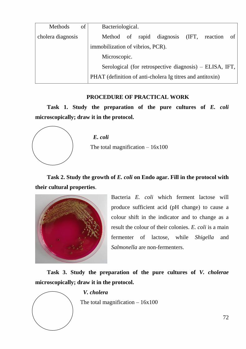
72
Methods of
cholera diagnosis
Bacteriological.
Method of rapid diagnosis (IFT, reaction of
immobilization of vibrios, PCR).
Microscopic.
Serological (for retrospective diagnosis) – ELISA, IFT,
PHAT (definition of anti-cholera Ig titres and antitoxin)
PROCEDURE OF PRACTICAL WORK
Task 1. Study the preparation of the pure cultures of E. coli
microscopically; draw it in the protocol.
E. coli
The total magnification – 16x100
Task 2. Study the growth of E. coli on Endo agar. Fill in the protocol with
their cultural properties.
Bacteria E. coli which ferment lactose will
produce sufficient acid (pH change) to cause a
colour shift in the indicator and to change as a
result the colour of their colonies. E. coli is a main
fermenter of lactose, while Shigella and
Salmonella are non-fermenters.
Task 3. Study the preparation of the pure cultures of V. cholerae
microscopically; draw it in the protocol.
V. cholera
The total magnification – 16x100
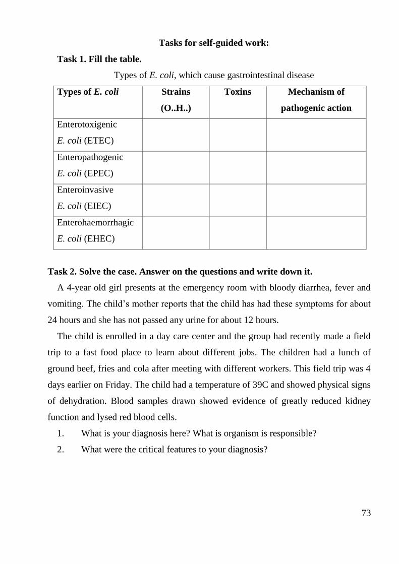
73
Tasks for self-guided work:
Task 1. Fill the table.
Types of E. coli, which cause gastrointestinal disease
Types of E. coli Strains
(O..H..)
Toxins Mechanism of
pathogenic action
Enterotoxigenic
E. coli (ETEC)
Enteropathogenic
E. coli (EPEC)
Enteroinvasive
E. coli (EIEC)
Enterohaemorrhagic
E. coli (EHEC)
Task 2. Solve the case. Answer on the questions and write down it.
A 4-year old girl presents at the emergency room with bloody diarrhea, fever and
vomiting. The child’s mother reports that the child has had these symptoms for about
24 hours and she has not passed any urine for about 12 hours.
The child is enrolled in a day care center and the group had recently made a field
trip to a fast food place to learn about different jobs. The children had a lunch of
ground beef, fries and cola after meeting with different workers. This field trip was 4
days earlier on Friday. The child had a temperature of 39C and showed physical signs
of dehydration. Blood samples drawn showed evidence of greatly reduced kidney
function and lysed red blood cells.
1. What is your diagnosis here? What is organism is responsible?
2. What were the critical features to your diagnosis?
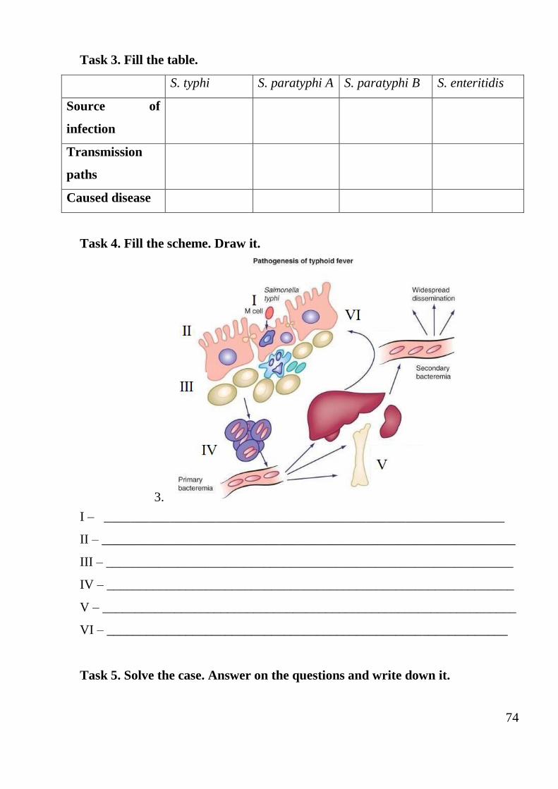
74
Task 3. Fill the table.
S. typhi S. paratyphi A S. paratyphi B S. enteritidis
Source of
infection
Transmission
paths
Caused disease
Task 4. Fill the scheme. Draw it.
3.
I – _____________________________________________________________
II – _______________________________________________________________
III – ______________________________________________________________
IV – ______________________________________________________________
V – _______________________________________________________________
VI – _____________________________________________________________
Task 5. Solve the case. Answer on the questions and write down it.
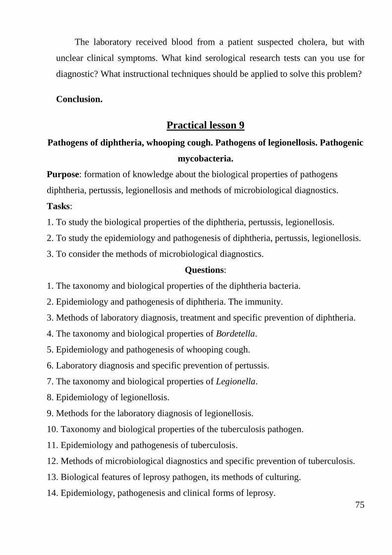
75
The laboratory received blood from a patient suspected cholera, but with
unclear clinical symptoms. What kind serological research tests can you use for
diagnostic? What instructional techniques should be applied to solve this problem?
Conclusion.
Practical lesson 9
Pathogens of diphtheria, whooping cough. Pathogens of legionellosis. Pathogenic
mycobacteria.
Purpose: formation of knowledge about the biological properties of pathogens
diphtheria, pertussis, legionellosis and methods of microbiological diagnostics.
Tasks:
1. To study the biological properties of the diphtheria, pertussis, legionellosis.
2. To study the epidemiology and pathogenesis of diphtheria, pertussis, legionellosis.
3. To consider the methods of microbiological diagnostics.
Questions:
1. The taxonomy and biological properties of the diphtheria bacteria.
2. Epidemiology and pathogenesis of diphtheria. The immunity.
3. Methods of laboratory diagnosis, treatment and specific prevention of diphtheria.
4. The taxonomy and biological properties of Bordetella.
5. Epidemiology and pathogenesis of whooping cough.
6. Laboratory diagnosis and specific prevention of pertussis.
7. The taxonomy and biological properties of Legionella.
8. Epidemiology of legionellosis.
9. Methods for the laboratory diagnosis of legionellosis.
10. Taxonomy and biological properties of the tuberculosis pathogen.
11. Epidemiology and pathogenesis of tuberculosis.
12. Methods of microbiological diagnostics and specific prevention of tuberculosis.
13. Biological features of leprosy pathogen, its methods of culturing.
14. Epidemiology, pathogenesis and clinical forms of leprosy.
76
15. Microbiological diagnostics of leprosy. Treatment and prevention of leprosy.
Laboratory diagnosis of the diphtheria and pertussis
Notion Definition/explanation
Diphtheria
Corynebacterium diphtheriae causes diphtheria. Usually the
bacteria multiply on the surface of the mucous membranes of
the throat where they
Notion Definition/explanation
cause inflammation. The inflammation may spread to
the voice box (larynx) and may cause throat swelling, and
airway narrowing. Disease-causing strains of C. diphtheriae
release a damaging substance (toxin), which can also involve
the heart, brain and nerves.
The bacteria may cause a thick, gray covering in the
nose, throat or airway a marker of diphtheria that
distinguishes it among other respiratory diseases. This
covering is usually fuzzy gray or black and causes respiratory
difficulties and painful swallowing
Morphology,
staining, and
culturing of
Corynebacterium
diphtheriae
Diphtheria bacteria are gram-positive, pleomorphic,
often club-shaped rods. The individual cells tend to group in
V, Y, or palisade arrangements.
Loeffler’s nutrient medium, which consists of
coagulated serum and nutrient broth, is used for the primary
cultures.
Transmition of
diphtheria
The disease is usually transmitted by droplet infection,
or less frequently indirectly via contaminated objects
Laboratory
diagnosis of
diphtheria
Specimens obtained depend on the disease process and
include a nose, throat, nasopharyngeal, and wound swab.
Specimens are cultivated on cysteine-tellurite agar and
77
Loeffler’s coagulated serum. In addition, differential
diagnosis necessitates primary cultivation on blood and
chocolate agar.
Treatment of
diphtheria
Specific antitoxic serum must be administered
immediately. Inasmuch as the antitoxin is generated in
horses, a skin test to ensure the absence of hypersensitivity to
horse protein is essential prior to its use. Although antibiotics
have no effect upon the toxemic disease process, penicillin or
erythromycin is effective in killing organism and thus
preventing further toxin production
Prevention and
control of
diphtheria
1. Active immunization with formalin-inactivated
toxoid. Vaccination is highly effective. It is first
administered early in infancy along with tetanus toxoid and
killed Bordetella pertusis (DTaP) and must be followed by
periodic toxoid boosters throughout childhood and
adulthood.
2. The schick test
Bordetella
pertussis
B. pertussis organisms do not grow on universal laboratory
media. Bordet-Gengou medium contains potatoes, glycerol,
and sheep blood. It is traditionally the medium of choice.
These organisms are nonmotile and oxidize amino acids but
do not ferment carbohydrates. The other bordetella species
are less fastidious and can grow on blood and MacConkey
agars
Laboratory
diagnosis of
pertussis
Secretions collected on nasopharyngeal smears
consisting of calcium alginate or dacron on wire handles are
the best specimen. The smears must be inoculated
immediately onto media at the patient’s bedside or
78
transported in a moist, protective medium to the laboratory
for cultivation.
Direct fluorescent antibody staining of the organism is
the fastest diagnostic tool. Although method is only 60%
sensitive, it is very specific.
Treatment of
pertussis
Treatment with macrolide (erythromycin, azithromycin)
is effective in eradicating organisms and reduces the duration
of infectious stage. Treatment does not alleviate symptoms
Laboratory diagnostics of the tuberculosis and leprosy
Notion Definition/explanation
Etiopathogenesis
of tuberculosis
Tuberculosis is an infectious disease and its causative
agent is Mycobacterium tuberculosis (MBT) of the genus
Mycobacterium of Mycobacteriaceae family
Morphology of
Mycobacterium
tuberculosis
MBT looks like a bacillus. MBT can exist in various
forms: typical rods, chips, L-forms and filtrating forms
Groups of
mycobacteria
3 groups of mycobacteria are distinguished: true
(pathogenic for a human being), atypical and acidic-stable
saprophytes. There are three types (species) of pathogenic
MBT: human (M. tuberculosis), bovine (M. bovis) and African
(M. africanum)
Sourse of
tuberculosis
infection
Sick people and animals, secreting MBT, are the source of
human tuberculosis infestation. A pathogene, depending on the
affected organ, is secreted into the environment with sputum,
excrements, urine, milk, sperm, etc. Infectation occurs most
often by aero-genic (90%), contact (5–6%), rarely alimentary
(1–2 %) and extremely rarely by intrauterine route
Matherial for The materials for MBT revealing are sputum, bronchial
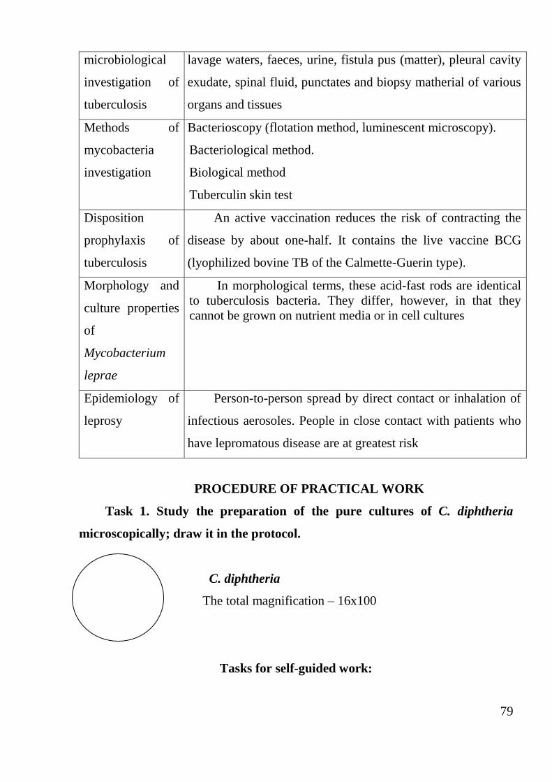
79
microbiological
investigation of
tuberculosis
lavage waters, faeces, urine, fistula pus (matter), pleural cavity
exudate, spinal fluid, punctates and biopsy matherial of various
organs and tissues
Methods of
mycobacteria
investigation
Bacterioscopy (flotation method, luminescent microscopy).
Bacteriological method.
Biological method
Tuberculin skin test
Disposition
prophylaxis of
tuberculosis
An active vaccination reduces the risk of contracting the
disease by about one-half. It contains the live vaccine BCG
(lyophilized bovine TB of the Calmette-Guerin type).
Morphology and
culture properties
of
Mycobacterium
leprae
In morphological terms, these acid-fast rods are identical
to tuberculosis bacteria. They differ, however, in that they
cannot be grown on nutrient media or in cell cultures
Epidemiology of
leprosy
Person-to-person spread by direct contact or inhalation of
infectious aerosoles. People in close contact with patients who
have lepromatous disease are at greatest risk
PROCEDURE OF PRACTICAL WORK
Task 1. Study the preparation of the pure cultures of C. diphtheria
microscopically; draw it in the protocol.
C. diphtheria
The total magnification – 16x100
Tasks for self-guided work:
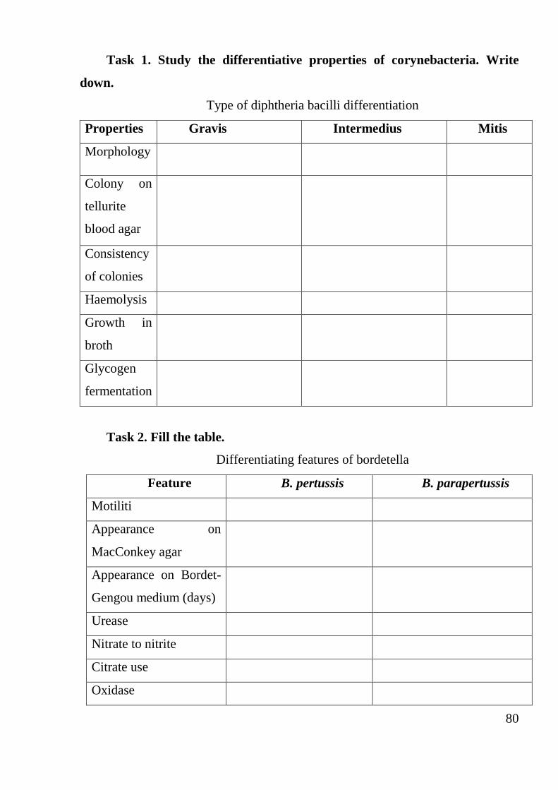
80
Task 1. Study the differentiative properties of corynebacteria. Write
down.
Type of diphtheria bacilli differentiation
Properties Gravis Intermedius Mitis
Morphology
Colony on
tellurite
blood agar
Consistency
of colonies
Haemolysis
Growth in
broth
Glycogen
fermentation
Task 2. Fill the table.
Differentiating features of bordetella
Feature B. pertussis B. parapertussis
Motiliti
Appearance on
MacConkey agar
Appearance on Bordet-
Gengou medium (days)
Urease
Nitrate to nitrite
Citrate use
Oxidase
Antitoxic
antidiphtheria serum
Colonies of
toxigenic
Corynebacteria
diphtheriae
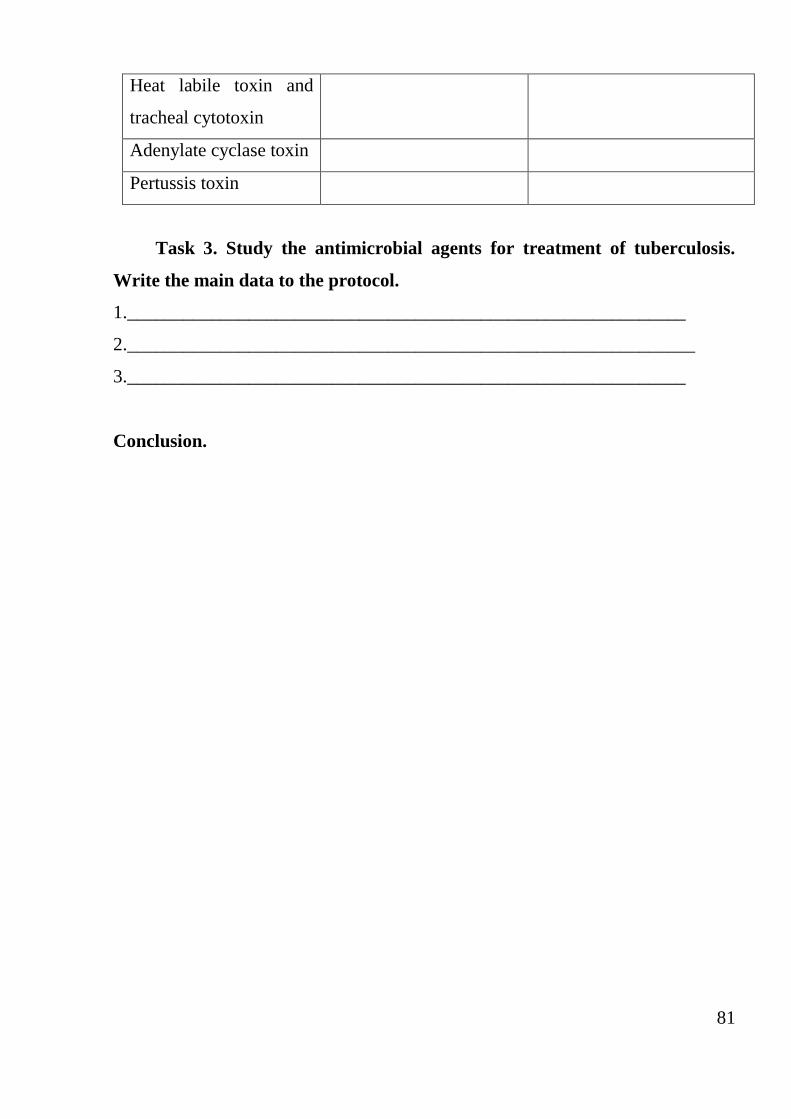
81
Heat labile toxin and
tracheal cytotoxin
Adenylate cyclase toxin
Pertussis toxin
Task 3. Study the antimicrobial agents for treatment of tuberculosis.
Write the main data to the protocol.
1.____________________________________________________________
2._____________________________________________________________
3.____________________________________________________________
Conclusion.
82
Questions for the final lesson.
1. Value of medical microbiology in practical activities of the doctor.
2. Structure and a chemical compound of a bacterial cell. Peculiarities of a
structure of Gram-positive and Gram- negative bacteria.
3. Tinctorial properties of bacteria. Methods of coloring.
4. Structure and a chemical compound of a bacterial cell. Capsules, flagella,
sporulation.
5. Peculiarities of biology of viruses.
6. Structure and a chemical compound of viruses and bacteriophages.
7. Types of virus and cell interaction. Stages of multiplication of viruses.
8. Bacteriophagia. Interaction of a phage with a bacterial cell. Temperate and
virulence bacteriophages. Lysogeny.
9. Application of phages in medicine and biotechnologies.
10. Artificial nutrient mediums, their classification. Requirements. Showed to
nutrient mediums.
11. Microbial growth and multiplication of bacteria.
12. Methods for cultivation of anaerobes.
13. Principles and methods of isolation of pure cultures of bacteria.
14. Enzymes of bacteria, their value in identification of bacteria.
15. Methods of cultivation of viruses.
16. Normal microbial flora of human body. Dysbioses. Probiotics.
17. Microflora of air and methods of its research.
18. Methods of sanitary - bacteriological research of water: definition of
microbic number.
19. Concept of disinfection. Methods. Disinfectants.
20. Concept of sterilizations, methods, the equipment.
21. Concept of chemotherapy and antibiotics. The mechanism of action of
antibiotics.
83
22. Antibiotics, classification bayed on a source of reception, a way of
reception.
23. Antibiotics. Sources and ways of reception. Classes of antibiotics.
24. Classification of antibiotics on chemical structure, the mechanism and a
spectrum of activity.
25. Methods of antibiotic sensitivity test.
26. A structure of bacterial genome. Concept of a genotype and a phenotype.
Kinds of variability.
27. Plasmids, their functions and properties. Use plasmids in genetic
engineering.
28. The mechanism of transfer of a genetic information.
29. Concept of an infection. Pathogenicity and virulence of bacteria. Factors of
pathogenicity.
30. Toxins of bacteria, their nature, properties, reception.
31. Concept of infectious disease. Stages of development and characteristic
properties.
32. Concept of clinical microbiology. A role of opportunictic microorganisms in
a pathology of human.
33. Methods of microbiological diagnostics of infectious diseases.
34. Concept of immunity. Classification of immunity.
35. Antibodies, their characteristics.
36. Meningococci. Taxonomy. The characteristic of biological properties.
Pathogenesis. Forms of an infection. Microbiological diagnostics. Principles
of specific prophylaxis and treatment.
37. Causative agents of a gonorrhoeae. Taxonomy. Microbiological diagnostics.
Specific treatment.
38. Staphylococci. Taxonomy. The characteristic of biological properties.
Microbiological diagnostics of the diseases caused by staphylococci.
Principles of specific prophylaxis and treatment.
84
39. Streptococci. Taxonomy. The characteristic. Microbiological diagnostics of
streptococcal infections. Principles of specific prophylaxis and treatment. A
role of streptococci at a scarlet fever.
40. Causative agents of tuberculosis. Taxonomy. The characteristic.
Microbiological diagnostics of a tuberculosis. Principles of specific
prophylaxis at children.
41. Causative agents of tularemia. The characteristic. Microbiological
diagnostics. Principles of specific prophylaxis and treatment.
42. Causative agents of anthrax. The characteristic. Microbiological diagnostics.
Principles of specific prophylaxis and treatment.
43. Causative agents of brucellosis. Taxonomy. The characteristic.
Microbiological diagnostics. Principles of specific prophylaxis and
treatment.
44. Causative agents of a plague. Taxonomy. The characteristic.
Microbiological diagnostics. Principles of specific prophylaxis and
treatment.
45. Causative agents of wooping cough. Taxonomy. The characteristic.
Microbiological diagnostics. Principles of specific prophylaxis and
treatment.
46. A problem of the hospital infections.
85
List of literature
1. Brooks G.F., Carroll K.C. & others. Jawetz, Melnick, Adelberg's Medical
microbiology-McGraw-Hill Education Medical .- 2015. - 864 р.
2. Chamberlain N. The Big Picture. Medical Microbiology -The McGraw-Hill
Companies.- 2009. - 456 p.
3. Hart C. Anthony, Shears Paul. Color Atlas of Medical Microbiology- Elsevier-
2004. - 698 p.
4. Murray P.R., Rosenthal K.S., Pfaller M.A. Medical Microbiology-
Saunders/Elsevier. - 2013. -1023 p.
5. Ryan, Kenneth, Sherris. Medical Microbiology - McGraw-Hill Medical.- 2010.-
1040 p.
6. Tortora G.J., Funke B.R., Case C.L. Microbiology. An Introduction - Pearson
Education.- 2012. - 960 p.