Embed Size (px)
Citation preview
APPLIED AND ENVIRONMENTAL MICROBIOLOGY, Sept. 1986, p. 527-5300099-2240/86/090527-04$02.00/0Copyright © 1986, American Society for Microbiology
Microcetus lappus gen. nov., sp. nov.: New Species of CiliatedProtozoon from the Bovine Rumen
COLIN G. ORPIN'* AND SVEIN D. MATHIESEN2Department ofBiochemistry, Agricultural and Food Research Council, Institute ofAnimal Physiology, Babraham,
Cambridge CB2 4AT, United Kingdom,' and Department of Arctic Biology and Institute of Medical Biology, University ofTroms0, 9001 Troms0, Norway2
Received 17 March 1986/Accepted 1 April 1986
A new species of small, ciliated protozoon, Microcetus lappus gen. nov., sp. nov., from the rumen of
Norwegian Red cattle is described. M. lappus possesses a novel cytopharyngeal apparatus of two rod-shapedstructures, one situated on the dorsal side of the buccal cavity and one on the ventral side, suggesting that itbelongs to a previously undescribed taxon.
Ruminants normally harbor a ruminal microbial popula-tion containing a mixture of ciliated protozoa composed ofentodiniomorphid species and other species with cilia lo-cated over part or all of the body surface. The most commonspecies of uniformly ciliated protozoa are Isotricha intesti-nalis, Isotricha prostoma, and Dasytricha ruminantium (8,15), which are members of the order Trichostomatida. Do-mestic ruminants have occasionally been described whichhave ruminal ciliate populations containing other specieswith cilia located over part of the body surface, includingBuetschlia spp. (4, 17), Charonina ventriculi (6, 13), Oligo-isotricha bubali (9, 10, 12), and Parabundleia ruminantium(11). All of these species have been found in cattle. Wereport here a previously undescribed species of protozoon,with nearly uniform surface ciliation, from the rumen ofNorwegian Red cattle.
MATERIALS AND METHODS
Rumen contents. Rumen contents were obtained by aspi-ration via permanent rumen cannulae from two NorwegianRed cattle from Bod0, which is located on the arctic circle inNorway. The animals were fed a ration of high-qualitytimothy hay and 1 kg of concentrates daily. The rumen fluidwas fixed immediately with 2% glutaraldehyde.
Preparation of samples. The rumen contents were filteredthrough two layers of muslin and examined with a lightmicroscope. Many of the protozoa were aggregated withparticulate matter; therefore, methylcellulose (final concen-
tration, 1% [wt/vol]) was added, and the suspension was
mixed by vortexing for 30 s. Subsamples were stained withmethyl green (15) and iodine (2) to visualize macronuclei andintracellular polysaccharides, respectively.
Suspensions of ciliates for examination by scanning elec-tron microscopy were prepared by twice vortexing the fixedpreparation for 30 s, filtering the suspension through nylonmesh (maximum pore size, 45 ,um), and centrifuging thefiltrate for 5 min at 250 x g. The pellet was suspended indistilled water and was washed by repeated centrifugation at250 x g for 3 min until the supernatant fluid was clear. Thecells were dehydrated (16), critical-point dried (1), spluttercoated with gold in a Polaron coating apparatus mounted on
a double-sided adhesive tape on a standard aluminium stub,and examined with a JSM 2 scanning electron microscope.
* Corresponding author.
Determination of population densities of protozoa. Thepopulation densities of ruminal ciliates in the twice-filtered,methylcellulose-treated samples were determined by micros-copy (2). Protozoa were identified by their morphology (15).Cell dimensions were measured with a light microscopefitted with a micrometer eyepiece calibrated against a
hemacytometer grid.
RESULTS AND DISCUSSION
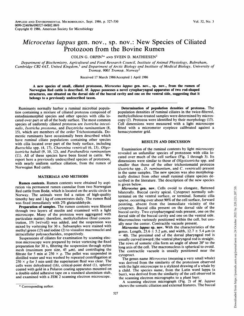
Examination of the ruminal contents by light microscopyrevealed an unfamiliar species of protozoon with cilia lo-cated over much of the cell surface (Fig. 1 through 3). Itsdimensions were similar to those of Oligoisotricha spp. andsmaller than those of the other trichostomatid protozoa(Isotricha spp., D. ruminantium, and C. ventriculi) presentin the same samples. The new species was also morpholog-ically distinct from other small ruminal ciliate species de-scribed in the literature. The description of the new speciesis giveri below.
Microcetus gen. nov. Cells ovoid to elongate, flattenedbilaterally. Buccal cavity apical. Cytoproct normally sub-terminal on the ventral surface, or terminal. Somatic ciliasparse, occurring over about 90% of the cell surface, forwardpointing, absent from the immediate vicinity of thecytoproct. Buccal cilia present on the dorsal side of thebuccal cavity. Two cytopharyngeal rods present, one on thedorsal side of the buccal cavity and one on the ventral side.Macronucleus variously positioned within the cell, but usu-
ally near the center. Contractile vacuole present.Microcetus lappus sp. nov. With the characteristics of the
genus. Length, 23.6 + 5.2 ,um, and width, 12.7 + 5.4 ,um (n= 40). The proximal end uf the dorsal pharyngeal rod isusually curved inward; the ventral pharyngeal rod is straight.The rows of somatic cilia form an angle of about 200 to thelong axis of the cell. The macronucleus is spherical to ovoid.The contractile vacuole is usually positioned near thecytoproct.The genus name Microcetus (meaning a very small whale)
was derived from the similarity of the protozoon observedwith the light microscope to a stylized drawing of a whale bya child. The species name, from the Latin word lappa (aburr), was derived from the similarity of the cell observed in
the scanning electron micrographs to a plant burr.A scanning electron micrograph (Fig. 2) of M. lappus
shows the somatic ciliation and external features. The buccal
527
Vol. 52, No. 3
Dow
nloa
ded
from
http
s://j
ourn
als.
asm
.org
/jour
nal/a
em o
n 24
Oct
ober
202
1 by
220
.221
.207
.59.
528 ORPIN AND MATHIESEN APPL. ENVIRON. MICROBIOL.
Dow
nloa
ded
from
http
s://j
ourn
als.
asm
.org
/jour
nal/a
em o
n 24
Oct
ober
202
1 by
220
.221
.207
.59.
NEW RUMINAL PROTOZOON 529
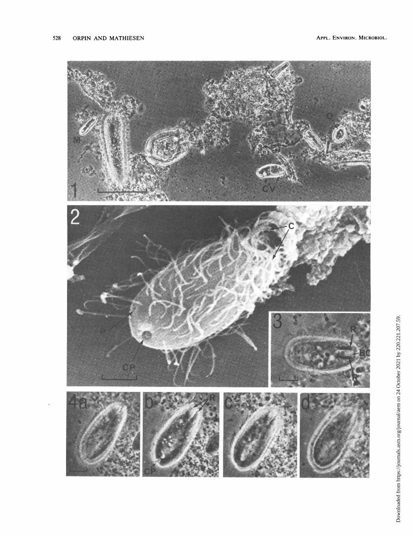
FIG. 5. Phase-contrast light micrographs of M. lappus, showing the macronucleus (M) as normally observed (a) and after division (b); thecontractile vacuole (V) is also visible. The spiral alignment of the cilia (arrows) (c) and engulfed starch grains (S) (d) are also shown. Bar =5 ,um.
cavity, buccal cilia, and cytopharyngeal apparatus wererevealed by light microscopy (Fig. 3).
Light micrographs taken at different focal planes showedthat the cytopharyngeal apparatus consisted of two longitu-dinal rods (Fig. 4a to d) , with no evidence of a more complexrhabdos or cyrtos structure. The cytopharyngeal rods couldbe seen with both transmitted-light and phase-contrast op-tics. The cytoproct can be clearly seen in Fig. 4b in acharacteristic subterminal position. Staining with methylgreen revealed the macronucleus (Fig. 5a), which was nor-mally ovoid, but in a few cells (probably nearing division)two adjacent spherical macronuclei were seen (Fig. 5b). Inabout 20% of the cells a structure which we believe to be acontractile vacuole was found in the posterior region; thestructure is just visible in Fig. Sb and c. Many cells containedstarch grains, probably of dietary origin (Fig. Sd), and otherintracellular bodies which were identified by their morphol-ogy as large ruminal bacteria, suggesting that particle engulf-ment is important in the nutrition of this species.The external characteristics of M. lappus are similar to
those described by Dogiel (7) for Isotricha bubali, laternamed 0. bubali (9), but 0. bubali does not possesscytopharyngeal rods and has a striated vestibulum. M.lappus is easily distinguished from Buetschlia spp. (4, 17), C.v,entriculi (6), and P. ruminantium (11) by the possession ofsomatic ciliation over most of the cell surface and by itscytopharyngeal apparatus.
It is likely that the 0. bubali reported (5) to occur in cattlein the United States was, in fact, M. lappius, because two
cytopharyngeal rods can be clearly seen in the photomicro-graphs of these ciliates and the posterior of the cell issmoothly rounded. Although these structural features areclearly at variance with the original description of 0. bubali(7), the investigators did not comment on these anomalies.M. lappus occurred in both of the Bod0 cattle examined.
The total ciliate population density in the two animals was3.4 x 104 ml-1 and 2.25 x 10⁴ ml-1 (mean values of eightestimates); of these, M. lappius represented 12 and 15%,respectively. We also examined the ruminal contents of fivecattle from near Troms0 (latitude, 700 N), and the newspecies was not present. Therefore, M. lappius is not uni-formly distributed in Norwegian Red cattle, and we are nowattempting to determine its geographical distribution.The taxonomic position of the genus Microceti's is as yet
unknown. Further work with freshly stained material toindicate the silver-line system and micronucleus and withtransmission electron microscopy to determine the nature ofthe cytopharyngeal rods and the origin and structure of thebuccal ciliation is necessary before any conclusions can bereached regarding the taxonomy of Microcetus. No taxo-nomic group currently exists which embraces protozoapossessing the type of cytopharyngeal apparatus found inMicrocetus. The nearly uniform somatic ciliation, the nearlyapical cytostome preceded by a vestibulum, and the pres-ence of oral ciliation suggest that Microcetus should beplaced in the class Kinetofragminophora de Puytorac et al.1974, according to recent classification systems (3, 14).Within this class, two subclasses, the Vestibulifera and the
FIG. 1. Phase-contrast light micrograph of rumen contents of Norwegian Red cattle, showing the newly described ciliate in relation toother rumen ciliates. M, M. lappus; 0, Oligoisotricha sp.; D, D. rurminantium; E, Entodiniumn longinucleatum; CV, C. ventriculi. Bar = 50pum.
FIG. 2. Scanning electron micrograph of M. lappius, showing cilia (C), the cytoproct (CP), and what is probably the external pore of thecontractile vacuole (P). Bar = 5 p.m.
FIG. 3. Phase-contrast light micrograph of M. lapplus, showing the prominent cytopharyngeal rods (R) and buccal ciliation (BC). Bar =5 p.m.FIG. 4. Phase-contrast light micrographs taken at different focal planes through a cell of M. lappius. showing only two cytopharyngeal rods
(R), the cytoproct (CP), and sparse somatic cilia. Bar = 5 p.m.
VOL. 52, 1986
Dow
nloa
ded
from
http
s://j
ourn
als.
asm
.org
/jour
nal/a
em o
n 24
Oct
ober
202
1 by
220
.221
.207
.59.
530 ORPIN AND MATHIESEN
Hypostomata, possess cytopharyngeal strengthening struc-tures. The cytopharynx of members of the Vestibulifera isusually strengthened by several nematodesmata to form arhabdoslike structure; members of the Hypostomata typi-cally possess a cytopharyngeal apparatus of the more com-plex cyrtos type (a cone-shaped, basketlike structure, some-times curved, composed of nematodesmata). Both types areclearly different from that found in Microcetus. Because ofits characteristic cytopharyngeal apparatus, Microcetus mayrepresent a previously undescribed taxon.Two other species of small ciliates, C. ventriculi and a
species putatively identified as Oligoisotricha sp., werepresent in the ruminal contents of the Norwegian Red cattleat Bod0. Whereas C. ventriculi has been recorded as occur-ring in ruminants in a variety of geographical locations (6, 8,13), 0. bubali has been positively identified only in waterbuffalo (9, 10) and in cattle in Japan (12). In all of ourpreparations to date the putative Oligoisotricha sp. has beenstrongly associated with particulate material, thus prevent-ing the use of scanning electron microscopy. The Oligo-isotricha sp., like 0. bubali (7), possesses a striatedvestibulum and lacks the cytopharyngeal rods of M. lappus,
but unlike 0. bubali, the posterior end of the cells issmoothly rounded. This organism, like M. lappus, may be anew species.
ACKNOWLEDGMENTS
We thank F. J. Hall for performing the electron microscopy and J.Reid Hole for allowing us to use the cattle with fistulated rumens atVag0nes Agriculture Station, Vag0nes, Norway.
LITERATURE CITED1. Anderson, T. F. 1951. Techniques for the preservation of the
three-dimensional structure in preparing specimens for theelectron microscope. Trans. N.Y. Acad. Sci. 13:130.
2. Coleman, G. S. 1978. Rumen entodiniomorphid protozoa, p.
39-44. In A. E. R. Taylor and J. R. Baker (ed.), Methods ofcultivating parasites in vitro. Academic Press, Inc. (London),Ltd., London.
3. Corliss, J. 0. 1974. The ciliated protozoa: characterization,
classification and guide to the literature, 2nd ed. PergamonPress, Ltd., Oxford.
4. Dehority, B. A. 1970. Occurrence of the ciliate protozoaButschlia parva Schuberg in the rumen of the ovine. Appl.Microbiol. 19:179-181.
5. Dehority, B. A., W. S. Damron, and J. B. McLaren. 1983.Occurrence of the rumen ciliate Oligoisotricha bubali in domes-tic cattle (Bos taurus). Appl. Environ. Microbiol. 45:1394-1397.
6. Dehority, B. A., and W. R. S. Mattos. 1978. Diurnal changes andeffect of ration on concentrations of the rumen ciliate Charonventriculi. Appl. Environ. Microbiol. 36:953-958.
7. Dogiel, V. 1928. La faune d'infusoires habitant l'estomac dubuffle et du dromadaire. Ann. Parasitol. 6:323-338.
8. Hungate, R. E. 1966. The rumen and its microbes. AcademicPress, Inc. (London), Ltd., London.
9. Imai, S. 1981. Four new rumen ciliates, Entodinium ogimotoisp. n., E. bubalum sp. n., E. fujitai sp. n., and E. tsunodai sp.n. and Oligoisotricha bubali (Dogiel 1928) n. comb. Jpn. J. Vet.Sci. 43:201-209.
10. Imai, S., C. H. Chang, J.-S. Wang, K. Ogimoto, and J. Kujita.1981. Rumen ciliate protozoal fauna of the water buffalo(Bubalus bubalis) in Taiwan. Jpn. J. Zootech. Sci. 30:77-81.
11. Imai, S., and K. Ogimoto. 1983. Parabundleia ruminantium gen.n., sp. n., Diplodinium mahidoli sp. n. with two forma, andEntodinium parvum forma monospinosum forma n. from thezebu cattle (Bos indicus L. 1758) in Thailand. Jpn. J. Vet. Sci.45:585-591.
12. Imai, S., M. Shimizu, M. Konishita, M. Toguchi, T. Ishii, and J.Fujita. 1982. Rumen ciliate protozoal fauna and composition ofthe cattle in Japan. Bull. Nippon Vet. Zootech. Coll. 31:70-74.
13. Jameson, A. P. 1925. A new ciliate, Charon ventriculi n.g., n.sp., from the stomach of ruminants. Parasitology 17:403-405.
14. Levine, N. D., J. 0. Corliss, F. E. G. Cox, G. Deroux, J. Grain,B. M. Honigberg, G. F. Leedale, A. R. Loeblich III, J. Lom, D.Lynn, E. G. Merinfeld, F. C. Page, G. PoUansky, V. Sprague, J.Vavra, and F. G. Wallace. 1980. A newly revised classificationof the protozoa. J. Protozool. 27:37-58.
15. Ogimoto, K., and S. Imai. 1981. Atlas of rumen microbiology.Japan Scientific Societies Press, Tokyo.
16. Orpin, C. G., and F. J. Hall. 1983. Surface structures of therumen holotrich protozoon Isotricha intestinalis with particularreference to the attachment zone. Curr. Microbiol. 8:321-325.
17. Schuberg, A. 1888. Die Protozoen des Wiederkauermagens. 1.Butschlia, Isotricha, Dasytricha, Entodinium. Zool. Jahrb. Abt.Allg. Zool. Physiol. Tiere 3:365-418.
APPL. ENVIRON. MICROBIOL.
Dow
nloa
ded
from
http
s://j
ourn
als.
asm
.org
/jour
nal/a
em o
n 24
Oct
ober
202
1 by
220
.221
.207
.59.