Embed Size (px)
Citation preview

Radiation Research
An International Journal Editor-in-Chief
R. J . M. Fry
Managing: Edi tor
Martha Edington
Volume 132, 1992
ACADEMIC P R E S S , INC. H a r c o u r t . B r a c e J o v a n o v i c h , P u b l i s h e r s
San Diego New York Boston London Svclnev Tok> Toronto
Copyright © 1992 by Academic Press, Inc. All rights reserved
No part of this publication may be reproduced or transmitted in any form or by any means, electronic or mechanical, including photocopy, recording, or any information storage and retrieval system, without permission in writing from the copyright owner.
The appearance of the code at the bottom of the first page of an article in this journal indicates the copyright owner's consent that copies of the article may be made for personal or internal use, or for the personal or internal use of specific clients. This consent is given on the condition, however, that the copier pay the stated per copy fee through the Copyright Clearance Center, Inc. (27 Congress Street, Salem, Massachusetts 01970), for copying beyond that permitted by Sections 107 or 108 of the U.S. Copyright Law. This consent does not extend to other kinds of copying, such as copying for general distribution, for advertising or promotional purposes, for creating new collective works, or for resale. Copy fees for pre-1992 articles are as shown on the article title pages; i f no fee code appears on the title page, the copy fee is the same as for current articles.
0033-7587/92 $5.00
M A D E IN T H E U N I T E D S T A T E S O F A M E R I C A
This journal is printed on acid-free paper.
RADIATION RESEARCH
O F F I C I A L J O U R N A L O F T H E R A D I A T I O N R E S E A R C H S O C I E T Y
Ediior-in-Chief: R. J. M . FRY, Biology Division, Oak Ridge National Laboratory, P.O. Box 2009, Oak Ridge, Tennessee 37831-8077
Managing Editor: M A R T H A EDINGTON, University of Tennessee-Oak Ridge Graduate School of Biomedical Sciences, Biology Division, Oak Ridge National Laboratory, P.O. Box 2009,
Oak Ridge, Tennessee 37831-8077
ASSOCIATE EDITORS
S. J . A D E L S T E I N , Harvard Medical School
W. A. B E R N H A R D . University of Rochester
M. W. D E W H I R S T , Duke University
J. F . D I C E L L O . Clarkson University
P. W. D O E T S C H , Emory University
H. S. D U C O F F , University of Illinois
E. G L A T S T E I N . University of Texas
D . T . G O O D H E A D , Medical Research Council, Chilton, Didcot, Oxfordshire. England
M. N. G O U L D , University of Wisconsin
F. F . HAHN, Lovelace Inhalation Toxicology Research Institute
W. D. H E N N E R . The Oregon Health Sciences University
R. P. HIL1 Ontario Cancer Institute. Toronto. Canada
C. D. JONAH, Argonne National Laboratory
C. J . K O C H . University of Pennsylvania
J. R. L E P O C K , University of Waterloo, Ontario, Canada
W. H. M C B R I D E , University of California, Los Angeles
P. L . O L I V E , British Columbia Cancer Research Centre, Vancouver, Canada
R. J . P R E S T O N , C U T , Research Triangle Park
J. S. R A S E Y , University of Washington
M. A. R I T T E R , University of Wisconsin
S. R O C K W E L L . Yale University
M. D. S E V I L L A , Oakland University
J . E . T U R N E R , Oak Ridge National Laboratory
R. L . WÄRTERS, University of Utah
F. M. W A T E R M A N . Thomas Jefferson University Hospital
CONSULTANT STATISTICIANS
E. I F R O M E , Oak Ridge National Laboratory M. D. MORRIS , Oak Ridge National Laboratory
OFFICERS OF THE SOCIETY
President: J A M E S A. B E L L I , Department of Radiation Therapy, University of Texas Medical Branch, Galveston, Texas 77550
Vice President and President-Elect: G O R D O N F . W H I T M O R E , Physics Division, Ontario Cancer Institute, 500 Sherbourne Street, Toronto, Ontario, Canada M4X 1K9
Secretary-Treasurer: D A V I D J . G R D I N A , Argonne National Laboratory, 700 South Cass Avenue, Argonne, Illinois 60439-4833
Secretary-Treasurer-Elect: W A Y N E R. HANSON. Loyola-Hines Department of Radiotherapy, Hines VA Medical Center, 5th Avenue and Roosevelt Road,
Hines, Illinois 60141
Editor-in-Chief: R. J . M. F R Y , Biology Division. Oak Ridge National Laboratory', P.O. Box 2009, Oak Ridge, Tennessee 37831-8077
Administrative Director: L A U R A F L E M I N G JONES, 1891 Preston White Drive, Reston, Virginia 22091
Titus C . Evans, Editor-in-Chief Volumes 1-50 Oddvar F. Nygaard, Editor-in-Chief Volumes 51-79
Daniel Billen, Editor-in-Chief Volumes 80-113
V O L U M E 132, 1992

C o u n c i l o r s , R a d i a t i o n R e s e a r c h S o c i e t y 1 9 9 2 - 1 9 9 3
P H Y S I C S
W. A . Bernhard, University of Rochester A . Chatterjee, Lawrence Berkeley Laboratory
B I O L O G Y
C. R. Geard, Columbia University H. D. Thames, University of Texas
M E D I C I N E
C. N . Coleman, Harvard Medical School M . W. Dewhirst, Duke University
C H E M I S T R Y
R. C. Fahey, University of California, San Diego M . E. Varnes, Case Western Reserve University
A T - L A R G E
B. F. Kimler, University of Kansas J. L. Roti Roti, Washington University
C O N T E N T S O F V O L U M E 132
N U M B E R l , OCTOBER 1992
V. Noethig-Laslo, T. A. Himdan, and Paramagnetic Molecular Centers in 7-Irradiated H . Bilinski Precipitates in the System A l C l 3 - D L - a - V a l i n e -
NaOH 1
A. Saha, P. C. Mandal, and Radiation-Induced Inactivation of Dihydroorotate S. N . Bhattacharyya Dehydrogenase in Dilute Aqueous Solution . . . 7
L . J. Shyr and B. A. Müggenburg A Comparison of the Predicted Risks of Developing Osteosarcoma for Dogs Exposed to 2 3 8 P u 0 2
Based on Average Bone Dose or Endosteal Cell Dose 13
John R. J. Sorenson Essential Metalloelement Metabolism and Radiation Protection and Recovery 19
Lynn Harrison, Mi l an Skorvaga, Transfection of the Escherichia coli nth Gene into Richard P. Cunningham, Radiosensitive Chinese Hamster Cells: Effects on Jolyon H . Hendry, and Sensitivity to Radiation, Hydrogen Peroxide, and Geoffrey P. Margison Bleomycin Sulfate 30
C. J. Koch and K. A. Skov Comparisons of Cellular Radiation Response Using Absolute Rather Than Relative Parameters 40
Mary Avery, S. David Nathanson, and Lymph Flow from Murine Footpad Tumors be-Fred W. Hetzel fore and after Sublethal Hyperthermia 50
Louis K. Wagner, Dennis A. Johnston, Radiation-Induced Micrencephaly in Guinea Pigs 54 and Daniel J. Felleman
Mark W. Dewhirst, Edgardo T . Ong, Effects of the Calcium Channel Blocker Flunari-David Madwed, Bruce Kli tzman, zine on the Hemodynamics and Oxygenation of Timothy Secomb, David Brizel, Tumor Microvasculature 61 Joseph Bonaventura, Gary Rosner, Brian Kavanagh, Jeri Edwards, and Joseph Gross
Jamie R. Mil l igan, Allister D. Arnold, The Effect of Superhelical Density on the Yield of and John F. Ward Single-Strand Breaks in 7-Irradiated Plasmid DNA 69
Candace D. Carter and Thrombopoietin from Human Embryonic Kidney Ted P. McDonald Cells Causes Increased Thrombocytopoiesis in
Sublethally Irradiated Mice 74
R. W. M . van der Maazen, Repopulation of 0-2A Progenitor Cells after Irra-I . Verhagen, B. J. Kleiboer, and diation of the Adult Rat Optic Nerve Analyzed by A. J. van der Kogel an in Vitro Clonogenic Assay 82
Stephen M . Hahn, Lynn Wilson, Identification of Nitroxide Radioprotectors . . . 87 C. Mural i Krishna, James Liebmann, Wi l l i am DeGraff, Janet Gamson, Amram Samuni, David Venzon, and James B. Mitchell
Edward A. Bump, Beth A. Cerce, Ramsey Al-Sarraf, Susan M , Pierce, and Cameron J. Koch
Radioprotection of DNA in Isolated Nuclei by Naturally Occurring Thiols at Intermediate Oxygen Tension 94
Y i n Zhang, Kevin M . Sweet, Interaction between Radiation and Drug Damage Marguerite A . Sognier, and in Mammalian Cells. V I . Radiation and Doxorubi-James A. Belli ein Age-Response Function of Doxorubicin-Sen-
sitive and-Resistant Chinese Hamster Cells . . . 105
SHORT COMMUNICATIONS Björn Zackrisson and Mikael Karlsson Relative Biological Effectiveness of 50-M V X Rays
on Jejunal Crypt Survival in Vivo 112
J. H . Hendry, S. A. Roberts, and The Clonogen Content of Murine Intestinal C. S. Potten Crypts: Dependence on Radiation Dose Used in Its
Determination 115
Q. Chen, M . Chopp, M . O. Dereski, The Effect of Light Fluence Rate in Photodynamic B. C. Wilson, M . S. Patterson, Therapy of Normal Rat Brain 120 A. Schreiber, and F. W. Hetzel
LETTERS TO THE EDITOR Nandanuri M . S. Reddy and Comments on "Radiation-Induced D N A U n -Christopher S. Lange winding Is Influenced by Cell Shape and Trypsin" 124
P. L . Olive and S. H . MacPhail Reply to Comments on "Radiation-Induced D N A Unwinding Is Influenced by Cell Shape and Trypsin" 125
ERRATUM M . P. Li t t le , M . M . Hawkins, Volume 126, Number 3 (1991): "Time Variations R. E. Shore, M . W. Charles, and in the Risk of Cancer following Irradiation in N . G. Hildreth Childhood," pp. 304-316 126
ANNOUNCEMENTS 127
NUMBER 2, NOVEMBER 1992
FAILLA MEMORIAL LECTURE
G. E. Adams Redox, Radiation, and Reductive Bioactivation 129
SYMPOSIUM: THE MOLECULAR BASIS FOR RADIATION EFFECTS ON CELL PROGRESSION THROUGH THE CELL CYCLE J. L . Roti Roti Introduction 140
Ted A. Weinert Dual Cell Cycle Checkpoints Sensitive to Chromosome Replication and D N A Damage in the Budding Yeast Saccharomyces cerevisiae 141
Roy Rowley Radiation-Induced Mitotic Delay: A Genetic Characterization in the Fission Yeast 144
Ruth J. Muschel, Hong Bing Zhang, Effects of Ionizing Radiation on Cyclin Expression George Iliakis, and in HeLa Cells 153 W. Gillies McKenna
R. B. Mikkelsen and C. Gentry A Radiation-Induced Inhibitor of Chromosome Condensation and Nuclear Envelope Breakdown in HeLa Cells 158
P. C. Goswami, M . H i l l , The Suppression of the Synthesis of a Nuclear Pro-R. Higashikubo, W . D . Wright, tein in Cells Blocked in G 2 Phase: Identification of and J. L . Roti Roti NP-170 as Topoisomerase I I 162
REGULAR ARTICLES C. Malamut, P. J. Paes-Leme, Diffusion of Low-Energy Electrons in Tissue-like and A. S. Paschoa Liquids 168
Harald H . Rossi and Marco Zaider Compound Dual Radiation Action. I . General Aspects 178
A. Rodriguez, E. L . Alpen, The RBE-LET Relationship for Rodent Intestinal and P. Powers-Risius Crypt Cell Survival, Testes Weight Loss, and Mul
ticellular Spheroid Cell Survival after Heavy-Ion Irradiation 184
Y i Jin, Fredde J. Burns, Oncogene Amplification Detected by in Situ Hy-and Seymour J. Garte bridization in Radiation-Induced Skin Cancers in
Rats 193
John M . Buatti, Luis R. Rivero, and Radiation-Induced DNA Single-Strand Breaks in Timothy J. Jorgensen Freshly Isolated Human Leukocytes 200
M . P. Lit t le , M . M . Hawkins, Fitting the Armitage-Doll Model to Radiation-Ex-M . W. Charles, and N . G. Hildreth posed Cohorts and Implications for Population
Cancer Risks 207
K. A. Kunugi, E. M . Mil ler , Low pH Does Not Affect the Dose Response M . A. Vazquez-Padua, for 5 -Amino-5'-deoxythymidine Modulation of and T. J. Kinsella IdUrd D N A Incorporation and Radiosensitization
in a Human Bladder Cancer Cell Line 222
Dominique Bonnefont-Rousselot, Effect of pH on Low-Density Lipoprotein Oxida-Monique Gardes-Albert, t ion by O j / H O ; Free Radicals Produced by Sylvie Lepage, Jacques Delattre, Gamma Radiolysis 228 and Christiane Ferradini
Celso Luiz S. Lage and Developmental Changes Induced by 7 Irradiation Maria Apparecida Esquibel in Ipomoea batatas L. Lam (Sweet Potato) . . . 237
Danuta Wlodek and Peggy L . Olive Neutral Filter Elution Detects Differences in Chromatin Organization Which Can Influence Cellular Radiosensitivity 242
W. H . Y. Roa and J. D . Chapman Kil l ing of EMT-6 Cells by Decays from Isotopes Incorporated on Sensitizer Adducts 248
Colin A. B i l l , Brigitte M . Grochan, Decreased Repair of Radiation-Induced D N A Eduard Vrdoljak, Double-Strand Breaks with Cellular Differentia-Elizabeth A. Mendoza, and tion 254 Philip J. Tofilon
SHORT COMMUNICATIONS Sydney M . Evans, Protection against Metastasis of Radiation-In-Thomas Van Winkle, duced Thymic Lymphosarcoma and Weight Loss Bernard F. Szuhaj, in C57Bl/6NCrl BR Mice by an Autoclave-Resis-Kathryn E. Michel, tant Factor Present in Soybeans 259 and Ann R. Kennedy
John T. Leith, Gia Padfield, Effects of Partial Hepatectomy on the Growth and Seth Michelson Characteristics and Hypoxic Fractions of Xeno-
grafted DLD-2 Human Colon Cancers 263
IN MEMORIAM G. E. Adams John Freeman Loutit 269
N U M B E R 3, D E C E M B E R 1992
J. Chen, H . Roos, and A. M . Kellerer Microdosimetry of Diagnostic X Rays: Applications of the Variance-Covariance Method . . . . 271
Nestor Azziz, Ferenc Hajnal, Stopping Power and the Concept of Effective Ion Wayne M . Lowder, and Phil Murley Charge at Low Energies 277
Michael A. Xapsos A Spatially Restricted Linear Energy Transfer Equation 282
Edwin E. Budzinski, Analysis of D N A Damage at the Dinucleoside Alexander E. Maccubbin, Monophosphate Level: Application to the Forma-Marianne S. Evans, mido Lesion 288 Mol ly Kulesz-Martin, and Harold C. Box
Björn Anderstam, Carlos Vaca, Lipid Peroxide Levels in a Murine Adenocarci-and Mats Harms-Ringdahl noma Exposed to Hyperthermia: The Role of Glu
tathione Depletion 296
Brian Endlich, Reza Salavati, Rat Embryo Cells Immortalized with Transfected Thomas Sullivan, and C. Clifton Ling Oncogenes Are Transformed by 7 Irradiation . . 301
D. L . Lundgren, F. F. Hahn, Repeated Inhalation Exposure of Rats to Aerosols J. H . Diel, and M . B. Snipes of 1 4 4 C e 0 2 . I . Lung, Liver, and Skeletal Dosimetry 312
D. L. Lundgren, F. F. Hahn, Repeated Inhalation Exposure of Rats to Aerosols and J. H . Diel of , 4 4 C e 0 2 . I I . Effects on Survival and Lung, Liver,
and Skeletal Neoplasms 325
Qiu-mei Zhang, Shuji Yonei, Multiple Pathways for Repair of Oxidative D N A and Mik i t a Kato Damages Caused by X Rays and Hydrogen Perox
ide in Escherichia coli 334
N . F. Metting, S. T. Palayoor, Induction of Mutations by Bismuth-212 a Parti-R. M . Macklis, R. W. Atcher, cles at Two Genetic Loci in Human B-Lympho-H . L . Liber, and J. B. Little blasts 339
Eric P. Cohen, Brian L . Fish, Treatment of Radiation Nephropathy with and John E. Moulder Captopril 346
Virgina K. Langmuir The Combined Use of 1 3 1I-Labeled Antibody and and Holly L . Mendonca the Hypoxic Cytotoxin SR 4233 in Vitro and
in Vivo 351
Thomas L . Waiden, Jr. Leukotriene C 4-Induced Radioprotection: The Role of Hypoxia 359
R. H . Mannan, J. R. Mercer, L. I . Radioiodinated l-(2-Fluoro-4-iodo-2,4-dideoxy-ß-Wiebe, V . V. Somayaji, and J. D. L-xylopyranosyl)-2-nitroimidazole: A Novel Probe Chapman for the Noninvasive Assessment of Tumor Hyp
oxia 368
SHORT COMMUNICATION H . Nagasawa, J. B. Lit t le , Effect of Dose Rate on the Survival of Irradiated N . M . Tsang, E. Saunders, J. Tesmer, Human Skin Fibroblasts 375 and G. F. Strniste
AUTHOR INDEX FOR VOLUME 132 380
CUMULATIVE AUTHOR INDEX FOR VOLUMES 129-132 381
CUMULATIVE SUBJECT INDEX FOR VOLUMES 129-132 384
RADIATION RESEARCH 132, 27 1-276 ( 1992)
Microdosimetry of Diagnostic X Rays: Applications of the Variance-Covariance Method
J . C H E N , * H . R o o s , f A N D A . M . K E L L E R E R * ^
^Institute for Radiation Biology, GSF, Neuherberg, Germany; and ^Radiobiological Institute, University of Munich, Germany
C H E N , J . , Roos, H . , AND K E L L E R E R , A. M . Microdosimetry of Diagnostic X Rays: Applications of the Variance-Covariance Method. Radial. Res. 132, 271-276 (1992).
Microdosimetric measurements in beams of diagnostic X rays (between 30 and 125 kV) have been performed. In these pulsed radiation fields, microdosimetric measurements are possible only by application of the variance-covariance technique. The dose mean lineal energy, j> D , is determined for various simulated diameters, at different depths in the absorber, and at different points within the pulse intervals. From the measured temporal dependences one can also obtain values of yD for different X-ray pulse generators. The results demonstrate the potential of the variance-covariance method for a diversity of microdosimetric measurements in radiation protection and in the quality control of radiation beams. © 1992 Academic Press, Inc.
INTRODUCTION
The quality factor in radiation protection is currently defined as a function of the linear energy transfer (L) which can be calculated in radiation fields of known spectral distribution. But the experimental determination of the quality factor, especially for radiation fields with incompletely known spectra, needs to be based on the measurable microdosimetric parameter dose mean lineal energy, y D , which is closely related to the dose mean linear energy transfer. This parameter therefore becomes increasingly important in the practice of radiation measurements.
The variance-covariance method permits one to determine yD in time-varying radiation fields (7). Unt i l now, several applications of this method have been made. Some experiments were performed in the laboratory with low dose rate (2, i ) , some were performed with neutron beams (4, 5), and some were done with therapeutic electron beams (6. 7). A l l of the experiments show the potential of the variance-covariance method and its applicability in radiation protection and radiation beam control.
However, while microdosimetry in its conventional form has become more and more important in their fields, the variance-covariance method has still not entered into routine practice. We felt, therefore, that a new investigation with added features would be useful.
X rays are widely used in diagnostic radiology, but there are very few microdosimetric measurements so far for such radiation fields. In the pulsed diagnostic radiation fields, the variance method cannot be used, and measurements of single-event spectra would be possible only i f one reduced the dose rate sufficiently by increasing the source-to-detector distance, which is usually impracticable. Furthermore, measurements with the single-event technique would require impracticable time for this kind of radiation instrument. With the variance-covariance method one avoids this dilemma. Therefore, a special aim of this paper is to demonstrate the potential of the variance-covariance method in pulsed diagnostic radiation fields. We give new data for these commonly used radiation fields and show that not only the averaged information for a whole pulse train but also the microdosimetric characteristics of individual subpulses can be determined by the variance-covariance method. We achieve this by the use of a sufficiently high sampling frequency.
The measurements with the variance-covariance method were performed in the field of a "two-pulse" X-ray generator for radiography. It wil l be seen subsequently that measurements on this type of generator provide microdosimetric information even for other pulsed X-ray generators.
MATERIALS AND METHODS
We chose to determine the quality of the diagnostic X rays produced by a Siemens "Nanomobil 2" X-ray generator. It is a two-pulse generator, i.e., there are two sinusoidal pulses within each 20-ms period (see Fig. 1). The anode voltage1 of this type of instrument can be varied between 30 and 125 kV.
A pair of tissue-equivalent cylindrical proportional counters (height equal to diameter) with methane-based tissue-equivalent gas was employed for the measurements. The geometry and construction of the detectors have been described earlier (J). They have tissue-equivalent walls 12 mm thick. The counters are linked to the same gas-flow system and to the same high voltage supply; any fluctuations of gas gain in the two detectors are therefore correlated and can, as is the case with the dose fluctuations, be eliminated with the variance-covariance technique. The proportional counters are calibrated with a collimated 2 4 1 Am a-particle source for different gas pressures and electrode voltages.
The electronic signal processing system is represented in Fig. 2. Two detectors, A and B, are exposed to the same radiation field and register
1 The term anode voltage is here used for the maximum of the pulsed potential.
271 0033-7587/92 $5.00
Copyright © 1992 by Academic Press. Inc. All rights of reproduction in any form reserved.
272 C H E N . ROOS, A N D K E L L E R E R
3 1-5
0)"
> 0 TD O § 0.5
0 10 20 30
Time (ms)
F I G . 1. Pulse form of anode voltage of a two-pulse X-ray generator.
synchronously the electric signals which are proportional to the energy imparted within each of the detectors. The currents from the detectors are measured by electrometers in an integrating mode. The current and the voltage noise of the electrometers are 10"15 A and 20 p\ (rms), respectively. To achieve optimal resolution, the signals of each irradiation pulse are further amplified to cover most of the dynamic range of the analog-to-digital converter (12-bit). The influences of electronic noise and of the converter's resolution on the measured signals are below 5%.
The digitized data are stored in the computer as time sequences. The variance-covariance method does not require information on the temporal interrelationship of the specific energies registered in consecutive measurement intervals. The information can, however, be used to determine from the data for the two-pulse generator the dose mean lineal energy as a function of instantaneous voltage, and thus the variations of yD within irradiation pulses. Figure 3 shows, as an example, the measured data from three irradiation pulses for two detectors; the duration of the sampling intervals was 100 ps. Due to attenuation of the softer X rays as well as the decline of photon yield at low anode voltages, Fig. 3 shows "zero" energy imparted during an appreciable portion of each cycle.
From paired data for the two detectors the mean, t, of the energy imparted, its relative variance, K, and its relative covariance, C, are determined. The dose mean of the energy imparted per energy deposition event is given by the variance-covariance method.
eD = (V-C)-e
The corresponding values of the dose mean lineal energy, yD, are given by
where I is the mean chord length of the simulated volume. Figure 3 indicates that detector B registered a somewhat higher dose rate
than detector A. However, the values of the microdosimetric parameters,
> CD
0 •e CO Q -
E
i— CD C
LU
Time, t (ms)
F I G . 3. Data for the energy imparted in sampling intervals of 100 /is from the pair of detectors. The peak voltage on the anode is 90 kV; the simulated diameter of the cylindrical detector volume is 0.5 ^m.
as well as those of the relative variance and the relative covariance, are not dependent on dose rate, and equality of the dose rate in the two detectors or centering of two detectors in radiation field is therefore not a requirement of the variance-covariance method.
Another advantage of the method is the correction for correlated additive disturbances (5, 4, 7), such as pick-up, that affect the two detectors equally. As shown in the Appendix, such disturbances are eliminated if the parameters for the two detectors are averaged.
>'d = ( ^ D . A + ^ D . B ) / 2
yD FOR VARIOUS SITE SIZES AND DIFFERENT ANODE VOLTAGES
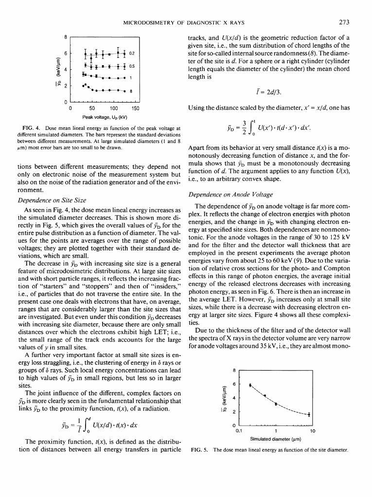
The dose mean lineal energies, yDj for simulated diameters of 0.2, 0.5, 1, and 8 were measured at different anode voltages between 30 or 35 and 125 kV (Table I). For simulated diameters smaller than 1 pm signals at 30 kV were too weak to be measured with the required accuracy. Al l measurements were performed with a filter equivalent to 2 m m of aluminum. Figure 4 and Table I summarize the results. The standard deviations given in Fig. 4 are devia-
T A B L E I The Dose Mean Lineal Energy (keV/^im) at Different Anode
Voltages (kV) for Two-Pulse X-Ray Generator
D e - t e c - t o r s E l e c t r o m e t e r , A m p l i f i e r C o m p u t e r
F I G . 2. Electronic signal processing system to perform measurements with the variance-covariance technique.
Voltage 0.2 M m 0.5 fim 1 fim 8
30 3.42 ± 0.25 1.98 ± 0 . 0 3 35 5.89 ± 0.60 4.55 ± 0.24 3.35 ± 0 . 1 1 2.17 ± 0 . 1 2 42 1.84 ± 0 . 0 7 48 5.37 ± 0 . 5 4 4.30 ± 0.28 3.21 ± 0 . 0 9 51 1.50 ± 0 . 0 2 60 5.98 ± 0.48 4.27 ± 0.25 3.19 zb 0.16 1.41 ± 0.06 75 5.82 ± 0.48 4.38 ± 0 . 1 3 2.94 ± 0.05 1.46 ± 0.06 90 5.86 ± 0 . 1 0 4.36 ± 0 . 1 9 2.97 ± 0.03 1.47 ± 0.08
110 6.10 ± 0 . 6 2 4.37 ± 0.27 2.94 ± 0.04 1.47 ± 0.09 125 5.93 ± 0.44 4.38 ± 0.35 2.99 ± 0.04 1.35 ± 0.04
M I C R O D O S I M E T R Y O F D I A G N O S T I C X R A Y S 273
E >
. . • . .»• 1
0 50 100 150
Peak voltage, U p (kV)
F I G . 4. Dose mean lineal energy as function of the peak voltage at different simulated diameters. The bars represent the standard deviations between different measurements. At large simulated diameters (1 and 8 ^m) most error bars are too small to be drawn.
tions between different measurements; they depend not only on electronic noise of the measurement system but also on the noise of the radiation generator and of the environment.
Dependence on Site Size As seen in Fig. 4, the dose mean lineal energy increases as
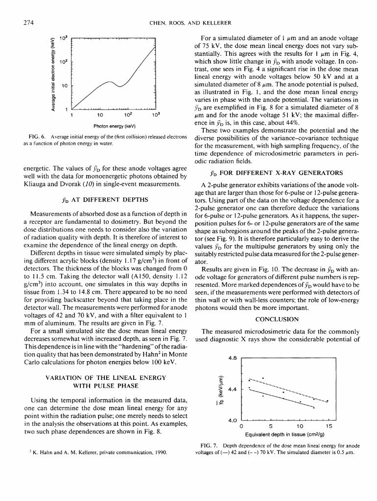
the simulated diameter decreases. This is shown more directly in Fig. 5, which gives the overall values of y D for the entire pulse distribution as a function of diameter. The values for the points are averages over the range of possible voltages; they are plotted together with their standard deviations, which are small.
The decrease in j7D with increasing site size is a general feature of microdosimetric distributions. At large site sizes and with short particle ranges, it reflects the increasing fraction of "starters" and "stoppers" and then of "insiders," i.e., of particles that do not traverse the entire site. In the present case one deals with electrons that have, on average, ranges that are considerably larger than the site sizes that are investigated. But even under this condition yD decreases with increasing site diameter, because there are only small distances over which the electrons exhibit high LET; i.e., the small range of the track ends accounts for the large values of y in small sites.
A further very important factor at small site sizes is energy loss straggling, i.e., the clustering of energy in b rays or groups of b rays. Such local energy concentrations can lead to high values of yu in small regions, but less so in larger sites.
The joint influence of the different, complex factors on j7D is more clearly seen in the fundamental relationship that links yD to the proximity function, t(x), of a radiation.
/"Jo U(x/d)-t(x)-dx
The proximity function, t(x), is defined as the distribution of distances between all energy transfers in particle
tracks, and U(x/d) is the geometric reduction factor of a given site, i.e., the sum distribution of chord lengths of the site for so-called internal source randomness (8). The diameter of the site is d. For a sphere or a right cylinder (cylinder length equals the diameter of the cylinder) the mean chord length is
/"= 2d/3.
Using the distance scaled by the diameter, x' = x/d, one has
Ä> = i f U{x')-t(d-x'ydx: Z Jo
Apart from its behavior at very small distance t(x) is a monotonously decreasing function of distance x, and the formula shows that yD must be a monotonously decreasing function of d. The argument applies to any function U(x\ i.e., to an arbitrary convex shape.
Dependence on Anode Voltage
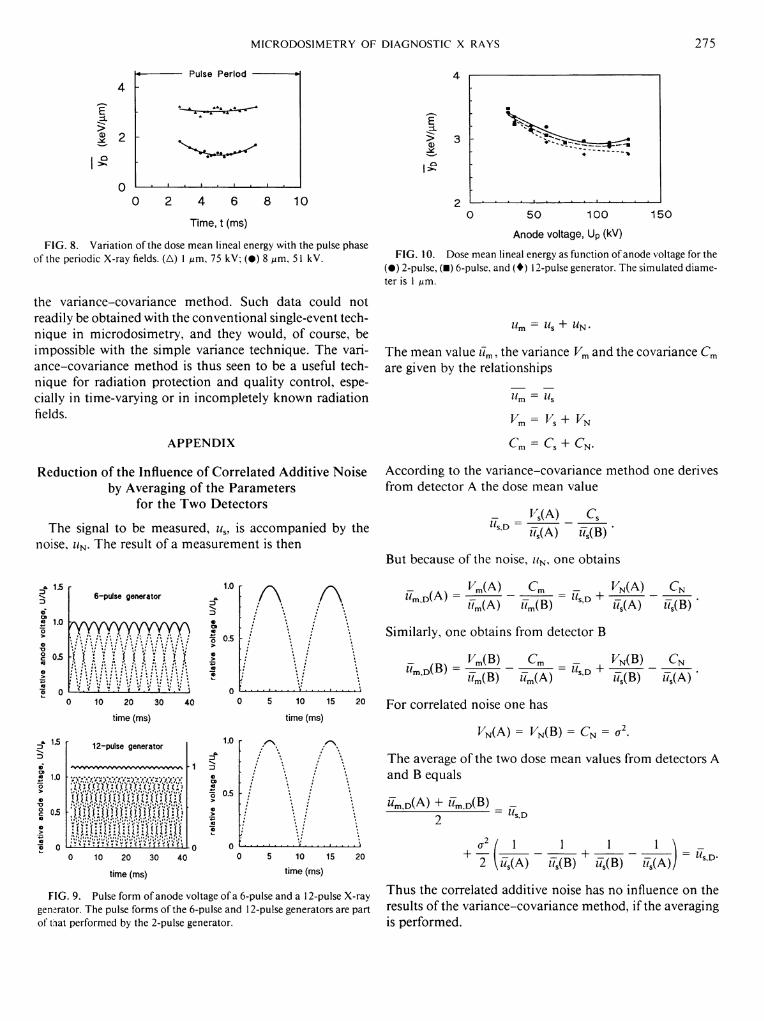
The dependence of yD on anode voltage is far more complex. It reflects the change of electron energies with photon energies, and the change in y D with changing electron energy at specified site sizes. Both dependences are nonmonotonic. For the anode voltages in the range of 30 to 125 kV and for the filter and the detector wall thickness that are employed in the present experiments the average photon energies vary from about 25 to 60 keV (9). Due to the variation of relative cross sections for the photo- and Compton effects in this range of photon energies, the average initial energy of the released electrons decreases with increasing photon energy, as seen in Fig. 6. There is then an increase in the average LET. However, yD increases only at small site sizes, while there is a decrease with decreasing electron energy at larger site sizes. Figure 4 shows all these complexities.
Due to the thickness of the filter and of the detector wall the spectra of X rays in the detector volume are very narrow for anode voltages around 35 kV, i.e., they are almost mono-
E 3 .
8
6
> 4
0
N.
• . 1 i . . . . 1
0.1 1 10
Simulated diameter (urn)
F I G . 5. The dose mean lineal energy as function of the site diameter.
274 C H E N , ROOS, A N D K E L L E R E R
1 10 10 2 10 3
Photon energy (keV)
FIG. 6. Average initial energy of the (first collision) released electrons as a function of photon energy in water.
energetic. The values of v D for these anode voltages agree well with the data for monoenergetic photons obtained by Kliauga and Dvorak (70) in single-event measurements.
j ; D AT DIFFERENT DEPTHS
Measurements of absorbed dose as a function of depth in a receptor are fundamental to dosimetry. But beyond the dose distributions one needs to consider also the variation of radiation quality with depth. It is therefore of interest to examine the dependence of the lineal energy on depth.
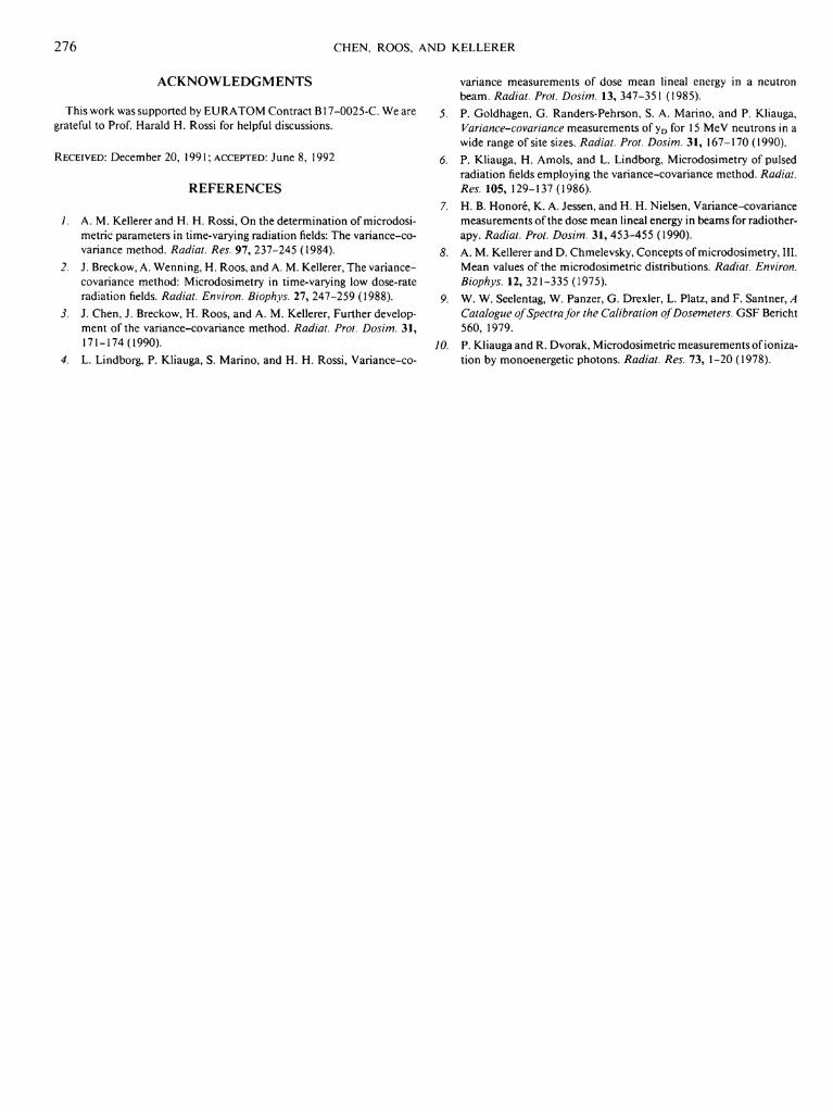
Different depths in tissue were simulated simply by placing different acrylic blocks (density 1.17 g/cm 3) in front of detectors. The thickness of the blocks was changed from 0 to 11.5 cm. Taking the detector wall (A 150, density 1.12 g/cm 3) into account, one simulates in this way depths in tissue from 1.34 to 14.8 cm. There appeared to be no need for providing backscatter beyond that taking place in the detector wall. The measurements were performed for anode voltages of 42 and 70 kV, and with a filter equivalent to 1 mm of aluminum. The results are given in Fig. 7.
For a small simulated site the dose mean lineal energy decreases somewhat with increased depth, as seen in Fig. 7. This dependence is in line with the "hardening" of the radiation quality that has been demonstrated by Hahn 2 in Monte Carlo calculations for photon energies below 100 keV.
VARIATION OF THE LINEAL ENERGY WITH PULSE PHASE
Using the temporal information in the measured data, one can determine the dose mean lineal energy for any point within the radiation pulse; one merely needs to select in the analysis the observations at this point. As examples, two such phase dependences are shown in Fig. 8.
2 K . Hahn and A. M. Kellerer, private communication, 1990.
For a simulated diameter of 1 pm and an anode voltage of 75 kV, the dose mean lineal energy does not vary substantially. This agrees with the results for 1 pm in Fig. 4, which show little change in yD with anode voltage. In contrast, one sees in Fig. 4 a significant rise in the dose mean lineal energy with anode voltages below 50 kV and at a simulated diameter of 8 / im. The anode potential is pulsed, as illustrated in Fig. 1, and the dose mean lineal energy varies in phase with the anode potential. The variations in y D are exemplified in Fig. 8 for a simulated diameter of 8 pm and for the anode voltage 51 kV; the maximal difference in yD is, in this case, about 44%.
These two examples demonstrate the potential and the diverse possibilities of the variance-covariance technique for the measurement, with high sampling frequency, of the time dependence of microdosimetric parameters in periodic radiation fields.
yD FOR DIFFERENT X-RAY GENERATORS
A 2-pulse generator exhibits variations of the anode voltage that are larger than those for 6-pulse or 12-pulse generators. Using part of the data on the voltage dependence for a 2-pulse generator one can therefore deduce the variations for 6-pulse or 12-pulse generators. As it happens, the superposition pulses for 6- or 12-pulse generators are of the same shape as subregions around the peaks of the 2-pulse generator (see Fig. 9). It is therefore particularly easy to derive the values yD for the multipulse generators by using only the suitably restricted pulse data measured for the 2-pulse generator.
Results are given in Fig. 10. The decrease in yD with anode voltage for generators of different pulse numbers is represented. More marked dependences of yD would have to be seen, i f the measurements were performed with detectors of thin wall or with wall-less counters; the role of low-energy photons would then be more important.
CONCLUSION
The measured microdosimetric data for the commonly used diagnostic X rays show the considerable potential of
4 .8 i 1
4 . 0 1 1 — 1
0 5 10 15
Equivalent depth in tissue (cm2/g)
FIG. 7. Depth dependence of the dose mean lineal energy for anode voltages of (—) 42 and (—) 70 kV. The simulated diameter is 0.5 ^m.
M I C R O D O S I M E T R Y O F D I A G N O S T I C X R A Y S 275
E
> &
Pulse Period
2 4 6 8
Time, t (ms)
10
F I G . 8. Variation of the dose mean lineal energy with the pulse phase of the periodic X-ray fields. (A) I /urn, 75 kV; ( • ) 8 urn, 51 kV.
the variance-covariance method. Such data could not readily be obtained with the conventional single-event technique in microdosimetry, and they would, of course, be impossible with the simple variance technique. The variance-covariance method is thus seen to be a useful technique for radiation protection and quality control, especially in time-varying or in incompletely known radiation fields.
APPENDIX
Reduction of the Influence of Correlated Additive Noise by Averaging of the Parameters
for the Two Detectors
The signal to be measured, us, is accompanied by the noise, uN. The result of a measurement is then
15
2 1.0
05
6-pulse generator
Willi 11 mi OS
A A
15
1.0
05
) 10 20 30 40
time (ms)
12-pulse generator
'vwwywwwwywvwvwN
10 15
time (ms)
20
1.0
3
0.5 - ;
10 20 30
time (ms)
40 10 15
time (ms)
20
F I G . 9. Pulse form of anode voltage of a 6-pulse and a 12-pulse X-ray generator. The pulse forms of the 6-pulse and 12-pulse generators are part ofthat performed by the 2-pulse generator.
E n
•—-> d)
i Q
5 0 1 0 0 1 5 0
Anode voltage, Up (kV)
F I G . 10. Dose mean lineal energy as function of anode voltage for the ( • ) 2-pulse, (•) 6-pulse, and (4)12-pulse generator. The simulated diameter is 1 jum.
Um = Us + W N .
The mean value üm, the variance Vm and the covariance C m
are given by the relationships
^ = Us
y m y s ' Y N
C m = C s + C N .
According to the variance-covariance method one derives from detector A the dose mean value
n(A) C s s D *7S(A) z7s(B) *
But because of the noise, // N , one obtains
r / A X ^m(A) C m K N (A) C N w m , D (A) = -=—T~ ~ T T — = M^D + z7m(A) ilm(B) ^ u ' w8(A) iT8(B) *
Similarly, one obtains from detector B
- / m VrJß) Cm _ F N(B) C N
*7m(B) *7m(A) s ' " i7s(B) z7s(A)
For correlated noise one has
^ N ( A ) = K N(B) = CN = a2.
The average of the two dose mean values from detectors A and B equals
ä m , D ( A ) + i7m,D(B) _ 2 ~ s D
1 1 _ 1 1 _ \ = _ 2 \ÜS(A) ÜS(B) + *7S(B) üs(A)j
Thus the correlated additive noise has no influence on the results of the variance-covariance method, i f the averaging is performed.
276 C H E N , ROOS, A N D K E L L E R E R
ACKNOWLEDGMENTS
This work was supported by E U R A T O M Contract B17-0025-C. We are grateful to Prof Harald H. Rossi for helpful discussions.
R E C E I V E D : December 20, 1991; A C C E P T E D : June 8, 1992
REFERENCES
/. A. M. Kellerer and H. H. Rossi, On the determination of microdosimetric parameters in time-varying radiation fields: The variance-covariance method. Radial. Res. 9 7 , 237-245 (1984).
2. J . Breckow, A. Wenning, H. Roos, and A. M. Kellerer, The variance-covariance method: Microdosimetry in time-varying low dose-rate radiation fields. Radial. Environ. Biophys. 2 7 , 247-259 (1988).
3. J . Chen, J . Breckow, H . Roos, and A. M. Kellerer, Further development of the variance-covariance method. Radial. Prot. Dosim. 3 1 , 171-174(1990).
4. L . Lindborg, P. Kliauga, S. Marino, and H . H . Rossi, Variance-co
variance measurements of dose mean lineal energy in a neutron beam. Radial. Prot. Dosim. 13 , 347-351 (1985).
5. P. Goldhagen, G . Randers-Pehrson, S. A. Marino, and P. Kliauga, Variance-covariance measurements of y D for 15 MeV neutrons in a wide range of site sizes. Radial. Prot. Dosim. 3 1 , 167-170 (1990).
6. P. Kliauga, H. Amols, and L . Lindborg, Microdosimetry of pulsed radiation fields employing the variance-covariance method. Radial. Res. 1 0 5 , 129-137 (1986).
7. H . B. Honore, K . A. Jessen, and H. H . Nielsen, Variance-covariance measurements of the dose mean lineal energy in beams for radiotherapy. Radial. Prot. Dosim. 3 1 , 453-455 (1990).
8. A. M. Kellerer and D. Chmelevsky, Concepts of microdosimetry, III. Mean values of the microdosimetric distributions. Radial. Environ. Biophys. 12 , 321-335 (1975).
9. W. W. Seelentag, W. Panzer, G . Drexler, L . Platz, and F. Santner, A Catalogue of Spectra for the Calibration of Dosemeters. G S F Bericht 560, 1979.
10. P. Kliauga and R. Dvorak, Microdosimetric measurements of ionization by monoenergetic photons. Radial. Res. 7 3 , 1-20 (1978).