Embed Size (px)
Citation preview
Dissertation presented to obtain a Master degree in
Biotechnology at the Universidade Nova de Lisboa,
Faculdade de Ciências e Tecnologia
Supervisor: Dr. Paula Alves,
Animal Cell Technology Unit, IBET/ITQB-UNL
Universidade Nova de Lisboa
Faculdade de Ciências e Tecnologia
Departamento de Química
Microencapsulation technology: a powerful tool for
human embryonic stem cells expansion and
cryopreservation
Cláudia Susana Pedreira Correia
Monte de Caparica, 2010


Acknowledgements
i
Acknowledgements
I would like to acknowledge all the people directly or indirectly involved in this thesis.
To Dr. Paula Alves, for giving me the opportunity to do my master thesis at the Animal Cell
Technology Unit at ITQB/IBET, for the good working conditions offered and for being a
strong example of leadership. Also, I would like to thank the chance of attending the
international symposium “Stem cells in biology and disease” as well as the iPS Cells
Technology course that positively contributed to my scientific formation.
To Prof. Susana Barreiros for always being available to help during the entire Master degree.
To Prof. António Laires for accepting to be jury of my thesis.
To Dr. Heiko Zimmermann and his group at Fraunhoffer-IBMT for kindly providing the cGMP
grade ultra high viscous alginate.
To Margarida Serra for having taught me the majority of what I learnt this year and for
always being there to help me. A deep thanks for your confidence, support, constant
encouragement and friendship. It is a pleasure to work with you and to be your friend.
To all the TCA colleagues for the good working environment. A special acknowledgment to:
Eng. Marcos Sousa and Dr. Catarina Brito, for always being available to help in anything; Rui
Tostões for the help with confocal images; Rita Malpique for helpful discussions on
cryopreservation studies; Carina Silva for the initial formation.
A todos os amigos que fiz durante os meus cinco anos académicos na FCT, pelos bons
momentos passados dentro e fora desta instituição. Um agradecimento especial à Joana, à
Cris, à Lúcia, ao Hélio, ao André e à Vera pela amizade e por me fazerem sempre rir.
Aos amigos de longa data, dos quais nunca me esqueço.
À minha cara-metade por estar sempre presente, por todo o apoio durante estes últimos
anos e por ter partilhado comigo alguns dos momentos mais felizes da minha vida.
À minha família, em particular aos meus pais a quem devo tudo. Obrigada por me terem
transmitido os vossos valores e por me terem ajudado e apoiado sempre em todos os
momentos. À minha querida avó, de quem tanto gosto, por ajudar a cuidar de nós. À minha
sis, por ser quem é, para mim um exemplo de coragem e de perseverança, e por nunca se
esquecer de mim.

Preface
ii
Preface This work was performed in the scope of the project - Integrated strategy for expansion,
neuronal differentiation and cryopreservation of human embryonic stem cells
(PTDC/BIO/72755/2006) funded by FCT (Fundação para a Ciência e Tecnologia). The main
aims of this project are:
i) Scale-up of undifferentiated hESCs production, with the goal of obtaining large
expansion of healthy undifferentiated hESCs.
ii) Optimize scalable protocols to direct and control differentiation of hESCs into
neuronal lineages.
iii) Improvement of cryopreservation protocols to successful storage and transport of
viable and genetically stable stocks of both undifferentiated and differentiated hESCs.
This thesis contributed to achieve the objectives i) and iii).
This work led to the following article:
Serra, M., Correia, C., Malpique, R., Brito, C., Bjorquist, P., Carrondo, M.J.T., and Alves, P.M.
Microencapsulation technology: a powerful tool to integrate expansion and
cryopreservation of pluripotent human embryonic stem cells. Accepted in PLoS One.
Abstract
iii
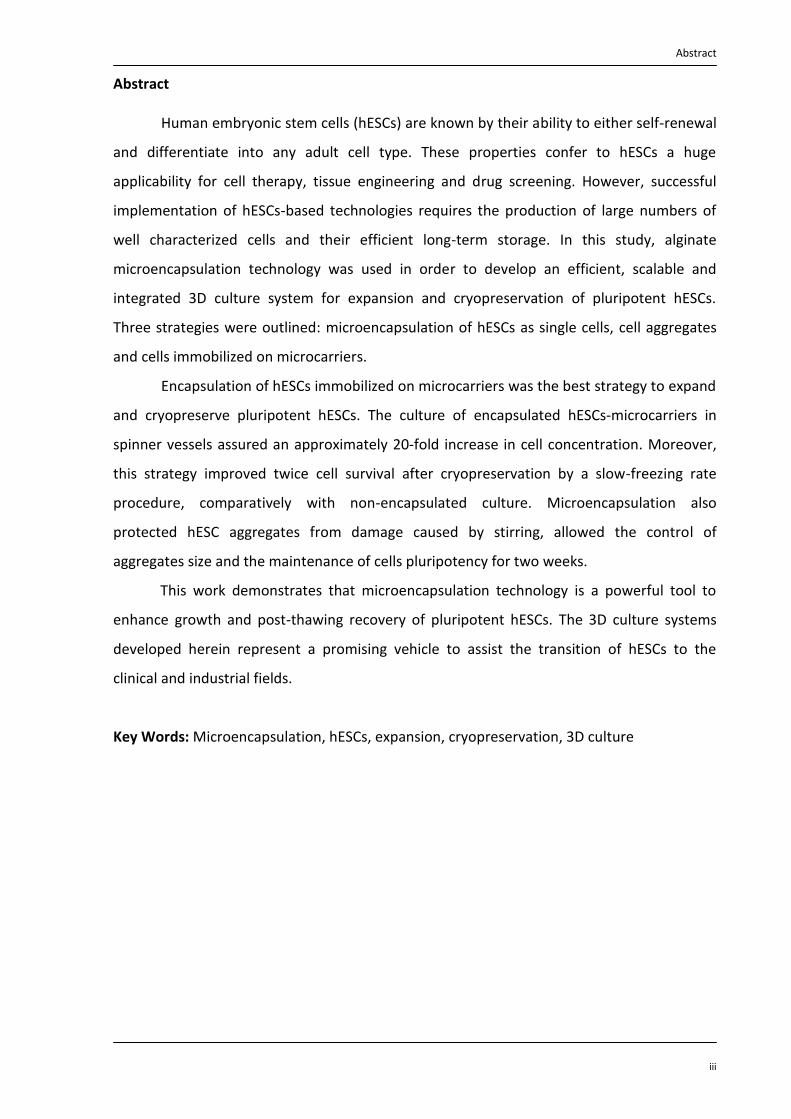
Abstract
Human embryonic stem cells (hESCs) are known by their ability to either self-renewal
and differentiate into any adult cell type. These properties confer to hESCs a huge
applicability for cell therapy, tissue engineering and drug screening. However, successful
implementation of hESCs-based technologies requires the production of large numbers of
well characterized cells and their efficient long-term storage. In this study, alginate
microencapsulation technology was used in order to develop an efficient, scalable and
integrated 3D culture system for expansion and cryopreservation of pluripotent hESCs.
Three strategies were outlined: microencapsulation of hESCs as single cells, cell aggregates
and cells immobilized on microcarriers.
Encapsulation of hESCs immobilized on microcarriers was the best strategy to expand
and cryopreserve pluripotent hESCs. The culture of encapsulated hESCs-microcarriers in
spinner vessels assured an approximately 20-fold increase in cell concentration. Moreover,
this strategy improved twice cell survival after cryopreservation by a slow-freezing rate
procedure, comparatively with non-encapsulated culture. Microencapsulation also
protected hESC aggregates from damage caused by stirring, allowed the control of
aggregates size and the maintenance of cells pluripotency for two weeks.
This work demonstrates that microencapsulation technology is a powerful tool to
enhance growth and post-thawing recovery of pluripotent hESCs. The 3D culture systems
developed herein represent a promising vehicle to assist the transition of hESCs to the
clinical and industrial fields.
Key Words: Microencapsulation, hESCs, expansion, cryopreservation, 3D culture
Resumo
iv
Resumo
As células estaminais embrionárias humanas (hESCs) são conhecidas pelas
capacidades de proliferar indefinidamente em cultura e de diferenciar em qualquer tipo de
célula adulta. Estas propriedades conferem às hESCs uma enorme aplicabilidade em terapia
celular, engenharia de tecidos e no desenvolvimento de novas drogas. No entanto, para que
a implementação de tecnologias baseadas em hESCs seja bem-sucedida, é necessário
assegurar a produção de um elevado número de células bem caracterizadas e o seu
armazenamento adequado a longo prazo. Neste estudo, a tecnologia de microencapsulação
em alginato foi utilizada para desenvolver um sistema de cultura 3D eficiente, escalonável e
integrado para a expansão e criopreservação de hESCs pluripotentes. Três estratégias foram
delineadas: encapsulação de hESCs como células individuais, encapsulação de agregados de
hESCs e encapsulação de hESCs imobilizadas em microsuportes.
A encapsulação de hESCs imobilizadas em microsuportes revelou ser a melhor
estratégia para expandir e criopreservar hESCs pluripotentes. A cultura de hESCs-
microsuportes encapsulados em vasos agitados permitiu um aumento de aproximadamente
20 vezes na concentração celular. Além disso, com esta estratégia de cultura 3D, conseguiu-
se duplicar a percentagem de sobrevivência das células, imediatamente após
criopreservação por um processo de congelamento lento, em relação à cultura não-
encapsulada. É importante realçar que a microencapsulação permitiu proteger os agregados
de hESCs dos danos causados pela agitação, controlar o tamanho dos mesmos e manter o
estado pluripotente das células por duas semanas.
Em suma, este trabalho demonstra que aplicando a tecnologia de microencapsulação
é possível melhorar o crescimento de hESCs pluripotentes e a recuperação das células após
os processos de congelamento/descongelamento. Desta forma, os sistemas de cultura 3D
aqui desenvolvidos constituem uma estratégia promissora para acelerar a transição de
hESCs para a área clínica e industrial.
Palavras-Chave: Microencapsulação, hESCs, expansão, criopreservação, cultura 3D
Abbreviations list
v
Abbreviations list ASC adult stem cells ASMA α‐smooth muscle actin bFGF basic fibroblast growth factor DAPI 4,6‐diamidino‐2‐phenylindole DMEM Dulbecco’s minimum essential medium DMSO dimethyl sulfoxide CPA cryoprotective agent EB embryoid bodies ECM extracellular matrix EG ethylene glycol FBS fetal bovine serum FDA fluoresceíne diacetate FOXA2 forkheadbox A2 HSA human serum albumin HEPES 4‐(2‐hydroxyethyl)‐1‐piperazineethanesulfonic acid hESCs human embryonic stem cells ESCs embryonic stem cells hFFs human foreskin fibroblasts ICM inner cell mass IgG immunoglobulin G IgM immunoglobulin M IVF in vitro fertilization KO-SR knockout serum replacement LN2 liquid nitrogen mEFs mouse embryonic fibroblasts mEFs-CM conditioned media in mEFs mESCs mouse embryonic stem cells NEAA non-essential aminoacids non-CM non-conditioned media PBS phosphate buffered saline Pen/Strep Penicillin/Streptomycin PI propidium iodide PFA paraformaldehyde Oct‐4 transcription factor octamer‐4 OPS open pulled straw ROCKi Rho-associated kinase (ROCK) inhibitor, Y-27632 SCED single cell enzymatic dissociation SD standard deviation SSEA‐1 stage‐specific embryonic antigen‐1 SSEA‐4 stage‐specific embryonic antigen‐4 TRA‐1‐60 tumour rejection antigen‐1‐60 TX‐100 Triton X‐100 UHV ultra‐high viscous VS vitrification solution WS warming solution
2D two dimensional
3D three dimensional
Contents
vi
Contents
Acknowledgements……………………………………………………………………………………………………………….……….…..i
Preface……………….…………………………………………………………………………………………………………..……….………..ii
Abstract…….…………………………………………………………………………………………………………………………..………....iii
Resumo…….………………………………………………………………………………………………………………………….………......iv
Abbreviations list………………………………………………………………………………………………………………………….......v
1. Introduction ......................................................................................................................................... 1
1.1. Human embryonic stem cells (hESCs) .......................................................................................... 1
1.1.1. Sources of pluripotent stem cells .......................................................................................... 1
1.1.2. Advantages of using hESCs instead ASCs or primary cells ..................................................... 4
1.1.3. hESCs applications ................................................................................................................. 4
1.1.4. Using hESCs in the clinic: critical issues ................................................................................. 5
1.2. 3D models for hESCs culture ........................................................................................................ 8
1.2.1. Culture of hESCs as aggregates ............................................................................................. 9
1.2.2. Culture of hESCs on microcarriers ....................................................................................... 10
1.3. hESCs cryopreservation .............................................................................................................. 10
1.3.1. Cryopreservation methodology ........................................................................................... 11
1.3.2. Techniques to cryopreserve hESCs ...................................................................................... 12
1.4. Cells microencapsulation ............................................................................................................ 14
1.4.1. History and the concept ...................................................................................................... 14
1.4.2. Using cell microencapsulation in the clinic: critical issues .................................................. 16
1.4.3. Microcapsule materials ....................................................................................................... 17
1.4.4. Microcapsule formation ...................................................................................................... 19
1.4.5. Microencapsulation of stem cells for clinical application ................................................... 19
1.5. Microencapsulation in stem cell bioprocessing ......................................................................... 19
1.5.1. Expansion of encapsulated hESCs ....................................................................................... 20
2. Aim of the thesis ................................................................................................................................ 24
3. Material and Methods ....................................................................................................................... 25
3.1. hESCs culture on feeder layer..................................................................................................... 25
3.2. Preparation of mEFs conditioned medium................................................................................. 25
3.3. Encapsulation of human embryonic stem cells .......................................................................... 25
3.4. Three-dimensional hESC cultures ............................................................................................... 26
3.4.1. Culture of hESCs in Erlenmeyer ........................................................................................... 26
3.4.2. Culture of hESCs in spinner vessels ..................................................................................... 27
3.5. Cell cryopreservation .................................................................................................................. 29
3.5.1. Slow Freezing Rate............................................................................................................... 29
Contents
vii
3.5.2. Vitrification .......................................................................................................................... 29
3.5.3. Assessment of hESCs survival after thawing ....................................................................... 30
3.6. Evaluation of cellular viability .................................................................................................... 30
3.7. Evaluation of the culture metabolic activity .............................................................................. 31
3.8. Evaluation of cell growth ............................................................................................................ 31
3.9. Cell characterization ................................................................................................................... 31
3.10. In vitro pluripotency test via embryoid bodies (EBs) formation .............................................. 32
4. Results ............................................................................................................................................... 33
4.1. Evaluation of two different UHVNT alginate concentrations ...................................................... 33
4.2. Strategy I – Encapsulation of hESCs as single-cells .................................................................... 33
4.2.1. Culture of encapsulated hESCs as single cells in Erlenmeyer .............................................. 33
4.3. Strategy II – Encapsulation of hESCs as aggregates ................................................................... 35
4.3.1. Culture of encapsulated hESCs as aggregates in Erlenmeyer ............................................. 35
4.3.2. Expansion of encapsulated hESC aggregates in spinner vessels ......................................... 36
4.3.3. Effect of two alginate types in expansion of hESC aggregates in spinner vessels .............. 37
4.3.4. Characterization of expanded hESC aggregates.................................................................. 38
4.3.5. Cryopreservation of encapsulated hESCs aggregates ......................................................... 40
4.4. Strategy III – Encapsulation of hESCs immobilized on microcarriers ......................................... 41
4.4.1. Culture of hESCs immobilized on microcarriers in Erlenmeyer .......................................... 41
4.4.2. Expansion of encapsulated hESCs immobilized on microcarriers in spinner vessels .......... 44
4.4.3. Characterization of expanded hESCs ................................................................................... 47
4.4.4. Cryopreservation of encapsulated hESCs immobilized on microcarriers ........................... 49
5. Discussion .......................................................................................................................................... 53
6. Conclusion ......................................................................................................................................... 61
7. References ......................................................................................................................................... 62
Introduction
1
1. Introduction
1.1. Human embryonic stem cells (hESCs)
Human embryonic stem cells (hESCs) have an enormous potential for cell-based
therapy, due to their ability to self-renew indefinitely and to differentiate into all mature cell
types of the human body (pluripotent nature). The first reports describing hESCs potential
were published in 19841 and 19942, but it was only in 1998 that Thomson and co-workers
described the isolation of hESCs from blastocysts and the creation of the first permanent and
characterized hESC lines for research3.
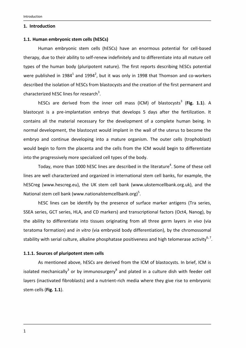
hESCs are derived from the inner cell mass (ICM) of blastocysts3 (Fig. 1.1). A
blastocyst is a pre-implantation embryo that develops 5 days after the fertilization. It
contains all the material necessary for the development of a complete human being. In
normal development, the blastocyst would implant in the wall of the uterus to become the
embryo and continue developing into a mature organism. The outer cells (trophoblast)
would begin to form the placenta and the cells from the ICM would begin to differentiate
into the progressively more specialized cell types of the body.
Today, more than 1000 hESC lines are described in the literature4. Some of these cell
lines are well characterized and organized in international stem cell banks, for example, the
hESCreg (www.hescreg.eu), the UK stem cell bank (www.ukstemcellbank.org.uk), and the
National stem cell bank (www.nationalstemcellbank.org)5.
hESC lines can be identify by the presence of surface marker antigens (Tra series,
SSEA series, GCT series, HLA, and CD markers) and transcriptional factors (Oct4, Nanog), by
the ability to differentiate into tissues originating from all three germ layers in vivo (via
teratoma formation) and in vitro (via embryoid body differentiation), by the chromossomal
stability with serial culture, alkaline phosphatase positiveness and high telomerase activity6, 7.
1.1.1. Sources of pluripotent stem cells
As mentioned above, hESCs are derived from the ICM of blastocysts. In brief, ICM is
isolated mechanically3 or by immunosurgery8 and plated in a culture dish with feeder cell
layers (inactivated fibroblasts) and a nutrient-rich media where they give rise to embryonic
stem cells (Fig. 1.1).
Microencapsulation technology: a powerful tool for hESCs expansion and cryopreservation
2
Fig. 1.1 - hESC line derivation. Main steps of the protocol for establishment of hESCs lines
9.
The main suppliers of blastocysts for stem cell
research are in vitro fertilization (IVF) clinics. The process of
IVF requires the retrieval of a woman’s eggs via a surgical
procedure after undergoing an intensive regimen of
“fertility drugs”. When IVF is used for reproductive
purposes, all of the donated eggs are fertilized in order to
maximize their chance of producing a viable blastocyst that
can be implanted. Because not all the fertilized eggs are
implanted, this has resulted in a large bank of excess
blastocysts that are currently stored for use in medical
research. Importantly, the unused blastocysts can only be
utilized for research purposes with the written informed
consent of the donors.
The creation of stem cells specifically for research
using IVF rises ethical issues because it involves the
destruction of human embryos. Accordingly, other
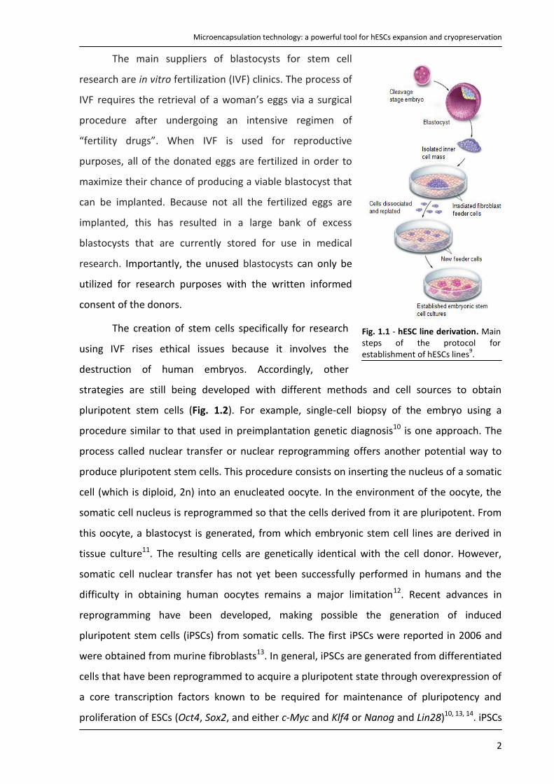
strategies are still being developed with different methods and cell sources to obtain
pluripotent stem cells (Fig. 1.2). For example, single-cell biopsy of the embryo using a
procedure similar to that used in preimplantation genetic diagnosis10 is one approach. The
process called nuclear transfer or nuclear reprogramming offers another potential way to
produce pluripotent stem cells. This procedure consists on inserting the nucleus of a somatic
cell (which is diploid, 2n) into an enucleated oocyte. In the environment of the oocyte, the
somatic cell nucleus is reprogrammed so that the cells derived from it are pluripotent. From
this oocyte, a blastocyst is generated, from which embryonic stem cell lines are derived in
tissue culture11. The resulting cells are genetically identical with the cell donor. However,
somatic cell nuclear transfer has not yet been successfully performed in humans and the
difficulty in obtaining human oocytes remains a major limitation12. Recent advances in
reprogramming have been developed, making possible the generation of induced
pluripotent stem cells (iPSCs) from somatic cells. The first iPSCs were reported in 2006 and
were obtained from murine fibroblasts13. In general, iPSCs are generated from differentiated
cells that have been reprogrammed to acquire a pluripotent state through overexpression of
a core transcription factors known to be required for maintenance of pluripotency and
proliferation of ESCs (Oct4, Sox2, and either c-Myc and Klf4 or Nanog and Lin28)10, 13, 14. iPSCs
Introduction
3
exhibit similar features to embryonic stem cells, including cell morphology, cell-surface
markers, growth properties, telomerase activity expression, and epigenetic marks of
pluripotent cell–specific genes13, 14 and can give rise to cells derived from all three germ
layers in vitro and in vivo. This technology can be used to generate patient-specific cell
types11, 15, opening the door to “personalized” cell-based therapy. However, generation of
iPSCs still suffers from low efficiency and high cost10. Furthermore the viral expression
vectors used to obtain iPSCs16, the potential for insertional mutagenis11 and the recent
knowledge that hiPSCs expresses cancer hallmarks17 have raised additional concerns
regarding the safety of these cells for clinical applications.
Fig. 1.2 – Isolation/generation, culture and differentiation of pluripotent stem cells. The development of pluripotent stem cells by A) in vitro fertilization, B) somatic cell nuclear transfer and C) generation of induced pluripotent stem cells is illustrated. Typically pluripotent cells are expanded in culture on feeder cell layers. When removed from feeders and transferred to suspension cultures, hESCs begin to form 3D aggregates of differentiated and undifferentiated cells that resemble early post-implantation embryos, named embryoid bodies. Plated cultures of embryoid bodies spontaneously display a variety of cellular types from the 3 germ lineages at various differentiation stages. Progenitors of differentiated cells can then be identified and sorted by specific markers, expression of reporter genes and characteristic morphology. Enriched culture in progenitor cells should then be cultured in a way that allowed their differentiation into more mature cell types, normally in the presence of specific growth factors. (Adapted from Brignier et al.
10).
Microencapsulation technology: a powerful tool for hESCs expansion and cryopreservation
4
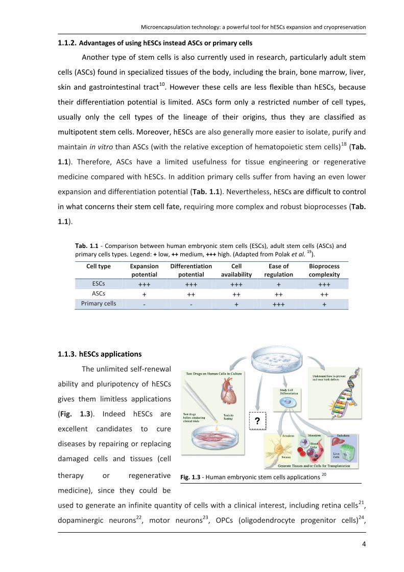
Fig. 1.3 - Human embryonic stem cells applications 20
1.1.2. Advantages of using hESCs instead ASCs or primary cells
Another type of stem cells is also currently used in research, particularly adult stem
cells (ASCs) found in specialized tissues of the body, including the brain, bone marrow, liver,
skin and gastrointestinal tract10. However these cells are less flexible than hESCs, because
their differentiation potential is limited. ASCs form only a restricted number of cell types,
usually only the cell types of the lineage of their origins, thus they are classified as
multipotent stem cells. Moreover, hESCs are also generally more easier to isolate, purify and
maintain in vitro than ASCs (with the relative exception of hematopoietic stem cells)18 (Tab.
1.1). Therefore, ASCs have a limited usefulness for tissue engineering or regenerative
medicine compared with hESCs. In addition primary cells suffer from having an even lower
expansion and differentiation potential (Tab. 1.1). Nevertheless, hESCs are difficult to control
in what concerns their stem cell fate, requiring more complex and robust bioprocesses (Tab.
1.1).
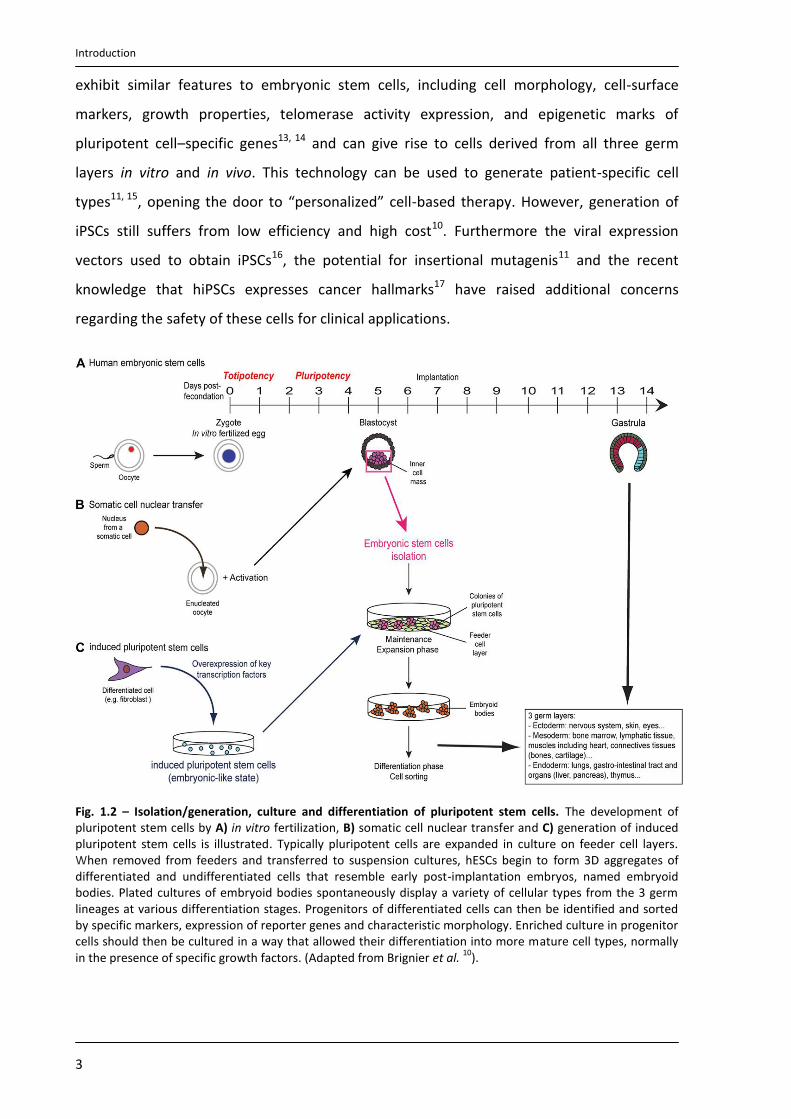
Tab. 1.1 - Comparison between human embryonic stem cells (ESCs), adult stem cells (ASCs) and primary cells types. Legend: + low, ++ medium, +++ high. (Adapted from Polak et al.
19).
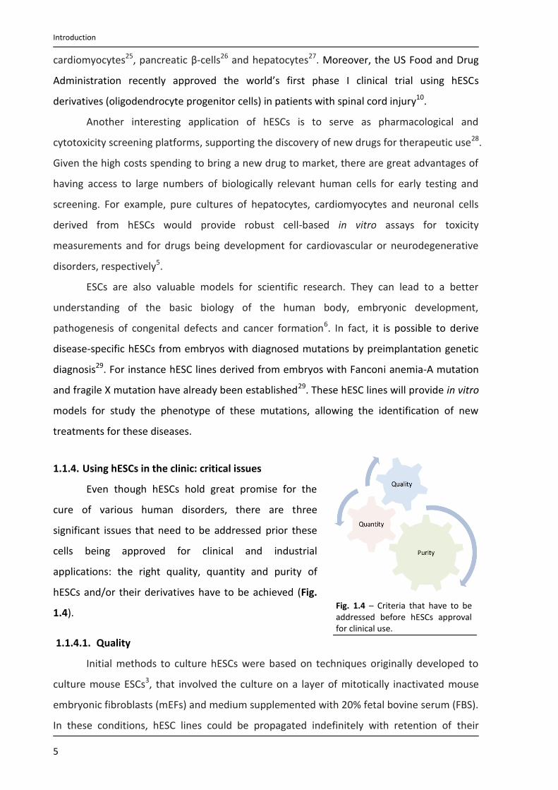
1.1.3. hESCs applications
The unlimited self-renewal
ability and pluripotency of hESCs
gives them limitless applications
(Fig. 1.3). Indeed hESCs are
excellent candidates to cure
diseases by repairing or replacing
damaged cells and tissues (cell
therapy or regenerative
medicine), since they could be
used to generate an infinite quantity of cells with a clinical interest, including retina cells21,
dopaminergic neurons22, motor neurons23, OPCs (oligodendrocyte progenitor cells)24,
Cell type Expansion potential
Differentiation potential
Cell availability
Ease of regulation
Bioprocess complexity
ESCs +++ +++ +++ + +++ ASCs + ++ ++ ++ ++
Primary cells - - + +++ +
Introduction
5
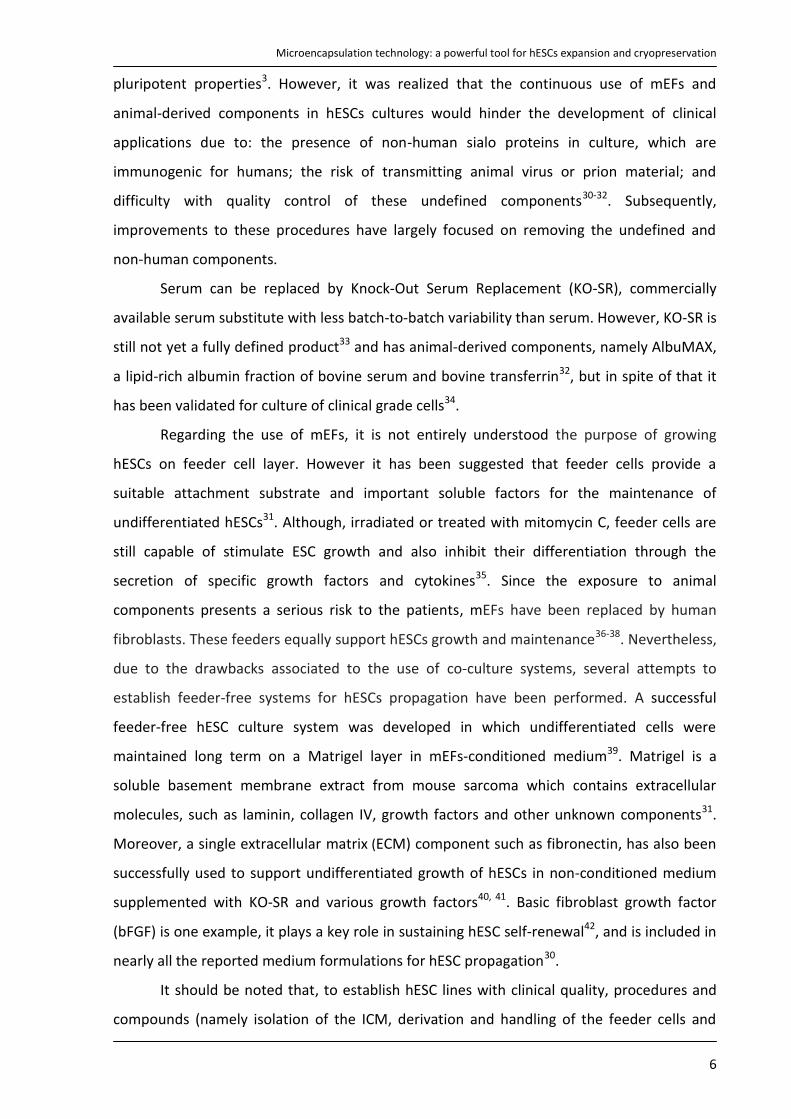
Fig. 1.4 – Criteria that have to be addressed before hESCs approval for clinical use.
cardiomyocytes25, pancreatic β-cells26 and hepatocytes27. Moreover, the US Food and Drug
Administration recently approved the world’s first phase I clinical trial using hESCs
derivatives (oligodendrocyte progenitor cells) in patients with spinal cord injury10.
Another interesting application of hESCs is to serve as pharmacological and
cytotoxicity screening platforms, supporting the discovery of new drugs for therapeutic use28.
Given the high costs spending to bring a new drug to market, there are great advantages of
having access to large numbers of biologically relevant human cells for early testing and
screening. For example, pure cultures of hepatocytes, cardiomyocytes and neuronal cells
derived from hESCs would provide robust cell-based in vitro assays for toxicity
measurements and for drugs being development for cardiovascular or neurodegenerative
disorders, respectively5.
ESCs are also valuable models for scientific research. They can lead to a better
understanding of the basic biology of the human body, embryonic development,
pathogenesis of congenital defects and cancer formation6. In fact, it is possible to derive
disease-specific hESCs from embryos with diagnosed mutations by preimplantation genetic
diagnosis29. For instance hESC lines derived from embryos with Fanconi anemia-A mutation
and fragile X mutation have already been established29. These hESC lines will provide in vitro
models for study the phenotype of these mutations, allowing the identification of new
treatments for these diseases.
1.1.4. Using hESCs in the clinic: critical issues
Even though hESCs hold great promise for the
cure of various human disorders, there are three
significant issues that need to be addressed prior these
cells being approved for clinical and industrial
applications: the right quality, quantity and purity of
hESCs and/or their derivatives have to be achieved (Fig.
1.4).
1.1.4.1. Quality
Initial methods to culture hESCs were based on techniques originally developed to
culture mouse ESCs3, that involved the culture on a layer of mitotically inactivated mouse
embryonic fibroblasts (mEFs) and medium supplemented with 20% fetal bovine serum (FBS).
In these conditions, hESC lines could be propagated indefinitely with retention of their
Microencapsulation technology: a powerful tool for hESCs expansion and cryopreservation
6
pluripotent properties3. However, it was realized that the continuous use of mEFs and
animal-derived components in hESCs cultures would hinder the development of clinical
applications due to: the presence of non-human sialo proteins in culture, which are
immunogenic for humans; the risk of transmitting animal virus or prion material; and
difficulty with quality control of these undefined components30-32. Subsequently,
improvements to these procedures have largely focused on removing the undefined and
non-human components.
Serum can be replaced by Knock-Out Serum Replacement (KO-SR), commercially
available serum substitute with less batch-to-batch variability than serum. However, KO-SR is
still not yet a fully defined product33 and has animal-derived components, namely AlbuMAX,
a lipid-rich albumin fraction of bovine serum and bovine transferrin32, but in spite of that it
has been validated for culture of clinical grade cells34.
Regarding the use of mEFs, it is not entirely understood the purpose of growing
hESCs on feeder cell layer. However it has been suggested that feeder cells provide a
suitable attachment substrate and important soluble factors for the maintenance of
undifferentiated hESCs31. Although, irradiated or treated with mitomycin C, feeder cells are
still capable of stimulate ESC growth and also inhibit their differentiation through the
secretion of specific growth factors and cytokines35. Since the exposure to animal
components presents a serious risk to the patients, mEFs have been replaced by human
fibroblasts. These feeders equally support hESCs growth and maintenance36-38. Nevertheless,
due to the drawbacks associated to the use of co-culture systems, several attempts to
establish feeder-free systems for hESCs propagation have been performed. A successful
feeder-free hESC culture system was developed in which undifferentiated cells were
maintained long term on a Matrigel layer in mEFs-conditioned medium39. Matrigel is a
soluble basement membrane extract from mouse sarcoma which contains extracellular
molecules, such as laminin, collagen IV, growth factors and other unknown components31.
Moreover, a single extracellular matrix (ECM) component such as fibronectin, has also been
successfully used to support undifferentiated growth of hESCs in non-conditioned medium
supplemented with KO-SR and various growth factors40, 41. Basic fibroblast growth factor
(bFGF) is one example, it plays a key role in sustaining hESC self-renewal42, and is included in
nearly all the reported medium formulations for hESC propagation30.
It should be noted that, to establish hESC lines with clinical quality, procedures and
compounds (namely isolation of the ICM, derivation and handling of the feeder cells and
Introduction
7
culture matrices, culture media, hESCs passaging and cryopreservation) have to minutely
follow the FDA (Food and Drug Administration) regulations43. Importantly, cell phenotype
and function should be characterized and evaluated during culture. Undifferentiated hESCs
have to maintain their pluripotency and karyotype stability after expansion while hESCs
derivatives must express markers of the specific cell lineage and be fully functional after
differentiation.
1.1.4.2. Quantity
Another important challenge is to achieve sufficient numbers of stem cell for an
effective therapy. For example, 1x10⁹ to 2x10⁹ cardiomyocytes are required to replace
damaged cardiac tissue after myocardial infarction44 and 1.3x10⁹ insulin producing β-cells
per 70-kg patient45 are needed for insulin independence after islet transplantation. To
achieve these high cell numbers scalable bioprocesses need to be developed.
The requirement of two-dimensional (2D) surfaces, such as well-plates and tissue-
culture flasks for cell attachment is the first hurdle to limit large stem cell production and
clinical applications19. Indeed there are several drawbacks related with the 2D culture
systems namely: i) low reproducibility due to the uncontrolled culture conditions, ii)
achievement of low cell yields, iii) limitations in scaling-up, iv) labor-intensive procedures, v)
inability to support complex cellular growth configurations19. Therefore, the establishment
of novel three-dimensional (3D) culture systems with a high available surface area for
cellular attachment and growth that resemble the in vivo conditions by accounting for the
cell–cell, cell–matrix and cell–growth factor interactions (see section 1.2) became a priority.
In addition, in order to improve cell density or the expansion rate of the 3D systems
cultures, bioreactors or stirred vessels that can accommodate dynamic culture conditions
have been used46, 47. Stirred suspension bioreactors makes possible to overcome the mass
transport limitations of static cultures due to the constant agitation rate promoted for
example by an impeller. However, they require careful impeller design and the delineation
of the optimum stirring speed for each culture. Distinct cell types have different
sensitivities/necessities in terms of the shear stress (force exerted over the cells due to the
flow of the media)46. Consequently, an inappropriate agitation could cause unsuitable shear
stress that can damage the cells.
Moreover, bioreactors allow the production of high cell numbers in a well-regulated
environment, with controlled oxygen, pH, temperature and nutrition supply. They are
hidrodynamically well characterized, can be easily scaled-up and the risk of contamination is
Microencapsulation technology: a powerful tool for hESCs expansion and cryopreservation
8
low46, 48. So far, fully controlled stirred tank bioreactors appear as promising candidates for
the expansion of hESCs since at least a 10-fold increase in cell density is achieved when
compared with the traditional 2D culture systems47.
Another worth mentioning aspect is that clonal efficiency of hESC is extremely low.
These cells are very sensitive to single-cell dissociation and recover poorly when plated at
clonal density after a passage30, which hinders cell passage from 2D to 3D culture system. In
fact, cell-cell interactions seem critical for efficient hESCs propagation, since it was
demonstrated that the loss of gap junctions between hESCs can increase cell apoptosis and
inhibit colony growth49. For these reasons, hESCs have been routinely passaged in cell
clumps. In order to facilitate up-scaling of hESCs culture, it was developed a new single-cell
enzymatic dissociation (SCED) culture system using recombinant protein-based TrypLE
Select50. Nevertheless, clonal survival of hESCs can also be enhanced by culturing in the
presence of Rho-associated kinase inhibitor (ROCKi)51.
1.1.4.3. Purity
The tumorigenic potential of pluripotent cells is other important hurdle in the safety
utilization of these cells. At present, protocols for the differentiation of hESCs are generally
inefficient, resulting in low differentiated cell yields and contamination by other cell types.
Of greater concern is the persistence of undifferentiated hESCs and the possibility of these
cells form malignant tumors when transplanted in the host52. Therefore the use of robust
methods for i) differentiation, ii) selection of pure populations of specialized cells and iii)
demonstration of their genetic and epigenetic stability will be essential before these cells
being used clinically10.
1.2. 3D models for hESCs culture
The proliferation and differentiation of stem cells depends largely on cell adhesion
and the provision of a three-dimensional growth environment that mimic the physiological
(in vivo) milieu in developing and adult tissues47, 53. In vivo cells microenvironment is
composed by the extra cellular matrix (ECM), soluble growth factors, cell-cell interactions
and mechanical stimuli54, 55 (Fig. 1.5). ECM provides structural support and physical
environmental for cells56. The architecture of the ECM can guide morphological changes and
cellular organization53. Specific signaling molecules on the ECM itself can affect cell behavior
and direct cell differentiation into a particular lineage57. ECM proteins bind to specific
integrin cell surface receptors, activating intracellular signaling pathways that control gene

Introduction
9
expression, cytoskeletal organization and cell morphology47. Besides that, cells constantly
remodel local ECM by degrading or synthesizing new ECM elements58.
Fig. 1.5 – Components of the cellular in vivo microenvironment. The synergy of biochemical (signaling molecules), biophysical (ECM or substrates) and mechanical (hemodynamic forces or shear forces) factors with cell-cell interactions determine the fate of pluripotent stem cells. (Adapted from Abraham et al.
55)
Thus, culturing stem cells in 3D microstructures that closely mimic stem cells native
microenvironment is imperative to enhance cells performance and fully exploit cells
potential.
hESCs are highly anchorage dependent and need a surface to attach and proliferate.
Therefore, growth of these cells in a 3D configuration (Fig. 1.6)59 requires normally either the
use of a support system, such as
microcarriers, or the formation of small
aggregates. Moreover, polymeric
matrices like alginate hydrogels can also
be a good option for hESCs cultivation
since this type of matrices better
represents the geometry, chemistry and
signaling of in vivo environment than
2D cultures60 (see section 1.5.).
1.2.1. Culture of hESCs as aggregates
The first attempts to grow ESCs in scalable suspension cultures were based on the
tendency of undifferentiated stem cells to form embryoid bodies in a feeder free or non-
adherent system61. The culture of hESCs in suspension bioreactors was first reported by
Gerecht-Nir et al.62. This group cultured EBs of hESCs in slow-turning lateral vessels (STLVs).
Fig. 1.6 – 2D and 3D systems for cultivation of human embryonic stem cells. (Adapted from Serra et al.
59)
Microencapsulation technology: a powerful tool for hESCs expansion and cryopreservation
10
The cells grew 70-fold, reaching 3.6x10⁷ cell/mL after 28 days and gave rise to cells of the
three germ layers.
hESCs differentiate extensively when cultured as aggregates61, therefore is difficult to
control the expansion of undifferentiated hESCs and their further directed differentiation
into specific cell types. Indeed, there are only a reduced number of studies that reported the
expansion of undifferentiated hESCs as aggregates (see Tab. 1.2; Page 22).
1.2.2. Culture of hESCs on microcarriers
Microcarriers are spherical particles, composed of various materials including
cellulose, glass, plastic, and polyester. They have a typical diameter of 100-250 μm and can
be compact or porous63. Currently many types of microcarriers are commercial available and
they are used for the growth of several adherent cell types. Indeed, the attributes of
microcarriers make them also attractive for culture and directed differentiation of hESCs.
First, they allow the translation of anchorage-dependent hESCs from 2D culture into
suspension culture by providing a high attachment surface. Also, microcarrier cultures are
characterized by high surface-to-volume ratio, which allows higher cell densities than 2D
cultures. More important, the available area for cell growth can be adjusted easily by
changing the amount, porosity and size of microcarriers61, 63.
Normally microcarriers have to be customized, for example, by attaching synthetic
peptides or extracellular matrix molecules (collagen, Matrigel, fibronectin…) in order to
improve the adhesion of hESCs. In fact, different types of microcarriers were already tested
to culture hESCs and some distinct results were obtained. For example, Nie et al. reported
that Cytodex 3 beads appeared to promote better attachment and viability of hESCs63,
whereas others observed that hESCs on these beads exhibited the poorest growth with little
or no recovery of viable cells after 48–72 hours64. Such discrepancies may be due to the use
of different hESC lines. Nevertheless, the adhesion efficiency of hESCs increased when the
beads were coated with Matrigel or seeded with mouse embryonic fibroblasts (mEFs)63. For
instance, our group recently demonstrates a 12-fold in yield of hESCs cultured in Cytodex3
microcarriers coated with Matrigel in 300mL perfused bioreactors fully controlled, operating
with pO2 at 30% air saturation65. See Tab. 1.2 (Page 22) for more examples.
1.3. hESCs cryopreservation
To fully exploit the potential of hESCs in medicine and research, freezing, cryostorage
and thawing technology is also of major importance.
Introduction
11
Cryopreservation is traditionally defined as the maintenance of biologics at
temperatures typically bellow the glass transition of pure water (‐132°C), at which biological
metabolism is dramatically diminished66.
An effective method for hESCs cryopreservation is critical for their use in clinical and
research applications. A suitable cryopreservation enables a good storage and transportation
of the cells between the sites of collection, processing and clinical administration67. This
makes possible the exchange of cells between research centers, which promotes scientific
collaboration and facilitates widespread use of hESCs. Also, the ability to preserve cells
permits the banking of stem cells until later use. An inefficient hESCs cryopreservation
increases time between cell storage and use in experimental or clinical settings, because of
an extended lag in establishing a viable highly populated culture following thawing. Even
though hESCs are self-renewable, aging cultures can acquire chromosomal abnormalities and
lose their differentiation potential, thus efficient procedures to preserve hESCs in low
passage numbers are indispensable.
1.3.1. Cryopreservation methodology
Typically, cryopreservation procedure involves the steps described in Fig. 1.7. First
cells are subjected to a pre‐freezing treatment in order to leave the cells in the state that
they will be frozen (e.g. single‐cell suspension, cell aggregates, adherent cell monolayers)
and the cell viability is evaluated. Second cells are transferred into the cryovessel on which
they will be frozen (e.g. vial, well‐plate, straw). Before freezing, samples are loaded with a
cryoprotective agent (CPA) like DMSO or glycerol to help minimizing the injury to cells during
freezing and thawing. The cells are then frozen at the desired cooling rate to the storage
temperature at which they will be stored. After thawing CPAs are removed from the sample
by dilution. At this time post-thaw cell recovery is evaluated.
CPAs are usually classified based on their ability to diffuse across the plasm
membrane of cell in penetrating (e.g. DMSO, glycerol and 1,2‐propanediol) or non-
penetrating CPAs (hydroxyethyl starch, sucrose and polyssaccharides).
The exact mechanisms by which penetrating CPAs are able to protect cells from cryo
injury are not fully understood. However, it was proposed that penetrating CPAs reduce
colligatively the concentration of damaging electrolytes at a given subzero temperature68
and reduce the extent of the cell volume change during slow‐rate freezing and thawing69.

Microencapsulation technology: a powerful tool for hESCs expansion and cryopreservation
12
Non‐penetrating CPAs are generally relatively high molecular weight, long chain
polymers that are soluble in water and can only be taken up by cells through endocytosis or
induced processes. They are thought to act by dehydrating the cells before freezing, thereby
reducing the amount of water that the cell needs to lose to remain close to the osmotic
equilibrium during freezing70. On the other hand, trehalose and other polysaccharides
protect membranes and proteins against the destructive effects of dehydration by serving as
a substitute for structural water associated with their surface71.
Fig. 1.7 – Main steps composing of a cryopreservation procedure for mammalian cells. For each step the most relevant parameters are listed
1.3.2. Techniques to cryopreserve hESCs
Two different methods have generally been used to preserve hESCs, namely slow
freezing-rapid thawing (for larger numbers of cells) and vitrification (for smaller number of
cells)72. These procedures are based on well-established protocols developed for mESCs and
embryos, respectively73.
1.3.2.1. Slow freezing rate
The conventional slow‐freezing rate consists in freezing the samples in the presence
of a CPA at a slower cooling rate to minimize the probability of intracellular ice formation,
which is likely to damage the cells. Slow cooling leads to a better cell dehydration during the
cryopreservation process74. Briefly, hESCs colonies or single cell suspension are exposed to
the CPA solution (normally 5-20% dimethyl sulfoxide (DMSO) in culture medium or serum)
Pre-freezing processing
Cell manipulation (e.g. detachment by enzymatic and /or mechanical means, entrapment in polymeric gels) Cell state (e.g. single-cell suspension, adherent cells, cell aggregates) CPA (e.g. DMSO, glycerol, trehalose) and cryopreservation solution (e.g. serum-containing / serum-free medium, commercial solutions) Evaluation of cell viability and/or functionality Cryovessel (e.g. vial, well-plate, freezing bag, straw) Cooling rate (slow-rate freezing versus vitrification) Cooling apparatus (isopropanol-based freezing system, programmed rate freezer)
Freezing
Storage temperature (-80ºC, vapor-phase of LN2 (-130ºC to -170ºC), LN2 (-196ºC))
Storage
Warming rate (e.g. water bath, CO2 incubator) CPA dilution (single-step versus stepwise dilutions)
Thawing
Evaluation of cell recovery
Time for assessment of recovery Viability / functionality assays
Introduction
13
and placed in a programmed rate freezer or in an isopropanol bath intended to cool the
sample at 1°C/min. Slow cooling protocols are effective for the preservation of mESCs,
however, applying these to hESCs, has met serious difficulties. Normally these procedures
result in low post‐thaw survival, low plating efficiencies, high differentiation rates and loss of
pluripotency, presumably due to ice crystal formation that disrupts cell‐cell adhesion75-77.
For instance, in early studies colony recovery after thawing were very low: 16%77, 23%78 and
30%79. Trying to improve post thaw recovery it has been tested the additions of other
components to the cooling solution. For instance, extracellular matrix molecules (human
type IV collagen or laminin) and trehalose were found to improve post thaw recovery and
reduce differentiation of the colonies80, 81.
In all referred studies, hESCs were cryopreserved in small clumps in order to prevent
cell loss from apoptosis after single cell dissociation. However, recently it was reported the
cryopreservation of hESCs as single cells suspension by slow freezing rate using ROCKi. Cell
survival increased from 30.5±5.2% (absence of ROCKi) to 56.4±7.2% (presence of ROCKi)82.
1.3.2.2. Vitrification
Vitrification procedures aim at preventing ice formation throughout the sample by
applying extremely high cooling rates (>104 °C/min) together with high concentrations of
CPAs (6 ‐ 8M)83. Increasing the concentration of non-penetrating CPAs that interact strongly
with water prevents water molecules from interacting to form ice. Moreover high cooling
rates through the temperature region of potential crystallisation allows reaching the
amorphous glassy state before ice crystals have the opportunity to form84. Therefore,
samples pass directly from a liquid phase to a glassy state without suffering any nucleation.
Briefly, the method consist in a brief stepwise exposure of hESCs colonies (100–400 cells) to
two vitrification solutions of increasing CPA concentrations, in which the common
components are DMSO, EG and sucrose. The colonies are then loaded into open pulled
straws and directly plunged into liquid nitrogen. To avoid ice crystallisation during thawing,
colonies are rewarmed as rapidly as possible by direct immersion into pre-warmed culture
medium solutions containing decreasing concentration of sucrose73. High recovery rates
(>75%) are described for vitrified undifferentiated colonies75, 77, 78.
1.3.2.3. Slow freezing rate vs. vitrification of hESCs
Despite vitrification protocols yield higher post thawing recoveries and lower
differentiation rates when compared to slow-rate freezing77, 78, 80, it presents several
Microencapsulation technology: a powerful tool for hESCs expansion and cryopreservation
14
limitations. There is an increased risk of microbiological contamination and transmission of
infection associated with the use of open/non sterile straws and the direct exposure to
liquid nitrogen85. Furthermore, the process uses high concentrations of a CPA that is toxic to
cells at room temperature and is difficult to apply to bulk quantities of hESCs. Typically, each
straw will hold only 8–12 colony fragments which must be prepared, transferred through the
vitrification solutions, loaded into a straw and plunged into liquid nitrogen. The process is
extremely labor-intensive and operator-dependent, thus is difficult to reproduce73. All this
makes current vitrification protocols unsuitable for development of a scalable process and
enable its use in hESC banks67.
Using the slow freezing method, large cell numbers can be frozen in one vial, making
easy handling of bulk quantities of hESCs. Therefore, an efficient slow freezing of hESCs in
suspension (single cells or hESCs growing in 3D), which can easily and efficiently be stored in
cryovials seems to be the optimal scalable system to preserve hESCs. Recently, Nie et al.
developed a scalable method to cryopreserve hESCs in cryovials. They reported that the
cryopreservation of hESCs adherent on mEFs coated microcarriers using slow freezing
method, improved post thaw recovery when compared to standard cryopreservation of
hESCs colonies63.
1.4. Cells microencapsulation
1.4.1. History and the concept
The concept of enclosing transplantable cells within a semi-permeable polymeric
capsule to protect them from immune rejection was proposed by Chang in 196486. Twenty
years later encapsulation was successfully used to immobilize xenograft islet to aid in
glucose control for diabetes in rats. Diabetic state was corrected for several weeks87.
Many different encapsulation systems have been studied. The most commonly used
is the encapsulation of cells in microcapsules, because of their spherical shape and small size
that offers an optimal surface to volume ratio and an appropriate diffusion capacity when
compared for instance with macrocapsules or an intravascular implant88.
Microencapsulation also minimizes the risk of imunoprotection failure by using thousands of
microcapsules instead of a single large macrocapsule. Moreover, microcapsules can be
injected directly or transplantable with minimal-invasive surgery into the muscle, peritoneal
cavity, liver or elsewhere89.
Introduction
15
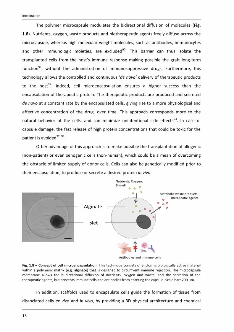
The polymer microcapsule modulates the bidirectional diffusion of molecules (Fig.
1.8). Nutrients, oxygen, waste products and biotherapeutic agents freely diffuse across the
microcapsule, whereas high molecular weight molecules, such as antibodies, immunocytes
and other immunologic moieties, are excluded90. This barrier can thus isolate the
transplanted cells from the host’s immune response making possible the graft long-term
function91, without the administration of immunosuppressive drugs. Furthermore, this
technology allows the controlled and continuous ‘de novo’ delivery of therapeutic products
to the host92. Indeed, cell microencapsulation ensures a higher success than the
encapsulation of therapeutic protein. The therapeutic products are produced and secreted
de novo at a constant rate by the encapsulated cells, giving rise to a more physiological and
effective concentration of the drug, over time. This approach corresponds more to the
natural behavior of the cells, and can minimize unintentional side effects93. In case of
capsule damage, the fast release of high protein concentrations that could be toxic for the
patient is avoided92, 93.
Other advantage of this approach is to make possible the transplantation of allogenic
(non-patient) or even xenogenic cells (non-human), which could be a mean of overcoming
the obstacle of limited supply of donor cells. Cells can also be genetically modified prior to
their encapsulation, to produce or secrete a desired protein in vivo.
Fig. 1.8 – Concept of cell microencapsulation. This technique consists of enclosing biologically active material within a polymeric matrix (e.g. alginate) that is designed to circumvent immune rejection. The microcapsule membrane allows the bi-directional diffusion of nutrients, oxygen and waste, and the secretion of the therapeutic agents, but prevents immune cells and antibodies from entering the capsule. Scale bar: 200 µm.
In addition, scaffolds used to encapsulate cells guide the formation of tissue from
dissociated cells ex vivo and in vivo, by providing a 3D physical architecture and chemical
Microencapsulation technology: a powerful tool for hESCs expansion and cryopreservation
16
environment wherein cells can grow to mimic the in vivo process, this makes the
microencapsulation technique also very attractive in regenerative medicine19.
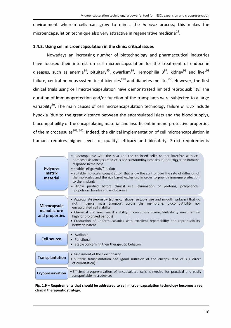
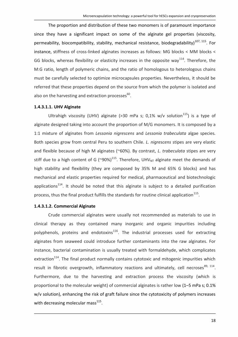
1.4.2. Using cell microencapsulation in the clinic: critical issues
Nowadays an increasing number of biotechnology and pharmaceutical industries
have focused their interest on cell microencapsulation for the treatment of endocrine
diseases, such as anemia94, pituitary95, dwarfism96, Hemophilia B97, kidney98 and liver99
failure, central nervous system insufficiencies100 and diabetes mellitus87. However, the first
clinical trials using cell microencapsulation have demonstrated limited reproducibility. The
duration of immunoprotection and/or function of the transplants were subjected to a large
variability89. The main causes of cell microencapsulation technology failure in vivo include
hypoxia (due to the great distance between the encapsulated islets and the blood supply),
biocompatibility of the encapsulating material and insufficient immune-protective properties
of the microcapsules101, 102. Indeed, the clinical implementation of cell microencapsulation in
humans requires higher levels of quality, efficacy and biosafety. Strict requirements
Fig. 1.9 – Requirements that should be addressed to cell microencapsulation technology becomes a real clinical therapeutic strategy.
Introduction
17
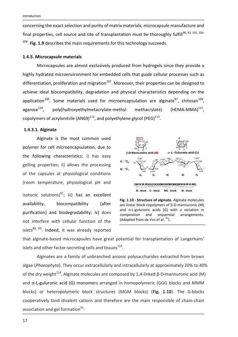
Fig. 1.10 - Structure of alginate. Alginate molecules are linear block copolymers of β-D-mannuronic (M) and α-L-guluronic acids (G) with a variation in composition and sequential arrangements. (Adapted from de Vos et al.
91).
concerning the exact selection and purity of matrix materials, microcapsule manufacture and
final properties, cell source and site of transplantation must be thoroughly fulfill90, 92, 101, 103-
106. Fig. 1.9 describes the main requirements for this technology succeeds.
1.4.3. Microcapsule materials
Microcapsules are almost exclusively produced from hydrogels since they provide a
highly hydrated microenvironment for embedded cells that guide cellular processes such as
differentiation, proliferation and migration107. Moreover, their properties can be designed to
achieve ideal biocompatibility, degradation and physical characteristics depending on the
application108. Some materials used for microencapsulation are alginate87, chitosan109,
agarose110, poly(hydroxyethylmetacrylate-methyl methacrylate) (HEMA-MMA)111,
copolymers of acrylonitrile (AN69)112, and polyethylene glycol (PEG)113.
1.4.3.1. Alginate
Alginate is the most common used
polymer for cell microencapsulation, due to
the following characteristics: i) has easy
gelling properties; ii) allows the processing
of the capsules at physiological conditions
(room temperature, physiological pH and
isotonic solutions)91; iii) has an excellent
availability, biocompatibility (after
purification) and biodegradability; iv) does
not interfere with cellular function of the
islets89, 93. Indeed, it was already reported
that alginate-based microcapsules have great potential for transplantation of Langerhans’
islets and other factor-secreting cells and tissues114.
Alginates are a family of unbranched anionic polysaccharides extracted from brown
algae (Phaeophyta). They occur extracellularly and intracellularly at approximately 20% to 40%
of the dry weight114. Alginate molecules are composed by 1,4-linked β-D-mannuronic acid (M)
and α-L-guluronic acid (G) monomers arranged in homopolymeric (GGG blocks and MMM
blocks) or heteropolymeric block structures (MGM blocks) (Fig. 1.10). The G-blocks
cooperatively bind divalent cations and therefore are the main responsible of chain-chain
association and gel formation91.
Microencapsulation technology: a powerful tool for hESCs expansion and cryopreservation
18
The proportion and distribution of these two monomers is of paramount importance
since they have a significant impact on some of the alginate gel properties (viscosity,
permeability, biocompatibility, stability, mechanical resistance, biodegradability)107, 115. For
instance, stiffness of cross-linked alginates increases as follows: MG blocks < MM blocks <
GG blocks, whereas flexibility or elasticity increases in the opposite way114. Therefore, the
M:G ratio, length of polymeric chains, and the ratio of homologous to heterologous chains
must be carefully selected to optimize microcapsules properties. Nevertheless, it should be
referred that these properties depend on the source from which the polymer is isolated and
also on the harvesting and extraction processes60.
1.4.3.1.1. UHV Alginate
Ultrahigh viscosity (UHV) alginate (>30 mPa s; 0,1% w/v solution115) is a type of
alginate designed taking into account the proportion of M/G monomers. It is composed by a
1:1 mixture of alginates from Lessonia nigrescens and Lessonia trabeculata algae species.
Both species grow from central Peru to southern Chile. L. nigrescens stipes are very elastic
and flexible because of high M alginates (~60%). By contrast, L. trabeculata stipes are very
stiff due to a high content of G (~90%)115. Therefore, UHVNT alginate meet the demands of
high stability and flexibility (they are composed by 35% M and 65% G blocks) and has
mechanical and elastic properties required for medical, pharmaceutical and biotechnologic
applications114. It should be noted that this alginate is subject to a detailed purification
process, thus the final product fulfills the standards for routine clinical application115.
1.4.3.1.2. Commercial Alginate
Crude commercial alginates were usually not recommended as materials to use in
clinical therapy as they contained many inorganic and organic impurities including
polyphenols, proteins and endotoxins116. The industrial processes used for extracting
alginates from seaweed could introduce further contaminants into the raw alginates. For
instance, bacterial contamination is usually treated with formaldehyde, which complicates
extraction114. The final product normally contains cytotoxic and mitogenic impurities which
result in fibrotic overgrowth, inflammatory reactions and ultimately, cell necroses89, 114.
Furthermore, due to the harvesting and extraction process the viscosity (which is
proportional to the molecular weight) of commercial alginates is rather low (1–5 mPa s; 0.1%
w/v solution), enhancing the risk of graft failure since the cytotoxicity of polymers increases
with decreasing molecular mass115.
Introduction
19
However, some companies have focused on the purification of alginate allowing to
guarantee clinical-grade qualities. For instance it is now possible to buy ultrapure alginates
with endotoxin levels of less than 100EU/g91 and lacking immunogenic effects. Pronova UP
MVG alginate (Pronova Biomedical) is an example. It is isolated from Laminaria hyperborea
stipe and has a high G content, a high viscosity (316 mPa s; 1% w/v solution) and a high
molecular weight (231 000 g/mole)117.
1.4.4. Microcapsule formation
Microcapsules are normally produced by forcing the alginate-cells suspension (by
using a syringe or a motor-driven prison) through a nozzle with a coaxial air jet to facilitate
break-off (Fig. 2.1). The resulting droplet is transformed into a rigid bead by gelification in an
oppositely charged, di- or trivalent ion solution. The most used cross-linkers of the polymeric
chains are Ba2+ and Ca2+ 88. Microcapsule size is controlled via the air and alginate flows. Air-
jet generators however can create “tails” during break-off that can elicit immunologic
reactions, and in some cases small air bubbles can be trapped in the alginate, limiting
diffusion and leading to poor long-term stability114. Therefore, tails and air bubbles should be
avoided during the process.
1.4.5. Microencapsulation of stem cells for clinical application
Although the availability of allogenic or xenogenic mature cells is not a major
problem, cell therapy based on encapsulated mature cells still has some drawbacks. The
time of secretion of therapeutic proteins is often limited93. Still, mature differentiated cells
cannot be expanded easily as stem cells. So the combination of stem cells and encapsulation
technology has the potential to expand the current application range of this approach.
Although most of the published works about stem cells microencapsulation are only in vitro
approaches, some in vivo studies have been developed such as the implantation of
encapsulated bone marrow mesenchymal stem cells to improve the formation of the
osseous and cartilaginous architecture118-120. Moreover, the encapsulation of these cells with
hepatocytes improved hepatocyte-specific functions in vivo121.
1.5. Microencapsulation in stem cell bioprocessing
Cell microencapsulation is an attractive tool to achieve stem cell bioprocesses issues
like control, reproducibility, validation and safety. In addition, it could be a valuable strategy
to integrate expansion, differentiation and cryopreservation of human embryonic stem cells.
Microencapsulation technology: a powerful tool for hESCs expansion and cryopreservation
20
1.5.1. Expansion of encapsulated hESCs
Cell encapsulation has been shown to be a good strategy to culture cells since it
allows higher cell survival during long-term suspension culture91. Cell encapsulation provides
a growth support for the islets, prevents excessive aggregation of free cells which can
interfere with availability of nutrients and oxygen for the cells in the core of the aggregates91
and protects cells from shear stress122. In fact, 3D cultures are normally highly dependent of
the agitation rate. Low stirring speeds result in a few oversized aggregates and/or excessive
agglomeration of microcarriers which may cause the formation of necrotic centers. More
intense agitation rates induce high shear, compromising cell viability, morphology, gene
expression and differentiation potential123. Cell encapsulation overcomes this hurdle by
protecting the cells from the damage caused by stirring. Moreover, the scaffold environment
can be customized by the incorporation of primordial tissue124, growth factors125, and small
functional groups126, to design suitable microenvironments for hESCs attachment, growth
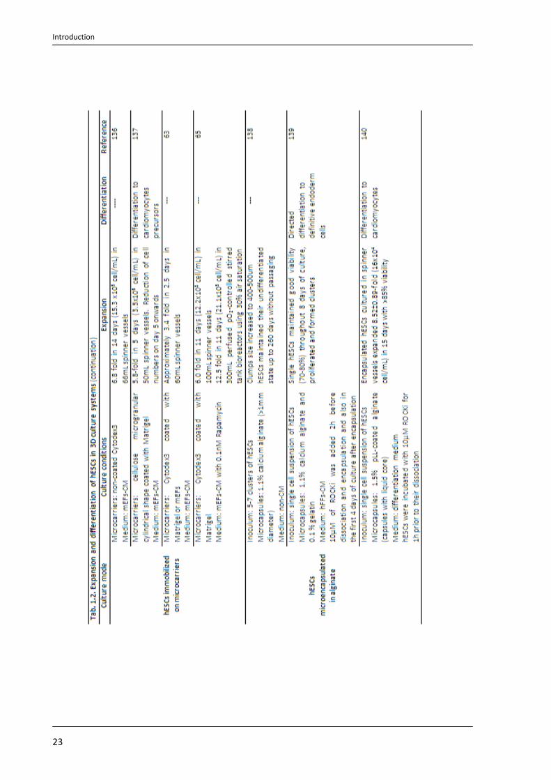
and/or differentiation. The main studies of culture hESCs encapsulated in alginate are
described in Tab. 1.2 (Page 23).
1.5.2. Cryopreservation of encapsulated cells
The numerous applications of cell microencapsulation make imperative the
development of effective cryopreservation protocols for microcapsules that would allow the
good storage of encapsulated cells until their use.
The relatively large size (500-600 μm) and fragile semipermeable membrane of the
microcapsules makes them particularly prone to cryodamage by ice crystallisation127.
Nevertheless, same studies have been reported in which encapsulated cells were
successfully cryopreserved. Stensvaag et al. cryopreserve recombinant human embryonic
kidney cells (HEK 293 cells) secreting endostatin, encapsulated in alginate beads, using a
slow freezing procedure. Cellular viability, alginate structure, and protein secretion were
maintained128. Wu et al. vitrified hepatocytes encapsulated in 2 types of engineered collagen
matrices and achieved post-thaw viabilities of 86%129. Heng et al. developed an efficient
cryopreservation medium for microencapsulated cells (2.8M (20%) DMSO and 0.25M
ssucrose) that could give high post-thaw cell viability (>95%)127. Malpique et al.
cryopreserved microencapsulated primary brain neurospheres in ultra-high viscous alginate
and enhance post-thaw cell survival and recovery without affecting neurospheres
metabolism and differentiation patterns in culture130.
Introduction
21
Although the biophysics associated with the cryopreservation process of
microencapsulated cells has not well studied, there are some advantages in using
microencapsulation in cryopreservation that could justify the good results described above.
Encapsulation of the cells avoids loss of cell-cell and cell-matrix interactions130 and reduces
the fragmentation of islets or other multicellular spheroids114 which in consequence would
lead to a decrease in cell viability after thawing. Also, the prolonged exposure of
microencapsulated cells to high cryoprotectant concentrations may not be as detrimental as
direct prolonged exposure of non-encapsulated cells, because the semipermeable
membrane would slow down or even reduce the exposure of the cells to the
cryoprotectant127. Therefore, encapsulation of hESCs could be also useful to improve their
post-thawing recovery.

Microencapsulation technology: a powerful tool for hESCs expansion and cryopreservation
22
Introduction
23
Aim of the thesis
24
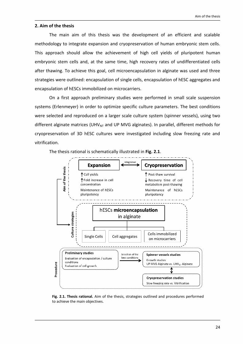
2. Aim of the thesis
The main aim of this thesis was the development of an efficient and scalable
methodology to integrate expansion and cryopreservation of human embryonic stem cells.
This approach should allow the achievement of high cell yields of pluripotent human
embryonic stem cells and, at the same time, high recovery rates of undifferentiated cells
after thawing. To achieve this goal, cell microencapsulation in alginate was used and three
strategies were outlined: encapsulation of single cells, encapsulation of hESC aggregates and
encapsulation of hESCs immobilized on microcarriers.
On a first approach preliminary studies were performed in small scale suspension
systems (Erlenmeyer) in order to optimize specific culture parameters. The best conditions
were selected and reproduced on a larger scale culture system (spinner vessels), using two
different alginate matrices (UHVNT and UP MVG alginates). In parallel, different methods for
cryopreservation of 3D hESC cultures were investigated including slow freezing rate and
vitrification.
The thesis rational is schematically illustrated in Fig. 2.1.
Fig. 2.1. Thesis rational. Aim of the thesis, strategies outlined and procedures performed to achieve the main objectives.
Material and Methods
25
3. Material and Methods
3.1. hESCs culture on feeder layer
hESCs cells (SCEDTM461, Cellartis AB) were routinely propagated as colonies in static
conditions (6 well-plates) on a feeder layer of human foreskin fibroblasts (ATCC collection,
Cat. No. CRL‐2429) inactivated with mitomycin C (ihFF) in standard culture medium (DMEM-
KO) (DMEM-KO supplemented with 20% (v/v) KO-SR, 1% (v/v) MEM-NEAA, 0.1mM 2-
mercaptoethanol, 2mM Glutamax, 1% (v/v) Pen/Strep, 0.5% (v/v) Gentamycin (all from
Invitrogen) and 10 ng/mL bFGF (Perprotech), as previously reported50. Every 10-12 days, i.e.
when the hESC colonies covered approximately 75-85% of the surface area of the culture
well, the colonies were digested with TrypLETM Select (Invitrogen) for 6‐8 min, and the
resulting single cell suspension was transferred to fresh ihFF feeders (at splitting ratios
between 1:4 and 1:24). Culture medium was replaced with fresh medium every 1–3 days.
3.2. Preparation of mEFs conditioned medium
For the production of the conditioned medium (mEFs-CM) for use in 3D hESC cultures,
mouse embryonic fibroblast (mEFs, Millipore) were mitotically inactivated and replated on
gelatin-coated T-flasks at 5.5x10⁴ cell/cm2 in DMEM-KO medium without bFGF (0.5mL/cm2).
mEFs were cultured at 37ºC with 5% (v/v) CO2 and conditionated media were collected daily
for a total of 10 days per batch. Before feeding hESCs cultures, mEFs-CM was filtered and
supplemented with 10 ng/ml bFGF and 0.1 nM Rapamycin (Sigma).
3.3. Encapsulation of human embryonic stem cells
Alginates: A 1:1 mixture of purified UHV alginates from L. nigrescens (UHVN) and L.
trabeculata (UHVT) - UHVNT alginate - was used at 0.4% or 0.7% (w/v) in 0.9% NaCl solution115,
130, 141. UP MVG alginate (Pronova UP MVG NovaMatrix) was used at 1.1% (w/v) in 0.9% NaCl
solution139.
Microcapsules formation: Microcapsules were formed by passing the alginate-cells mixture
using 1 mL syringe through an air‐jet generator (Fig. 3.1; Page 28), at an air flow rate of 2-3.5
L/min and an air pressure of 1 bar. These encapsulation parameters yielded microcapsules
with a diameter of approximately 500-700 µm. For cross‐linkage of the UHVNT alginate,
microcapsules were dropped directly from the droplet generator’s nozzle into a 20 mM BaCl2
solution, adjusted to 290 mOsm using NaCl buffered at pH 7 with 5 mM histidine. For
cross‐linkage of the UP MVG alginate, a 100 mM CaCl2/10 mM HEPES solution adjusted to pH
Microencapsulation technology: a powerful tool for hESCs expansion and cryopreservation
26
7.4 was used. Both alginates microcapsules were washed twice with 0.9% NaCl solution and
one time with DMEM-KO medium before being transferred to culture systems.
Alginate dissociation: Ba2+-UHVNT alginate was dissolved by incubating the microcapsules in a
20 mM Na2SO4 solution for 20 min at 37°C, in a humidified atmosphere of 5% CO2 in air130.
Ca2+-UP MVG alginate was dissolved by incubating the microcapsules with a chelating
solution (50 mM EDTA and 10 mM HEPES in PBS) for 5 min at 37ºC139.
3.4. Three-dimensional hESC cultures
The 3D hESC strategies and culture parameters tested are summarized in Tab. 3.1 and 3.2.
3.4.1. Culture of hESCs in Erlenmeyer
Single Cells: Before dissociation from 2D static cultures, hESCs colonies were pre-treated for
1 hour with 5 µM Y-27632, a ROCK inhibitor (ROCKi, Calbiochem). The single cell suspension,
resulted from colonies dissociation using Tryple Select, was immediately encapsulated at
different concentrations (0.75, 2 and 3 x10⁶ cell/mL alginate) using two alginate types (Tab.
3.1). In all cultures the final hESCs concentration was 1.5 x10⁶ cell/mL. hESCs-microcapsules
were then inoculated into 125 mL Erlenmeyer (Corning), cultured in 15 mL mEFs-CM
supplemented with 10 µM ROCKi, at 37ºC and 5% CO2 stirred at 70 rpm using an orbital
agitation.
hESC aggregates: hESCs were dissociated from the 2D static cultures and inoculated as single
cells at 1.5 105 cell/mL into 125 mL Erlenmeyer. Cells were cultured in 10 mL mEFs-CM
supplemented with 10 µM ROCKi, at 37ºC and 5% CO2, using an orbital agitation of 70 rpm.
Encapsulation was performed at day 2 or 5 (Tab. 3.1); aggregates were pre-treated with 5
µM ROCKi for 1 hour and then transferred to 15mL falcon tubes to allow their deposition
and culture medium removal. The encapsulation procedure was done after ressuspension of
the aggregates with equal volume of alginate solution. Microcapsules were cultured in
Erlenmeyers, with 15 mL of mEFs-CM supplemented with 10 µM ROCKi.
hESCs immobilized on microcarriers: hESCs were inoculated at 4.5 105 cell/mL into 125 mL
Erlenmeyer containing 3 g/L (dry weight) Cytodex3 microcarriers (GE Healthcare) coated
with Matrigel (BD Biosciences) or human plasma fibronectin (Millipore). The microcarriers
were prepared and sterilized according to the manufacture’s recommendation and coated
with Matrigel and fibronectin as described in the literature63, 65, 135, 137. Cells and supports
were inoculated in 4 mL mEFs-CM/non-CM supplemented with 10 µM ROCKi and
Material and Methods
27
Erlenmeyers were placed inside an incubator (37ºC, 5%CO2) under intermittent stirring in
order to obtain a homogeneous cell distribution on the supports. After 6h, 2.5 mL of fresh
medium was added to cultures and agitation rate set to 70 rpm. By day 3, the volume was
adjusted to 15 mL. The encapsulation was performed at different time points (Tab. 3.2)
using the same procedure described for aggregate cultures.
In all strategies, non-encapsulated cells culture was performed and run in parallel using the
same culture conditions. The media were only partially (50%) replaced when necessary.
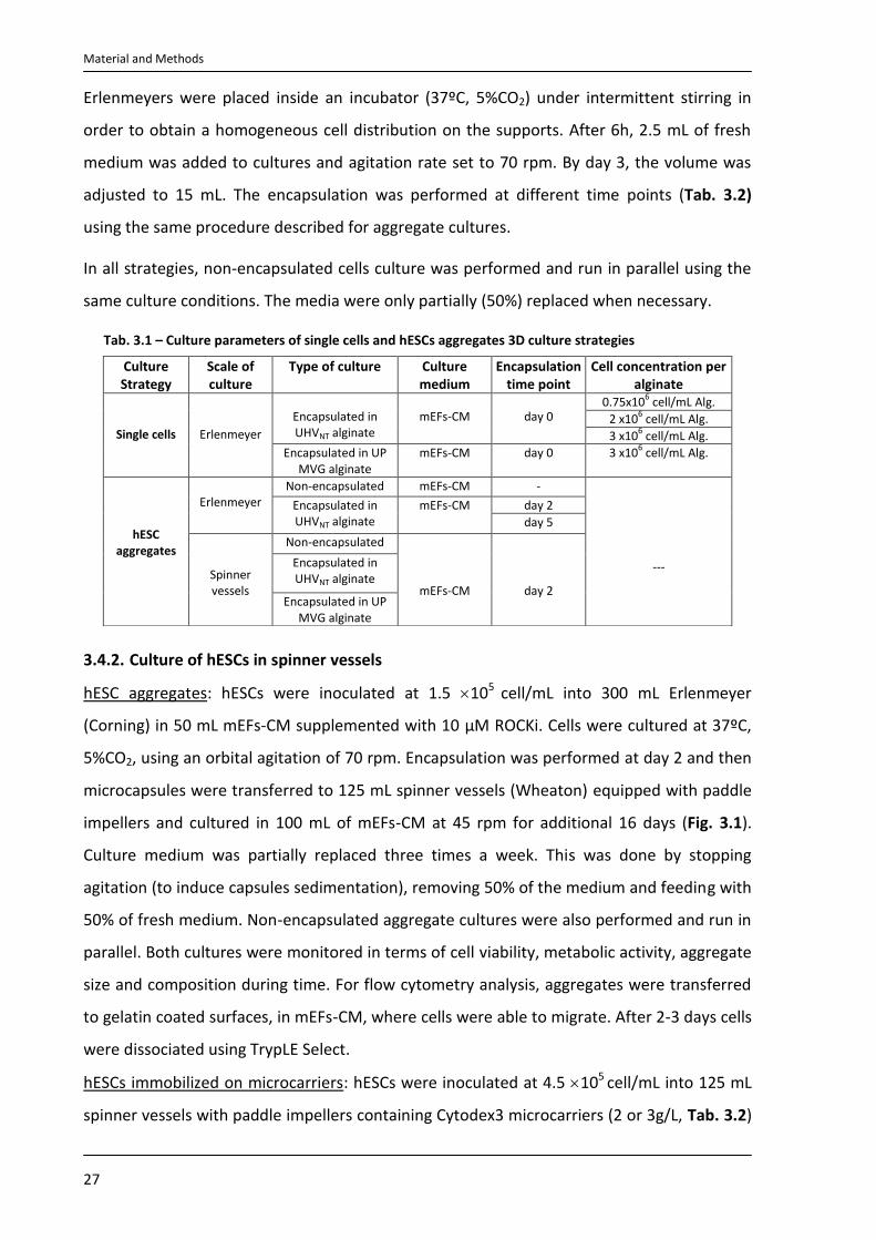
Tab. 3.1 – Culture parameters of single cells and hESCs aggregates 3D culture strategies
3.4.2. Culture of hESCs in spinner vessels
hESC aggregates: hESCs were inoculated at 1.5 105 cell/mL into 300 mL Erlenmeyer
(Corning) in 50 mL mEFs-CM supplemented with 10 µM ROCKi. Cells were cultured at 37ºC,
5%CO2, using an orbital agitation of 70 rpm. Encapsulation was performed at day 2 and then
microcapsules were transferred to 125 mL spinner vessels (Wheaton) equipped with paddle
impellers and cultured in 100 mL of mEFs-CM at 45 rpm for additional 16 days (Fig. 3.1).
Culture medium was partially replaced three times a week. This was done by stopping
agitation (to induce capsules sedimentation), removing 50% of the medium and feeding with
50% of fresh medium. Non-encapsulated aggregate cultures were also performed and run in
parallel. Both cultures were monitored in terms of cell viability, metabolic activity, aggregate
size and composition during time. For flow cytometry analysis, aggregates were transferred
to gelatin coated surfaces, in mEFs-CM, where cells were able to migrate. After 2-3 days cells
were dissociated using TrypLE Select.
hESCs immobilized on microcarriers: hESCs were inoculated at 4.5 105 cell/mL into 125 mL
spinner vessels with paddle impellers containing Cytodex3 microcarriers (2 or 3g/L, Tab. 3.2)
Culture Strategy
Scale of culture
Type of culture Culture medium
Encapsulation time point
Cell concentration per alginate
Single cells
Erlenmeyer
Encapsulated in UHVNT alginate
mEFs-CM
day 0 0.75x10
6 cell/mL Alg.
2 x10⁶ cell/mL Alg.
3 x10⁶ cell/mL Alg.
Encapsulated in UP MVG alginate
mEFs-CM day 0 3 x10⁶ cell/mL Alg.
hESC aggregates
Erlenmeyer
Non-encapsulated mEFs-CM -
---
Encapsulated in UHVNT alginate
mEFs-CM day 2
day 5
Spinner vessels
Non-encapsulated
mEFs-CM
day 2
Encapsulated in UHVNT alginate
Encapsulated in UP MVG alginate
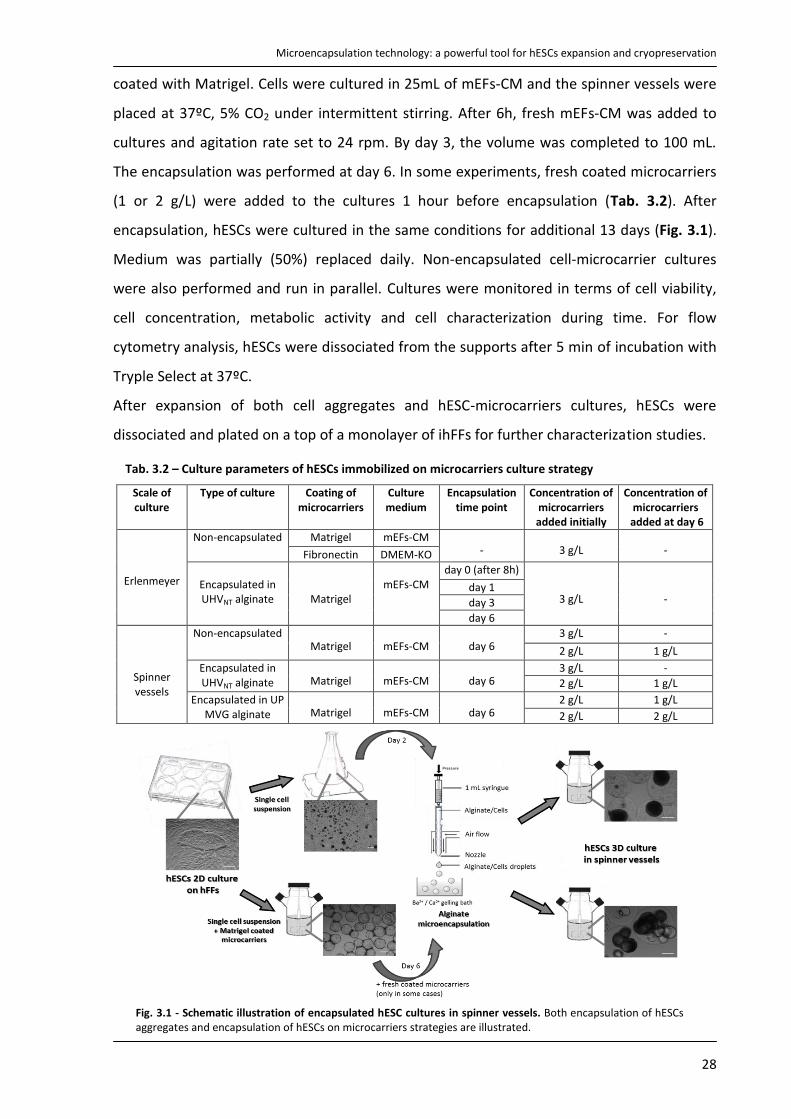
Microencapsulation technology: a powerful tool for hESCs expansion and cryopreservation
28
coated with Matrigel. Cells were cultured in 25mL of mEFs-CM and the spinner vessels were
placed at 37ºC, 5% CO2 under intermittent stirring. After 6h, fresh mEFs-CM was added to
cultures and agitation rate set to 24 rpm. By day 3, the volume was completed to 100 mL.
The encapsulation was performed at day 6. In some experiments, fresh coated microcarriers
(1 or 2 g/L) were added to the cultures 1 hour before encapsulation (Tab. 3.2). After
encapsulation, hESCs were cultured in the same conditions for additional 13 days (Fig. 3.1).
Medium was partially (50%) replaced daily. Non-encapsulated cell-microcarrier cultures
were also performed and run in parallel. Cultures were monitored in terms of cell viability,
cell concentration, metabolic activity and cell characterization during time. For flow
cytometry analysis, hESCs were dissociated from the supports after 5 min of incubation with
Tryple Select at 37ºC.
After expansion of both cell aggregates and hESC-microcarriers cultures, hESCs were
dissociated and plated on a top of a monolayer of ihFFs for further characterization studies.
Tab. 3.2 – Culture parameters of hESCs immobilized on microcarriers culture strategy
Scale of culture
Type of culture Coating of microcarriers
Culture medium
Encapsulation time point
Concentration of microcarriers added initially
Concentration of microcarriers
added at day 6
Erlenmeyer
Non-encapsulated Matrigel mEFs-CM
-
3 g/L
- Fibronectin DMEM-KO
Encapsulated in UHVNT alginate
Matrigel
mEFs-CM
day 0 (after 8h)
3 g/L
-
day 1
day 3
day 6
Spinner vessels
Non-encapsulated
Matrigel
mEFs-CM
day 6 3 g/L -
2 g/L 1 g/L
Encapsulated in UHVNT alginate
Matrigel
mEFs-CM
day 6 3 g/L -
2 g/L 1 g/L
Encapsulated in UP MVG alginate
Matrigel
mEFs-CM
day 6 2 g/L 1 g/L
2 g/L 2 g/L
Fig. 3.1 - Schematic illustration of encapsulated hESC cultures in spinner vessels. Both encapsulation of hESCs aggregates and encapsulation of hESCs on microcarriers strategies are illustrated.
Material and Methods
29
3.5. Cell cryopreservation
Cultures of non-encapsulated and encapsulated hESCs (cell aggregates and cell immobilized
on microcarriers) were harvested from the spinner vessels at days 14 and 13, respectively,
and cryopreserved using two different procedures: slow freezing rate and vitrification.
Before cryopreservation, hESCs were pre-treated with 5 µM ROCKi for one hour.
3.5.1. Slow Freezing Rate
Freezing: At the moment of freezing, after deposition of the microcapsules, culture medium
was removed and cryopreservation medium (90% KO-SR and 10% (v/v) DMSO (Sigma)
supplemented with ROCKi (5µM)) was added. Cell suspension obtained was then transferred
to cryovials (1 mL/vial). The cells were allowed to equilibrate in the cryopreservation
medium for 20 min at 4ºC. Samples were frozen to -80ºC in an isopropanol-based freezing
system, (“Mr. Frosty”, Nalgene) at a rate of cooling very close to 1ºC per minute, and stored
in the gas phase of a liquid nitrogen (LN2) reservoir until their thawing.
Thawing: Following storage, cells were quickly thawed by placing the cryovials in a 37ºC
water bath, a stepwise dilution (1:1, 1:2, 1:4) in mEFs-CM was performed immediately after
thawing in order to dilute the DMSO while reducing the osmotic shock. Cells were further
cultured for post-thaw studies (cell viability, metabolic activity and differentiation state) in
petri-dishes in mEFs-CM suppletemented with 5 M of ROCKi. Changes of media were
performed daily. Whenever possible microcapsules were dissolved, hESCs were dissociated
with TrypLE Select, transferred to ihFFs monolayers and maintained in culture for several
passages for post-thaw studies of growth and pluripotency.
3.5.2. Vitrification
Freezing: Two serum‐free vitrification solutions (VS) were used in the freezing process: VS1
included 10% DMSO and 10% ethyleneglycol (EG); VS2 contained 0.5 M sucrose, 30% DMSO
and 30% EG. Cells/capsules were incubated in VS1 for 1 min followed by VS2 for 5 seconds
and by 10 seconds in LN2. Cell strainers of 70 µm (BD) were used to transfer the
microcapsules between solutions. As these were preliminary studies the cells were thawed
immediately after the cooling process instead of being stored in the vapor‐phase of LN2.
Thawing: Two warming solutions (WS) were used in CPA dilution of the vitrified samples:
WS1, consisting of 0.4 M sucrose in VitroHES culture media (Vitrolife) and WS2, consisting of
0.1 M sucrose in VitroHES. Vitrified samples were quickly thawed by an immediate

Microencapsulation technology: a powerful tool for hESCs expansion and cryopreservation
30
incubation for 1 min in 2x diluted WS1, followed by 5 min incubation in WS2. Cells were then
cultured in mEFs-CM supplemented with 5 M of ROCKi.
3.5.3. Assessment of hESCs survival after thawing
The percentage of hESCs survival/recovery after thawing was determined by calculating the
ratio between the number of viable hESCs after cryopreservation and the number of initially
frozen viable hESCs, counted using a Fuchs-Rosenthal haemocytometer chamber (Invitrogen)
and the Trypan Blue (Invitrogen) exclusion method.
3.6. Evaluation of cell viability
Cell membrane integrity assay: The qualitative assessment of the cell plasma membrane
integrity during culture was done using the enzyme substrate fluorescein diacetate (FDA;
Sigma-Aldrich) and propidium iodide (PI; Sigma-Aldrich). Cells were incubated with 20 µg/mL
FDA and 10 µg/mL PI in PBS for 2-5 min and then visualized using fluorescence microscopy
(Leica Microsystems GmbH). FDA is a non-polar, non-fluorescent fluorescein analogue which
enters all cells freely. In viable cells, FDA is converted by intracellular esterases to highly
fluorescent fluorescein. Being highly polar, fluorescein becomes trapped within cells that
possess membrane integrity (viable cells) and confers to them green fluorescence. PI is a
polar, highly fluorescent (red color) compound which can only enter in cells that lack
membrane integrity. It intercalates into the major groove of the DNA, therefore nucleus
dying/dead cells stain red142.
Trypan Blue exclusion method: The total number of viable cells was determined by counting
the colorless cells in a Fuchs-Rosenthal haemocytometer chamber after incubation with
Trypan Blue dye (0.1% (v/v) in PBS). Dead cells with damaged membranes stain blue in the
presence of this compound.
Lactate dehydrogenase (LDH) activity: LDH activity from the culture supernatant was
dermined spectrophotometrically (at 340 nm) following the rate of oxidation of NADH to
NAD+ coupled with the reduction of pyruvate to lactate. The specific rate of LDH release
(qLDH) was calculated for each time interval using the following equaton:
, where is the change in LDH activity over the time period , and
is the average of the total cell number during the same period. The cumulative value
qLDHcum was estimated by qLDHcum i+1 = qLDH i + qLDH i+1. This assay is useful to monitor cell lysis

Material and Methods
31
throughout culture since LDH is an intracellular enzyme that is only presented in culture
supernatant when the cell membrane is damaged.
3.7. Evaluation of the culture metabolic activity
AlamarBlueTM assay: hESCs metabolic activity was assessed using the metabolic indicator
alamarBlue following the manufacture’s recommendation (Invitrogen). Briefly, 2 mL of hESC
culture were incubated over night with fresh medium containing 10% (v/v) alamarBlue.
Fluorescence was measured (excitation wavelength: 570 nm, emission wavelength: 585 nm)
in 96-well plates using a microwell plate fluorescence reader (MicroMax 384, Horiba Jobin
yvon). The active ingredient of alamarBlue (resazurin) is a nontoxic non-fluorescence
indicator dye that is converted to bright red-fluorescent resorufin via the reduction reactions
of metabolically active cells. The amount of fluorescence produced is proportional to the
number of live and metabolic active cells.
3.8. Evaluation of cell growth
Apparent growth rate (µ): µ was estimated using a simple first-order kinetic model for cell
expansion: dX/dt = µX, where t (day) is the culture time and X (cell) is the value of viable cells
for a specific t. The value of µ was estimated using this model applied to the slope of the
curves during the exponential phase.
Fold increase in cell expansion (FI): was evaluated based on the ratio XMAX/X0, where XMAX is
the peak of cell density (cell/mL) and X0 is the lowest cell density (cell/mL).
3.9. Characterization of hESCs
Alkaline Phosphatase (AP) Staining: Undifferentiated embryonic stem cells are characterized
by high expression levels of alkaline phosphatase (AP). Cultures were stained using an AP
activity detection kit (Millipore) according to the manufacturer’s instructions and observed
under phase contrast microscopy (Leica Microsystems GmbH). Positively stained hESCs
(purple) were considered undifferentiated cells.
Immunocytochemistry: hESC cultures were fixed in 4% (w/v) paraformaldehyde (PFA) in
phosphate buffered saline (PBS) for 20 min, permeabilized (only for detection of intracellular
marker OCT-4) for 5 min in 0.1% (w/v) Triton X‐100 solution (Sigma‐Aldrich) and
subsequently incubated with primary antibody overnight at 4°C. Cells were washed three
times in PBS and the secondary antibodies were applied to the cells for 60 min at room
Microencapsulation technology: a powerful tool for hESCs expansion and cryopreservation
32
temperature in the dark. After three washes with PBS, cell nuclei were counterstained with
4,6-diamidino-2-phenylindole (DAPI, Sigma-Aldrich). Cells were visualized using spinning disk
confocal (Nikon Eclipse Ti-E, confocal scanner: Yokogawa CSU-x1) and inverted fluorescence
microscopy (Leica Microsystems GmbH). In aggregate samples, an additional
permeabilization step was performed before the addition of primary antibodies; samples
were incubated with 0.2% fish skin gelatine and 0.1% TX-100 in PBS for 2 h at room
temperature. Primary antibodies used were: Tra-1-60, SSEA-4, Oct-4 (1:50, all Santa Cruz
Biotechnology) and hESCellectTM (1:1000, Cellartis AB). Secondary antibodies used were:
goat anti-mouse IgM-AlexaFluor488 and goat anti-mouse IgG-AlexaFluor 488 (1:500, all from
Invitrogen). All antibodies were diluted in PBS.
Flow cytometry analysis: cells were dissociated with TrypLETM Select and the single cell
suspension was resuspended in washing buffer (WB) solution (5% (v/v) FBS in PBS). After
two washing steps, cells were incubated with primary antibody for 1 hour at 4ºC, washed
three times in WB and then incubated with the secondary antibody for additional 30 min at
4ºC. After 2 washing steps with WB, cells were suspended in WB and analyzed in a flow
cytometry instrument (CyFlow‐space, Partec GmbH). Ten thousand events were registered
per sample. Primary antibodies used were: Tra-1-60, SSEA-4, SSEA-1 and isotype control
antibodies (1:10, all Santa Cruz Biotechnology) and hESCellectTM, hFFCellectTM (1:1000,
Cellartis AB). Secondary antibodies used were: goat anti-mouse IgM-AlexaFluor488 and goat
anti-mouse IgG-AlexaFluor 488 (1:200, all from Invitrogen). All antibodies were diluted in WB.
3.10. In vitro pluripotency test via embryoid bodies (EBs) formation
hESCs were dissociated, transferred to non-adherent petri dishes (5x10⁵ cell/mL) and
cultured in suspension for 1 week in DMEM-KO medium without bFGF. EBs formed during
this time were harvested and cultured in gelatin‐coated plates for additional 2 weeks
(medium was changed three times a week). Cells were then assessed for spontaneous
differentiation into derivatives of all three embryonic germ layer. Differentiated cells were
identified using immunocytochemistry (protocol described above) with markers for
mesoderm (α-smooth muscle actin – ASMA, DAKO 1:200), endoderm (Forkhead box A2 ‐
FOX-A2, Santa Cruz 1:500) and ectoderm (β tubulin type III - βIII‐Tub, Chemicon 1:100).
Secondary antibodies used were: goat anti-mouse IgG-AlexaFluor488 and donkey anti‐goat
IgG-AlexaFluor594 (Invitrogen, 1:500).
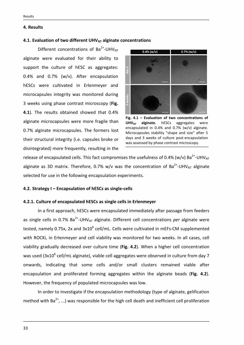
Results
33
4. Results
4.1. Evaluation of two different UHVNT alginate concentrations
Different concentrations of Ba2+-UHVNT
alginate were evaluated for their ability to
support the culture of hESC as aggregates:
0.4% and 0.7% (w/v). After encapsulation
hESCs were cultivated in Erlenmeyer and
microcapsules integrity was monitored during
3 weeks using phase contrast microscopy (Fig.
4.1). The results obtained showed that 0.4%
alginate microcapsules were more fragile than
0.7% alginate microcapsules. The formers lost
their structural integrity (i.e. capsules broke or
disintegrated) more frequently, resulting in the
release of encapsulated cells. This fact compromises the usefulness of 0.4% (w/v) Ba2+-UHVNT
alginate as 3D matrix. Therefore, 0.7% w/v was the concentration of Ba2+-UHVNT alginate
selected for use in the following encapsulation experiments.
4.2. Strategy I – Encapsulation of hESCs as single-cells
4.2.1. Culture of encapsulated hESCs as single cells in Erlenmeyer
In a first approach, hESCs were encapsulated immediately after passage from feeders
as single cells in 0.7% Ba2+-UHVNT alginate. Different cell concentrations per alginate were
tested, namely 0.75x, 2x and 3x10⁶ cell/mL. Cells were cultivated in mEFs-CM supplemented
with ROCKi, in Erlenmeyer and cell viability was monitored for two weeks. In all cases, cell
viability gradually decreased over culture time (Fig. 4.2). When a higher cell concentration
was used (3x10⁶ cell/mL alginate), viable cell aggregates were observed in culture from day 7
onwards, indicating that some cells and/or small clusters remained viable after
encapsulation and proliferated forming aggregates within the alginate beads (Fig. 4.2).
However, the frequency of populated microcapsules was low.
In order to investigate if the encapsulation methodology (type of alginate, gelification
method with Ba2+, ...) was responsible for the high cell death and inefficient cell proliferation
Fig. 4.1 – Evaluation of two concentrations of UHVNT alginate. hESCs aggregates were encapsulated in 0.4% and 0.7% (w/v) alginate. Microcapsules stability "shape and size" after 5 days and 3 weeks of culture post-encapsulation was assessed by phase contrast microscopy.
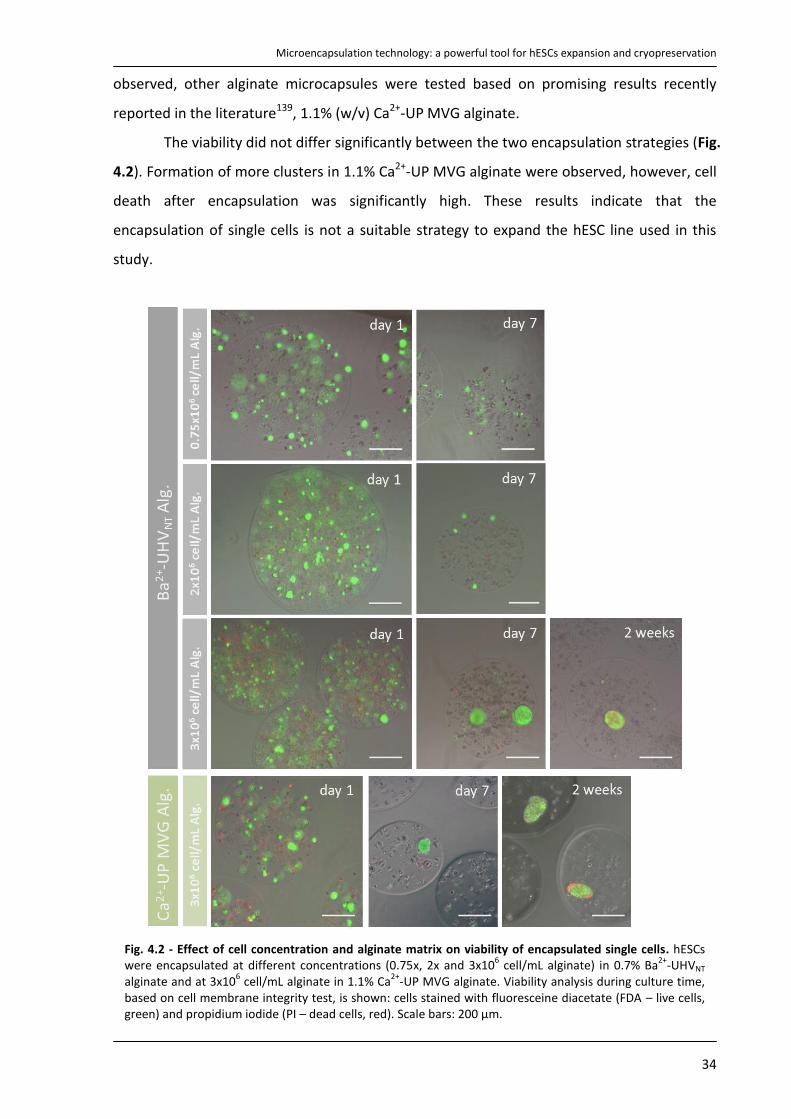
Microencapsulation technology: a powerful tool for hESCs expansion and cryopreservation
34
observed, other alginate microcapsules were tested based on promising results recently
reported in the literature139, 1.1% (w/v) Ca2+-UP MVG alginate.
The viability did not differ significantly between the two encapsulation strategies (Fig.
4.2). Formation of more clusters in 1.1% Ca2+-UP MVG alginate were observed, however, cell
death after encapsulation was significantly high. These results indicate that the
encapsulation of single cells is not a suitable strategy to expand the hESC line used in this
study.
Fig. 4.2 - Effect of cell concentration and alginate matrix on viability of encapsulated single cells. hESCs were encapsulated at different concentrations (0.75x, 2x and 3x10
6 cell/mL alginate) in 0.7% Ba
2+-UHVNT
alginate and at 3x10⁶ cell/mL alginate in 1.1% Ca
2+-UP MVG alginate. Viability analysis during culture time,
based on cell membrane integrity test, is shown: cells stained with fluoresceine diacetate (FDA – live cells, green) and propidium iodide (PI – dead cells, red). Scale bars: 200 µm.
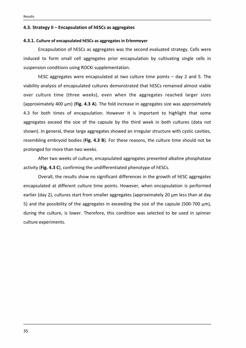
Results
35
4.3. Strategy II – Encapsulation of hESCs as aggregates
4.3.1. Culture of encapsulated hESCs as aggregates in Erlenmeyer
Encapsulation of hESCs as aggregates was the second evaluated strategy. Cells were
induced to form small cell aggregates prior encapsulation by cultivating single cells in
suspension conditions using ROCKi supplementation.
hESC aggregates were encapsulated at two culture time points – day 2 and 5. The
viability analysis of encapsulated cultures demonstrated that hESCs remained almost viable
over culture time (three weeks), even when the aggregates reached larger sizes
(approximately 400 µm) (Fig. 4.3 A). The fold increase in aggregates size was approximately
4.3 for both times of encapsulation. However it is important to highlight that some
aggregates exceed the size of the capsule by the third week in both cultures (data not
shown). In general, these large aggregates showed an irregular structure with cystic cavities,
resembling embryoid bodies (Fig. 4.3 B). For these reasons, the culture time should not be
prolonged for more than two weeks.
After two weeks of culture, encapsulated aggregates presented alkaline phosphatase
activity (Fig. 4.3 C), confirming the undifferentiated phenotype of hESCs.
Overall, the results show no significant differences in the growth of hESC aggregates
encapsulated at different culture time points. However, when encapsulation is performed
earlier (day 2), cultures start from smaller aggregates (approximately 20 µm less than at day
5) and the possibility of the aggregates in exceeding the size of the capsule (500-700 µm),
during the culture, is lower. Therefore, this condition was selected to be used in spinner
culture experiments.
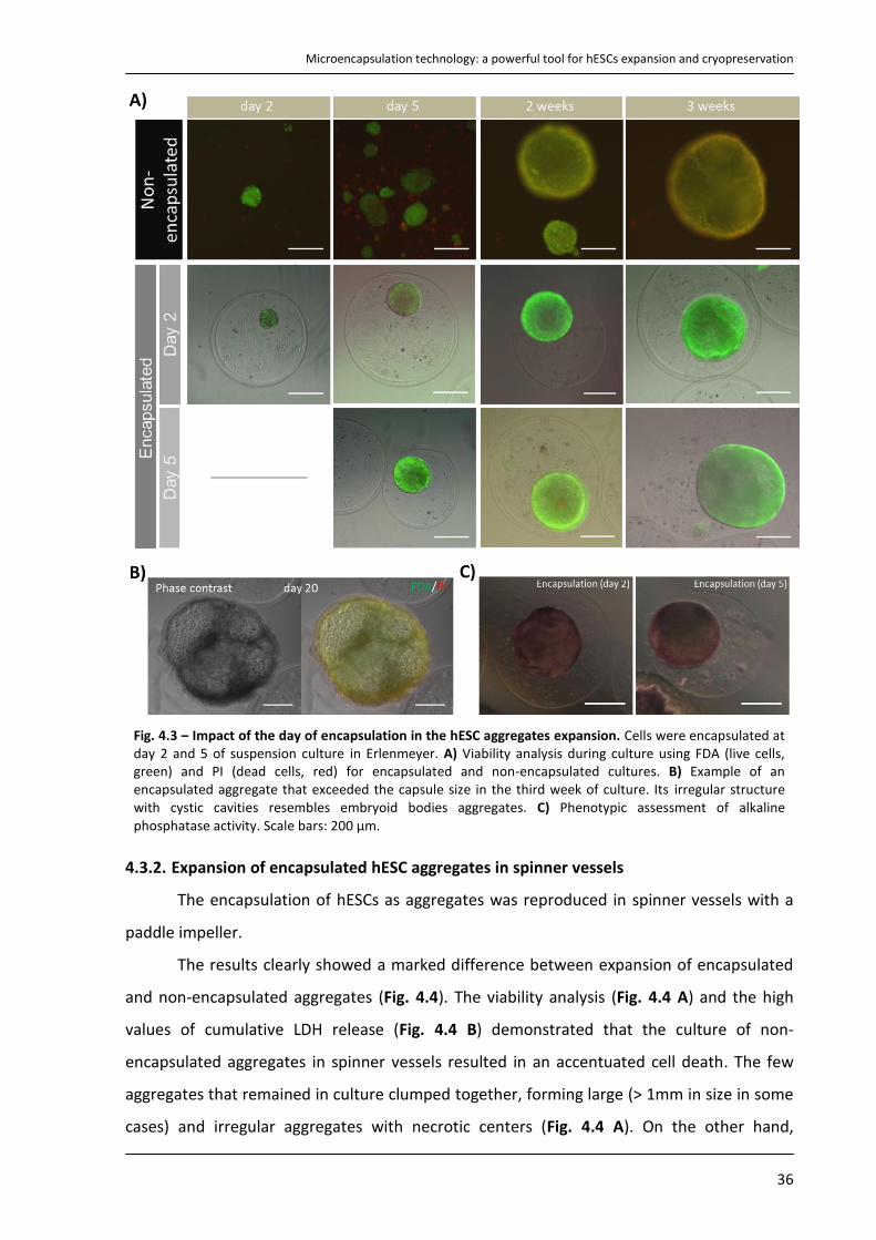
Microencapsulation technology: a powerful tool for hESCs expansion and cryopreservation
36
Fig. 4.3 – Impact of the day of encapsulation in the hESC aggregates expansion. Cells were encapsulated at day 2 and 5 of suspension culture in Erlenmeyer. A) Viability analysis during culture using FDA (live cells, green) and PI (dead cells, red) for encapsulated and non-encapsulated cultures. B) Example of an encapsulated aggregate that exceeded the capsule size in the third week of culture. Its irregular structure with cystic cavities resembles embryoid bodies aggregates. C) Phenotypic assessment of alkaline phosphatase activity. Scale bars: 200 µm.
4.3.2. Expansion of encapsulated hESC aggregates in spinner vessels
The encapsulation of hESCs as aggregates was reproduced in spinner vessels with a
paddle impeller.
The results clearly showed a marked difference between expansion of encapsulated
and non-encapsulated aggregates (Fig. 4.4). The viability analysis (Fig. 4.4 A) and the high
values of cumulative LDH release (Fig. 4.4 B) demonstrated that the culture of non-
encapsulated aggregates in spinner vessels resulted in an accentuated cell death. The few
aggregates that remained in culture clumped together, forming large (> 1mm in size in some
cases) and irregular aggregates with necrotic centers (Fig. 4.4 A). On the other hand,
Results
37
encapsulated aggregates were homogeneous, presented a spherical shape and a compact
structure throughout time (Fig. 4.4 A). The concentration of aggregates was always higher in
encapsulated cultures (Fig. 4.4 C). However, from day 14 onwards the number of
encapsulated aggregates decreased, probably due to the aggregation of small clumps within
the same capsule.
Fig. 4.4 – Effect of alginate microencapsulation on the expansion of hESC aggregates in spinner vessels. Small hESC aggregates were encapsulated at day 2 in 0.7% Ba
2+-UHVNT Alginate. A) Viability analysis of
encapsulated and non-encapsulated aggregates stained with FDA (live cells, green) and PI (dead cells, red). Scale bars: 200 µm B) Cumulative values of LDH release per aggregate during time, for encapsulated and non-encapsulated cultures. C) Number of encapsulated and non-encapsulated aggregates per volume of medium at days 3, 7, 14 and 18 of culture.
4.3.3. Effect of two alginate types in expansion of hESC aggregates in spinner vessels
The proliferation of hESCs aggregates in two types of alginate microcapsules (0.7%
Ba2+-UHVNT and 1.1% Ca2+-UP MVG) was investigated. The growth profile of encapsulated
hESCs was very similar, both in aggregate size and metabolic activity (Fig. 4.5 A, B). The fold
increase in aggregates size was 5.3 and 6.8 for aggregates encapsulated in Ba2+-UHVNT and
Ca2+-UP MVG alginate, respectively. Metabolic activity, assessed by alamarBlue test (Fig. 4.5
B), increased 2 times from the day after encapsulation (day 3) to day 15, in both

Microencapsulation technology: a powerful tool for hESCs expansion and cryopreservation
38
encapsulated cultures. In contrast, in non-encapsulated cultures the metabolic activity
decreased significantly (Fig. 4.5 B). Moreover, hESCs aggregates presented high cell
viabilities in both encapsulated cultures (Fig 4.5 C).
After microcapsules dissociation, hESCs aggregates recovered from Ba2+-UHVNT
alginate beads presented a high percentage of dead cells that compromised aggregate
integrity. On the other hand, when the UP MVG alginate was dissolved, aggregates
maintained their cohesiveness and viability (Fig. 4.5 D), thus assuring an efficient cell
recovery and characterization.
Fig. 4.5 – Impact of alginate type on the expansion of hESC aggregates in spinner vessels. Small aggregates were encapsulated at day 2 in 0.7% Ba
2+-UHVNT and in 1.1% Ca
2+-UP MVG alginates. A) Average size of
aggregates encapsulated in both alginates, during time. Error bars denote SD of up to 20 measurements. B) AlamarBlue metabolism test of non-encapsulated and encapsulated cultures on the day after encapsulation (day 3) and at day 15. Error bars denote SD of 3 measurements. C) Viability analysis of hESCs aggregates at day 9 for both encapsulated cultures. Cells were stained with FDA (live cells, green) and PI (dead cells, red). D) Viability test of hESCs aggregates immediately after dissociation. Scale bars: 200 µm
4.3.4. Characterization of expanded hESC aggregates
In order to confirm the undifferentiated state and pluripotency of aggregates during
and after culture in spinner vessels, immunofluorescence microscopy, flow cytometry
analysis, alkaline phosphatase activity test and embryoid bodies were performed (Fig. 4.6).
The results confirmed that hESCs expanded as encapsulated 3D aggregates (in UP
MVG alginate) retained their undifferentiated phenotype and pluripotency, evaluated by
immunofluorescence microscopy (Fig. 4.6 A) and detection of alkaline phosphatase activity.
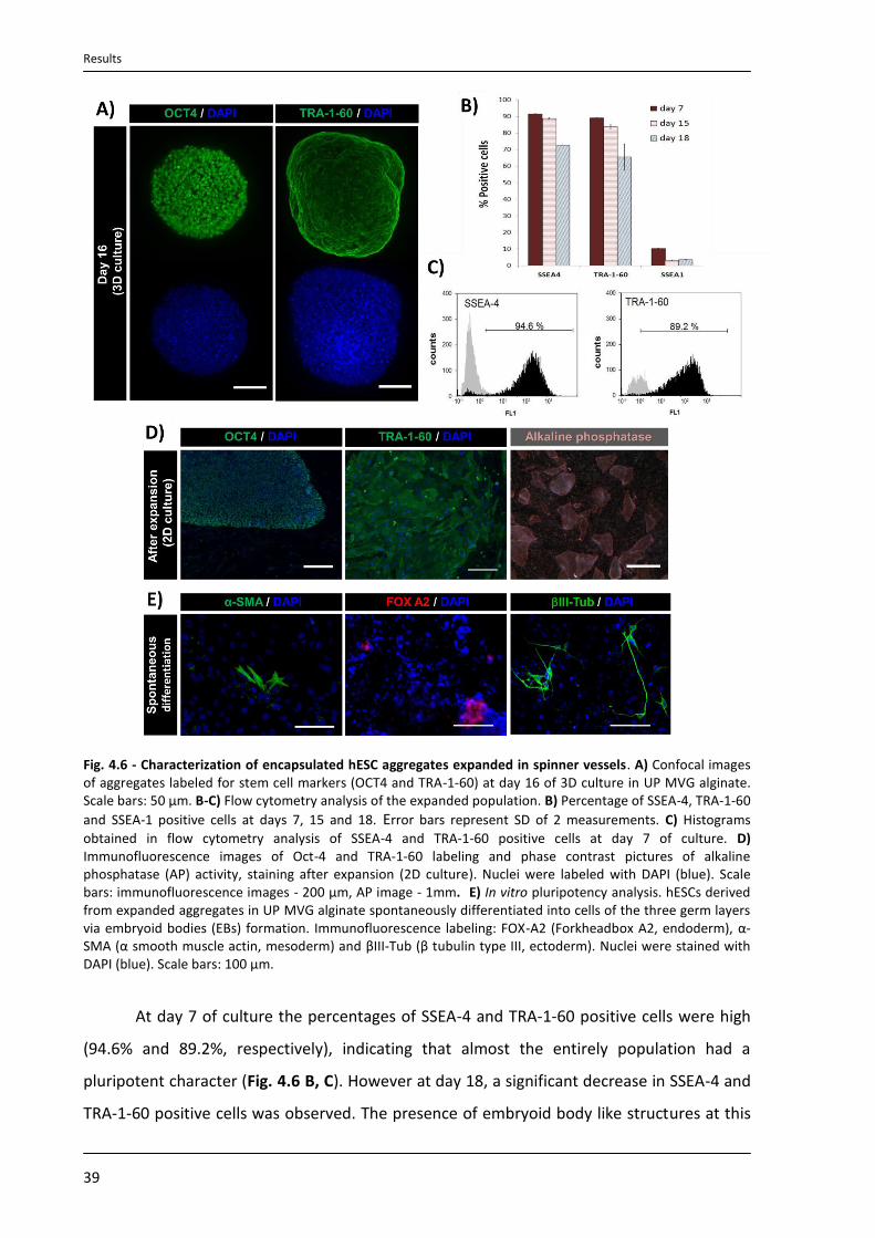
Results
39
Fig. 4.6 - Characterization of encapsulated hESC aggregates expanded in spinner vessels. A) Confocal images of aggregates labeled for stem cell markers (OCT4 and TRA-1-60) at day 16 of 3D culture in UP MVG alginate. Scale bars: 50 µm. B-C) Flow cytometry analysis of the expanded population. B) Percentage of SSEA-4, TRA-1-60
and SSEA-1 positive cells at days 7, 15 and 18. Error bars represent SD of 2 measurements. C) Histograms
obtained in flow cytometry analysis of SSEA-4 and TRA-1-60 positive cells at day 7 of culture. D) Immunofluorescence images of Oct-4 and TRA-1-60 labeling and phase contrast pictures of alkaline phosphatase (AP) activity, staining after expansion (2D culture). Nuclei were labeled with DAPI (blue). Scale bars: immunofluorescence images - 200 μm, AP image - 1mm. E) In vitro pluripotency analysis. hESCs derived from expanded aggregates in UP MVG alginate spontaneously differentiated into cells of the three germ layers via embryoid bodies (EBs) formation. Immunofluorescence labeling: FOX-A2 (Forkheadbox A2, endoderm), α-SMA (α smooth muscle actin, mesoderm) and βIII-Tub (β tubulin type III, ectoderm). Nuclei were stained with DAPI (blue). Scale bars: 100 μm.
At day 7 of culture the percentages of SSEA-4 and TRA-1-60 positive cells were high
(94.6% and 89.2%, respectively), indicating that almost the entirely population had a
pluripotent character (Fig. 4.6 B, C). However at day 18, a significant decrease in SSEA-4 and
TRA-1-60 positive cells was observed. The presence of embryoid body like structures at this
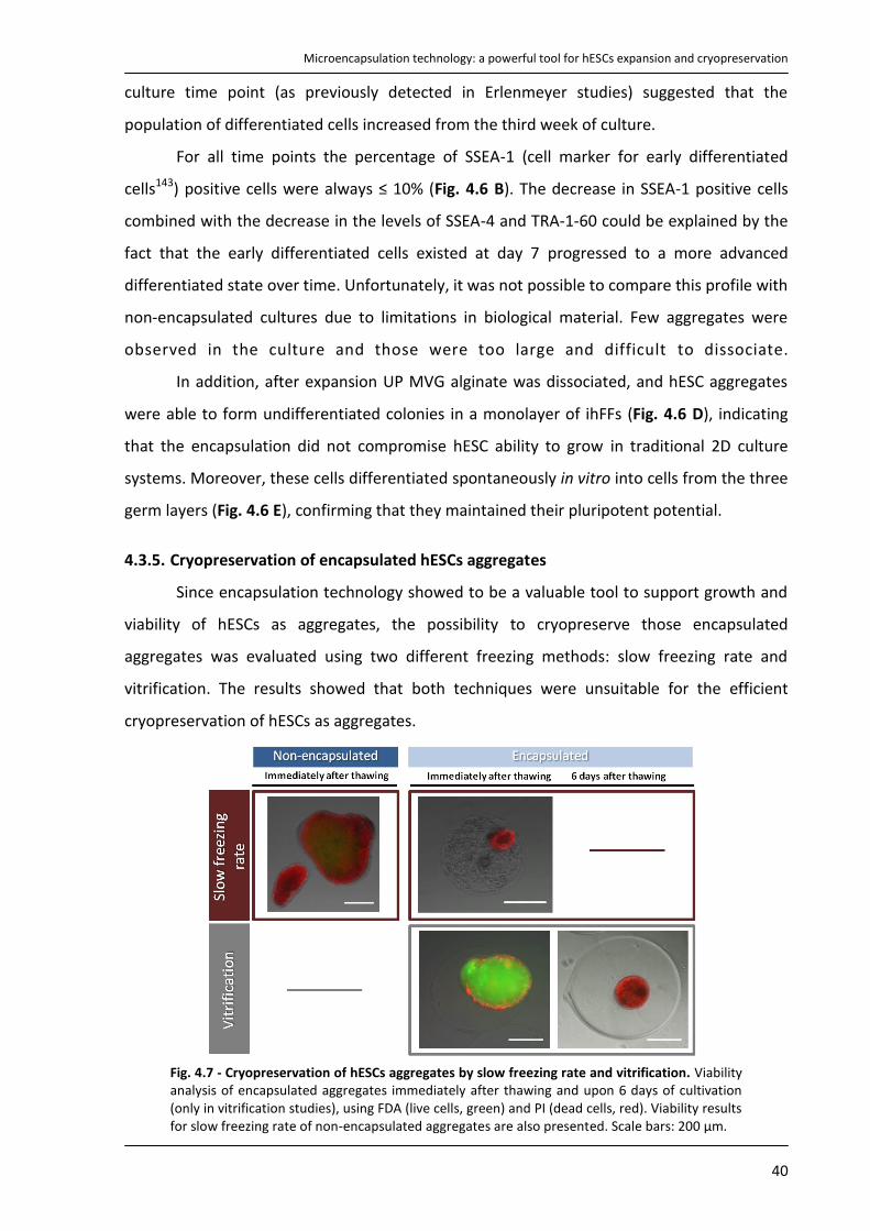
Microencapsulation technology: a powerful tool for hESCs expansion and cryopreservation
40
culture time point (as previously detected in Erlenmeyer studies) suggested that the
population of differentiated cells increased from the third week of culture.
For all time points the percentage of SSEA-1 (cell marker for early differentiated
cells143) positive cells were always ≤ 10% (Fig. 4.6 B). The decrease in SSEA-1 positive cells
combined with the decrease in the levels of SSEA-4 and TRA-1-60 could be explained by the
fact that the early differentiated cells existed at day 7 progressed to a more advanced
differentiated state over time. Unfortunately, it was not possible to compare this profile with
non-encapsulated cultures due to limitations in biological material. Few aggregates were
observed in the culture and those were too large and difficult to dissociate.
In addition, after expansion UP MVG alginate was dissociated, and hESC aggregates
were able to form undifferentiated colonies in a monolayer of ihFFs (Fig. 4.6 D), indicating
that the encapsulation did not compromise hESC ability to grow in traditional 2D culture
systems. Moreover, these cells differentiated spontaneously in vitro into cells from the three
germ layers (Fig. 4.6 E), confirming that they maintained their pluripotent potential.
4.3.5. Cryopreservation of encapsulated hESCs aggregates
Since encapsulation technology showed to be a valuable tool to support growth and
viability of hESCs as aggregates, the possibility to cryopreserve those encapsulated
aggregates was evaluated using two different freezing methods: slow freezing rate and
vitrification. The results showed that both techniques were unsuitable for the efficient
cryopreservation of hESCs as aggregates.
Fig. 4.7 - Cryopreservation of hESCs aggregates by slow freezing rate and vitrification. Viability analysis of encapsulated aggregates immediately after thawing and upon 6 days of cultivation (only in vitrification studies), using FDA (live cells, green) and PI (dead cells, red). Viability results for slow freezing rate of non-encapsulated aggregates are also presented. Scale bars: 200 μm.
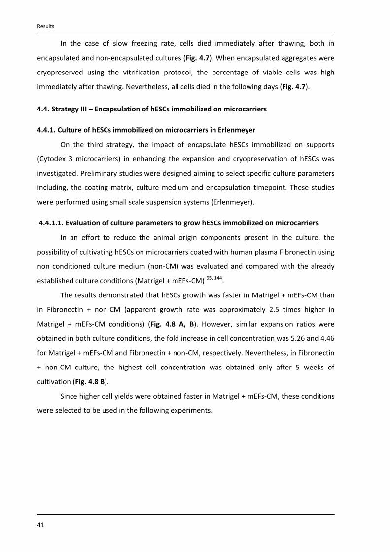
Results
41
In the case of slow freezing rate, cells died immediately after thawing, both in
encapsulated and non-encapsulated cultures (Fig. 4.7). When encapsulated aggregates were
cryopreserved using the vitrification protocol, the percentage of viable cells was high
immediately after thawing. Nevertheless, all cells died in the following days (Fig. 4.7).
4.4. Strategy III – Encapsulation of hESCs immobilized on microcarriers
4.4.1. Culture of hESCs immobilized on microcarriers in Erlenmeyer
On the third strategy, the impact of encapsulate hESCs immobilized on supports
(Cytodex 3 microcarriers) in enhancing the expansion and cryopreservation of hESCs was
investigated. Preliminary studies were designed aiming to select specific culture parameters
including, the coating matrix, culture medium and encapsulation timepoint. These studies
were performed using small scale suspension systems (Erlenmeyer).
4.4.1.1. Evaluation of culture parameters to grow hESCs immobilized on microcarriers
In an effort to reduce the animal origin components present in the culture, the
possibility of cultivating hESCs on microcarriers coated with human plasma Fibronectin using
non conditioned culture medium (non-CM) was evaluated and compared with the already
established culture conditions (Matrigel + mEFs-CM) 65, 144.
The results demonstrated that hESCs growth was faster in Matrigel + mEFs-CM than
in Fibronectin + non-CM (apparent growth rate was approximately 2.5 times higher in
Matrigel + mEFs-CM conditions) (Fig. 4.8 A, B). However, similar expansion ratios were
obtained in both culture conditions, the fold increase in cell concentration was 5.26 and 4.46
for Matrigel + mEFs-CM and Fibronectin + non-CM, respectively. Nevertheless, in Fibronectin
+ non-CM culture, the highest cell concentration was obtained only after 5 weeks of
cultivation (Fig. 4.8 B).
Since higher cell yields were obtained faster in Matrigel + mEFs-CM, these conditions
were selected to be used in the following experiments.
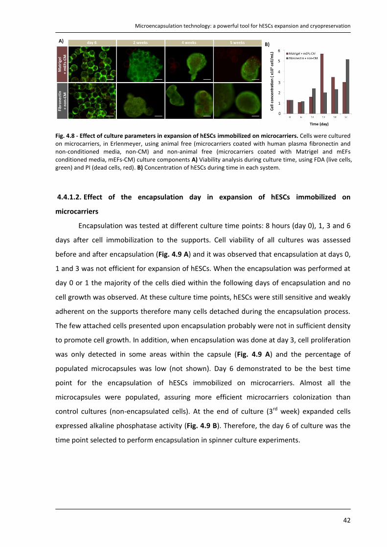
Microencapsulation technology: a powerful tool for hESCs expansion and cryopreservation
42
Fig. 4.8 - Effect of culture parameters in expansion of hESCs immobilized on microcarriers. Cells were cultured on microcarriers, in Erlenmeyer, using animal free (microcarriers coated with human plasma fibronectin and non-conditioned media, non-CM) and non-animal free (microcarriers coated with Matrigel and mEFs conditioned media, mEFs-CM) culture components A) Viability analysis during culture time, using FDA (live cells, green) and PI (dead cells, red). B) Concentration of hESCs during time in each system.
4.4.1.2. Effect of the encapsulation day in expansion of hESCs immobilized on
microcarriers
Encapsulation was tested at different culture time points: 8 hours (day 0), 1, 3 and 6
days after cell immobilization to the supports. Cell viability of all cultures was assessed
before and after encapsulation (Fig. 4.9 A) and it was observed that encapsulation at days 0,
1 and 3 was not efficient for expansion of hESCs. When the encapsulation was performed at
day 0 or 1 the majority of the cells died within the following days of encapsulation and no
cell growth was observed. At these culture time points, hESCs were still sensitive and weakly
adherent on the supports therefore many cells detached during the encapsulation process.
The few attached cells presented upon encapsulation probably were not in sufficient density
to promote cell growth. In addition, when encapsulation was done at day 3, cell proliferation
was only detected in some areas within the capsule (Fig. 4.9 A) and the percentage of
populated microcapsules was low (not shown). Day 6 demonstrated to be the best time
point for the encapsulation of hESCs immobilized on microcarriers. Almost all the
microcapsules were populated, assuring more efficient microcarriers colonization than
control cultures (non-encapsulated cells). At the end of culture (3rd week) expanded cells
expressed alkaline phosphatase activity (Fig. 4.9 B). Therefore, the day 6 of culture was the
time point selected to perform encapsulation in spinner culture experiments.
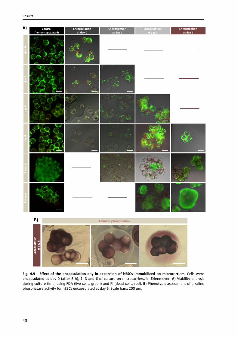
Results
43
Fig. 4.9 - Effect of the encapsulation day in expansion of hESCs immobilized on microcarriers. Cells were encapsulated at day 0 (after 8 h), 1, 3 and 6 of culture on microcarriers, in Erlenmeyer. A) Viability analysis during culture time, using FDA (live cells, green) and PI (dead cells, red). B) Phenotypic assessment of alkaline phosphatase activity for hESCs encapsulated at day 6. Scale bars: 200 µm.
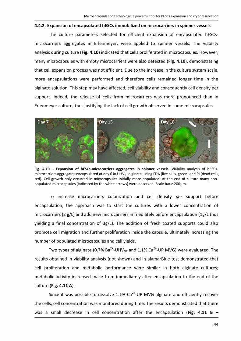
Microencapsulation technology: a powerful tool for hESCs expansion and cryopreservation
44
4.4.2. Expansion of encapsulated hESCs immobilized on microcarriers in spinner vessels
The culture parameters selected for efficient expansion of encapsulated hESCs-
microcarriers aggregates in Erlenmeyer, were applied to spinner vessels. The viability
analysis during culture (Fig. 4.10) indicated that cells proliferated in microcapsules. However,
many microcapsules with empty microcarriers were also detected (Fig. 4.10), demonstrating
that cell expansion process was not efficient. Due to the increase in the culture system scale,
more encapsulations were performed and therefore cells remained longer time in the
alginate solution. This step may have affected, cell viability and consequently cell density per
support. Indeed, the release of cells from microcarriers was more pronounced than in
Erlenmeyer culture, thus justifying the lack of cell growth observed in some microcapsules.
Fig. 4.10 – Expansion of hESCs-microcarriers aggregates in spinner vessels. Viability analysis of hESCs-microcarriers aggregates encapsulated at day 6 in UHVNT alginate, using FDA (live cells, green) and PI (dead cells, red). Cell growth only occurred in microcapsules initially more populated. At the end of culture many non-populated microcapsules (indicated by the white arrows) were observed. Scale bars: 200μm.
To increase microcarriers colonization and cell density per support before
encapsulation, the approach was to start the cultures with a lower concentration of
microcarriers (2 g/L) and add new microcarriers immediately before encapsulation (1g/L thus
yielding a final concentration of 3g/L). The addition of fresh coated supports could also
promote cell migration and further proliferation inside the capsule, ultimately increasing the
number of populated microcapsules and cell yields.
Two types of alginate (0.7% Ba2+-UHVNT and 1.1% Ca2+-UP MVG) were evaluated. The
results obtained in viability analysis (not shown) and in alamarBlue test demonstrated that
cell proliferation and metabolic performance were similar in both alginate cultures;
metabolic activity increased twice from immediately after encapsulation to the end of the
culture (Fig. 4.11 A).
Since it was possible to dissolve 1.1% Ca2+-UP MVG alginate and efficiently recover
the cells, cell concentration was monitored during time. The results demonstrated that there
was a small decrease in cell concentration after the encapsulation (Fig. 4.11 B –
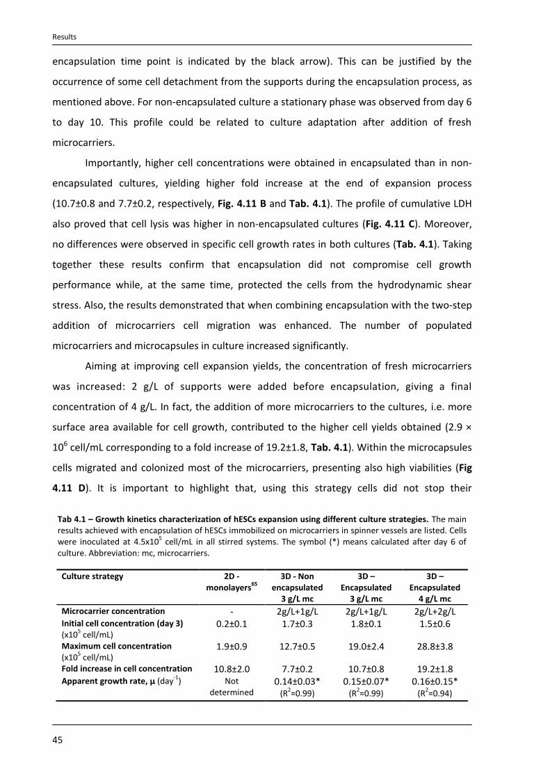
Results
45
encapsulation time point is indicated by the black arrow). This can be justified by the
occurrence of some cell detachment from the supports during the encapsulation process, as
mentioned above. For non-encapsulated culture a stationary phase was observed from day 6
to day 10. This profile could be related to culture adaptation after addition of fresh
microcarriers.
Importantly, higher cell concentrations were obtained in encapsulated than in non-
encapsulated cultures, yielding higher fold increase at the end of expansion process
(10.7±0.8 and 7.7±0.2, respectively, Fig. 4.11 B and Tab. 4.1). The profile of cumulative LDH
also proved that cell lysis was higher in non-encapsulated cultures (Fig. 4.11 C). Moreover,
no differences were observed in specific cell growth rates in both cultures (Tab. 4.1). Taking
together these results confirm that encapsulation did not compromise cell growth
performance while, at the same time, protected the cells from the hydrodynamic shear
stress. Also, the results demonstrated that when combining encapsulation with the two-step
addition of microcarriers cell migration was enhanced. The number of populated
microcarriers and microcapsules in culture increased significantly.
Aiming at improving cell expansion yields, the concentration of fresh microcarriers
was increased: 2 g/L of supports were added before encapsulation, giving a final
concentration of 4 g/L. In fact, the addition of more microcarriers to the cultures, i.e. more
surface area available for cell growth, contributed to the higher cell yields obtained (2.9 ×
106 cell/mL corresponding to a fold increase of 19.2±1.8, Tab. 4.1). Within the microcapsules
cells migrated and colonized most of the microcarriers, presenting also high viabilities (Fig
4.11 D). It is important to highlight that, using this strategy cells did not stop their
Tab 4.1 – Growth kinetics characterization of hESCs expansion using different culture strategies. The main results achieved with encapsulation of hESCs immobilized on microcarriers in spinner vessels are listed. Cells were inoculated at 4.5x10
5 cell/mL in all stirred systems. The symbol (*) means calculated after day 6 of
culture. Abbreviation: mc, microcarriers.
Culture strategy 2D -monolayers
65
3D - Non encapsulated
3 g/L mc
3D – Encapsulated
3 g/L mc
3D – Encapsulated
4 g/L mc
Microcarrier concentration - 2g/L+1g/L 2g/L+1g/L 2g/L+2g/L Initial cell concentration (day 3) (x10
5 cell/mL)
0.2±0.1 1.7±0.3 1.8±0.1 1.5±0.6
Maximum cell concentration (x10
5 cell/mL)
1.9±0.9 12.7±0.5 19.0±2.4 28.8±3.8
Fold increase in cell concentration 10.8±2.0 7.7±0.2 10.7±0.8 19.2±1.8 Apparent growth rate, μ (day
-1) Not
determined 0.14±0.03*
(R2=0.99)
0.15±0.07* (R
2=0.99)
0.16±0.15* (R
2=0.94)
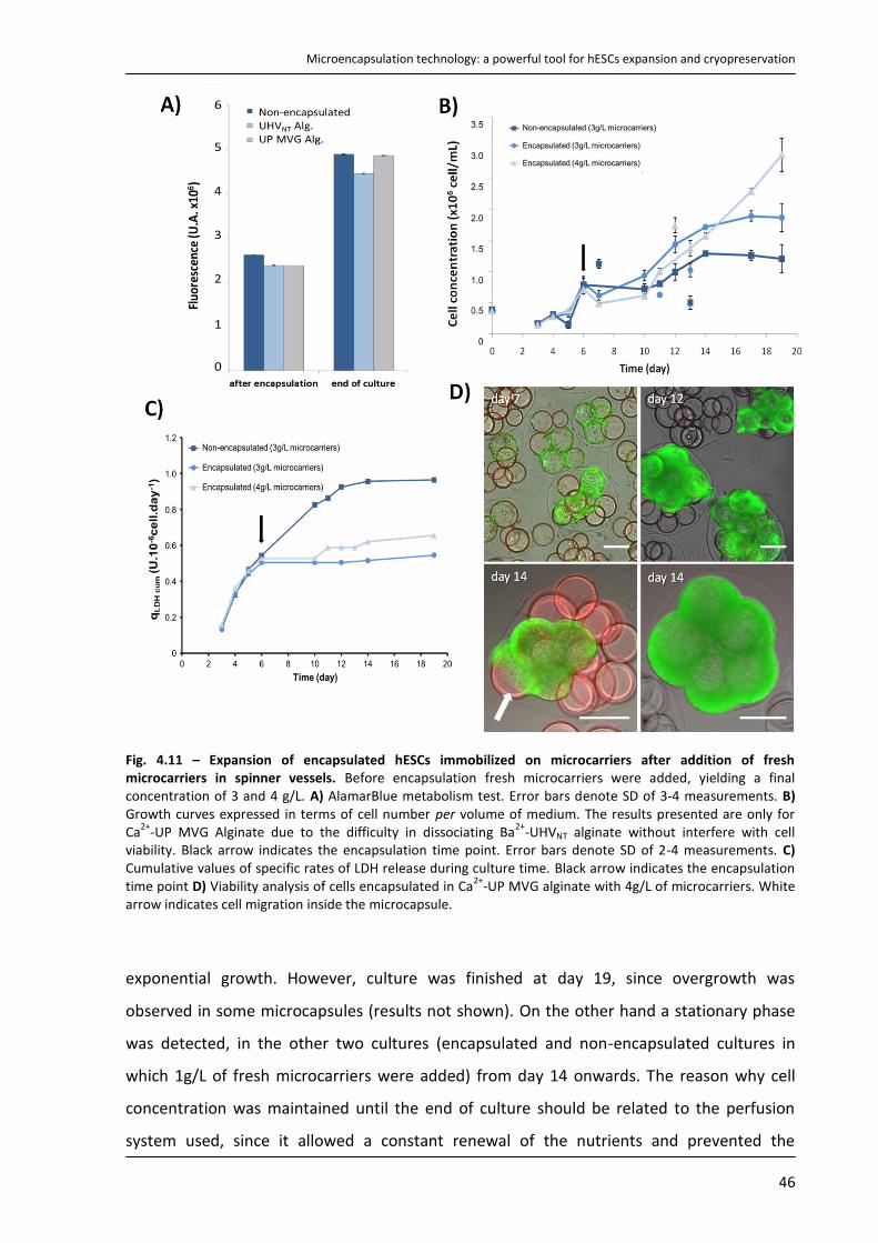
Microencapsulation technology: a powerful tool for hESCs expansion and cryopreservation
46
Fig. 4.11 – Expansion of encapsulated hESCs immobilized on microcarriers after addition of fresh microcarriers in spinner vessels. Before encapsulation fresh microcarriers were added, yielding a final concentration of 3 and 4 g/L. A) AlamarBlue metabolism test. Error bars denote SD of 3-4 measurements. B) Growth curves expressed in terms of cell number per volume of medium. The results presented are only for Ca
2+-UP MVG Alginate due to the difficulty in dissociating Ba
2+-UHVNT alginate without interfere with cell
viability. Black arrow indicates the encapsulation time point. Error bars denote SD of 2-4 measurements. C) Cumulative values of specific rates of LDH release during culture time. Black arrow indicates the encapsulation time point D) Viability analysis of cells encapsulated in Ca
2+-UP MVG alginate with 4g/L of microcarriers. White
arrow indicates cell migration inside the microcapsule.
exponential growth. However, culture was finished at day 19, since overgrowth was
observed in some microcapsules (results not shown). On the other hand a stationary phase
was detected, in the other two cultures (encapsulated and non-encapsulated cultures in
which 1g/L of fresh microcarriers were added) from day 14 onwards. The reason why cell
concentration was maintained until the end of culture should be related to the perfusion
system used, since it allowed a constant renewal of the nutrients and prevented the

Results
47
accumulation of toxic metabolites. Also, supplementation the medium with Rapamycin may
have enhanced hESC viabilities65, 134.
4.4.3. Characterization of expanded hESCs
After expansion in spinner vessels, hESCs were characterized for their
undifferentiated and pluripotent status. The results obtained by both immunofluorescence
microscopy and flow cytometry analysis (Fig. 4.12 A, C) demonstrated that high numbers of
hESCs that stained positive for specific stem cell markers (OCT4, SSEA-4, hESCellectTM and
TRA-1-60) were observed at the end of the expansion process. When compared to non-
encapsulated cultures, the flow cytometry analysis results for pluripotent stem cell markers
were very similar, except for TRA-1-60 where higher percentage of positive cells were
detected in encapsulated cultures (Fig. 4.12 C). This difference could be related to the
experimental protocol itself that could have affected the analysis/result.
Regarding SSEA1 detection, higher levels of positive cells were obtained in non-
encapsulated (13.0±0.4) than in encapsulated cultures (7.8±0.3) (Fig. 4.12 D), suggesting that,
at the end of the expansion process, more cells in an early differentiated state were
presented in the formers.
Encapsulated cells maintained the capacity to form undifferentiated colonies in 2D
traditional monolayer systems (Fig. 4.12 B) and presented in vitro pluripotency. Cells were
able to spontaneously differentiate into cells of the three embryonic germ layers (Fig. 4.12
D).
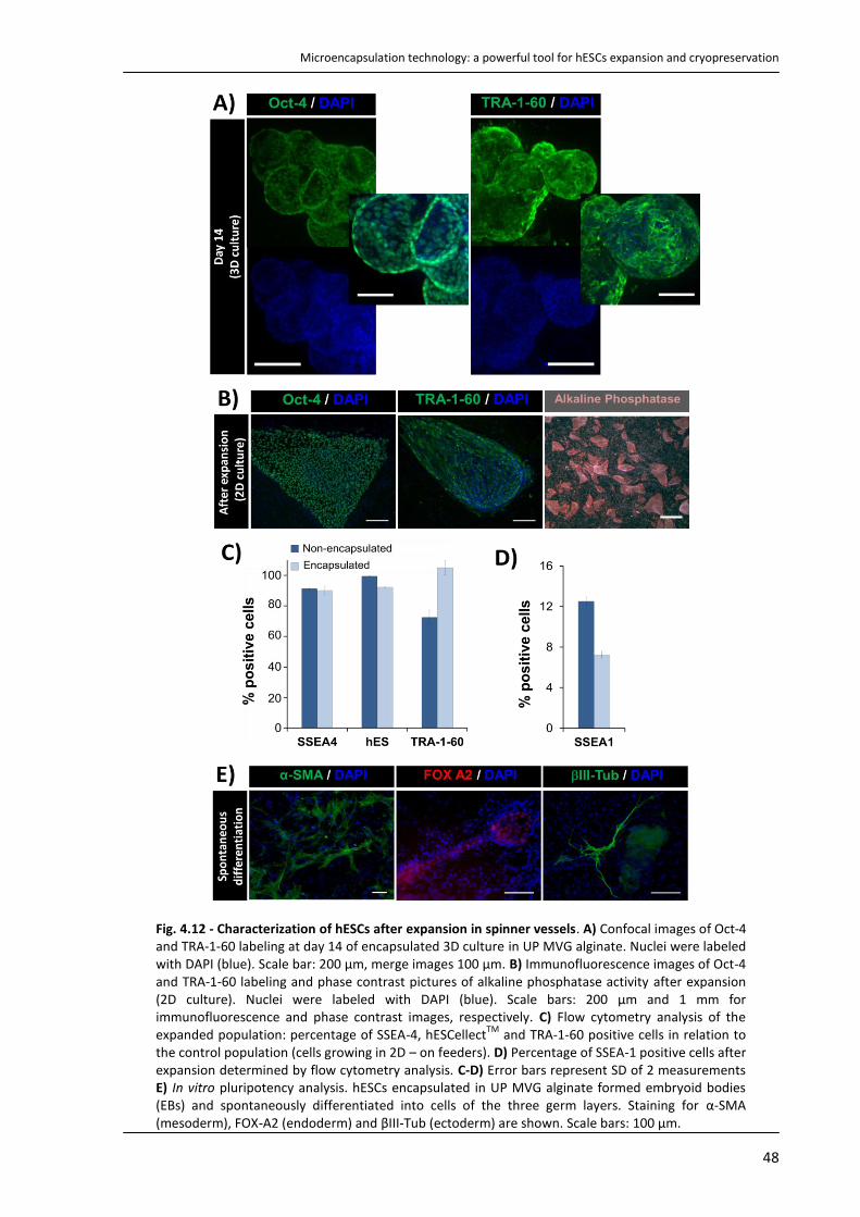
Microencapsulation technology: a powerful tool for hESCs expansion and cryopreservation
48
Fig. 4.12 - Characterization of hESCs after expansion in spinner vessels. A) Confocal images of Oct-4 and TRA-1-60 labeling at day 14 of encapsulated 3D culture in UP MVG alginate. Nuclei were labeled with DAPI (blue). Scale bar: 200 µm, merge images 100 µm. B) Immunofluorescence images of Oct-4 and TRA-1-60 labeling and phase contrast pictures of alkaline phosphatase activity after expansion (2D culture). Nuclei were labeled with DAPI (blue). Scale bars: 200 μm and 1 mm for immunofluorescence and phase contrast images, respectively. C) Flow cytometry analysis of the expanded population: percentage of SSEA-4, hESCellect
TM and TRA-1-60 positive cells in relation to
the control population (cells growing in 2D – on feeders). D) Percentage of SSEA-1 positive cells after expansion determined by flow cytometry analysis. C-D) Error bars represent SD of 2 measurements E) In vitro pluripotency analysis. hESCs encapsulated in UP MVG alginate formed embryoid bodies (EBs) and spontaneously differentiated into cells of the three germ layers. Staining for α-SMA (mesoderm), FOX-A2 (endoderm) and βIII-Tub (ectoderm) are shown. Scale bars: 100 μm.
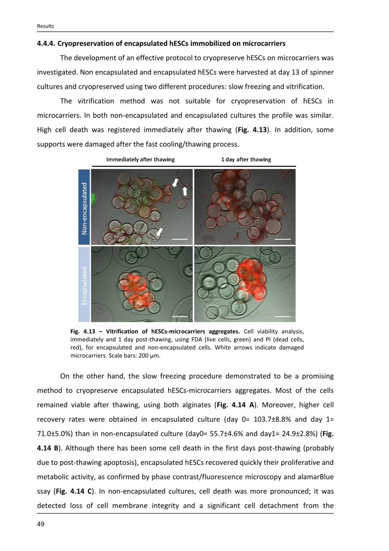
Results
49
4.4.4. Cryopreservation of encapsulated hESCs immobilized on microcarriers
The development of an effective protocol to cryopreserve hESCs on microcarriers was
investigated. Non encapsulated and encapsulated hESCs were harvested at day 13 of spinner
cultures and cryopreserved using two different procedures: slow freezing and vitrification.
The vitrification method was not suitable for cryopreservation of hESCs in
microcarriers. In both non-encapsulated and encapsulated cultures the profile was similar.
High cell death was registered immediately after thawing (Fig. 4.13). In addition, some
supports were damaged after the fast cooling/thawing process.
Fig. 4.13 – Vitrification of hESCs-microcarriers aggregates. Cell viability analysis, immediately and 1 day post-thawing, using FDA (live cells, green) and PI (dead cells, red), for encapsulated and non-encapsulated cells. White arrows indicate damaged microcarriers. Scale bars: 200 μm.
On the other hand, the slow freezing procedure demonstrated to be a promising
method to cryopreserve encapsulated hESCs-microcarriers aggregates. Most of the cells
remained viable after thawing, using both alginates (Fig. 4.14 A). Moreover, higher cell
recovery rates were obtained in encapsulated culture (day 0= 103.7±8.8% and day 1=
71.0±5.0%) than in non-encapsulated culture (day0= 55.7±4.6% and day1= 24.9±2.8%) (Fig.
4.14 B). Although there has been some cell death in the first days post-thawing (probably
due to post-thawing apoptosis), encapsulated hESCs recovered quickly their proliferative and
metabolic activity, as confirmed by phase contrast/fluorescence microscopy and alamarBlue
ssay (Fig. 4.14 C). In non-encapsulated cultures, cell death was more pronounced; it was
detected loss of cell membrane integrity and a significant cell detachment from the
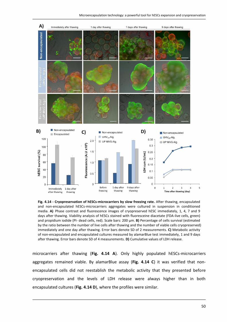
Microencapsulation technology: a powerful tool for hESCs expansion and cryopreservation
50
Fig. 4.14 - Cryopreservation of hESCs-microcarriers by slow freezing rate. After thawing, encapsulated and non-encapsulated hESCs-microcarriers aggregates were cultured in suspension in conditioned media. A) Phase contrast and fluorescence images of cryopreserved hESC immediately, 1, 4, 7 and 9 days after thawing. Viability analysis of hESCs stained with fluoresceine diacetate (FDA-live cells, green) and propidium iodide (PI- dead cells, red). Scale bars: 200 μm. B) Percentage of cells survival (estimated by the ratio between the number of live cells after thawing and the number of viable cells cryopreserved) immediately and one day after thawing. Error bars denote SD of 2 measurements. C) Metabolic activity of non-encapsulated and encapsulated cultures measured by alamarBlue test immediately, 1 and 9 days after thawing. Error bars denote SD of 4 measurements. D) Cumulative values of LDH release.
microcarriers after thawing (Fig. 4.14 A). Only highly populated hESCs-microcarriers
aggregates remained viable. By alamarBlue assay (Fig. 4.14 C) it was verified that non-
encapsulated cells did not reestablish the metabolic activity that they presented before
cryopreservation and the levels of LDH release were always higher than in both
encapsulated cultures (Fig. 4.14 D), where the profiles were similar.
Results
51
Cells encapsulated in UP MVG alginate were released from the
microcapsules/supports and plated onto ihFF monolayers immediately after thawing. These
cells maintained their ability to form undifferentiated pluripotent colonies for at least 5
passages as confirmed by immunofluorescence microscopy, flow cytometry, alkaline
phosphatase activity test and colonies morphology (Fig. 4.15 B, C). These cells also retained
the ability to differentiate in vitro into cells from the three germ layers (Fig. 4.15 D).
Immunofluorescence microscopy for stem cells markers of encapsulated cells in UP
MVG alginate at day 9 post-thawing, show that cells maintained the undifferentiated
character also when expanded after thawing (Fig. 4.15 A).

Microencapsulation technology: a powerful tool for hESCs expansion and cryopreservation
52
Fig. 4.15 - Characterization of hESCs after cryopreservation by slow freezing rate. Phenotype analysis of encapsulated hESC immobilized on microcarriers after (A) 9 days of culture post‐thawing and (B) after 2 and 5 cell passages in 2-D culture systems (P2 and P5, respectively). A, B) Immunofluorescence images of Oct-4 and TRA-1-60 labeling, and phase contrast pictures of alkaline phosphatase activity. Nuclei were labeled with DAPI (blue). Scale bars: (A) 100 μm and (B) 200 μm for immmunofluorescence images; (A, B) 1mm for phase contrast images. C) Fow cytometry analysis; percentage of SSEA-4, hES-Cellect
TM(hES),
TRA-1-60 and SSEA-1 positive cells. Error bars represent SD of 2 measurements. D) In vitro pluripotency analysis. Microcapsules were dissolved and hESCs were dissociated from the microcarriers and transferred to a monolayer of ihFF. At confluence, colonies were dissociated and hESCs were able to form embryoid bodies (EBs) in non-adherent conditions and differentiated into cells from all three germ layers. Phase contrast micrograph of human embryoid bodies and fluorescence images of differentiated cultures labeled for α–SMA (α smooth muscle actin, mesoderm), FOX-A2 (Forkheadbox A2, endoderm) and βIII-Tub (β tubulin type III, ectoderm). Nuclei were stained with DAPI (blue). Scale bars: 100 μm.
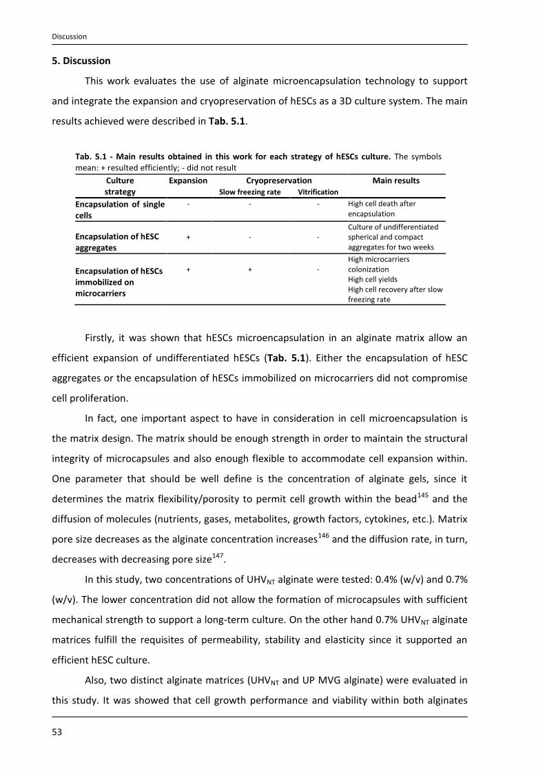
Discussion
53
5. Discussion
This work evaluates the use of alginate microencapsulation technology to support
and integrate the expansion and cryopreservation of hESCs as a 3D culture system. The main
results achieved were described in Tab. 5.1.
Tab. 5.1 - Main results obtained in this work for each strategy of hESCs culture. The symbols mean: + resulted efficiently; - did not result
Firstly, it was shown that hESCs microencapsulation in an alginate matrix allow an
efficient expansion of undifferentiated hESCs (Tab. 5.1). Either the encapsulation of hESC
aggregates or the encapsulation of hESCs immobilized on microcarriers did not compromise
cell proliferation.
In fact, one important aspect to have in consideration in cell microencapsulation is
the matrix design. The matrix should be enough strength in order to maintain the structural
integrity of microcapsules and also enough flexible to accommodate cell expansion within.
One parameter that should be well define is the concentration of alginate gels, since it
determines the matrix flexibility/porosity to permit cell growth within the bead145 and the
diffusion of molecules (nutrients, gases, metabolites, growth factors, cytokines, etc.). Matrix
pore size decreases as the alginate concentration increases146 and the diffusion rate, in turn,
decreases with decreasing pore size147.
In this study, two concentrations of UHVNT alginate were tested: 0.4% (w/v) and 0.7%
(w/v). The lower concentration did not allow the formation of microcapsules with sufficient
mechanical strength to support a long-term culture. On the other hand 0.7% UHVNT alginate
matrices fulfill the requisites of permeability, stability and elasticity since it supported an
efficient hESC culture.
Also, two distinct alginate matrices (UHVNT and UP MVG alginate) were evaluated in
this study. It was showed that cell growth performance and viability within both alginates
Culture strategy
Expansion Cryopreservation Main results Slow freezing rate Vitrification
Encapsulation of single cells
- - - High cell death after encapsulation
Encapsulation of hESC aggregates
+
-
-
Culture of undifferentiated spherical and compact aggregates for two weeks
Encapsulation of hESCs immobilized on microcarriers
+
+
-
High microcarriers colonization High cell yields High cell recovery after slow freezing rate
Microencapsulation technology: a powerful tool for hESCs expansion and cryopreservation
54
were similar. These observations can be explained by the fact that both polymers are high-G
content (>60)115, 117. In general, alginates with high content of G monomers have shown to
form stable gels with a high permeability91 and therefore have the necessary properties to
support an efficient cell growth.
The only difference observed between the two alginate matrices was related with
alginate dissociation and consequent recovery of hESCs. The dissociation of UHVNT alginate
significantly affects cell viability, preventing an efficient recovery of the cells. On the other
hand, UP MVG alginate dissociation does not compromise cell viability, allowing the recovery
of viable cells and their further characterization after expansion. This difference should be
related with the use of different crosslinkers. The gelification of UHVNT alginate was
performed using Ba2+ whereas UP MVG alginate microcapsules were formed in a Ca2+ bath.
In fact, it is known that Ba2+ ions give better long-term mechanical stability than Ca2+ ions do,
since Ba2+ forms stronger cross linkers with alginate than Ca2+ 148. However, this effect is only
observed for alginates with a high G content (more than 60% G)149. To dissociate the strong
linkers between Ba2+ and the UHVNT alginate, a solution of Na2SO4 was used. This procedure
changes drastically the osmotic pressure, leading to cell shrink and ultimately cell death. The
size of the cells recovered after decapsulation was so small that it was difficult to distinguish
them from cellular debris. Cross linkers between Ca2+ and the UP MVG alginate were easily
disrupted using an EDTA solution and cells maintained their viability.
Overall, these considerations support the assumption that 0.7% Ba2+-UHV alginate is
more appropriate to be used for long-term immunoprotection of differentiated cells during
transplantation. It will be useful for the treatment of human disorders such as hormone or
protein deficiencies. Ca2+-UP MVG alginate is more suitable to be integrated in stem cell
bioprocesses for efficient high production of cell-based products. Calcium forms weaker
alginate gels and is also more sensitive to chelators such as citrate, phosphate, and lactate89,
therefore long-term survival of Ca2+-alginate microcapsules in vivo should be limited.
However this can also be advantageous if the aim is the transplantation of autologous cells
for repair, remodeling or regeneration of tissues107. In this case, the capsule will only
minimize the initial immune-rejection of the cells by preventing surgery induced activation
of innate immunity and could enhance the graft delivery to a certain target. Indeed, easily
biodegradable alginates have been studied as promising approaches for tissue engineering
and regenerative medicine. It should be noted that Ca2+-UP MVG alginate was not tested yet
for routine clinical application. Although, this alginate is manufactured in compliance with
Discussion
55
current Good Manufacturing Practice and presents an endotoxin concentration less than
required for in vivo studies (≤100 EU/g)91.
The encapsulation of hESCs as single cells resulted in a pronounced cell death even
when different cell densities and alginate matrices were used. Thus this strategy is not useful
for hESCs expansion. This result is in agreement with other published studies139, 146,
confirming that cell–cell interactions are an important environmental factor that influences
hESCs survival and proliferation. In fact, the formation of hESCs aggregates within the
microcapsules was only observed when higher cell densities were used (3x10⁶ cell/mL
alginate), since some small clusters were already encapsulated and single cells within the
alginate matrix were closer to each other. It is important to highlight that hESCs are
vulnerable to apoptosis after complete dissociation51, therefore when the cells were handled
as single cells (before and after both harvesting and encapsulation steps) they were treated
with ROCKi. Previous reports shown that this compound prevent apoptosis and enhance the
survival and cloning efficiency of dissociated hESCs without affecting their pluripotency51.
However, in this case no significant improvements in cell viability/expansion were observed.
On the other hand, it was demonstrated that alginate microencapsulation of
aggregates is an efficient strategy for the expansion of hESCs in spinner vessels. Cells were
protected from the harmful effects of hydrodynamic shear stress, allowing a more efficient
cell growth, control of aggregate size and maintenance of pluripotent status for two weeks
of culture.
Compact, spherical and size uniform aggregates were formed in both alginate
cultures. Furthermore, cells presented high viability within the aggregates during the two
weeks of culture, confirming that limitations in the diffusion of nutrients and metabolites
within alginate matrices did not occur. In fact, a high improvement in culture performance
was achieved comparatively to non-encapsulated culture, where many dead cells were
observed in suspension as well as aggregate clumping. The formation of these large
aggregates compromised cell viability; necrotic centers were observed as a consequence of
diffusion limitations within the 3D structure. These observations confirmed that shear stress
(promoted by stirring) was harmful for the cells.
The expansion of undifferentiated hESCs as aggregates using spinner vessels has
been reported recently. Krawetz et al. cultivated hESC aggregates in stirred spinner vessels
(stirring rate - 100 rpm) using the repeated dissociation strategy (every 6 days the cultures

Microencapsulation technology: a powerful tool for hESCs expansion and cryopreservation
56
were dissociated into single cells and split at 1:5) in order to avoid the formation of large
aggregates and achieve high levels of pluripotency134. In another study, Singh et al.
successfully reported the culture of hESCs for 7 days as uniform spherical and viable
aggregates using spinner vessels equipped with a low shear impeller (single stirring
pendulum at 40 rpm)131. When compared to the conditions used herein for non-
encapsulated culture, these studies used less time of culture and less abrasive stirring, which
probably contributed to the efficient expansion of pluripotent hESCs as aggregates.
Importantly, the results obtained in this study demonstrated that the culture of
encapsulated aggregates seems to be a more efficient system for the expansion of hESCs.
Few reports of culturing hESCs on scaffolds or hydrogels have been published,
demonstrating that the culture of hESCs in these 3D matrixes assured the maintenance of
their undifferentiated phenotype for weeks without passaging. Gerecht et al. demonstrated
that encapsulated hESCs in hyaluronic acid hydrogel maintained their undifferentiated status
in the presence of conditioned medium from mEFs for more than 20 days in culture150. Siti-
Ismail et al. showed that encapsulation supported the maintenance of hESCs in the
undifferentiated state for 260 days, without the need for feeders or passaging. In this case
the encapsulated hESCs were cultured in non-conditioned medium supplemented with bFGF.
The larger capsule sizes used may allow such a prolonged culture of encapsulated hESCs138.
Nevertheless, in this study, after two weeks of culture, signals of cell differentiation
in encapsulated aggregates were observed. This can be explained by the increase in
aggregate size that could led to the formation of gradients in the concentration of gases and
regulatory molecules (e.g. bFGF) within the aggregate, thus promoting spontaneous
differentiation of some cells. Alternatively, this profile can be considered advantageous if the
aim is to integrate directed differentiation after a previous expansion step. Inducing directed
differentiation at this culture time point (2nd week), by changing specific culture conditions
(addition of inducible factors, medium replenishment, etc.), would bypass the step of EBs
formation, which is time-consuming and uncontrolled, and ultimately increase
differentiation efficiency and yields. Several studies have successful reported the
differentiation of encapsulated embryonic stem cells, for instance into definitive
endoderm139, pancreatic insulin-producing cells146, hepatocytes151, cardiomyocytes140, and
bone cells152.
Discussion
57
In this study it was evaluated for the first time the encapsulation oh hESCs adherent
on microcarriers. This 3D strategy demonstrated to be very efficient to the production of
high cell numbers required for clinical or pharmacological applications. At the end, it was
possible to achieve approximately 3x10⁶ cell/mL, corresponding to an improvement of 15.2-
fold in cell yields when compared to standard 2D protocols65.
It should be noted that the best result was obtained when 2g/L of empty and fresh
coated microcarriers were added to the hESCs-microcarriers aggregates cultures
immediately before encapsulation. The two-step addition of microcarriers allowed to
improve microcarriers colonization, microcapsules population and consequently enhance
cell yields in spinner vessels. Despite of being an effective system for the production of high
numbers of pluripotent hESCs, this strategy cannot be conjugated with transplantation
studies, due to the presence of supports within the capsules. Alternatively, cells could be
released from the matrix, detached from the supports and then used clinically. Moreover, a
biodegradable clinical approved support could eventually be used. For instance, Tatard et al.
developed pharmacologically active biodegradable microcarriers made with poly(D,L-lactic-
co-glycolic acid) (PLGA) and coated with adhesion molecules to serve as a support for cell
culture153. Gelatin microcarriers are also under study to be used in 3D cartilage and bone like
tissue engineering154.
Although the culture of hESCs on microcarriers has been reported to be an efficient
strategy to expand hESCs, future studies should be focus on the development of define
xeno-free culture conditions. For instance, coating the microcarriers with Matrigel has
proven effective in enhancing the poor adhesion of hESCs to the supports but its animal
origin makes it unsuitable for use in cell therapy. The same is true about the use of mEFs-
conditioned media. Preliminary studies performed in this work suggested that the use of
non-conditioned media and microcarriers coated with human plasm fibronectin is able to
support the proliferation of hESCs. However, the specific growth rate was reduced. This
preliminary knowledge might be helpful for the future development of an efficient xeno-free
culture system.
The feasibility of using microencapsulation technology for hESCs cryopreservation
was also investigated.
The results shown that the encapsulation of cells adherent on microcarriers is a
valuable strategy for the efficient cryopreservation of pluripotent hESCs, by slow freezing
Microencapsulation technology: a powerful tool for hESCs expansion and cryopreservation
58
rate. In fact, comparatively with non-encapsulated culture, higher cell survival was obtained
immediately and one day after thawing (improvement of 2- and 3- fold, respectively) and
hESCs recovered faster their proliferative and metabolic activity. These results indicate that
the encapsulation process had an important role in cell recovery. Indeed, cell
microencapsulation within alginate beads has previously been reported as a promising
strategy to improve post‐thaw viability of different cell types which were shown to poorly
survive in the cryopreservation process, such as hepatocytes155, 156, pancreatic islets110, 157
and neural cells130. Although the underlying mechanisms are still unclear, cell entrapment
may help protect cells from the adverse effects of cryopreservation by decreasing exposure
to cryoprotectants127, limiting extracellular ice growth near the cell’s membrane and the
initiation of intracellular ice formation158, 159, providing cell immobilization thus preventing
detachment of the cells from the supports and breakage of cell-cell contacts114, 130.
On the other hand, the recoveries reported after conventional slow freezing of hESCs
vary from 0% to only 48%75, 77-79, 81, 160. In addition Nie et al. verified that cryopreservation of
hESCs on mEFs-microcarriers improved in 1.5-1.9 times the recovery of hESCs frozen as free
colonies. However, when hESCs were cryopreserved adherent to Matrigel-microcarriers cell
recovery was not improved63. Cells in these microcarriers were more prone to detach from
the supports after thawing. This result is similar to that obtained in this study. These results
reinforce the assumption that maintaining both cell-cell and cell-matrix contacts improve
hESCs recovery following cryopreservation through slow-rate freezing63, 73, 76.
The slightly decrease obtained for cell recovery at day 1 after thawing, in
encapsulated cultures must be caused by post-thaw apoptotic and necrotic mechanisms161,
162. Therefore the addition of anti-apoptotic factors at this time should have a great
importance. In this study, ROCKi was used before, during and after the cryopreservation
process since it was reported that the addition of this inhibitor also increases the survival
rate after thawing163, 164.
Importantly, in the system developed herein, hESCs can be efficiently recovered after
cryopreservation without compromising their stem cell characteristics. These cells
maintained their pluripotency over at least five passages in standard culture conditions.
The slow freezing rate of both encapsulated and non-encapsulated hESC aggregates
proved to be an inefficient strategy. The higher cell death observed in aggregates in
comparison with cell-microcarrier cultures may be explained by the limitation in heat and
mass (water and cryoprotectant) diffusion within the aggregates derived from their larger
Discussion
59
size and more compact 3D structures. This results in different cryoprotection effect from the
surface to the center of the aggregate130, 166, possibly leading to cryodamage. Another
explanation is the presence of components of the extracellular matrix (ECM) when hESCs
were cryopreserved immobilized on Matrigel coated Cytodex3 microcarriers. This may have
contributed to enhance cell survival during slow freezing rate and thawing76, 80. Indeed, it has
been demonstrated that hESCs embedded in Matrigel exhibit an improvement in cell
recovery76, 82; Heng et al. argued that Matrigel entrapment improves hESCs colony recovery
by preserving gap junctions and other intercellular adhesion contacts during freezing and
thawing165.
It is important to highlight that cryopreservation of encapsulated hESCs using the
vitrification protocol did not work in all strategies tested in this study. The reasons why cells
lose their viability are not clear. However, it should be noted that only preliminary tests were
performed and the optimization of this technique using, for example, straws and by
improving specific steps during the fast cooling and/or thawing process(es) should be
considered. The addition of more apoptotic inhibitors may be also important, since some
encapsulated aggregates remained viable immediately after thawing process and only died
in the days after-thawing. Nevertheless, it is important to note that slow freezing rate is a
more appropriate procedure to cryopreserve hESCs than vitrification since the last technique
is labor consuming and impractical on a large scale; it requires special devices, is extremely
dependent on the expertise manipulation skills and is unsuitable for handling bulk quantities
of hESCs67, 73, 167. The high risk of contamination with pathogenic agents85 is another
disadvantage of the vitrification protocol.
In future, more fundamental studies regarding the physical-chemical and bio-physical
phenomena occurring during freezing/thawing of encapsulated hESCs should be performed
in order to improve the cryopreservation of hESCs aggregates.
Summing up, this work has shown that alginate cell microencapsulation is a
promising approach for the cultivation of hESCs because: it allows the culture of hESCs in a
3D conformation similarly to what occurs in vivo; it enables the culture of cells in fully
controlled bioreactors, where scalability automation and tight control of the culture
environment are combined to assure high cell yields; it protects cells from shear stress and
also prevents excessive clumping, permitting a tighter control of the process and a
prolonged cell culture; it allows the differentiation of the cells within; and finally, it allows

Microencapsulation technology: a powerful tool for hESCs expansion and cryopreservation
60
higher cell recovery of pluripotent hESCs after cryopreservation on microcarriers by a
scalable slow freezing procedure.
In addition microencapsulation technology is useful to protect cells from the
immune-response during transplants. However, to increase the efficiency of
microencapsulation technique so that it could be used in the clinic and industrial fields some
parameters have to be optimized. Bubbles as well as tails during the manufacture of the
microcapsules should be minimized or eliminated for instances by applying sinusoidal
pressure waves114. Ca2+-alginate microcapsules should be coated with polycation, such as
poly-L-lysine (PLL)91, to increase its integrity and stability during long-term culture. Immune
reactions should be avoided, for example, by covering microcapsules with a cell-free layer of
high M alginate, that could also guarantee long-term stability to the microcapsules114.
Prolonged incubation time periods with Ba2+ should be avoided since it inhibit the potassium
channels in the membranes of encapsulated cells, leading to their death115. Cells should be
symmetric positioned within the capsule, since thin layers of alginate do not
immunologically protect the entrapped cells and could also lead to breakage of the
microcapsules114. Knowing that cells tend to agglotinate during the encapsulation process
and induce the production of empty beads, another challenge is to separate these empty
microcapsules from the filled microcapsules. Finally, standardization and validation of the
entire manufacturing process from the material production site to the clinics should be
performed.
Conclusion
61
6. Conclusion
This thesis shows that alginate microencapsulation is a powerful tool to integrate
expansion and cryopreservation of pluripotent human embryonic stem cells. When
microencapsulation technology is combined with microcarriers to grow hESCs in stirred
systems, higher cell yields can be achieved (approximately 3×10⁶ hESC/mL) without
compromising cell pluripotency. Moreover, cells can be cryopreserved immediately after
expansion by using a simple and easy scalable technique, that assures high recoveries of
undifferentiated and pluripotent hESCs after thawing. The integrated strategy developed
herein would be amenable to scale-up (e.g. using large-scale stirred tank bioreactors),
therefore it would ensure the production, banking and distribution of clinical relevant
numbers of high-quality hESCs, in a scalable and straightforward manner.
Furthermore, this work also demonstrated that microencapsulation confers physical
protection to hESC aggregates from damage caused by stirring, allows the control of
aggregates size and the maintenance of pluripotency for two weeks. Also, this strategy is
very promising to integrate both expansion and directed differentiation steps in a controlled
bioprocess. In future, using a xeno-free, clinical-grade alginate (such as UHVNT alginate) the
differentiated cells derived from the process could be eventually used in (human)
transplantation studies.
Hopefully, the 3D culture strategies developed here represent a relevant step
forward in the promising transition of hESCs to regenerative medicine, tissue engineering
and toxicology fields.

References
62
7. References
1. Fishel, S.B., Edwards, R.G. & Evans, C.J. Human chorionic gonadotropin secreted by preimplantation embryos cultured in vitro. Science 223, 816-818 (1984).
2. Bongso, A., Fong, C.Y., Ng, S.C. & Ratnam, S. Isolation and culture of inner cell mass cells from human blastocysts. Hum Reprod 9, 2110-2117 (1994).
3. Thomson, J.A. et al. Embryonic stem cell lines derived from human blastocysts. Science 282, 1145-1147 (1998).
4. Löser, P., Schirm, J., Guhr, A., Wobus, A.M. & Kurtz, A. Human embryonic stem cell lines and their use in international research. Stem Cells 28, 240-246 (2010).
5. Jensen, J., Hyllner, J. & Björquist, P. Human embryonic stem cell technologies and drug discovery. J Cell Physiol 219, 513-519 (2009).
6. Bongso, A., Fong, C.Y. & Gauthaman, K. Taking stem cells to the clinic: Major challenges. J Cell Biochem 105, 1352-1360 (2008).
7. Allegrucci, C. & Young, L.E. Differences between human embryonic stem cell lines. Hum Reprod Update 13, 103-120 (2007).
8. Reubinoff, B.E., Pera, M.F., Fong, C.Y., Trounson, A. & Bongso, A. Embryonic stem cell lines from human blastocysts: somatic differentiation in vitro. Nat Biotechnol 18, 399-404 (2000).
9. Appendix C: Human Embryonic Stem Cells and Human Embryonic Germ Cells. In Stem Cell Information [World Wide Web site]. Bethesda, MD: National Institutes of Health, U.S. Department of Health and Human Services, version 15 September 2010. Available at <http://stemcells.nih.gov/info/2001report/appendixc>.
10. Brignier, A.C. & Gewirtz, A.M. Embryonic and adult stem cell therapy. J Allergy Clin Immunol 125, S336-344 (2010).
11. Yamanaka, S. & Blau, H.M. Nuclear reprogramming to a pluripotent state by three approaches. Nature 465, 704-712 (2010).
12. Teo, A.K. & Vallier, L. Emerging use of stem cells in regenerative medicine. Biochem J 428, 11-23 (2010). 13. Takahashi, K. et al. Induction of pluripotent stem cells from adult human fibroblasts by defined factors. Cell
131, 861-872 (2007). 14. Yu, J. et al. Induced pluripotent stem cell lines derived from human somatic cells. Science 318, 1917-1920
(2007). 15. Raya, A. et al. Disease-corrected haematopoietic progenitors from Fanconi anaemia induced pluripotent
stem cells. Nature 460, 53-59 (2009). 16. Fenno, L.E., Ptaszek, L.M. & Cowan, C.A. Human embryonic stem cells: emerging technologies and practical
applications. Curr Opin Genet Dev 18, 324-329 (2008). 17. Malchenko, S. et al. Cancer hallmarks in induced pluripotent cells: new insights. J Cell Physiol 225, 390-393
(2010). 18. Guillot, P.V., Cui, W., Fisk, N.M. & Polak, D.J. Stem cell differentiation and expansion for clinical
applications of tissue engineering. J Cell Mol Med 11, 935-944 (2007). 19. Polak, J.M. & Mantalaris, S. Stem cells bioprocessing: an important milestone to move regenerative
medicine research into the clinical arena. Pediatr Res 63, 461-466 (2008). 20. In Stem Cell Information [World Wide Web site]. Bethesda, MD: National Institutes of Health, U.S.
Department of Health and Human Services, version 11 August 2009. Available at <http://stemcells.nih.gov/info/media/promise>.
21. Lund, R.D. et al. Human embryonic stem cell-derived cells rescue visual function in dystrophic RCS rats. Cloning Stem Cells 8, 189-199 (2006).
22. Takagi, Y. et al. Dopaminergic neurons generated from monkey embryonic stem cells function in a Parkinson primate model. J Clin Invest 115, 102-109 (2005).
23. Wichterle, H., Lieberam, I., Porter, J.A. & Jessell, T.M. Directed differentiation of embryonic stem cells into motor neurons. Cell 110, 385-397 (2002).
24. Nistor, G.I., Totoiu, M.O., Haque, N., Carpenter, M.K. & Keirstead, H.S. Human embryonic stem cells differentiate into oligodendrocytes in high purity and myelinate after spinal cord transplantation. Glia 49, 385-396 (2005).
25. Caspi, O. et al. Transplantation of human embryonic stem cell-derived cardiomyocytes improves myocardial performance in infarcted rat hearts. J Am Coll Cardiol 50, 1884-1893 (2007).
26. Kroon, E. et al. Pancreatic endoderm derived from human embryonic stem cells generates glucose-responsive insulin-secreting cells in vivo. Nat Biotechnol 26, 443-452 (2008).
27. Duan, Y. et al. Differentiation and enrichment of hepatocyte-like cells from human embryonic stem cells in vitro and in vivo. Stem Cells 25, 3058-3068 (2007).
References
63
28. Ebert, A.D. & Svendsen, C.N. Human stem cells and drug screening: opportunities and challenges. Nat Rev Drug Discov 9, 367-372 (2010).
29. Trounson, A. The production and directed differentiation of human embryonic stem cells. Endocr Rev 27, 208-219 (2006).
30. Lei, T. et al. Xeno-free derivation and culture of human embryonic stem cells: current status, problems and challenges. Cell Res 17, 682-688 (2007).
31. Skottman, H. & Hovatta, O. Culture conditions for human embryonic stem cells. Reproduction 132, 691-698 (2006).
32. Unger, C., Skottman, H., Blomberg, P., Dilber, M.S. & Hovatta, O. Good manufacturing practice and clinical-grade human embryonic stem cell lines. Hum Mol Genet 17, R48-53 (2008).
33. Amit, M. et al. Clonally derived human embryonic stem cell lines maintain pluripotency and proliferative potential for prolonged periods of culture. Dev Biol 227, 271-278 (2000).
34. Crook, J.M. et al. The generation of six clinical-grade human embryonic stem cell lines. Cell stem cell 1, 490-494 (2007).
35. Stacey, G.N. et al. The development of 'feeder' cells for the preparation of clinical grade hES cell lines: challenges and solutions. J Biotechnol 125, 583-588 (2006).
36. Richards, M., Fong, C.Y., Chan, W.K., Wong, P.C. & Bongso, A. Human feeders support prolonged undifferentiated growth of human inner cell masses and embryonic stem cells. Nat Biotechnol 20, 933-936 (2002).
37. Amit, M. et al. Human feeder layers for human embryonic stem cells. Biol Reprod 68, 2150-2156 (2003). 38. Hovatta, O. et al. A culture system using human foreskin fibroblasts as feeder cells allows production of
human embryonic stem cells. Hum Reprod 18, 1404-1409 (2003). 39. Xu, C. et al. Feeder-Layer free growth of undifferentiated human embryonic stem cells. Nat Biotechnol 19,
971-974 (2001). 40. Amit, M., Shariki, C., Margulets, V. & Itskovitz-Eldor, J. Feeder and serum free culture system for human
embryonic stem cells. Biol Reprod 70, 837-845 (2004). 41. Amit, M. Feeder-layer free culture system for human embryonic stem cells. Methods Mol Biol 407, 11-20
(2007). 42. Levenstein, M.E. et al. Basic fibroblast growth factor support of human embryonic stem cell self-renewal.
Stem cells 24, 568-574 (2006). 43. Holm, F., Bergström, R., Ström, S. & Hovatta, O. Derivation, maintenance and cryostorage of human
embryonic stem cells. Drug Discov Today: Technol 5, e131-e137 (2010). 44. Jing, D., Parikh, A., Canty, J.M., Jr. & Tzanakakis, E.S. Stem cells for heart cell therapies. Tissue Eng Part B
Rev 14, 393-406 (2008). 45. Ryan, E.A. et al. Successful islet transplantation: continued insulin reserve provides long-term glycemic
control. Diabetes 51, 2148-2157 (2002). 46. King, J.A. & Miller, W.M. Bioreactor development for stem cell expansion and controlled differentiation.
Curr Opin Chem Biol 11, 394-398 (2008). 47. Placzek, M.R. et al. Stem cell bioprocessing: fundamentals and principles. J R Soc Interface 6, 209-232
(2009). 48. Ulloa-Montoya, F., Verfaillie, C. & Hu, W.-S. Culture systems for pluripotent stem cells. J Biosci Bioeng 100,
12-27 (2005). 49. Wong, R.C.B. et al. Gap junctions modulate apoptosis and colony growth of human embryonic stem cells
maintained in a serum-free system. Biochem Biophys Res Commun 344, 181-188 (2006). 50. Ellerström, C., Strehl, R., Noaksson, K., Hyllner, J. & Semb, H. Facilitated expansion of human embryonic
stem cells by single-cell enzymatic dissociation. Stem Cells 25, 1690-1696 (2007). 51. Watanabe, K. et al. A ROCK inhibitor permits survival of dissociated human embryonic stem cells. Nat
Biotechnol 25, 681-686 (2007). 52. Fujikawa, T. et al. Teratoma formation leads to failure of treatment for type I diabetes using embryonic
stem cell-derived insulin-producing cells. Am J Pathol 166, 1781-1791 (2005). 53. Godier, A.F.G. et al. Engineered microenvironments for human stem cells. Birth Defects Res C Embryo
Today 84, 335-347 (2008). 54. Hwang, N., Varghese, S. & Elisseeff, J. Controlled differentiation of stem cells. Adv Drug Deliv Rev 60, 199-
214 (2008). 55. Abraham, S., Ola, E.N. & Rao, R.R. Role of bioinspired polymers in determination of pluripotent stem cell
fate. Regen Med 4, 561-578 (2009). 56. Chan, B.P. & Leong, K.W. Scaffolding in tissue engineering: general approaches and tissue-specifc
considerations. Eur Spine J 17, 467-479 (2008).
Microencapsulation technology: a powerful tool for hESCs expansion and cryopreservation
64
57. Lukashev, M. & Werb, Z. ECM signalling: orchestrating cell behaviour and misbehaviour. Trends Cell Biol 8, 437-441 (1998).
58. Even-Ram, S. & Yamada, K.M. Cell migration in 3D matrix Curr Opin Cell Biol 17, 524-532 ( 2005). 59. Serra, M., Brito, C. & Alves, P.A. Bioengineering strategies for stem cell expansion and differentiation.
Canal Bioquímica in press (2010). 60. Ghidoni, I. et al. Alginate cell encapsulation: new advances in reproduction and cartilage regenerative
medicine. Cytotechnology 58, 49-56 (2008). 61. Kehoe, D.E., Jing, D., Lock, L.T. & Tzanakakis, E.S. Scalable stirred-suspension bioreactor culture of human
pluripotent stem cells. Tissue Eng Part A 16, 405-421 (2010). 62. Gerecht-Nir, S., Cohen, S. & Itskovitz-Eldor, J. Bioreactor cultivation enhances the efficiency of human
embryoid body (hEB) formation and differentiation. Biotechnol Bioeng 86, 493-502 (2004). 63. Nie, Y., Bergendahl, V., Hei, D.J., Jones, J.M. & Palecek, S.P. Scalable culture and cryopreservation of
human embryonic stem cells on microcarriers. Biotechnol Prog 25, 20-31 (2009). 64. Phillips, B.W. et al. Attachment and growth of human embryonic stem cells on microcarriers. J Biotechnol
138, 24-32 (2008). 65. Serra, M. et al. Improving expansion of pluripotent human embryonic stem cells in perfused bioreactors
through oxygen control. J Biotechnol 148, 208-215 (2010). 66. Baust, J.G. & Baust, J.M. in Advances in Biopreservation. (eds. J.G. Baust & J.M. Baust) 15-62 (CRC Press-
Taylor and Francis Publishing New York; 2006). 67. Hanna, J. & Hubel, A. Preservation of stem cells. Organogenesis 5, 134-137 (2009). 68. Lovelock, J.E. The mechanism of the protective action of glycerol against haemolysis by freezing and
thawing. Biochim Biophys Acta 11, 28-36 (1953). 69. Pegg, D.E. The current status of tissue cryopreservation. Cryo Letters 22, 105-114 (2001). 70. Muldrew, K., Acker, J.P., Elliot, J.A.W. & McGann, L.E. in Life in The Frozen State. (eds. B.J. Fuller, N. Lane &
E.E. Benson) 67-107 (CRC Press, 2004). 71. Clegg, J.S., Seitz, P., Seitz, W. & Hazlewood, C.F. Cellular responses to extreme water loss: the water
replacement hypothesis. Cryobiology 19, 306-316 (1982). 72. Holm, F. et al. An effective serum- and xeno-free chemically defined freezing procedure for human
embryonic and induced pluripotent stem cells. Hum Reprod 25, 1271-1279 (2010). 73. Hunt, C.J. The Banking and Cryopreservation of Human Embryonic Stem Cells. Transfus Med Hemother 34,
293-304 (2007). 74. Mazur, P. Cryobiology: the freezing of biological systems. Science 168, 939-949 (1970). 75. Richards, M., Fong, C.Y., Tan, S., Chan, W.K. & Bongso, A. An efficient and safe xeno-free cryopreservation
method for the storage of human embryonic stem cells. Stem Cells 22, 779-789 (2004). 76. Ji, L., Pablo, J.J.d. & Palecek, S.P. Cryopreservation of adherent human embryonic stem cells. Biotechnol
Bioeng 88, 299-312 (2004). 77. Reubinoff, B.E., Pera, M.F., Vajta, G. & Trounson, A.O. Effective cryopreservation of human embryonic
stem cells by the open pulled straw vitrification method. Hum Reprod 16, 2187-2194 (2001). 78. Zhou, C.Q., Mai, Q.Y., Li, T. & Zhaung, G.J. Cryopreservation of human embryonic stem cells by vitrification.
Chin Med J 117, 1050-1055 (2004). 79. Ha, S.Y. et al. Cryopreservation of human embryonic stem cells without the use of a programmable freezer.
Hum Reprod 20, 463–707 (2005). 80. Kim, S.J. & Park, J.H. Effects of type IV collagen and laminin on the cryopreservation of human embryonic
stem cells. Stem Cells 22, 950-961 (2004). 81. Wu, C.F. et al. Improved cryopreservation of human embryonic stem cells with trehalose. Reprod Biomed
11, 733-739 (2005). 82. Martin-Ibañez, R. et al. Novel cryopreservation method for dissociated human embryonic stem cells in the
presence of a ROCK inhibitor. Hum Reprod 23, 2744-2754 (2008). 83. Fahy, G.M., MacFarlane, D.R., Angell, C.A. & Meryman, H.T. Vitrification as an approach to
cryopreservation. Cryobiology 21, 407-426 (1984). 84. Taylor, M.J., Song, Y.C. & Brockbank, K.G.M. in Life in the Frozen State. (eds. B.J. Fuller, N. Lane & E.E.
Benson) 603-641 (CRC Press, 2004). 85. Tedder, R.S. et al. Hepatitis B transmission from contaminated cryopreservation tank. Lancet 346, 137-140
(1995). 86. Chang, T.M.S. Semipermeable Microcapsules. Science 146, 524 - 525 (1964). 87. Lim, F. & Sun, A. Microencapsulated islets as bioartificial endocrine pancreas Science 210, 908-910 (1980). 88. de Vos, P. & Marchetti, P. Encapsulation of pancreatic islets for transplantation in diabetes: the
untouchable islets. Trends Mol Med 8, 363-366 (2002). 89. Zimmermann, U. et al. in Bioencapsulation Techniques 548-566.
References
65
90. Orive, G., Hernández, R.M., Gascón, A.R., Igartua, M. & Pedraz, J.L. Encapsulated cell technology: from research to market. Trends Biotechnol 20, 382-387 (2002).
91. de Vos, P., Faas, M.M., Strand, B. & Calafiore, R. Alginate-based microcapsules for immunoisolation of pancreatic islets. Biomaterials 27, 5603-5617 (2006).
92. Murua, A. et al. Cell microencapsulation technology: towards clinical application. J Control Release 132, 76-83 (2008).
93. Freimark, D. et al. Use of Encapsulated Stem Cells to Overcome the Bottleneck of Cell Availability for Cell Therapy Approaches. Transfus Med Hemother 37, 66-73 (2010).
94. Koo, J. & Chang, T.S.M. Secretion of erythropoietin from microencapsulated rat kidney cells: preliminary results. Int J Artif Organs 16, 557-560 (1993).
95. Aebischer, P. et al. A bioartificial parathyroid. ASAIO Trans 32, 134-137 (1986). 96. Chang, P.L., Shen, N. & Westcott, A.J. Delivery of recombinant gene products with microencapsulated cells
in vivo. Hum Gene Ther 4, 433-440 (1993). 97. Liu, H.W., Ofosu, F.A. & Chang, P.L. Expression of human factor IX by microencapsulated recombinant
fibroblasts. Hum Gene Ther 4, 291-301 (1993). 98. Cieslinski, D.A. & Humes, H.D. Tissue engineering of a bioartificial kidney. Biotechnol Bioeng 43, 678-681
(1994). 99. Wong, H. & Chang, T.M. Bioartificial liver: implanted artificial cells microencapsulated living hepatocytes
increases survival of liver failure rats. Int J Artif Organs 9, 335-336 (1986). 100. Aebischer, P., Goddard, M., Signore, A.P. & Timpson, R.L. Functional recovery in hemiparkinsonian
primates transplanted with polymerencapsulated PC12 cells. Exp Neurol 126, 151-158 (1994). 101. Beck, J. et al. Islet encapsulation: strategies to enhance islet cell functions. Tissue Eng 13, 589-599 (2007). 102. Wilson, J.T. & Chaikof, E.L. Challenges and emerging technologies in the immunoisolation of cells and
tissues. Adv Drug Deliv Rev 60, 124-145 (2008). 103. Rabanel, J.-m., Banquy, X., Zouaoui, H., Mokhtar, M. & Hildgen, P. Progress technology in
microencapsulation methods for cell therapy. Biotechnol Prog 25, 946-963 (2009). 104. Orive, G. et al. History, challenges and perspectives of cell microencapsulation. Trends Biotechnol 22, 87-
92 (2004). 105. Orive, G. et al. Cell encapsulation: promise and progress. Nat Med 9, 104-107 (2003). 106. de Groot, M., Schuurs, T.A. & van Schilfgaarde, R. Causes of limited survival of microencapsulated
pancreatic islet grafts. J Surg Res 121, 141-150 (2004). 107. Hernández, R.M., Orive, G., Murua, A. & Pedraz, J.L. Microcapsules and microcarriers for in situ cell
delivery. Adv Drug Deliv Rev 62, 711-730 (2010). 108. Li, R.H. Materials for immunoisolated cell transplantation. Adv Drug Deliv Rev 33, 87-109 (1998). 109. Zielinski, B.A. & Aebischer, P. Chitosan as a matrix for mammalian cell encapsulation. Biomaterials 15,
1049-1056 (1994). 110. Iwata, H. et al. Evaluation of microencapsulated islets in agarose gel as bioartificial pancreas by studies of
hormone secretion in culture and by xenotransplantation. Diabetes 38 Suppl 1, 224-225 (1989). 111. Dawson, R.M., Broughton, R.L., Stevenson, W.T. & Sefton, M.V. Microencapsulation of CHO cells in a
hydroxyethyl methacrylate-methyl methacrylate copolyme. Biomaterials 8, 360-366 (1987). 112. Kessler, L., Pinget, M., Aprahamian, M., Dejardin, P. & Damge, C. In vitro and in vivo studies of the
properties of an artificial membrane for pancreatic islet encapsulation. Horm Metab Res 23, 312-317 (1991).
113. Cruise, G.M. et al. In vitro and in vivo performance of porcine islets encapsulated in interfacially photopolymerized poly(ethylene glycol) diacrylate membranes. Cell Transplant 8, 293-306 (1999).
114. Zimmermann, H., Shirley, S.G. & Zimmermann, U. Alginate-based encapsulation of cells: past, present, and future. Curr Diab Rep 7, 314-320 (2007).
115. Zimmermann, H. et al. Towards a medically approved technology for alginate-based microcapsules allowing long-term immunoisolated transplantation. J Mater Sci 16, 491 - 501 (2005).
116. Storz, H. et al. Physicochemical features of ultra-high viscosity alginates. Carbohydr Res 344, 985-995 (2009).
117. Klokk, T.I. & Melvik, J.E. Controlling the size of alginate gel beads by use of a high electrostatic potential. J Microencapsul 19, 415-424 (2002).
118. Park, J.S. et al. Chondrogenesis of human mesenchymal stem cells encapsulated in a hydrogel construct: Neocartilage formation in animal models as both mice and rabbits. J Biomed Mater Res A 92A, 988-996 (2010).
119. Cai, X. et al. Ectopic osteogenesis and chondrogenesis of bone marrow stromal stem cells in alginate system. Cell Biol Int 31, 776-783 (2007).
Microencapsulation technology: a powerful tool for hESCs expansion and cryopreservation
66
120. Kaigler, D. et al. Transplanted endothelial cells enhance orthotopic bone regeneration. J Dent Res 85, 633-637 (2006).
121. Shi, X.L., Zhang, Y., Gu, J.Y. & Ding, Y.T. Coencapsulation of hepatocytes with bone marrow mesenchymal stem cells improves hepatocyte-specific functions. Transplantation 88, 1178-1185 (2009).
122. Trouche, E. et al. Evaluation of alginate microspheres for mesenchymal stem cells engraftment on solid organ. Cell Transplant (2010).
123. Sargent, C.Y. et al. Hydrodynamic modulation of embryonic stem cell differentiation by rotary orbital suspension culture. Biotechnol Bioeng 105, 611-626.
124. Anjomshoa, M. et al. Generation of motor neurons by coculture of retinoic acid-pretreated embryonic stem cells with chicken notochords. Stem Cells Dev 18, 259-268 (2009).
125. Seliktar, D., Zisch, A.H., Lutolf, M.P., Wrana, J.L. & Hubbell, A. MMP-2 sensitive, VEGF-bearing bioactive hydrogels for promotion of vascular healing. J Biomed Mater Res A 68, 704-716 (2004).
126. Benoit, D.S.W., Schwartz, M.P., Durney, A.R. & Anseth, K.S. Small functional groups for controlled differentiation of hydrogel-encapsulated human mesenchymal stem cells. Nat Mater 7, 816 - 823 (2008).
127. Chin Heng, B., Yu, H. & Chye Ng, S. Strategies for the cryopreservation of microencapsulated cells. Biotechnol Bioeng 85, 202-213 (2004).
128. Stensvaag, V. et al. Cryopreservation of alginate-encapsulated recombinant cells for antiangiogenic therapy. Cell Transplant 13, 35-44 (2004).
129. Wu, Y., Yu, H., Chang, S., Magalhães, R. & Kuleshova, L.L. Vitreous Cryopreservation of Cell–Biomaterial Constructs Involving Encapsulated Hepatocytes. Tissue Eng 13, 649-658 (2007).
130. Malpique, R. et al. Alginate encapsulation as a novel strategy for the cryopreservation of neurospheres. Tissue Eng Part C Methods 16, 965-977 (2010).
131. Singh, H., Mok, P., Balakrishnan, T., Rahmat, S.N.B. & Zweigerdt, R. Up-scaling single cell-inoculated suspension culture of human embryonic stem cells. Stem Cell Res 4, 165-179 (2010).
132. Olmer, R. et al. Long term expansion of undifferentiated human iPS and ES cells in suspension culture using a defined medium Stem Cell Res 5, 51-64 (2010).
133. Amit, M. et al. Suspension culture of undifferentiated human embryonic and induced pluripotent stem cells. Stem Cell Rev 6, 248-259 (2010).
134. Krawetz, R. et al. Large-scale expansion of pluripotent human embryonic stem cells in stirred-suspension bioreactors. Tissue Eng Part C Methods 16, 573-582 (2010).
135. Lock, L.T. & Tzanakakis, E.S. Expansion and differentiation of human embryonic stem cells to endoderm progeny in a microcarrier stirred-suspension culture. Tissue Eng Part A 15, 2051-2063 (2009).
136. Fernandes, A.M. et al. Successful scale-up of human embryonic stem cell production in a stirred microcarrier culture system. Braz J Med Biol Res 42, 515-522 (2009).
137. Oh, S.K. et al. Long-term microcarrier suspension cultures of human embryonic stem cells. Stem Cell Res 2, 219-230 (2009).
138. Siti-Ismail, N., Bishop, A.E., Polak, J.M. & Mantalaris, A. The benefit of human embryonic stem cell encapsulation for prolonged feeder-free maintenance. Biomaterials 29, 3946-3952 (2008).
139. Chayosumrit, M., Tuch, B. & Sidhu, K. Alginate microcapsule for propagation and directed differentiation of hESCs to definitive endoderm. Biomaterials 31, 505-514 (2010).
140. Jing, D., Parikh, A. & Tzanakakis, E.S. Cardiac Cell Generation from Encapsulated Embryonic Stem Cells in Static and Scalable Culture Systems. Cell Transplant (2010).
141. Zimmermann, H. et al. Physical and biological properties of barium cross-linked alginate membranes. Biomaterials 28, 1327-1345 (2007).
142. Ross, D.D. et al. Estimation of cell survival by flow cytometric quantification of fluorescein diacetate/propidium iodide viable cell number. Cancer Res 49, 3776-3782 (1989).
143. International Stem Cell Initiative. Characterization of human embryonic stem cell lines by the International Stem Cell Initiative. Nat Biotechnol 25, 803-816 (2007).
144. Nie, Y., Bergendahl, V., Hei, D.J., Jones, J.M. & Palecek, S.P. Scalable Culture and Cryopreservation of Human Embryonic Stem Cells on Microcarriers. Biotechnol. Prog. 25, 20-31 ( 2009).
145. Dembczynski, R. & Jankowski, T. Determination of pore diameter and molecular weight cut-off of hydrogel-membrane liquid-core capsules for immunoisolation. J Biomater Sci Polym Ed 12, 1051-1058 (2001).
146. Wang, N., Adams, G., Buttery, L., Falcone, F.H. & Stolnik, S. Alginate encapsulation technology supports embryonic stem cells differentiation into insulin-producing cells. J Biotechnol 144, 304-312 (2009).
147. Moon, J.S., Jeon, H.M., Meng, W., Akaike, T. & Kang, I.K. Morphology and metabolism of hepatocytes microencapsulated with acrylic terpolymer-alginate using gelatin and poly(vinyl alcohol) as extracellular matrices. J Biomater Sci Polym Ed 16, 1245-1259 (2005).
References
67
148. Smidsrod, O. Molecular basis for some physical properties of alginates in the gel state. J Chem Soc Faraday Trans 57, 263-274 (1974).
149.Morch, Y.A., Donati, I., Strand, B.L. & Skjak-Braek, G. Effect of Ca2+, Ba2+, and Sr2+ on alginate microbeads. Biomacromolecules 7, 1471-1480 (2006).
150. Gerecht, S. et al. Hyaluronic acid hydrogel for controlled self-renewal and differentiation of human embryonic stem cells. Proc Natl Acad Sci U S A 104, 11298-11303 (2007).
151. Maguire, T., Novik, E., Schloss, R. & Yarmush, M. Alginate-PLL Microencapsulation : Effect on the Differentiation of Embryonic Stem Cells Into Hepatocytes. Biotechnology (2005).
152. Hwang, Y.-s. et al. The use of murine embryonic stem cells , alginate encapsulation , and rotary microgravity bioreactor in bone tissue engineering. Biomaterials 30, 499-507 (2009).
153. Tatard, V.M. et al. Pharmacologically active microcarriers: a tool for cell therapy. Biomaterials 26, 3727-3737 (2005).
154. Sommara, P. et al. Engineering three-dimensional cartilage- and bone-like tissues using human dermal fibroblasts and macroporous gelatine microcarriers. JPRAS 63, 1036-1046 (2010).
155. Rialland, L., Guyomard, C., Scotte, M., Chesne, C. & Guillouzo, A. Viability and drug metabolism capacity of alginate entrapped hepatocytes after cryopreservation. Cell Biol Toxicol 16, 105-116 (2000).
156. Guyomard, C., Rialland, L. & al, e. Influence of alginate gel entrapment and cryopreservation on survival and xenobiotic metabolism capacity of rat hepatocytes. Toxicol Appl Pharmacol 141, 349-356 (1996).
157. Zhou, D., Vacek, I. & al., e. Cryopreservation of microencapsulated porcine pancreatic islets: in vitro and in vivo studies. Transplantation 64, 1112-1116 (1997).
158. Murase, N., Inoue, T. & Ruike, M. Equilibrium and non-equilibrium freezing of water in cross-linked dextran gels. Cryo Letters 18, 157-164 (1997).
159. Toner, M., Cravalho, E.G. & et al. Cellular response of mouse oocytes to freezing stress: prediction of intracellular ice formation. J Biomech Eng 115, 169-174 (1993).
160. Heng, B.C., Clement, M.V. & Cao, T. Caspase inhibitor z-vad-fmk enhances the freeze-thaw survival rate of human embryonic stem cells. Biosci Rep 27, 257-264 (2007).
161. Frim, J., Snyder, R.A. & et al. Growth kinetics of cells following freezing in liquid nitrogen. Cryobiology 15, 502-516 (1978).
162. Baust, J.M., Van, B. & Baust, J.G. Cell viability improves following inhibition of cryopreservation-induced apoptosis. In Vitro Cell Dev Biol Anim 36, 262-270 (2000).
163. Li, X., Krawetz, R., Liu, S., Meng, G. & Rancourt, D.E. ROCK inhibitor improves survival of cryopreserved serum/feeder-free single human embryonic stem cells. Hum Reprod 24, 580-589 (2009).
164. Li, X. et al. The ROCK inhibitor Y-27632 enhances the survival rate of human embryonic stem cells following cryopreservation. Stem Cells Dev 17, 1079-1085 (2008).
165. Heng, B.C. et al. Loss of viability during freeze-thaw of intact and adherent human embryonic stem cells with conventional slow-cooling protocols is predominantly due to apoptosis rather than cellular necrosis. J Biomed Sci 13, 433-445 (2006).
166. Karlsson, J.O.M. & Toner, M. Long-term storage of tissues by cryopreservation: critical issues.
Biomaterials 17, 243-256 (1996).
167. Li, Y., D, P., Tan, J.-c. & Li, L.-s. Comparison of three methods for cryopreservation of human embryonic stem cells. Fertil Steril 93, 999-1005 (2010).