Embed Size (px)
Citation preview

Microfluidic aqueous two-phase system for continuous partitioning of bacteria
Prem Kumar Periyannan Rajeswari
Degree Project in Industrial Biotechnology, Second Level, 2011
Supervisor: Aman Russom, Division of Cell Physics, KTH
Examiner: Andres Veide, Department of Bioprocess Technology, KTH

2
Summary … 3
Abbreviations … 4
1. Introduction … 5
1.1 Bacterial separation … 5
1.2 Aqueous two-phase system ... 6
1.3 Microfluidics … 8
1.3.1 Laminar flow … 8
1.4 Microfluidic aqueous two-phase system … 9
2. Theory … 11
2.1 Principle of partitioning … 11
2.2 Principle of continuous partitioning in microfluidic channel … 12
3. Experimental … 13
3.1 Bacterial strains and cultivation … 13
3.2 Conventional aqueous two-phase system partitioning … 13
3.3 Microfluidic chip design and fabrication … 14
3.4 Microfluidic aqueous two-phase system partitioning … 15
4. Results … 16
4.1 Conventional aqueous two-phase system partitioning … 16
4.2 Microfluidic chip design … 17
4.3 Microfluidic aqueous two-phase system partitioning … 18
5. Discussion … 19
5.1 Conventional aqueous two-phase system partitioning … 19
5.2 Microfluidic aqueous two-phase system partitioning … 19
6. Conclusion … 21
Acknowledgements … 22
References … 23
Appendix … 25

3
Summary
Bacterial contamination is a major issue in healthcare, food and process industry. It is responsible for several life threatening diseases, food spoilage and reduction in yield of desired products. Detecting the contaminating bacteria is important to give proper and effective treatment. The efficiency of the detection techniques available today can be improved manifold by separating the target bacteria from the sample matrix. Most of the separation techniques available today cannot separate target bacteria from other bacterial strains which have similar morphological features and may lead to false positive results in detection test.
The interaction of bacterial cells with surrounding environment depends on its surface characteristics such as hydrophobicity, hydrophilicity balance and net charge. Aqueous two-phase system (ATPS) is widely used to separate biomaterials based on their surface properties. In ATPS, biomaterials are partitioned based on their affinity for fluid streams formed by aqueous polymers such as polyethylene glycol (PEG) and dextran (Dex). Separation of bacteria using ATPS technique in microfluidics has not been studied before. The aim of this degree project was to develop a microfluidic aqueous two-phase system (µATPS) for continuously partitioning two strains of E. coli based on their surface property differences.
The E. coli cells were partitioned in conventional ATPS initially to optimize the phase composition of the system. Microfluidic chips for µATPS were designed and the optimized condition was tested in microscale. The flow rates of the phases were optimized to achieve stable two-phase system and for efficient sample recovery. The partitioning efficiency achieved in conventional system was not achieved in microfluidic system. Further investigation of channel geometries and flow rate is needed to achieve better partitioning. Although more work is required, µATPS is a simple and rapid method for partitioning of bacteria which opens up the possibility to be integrated in a lab-on-a-chip (LOC) device for simultaneous partitioning and detection of bacterial strain.
Keywords: Aqueous two-phase system, Bacterial separation, Continuous partition, Microfluidics, Lab-on-a-chip

4
Abbreviations
ATPS Aqueous two-phase system
µATPS Microfluidic aqueous two-phase system
PEG Polyethylene glycol
Dex Dextran
PDMS Polydimethylsiloxane
PVC Polyvinyl chloride
PMMA Polymethyl methacrylate
OD600 Optical density at 600nm
µTAS Micro total chemical analysis system
LOC Lab-on-a-chip
5
1. Introduction
1.1 Bacterial separation
The detection and identification of contaminating bacteria is critical in healthcare, food and process industry. In food industry, bacterial contamination can lead to food spoilage and reduce the shelf life of food products. Bacterial contamination in process industry can reduce the yield and productivity of the desired product. Pathogenic bacteria on entering human body can lead to the development life threatening diseases like sepsis, tuberculosis and pneumonia. Enumerative methods like direct plate count are widely used by microbiologists to measure the total number of bacteria present in the tested sample. These methods are unspecific since it supports growth of all bacteria present in the sample. It is both quantitative and semi-quantitative method. Identification of bacteria is generally done by selective and differential plating methods. In this method, the culture medium is enriched to favor the growth of specific bacterial strain and restrict the growth of other microorganisms present in the tested sample (Dziezak 1987; Stevens & Jaykus 2004). Positive or negative result from the test will show the presence or the absence of specific bacteria in the sample. The current bacterial detection and identification tests continue to depend on these time consuming labor intensive methods.
Separation of bacteria from complex matrix can aid in development of rapid detection technique since, it would reduce the sample volume and also remove the inhibiting substance present in the sample matrix (Swaminathan & Feng 1994). Separating target bacteria from food samples will remove food components which can hinder the performance of the detection methods. In clinical applications, separation of target bacteria can eliminate the need for time consuming selective and differential plating methods used for bacterial detection. In general, the separation process should isolate the target bacteria from the sample matrix without affecting its viability.
Bacterial separation methods are classified in to physical, chemical and biological methods (Stevens & Jaykus 2004). They are employed separately or in combinations to achieve separation of target bacteria. Ion exchange resins, aqueous two-phase partitioning and affinity separation are examples of chemical methods for bacterial separation. They utilize the chemical properties of the bacterial cell surface to achieve separation. In ion exchange resins, negatively charged bacteria binds to cation exchange resins and are later removed from the resin by altering the pH. In aqueous two-phase system (ATPS), the bacterial cells partitions in one of the two immiscible polymer phases based on its interaction with the system. Whereas in affinity separation, the bacteria is trapped or gets attached to solid supports like agarose beads or affinity columns. The chemical separation techniques are rapid and relatively inexpensive (Stevens & Jaykus 2004).
Centrifugation and filtration are examples of physical methods for separation of bacteria. Centrifugation uses centrifugal force to sediment the suspended particles. It can also be used to

6
separate one species from another if they have density difference. In filtration method, the sample is passed through a filter to separate the bacteria from the sample matrix. The pore size and shape of the filter can be adjusted to retain either the bacteria or the matrix in the filtrate. Physical separation methods are rapid, inexpensive and highly nonspecific.
Immunoseparation is a rapid biological method for bacterial separation. Monoclonal antibodies immobilized to magnetic or polystyrene beads are used to capture target bacteria from sample matrix. This method has high specificity since the monoclonal antibody will bind only to specific surface antigen of the bacteria. Cost and the need for sample pre-treatment before separation are the limitations of this method (Stevens & Jaykus 2004).
The separation techniques mentioned above can effectively reduce sample volume and aid in improving efficiency of rapid detection technologies. However, most of the separation techniques are less specific since they are not efficient in separating target bacteria from bacterial strains which have similar morphological features. Ion exchange resins, affinity separation, filtration and centrifugation cannot separate two bacterial strains from each other if they have similar separation influencing properties. For example, centrifugation cannot separate two bacterial strains if they have similar density; and filtration process will not be efficient if the bacterial strains have similar size and shape. The presence of bacterial strains other than target bacteria in the separated sample may lead to false positive results in the detection test.
Immunoseparation can be used to separate target bacteria from other similar bacterial strains using monoclonal antibodies which are specific for target bacteria. However, the cost of developing and using monoclonal antibodies can be quite high. Additionally, the need for sample pre-treatment to reduce sample size prior to adding magnetic or polystyrene beads is also a major limitation of this method. On the other hand, ATPS can be used to partition bacterial strains even with small difference in surface properties in different phases (Albertsson 1986). ATPS being a simple and rapid separation technique offers wide range of possibilities to improve the specificity of target bacterial separation and also in improving the efficiency of detection method. In this work, ATPS partitioning of two E. coli strains (E. coli ML308 and E. coli BL21) in a continuous flow microfluidic system was studied.
1.2 Aqueous two-phase system
ATPS is formed by mixing two immiscible aqueous polymer solutions or one aqueous polymer solution and a salt solution in certain concentration and temperature. This method is widely used for separation of biomaterials such as proteins, DNAs, bacteria, cell membranes, cell vesicles, animal cells and bone marrow cells from complex mixture in macro scale (Albertsson 1986; Nam et al., 2005). The advantage of high selectivity and high biocompatibility makes ATPS more convenient and effective method for bacterial separation than other methods (Albertsson 1986).
7
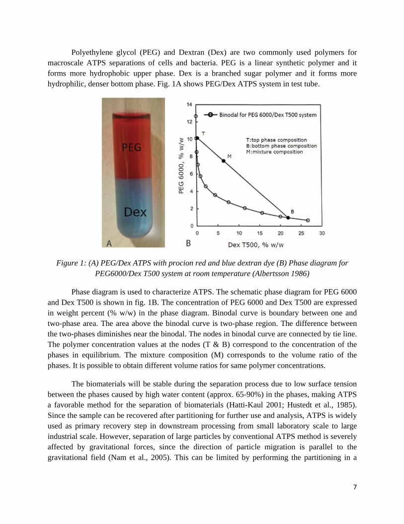
Polyethylene glycol (PEG) and Dextran (Dex) are two commonly used polymers for macroscale ATPS separations of cells and bacteria. PEG is a linear synthetic polymer and it forms more hydrophobic upper phase. Dex is a branched sugar polymer and it forms more hydrophilic, denser bottom phase. Fig. 1A shows PEG/Dex ATPS system in test tube.
Figure 1: (A) PEG/Dex ATPS with procion red and blue dextran dye (B) Phase diagram for PEG6000/Dex T500 system at room temperature (Albertsson 1986)
Phase diagram is used to characterize ATPS. The schematic phase diagram for PEG 6000 and Dex T500 is shown in fig. 1B. The concentration of PEG 6000 and Dex T500 are expressed in weight percent (% w/w) in the phase diagram. Binodal curve is boundary between one and two-phase area. The area above the binodal curve is two-phase region. The difference between the two-phases diminishes near the binodal. The nodes in binodal curve are connected by tie line. The polymer concentration values at the nodes (T & B) correspond to the concentration of the phases in equilibrium. The mixture composition (M) corresponds to the volume ratio of the phases. It is possible to obtain different volume ratios for same polymer concentrations.
The biomaterials will be stable during the separation process due to low surface tension between the phases caused by high water content (approx. 65-90%) in the phases, making ATPS a favorable method for the separation of biomaterials (Hatti-Kaul 2001; Hustedt et al., 1985). Since the sample can be recovered after partitioning for further use and analysis, ATPS is widely used as primary recovery step in downstream processing from small laboratory scale to large industrial scale. However, separation of large particles by conventional ATPS method is severely affected by gravitational forces, since the direction of particle migration is parallel to the gravitational field (Nam et al., 2005). This can be limited by performing the partitioning in a

8
microfluidic system. It is also difficult to standardize ATPS due to its sensitivity to temperature and phase composition (Stevens and Jaykus 2004).
1.3 Microfluidics
Microfluidics is the field of science that involves handling and manipulation of fluids at microliter scale in micrometer sized channels (Atencia & Beebe 2005; Whitesides 2006). The advancement in microfabrication technology in semiconductor industry led to development of smaller and smart sensors, which could be integrated in the microfluidic devices for controlling the flow of fluids in microchannel. Microfluidic devices for chemical and biological analysis have several advantages such as low sample volume; faster analysis; low cost; simultaneous detection and separation with high sensitivity.
Microfluidic devices are fabricated by standard microfabrication techniques using silicon, glass or polymers like Polydimethylsiloxane (PDMS). Silicon, which is used as standard fabrication material of integrated circuits, is also used to fabricate microfluidic chips. However, the use of silicon is limited since it is opaque to visible & UV light, making it not ideal for microfluidic devices which use optical methods for detection and it is also expensive. Glass is a preferred material for fabrication of microfluidic devices due to its favorable optical properties and resistance to high pressure. Microfluidic chip fabrication using glass in academic laboratories is however limited since the fabrication process is time consuming and expensive (Sollier et al., 2011). Polymers have the advantage of being cheaper, robust, rapid fabrication, non-toxic, impermeable to water, permeable to gases and it is also compatible for optical detection making it suitable for analysis of biological materials (Lima & Zhang 2007). Polymers such as Polydimethylsiloxane (PDMS), polyvinyl chloride (PVC) and Polymethyl methacrylate (PMMA) are generally used. Among them PDMS is most commonly used polymer for fabrication of microfluidic devices (Younan & Whitesides 1998). Despite number of advantages of microfluidic chip fabrication using PDMS, it has some limitations which affect its range of applications. (i) Poor chemical compatibility with some organic solvents. (ii) Small molecules like DNA are adsorbed by PDMS. (iii) PDMS deforms in high pressure making a substantial change in the channel geometry and affecting the overall process.
1.3.1 Fluid behavior in microfluidic channel
The fluid behavior in micro scale is different from their behavior in macro scale. At micrometer scale, the inertial forces of the fluid begin to dominate the viscous forces causing stable laminar flow when the fluid layers slides over one another and flow in parallel to the channel wall. Figure 2 shows the flow profile of a fluid in a microfluidic channel.

9
Figure 2: Laminar flow in microfluidic channel
Reynolds number relates the inertial forces to viscous forces of a fluid,
Re = ρvDH / µ = Inertial force / Viscous force
Where, “ρ” is the density of the fluid, “v” is the flow rate of the fluid, “DH” is the characteristic distance of the system and “µ” is the kinematic viscosity of the fluid. Since the characteristic distance of microfluidic device is very small (micrometer scale), the Reynolds number of the device is very low (typically less than 1) resulting in laminar flow where the fluids move in parallel layers. Due to stable laminar flow, diffusion of particles becomes the driving force for separation in straight micro channels (Nam et al., 2005). The microfluidic devices can be integrated into micro total chemical analysis systems (µTAS) or Lab-on-a-chip devices for increased functionality (Manz et al., 1990; Yamada et al., 2004).
1.4 Microfluidic aqueous two-phase system
Stable laminar flow formed in microfluidic channel due to low Reynolds number enables formation of stable two-phase system without diffusive mixing (Nam et al., 2005). µATPS device has been shown effective in separating plant cells (Yamada et al., 2004), leukocytes (Soohoo & Walker. 2009), CHO cells (Nam et al., 2005), leukocytes and erythrocytes (Tsukamoto et al., 2009), membrane proteins (Hu et al., 2010). However, µATPS has not yet been developed to partition bacterial cells. Partitioning bacterial cells in macro scale would require large sample volume. However, since µATPS works in continuous flow mode, it can be used to efficiently partition small amount of bacteria. For separating larger volumes of bacterial sample, microfluidic system with parallel channels can be used (Hansson et al., 2011). µATPS can potentially improve the partitioning because of high surface to volume ratio, which decreases the distance a cell has to travel to get attached to the interface (Soohoo & Walker. 2009). The damage caused to the cell by shear stress in conventional ATPS can be eliminated in µATPS since it is not essential to mix the phases in µATPS (Nam et al., 2005). Moreover, in a microfluidic channel, the direction of particle migration is perpendicular to the direction of gravity and it eliminates the effect of gravity on partitioning. The partitioning of biomaterials in microfluidic channel is governed by the flow rate of the fluids and by the dimensions of the channel. The flow rates are adjusted using syringe pumps to get stable laminar flow in the channel.
10
In this study, µATPS for continuously partitioning bacterial strains based on their difference in surface properties is introduced. The development of simple and rapid bacterial separation technology can improve the detection techniques. Optimum phase composition and ionic strength were determined by conventional ATPS separation of bacterial cells. The condition was reproduced in µATPS and the fractionation efficiency was compared with conventional ATPS results. This technique has the potential to be integrated with micro total chemical analysis system (µTAS) for rapid separation and detection of bacteria.

11
2. Theory
2.1 Principle of partitioning
In ATPS, each phase has distinctive physiochemical properties like charge and surface energy and it governs the partitioning of biomaterials when they are introduced into the system. PEG and Dex phase are non-ionic in nature. Physiological osmolarity is introduced in the phase system by adding salts. In addition, salts are also be added to adjust electrostatic potential differences between the phases. For example, phosphate ions partitions unevenly in the phase system making PEG phase relatively positive compared to Dex phase (Soohoo & Walker. 2008) whereas, sodium chloride salt preferentially partitions to Dex rich bottom phase (Hartounian et al., 1994). PEG rich phase is more hydrophobic than Dex rich phase (Albertsson 1986).
The cells get partitioned only after it gets attached to the interface. In macro scale ATPS, mixing the phases improves the chances of getting the cells attached to the interface and improves the speed of partitioning. Electrostatic potential difference of the system interacts with the net surface charge of the biomaterials and drives them to energetically favorable position in the system (Soohoo & Walker. 2008). Fig. 3 shows the electrostatic potential and surface energy forces acting on a bacterial cell at the interface in ATPS. The cell will migrate to one of the two bulk phases or stay at the interface depending on its interaction with the system (Payne & Kroll 1991; Pedersen et al. 1998). The distribution of cells between the two phases is described by partition coefficient (P).
P = Ct / Cb
Where, Ct and Cb are concentrations of cells in the top and bottom phase respectively.
Figure 3: The forces acting on a bacterial cell at the interface of ATPS. PEG phase is more
positive than Dex phase because of uneven partitioning of phosphate ions between the phases

12
2.2 Principle of continuous partitioning in microfluidic channel
The laminar flow property of microfluidic device is utilized to get stable ATPS by introducing two immiscible polymer solutions (PEG and Dex) through different inlets into the microfluidic channel. Figure 4 shows the cross section of a microfluidic ATPS device and the movement of the phases in the channel.
Figure 4: Principle of continuous partitioning of cells in a microfluidic channel. The direction of cell migration in a microfluidic channel is parallel to the direction of gravitational forces.
Partitioning of cells occur after they get attached to the interface. The bacterial cells were introduced in the middle inlet to have the cells in the interface of the two-phase system. The bacterial suspension is flown at lower rate than other two phases, to prevent it from diluting the two-phase system. The flow rate of PEG and Dex stream are kept low to give more time for the cells to interact with the phase system and get partitioned. Unlike conventional ATPS, the phases are beside one another in microfluidic ATPS. The direction of cell migration is parallel to the direction of gravity; therefore the partitioning of cell is not influenced by gravitational forces.

13
3. Materials and methods
3.1 Bacterial strains and cultivation
E. coli strains ML308 and BL21 were used in this project. The cultivation medium composed of (NH4)2SO2, 7.0 g/l; KH2PO4, 1.6 g/l; Na2HPO4.2H2O, 6.6 g/l; (NH4)2-H-citrate, 0.5 g/l; glycerol, 10 g/l in water (Sanden et al. 2003). The cultivation medium was autoclaved at 121 ˚C and cooled before supplemented with 1 ml/l of sterile filtrated trace elements solution and 1 ml/l of sterile filtrated 1.0 M MgSO4. The trace elements solution contained: CaCl2.2H2O, 0.5 g/l; FeCl3.6H2O, 16.7 g/l; ZnSo4.7H2O, 0.18 g/l, CuSo4.5H2O, 0.16 g/l; MnSO4.4H2O, 0.15 g/l; CoCl2.6H2O, 0.18 g/l; Na-EDTA, 20 g/l (Sanden et al. 2003).
Test cultivation of E. coli ML308 and BL21, showed that the growth rate of E. coli ML308 strain was higher than that of BL21. Therefore for cultivation of bacterial cells for ATPS partitioning, inoculation volume and incubation temperature was different for both the strains. 350 µl of E. coli ML308 stock solution was inoculated in 100 ml culture medium and incubated at 25̊ C, 180 rpm overnight (about 16 hours). For cultivation of E. coli BL21, 40 µl of stock solution was inoculated in 100 ml culture medium and incubated at 37̊C, 180 rpm overnight. The cell culture was harvested after reaching an optical density (OD600) value of 2-3 by centrifuging for 10 minutes at 4500 rpm (about 3400 xg). The cell sediment was resuspended in 10 mM sodium phosphate buffer, pH 7.4. The weight of the buffer added is approximately three times the weight of the pellet. This sample was then used for ATPS partitioning experiments.
3.2 Conventional aqueous two-phase system partitioning
The ATPS was prepared with 4.5% w/w PEG 6000 and 5.5% w/w Dex T500 in 10mM sodium phosphate buffer, pH 7.4. The selected phase concentration was in the two-phase area above the binodal curve in fig. 1B. Phase systems with different ionic strength (0 mM, 10 mM, 25 mM, 50 mM, 100 mM and 200 mM of NaCl salt) were used in this experiment to study the change in partitioning behavior of E. coli strains in different ionic condition. Bulk phase system was prepared in separating funnel and left to phase separate overnight at room temperature. The less dense, more hydrophobic PEG rich top phase and denser, more hydrophilic Dex rich bottom phase were separated and stored for preparing phase systems.
For conventional ATPS partitioning, 50 µl of bacterial sample was added to 2.5 ml of PEG and Dex phase solutions in a test tube. The contents in the tube was mixed in rotary mixer at 60 rpm for 5 minutes and left for 30 minutes to phase separate. The amount of bacteria in each phase was determined by measuring OD600 value of samples from respective phases. The experimental procedure was repeated in phase systems with different salt concentration and the partitioning efficiency was compared.

14
3.3 Microfluidic chip design and fabrication
Microfluidic chips for µATPS were fabricated by standard soft lithographic techniques using polydimethylsiloxane (PDMS) as shown in fig.5.
Figure 5: Overview of the fabrication process. (A) Master with SU-8 structure. (B) PDMS and curing agent (10:1 ratio) is mixed and poured over the master. (C) Desiccator is used to remove bubbles. (D) The chips are kept in oven for 6 hours at 37°C. (E) The PDMS structure is peeled off from the master. Holes for inlet and outlet channels are punched. (F) The glass slides and PDMS surfaces are plasma oxidized. (G) PDMS surfaces are bound irreversibly to the glass
slides by baking in hot plate at 75°C.
Negative photoresist SU-8 polymer was spin coated on a silicon wafer basal plate to a thickness of 57 µm. The substrate was then baked in oven at 65°C for 5 minutes and then at 95°C for 15 minutes. Printed photo mask film of the microfluidic chips was placed over the substrate and exposed to UV rays in UV transilluminator. UV exposure makes SU-8’s long molecular chains to cross link and harden the exposed microchannel structure. After exposing the substrate to UV rays, the unexposed material are washed away leaving the exposed channel structure behind (Fig. 5A). PDMS and curing agent (10:1 ratio) was mixed and cast on the patterned master (Fig. 5B). It was then desiccated using vacuum pump to remove air bubbles (Fig. 5C) and cured in oven for 6 hours at 37 °C (Fig. 5D). The PDMS was then peeled off from the master and were cut using sharp knife. The holes for inlet and outlet section were then punched in the PDMS structure (Fig. 5E). The PDMS structure was bonded irreversibly to glass slide by oxygen plasma

15
activation (Fig 5F) to form microchannel structures and baked at 75°C for 10 minutes in hot plate (Fig. 5G).
Microfluidic chips with three different geometries were used for µATPS partitioning. The only difference being the length of the microchannel (25 mm, 20 mm and 15 mm). The height and width of the microchannel were 57 µm and 150 µm respectively.
3.4 Microfluidic aqueous two-phase system partitioning
Separated bulk phase system used in conventional ATPS experiments was also used for µATPS partitioning experiments. PEG and Dex rich phase solutions were introduced separately into the microchannel using syringe pumps. Bacterial cell suspended in 10 mM sodium phosphate buffer was introduced through the middle inlet. The flow rates of all three inputs (PEG 3 µl/min, Dex 0.5 µl/min and bacterial solution 0.25 µl/min) were controlled independently to create a stable two-phase system. Movement of the phases in the microchannel was observed using a microscope. The samples from three outputs were collected separately and were used for cell counting using plating techniques to analyze the partitioning behavior of the cells.

16
4. Results
4.1 Conventional aqueous two-phase system partitioning
The optical density (OD600) of PEG rich and Dex rich phase from conventional ATPS partitioning experiments were measured and compared with the concentration of the bacterial cells in the system to measure the percentage of cells partitioned in each phase. The remaining cells are believed to be attached to the interface. Fig. 6 and 7 shows the partitioning behavior of E. coli ML308 and E. coli BL21 respectively in conventional ATPS with varying ionic strength.
Figure 6: E. coli ML308 cells partitioned in PEG rich top phase in all ionic conditions. The partitioning was not affected by the salt concentration in the phase system
Figure 7: E. coli BL21 cells showed affinity for Dex rich bottom phase with increasing salt concentration in the phase system
17
E. coli ML308 partitioned in hydrophobic PEG rich top phase and the partitioning was not influenced by ionic strength of the system. About 99% of the cells migrated to PEG rich phase in ATPSs with 0 mM, 50 mM, 100 mM and 200 mM NaCl concentration. Less than 1% of the cells were recovered from Dex rich phase under all conditions. Partitioning preference of E. coli ML308 to PEG rich phase could be observed from the visual cloudiness of top phase in the experiment tube. Partitioning behavior of E. coli BL21 was different from that of E. coli ML308. Under non-ionic condition, about 94% of the cells were recovered from PEG rich top phase. The affinity of E. coli BL21 to PEG rich phase declined when ionic strength of the system was increased. In phase system with 10 mM NaCl concentration, about 80% of the cells partitioned to the interface and about 16% of the cells partitioned to PEG rich top phase. The cells showed affinity for interface in the systems with 25 mM, 50 mM and 100 mM NaCl concentration. In 200 mM NaCl ATPS system, the cells were equally distributed between the interface and Dex rich bottom phase.
4.2 Microfluidic chip design
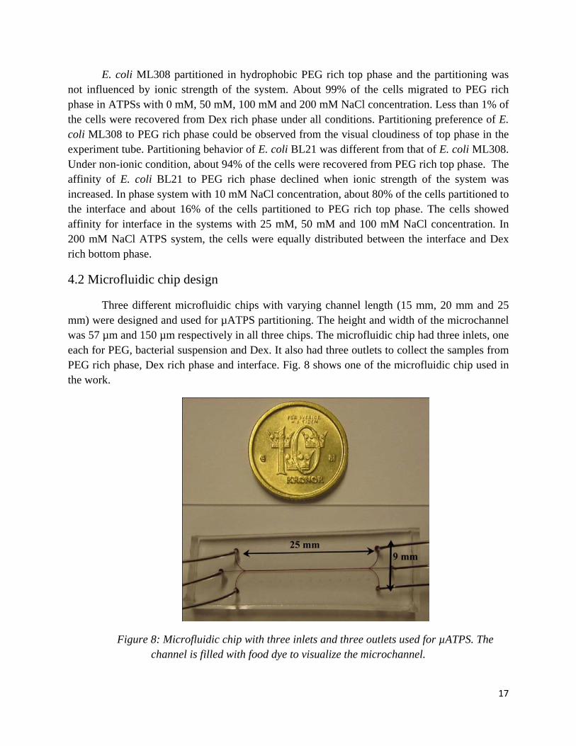
Three different microfluidic chips with varying channel length (15 mm, 20 mm and 25 mm) were designed and used for µATPS partitioning. The height and width of the microchannel was 57 µm and 150 µm respectively in all three chips. The microfluidic chip had three inlets, one each for PEG, bacterial suspension and Dex. It also had three outlets to collect the samples from PEG rich phase, Dex rich phase and interface. Fig. 8 shows one of the microfluidic chip used in the work.
Figure 8: Microfluidic chip with three inlets and three outlets used for µATPS. The channel is filled with food dye to visualize the microchannel.

18
4.3 Microfluidic aqueous two-phase system partitioning
Figure 9 shows the inlet and outlet section of the µATPS device. The flow rate of all three inlets were adjusted to get stable ATPS and to recover the interface from the middle outlet. The flow rate of PEG, bacterial suspension and Dex was optimized at 3µl/min, 0.25µl/min and 0.5µl/min respectively.
Figure 9: Inlet and outlet section of a microfluidic ATPS device. The phases and their respective flow rates are mentioned.
About 98% of E. coli ML308 was recovered from the middle outlet both in non-ionic and in 100 mM salt system. In the case of E. coli BL21, about 96% of the cells were recovered from the middle outlet in non-ionic system as expected. However, 92% of the cells attached to the interface even in phase system with 100 mM NaCl concentration.

19
5. Discussion
5.1 Conventional aqueous two-phase system partitioning
The partitioning behavior of E. coli ML308 and E. coli BL21 was similar in non-ionic condition where about 90% of the cells partitioned in PEG rich top phase. PEG phase is more hydrophobic than Dex phase in non-ionic condition and it has positive electric potential compared to Dex rich bottom phase, i.e. the top phase is more positive than the bottom phase (Johansson 1974). Therefore, the partitioning behavior of bacterial strains with negative net surface charge in non-ionic condition could be explained by the surface charge influenced partitioning. However, when NaCl was added to the phase system, E. coli BL21 cells started showing affinity towards Dex rich phase. NaCl salt has preference to partition into Dex rich bottom phase (Hartounian et al., 1994). The partition of NaCl to Dex phase reversed the electric potential of the phases, making Dex rich phase more positive compared to PEG rich phase (Johansson 1974; Hartounian et al., 1994). The change in electrical potential of the phases, made E. coli BL21 cells which has negative net surface charge to partition in the interface at low salt concentration and in the bottom phase at higher salt concentration. However, about 99% of E. coli ML308 cells partitioned in PEG rich top phase in ionic condition. This partition could have been influenced by the hydrophobic property of the bacterial cell since it lacks net surface charge (Råvik M., 2007).
Although conventional ATPS is a simple method for partitioning E. coli ML308 and E. coli BL21 cells based on their surface properties compared to other separation techniques, the influence of gravitational forces on partitioning is a major disadvantage. Prolonging the phase separation period over 30 minutes would make the cells to settle down and affect the partitioning efficiency in conventional ATPS.
5.2 Microfluidic aqueous two-phase system partitioning
The key aspect in µATPS is to achieve stable phase system for efficient recovery of phases, meaning that the samples collected at the outlets are without contamination of other phase components (Hardt & Hahn, 2011). The formation of stable phase system in a microfluidic channel is governed by the geometries of the channel and flow rates of the phases. These parameters also govern the partitioning of bacterial cells in the micro-channel. By reducing the flow rate of the phases, the bacterial cells will be attached to the interface for a longer time which in turn can increase the partitioning efficiency. The stability of the phases will be affected if the channels are too high.
In µATPS, the optimum flow rate of PEG phase, bacterial suspension and Dex phase was determined to achieve stable two-phase system and to recover the interface from the middle inlet. Since Dex phase is more viscous than PEG phase, the flow rate of Dex (0.5µl/min) was kept comparatively lower than the flow rate of PEG phase (3µl/min), to recover the interface from the

20
middle inlet and collect the PEG and Dex phases separately. The flow rate of bacterial suspension was kept very low (0.25µl/min) to avoid dilution of two-phase system. The low flow rate of bacterial suspension helped to mimic conventional ATPS condition by allowing the cells to get attached to the interface in contact with PEG and Dex phases, which is essential for the partition to take place.
In µATPS partitioning, about 98% of E. coli ML308 cells was recovered from the middle outlet as expected in phase system with 100 mM salt concentration. However, even in non-ionic and ionic conditions, E. coli ML308 and BL21 cells were recovered from the middle outlet. It indicates the cells are either (i) not migrating from the interface or (ii) the cells are partitioning to PEG and Dex phases but stay close to the interface and get recovered from the middle outlet. The sample collected from middle outlet had considerable amount of PEG and Dex phase. Therefore, the cells that might have partitioned and stayed close to the interface would have been collected from middle outlet. In this case, further fractionation of sample from middle outlet to separate the interface from PEG and Dex phase components can effectively increase the portioning efficiency.
Several factors have to be taken in to account and analyzed to improve the partitioning efficiency of µATPS. The major concern in µATPS is drifting of interface from middle outlet at low flow rates. However, flowing the phases at low rate is essential to get better partitioning. Therefore the geometries of the micro-channel have to be further optimized to get stable two-phase system even at low flow rate. The partitioning theory in micro-scale is not well studied and more work is needed to understand the principle of partitioning in µATPS. The viscosity difference of PEG and Dex also becomes a major issue in micro-scale compared to conventional ATPS setup. The partitioning behavior of biomaterials in µATPS can be studied by using fluorescent beads or bacteria expressing fluorescent proteins in a model µATPS. Their migration behavior can be studied by observing the channel using optical microscope. This could give a clear picture of the migration of biomaterials in µATPS and can be implemented for partitioning of bacteria and other biomaterials of choice.

21
6. Conclusion
Conventional ATPS is simple and rapid method for separation of bacteria based on surface property difference. The biocompatibility and bioselectivity of this technique makes it more suitable for bioseparation than other techniques. In this project, a novel µATPS device for partitioning E.coli strains based on their surface property was analyzed. A stable µATPS was achieved by optimizing the flow rate. The partitioning efficiency achieved in conventional system was not reproduced in microfluidic system. Further fractionation of sample could be a key element to improve the partitioning efficiency of the system. The microfluidic device can continuously partition bacterial cells even in low sample volumes which cannot be achieved by conventional ATPS. Once developed, this device has potential to be integrated into lab-on-a-chip device for rapid bacterial separation and detection.

22
Acknowledgements
I would like to thank my supervisor Aman Russom for giving me opportunity to work in this project and for his guidance. I would also like to thank my examiner and co-supervisor Andres Veide for all his wise inputs about ATPS. Special thanks to people at Division of Nano Biotechnology for allowing me to use droplet station for microfluidic experiments.
Thanks to Johan Norén, Harisha Ramachandraiah, Jonas Hansson and Sahar Ardabili for your contribution to the work. Finally I wish to thank everyone that I happened to meet in Division of Bioprocess Technology and Division of Cell Physics, for their company and for giving a very pleasant working atmosphere.

23
7. Reference
Albertsson PÅ. 1986. Partition of cell particles and macromolecules. 2nd edition. John Wiley and Sons, New York
Atencia J., Beebe D.J. 2005. Controlled microfluidic interfaces. Nature 437: 648-655
Dziezak J.D. 1987. Rapid methods for microbiological analysis of foods. Food Technol. 41: 56
Hansson J., Karlsson J.M., Haraldsson T., Wijngaart W.VD., Russom A. 2011. Inertial particle focusing in parallel microfluidic channels for high-throughput filtration. Conference manuscript presented at the 16th International Conference on Solid-State Sensors, Actuators and Microsystems (Transducers 2011), Beijing, China
Hardt S., Hahn T. 2011. Microfluidics with aqueous two-phase systems. Lab on a chip 12:434-442
Hartounian H., Sandler S.I., Kaler E.W. 1994. Aqueous two-phase systems. 1. Salt partitioning. Ind. Eng. Chem. Res. 33: 2288-2293
Hatti-Kaul R., 2001, Aqueous two-phase systems, Molecular biotechnology 19: 269-277
Hu R., Feng X., Chen P., Fu M., Chen H., Guo L., Liu BF. 2010. Rapid, highly efficient extraction and purification of membrane proteins using a microfluidic continuous-flow based aqueous two-phase system. Journal of Chromatography A 1218: 171-177
Hustedt H., Kroner K.H., Menge U., Kula M.R. 1985. Protein recovery using two-phase systems. Trends in Biotechnology 3(6): 139-144
Johansson G. 1974. Effects of salt on the partition of proteins in aqueous polymeric biphasic systems. Acta Chem Scand Ser A Phys & Inorg Chem 28: 873-882
Lima C.T., Zhang Y. 2007. Bead-based microfluidic immunoassays: The next generation Biosensors and Bioelectronics 22; 1197–1204.
Manz A., Graber N., Widmer H.M. 1990. Miniaturized total chemical analysis systems: a novel concept for chemical sensing. Sens. Actuators B: Chemical 1: 244–248.
Nam KH., Chang WJ., Hong H., Lim SM., Kim DI., Koo YM. 2005. Continuous-flow fractionation of animal cells in microfluidic device using aqueous two-phase extraction. Biomedical microdevices 7(3): 189-195.
Payne M.J., Kroll R.G. 1991. Methods for the separation and concentration of bacteria from foods. Trends Food Sci. Technol. 2: 315-319.
24
Pedersen L.K., Skouboe P., Rossen L., Rasmussen O.F. 1998. Separation of Listeria monocytogenes and Salmonella berta from a complex food matrix by aqueous polymer two-phase partitioning. Letters in Applied Microbiology 26: 47.
Råvik M., Cimander C., Elofsson U., Veide A. 2007. A method for microbial cell surface fingerprinting based on surface plasmon resonance. Journal of Biochemical and Biophysical methods 70: 595-604.
Sanden A.M., Prytz I., Tubulekas I., Forberg C., Le H., Hekor A., Neubauer P., Pragai Z., Harwood C., Ward A., Picon A., Mattos J.TD., Postma P., Farewell A., Nystrom T., Reeh S., Pedersen S., Larsson G. 2003. Limiting factors in Escherichia coli fed-batch production of recombinant proteins. Biotechnol Bioeng 81: 158-166
Sollier E., Murray C., Maoddi P., Carlo DD. 2011. Rapid prototyping polymers for microfluidic devices and high pressure injections. Lab on a chip 11: 3752-3765
Soohoo J.R., Walker G.M. 2008. Microfluidic aqueous two phase system for leukocyte concentration from whole blood. Biomed Microdevices 11: 323-329
Stevens K.A., Jaykus L.A. 2004. Bacterial separation and concentration from complex sample matrices: A reviw. Critical reviews in Microbiology 30(1): 7-24.
Swaminathan B., Feng P. 1994. Rapid detection of food-borne pathogenic bacteria. Annu. Rev. Microbiol. 48:401-426.
Tsukamoto M., Taira S., Yamamura S., Morita Y., Nagatani N., Takamura Y., Tamiya E. 2009. Cell separation by an aqueous two-phase system in a microfluidic device. Analyst 134: 1994-1998
Whitesides G.M. 2006. The origins and the future of microfluidics. Nature 442: 368-373
Yamada M., Kasim V., Nakashima M., Edahiro J., Seki M. 2004. Continuous cell partitioning using an aqueous two-phase flow system in microfluidic devices. Biotechnology and Bioengineering 88(4):489-494.
Younan X., Whitesides G.M. 1998. Soft Lithography. Angew. Chem. Int. Ed 37:550 – 575

25
APPENDIX
Conference manuscript: MicroTAS 2011, Seattle, Washington, October 2011

DEVELOPMENT OF MICROFLUIDIC AQUEOUS TWO-PHASE SYSTEM FOR CONTINUOUS PARTITIONING OF E. coli STRAINS
Prem Kumar Periyannan Rajeswari1,2, Harisha Ramachandraiah2, Jonas Hansson2, Sahar Ardabili2, Andres Veide1, Aman Russom2*
1Department of Bioprocess Technology, School of Biotechnology and 2Division of Cell Physics, Department of Applied Physics, KTH Royal Institute of Technology, Stockholm, SWEDEN
ABSTRACT
The interaction of bacterial cells with surrounding environment depends on its surface characteristics such as hydrophobicity, hydrophilicity balance and net charge. In this paper, aqueous two-phase system partitioning of Escherichia coli strains based on their difference in surface properties is introduced in a microfluidic system. While aqueous two-phase system is widely use to separate biomolecules on macroscale, the method has not been adapted in microfluidic system. The bacterial cells are partitioned based on their affinity for streams formed by aqueous polymers polyethylene glycol (PEG) and dextran (Dex). Partitioning efficiency of two Escherichia coli strains is currently being optimized.
KEYWORDS: Microfluidics, Aqueous two phase system, Escherichia coli, Continuous partitioning, PEG, Dex INTRODUCTION
Understanding and analysis of the surface of microbial cells is important in many fields like biotechnology, process engineering, medicine, environmental protection and material technology. For instance, the host immune response is triggered by the cell surface. Additionally, in down stream processing of proteins using bacteria or other cells, non-specific adsorption as well as the existence of multiple bacterial strains can affect the overall operational efficiency. Although bacterial concentration methods such as centrifugation, filtration, and immunomagnetic separation have been reported for food systems, the separation and subsequent concentration of bacterial cells from a food sample during sample preparation continues to be a stumbling block in the advancement of molecular methods for the detection of foodborne pathogens [1].
Although several methods are available for surface characterization, aqueous two-phase system (ATPS) partitioning method has advantage of high selectivity and biocompatibility [2]. In this paper, we use this method to separate two bacteria strains based on their surface property. ATPS is formed by two immiscible aqueous polymer solutions or one aqueous polymer solution and a salt solution. The separation of biomaterials is based on its surface characteristics. Partition of large particles in macro ATPS is severely affected by gravity and it is also difficult to perform continuous partitioning. In microfluidic based system (!ATPS), the influence of gravitational force on partitioning is negligible since the direction of particle migration will be perpendicular to gravitational field [3]. In addition, the continuous-flow microfluidic system could potentially enable partitioning of bacterial strains in sample volumes not possible using conventional systems. Moreover in !ATPS, the larger surface to volume ratio will decrease the distance a cell has to travel for partitioning to occur [3]. In this paper, we introduce !ATPS for partitioning of two E.coli strains (ML308 and BL21).
THEORY
In ATPS, the top phase will be PEG rich and the bottom phase will be Dex rich at equilibrium. PEG and Dex are immiscible non-ionic polymers in nature. Salt is added to give physiological osmolarity to the system and some salts have affinity for particular phase. Uneven migration of ions would leave one of the phases more positively charged than the other and it would introduce electrostatic potential difference between the phases [4]. Each phase has distinctive physiochemical properties like hydrophobicity, hydrophilicity, charge and surface energy and it controls the partitioning of biomaterials when they are introduced in to the system. Fig. 1 shows the electrostatic potential and surface energy forces acting on the cell in the interface. The cells migrate to any of the two phases or stay at the interface depending on its interaction with the system. The distribution of cells between the two phases is described by partition coefficient (P) defined as in equation (1).
P = Ct / Cb (1)
Where, Ct and Cb are concentrations of cells in the top and bottom phase respectively.

Figure 1: The forces acting on the cell in the interface of the two phase system. PEG phase is more positive than Dex phase because of uneven partitioning of phosphate ions between the phases
EXPERIMENTAL
E.coli strains ML308 and BL21 were cultivated overnight and harvested after reaching an OD600 value of 2 to 3 to be used in ATPS experiments. Macro ATPS was prepared with 4.5% (w/w) PEG 6000 and 5.5% (w/w) Dex T500 in 10 mM sodium phosphate buffer, pH 7.4. Bulk PEG and Dex phase was prepared in separating funnel after overnight phase separation. Different concentration of sodium chloride (NaCl) was used to change the ionic strength of the phase system. 50 !l of bacterial sample was added to phase system containing 2.5 ml of both phases in a tube. The contents in the tube was mixed and left for 30 minutes to phase separate. The amount of bacteria in each phase was determined by measuring OD600 value of samples from respective phases. The experiment was repeated in phase systems with different salt concentration and the results were compared with other systems.
Microfluidic devices for !ATPS and were fabricated by standard soft lithographic techniques using PDMS. Fig. 2 shows an example of a three inlet-three outlet chip used in this work. 4.5% (w/w) PEG 6000, 5.5% (w/w) Dex T500 solution and bacterial cells suspended in 10 mM sodium phosphate buffer solution were individually introduced in to the microfluidic chip using syringe pumps. Fig. 3 shows the inlet and outlet section of the microfluidic chip during the experiment. The flow rates of all three inlets (3; 0.5 and 0,25 µl/min for PEG, Dex and bacteria respective) were manipulated to get stable phase system.
Figure 2: Microfluidic chip with three inlets and three outlets. The length, width and height of the main channel are 25 mm 150 !m and 57 !m respectively. Two other chips with different lengths (20 mm and 15 mm) were also used in this work.
Figure 3: Inlet and outlet section of a microfluidic device. The phases and their respective flow rates are mentioned. A stable two-phase system was established, enabling the extraction of the interface through the center outlet.
RESULTS AND DISCUSSION
Initially, the partitioning efficiency of each E.coli strain was evaluated in a macro ATPS. After partitioning, each phase was separated and analyzed. As can be seen in Figure 4, E.coli ML308 preferred to migrate to hydrophobic PEG phase irrespective of ionic strength of the system. About 99% of the bacterial cell partitioned to the PEG rich top phase. In the case of E.coli BL21 strain, the cells showed affinity for PEG phase only in non-ionic conditions. When salt is added to the system, the bacteria started to migrate towards the interface. Further increase in ionic strength resulted in cell partitioning in

the dextran phase (Fig. 5). About 80% of the E.coli BL21 cells migrated to the interface in phase system containing 10 mM NaCl to 100 mM NaCl concentration. 200 mM NaCl phase system resulted in about 50% of the cells partitioned in the Dex rich bottom phase.
Figure 4. Conventional ATPS partitioning of E. coli ML308. Figure 5. Conventional ATPS partitioning of E. coli BL21
Using the optimized PEG and Dex concentrations, !ATPS partitioning of E.coli BL21 was examined (Table 1). To keep
the interface in the center, the PEGphase was flown at a higher flow rate (3µl/min) while the Dex was running at 0.5 µl/min (see Fig.2). The cells are introduced through the center inlet. The collecdet sample from the three outlets are plated over night and counted. As expected, the cells remained in the interface for the 100 mM salt system (Table 1). However, the cells also stayed in interface even in non-ionic system, indicating the cells are either (i) not migrating away from interface or (ii) the cells are migrating and partitioning in the PEG phase but close enough the middle outlet and extracted from the center.
Table 1: !ATPS partitioning of E. coli BL21
Phase % of cells in 0 mM NaCl system % of cells in 100 mM NaCl system
PEG rich phase 2.7 0.3 Dex rich phase 1.3 6.9
Interface 96 92.8 Partitioning in !ATPS depends on various factors like flow rate of the phases and dimensions of the channel in addition
to affinity of the particle to the phases. For improved performance, the flow can be further decreased to increase the time of interaction of the cells with the phase system for increased partitioning efficiency of the bacterial cells. In addition, the ions can be partitioned overnight in bulk system prior the !ATPS experiment. Currently, we are optimizing the flow conditions for a stabile system using a better syringe pump (low pulsing) to improve the partitioning efficiency.
CONCLUSION
In summary, we introduce microfluidic based aqueous two-phase system to separate bacteria strains. This device once developed can be used as a rapid analytical tool for detecting bacterial contamination in food and process industry. It can also be integrated to lab-on-a-chip (LOC) systems for downstream processing. REFERENCES
[1] Stevens, K. A. and L. A. Jaykus (2004). "Bacterial separation and concentration from complex sample matrices: A review." Critical Reviews in Microbiology 30(1): 7-24.
[2] Albertsson PÅ (1986). Partition of cell particles and macromolecules, John Wiley and Sons, New York [3] Yamada, M., V. Kasim, et al. (2004). "Continuous cell partitioning using an aqueous two-phase flow system in
microfluidic devices." Biotechnology and Bioengineering 88(4): 489-494. [4] SooHoo, J. R. and G. M. Walker (2009). "Microfluidic aqueous two phase system for leukocyte concentration from
whole blood." Biomedical Microdevices 11(2): 323-329. CONTACT: *Aman Russom, Tel: +46-709509684; Email: [email protected]