Embed Size (px)
Citation preview
Microfluidic cell cultureMatthias Mehling and Savas Tay
Available online at www.sciencedirect.com
ScienceDirect
Microfluidic techniques allow precise control of fluids and
particles at the nanoliter scale and facilitate simultaneous
manipulation and analysis of cultured cells, starting from a
single cell to larger populations and to intact tissues. The use of
integrated microfluidic devices has considerably advanced the
fields of quantitative and systems biology. In this review, we
survey the recent developments in microfluidic cell culture, and
discuss not only the advantages but also limitations of using
such systems, and give an outlook on potential future
developments.
Addresses
Department of Biosystems Science and Engineering, ETH Zurich,
Switzerland
Corresponding authors: Tay, Savas ([email protected])
Current Opinion in Biotechnology 2014, 25:95–102
This review comes from a themed issue on Analytical biotechnology
Edited by Frank L Jaksch and Savas Tay
For a complete overview see the Issue and the Editorial
Available online 12th November 2013
0958-1669/$ – see front matter, # 2013 Elsevier Ltd. All rights
reserved.
http://dx.doi.org/10.1016/j.copbio.2013.10.005
IntroductionThe establishment of techniques to maintain and grow
cells in vitro is a major milestone in biological sciences.
Since its introduction in 1912 [1], methods have been
developed for culturing, expanding, differentiating and
de-differentiating cells [2]. The basic tools and require-
ments for cell culture (Table 1) have not changed
significantly since then. Besides substantial advances
in controlling for contamination with bacteria, mold and
yeast, most improvements in cell-culture have been
made to the culture media and the materials used for
production of the cell culture dish. Interestingly, even
the shape of the cell culture dish remained mostly
unchanged for nearly a century. The recent use of
robotics to eliminate time-consuming manual pipetting
steps resulted in increased throughput and accuracy,
and constitutes a rare yet exceptional improvement of
the technique. Despite their bulky and complicated
nature and the substantial costs associated with their
use, robotic systems have now become a cornerstone
of large-scale industrial applications of cell culture.
Nevertheless, the principles of traditional cell culture
techniques remain mostly unchanged by these modifi-
cations.
www.sciencedirect.com
Recent insight from the emerging fields of quantitative
and systems biology highlighted the importance of ana-
lyzing individual cells instead of just bulk cultures.
Because of natural cell-to-cell variability in biochemical
parameters such as mRNA and protein expression levels,
and the inevitable stochastic molecular noise, population-
averaged bulk assays are often inaccurate or misleading.
Further, cellular biochemical parameters and signaling
states constantly change, making dynamical analysis of
cells crucial in understanding how biological systems
operate. Basic methods in single-cell and dynamical
analysis have mostly been limited to conventional cell
culture techniques using the traditional dish and the
hand-held pipette. Recent introduction of microfluidics
to biological sciences allowed addressing these funda-
mental limitations (Table 1). Microfluidic cell culture
allows controlling fluid flow in the micrometer and nano-
liter scale in precisely defined geometries and facilitates
simultaneous manipulation and analysis starting from a
single cell level, to larger cell populations and up to
tissues cultured on fully integrated and automated chips.
Among the multitude of approaches for manufacturing
microfluidic devices, soft lithography of poly-dimethylsi-
loxane (PDMS) has become standard for cell culture
applications. With this technique, structures of
micrometer resolution are molded from a hard master
into PDMS. Many advanced microfluidic chips use min-
iaturized micromechanical membrane valves made from
PDMS to efficiently manipulate fluids at the microliter
scale. Comparable to transistors in electronic devices,
these valves allow exact spatial and temporal control of
fluid flow and delivery of media, drugs and signaling
factors to live cells. Parallelized fabrication using optical
lithography and careful alignment of flow and control
layers allow rapid construction of various types of chan-
nels, chambers and valves in fully integrated, compact
devices (Figure 1) [3]. The advent of PDMS microme-
chanical valves along with multiplexing methods allowed
the development of truly ‘lab-on-a-chip devices’ that
improve the accuracy and throughput of biological assays
by orders of magnitude. The time from computerized
chip design to actual laboratory use of PDMS chips is
typically a few weeks. Integration of thousands of micro-
mechanical valves in compact platforms and their auto-
mation allows performing tedious, manual-labor intensive
tasks efficiently, rapidly and with minimal intervention
[4]. The use of nanoliter-sized assay chambers results in
increased precision, and reduced consumption of costly
reagents. Microfluidics offers the possibility to deliver not
only chemical but also mechanical signals, providing an
extra degree of control over cultured cells. Examples for
Current Opinion in Biotechnology 2014, 25:95–102
96 Analytical biotechnology
Table 1
Basic requirements for cell culture, and improvements when microfluidic methods are used
Requirements Conventional cell culture Microfluidic cell culture
Control of temperature and gasses Large fluid volumes prevent fast changes Small volumes allow dynamic control
Addition of nutrients and removal
of metabolites
Infrequent, manual exchange of large volumes Precisely measured, continuous or transient
exchange of media
Stimulation with drugs/proteins and
simultaneous imaging
Mostly not feasible Feasible
Parallelization of cellular assays Not feasible High capability for parallelization
Automation of cell culture tasks Bulky, expensive fluid-handling robots must be used High capability for automation in compact,
inexpensive format
Single-cell manipulation and analysis Manually involved, inaccurate, low throughput Accurate and high-throughput
Figure 1
Conditions
Input manifold & mixer
Chambers
Multiplexers
Control channels
Peristaltic pump
Cell inputMixer flushoutput
1mm
Flow
Chambers(1.12mm × 0.9mm × 40μm)
Output sieve
Input sieve
Flushchannel
Chambers
Chaotic mixer
Input manifold
1mm
1cm
Rep
licat
es
(a)
(b)
flow
OutletsInlets
control
Current Opinion in Biotechnology
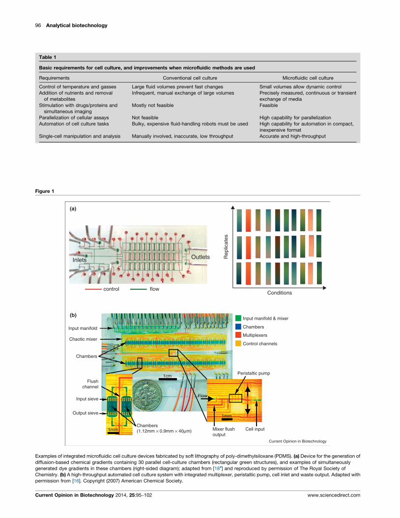
Examples of integrated microfluidic cell culture devices fabricated by soft lithography of poly-dimethylsiloxane (PDMS). (a) Device for the generation of
diffusion-based chemical gradients containing 30 parallel cell-culture chambers (rectangular green structures), and examples of simultaneously
generated dye gradients in these chambers (right-sided diagram); adapted from [18�] and reproduced by permission of The Royal Society of
Chemistry. (b) A high-throughput automated cell culture system with integrated multiplexer, peristaltic pump, cell inlet and waste output. Adapted with
permission from [16]. Copyright (2007) American Chemical Society.
Current Opinion in Biotechnology 2014, 25:95–102 www.sciencedirect.com

Microfluidic cell culture Mehling and Tay 97
mechanical signals are gradients of the surface-stiffness
[5] of cell culture devices or exact regulation of shear
stress forces [6].
In this review we discuss firstly the advantages, secondly
the issues and troubleshooting of the use of microfluidics
in cell culture, thirdly the recent developments of micro-
fluidic cell culture techniques, and finally an outlook on
open questions and potential developments in the field.
Advantages of microfluidic cell cultureMicrofluidics provide high degree of control over cell
culture conditions in various aspects (Table 1). Given
the small geometrical dimensions in the microscale and
nanoscale, the movement of fluids is laminar, and place-
ment of fluid volumes in the nL-range, pL-range and
even fL-range is possible [7]. The ability of exactly
timing fluid flow using in-chip membrane valves allows
precise chemical and physical control of the microenvir-
onment (the response time of valves can be as short as
1 ms). The doses delivered to cells can be measured in
nanoliters to femtoliters, representing a significant im-
provement in precision compared to the traditional pip-
ette that can measure microliters at best. Examples are
controlling of glucose [8] and oxygen concentrations [9]
with the potential to characterize cellular responses of
single cells to these changes. Microfluidic cell culture
devices also allow precise control of cell numbers and
density in a given area or volume, and can provide
placement of cells in complex geometries [10], their
monitoring with high spatial and temporal resolution
and their individual retrieval during or following exper-
iments. Further, cells can be organized into three-dimen-
sional geometries in matrices such as hydrogels, allowing
culture of cells in structures resembling those in tissues
[11].
The small dimensions of spatially separated microfluidic
compartments allow assembly of a multitude of indivi-
dually controllable cell culture chambers on a single
device. This facilitates high parallelization of exper-
iments, high throughput of samples and reactions and
thus improvement of reproducibility, as well as a
reduction in reagent costs [12,13].
Stem cells that are difficult to culture with conventional
techniques can be expanded relatively rapidly in micro-
fluidic culture [14]. Parallelization of experimental con-
ditions allows for enhanced cell-based screening assays,
such as immunophenotyping assays monitoring single
cell cytokine production in response to external stimuli
[15].
Microfluidic systems can be automated to an immense
extent. Automation of microfluidic cell culture systems
allows culturing cells for several weeks under precisely
defined conditions without manual intervention [7,16].
www.sciencedirect.com
Non-dividing or slowly dividing cells can be cultured
continuously in the same cell culture chamber by regular
or constant replacement of media. Automation of cell
culture systems leads to standardized manipulation,
monitoring and sampling of cultured cells. This allows
strict adherence to the timing of protocols, which is of
particular value when dynamic processes in smaller time
intervals, for example, seconds or minutes, are character-
ized.
PDMS based microfluidics provide excellent live cell
imaging conditions as PDMS offers transparency and
stable optical features, and the optical aberrations and
auto-fluorescence induced by small volumes of cell cul-
ture medium in such devices are generally negligible.
Efficient tracking of migrating cells [17��] and phenotyp-
ing of resting cells in response to predefined stimuli
have been demonstrated. In combination with fluorescent
live cell imaging, microfluidic cell culture devices there-
fore allow powerful characterization of a multitude of
cellular responses on a single cell as well as population
level.
Resulting from the above-mentioned advantages (con-
trollability, parallelization, automation, excellent ima-
ging properties), microfluidics has become particularly
valuable for analysis of single cell dynamics. With the
help of microfluidic devices cell growth and regulation of
cell size can be directly observed [8,18�,19,20�] and
lineages of single cells can be tracked for several gener-
ations [21–23]. On a molecular level microfluidics allow
the characterization of transcription factor and gene
expression dynamics in single-cells thereby adding sub-
stantially to our understanding of the function of bio-
logical systems [24–26]. Further, the dynamics of protein
secretion [27��,28] and the dynamic analysis of signaling
pathways have been addressed with the help of micro-
fluidic cell culture devices [24].
Issues and troubleshooting with PDMS basedmicrofluidic cell culture systemsPDMS offers many advantages for manufacturing micro-
fluidic cell culture systems. It allows implementation of
robust geometric structures and pneumatic membrane
valves and has a low level of auto-fluorescence and is
transparent. However, PDMS is a hydrophobic and por-
ous material, which results in the absorption of hydro-
phobic molecules such as lipids or small molecules [29]
from culture media into PDMS (Table 2). To maintain
stable cell culture conditions and to reduce the effects of
absorption into PDMS, regular replacement of culture
media is necessary. Pretreatment of PDMS devices with
sol–gel chemistry can reduce small-molecule absorption
[30]. Also, small volumes of media in microfluidic cell
culture devices can result in faster consumption of nutri-
ents and in an increase of the concentration of metab-
olites or secreted molecules, especially compared to
Current Opinion in Biotechnology 2014, 25:95–102
98 Analytical biotechnology
Table 2
Major issues with PDMS microfluidic cell culture devices and troubleshooting
Issues Potential solutions
Absorption of hydrophobic molecules into PDMS Sol-gel methods to block PDMS pores
PDMS water permeability and evaporation lead to
reduction of culture medium
Use of humidifiers or integrated media baths
PDMS toxicity Extended postproduction baking, autoclaving of devices,
or chemical extraction of uncrosslinked species
Insufficient cell adhesion Pretreatment with adhesion molecules like fibronectin
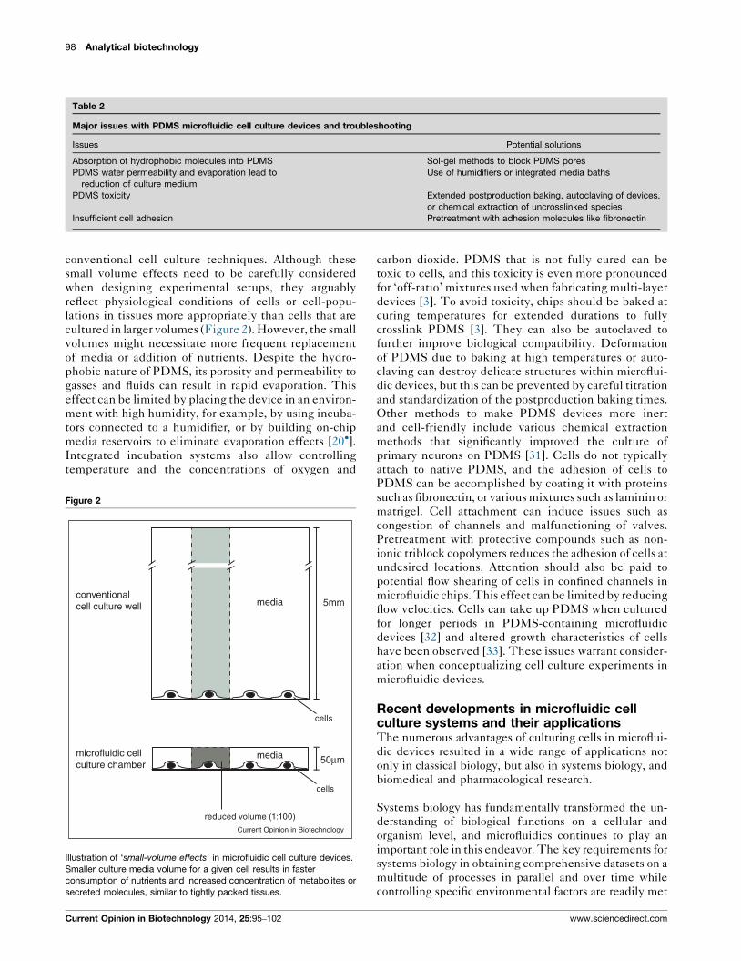
conventional cell culture techniques. Although these
small volume effects need to be carefully considered
when designing experimental setups, they arguably
reflect physiological conditions of cells or cell-popu-
lations in tissues more appropriately than cells that are
cultured in larger volumes (Figure 2). However, the small
volumes might necessitate more frequent replacement
of media or addition of nutrients. Despite the hydro-
phobic nature of PDMS, its porosity and permeability to
gasses and fluids can result in rapid evaporation. This
effect can be limited by placing the device in an environ-
ment with high humidity, for example, by using incuba-
tors connected to a humidifier, or by building on-chip
media reservoirs to eliminate evaporation effects [20�].Integrated incubation systems also allow controlling
temperature and the concentrations of oxygen and
Figure 2
50μm
5mm
Current Opinion in Biotechnology
reduced volume (1:100)
conventional cell culture well
microfluidic cell culture chamber
media
media
cells
cells
Illustration of ‘small-volume effects’ in microfluidic cell culture devices.
Smaller culture media volume for a given cell results in faster
consumption of nutrients and increased concentration of metabolites or
secreted molecules, similar to tightly packed tissues.
Current Opinion in Biotechnology 2014, 25:95–102
carbon dioxide. PDMS that is not fully cured can be
toxic to cells, and this toxicity is even more pronounced
for ‘off-ratio’ mixtures used when fabricating multi-layer
devices [3]. To avoid toxicity, chips should be baked at
curing temperatures for extended durations to fully
crosslink PDMS [3]. They can also be autoclaved to
further improve biological compatibility. Deformation
of PDMS due to baking at high temperatures or auto-
claving can destroy delicate structures within microflui-
dic devices, but this can be prevented by careful titration
and standardization of the postproduction baking times.
Other methods to make PDMS devices more inert
and cell-friendly include various chemical extraction
methods that significantly improved the culture of
primary neurons on PDMS [31]. Cells do not typically
attach to native PDMS, and the adhesion of cells to
PDMS can be accomplished by coating it with proteins
such as fibronectin, or various mixtures such as laminin or
matrigel. Cell attachment can induce issues such as
congestion of channels and malfunctioning of valves.
Pretreatment with protective compounds such as non-
ionic triblock copolymers reduces the adhesion of cells at
undesired locations. Attention should also be paid to
potential flow shearing of cells in confined channels in
microfluidic chips. This effect can be limited by reducing
flow velocities. Cells can take up PDMS when cultured
for longer periods in PDMS-containing microfluidic
devices [32] and altered growth characteristics of cells
have been observed [33]. These issues warrant consider-
ation when conceptualizing cell culture experiments in
microfluidic devices.
Recent developments in microfluidic cellculture systems and their applicationsThe numerous advantages of culturing cells in microflui-
dic devices resulted in a wide range of applications not
only in classical biology, but also in systems biology, and
biomedical and pharmacological research.
Systems biology has fundamentally transformed the un-
derstanding of biological functions on a cellular and
organism level, and microfluidics continues to play an
important role in this endeavor. The key requirements for
systems biology in obtaining comprehensive datasets on a
multitude of processes in parallel and over time while
controlling specific environmental factors are readily met
www.sciencedirect.com

Microfluidic cell culture Mehling and Tay 99
Figure 3
(a)
(b)
Nuc
lear
NF
-κB
(a.
u.)
0 50 100 150 200 250 3000
0.1
0.2
0.3
Time (min)
0.01 ng ml–1
5 h
Current Opinion in Biotechnology
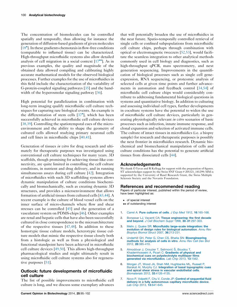
Microfluidic cell culture devices allow precise dynamical quantification of biological processes at the single cell level. (a) Longitudinal real-time
fluorescent images of fibroblasts expressing the fluorescent fusion protein p65–DsRed under control of the endogenous mouse p65-subunit of the NF-
kB transcription complex (arrows illustrate activated cell nuclei). (b) Representative traces for NF–kB activity following stimulation with 0.01 ng/ml of
tumor necrosis factor-alpha in single cells (solid lines), when only active cells are averaged (black squares), and when active and non-active cells are
averaged together (red squares). Combining the traces of all cells shows a false picture of reduced activity. In contrast, single-cell traces reflect the
correct digital nature and variable dynamics of NF–kB under low signal intensity.
Adapted by permission from Macmillan Publishers Ltd: Nature [24], copyright 2010.
by microfluidic cell culture. The use of microfluidics
allows single cell level quantification of cellular responses
to internal or external stimuli with a high temporal
resolution (<1 s) over prolonged cell culture periods
(Figure 3a). Parallelization allows the recording of
millions of data points of given biological processes
during a single experiment. A recent example is the
characterization of the innate immune response of fibro-
blasts to the inflammatory cytokine tumor necrosis factor
alpha (TNF-a) on a single cell level [24]. The use of high
throughput microfluidic cell culture systems [16] allowed
determining the effects of the magnitude, timing and
duration of TNF-a exposure of fibroblasts on the activity
of the transcription factor NF-kB. In contrast to popu-
lation-level studies with bulk-assays, characterizing the
activity of NF-kB in thousands of live cells by micro-
fluidic cell culture and fluorescence time-lapse micro-
scopy revealed that activation of cells is a digital process at
www.sciencedirect.com
the single cell level, subject to a great degree of variability
or ‘biological noise’ (Figure 3b). Microfluidics further
allow the integration of external feedback to control
the expression of given genes over many cell generations
[34]. In addition to capturing intracellular processes,
microfluidics also provides quantification of extracellular
responses to specific stimuli such as the production of
cytokines from single cells [27��]. This allowed the
quantification of temporal dynamics of the secretion of
specific cytokines, revealing that T-cells produce cyto-
kines asynchronously and in a sequential manner.
Together these examples illustrate the potential of micro-
fluidic cell culture systems to quantify biological pro-
cesses at various levels, at a single cell level and with
high temporal resolution.
Microfluidics also allows control of the cellular microen-
vironment, and does it in a highly parallelized fashion.
Current Opinion in Biotechnology 2014, 25:95–102
100 Analytical biotechnology
The concentration of biomolecules can be controlled
spatially and temporally, thus allowing for instance the
generation of diffusion-based gradients of given molecules
[18�]. In these gradients chemotaxis in flow-free conditions
(comparable to inflamed tissue) can be characterized.
High-throughput microfluidic systems also allow detailed
analysis of cell migration in a social context [17��]. As in
previous examples, the quality and magnitude of the
obtained data allowed compiling and calibrating highly
accurate mathematical models for the observed biological
processes. Further examples for the use of microfluidics in
this field include the characterization of the variability of
G-protein-coupled signaling pathways [35] and the band-
width of the hyperosmolar signaling pathway [36].
High potential for parallelization in combination with
long-term imaging qualify microfluidic cell culture tech-
niques for capturing rare biological events. An example is
the differentiation of stem cells [37�], which has been
successfully achieved in microfluidic cell culture devices
[38,39]. Controlling the spatiotemporal cues of the micro-
environment and the ability to shape the geometry of
cultured cells allowed studying primary neuronal cells
and cell lines in microfluidic chips [40,41].
Generation of tissues in vitro for drug research and ulti-
mately for therapeutic purposes was investigated using
conventional cell culture techniques for many years. 3D
scaffolds, though promising for achieving tissue-like con-
nectivity, are quite limited in controlling the cell culture
conditions, in nutrient and drug delivery, and in running
simultaneous assays during cell culture [42]. Integration
of microfluidics with such 3D scaffolding systems allows
dynamic manipulation of culture conditions biochemi-
cally and biomechanically, such as creating dynamic 3D
structures, and provides a microenvironment that allows
formation of artificial tissues from cultured cells [43,44]. A
recent example is the culture of blood vessel cells on the
inner surface of micro-channels where flow and shear
stresses can be controlled [45] and the generation of a
vasculature system on PDMS-chips [46]. Other examples
are renal and hepatic cells that have also been successfully
cultured in close correspondence to the microarchitecture
of the respective tissues [47,48]. In addition to these
homotypic tissue culture models, heterotypic tissue cul-
ture models that mimic the respective tissue closely both
from a histologic as well as from a physiological and
functional standpoint have been achieved in microfluidic
cell culture devices [49,50]. This allows high-throughput
pharmacological studies and might ultimately result in
using microfluidic cell culture systems also for regenera-
tive purposes [51].
Outlook: future developments of microfluidiccell cultureThe list of possible improvements to microfluidic cell
culture is long, and we discuss some exemplary advances
Current Opinion in Biotechnology 2014, 25:95–102
that will potentially broaden the use of microfluidics in
the near future. Spatio-temporally controlled retrieval of
single cells or confined subpopulations from microfluidic
cell culture chips, perhaps through combination with
optical or electromagnetic tweezers [52,53], would facili-
tate the seamless integration to other analytical methods
commonly used in cell biology and diagnostics, such as
high-throughput qPCR, mass spectrometry, and next
generation sequencing. Improvements in the quantifi-
cation of biological processes such as single cell gene-
expression, RNA sequencing, or proteomic analysis of
selected cells at given time points and further advance-
ments in automation and feedback control [34,54] of
microfluidic cell culture chips would considerably con-
tribute to addressing fundamental biological questions in
systems and quantitative biology. In addition to culturing
and assessing individual cell types, further developments
in coculture systems have the potential to widen the use
of microfluidic cell culture devices, particularly in gen-
erating physiologically relevant in vitro scenarios of basic
processes such as infection, innate immune response, and
clonal expansion and selection of activated immune cells.
The culture of intact tissues in microfluidics (i.e. a biopsy
sample) for research and therapeutic purposes is possibly
the next frontier in microfluidics research. Dynamic bio-
chemical and biomechanical manipulation of cells and
culture conditions has the potential to generate artificial
tissues from dissociated cells [44].
AcknowledgementsWe thank O Yavas and R Kellogg for support with the preparation of figures.ST acknowledges support by the Swiss SNF Grant # 205321_141299; MM issupported by the University of Basel Research Grant, the Swiss MultipleSclerosis Society and the Novartis Foundation.
References and recommended readingPapers of particular interest, published within the period of review,have been highlighted as:
� of special interest�� of outstanding interest
1. Carrel A: Pure cultures of cells. J Exp Med 1912, 16:165-168.
2. Bonassar LJ, Vacanti CA: Tissue engineering: the first decadeand beyond. J Cell Biochem Suppl 1998, 30–31:297-303.
3. Melin J, Quake SR: Microfluidic large-scale integration: theevolution of design rules for biological automation. Annu RevBiophys Biomol Struct 2007, 36:213-231.
4. Underhill GH, Peter G, Chen CS, Bhatia SN: Bioengineeringmethods for analysis of cells in vitro. Annu Rev Cell Dev Biol2012, 28:385-410.
5. Almodovar J, Crouzier T, Selimovic S, Boudou T,Khademhosseini A, Picart C: Gradients of physical andbiochemical cues on polyelectrolyte multilayer filmsgenerated via microfluidics. Lab Chip 2013, 13:1562.
6. Morgan JT, Wood JA, Shah NM, Hughbanks ML, Russell P,Barakat AI, Murphy CJ: Integration of basal topographic cuesand apical shear stress in vascular endothelial cells.Biomaterials 2012, 33:4126-4135.
7. Novo P, Volpetti F, Chu V, Conde JP: Control of sequential fluiddelivery in a fully autonomous capillary microfluidic device.Lab Chip 2013, 13:641-645.
www.sciencedirect.com
Microfluidic cell culture Mehling and Tay 101
8. Bennett MR, Pang WL, Ostroff NA, Baumgartner BL, Nayak S,Tsimring LS, Hasty J: Metabolic gene regulation in adynamically changing environment. Nature 2008, 454:1119-1122.
9. Chen Y-A, King AD, Shih H-C, Peng C-C, Wu C-Y, Liao W-H,Tung Y-C: Generation of oxygen gradients in microfluidicdevices for cell culture using spatially confined chemicalreactions. Lab Chip 2011, 11:3626-3633.
10. Chiu DT, Jeon NL, Huang S, Kane RS, Wargo CJ, Choi IS,Ingber DE, Whitesides GM: Patterned deposition of cells andproteins onto surfaces by using three-dimensionalmicrofluidic systems. Proc Natl Acad Sci U S A 2000, 97:2408-2413.
11. Gottwald E, Giselbrecht S, Augspurger C, Lahni B,Dambrowsky N, Truckenmuller R, Piotter V, Gietzelt T, Wendt O,Pfleging W et al.: A chip-based platform for the in vitrogeneration of tissues in three-dimensional organization. LabChip 2007, 7:777-785.
12. Woodruff K, Fidalgo LM, Gobaa S, Lutolf MP, Maerkl SJ: Livemammalian cell arrays. Nat Methods 2013 http://dx.doi.org/10.1038/nmeth.2473.
13. Vyawahare S, Griffiths AD, Merten CA: Miniaturization andparallelization of biological and chemical assays inmicrofluidic devices. Chem Biol 2010, 17:1052-1065.
14. Mata A, Boehm C, Fleischman AJ, Muschler G, Roy S: Growth ofconnective tissue progenitor cells on microtexturedpolydimethylsiloxane surfaces. J Biomed Mater Res 2002,62:499-506.
15. Huang N-T, Chen W, Oh B-R, Cornell TT, Shanley TP, Fu J,Kurabayashi K: An integrated microfluidic platform for in situcellular cytokine secretion immunophenotyping. Lab Chip2012, 12:4093-4101.
16. Gomez-Sjoberg R, Leyrat AA, Pirone DM, Chen CS, Quake SR:Versatile, fully automated, microfluidic cell culture system.Anal Chem 2007, 79:8557-8563.
17.��
Vedel S, Tay S, Johnston DM, Bruus H, Quake SR: Migration of cellsin a social context. Proc. Natl Acad Sci U S A 2013, 110:129-134.
This study exemplifies that microfluidic cell culture systems allow on thebasis of single cell migration analysis also detailed characterization ofmigration in a social context and illustrates the potential of microfluidics inthe field of systems biology.
18.�
Frank T, Tay S: Flow-switching allows independentlyprogrammable, extremely stable, high-throughput diffusion-based gradients. Lab Chip 2013 http://dx.doi.org/10.1039/c3lc4.1076.
This study reports an automated microfluidic cell culture platform tocreate and maintain diffusion-based gradients with a high level of controland the potential to monitor cells exposed to given cytokine-gradients.
19. Son S, Tzur A, Weng Y, Jorgensen P, Kim J, Kirschner MW,Manalis SR: Direct observation of mammalian cell growth andsize regulation. Nat Methods 2012, 9:910-912.
20.�
Lecault V, Vaninsberghe M, Sekulovic S, Knapp DJHF, Wohrer S,Bowden W, Viel F, McLaughlin T, Jarandehei A, Miller M et al.:High-throughput analysis of single hematopoietic stem cellproliferation in microfluidic cell culture arrays. Nat Methods2011, 8:581-586.
This study reports a microfluidic platform containing thousands of nano-scale chambers that allow monitoring of rare cell subpopulations overextended periods of time with a high level of control and automation.
21. Rowat AC, Bird JC, Agresti JJ, Rando OJ, Weitz DA: Trackinglineages of single cells in lines using a microfluidic device.Proc Natl Acad Sci U S A 2009, 106:18149-18154.
22. Falconnet D, Niemisto A, Taylor RJ, Ricicova M, Galitski T,Shmulevich I, Hansen CL: High-throughput tracking of singleyeast cells in a microfluidic imaging matrix. Lab Chip 2011,11:466-473.
23. Ricicova M, Hamidi M, Quiring A, Niemisto A, Emberly E,Hansen CL: Dissecting genealogy and cell cycle as sources ofcell-to-cell variability in MAPK signaling using high-throughput lineage tracking. Proc Natl Acad Sci U S A 2013,110:11403-11408.
www.sciencedirect.com
24. Tay S, Hughey JJ, Lee TK, Lipniacki T, Quake SR, Covert MW:Single-cell NF-kB dynamics reveal digital activation andanalogue information processing. Nature 2010, 466:267-271.
25. Cookson S, Ostroff N, Pang WL, Volfson D, Hasty J: Monitoringdynamics of single-cell gene expression over multiple cellcycles. Mol Syst Biol 2005, 1:2005.0024.
26. Taniguchi Y, Choi PJ, Li G-W, Chen H, Babu M, Hearn J, Emili A,Xie XS: Quantifying E. coli proteome and transcriptome withsingle-molecule sensitivity in single cells. Science 2010,329:533-538.
27.��
Han Q, Bagheri N, Bradshaw EM, Hafler DA, Lauffenburger DA,Love JC: Polyfunctional responses by human T cells resultfrom sequential release of cytokines. Proc Natl Acad Sci U S A2012, 109:1607-1612.
In this report the temporal dynamics of cytokine secretion from individualprimary human T cells are resolved. The use of a microfluidic cell culturesystem allows immune monitoring on a level that is less compromised bythe timing and duration of integrated measures.
28. Yamanaka YJ, Szeto GL, Gierahn TM, Forcier TL, Benedict KF,Brefo MSN, Lauffenburger DA, Irvine DJ, Love JC: Cellularbarcodes for efficiently profiling single-cell secretory responsesby microengraving. Anal Chem 2012, 84:10531-10536.
29. Toepke MW, Beebe DJ: PDMS absorption of small moleculesand consequences in microfluidic applications. Lab Chip 2006,6:1484-1486.
30. Gomez-Sjoberg R, Leyrat AA, Houseman BT, Shokat K, Quake SR:Biocompatibility and reduced drug absorption of sol–gel-treated poly(dimethyl siloxane) for microfluidic cell cultureapplications. Anal Chem 2010 http://dx.doi.org/10.1021/ac10.1870.
31. Millet LJ, Stewart ME, Sweedler JV, Nuzzo RG, Gillette MU:Microfluidic devices for culturing primary mammalian neuronsat low densities. Lab Chip 2007, 7:987-994.
32. Regehr KJ, Domenech M, Koepsel JT, Carver KC, Ellison-Zelski SJ, Murphy WL, Schuler LA, Alarid ET, Beebe DJ: Biologicalimplications of polydimethylsiloxane-based microfluidic cellculture. Lab Chip 2009, 9:2132-2139.
33. Paguirigan AL, Beebe DJ: From the cellular perspective:exploring differences in the cellular baseline in macroscaleand microfluidic cultures. Integr Biol 2009, 1:182-195.
34. Uhlendorf J, Miermont A, Delaveau T, Charvin G, Fages F,Bottani S, Batt G, Hersen P: Long-term model predictive controlof gene expression at the population and single-cell levels.Proc Natl Acad Sci U S A 2012, 109:14271-14276.
35. Bao XR, Fraser IDC, Wall EA, Quake SR, Simon MI: Variability inG-protein-coupled signaling studied with microfluidicdevices. Biophys J 2010, 99:2414-2422.
36. Hersen P, McClean MN, Mahadevan L, Ramanathan S: Signalprocessing by the HOG MAP kinase pathway. Sci Signal 2008,105:7165.
37.�
Schroeder T: Long-term single-cell imaging of mammalianstem cells. Nat Methods 2011, 8:S30-S35.
This report highlights the necessity of long-term imaging in parallel tocontrolling cell culture conditions for stem cell research. The use ofmicrofluidics has a great potential for meeting these requirements.
38. Tourovskaia A, Figueroa-Masot X, Folch A: Differentiation-on-a-chip: a microfluidic platform for long-term cell culture studies.Lab Chip 2005, 5:14-19.
39. Zheng W, Xie Y, Zhang W, Wang D, Ma W, Wang Z, Jiang X: Fluidflow stress induced contraction and re-spread ofmesenchymal stem cells: a microfluidic study. Integr Biol 2012,4:1102-1111.
40. Millet LJ, Stewart ME, Nuzzo RG, Gillette MU: Guiding neurondevelopment with planar surface gradients of substratecues deposited using microfluidic devices. Lab Chip 2010,10:1525-1535.
41. Taylor AM, Blurton-Jones M, Rhee SW, Cribbs DH, Cotman CW,Jeon NL: A microfluidic culture platform for CNS axonal injury,regeneration and transport. Nat Methods 2005, 2:599-605.
Current Opinion in Biotechnology 2014, 25:95–102
102 Analytical biotechnology
42. Beebe DJ, Ingber DE, Toonder den J: Organs on chips. Lab Chip2013, 13:3447-3448.
43. Trietsch SJ, Israels GD, Joore J, Hankemeier T, Vulto P:Microfluidic titer plate for stratified 3D cell culture. Lab Chip2013, 13:3548-3554.
44. Choi NW, Cabodi M, Held B, Gleghorn JP, Bonassar LJ,Stroock AD: Microfluidic scaffolds for tissue engineering. NatMater 2007, 6:908-915.
45. Fiddes LK, Raz N, Srigunapalan S, Tumarkan E, Simmons CA,Wheeler AR, Kumacheva E: A circular cross-section PDMSmicrofluidics system for replication of cardiovascular flowconditions. Biomaterials 2010, 31:3459-3464.
46. Schimek K, Busek M, Brincker S, Groth B, Hoffmann S, Lauster R,Lindner G, Lorenz A, Menzel U, Sonntag F et al.: Integratingbiological vasculature into a multi-organ-chip microsystem.Lab Chip 2013, 13:3588-3598.
47. Baudoin R, Griscom L, Monge M, Legallais C, Leclerc E:Development of a renal microchip for in vitro distal tubulemodels. Biotechnol Prog 2007, 23:1245-1253.
48. Lee PJ, Hung PJ, Lee LP: An artificial liver sinusoid with amicrofluidic endothelial-like barrier for primary hepatocyteculture. Biotechnol Bioeng 2007, 97:1340-1346.
Current Opinion in Biotechnology 2014, 25:95–102
49. Ho C-T, Lin R-Z, Chen R-J, Chin C-K, Gong S-E, Chang H-Y,Peng H-L, Hsu L, Yew T-R, Chang S-F et al.: Liver-cell patterninglab chip: mimicking the morphology of liver lobule tissue. LabChip 2013, 13:3578-3587.
50. Huh D, Matthews BD, Mammoto A, Montoya-Zavala M, Hsin HY,Ingber DE: Reconstituting organ-level lung functions on a chip.Science 2010, 328:1662-1668.
51. Harink B, Le Gac S, Truckenmuller R, van Blitterswijk C,Habibovic P: Regeneration-on-a-chip? The perspectives onuse of microfluidics in regenerative medicine. Lab Chip 2013,13:3512-3528.
52. Petit T, Zhang L, Peyer KE, Kratochvil BE, Nelson BJ: Selectivetrapping and manipulation of microscale objects using mobilemicrovortices. Nano Lett 2012, 12:156-160.
53. Fan D, Yin Z, Cheong R, Zhu FQ, Cammarata RC, Chien CL,Levchenko A: Subcellular-resolution delivery of a cytokinethrough precisely manipulated nanowires. Nat Nanotechnol2010, 5:545-551.
54. Menolascina F, di Bernardo M, di Bernardo D: Analysis, designand implementation of a novel scheme for in-vivo control ofsynthetic gene regulatory networks. Automatica 2011,47:1265-1270.
www.sciencedirect.com