Embed Size (px)
Citation preview
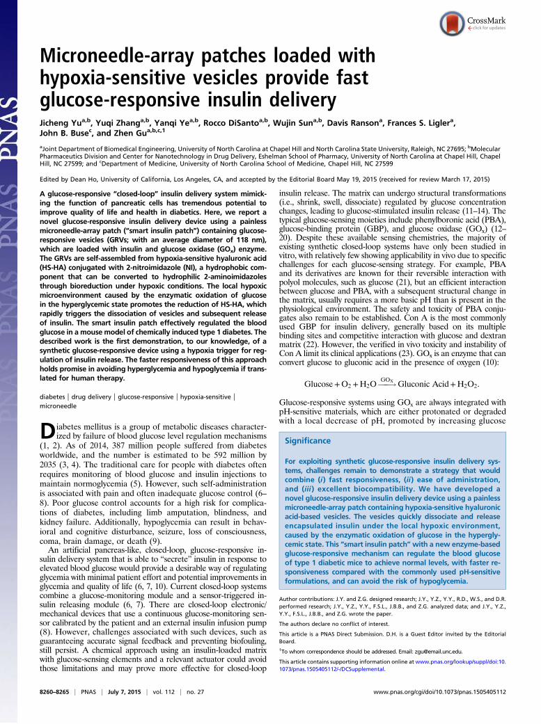
Microneedle-array patches loaded withhypoxia-sensitive vesicles provide fastglucose-responsive insulin deliveryJicheng Yua,b, Yuqi Zhanga,b, Yanqi Yea,b, Rocco DiSantoa,b, Wujin Suna,b, Davis Ransona, Frances S. Liglera,John B. Busec, and Zhen Gua,b,c,1
aJoint Department of Biomedical Engineering, University of North Carolina at Chapel Hill and North Carolina State University, Raleigh, NC 27695; bMolecularPharmaceutics Division and Center for Nanotechnology in Drug Delivery, Eshelman School of Pharmacy, University of North Carolina at Chapel Hill, ChapelHill, NC 27599; and cDepartment of Medicine, University of North Carolina School of Medicine, Chapel Hill, NC 27599
Edited by Dean Ho, University of California, Los Angeles, CA, and accepted by the Editorial Board May 19, 2015 (received for review March 17, 2015)
A glucose-responsive “closed-loop” insulin delivery system mimick-ing the function of pancreatic cells has tremendous potential toimprove quality of life and health in diabetics. Here, we report anovel glucose-responsive insulin delivery device using a painlessmicroneedle-array patch (“smart insulin patch”) containing glucose-responsive vesicles (GRVs; with an average diameter of 118 nm),which are loaded with insulin and glucose oxidase (GOx) enzyme.The GRVs are self-assembled from hypoxia-sensitive hyaluronic acid(HS-HA) conjugated with 2-nitroimidazole (NI), a hydrophobic com-ponent that can be converted to hydrophilic 2-aminoimidazolesthrough bioreduction under hypoxic conditions. The local hypoxicmicroenvironment caused by the enzymatic oxidation of glucosein the hyperglycemic state promotes the reduction of HS-HA, whichrapidly triggers the dissociation of vesicles and subsequent releaseof insulin. The smart insulin patch effectively regulated the bloodglucose in a mouse model of chemically induced type 1 diabetes. Thedescribed work is the first demonstration, to our knowledge, of asynthetic glucose-responsive device using a hypoxia trigger for reg-ulation of insulin release. The faster responsiveness of this approachholds promise in avoiding hyperglycemia and hypoglycemia if trans-lated for human therapy.
diabetes | drug delivery | glucose-responsive | hypoxia-sensitive |microneedle
Diabetes mellitus is a group of metabolic diseases character-ized by failure of blood glucose level regulation mechanisms
(1, 2). As of 2014, 387 million people suffered from diabetesworldwide, and the number is estimated to be 592 million by2035 (3, 4). The traditional care for people with diabetes oftenrequires monitoring of blood glucose and insulin injections tomaintain normoglycemia (5). However, such self-administrationis associated with pain and often inadequate glucose control (6–8). Poor glucose control accounts for a high risk for complica-tions of diabetes, including limb amputation, blindness, andkidney failure. Additionally, hypoglycemia can result in behav-ioral and cognitive disturbance, seizure, loss of consciousness,coma, brain damage, or death (9).An artificial pancreas-like, closed-loop, glucose-responsive in-
sulin delivery system that is able to “secrete” insulin in response toelevated blood glucose would provide a desirable way of regulatingglycemia with minimal patient effort and potential improvements inglycemia and quality of life (6, 7, 10). Current closed-loop systemscombine a glucose-monitoring module and a sensor-triggered in-sulin releasing module (6, 7). There are closed-loop electronic/mechanical devices that use a continuous glucose-monitoring sen-sor calibrated by the patient and an external insulin infusion pump(8). However, challenges associated with such devices, such asguaranteeing accurate signal feedback and preventing biofouling,still persist. A chemical approach using an insulin-loaded matrixwith glucose-sensing elements and a relevant actuator could avoidthose limitations and may prove more effective for closed-loop
insulin release. The matrix can undergo structural transformations(i.e., shrink, swell, dissociate) regulated by glucose concentrationchanges, leading to glucose-stimulated insulin release (11–14). Thetypical glucose-sensing moieties include phenylboronic acid (PBA),glucose-binding protein (GBP), and glucose oxidase (GOx) (12–20). Despite these available sensing chemistries, the majority ofexisting synthetic closed-loop systems have only been studied invitro, with relatively few showing applicability in vivo due to specificchallenges for each glucose-sensing strategy. For example, PBAand its derivatives are known for their reversible interaction withpolyol molecules, such as glucose (21), but an efficient interactionbetween glucose and PBA, with a subsequent structural change inthe matrix, usually requires a more basic pH than is present in thephysiological environment. The safety and toxicity of PBA conju-gates also remain to be established. Con A is the most commonlyused GBP for insulin delivery, generally based on its multiplebinding sites and competitive interaction with glucose and dextranmatrix (22). However, the verified in vivo toxicity and instability ofCon A limit its clinical applications (23). GOx is an enzyme that canconvert glucose to gluconic acid in the presence of oxygen (10):
Glucose+O2 +H2O ���!GOX Gluconic Acid+H2O2.
Glucose-responsive systems using GOx are always integrated withpH-sensitive materials, which are either protonated or degradedwith a local decrease of pH, promoted by increasing glucose
Significance
For exploiting synthetic glucose-responsive insulin delivery sys-tems, challenges remain to demonstrate a strategy that wouldcombine (i) fast responsiveness, (ii) ease of administration,and (iii ) excellent biocompatibility. We have developed anovel glucose-responsive insulin delivery device using a painlessmicroneedle-array patch containing hypoxia-sensitive hyaluronicacid-based vesicles. The vesicles quickly dissociate and releaseencapsulated insulin under the local hypoxic environment,caused by the enzymatic oxidation of glucose in the hypergly-cemic state. This “smart insulin patch”with a new enzyme-basedglucose-responsive mechanism can regulate the blood glucoseof type 1 diabetic mice to achieve normal levels, with faster re-sponsiveness compared with the commonly used pH-sensitiveformulations, and can avoid the risk of hypoglycemia.
Author contributions: J.Y. and Z.G. designed research; J.Y., Y.Z., Y.Y., R.D., W.S., and D.R.performed research; J.Y., Y.Z., Y.Y., F.S.L., J.B.B., and Z.G. analyzed data; and J.Y., Y.Z.,Y.Y., F.S.L., J.B.B., and Z.G. wrote the paper.
The authors declare no conflict of interest.
This article is a PNAS Direct Submission. D.H. is a Guest Editor invited by the EditorialBoard.1To whom correspondence should be addressed. Email: [email protected].
This article contains supporting information online at www.pnas.org/lookup/suppl/doi:10.1073/pnas.1505405112/-/DCSupplemental.
8260–8265 | PNAS | July 7, 2015 | vol. 112 | no. 27 www.pnas.org/cgi/doi/10.1073/pnas.1505405112
concentration. However, such pH decrease-dependent methodsare often compromised by slow responsiveness, especially in abuffered physiological environment (8). Taken together, chal-lenges remain to demonstrate a method that would combine(i) fast responsiveness with pharmacokinetics similar to normalpancreatic activity, (ii) ease of administration, and (iii) biocom-patibility without long-term side effects (3).Here, we report a novel glucose-responsive insulin delivery
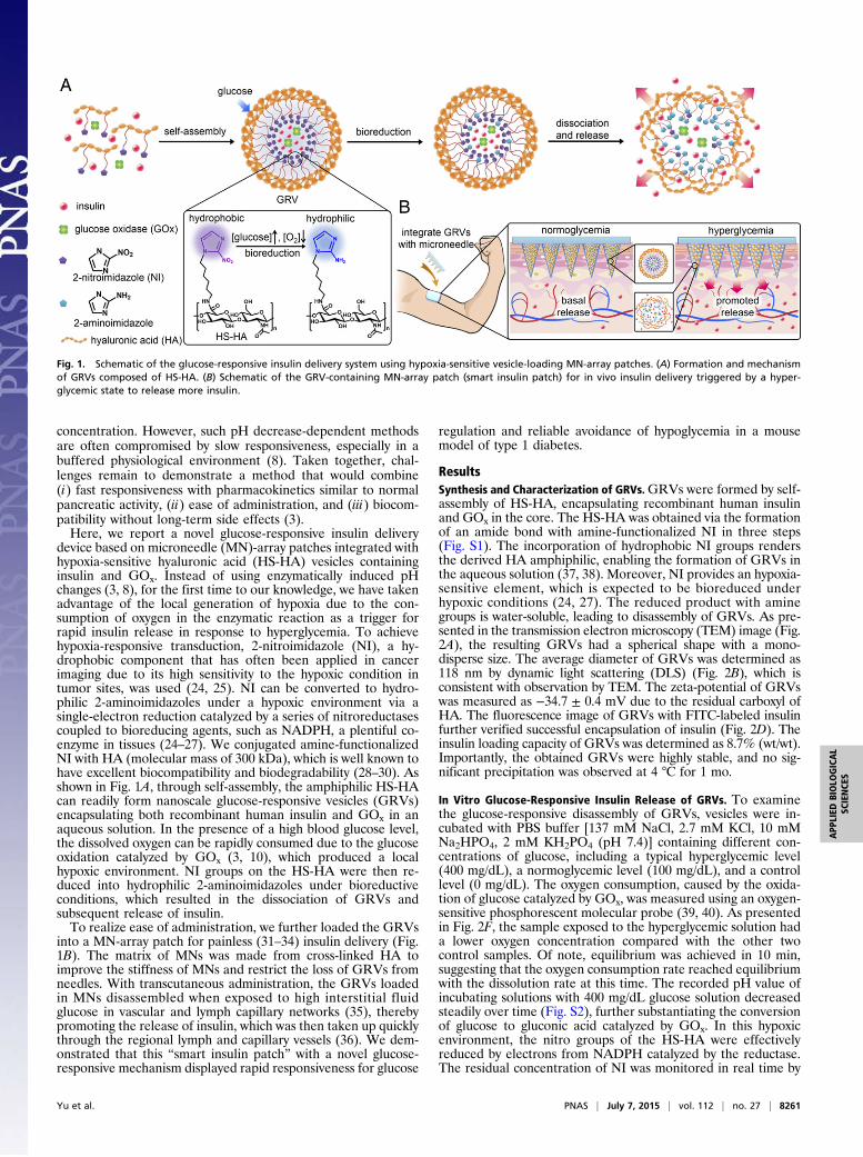
device based on microneedle (MN)-array patches integrated withhypoxia-sensitive hyaluronic acid (HS-HA) vesicles containinginsulin and GOx. Instead of using enzymatically induced pHchanges (3, 8), for the first time to our knowledge, we have takenadvantage of the local generation of hypoxia due to the con-sumption of oxygen in the enzymatic reaction as a trigger forrapid insulin release in response to hyperglycemia. To achievehypoxia-responsive transduction, 2-nitroimidazole (NI), a hy-drophobic component that has often been applied in cancerimaging due to its high sensitivity to the hypoxic condition intumor sites, was used (24, 25). NI can be converted to hydro-philic 2-aminoimidazoles under a hypoxic environment via asingle-electron reduction catalyzed by a series of nitroreductasescoupled to bioreducing agents, such as NADPH, a plentiful co-enzyme in tissues (24–27). We conjugated amine-functionalizedNI with HA (molecular mass of 300 kDa), which is well known tohave excellent biocompatibility and biodegradability (28–30). Asshown in Fig. 1A, through self-assembly, the amphiphilic HS-HAcan readily form nanoscale glucose-responsive vesicles (GRVs)encapsulating both recombinant human insulin and GOx in anaqueous solution. In the presence of a high blood glucose level,the dissolved oxygen can be rapidly consumed due to the glucoseoxidation catalyzed by GOx (3, 10), which produced a localhypoxic environment. NI groups on the HS-HA were then re-duced into hydrophilic 2-aminoimidazoles under bioreductiveconditions, which resulted in the dissociation of GRVs andsubsequent release of insulin.To realize ease of administration, we further loaded the GRVs
into a MN-array patch for painless (31–34) insulin delivery (Fig.1B). The matrix of MNs was made from cross-linked HA toimprove the stiffness of MNs and restrict the loss of GRVs fromneedles. With transcutaneous administration, the GRVs loadedin MNs disassembled when exposed to high interstitial fluidglucose in vascular and lymph capillary networks (35), therebypromoting the release of insulin, which was then taken up quicklythrough the regional lymph and capillary vessels (36). We dem-onstrated that this “smart insulin patch” with a novel glucose-responsive mechanism displayed rapid responsiveness for glucose
regulation and reliable avoidance of hypoglycemia in a mousemodel of type 1 diabetes.
ResultsSynthesis and Characterization of GRVs.GRVs were formed by self-assembly of HS-HA, encapsulating recombinant human insulinand GOx in the core. The HS-HA was obtained via the formationof an amide bond with amine-functionalized NI in three steps(Fig. S1). The incorporation of hydrophobic NI groups rendersthe derived HA amphiphilic, enabling the formation of GRVs inthe aqueous solution (37, 38). Moreover, NI provides an hypoxia-sensitive element, which is expected to be bioreduced underhypoxic conditions (24, 27). The reduced product with aminegroups is water-soluble, leading to disassembly of GRVs. As pre-sented in the transmission electron microscopy (TEM) image (Fig.2A), the resulting GRVs had a spherical shape with a mono-disperse size. The average diameter of GRVs was determined as118 nm by dynamic light scattering (DLS) (Fig. 2B), which isconsistent with observation by TEM. The zeta-potential of GRVswas measured as −34.7 ± 0.4 mV due to the residual carboxyl ofHA. The fluorescence image of GRVs with FITC-labeled insulinfurther verified successful encapsulation of insulin (Fig. 2D). Theinsulin loading capacity of GRVs was determined as 8.7% (wt/wt).Importantly, the obtained GRVs were highly stable, and no sig-nificant precipitation was observed at 4 °C for 1 mo.
In Vitro Glucose-Responsive Insulin Release of GRVs. To examinethe glucose-responsive disassembly of GRVs, vesicles were in-cubated with PBS buffer [137 mM NaCl, 2.7 mM KCl, 10 mMNa2HPO4, 2 mM KH2PO4 (pH 7.4)] containing different con-centrations of glucose, including a typical hyperglycemic level(400 mg/dL), a normoglycemic level (100 mg/dL), and a controllevel (0 mg/dL). The oxygen consumption, caused by the oxida-tion of glucose catalyzed by GOx, was measured using an oxygen-sensitive phosphorescent molecular probe (39, 40). As presentedin Fig. 2F, the sample exposed to the hyperglycemic solution hada lower oxygen concentration compared with the other twocontrol samples. Of note, equilibrium was achieved in 10 min,suggesting that the oxygen consumption rate reached equilibriumwith the dissolution rate at this time. The recorded pH value ofincubating solutions with 400 mg/dL glucose solution decreasedsteadily over time (Fig. S2), further substantiating the conversionof glucose to gluconic acid catalyzed by GOx. In this hypoxicenvironment, the nitro groups of the HS-HA were effectivelyreduced by electrons from NADPH catalyzed by the reductase.The residual concentration of NI was monitored in real time by
Fig. 1. Schematic of the glucose-responsive insulin delivery system using hypoxia-sensitive vesicle-loading MN-array patches. (A) Formation and mechanismof GRVs composed of HS-HA. (B) Schematic of the GRV-containing MN-array patch (smart insulin patch) for in vivo insulin delivery triggered by a hyper-glycemic state to release more insulin.
Yu et al. PNAS | July 7, 2015 | vol. 112 | no. 27 | 8261
APP
LIED
BIOLO
GICAL
SCIENCE
S
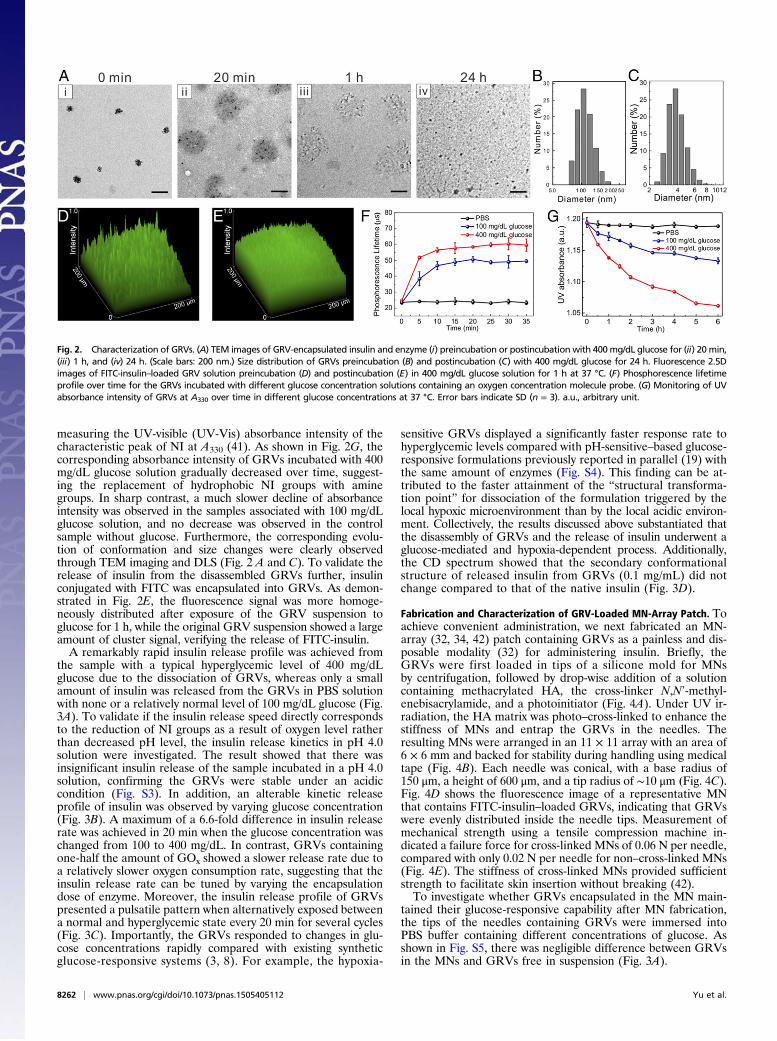
measuring the UV-visible (UV-Vis) absorbance intensity of thecharacteristic peak of NI at A330 (41). As shown in Fig. 2G, thecorresponding absorbance intensity of GRVs incubated with 400mg/dL glucose solution gradually decreased over time, suggest-ing the replacement of hydrophobic NI groups with aminegroups. In sharp contrast, a much slower decline of absorbanceintensity was observed in the samples associated with 100 mg/dLglucose solution, and no decrease was observed in the controlsample without glucose. Furthermore, the corresponding evolu-tion of conformation and size changes were clearly observedthrough TEM imaging and DLS (Fig. 2 A and C). To validate therelease of insulin from the disassembled GRVs further, insulinconjugated with FITC was encapsulated into GRVs. As demon-strated in Fig. 2E, the fluorescence signal was more homoge-neously distributed after exposure of the GRV suspension toglucose for 1 h, while the original GRV suspension showed a largeamount of cluster signal, verifying the release of FITC-insulin.A remarkably rapid insulin release profile was achieved from
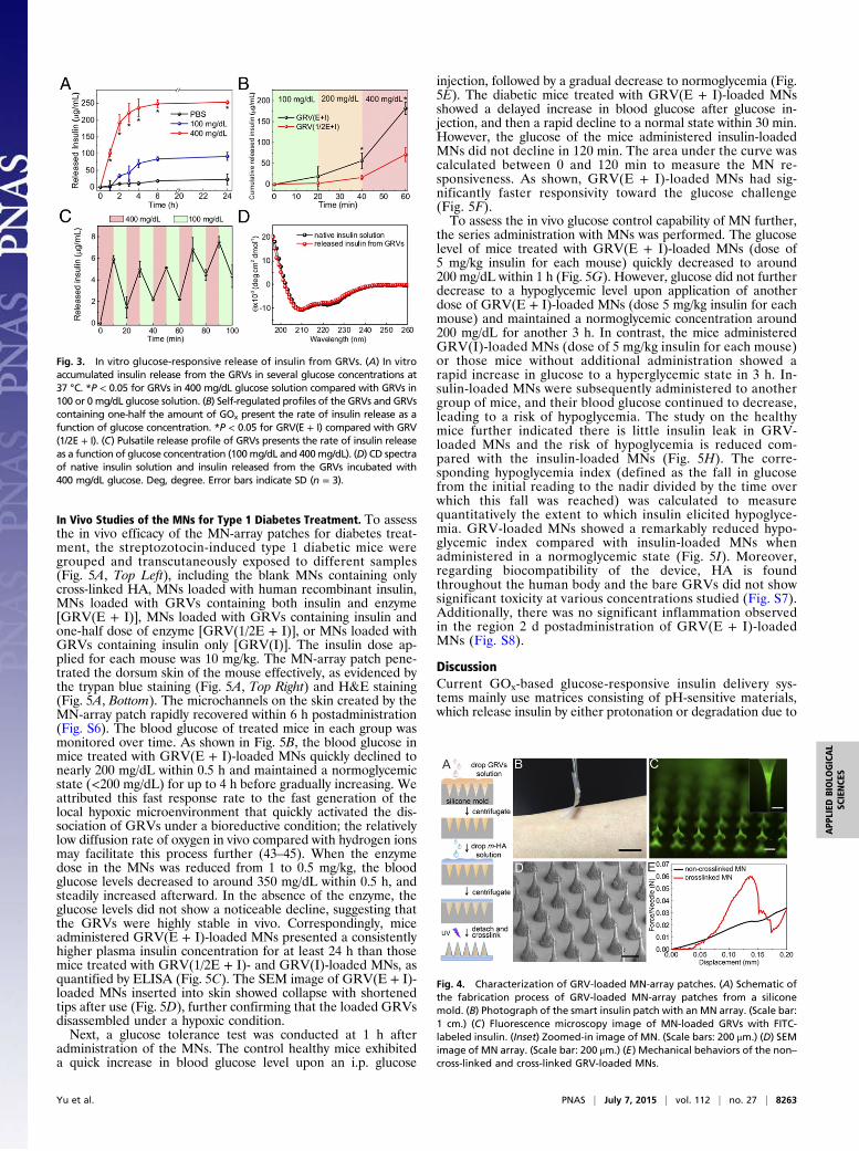
the sample with a typical hyperglycemic level of 400 mg/dLglucose due to the dissociation of GRVs, whereas only a smallamount of insulin was released from the GRVs in PBS solutionwith none or a relatively normal level of 100 mg/dL glucose (Fig.3A). To validate if the insulin release speed directly correspondsto the reduction of NI groups as a result of oxygen level ratherthan decreased pH level, the insulin release kinetics in pH 4.0solution were investigated. The result showed that there wasinsignificant insulin release of the sample incubated in a pH 4.0solution, confirming the GRVs were stable under an acidiccondition (Fig. S3). In addition, an alterable kinetic releaseprofile of insulin was observed by varying glucose concentration(Fig. 3B). A maximum of a 6.6-fold difference in insulin releaserate was achieved in 20 min when the glucose concentration waschanged from 100 to 400 mg/dL. In contrast, GRVs containingone-half the amount of GOx showed a slower release rate due toa relatively slower oxygen consumption rate, suggesting that theinsulin release rate can be tuned by varying the encapsulationdose of enzyme. Moreover, the insulin release profile of GRVspresented a pulsatile pattern when alternatively exposed betweena normal and hyperglycemic state every 20 min for several cycles(Fig. 3C). Importantly, the GRVs responded to changes in glu-cose concentrations rapidly compared with existing syntheticglucose-responsive systems (3, 8). For example, the hypoxia-
sensitive GRVs displayed a significantly faster response rate tohyperglycemic levels compared with pH-sensitive–based glucose-responsive formulations previously reported in parallel (19) withthe same amount of enzymes (Fig. S4). This finding can be at-tributed to the faster attainment of the “structural transforma-tion point” for dissociation of the formulation triggered by thelocal hypoxic microenvironment than by the local acidic environ-ment. Collectively, the results discussed above substantiated thatthe disassembly of GRVs and the release of insulin underwent aglucose-mediated and hypoxia-dependent process. Additionally,the CD spectrum showed that the secondary conformationalstructure of released insulin from GRVs (0.1 mg/mL) did notchange compared to that of the native insulin (Fig. 3D).
Fabrication and Characterization of GRV-Loaded MN-Array Patch. Toachieve convenient administration, we next fabricated an MN-array (32, 34, 42) patch containing GRVs as a painless and dis-posable modality (32) for administering insulin. Briefly, theGRVs were first loaded in tips of a silicone mold for MNsby centrifugation, followed by drop-wise addition of a solutioncontaining methacrylated HA, the cross-linker N,N’-methyl-enebisacrylamide, and a photoinitiator (Fig. 4A). Under UV ir-radiation, the HA matrix was photo–cross-linked to enhance thestiffness of MNs and entrap the GRVs in the needles. Theresulting MNs were arranged in an 11 × 11 array with an area of6 × 6 mm and backed for stability during handling using medicaltape (Fig. 4B). Each needle was conical, with a base radius of150 μm, a height of 600 μm, and a tip radius of ∼10 μm (Fig. 4C).Fig. 4D shows the fluorescence image of a representative MNthat contains FITC-insulin–loaded GRVs, indicating that GRVswere evenly distributed inside the needle tips. Measurement ofmechanical strength using a tensile compression machine in-dicated a failure force for cross-linked MNs of 0.06 N per needle,compared with only 0.02 N per needle for non–cross-linked MNs(Fig. 4E). The stiffness of cross-linked MNs provided sufficientstrength to facilitate skin insertion without breaking (42).To investigate whether GRVs encapsulated in the MN main-
tained their glucose-responsive capability after MN fabrication,the tips of the needles containing GRVs were immersed intoPBS buffer containing different concentrations of glucose. Asshown in Fig. S5, there was negligible difference between GRVsin the MNs and GRVs free in suspension (Fig. 3A).
Fig. 2. Characterization of GRVs. (A) TEM images of GRV-encapsulated insulin and enzyme (i) preincubation or postincubation with 400mg/dL glucose for (ii) 20 min,(iii) 1 h, and (iv) 24 h. (Scale bars: 200 nm.) Size distribution of GRVs preincubation (B) and postincubation (C) with 400 mg/dL glucose for 24 h. Fluorescence 2.5Dimages of FITC-insulin–loaded GRV solution preincubation (D) and postincubation (E) in 400 mg/dL glucose solution for 1 h at 37 °C. (F) Phosphorescence lifetimeprofile over time for the GRVs incubated with different glucose concentration solutions containing an oxygen concentration molecule probe. (G) Monitoring of UVabsorbance intensity of GRVs at A330 over time in different glucose concentrations at 37 °C. Error bars indicate SD (n = 3). a.u., arbitrary unit.
8262 | www.pnas.org/cgi/doi/10.1073/pnas.1505405112 Yu et al.
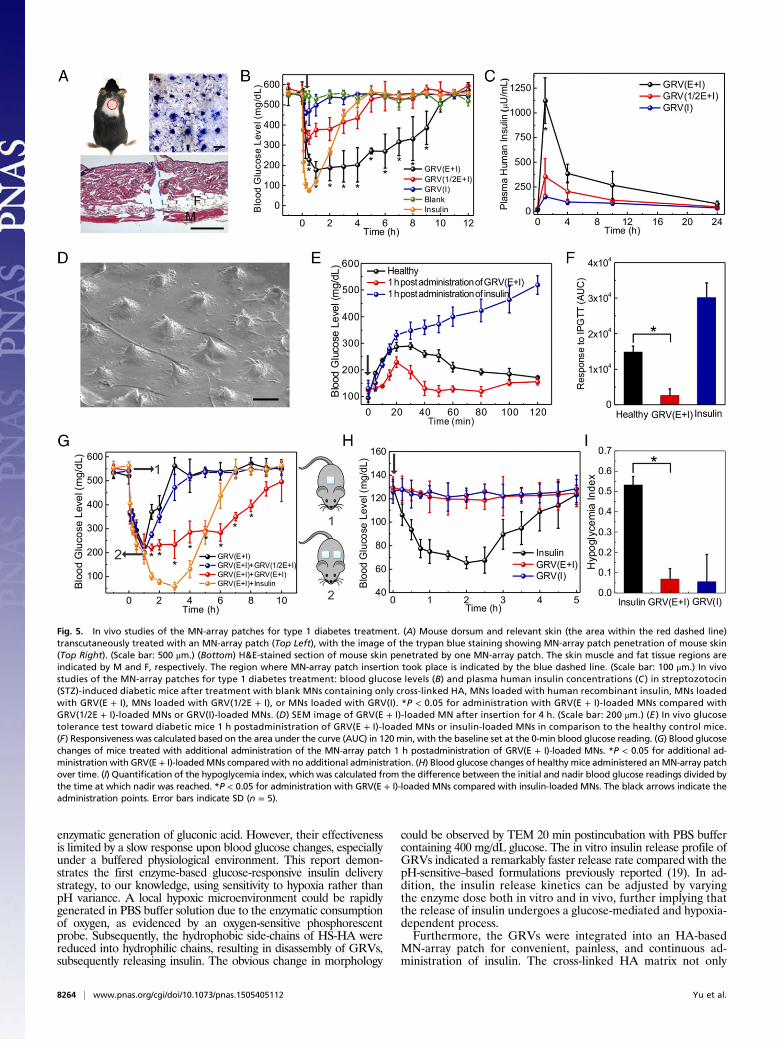
In Vivo Studies of the MNs for Type 1 Diabetes Treatment. To assessthe in vivo efficacy of the MN-array patches for diabetes treat-ment, the streptozotocin-induced type 1 diabetic mice weregrouped and transcutaneously exposed to different samples(Fig. 5A, Top Left), including the blank MNs containing onlycross-linked HA, MNs loaded with human recombinant insulin,MNs loaded with GRVs containing both insulin and enzyme[GRV(E + I)], MNs loaded with GRVs containing insulin andone-half dose of enzyme [GRV(1/2E + I)], or MNs loaded withGRVs containing insulin only [GRV(I)]. The insulin dose ap-plied for each mouse was 10 mg/kg. The MN-array patch pene-trated the dorsum skin of the mouse effectively, as evidenced bythe trypan blue staining (Fig. 5A, Top Right) and H&E staining(Fig. 5A, Bottom). The microchannels on the skin created by theMN-array patch rapidly recovered within 6 h postadministration(Fig. S6). The blood glucose of treated mice in each group wasmonitored over time. As shown in Fig. 5B, the blood glucose inmice treated with GRV(E + I)-loaded MNs quickly declined tonearly 200 mg/dL within 0.5 h and maintained a normoglycemicstate (<200 mg/dL) for up to 4 h before gradually increasing. Weattributed this fast response rate to the fast generation of thelocal hypoxic microenvironment that quickly activated the dis-sociation of GRVs under a bioreductive condition; the relativelylow diffusion rate of oxygen in vivo compared with hydrogen ionsmay facilitate this process further (43–45). When the enzymedose in the MNs was reduced from 1 to 0.5 mg/kg, the bloodglucose levels decreased to around 350 mg/dL within 0.5 h, andsteadily increased afterward. In the absence of the enzyme, theglucose levels did not show a noticeable decline, suggesting thatthe GRVs were highly stable in vivo. Correspondingly, miceadministered GRV(E + I)-loaded MNs presented a consistentlyhigher plasma insulin concentration for at least 24 h than thosemice treated with GRV(1/2E + I)- and GRV(I)-loaded MNs, asquantified by ELISA (Fig. 5C). The SEM image of GRV(E + I)-loaded MNs inserted into skin showed collapse with shortenedtips after use (Fig. 5D), further confirming that the loaded GRVsdisassembled under a hypoxic condition.Next, a glucose tolerance test was conducted at 1 h after
administration of the MNs. The control healthy mice exhibiteda quick increase in blood glucose level upon an i.p. glucose
injection, followed by a gradual decrease to normoglycemia (Fig.5E). The diabetic mice treated with GRV(E + I)-loaded MNsshowed a delayed increase in blood glucose after glucose in-jection, and then a rapid decline to a normal state within 30 min.However, the glucose of the mice administered insulin-loadedMNs did not decline in 120 min. The area under the curve wascalculated between 0 and 120 min to measure the MN re-sponsiveness. As shown, GRV(E + I)-loaded MNs had sig-nificantly faster responsivity toward the glucose challenge(Fig. 5F).To assess the in vivo glucose control capability of MN further,
the series administration with MNs was performed. The glucoselevel of mice treated with GRV(E + I)-loaded MNs (dose of5 mg/kg insulin for each mouse) quickly decreased to around200 mg/dL within 1 h (Fig. 5G). However, glucose did not furtherdecrease to a hypoglycemic level upon application of anotherdose of GRV(E + I)-loaded MNs (dose 5 mg/kg insulin for eachmouse) and maintained a normoglycemic concentration around200 mg/dL for another 3 h. In contrast, the mice administeredGRV(I)-loaded MNs (dose of 5 mg/kg insulin for each mouse)or those mice without additional administration showed arapid increase in glucose to a hyperglycemic state in 3 h. In-sulin-loaded MNs were subsequently administered to anothergroup of mice, and their blood glucose continued to decrease,leading to a risk of hypoglycemia. The study on the healthymice further indicated there is little insulin leak in GRV-loaded MNs and the risk of hypoglycemia is reduced com-pared with the insulin-loaded MNs (Fig. 5H). The corre-sponding hypoglycemia index (defined as the fall in glucosefrom the initial reading to the nadir divided by the time overwhich this fall was reached) was calculated to measurequantitatively the extent to which insulin elicited hypoglyce-mia. GRV-loaded MNs showed a remarkably reduced hypo-glycemic index compared with insulin-loaded MNs whenadministered in a normoglycemic state (Fig. 5I). Moreover,regarding biocompatibility of the device, HA is foundthroughout the human body and the bare GRVs did not showsignificant toxicity at various concentrations studied (Fig. S7).Additionally, there was no significant inflammation observedin the region 2 d postadministration of GRV(E + I)-loadedMNs (Fig. S8).
DiscussionCurrent GOx-based glucose-responsive insulin delivery sys-tems mainly use matrices consisting of pH-sensitive materials,which release insulin by either protonation or degradation due to
Fig. 3. In vitro glucose-responsive release of insulin from GRVs. (A) In vitroaccumulated insulin release from the GRVs in several glucose concentrations at37 °C. *P < 0.05 for GRVs in 400 mg/dL glucose solution compared with GRVs in100 or 0 mg/dL glucose solution. (B) Self-regulated profiles of the GRVs and GRVscontaining one-half the amount of GOx present the rate of insulin release as afunction of glucose concentration. *P < 0.05 for GRV(E + I) compared with GRV(1/2E + I). (C) Pulsatile release profile of GRVs presents the rate of insulin releaseas a function of glucose concentration (100mg/dL and 400mg/dL). (D) CD spectraof native insulin solution and insulin released from the GRVs incubated with400 mg/dL glucose. Deg, degree. Error bars indicate SD (n = 3).
Fig. 4. Characterization of GRV-loaded MN-array patches. (A) Schematic ofthe fabrication process of GRV-loaded MN-array patches from a siliconemold. (B) Photograph of the smart insulin patch with an MN array. (Scale bar:1 cm.) (C) Fluorescence microscopy image of MN-loaded GRVs with FITC-labeled insulin. (Inset) Zoomed-in image of MN. (Scale bars: 200 μm.) (D) SEMimage of MN array. (Scale bar: 200 μm.) (E) Mechanical behaviors of the non–cross-linked and cross-linked GRV-loaded MNs.
Yu et al. PNAS | July 7, 2015 | vol. 112 | no. 27 | 8263
APP
LIED
BIOLO
GICAL
SCIENCE
S
enzymatic generation of gluconic acid. However, their effectivenessis limited by a slow response upon blood glucose changes, especiallyunder a buffered physiological environment. This report demon-strates the first enzyme-based glucose-responsive insulin deliverystrategy, to our knowledge, using sensitivity to hypoxia rather thanpH variance. A local hypoxic microenvironment could be rapidlygenerated in PBS buffer solution due to the enzymatic consumptionof oxygen, as evidenced by an oxygen-sensitive phosphorescentprobe. Subsequently, the hydrophobic side-chains of HS-HA werereduced into hydrophilic chains, resulting in disassembly of GRVs,subsequently releasing insulin. The obvious change in morphology
could be observed by TEM 20 min postincubation with PBS buffercontaining 400 mg/dL glucose. The in vitro insulin release profile ofGRVs indicated a remarkably faster release rate compared with thepH-sensitive–based formulations previously reported (19). In ad-dition, the insulin release kinetics can be adjusted by varyingthe enzyme dose both in vitro and in vivo, further implying thatthe release of insulin undergoes a glucose-mediated and hypoxia-dependent process.Furthermore, the GRVs were integrated into an HA-based
MN-array patch for convenient, painless, and continuous ad-ministration of insulin. The cross-linked HA matrix not only
Fig. 5. In vivo studies of the MN-array patches for type 1 diabetes treatment. (A) Mouse dorsum and relevant skin (the area within the red dashed line)transcutaneously treated with an MN-array patch (Top Left), with the image of the trypan blue staining showing MN-array patch penetration of mouse skin(Top Right). (Scale bar: 500 μm.) (Bottom) H&E-stained section of mouse skin penetrated by one MN-array patch. The skin muscle and fat tissue regions areindicated by M and F, respectively. The region where MN-array patch insertion took place is indicated by the blue dashed line. (Scale bar: 100 μm.) In vivostudies of the MN-array patches for type 1 diabetes treatment: blood glucose levels (B) and plasma human insulin concentrations (C ) in streptozotocin(STZ)-induced diabetic mice after treatment with blank MNs containing only cross-linked HA, MNs loaded with human recombinant insulin, MNs loadedwith GRV(E + I), MNs loaded with GRV(1/2E + I), or MNs loaded with GRV(I). *P < 0.05 for administration with GRV(E + I)-loaded MNs compared withGRV(1/2E + I)-loaded MNs or GRV(I)-loaded MNs. (D) SEM image of GRV(E + I)-loaded MN after insertion for 4 h. (Scale bar: 200 μm.) (E ) In vivo glucosetolerance test toward diabetic mice 1 h postadministration of GRV(E + I)-loaded MNs or insulin-loaded MNs in comparison to the healthy control mice.(F) Responsiveness was calculated based on the area under the curve (AUC) in 120 min, with the baseline set at the 0-min blood glucose reading. (G) Blood glucosechanges of mice treated with additional administration of the MN-array patch 1 h postadministration of GRV(E + I)-loaded MNs. *P < 0.05 for additional ad-ministration with GRV(E + I)-loadedMNs compared with no additional administration. (H) Blood glucose changes of healthy mice administered anMN-array patchover time. (I) Quantification of the hypoglycemia index, which was calculated from the difference between the initial and nadir blood glucose readings divided bythe time at which nadir was reached. *P < 0.05 for administration with GRV(E + I)-loaded MNs compared with insulin-loaded MNs. The black arrows indicate theadministration points. Error bars indicate SD (n = 5).
8264 | www.pnas.org/cgi/doi/10.1073/pnas.1505405112 Yu et al.
helped to improve mechanical strength and skin penetrationcapability but also restricted the loss of GRVs to avoid burstrelease. Additionally, the framework of both needle patches andvesicles was made from HA, which is highly biocompatible. TheGRV(E + I)-loaded MNs exhibited excellent regulation of glu-cose into a normal range with fast responsiveness. Furthermore,in addition to the highly sensitive vesicles, the rapid uptake bythe lymphatics through transcutaneous administration may con-tribute to the fast responsivity. The in vivo glucose tolerance testdemonstrated not only that GRV-loaded MNs were responsiveto glucose challenge but that they could also efficiently minimizethe risk of hypoglycemia. In addition, the results of serial ad-ministration with MNs showed that it could precisely controlglucose in a normal range for prolonged periods. Also consid-ering that mice have reduced sensitivity to the human insulinused in this study, the real dose for potential human use will besignificantly lower. This smart insulin patch with its novel triggermechanism offers a clinical opportunity for closed-loop deliveryof insulin in a fast glucose-responsive, pain-free, and safe man-ner. It will also guide the development of a useful drug deliveryplatform for treating other diseases using artificial vesicles, thebehaviors of which can be “intelligently” activated and self-reg-ulated by the variation of physiological signals.
MethodsMaterials. All chemicals were purchased from Sigma–Aldrich unless otherwisespecified and were used as received. Sodium HA (molecular mass of 300 kDa)
was purchased from Freda Biochem Co., Ltd.. Human recombinant insulin(27.5 IU/mg of Zn salt) was purchased from Life Technology.
Preparation of GRVs. GRVs were prepared by self-assembly in aqueous so-lution. Briefly, 20 mg of amphiphilic HS-HA was dissolved in water/methanol(2:1 vol/vol), followed by addition of 10 mg of human insulin and 1.0 mg ofGOx. The emulsion was stirred at 4 °C for 2 h. Then, the methanol was re-moved by dialysis against deionized water for 1 d. The pH value of theresulting GRV suspension was adjusted to 5.3 (the pI of insulin) to removethe unloaded insulin by centrifugation at 6,200 × g for 10 min and furtherfiltered by a centrifugal filter (100,000 Da molecular mass cutoff, Millipore)at pH 7.4. The final GRV suspension was stored at 4 °C for later study. Theinsulin loading capacity of GRVs was determined by measuring the loadedinsulin content using a Coomassie Plus protein assay. The zeta-potential andsize distribution were measured on the Zetasizer (Nano ZS; Malvern). TheTEM images of GRVs were obtained on a JEOL 2000FX TEM instrument.
The animal study protocol was approved by the Institutional Animal Careand Use Committee at North Carolina State University and the University ofNorth Carolina at Chapel Hill.
ACKNOWLEDGMENTS. We thank Dr. Elizabeth Loboa, Dr. Glenn Walker,Dr. Tushar K. Ghosh, and Xiaomeng Fang for providing experimental facilitiesand assistance in device characterizations. We acknowledge the use of theAnalytical Instrumentation Facility at North Carolina State University, which issupported by the State of North Carolina and the National Science Foundation.This work was supported by Grants 1-14-JF-29 and 1-15-ACE-21 from theAmerican Diabetes Association (to Z.G.) and by the North Carolina Trans-lational and Clinical Sciences Institute, supported by the NIH Clinical andTranslational Science Awards program (NIH Grant 1UL1TR001111) at theUniversity of North Carolina at Chapel Hill.
1. Owens DR (2002) New horizons—Alternative routes for insulin therapy. Nat Rev DrugDiscov 1(7):529–540.
2. Stumvoll M, Goldstein BJ, van Haeften TW (2005) Type 2 diabetes: Principles ofpathogenesis and therapy. Lancet 365(9467):1333–1346.
3. Mo R, Jiang T, Di J, Tai W, Gu Z (2014) Emerging micro- and nanotechnology basedsynthetic approaches for insulin delivery. Chem Soc Rev 43(10):3595–3629.
4. Tabák AG, Herder C, RathmannW, Brunner EJ, Kivimäki M (2012) Prediabetes: A high-risk state for diabetes development. Lancet 379(9833):2279–2290.
5. Owens DR, Zinman B, Bolli GB (2001) Insulins today and beyond. Lancet 358(9283):739–746.
6. Bratlie KM, York RL, Invernale MA, Langer R, Anderson DG (2012) Materials for di-abetes therapeutics. Adv Healthc Mater 1(3):267–284.
7. Ravaine V, Ancla C, Catargi B (2008) Chemically controlled closed-loop insulin delivery.J Control Release 132(1):2–11.
8. Veiseh O, Tang BC, Whitehead KA, Anderson DG, Langer R (2015) Managing diabeteswith nanomedicine: Challenges and opportunities. Nat Rev Drug Discov 14(1):45–57.
9. Ohkubo Y, et al. (1995) Intensive insulin therapy prevents the progression of diabeticmicrovascular complications in Japanese patients with non-insulin-dependent di-abetes mellitus: A randomized prospective 6-year study. Diabetes Res Clin Pract 28(2):103–117.
10. Wu Q, Wang L, Yu H, Wang J, Chen Z (2011) Organization of glucose-responsivesystems and their properties. Chem Rev 111(12):7855–7875.
11. Gordijo CR, et al. (2011) Nanotechnology‐enabled closed loop insulin delivery device:In vitro and in vivo evaluation of glucose‐regulated insulin release for diabetes con-trol. Adv Funct Mater 21(1):73–82.
12. Gu Z, et al. (2013) Glucose-responsive microgels integrated with enzyme nano-capsules for closed-loop insulin delivery. ACS Nano 7(8):6758–6766.
13. Kataoka K, Miyazaki H, Bunya M, Okano T, Sakurai Y (1998) Totally synthetic polymergels responding to external glucose concentration: Their preparation and applicationto on-off regulation of insulin release. J Am Chem Soc 120(48):12694–12695.
14. Gu Z, et al. (2013) Injectable nano-network for glucose-mediated insulin delivery. ACSNano 7(5):4194–4201.
15. Chou DH, et al. (2015) Glucose-responsive insulin activity by covalent modificationwith aliphatic phenylboronic acid conjugates. Proc Natl Acad Sci USA 112(8):2401–2406.
16. Fischel-Ghodsian F, Brown L, Mathiowitz E, Brandenburg D, Langer R (1988) Enzy-matically controlled drug delivery. Proc Natl Acad Sci USA 85(7):2403–2406.
17. Podual K, Doyle F, Peppas N (2000) Preparation and dynamic response of cationiccopolymer hydrogels containing glucose oxidase. Polymer (Guildf) 41(11):3975–3983.
18. Podual K, Doyle FJ, 3rd, Peppas NA (2000) Glucose-sensitivity of glucose oxidase-containing cationic copolymer hydrogels having poly(ethylene glycol) grafts. J ControlRelease 67(1):9–17.
19. Tai W, et al. (2014) Bio-inspired synthetic nanovesicles for glucose-responsive releaseof insulin. Biomacromolecules 15(10):3495–3502.
20. Makino K, Mack EJ, Okano T, Kim SW (1990) A microcapsule self-regulating deliverysystem for insulin. J Control Release 12(3):235–239.
21. Aronoff S, Chen TC, Cheveldayoff M (1975) Complexation of D-glucose with borate.Carbohydr Res 40(2):299–309.
22. Kim SW, et al. (1990) Self-regulated glycosylated insulin delivery. J Control Release11(1):193–201.
23. Tiegs G, Hentschel J, Wendel A (1992) A T cell-dependent experimental liver injury inmice inducible by concanavalin A. J Clin Invest 90(1):196–203.
24. Nunn A, Linder K, Strauss HW (1995) Nitroimidazoles and imaging hypoxia. Eur J NuclMed 22(3):265–280.
25. Krohn KA, Link JM, Mason RP (2008) Molecular imaging of hypoxia. J Nucl Med 49(Suppl 2):129S–148S.
26. Edwards DI (1993) Nitroimidazole drugs—Action and resistance mechanisms. I.Mechanisms of action. J Antimicrob Chemother 31(1):9–20.
27. Takasawa M, Moustafa RR, Baron J-C (2008) Applications of nitroimidazole in vivohypoxia imaging in ischemic stroke. Stroke 39(5):1629–1637.
28. Chauhan VP, et al. (2013) Angiotensin inhibition enhances drug delivery and poten-tiates chemotherapy by decompressing tumour blood vessels. Nat Commun 4:2516.
29. Khetan S, et al. (2013) Degradation-mediated cellular traction directs stem cell fate incovalently crosslinked three-dimensional hydrogels. Nat Mater 12(5):458–465.
30. Mo R, Jiang T, DiSanto R, Tai W, Gu Z (2014) ATP-triggered anticancer drug delivery.Nat Commun 5:3364.
31. Martanto W, et al. (2004) Transdermal delivery of insulin using microneedles in vivo.Pharm Res 21(6):947–952.
32. Prausnitz MR, Langer R (2008) Transdermal drug delivery. Nat Biotechnol 26(11):1261–1268.
33. Yang SY, et al. (2013) A bio-inspired swellable microneedle adhesive for mechanicalinterlocking with tissue. Nat Commun 4:1702.
34. DeMuth PC, et al. (2013) Polymer multilayer tattooing for enhanced DNA vaccination.Nat Mater 12(4):367–376.
35. Heo YJ, Shibata H, Okitsu T, Kawanishi T, Takeuchi S (2011) Long-term in vivo glucosemonitoring using fluorescent hydrogel fibers. Proc Natl Acad Sci USA 108(33):13399–13403.
36. Harvey AJ, et al. (2011) Microneedle-based intradermal delivery enables rapid lym-phatic uptake and distribution of protein drugs. Pharm Res 28(1):107–116.
37. Chung JE, et al. (2014) Self-assembled micellar nanocomplexes comprising green teacatechin derivatives and protein drugs for cancer therapy. Nat Nanotechnol 9(11):907–912.
38. Yu H, Qiu X, Nunes SP, Peinemann K-V (2014) Biomimetic block copolymer particleswith gated nanopores and ultrahigh protein sorption capacity. Nat Commun 5:4110.
39. Will Y, Hynes J, Ogurtsov VI, Papkovsky DB (2006) Analysis of mitochondrial functionusing phosphorescent oxygen-sensitive probes. Nat Protoc 1(6):2563–2572.
40. Fercher A, Borisov SM, Zhdanov AV, Klimant I, Papkovsky DB (2011) Intracellular O2
sensing probe based on cell-penetrating phosphorescent nanoparticles. ACS Nano5(7):5499–5508.
41. Seki Y, Nakamura T, Okami Y (1970) Accumulation of 2-aminoimidazole by Strepto-myces eurocidicus. J Biochem 67(3):389–396.
42. Prausnitz MR (2004) Microneedles for transdermal drug delivery. Adv Drug Deliv Rev56(5):581–587.
43. Fletcher JE (1980) On facilitated oxygen diffusion in muscle tissues. Biophys J 29(3):437–458.
44. Michaels A, Chandrasekaran S, Shaw J (1975) Drug permeation through human skin:Theory and in vitro experimental measurement. AIChE J 21(5):985–996.
45. Dowd GS, Linge K, Bentley G (1983) Measurement of transcutaneous oxygen pressurein normal and ischaemic skin. J Bone Joint Surg Br 65(1):79–83.
Yu et al. PNAS | July 7, 2015 | vol. 112 | no. 27 | 8265
APP
LIED
BIOLO
GICAL
SCIENCE
S