Embed Size (px)
Citation preview
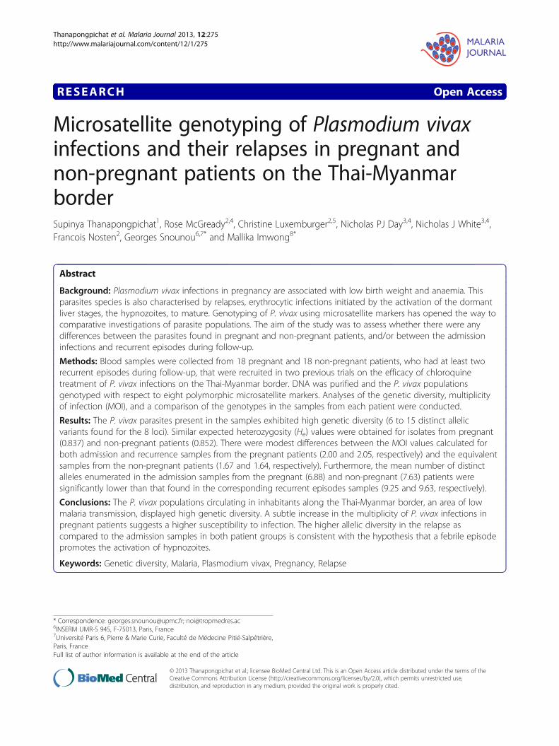
RESEARCH Open Access
Microsatellite genotyping of Plasmodium vivaxinfections and their relapses in pregnant andnon-pregnant patients on the Thai-MyanmarborderSupinya Thanapongpichat1, Rose McGready2,4, Christine Luxemburger2,5, Nicholas PJ Day3,4, Nicholas J White3,4,Francois Nosten2, Georges Snounou6,7* and Mallika Imwong8*
Abstract
Background: Plasmodium vivax infections in pregnancy are associated with low birth weight and anaemia. Thisparasites species is also characterised by relapses, erythrocytic infections initiated by the activation of the dormantliver stages, the hypnozoites, to mature. Genotyping of P. vivax using microsatellite markers has opened the way tocomparative investigations of parasite populations. The aim of the study was to assess whether there were anydifferences between the parasites found in pregnant and non-pregnant patients, and/or between the admissioninfections and recurrent episodes during follow-up.
Methods: Blood samples were collected from 18 pregnant and 18 non-pregnant patients, who had at least tworecurrent episodes during follow-up, that were recruited in two previous trials on the efficacy of chloroquinetreatment of P. vivax infections on the Thai-Myanmar border. DNA was purified and the P. vivax populationsgenotyped with respect to eight polymorphic microsatellite markers. Analyses of the genetic diversity, multiplicityof infection (MOI), and a comparison of the genotypes in the samples from each patient were conducted.
Results: The P. vivax parasites present in the samples exhibited high genetic diversity (6 to 15 distinct allelicvariants found for the 8 loci). Similar expected heterozygosity (He) values were obtained for isolates from pregnant(0.837) and non-pregnant patients (0.852). There were modest differences between the MOI values calculated forboth admission and recurrence samples from the pregnant patients (2.00 and 2.05, respectively) and the equivalentsamples from the non-pregnant patients (1.67 and 1.64, respectively). Furthermore, the mean number of distinctalleles enumerated in the admission samples from the pregnant (6.88) and non-pregnant (7.63) patients weresignificantly lower than that found in the corresponding recurrent episodes samples (9.25 and 9.63, respectively).
Conclusions: The P. vivax populations circulating in inhabitants along the Thai-Myanmar border, an area of lowmalaria transmission, displayed high genetic diversity. A subtle increase in the multiplicity of P. vivax infections inpregnant patients suggests a higher susceptibility to infection. The higher allelic diversity in the relapse ascompared to the admission samples in both patient groups is consistent with the hypothesis that a febrile episodepromotes the activation of hypnozoites.
Keywords: Genetic diversity, Malaria, Plasmodium vivax, Pregnancy, Relapse
* Correspondence: [email protected]; [email protected] UMR-S 945, F-75013, Paris, France7Université Paris 6, Pierre & Marie Curie, Faculté de Médecine Pitié-Salpêtrière,Paris, FranceFull list of author information is available at the end of the article
© 2013 Thanapongpichat et al.; licensee BioMed Central Ltd. This is an Open Access article distributed under the terms of theCreative Commons Attribution License (http://creativecommons.org/licenses/by/2.0), which permits unrestricted use,distribution, and reproduction in any medium, provided the original work is properly cited.
Thanapongpichat et al. Malaria Journal 2013, 12:275http://www.malariajournal.com/content/12/1/275
BackgroundMalaria infection during pregnancy substantially increasesthe risk of morbidity and mortality to the mother, herfoetus and the neonate, and thus constitutes an importantpublic health problem [1]. In areas of high endemicity, thenefarious effects of infection by Plasmodium falciparumare most pronounced in primigravidae, and though lesssevere, infections in subsequent pregnancies remain asso-ciated with anaemia and low birth weight [2]. In areas oflow endemicity, the relatively low levels of acquired im-munity against malaria parasites increase susceptibility tomore severe clinical falciparum episodes during pregnan-cies, which could result in foetal or maternal death [3].The impact of infection by Plasmodium vivax, the mostprevalent parasite in the Asia-Pacific region, is less pro-nounced than that associated with P. falciparum [4]. In alarge-scale study conducted on the western border ofThailand, an area of low endemicity, P. vivax infectionswere more common in primigravidae than in multigrav-idae and were associated with mild maternal anaemia andincreased risk of low birth weight [5]. Histopathologicalexamination of the placentas from P. vivax-infected andtreated women did not reveal any evidence of parasite se-questration or pathological changes [6], thus the patho-genic mechanisms remain unclear.One salient biological characteristic of P. vivax is the
formation of dormant liver stages, hypnozoites, by aproportion of the sporozoites inoculated by the infectedmosquito. Hypnozoites remain uninucleate and meta-bolically quiescent for varying durations, that can ex-tend to years, before resuming their development toform mature schizonts that then initiate a new erythro-cytic episode upon merozoite release. In tropical areas,P. vivax infections have a short latent period (2 to6 weeks), and the primary episode is often followed by asuccession of relapses (every 3 to 4 weeks) that wane infrequency with time [7]. In the mid 1990’s, the recur-rence rate for P. vivax infections treated with chloro-quine and followed up for 63 days was found to be 63%(95% CI 57-69%) in non pregnant patients on the Thai-Myanmar border [8]. In a similar study conducted to-wards the end of the 2000’s in adults and children in thesame region the recurrence rate following chloroquinetreatment increased to 79.1% (95% CI, 73.5%–84.8%),probably because of increased prevalence of chloro-quine resistant P. vivax [9]. Indeed, the first case of P.vivax high-grade resistance to chloroquine in pregnancywas reported recently from the same area [4]. The onlydrug available to eliminate hypnozoites, thus preventingrelapses, is primaquine. Thus, in 1995–1996 when thefirst P. vivax recurrence in non pregnant patients on theThai-Myanmar border was treated with primaquine andchloroquine the risk of having a further vivax episodewithin 2 months was reduced by 96% (95% CI 83-99%)
[8]. However, primaquine is contraindicated in preg-nancy and in 1986 to 1997 23% (149/634) of pregnantwomen had two or more parasitaemia episodes [5].In recent years, reliable methods to genotype P. vivax
populations using microsatellites have been developed[10,11]. Using this methodology it was revealed that P.vivax infections in patients from Thailand, Myanmar andIndia are often polyclonal, and that the genotype of the par-asites in the first relapse following chloroquine treatment[12] differs from that of the initial admission P. vivax popu-lation in more than half of the patients. This suggests heter-ologous activation of hypnozoites, most probably acquiredfrom earlier inoculations. This was supported by data froma study of relapses in infants and in their mothers pre- andpost-partum [13] that showed that whereas admission andrelapse infections are often genetically heterologous in themothers, those observed in the children are generallyhomogeneous. In a recent study P. vivax genetic diversitywas compared in pregnant and non pregnant patients inColombia, and found to be similar in both groups [14]. Thepurpose of the study presented here is to expand know-ledge on the genetic diversity of P. vivax in pregnancy be-yond the few studies quoted above. Given the alteredimmunological and physiological status in pregnancy, itwas considered important to ascertain whether the patternof relapse and the genetic diversity of the P. vivax popula-tions observed differed between pregnant and non-pregnant patients. To this end, a genetic analysis wasconducted on archived samples that had been collected inthe course of drug treatment studies conducted in villagesalong the Thai-Myanmar border.
MethodsStudy site and collection of blood samplesThe studies took place in the Shoklo Malaria ResearchUnit clinics on the Thailand-Myanmar border, an area oflow (estimated entomologic inoculation rate of one orless infectious bites per year) and seasonal malaria trans-mission [15]. The samples were derived from two previ-ous trials on the efficacy of chloroquine in the treatmentof P. vivax. The group of P. vivax-infected non-pregnantwomen represented all those patients with two relapsesrecruited between July 1995 and July 1996 [8], and inpregnant women recruited between November 1998 andJanuary 2000 [16]. In both patients groups P. vivax wasconfirmed by blood smear and given a treatment withchloroquine (Government Pharmaceutical Organization,Thailand) using the following schedule: 15 mg base/kgon the first day, followed by 5 mg base/kg daily on thesecond and third day (total 25 mg base/kg). The non-pregnant patients were followed up for 63 days and anyrecurrent parasitaemia was recorded, treated appropri-ately and the patient followed up for a further 63 days.Pregnant patients were followed up until delivery. At
Thanapongpichat et al. Malaria Journal 2013, 12:275 Page 2 of 9http://www.malariajournal.com/content/12/1/275
recurrence the treatment administered was the same asthat on admission, except in non-pregnant patients wherethe second recurrence was treated with chloroquine com-bined with primaquine (0.25 mg/kg daily for 14 days). Thestudy in non-pregnant women was approved by the EthicsCommittee of Mahidol University and the Karen RefugeeCommittee [8], and that in pregnant women [16] by theEthics Committee of the Faculty of Tropical Medicine ofMahidol University and the Ethics Committee of theLondon School of Hygiene & Tropical Medicine. Written
informed consent was obtained from the patient for thepublication of this report and any accompanying images.
DNA extraction and microsatellite genotypingThe blood samples from the non-pregnant patients werecollected and stored on filter paper (Whatman 3 MM),while those from the pregnant women were whole bloodwith EDTA as anticoagulant stored at −20°C. GenomicDNA was extracted from a punched out 13 mm diameterspot for the filter paper samples (equivalent to about 35 μl
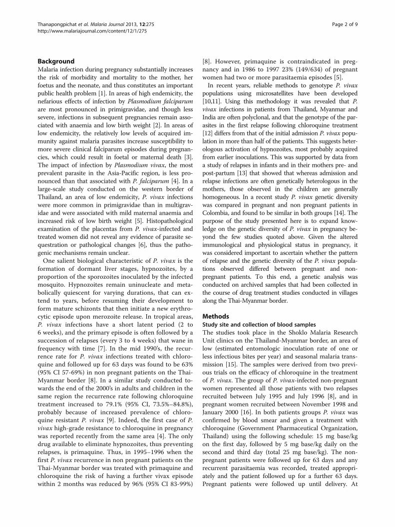
Table 1 Characteristics of pregnant and non-pregnant P. vivax patients
Characteristics Pregnant patients Non-pregnant patients p-value
N = 18 N = 18
Age (years)a 24 ± 7 (15–40) 14 ± 10 (5–46) <0.001
Weight (kg)a 48 ± 6 (41–62) 29 ± 12 (13–48) <0.001
Previous P. vivax malaria
During previous (year)b 5/18 (27.8) 15/18 (83.3) 0.003
>1 episode during previous (year)b 1/5 (20.0) 8/15 (53.3) 0.436
Time since the last episode (days)c 62 (21–102) 65 (36–218) 0.432
Previous P. falciparum
During previous (year)b 4/18 (22.2) 7/18 (38.9) 0.469
>1 episode during previous yearb 1/4 (25.0) 1/7 (14.3) 0.712
Time since the last episode (days)c 26 (21–98) 55 (44–362) 0.088
Characteristics of infection at 1st genotype on day admission N = 18 N = 18
Proportion with a history of feverb 13/18 (72.2) 18/18 (100.0) 0.205
Duration of fever (days)c 3 (1–7) 2 (1–7) 0.016
Proportion febrileb 6/18 (33.3) 10/18 (55.6) 0.314
Temperature (°C)a 37.1 ± 1.4 (35.5-39.7) 37.8 ± 1.0 (36.8-39.9) 0.134
Haematocrit (%)a 32 ± 6 (17–42) 37 ± 3 (31–42) 0.002
Parasitaemia (uL)d 943(32–11,492) 1,991(43–25,844) 0.248
Time to 1st reappearance (days)c 45 (25–71) 43 (35–52) 0.495
Characteristics of patients on day of follow up N = 44 N = 36
Proportion with a history of feverb 22/44 (50.0) 32/36 (88.9) <0.001
Duration of fever in days (range)e 2 (1–3) 1 (1–3) 0.013
Proportion febrileb 11/44 (25.0) 29/36 (80.6) <0.001
Mean temperature (°C)a 36.9 ± 1.1 (35.0-39.7) 38.1 ± 1.0 (35.8-40.8) <0.001
Haematocrit (%)a 31 ± 3 (23–39) 38 ± 3 (30–46) <0.001
Parasitaemia (uL)d 637 (16–3,624) 1,581 (36–22,272) 0.022
Interval times to reappearance (days)
1st reappearance to 2nd reappearance (range)c 51 (28–98) 48 (33–64) 0.468
2nd reappearance to 3rd reappearance (range)c 51(28–66) - -
3rd reappearance to 4th reappearance (range)c 32(28–35) - -a mean ± SD (min-max).b Number of patients (%).c Median (min-max).d Geometric mean (min-max).e Range.N = number of patient.
Thanapongpichat et al. Malaria Journal 2013, 12:275 Page 3 of 9http://www.malariajournal.com/content/12/1/275

of whole blood) and from 200 μl of the EDTA wholeblood, using the QiAamp Blood kit (Hilden, Germany)eluted in a final volume of 100 μl and stored at −20°Cuntil use. The presence of P. vivax was confirmed in allthe samples by nested PCR [17]. Eight extensively poly-morphic microsatellite markers: Pv1.501, Pv3.27, Pv3.502,Pv6.34, Pv 8.504 [10] and MS1, MS5 and MS7 [11], wereused to genotype the isolates using published protocols.Two microliters of the purified genomic DNA were usedas a template for amplification, and in cases where a sec-ondary amplification was carried out, it was initiated with1 μl of the primary amplification product. The amplified
fragments were analysed on an ABI 3130 Genetic Analyzerand by GeneMapper® software version 4.0 (AppliedBiosystems) to measure the variable length in the samples.
Data analysisIn any isolate the presence of one or more alleles at a par-ticular locus was interpreted as a co-infection with two ormore genetically distinct clones, i.e. multiple or polyclonalinfections [10,18]. A locus was classed as having multiplealleles when the score of the minor peak was at least one-third the height of the predominant allele present for thislocus. Samples from which the data was ambiguous were
Table 2 Genetic diversity of P. vivax infections in pregnant and non-pregnant women based on 8 microsatellite markers
Episodes Diversity Patients n Plasmodium vivax loci Mean SD SE p-valueaPv
1.501Pv3.27
Pv3.502
Pv6.34
Pv8.504
MS1 MS5 MS7
Admissionsamples
No. of distinct alleles(A)
P 18 9 9 7 7 6 5 6 6 6.88 1.46 0.52 0.265
NP 18 8 12 9 8 6 4 9 5 7.63 2.56 0.91
He P 18 0.919 0.895 0.889 0.739 0.856 0.772 0.838 0.742 0.831 0.07 0.03 0.533
NP 18 0.876 0.948 0.922 0.830 0.739 0.767 0.892 0.802 0.847 0.07 0.03
No. of distinct allelesper locus
P 18 1.59 1.61 1.50 1.22 1.56 1.35 1.27 1.38 1.44 0.14 0.05 <0.001
NP 18 1.28 1.33 1.11 1.17 1.22 1.13 1.13 1.07 1.18 0.09 0.03
Recurrentsamples
No. of distinct alleles(A)
P 44 9 11 10 12 8 6 9 9 9.25 1.83 0.65 0.487
NP 36 13 14 10 11 8 6 8 8 9.63 2.76 0.98
He P 44 0.882 0.824 0.872 0.901 0.846 0.736 0.848 0.839 0.844 0.05 0.02 0.316
NP 36 0.911 0.919 0.884 0.867 0.808 0.783 0.874 0.835 0.86 0.05 0.02
No. of distinct allelesper locus
P 44 1.49 1.40 1.30 1.50 1.36 1.26 1.27 1.51 1.38 1.11 0.04 <0.001
NP 36 1.26 1.31 1.11 1.17 1.11 1.06 1.10 1.23 1.17 0.09 0.03
Allsamples
No of distinct alleles(A)
P 62 9 12 10 12 8 7 9 10 9.63 1.77 0.63 0.320
NP 54 13 15 10 13 9 6 9 8 10.38 3.02 1.07
He P 62 0.885 0.846 0.869 0.864 0.845 0.736 0.834 0.820 0.837 0.05 0.02 0.340
NP 54 0.906 0.921 0.888 0.848 0.781 0.770 0.866 0.836 0.852 0.06 0.02
No. of distinct allelesper locus
P 62 1.52 1.46 1.35 1.42 1.42 1.28 1.29 1.47 1.40 0.09 0.03 <0.001
NP 54 1.26 1.31 1.11 1.17 1.15 1.08 1.11 1.18 1.17 0.08 0.03
P = pregnant women and NP = non-pregnant women.The No. of alleles and He values were calculated from a data set in which only the predominant alleles at each locus was considered.The No. of alleles per locus values were calculated from all detected alleles at each loci, divided by the total number of samples.a Paired t-test was used to compare the means between groups.
Table 3 Multiple clone infections and multiplicity of infection in P. vivax
Episodes of infection Pregnant women Non-pregnant women p-value
Pregnant women Non-pregnant women p-valueSamples with multiple clones MOI*, mean (±SD)
Admission 67% 44% 0.180 2.00 1.67 0.278
(12/18 isolates) (8/18 isolates) (±0.97) (±0.84)
Recurrence 55% 53% 0.875 2.05 1.64 0.110
(24/44 isolates) (19/36 isolates) (±1.41) (±0.80)
All episodes of infection 58% 50% 0.384 2.03 1.65 0.054
(36/62 isolates) (27/54 isolates) (± 1.30) (±0.81)
*MOI =multiplicity of infection.
Thanapongpichat et al. Malaria Journal 2013, 12:275 Page 4 of 9http://www.malariajournal.com/content/12/1/275
re-amplified (maximum peak height < 300 fluorescentunits). The genetic diversity was measured using thepredominated allele at each locus to calculate the expectedheterozygosity (He). The formula defined as He = 1/(1-n)(1-∑pi2) where p is the frequency of ith allele. Expected het-erozygosity (He) ranges between 0 and 1, a value close to 1indicated high genetic diversity levels in the population
[19]. The multiplicity of infection (MOI) was also calcu-lated as the maximum number of alleles observed at anylocus. Multilocus linkage disequilibrium (LD) was calcu-lated by using a standardized index of association (ISA)[20,21]. This test compares the variance (VD) of the num-ber of alleles shared between all pairs of haplotypes ob-served in the population (D) with the variance expectedunder random association of alleles (VE) as follows: ISA =(VD/VE-1) (r-1), where r is the number of loci analyzed[20]. VE is derived from 10,000 simulated data sets inwhich alleles were randomly reshuffled among haplotypes.Significant linkage disequilibrium is detected if VD isgreater than 95% of the values derived from the reshuffleddata sets. Data were analyzed with LIAN 3.1 [22]. Onlythe dominant alleles were considered to verify linkage.In order to detect possible bias due to equivocal assign-ment of haplotypes in multiple-clone infections, link-age disequilibrium (LD) was tested at three levels: (i)for all infections including those with more than onemulti-allelic locus, (ii) for single clone infection, and(iii) for unique haplotypes only. In order to assess re-latedness between isolates the genotype observed at
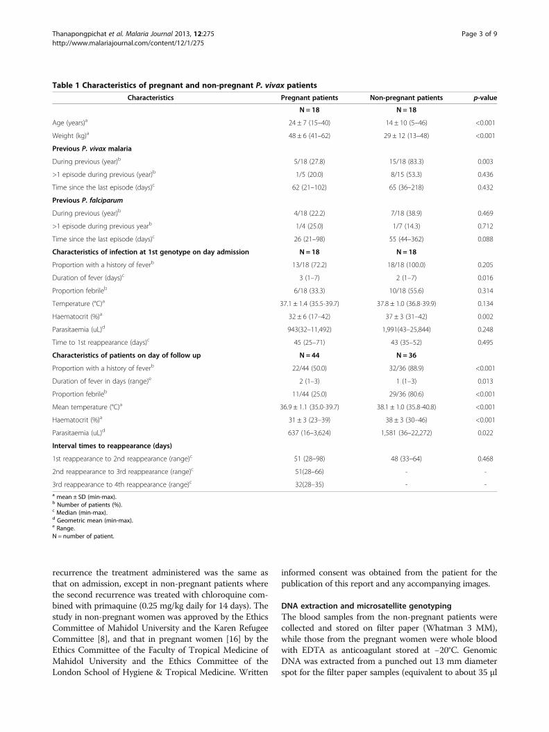
Figure 1 Frequency distribution of the number of loci with multiple alleles. The frequency of samples carrying multiple alleles for a givenlocus is plotted against the total number of loci in each sample found to be polyclonal.
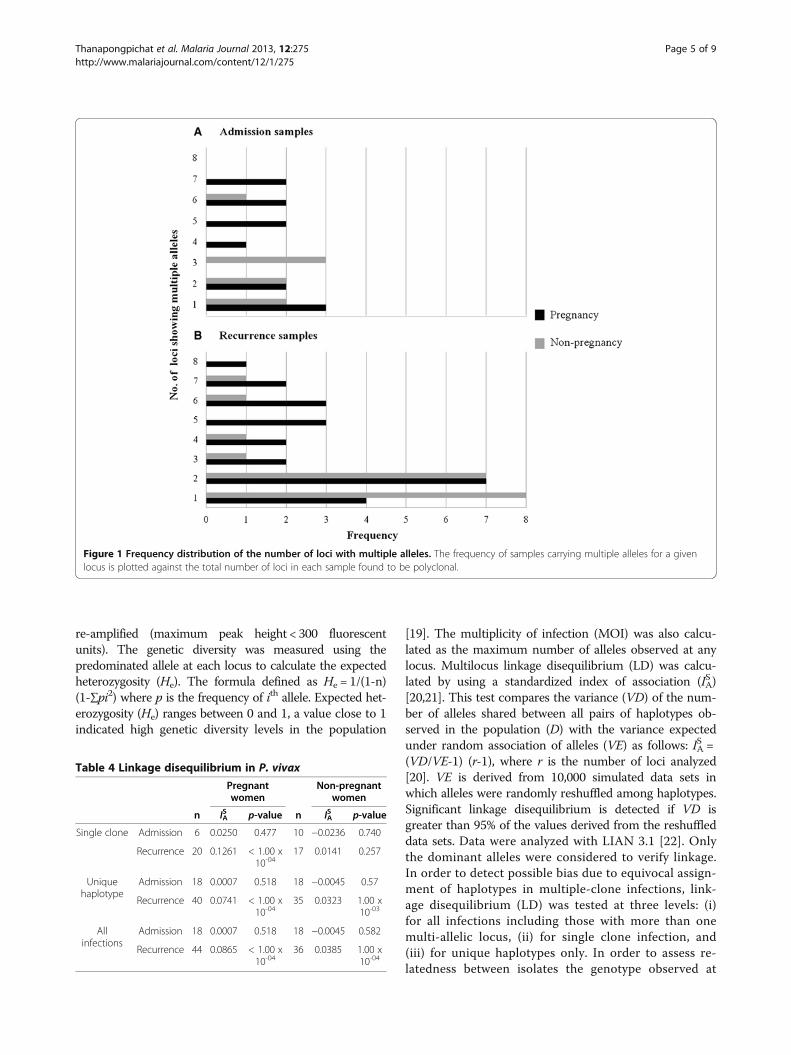
Table 4 Linkage disequilibrium in P. vivax
Pregnantwomen
Non-pregnantwomen
n IAS p-value n IA
S p-value
Single clone Admission 6 0.0250 0.477 10 −0.0236 0.740
Recurrence 20 0.1261 < 1.00 x10-04
17 0.0141 0.257
Uniquehaplotype
Admission 18 0.0007 0.518 18 −0.0045 0.57
Recurrence 40 0.0741 < 1.00 x10-04
35 0.0323 1.00 x10-03
Allinfections
Admission 18 0.0007 0.518 18 −0.0045 0.582
Recurrence 44 0.0865 < 1.00 x10-04
36 0.0385 1.00 x10-04
Thanapongpichat et al. Malaria Journal 2013, 12:275 Page 5 of 9http://www.malariajournal.com/content/12/1/275
reappearances with that one admission day were com-pared: two genotypes were classed as related if a geno-type was either a mixture of 2 adjacent microsatellitealleles at any one locus of which one was present in thepaired sample, or if alleles at one or two loci differedonly by one tandem repeat [13].
Results and discussionA total of 116 P. vivax isolates were selected for inclusionin this study: 54 were derived from 18 non-pregnant pa-tients who all had two recurrent episodes, and 62 wereobtained from 18 pregnant patients who all had two re-currences, four of whom then had a third and two more afourth recurrence. The characteristics of the patients areprovided in Table 1. Non-pregnant patients tended to beof lower age and to have a more frequent history of P.vivax infections in the year preceding the date of recruit-ment to the study than the pregnant patients. They also
tended to have higher temperature and parasitaemia onadmission. As expected, pregnant patients had a lowerhaematocrit than non-pregnant patients. The mean inter-val time to the first reappearance did not significantly dif-fer between the two groups, 45 days for the pregnant vs43 days for the non-pregnant patients, nor did it differ forthe second reappearance, 51 days vs 48 days. The secondrecurrence in non-pregnant women was treated withchloroquine and primaquine, and no further recurrencesoccurred. In the pregnant women the mean times to thethird and fourth reappearances were 51 days and 32 days,respectively.The microsatellite genotyping data were used to calculate
the mean number of distinct alleles (A), the heterozygosity(He) and the mean number of distinct allelic variants foreach locus in each sample (Table 2). The high number ofdistinguishable allelic forms observed for each locus andthe high value of heterozygosity indicated that overall the P.
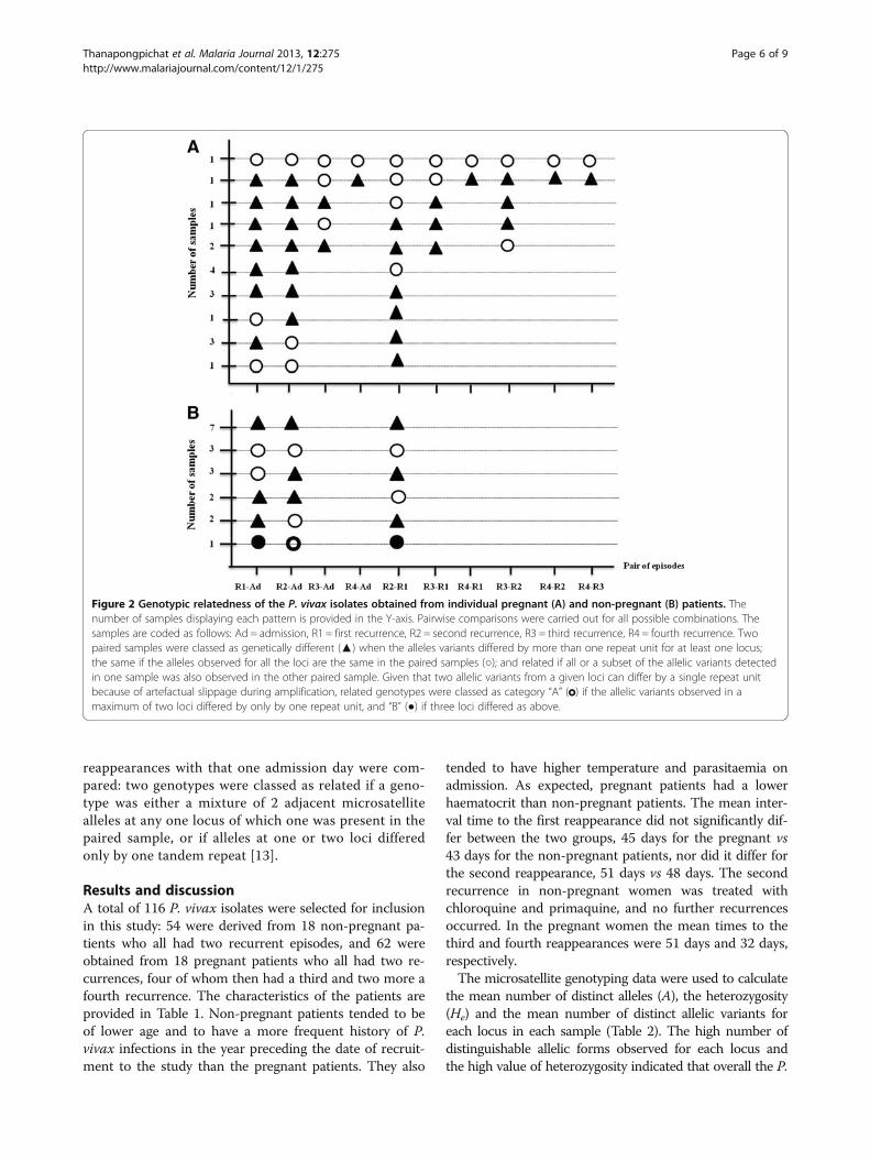
Figure 2 Genotypic relatedness of the P. vivax isolates obtained from individual pregnant (A) and non-pregnant (B) patients. Thenumber of samples displaying each pattern is provided in the Y-axis. Pairwise comparisons were carried out for all possible combinations. Thesamples are coded as follows: Ad = admission, R1 = first recurrence, R2 = second recurrence, R3 = third recurrence, R4 = fourth recurrence. Twopaired samples were classed as genetically different (▲) when the alleles variants differed by more than one repeat unit for at least one locus;the same if the alleles observed for all the loci are the same in the paired samples (○); and related if all or a subset of the allelic variants detectedin one sample was also observed in the other paired sample. Given that two allelic variants from a given loci can differ by a single repeat unitbecause of artefactual slippage during amplification, related genotypes were classed as category “A” ( ) if the allelic variants observed in amaximum of two loci differed by only by one repeat unit, and “B” (●) if three loci differed as above.
Thanapongpichat et al. Malaria Journal 2013, 12:275 Page 6 of 9http://www.malariajournal.com/content/12/1/275
vivax isolates circulating in the patients had a high de-gree of genetic diversity. The values did not signifi-cantly differ between parasites from pregnant and non-pregnant patients. There was a tendency that did notreach significance for samples from pregnant women tohave a lower mean number of distinguishable alleles(A) than those from non-pregnant patients. On theother hand the number of distinct alleles per locus wassignificantly higher in pregnant vs non-pregnant pa-tients. This was reflected in the higher MOI observedin the combined admission and recurrence samplesfrom the pregnant vs non-pregnant women (Table 3), adifference that nearly reached significance. Nonethe-less, the proportion of polyclonal infections was notstatistically different between the samples obtained
from either group (Table 3). When all the P. vivax sam-ples from the admission vs the recurrent samples werecompared (Table 2), there was a significant differencein the mean number of distinct alleles (A) observed forthe loci (7.25 vs 9.5, p-value 0.0001), and the isolatesfrom the recurrent episodes showed a higher propor-tion of loci for which more than one allelic variant wasnoted in each sample (Figure 1). Linkage disequilibriumwas assessed for clonal infections, infections with uniquehaplotype and all infections, in samples from pregnant pa-tients and non-pregnant patients that were subdivided asadmission and recurrence samples (Table 4). No evidentfor linkage disequilibrium was found for the parasites inthe samples obtained on admission, or in the subgroup ofrecurrence samples with a monoclonal infection. However,significant linkage disequilibrium was found for the recur-rent samples from the two groups when considered intheir entirety or for the subgroup that has a unique haplo-type (Table 4).The genetic relatedness of the parasites obtained from
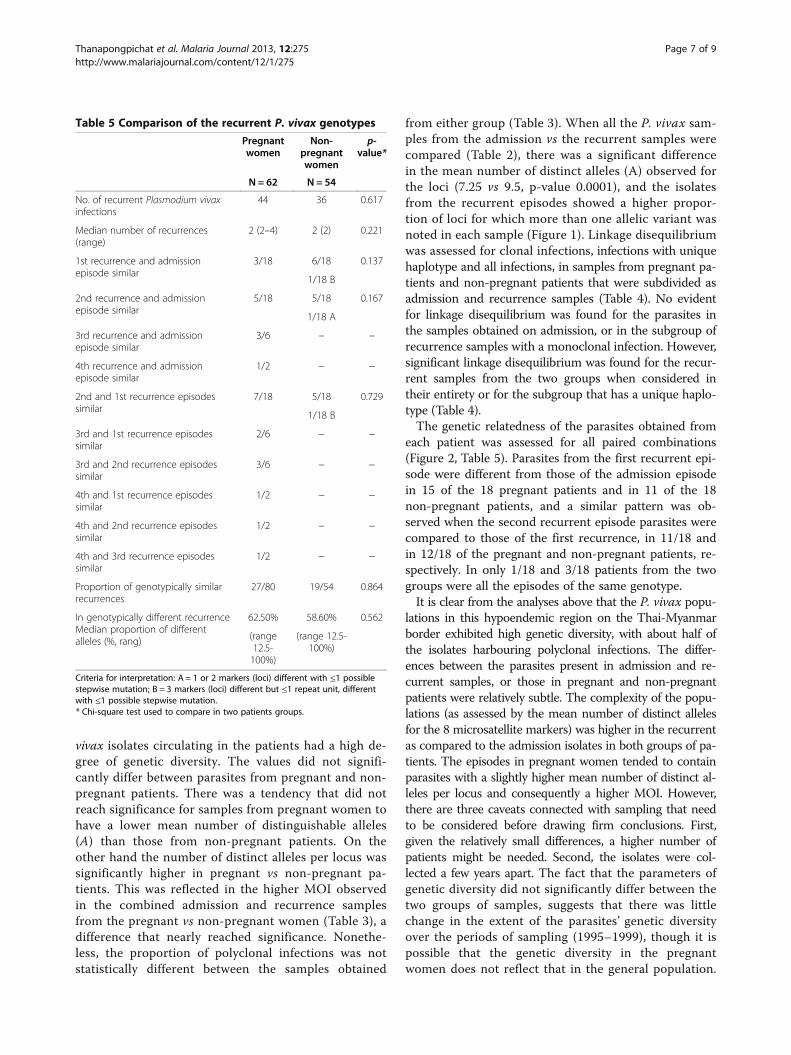
each patient was assessed for all paired combinations(Figure 2, Table 5). Parasites from the first recurrent epi-sode were different from those of the admission episodein 15 of the 18 pregnant patients and in 11 of the 18non-pregnant patients, and a similar pattern was ob-served when the second recurrent episode parasites werecompared to those of the first recurrence, in 11/18 andin 12/18 of the pregnant and non-pregnant patients, re-spectively. In only 1/18 and 3/18 patients from the twogroups were all the episodes of the same genotype.It is clear from the analyses above that the P. vivax popu-
lations in this hypoendemic region on the Thai-Myanmarborder exhibited high genetic diversity, with about half ofthe isolates harbouring polyclonal infections. The differ-ences between the parasites present in admission and re-current samples, or those in pregnant and non-pregnantpatients were relatively subtle. The complexity of the popu-lations (as assessed by the mean number of distinct allelesfor the 8 microsatellite markers) was higher in the recurrentas compared to the admission isolates in both groups of pa-tients. The episodes in pregnant women tended to containparasites with a slightly higher mean number of distinct al-leles per locus and consequently a higher MOI. However,there are three caveats connected with sampling that needto be considered before drawing firm conclusions. First,given the relatively small differences, a higher number ofpatients might be needed. Second, the isolates were col-lected a few years apart. The fact that the parameters ofgenetic diversity did not significantly differ between thetwo groups of samples, suggests that there was littlechange in the extent of the parasites’ genetic diversityover the periods of sampling (1995–1999), though it ispossible that the genetic diversity in the pregnantwomen does not reflect that in the general population.
Table 5 Comparison of the recurrent P. vivax genotypes
Pregnantwomen
Non-pregnantwomen
p-value*
N = 62 N = 54
No. of recurrent Plasmodium vivaxinfections
44 36 0.617
Median number of recurrences(range)
2 (2–4) 2 (2) 0.221
1st recurrence and admissionepisode similar
3/18 6/18 0.137
1/18 B
2nd recurrence and admissionepisode similar
5/18 5/18 0.167
1/18 A
3rd recurrence and admissionepisode similar
3/6 − −
4th recurrence and admissionepisode similar
1/2 − −
2nd and 1st recurrence episodessimilar
7/18 5/18 0.729
1/18 B
3rd and 1st recurrence episodessimilar
2/6 − −
3rd and 2nd recurrence episodessimilar
3/6 − −
4th and 1st recurrence episodessimilar
1/2 − −
4th and 2nd recurrence episodessimilar
1/2 − −
4th and 3rd recurrence episodessimilar
1/2 − −
Proportion of genotypically similarrecurrences
27/80 19/54 0.864
In genotypically different recurrenceMedian proportion of differentalleles (%, rang)
62.50% 58.60% 0.562
(range12.5-100%)
(range 12.5-100%)
Criteria for interpretation: A = 1 or 2 markers (loci) different with ≤1 possiblestepwise mutation; B = 3 markers (loci) different but ≤1 repeat unit, differentwith ≤1 possible stepwise mutation.* Chi-square test used to compare in two patients groups.
Thanapongpichat et al. Malaria Journal 2013, 12:275 Page 7 of 9http://www.malariajournal.com/content/12/1/275
Third, the amount of blood equivalent to the DNA tem-plate aliquots used for genotyping was smaller for thenon-pregnant (0.7 μl) than for the pregnant (4 μl) pa-tients. This was inherent to the nature of the archivedsamples (dried blood spots and whole frozen blood, re-spectively). This six-fold difference in the volume ofblood analyzed might account for the higher MOI inthe pregnant women’s episodes, though it should bemitigated by the fact that mean parasitaemia in preg-nant patients was half that in non-pregnant women,and that a 33% cut-off threshold was used to excludethe minor alleles. The main reason why this is unlikelyis that it is inconsistent with the increase in allelic di-versity in recurrent as compared to admission samples.In conclusion, the parasites causing episodes in pregnant
women had a higher genetic diversity than those in non-pregnant patients. This could be due to increased suscepti-bility to mosquito bites, reduced levels of immunity to in-fection, physiological changes in pregnancy especially ifthey affect reticulocyte dynamics, or a combination of thesefactors. In both patient groups the parasites at recurrencefollowing treatment of the admission episode displayed ahigher genetic complexity. In both pregnant and non-pregnant patients, the parasites at recurrence were genetic-ally distinct from those on admission in about half thecases, as were the parasites from the first and the secondrecurrence. This pattern is similar to that observed earlierfor relapsing P. vivax episodes in Thailand [12,13]. Differ-ential accumulation of P. vivax in the placenta has not beennoted in this area [6], though the phenomenon was ob-served in deliveries at Papua New Guinea [23], and in vitrocytoadhesion of P. vivax-infected red blood cells to ligandsfound in the placenta has been reported [24,25]. However,at the time when the samples analysed here were obtained,resistance of P. vivax to chloroquine, the treatment thatwas administered to both groups of patients, had not beenrecorded in Thailand [8,26]. Therefore, it is highly likelythat the recurrent episodes analysed in this study were re-lapses originating from hypnozoites. The frequent heterol-ogous nature of these relapse infections and their increasedgenetic diversity as compared to the admission infectionsare consistent with the activation of latent hypnozoites hy-pothesis [27].
Competing interestsThe authors declare that they have no competing interests.
Authors’ contributionsRM, FN, MI, GS, ND and NW were involved in the conception and design ofthe study. ST performed the laboratory experiments and the analysis. RM, FNand CL were involved in the samples collection. ST and MI wrote the firstdraft. GS and MI wrote the final draft of the manuscript. All authors read andapproved the final manuscript.
AcknowledgementsWe thank all pregnant women who provided a blood sample and all thestaff at the Shoklo Malaria Research Unit who contributed to this study.
Thanks also to Dr. Leopoldo Villegas who provided the blood samples ofpregnancies. This project was supported by the programme strategicscholarship for frontier research network for the joint Ph.D. Programme andthe office of the higher education commission, the Wellcome Trust of GreatBritain and Mahidol University and The Dean’s Research Fund, Faculty ofTropical Medicine.
Author details1Department of Clinical Tropical Medicine, Faculty of Tropical Medicine,Mahidol University, Bangkok, Thailand. 2Shoklo Malaria Research Unit, MaeSot, Tak Province, Thailand. 3Mahidol Oxford Tropical Research Unit, Facultyof Tropical Medicine, Mahidol University, Bangkok, Thailand. 4Centre forVaccinology and Tropical Medicine, Churchill Hospital, Oxford, UK. 5SanofiPasteur, Lyon, France. 6INSERM UMR-S 945, F-75013, Paris, France. 7UniversitéParis 6, Pierre & Marie Curie, Faculté de Médecine Pitié-Salpêtrière, Paris,France. 8Department of Molecular Tropical Medicine and Genetics, Faculty ofTropical Medicine, Mahidol University, Bangkok, Thailand.
Received: 3 April 2013 Accepted: 30 July 2013Published: 6 August 2013
References1. WHO: Malaria in pregnancy. Geneva: World Health Organization; 2007.2. Steketee RW, Wirima JJ, Campbell CC: Developing effective strategies for
malaria prevention programs for pregnant African women. Am J TropMed Hyg 1996, 55(1 Suppl):95–100.
3. Luxemburger C, Ricci F, Nosten F, Raimond D, Bathet S, White NJ: Theepidemiology of severe malaria in an area of low transmission inThailand. Trans R Soc Trop Med Hyg 1997, 91:256–262.
4. Rijken MJ, Boel ME, Russell B, Imwong M, Leimanis ML, Phyo AP,Muehlenbachs A, Lindegardh N, McGready R, Renia L, Snounou G,Singhasivanon P, Nosten F: Chloroquine resistant vivax malaria in apregnant woman on the western border of Thailand. Malar J2011, 10:113.
5. Nosten F, McGready R, Simpson JA, Thwai KL, Balkan S, Cho T, Hkirijaroen L,Looareesuwan S, White NJ: Effects of Plasmodium vivax malaria inpregnancy. Lancet 1999, 354:546–549.
6. McGready R, Davison BB, Stepniewska K, Cho T, Shee H, Brockman A,Udomsangpetch R, Looareesuwan S, White NJ, Meshnick SR, Nosten F: Theeffects of Plasmodium falciparum and P. vivax infections on placentalhistopathology in an area of low malaria transmission. Am J Trop MedHyg 2004, 70:398–407.
7. Cogswell FB: The hypnozoite and relapse in primate malaria. ClinMicrobiol Rev 1992, 5:26–35.
8. Luxemburger C, Van Vugt M, Jonathan S, McGready R, Looareesuwan S,White NJ, Nosten F: Treatment of vivax malaria on the western border ofThailand. Trans R Soc Trop Med Hyg 1999, 93:433–438.
9. Phyo AP, Lwin KM, Price RN, Ashley EA, Russell B, Sriprawat K, Lindegardh N,Singhasivanon P, White NJ, Nosten F: Dihydroartemisinin-piperaquineversus chloroquine in the treatment of Plasmodium vivax malaria inThailand: a randomized controlled trial. Clin Infect Dis 2011, 53:977–984.
10. Imwong M, Nair S, Pukrittayakamee S, Sudimack D, Williams JT, MayxayM, Newton PN, Kim JR, Nandy A, Osorio L, Carlton JM, White NJ, DayNP, Anderson TJ: Contrasting genetic structure in Plasmodium vivaxpopulations from Asia and South America. Int J Parasitol 2007,37:1013–1022.
11. Karunaweera NDFM, Hartl DL, Wirth DF: Fourteen polymorphicmicrosatellite DNA markers for the human malaria parasite Plasmodiumvivax. Mol Ecol Notes 2007, 7:172–175.
12. Imwong M, Snounou G, Pukrittayakamee S, Tanomsing N, Kim JR, Nandy A,Guthmann JP, Nosten F, Carlton J, Looareesuwan S, Nair S, Sudimack D, DayNP, Anderson TJ, White NJ: Relapses of Plasmodium vivax infection usuallyresult from activation of heterologous hypnozoites. J Infect Dis 2007,195:927–933.
13. Imwong M, Boel ME, Pagornrat W, Pimanpanarak M, McGready R, Day NP,Nosten F, White NJ: The first Plasmodium vivax relapses of life are usuallygenetically homologous. J Infect Dis 2012, 205:680–683.
14. Arango EM, Samuel R, Agudelo OM, Carmona-Fonseca J, Maestre A, YanowSK: Genotype comparison of Plasmodium vivax and Plasmodiumfalciparum clones from pregnant and non-pregnant populations inNorth-west Colombia. Malar J 2012, 11:392.
Thanapongpichat et al. Malaria Journal 2013, 12:275 Page 8 of 9http://www.malariajournal.com/content/12/1/275
15. Luxemburger C, Thwai KL, White NJ, Webster HK, Kyle DE, Maelankirri L,Chongsuphajaisiddhi T, Nosten F: The epidemiology of malaria in a Karenpopulation on the western border of Thailand. Trans R Soc Trop Med Hyg1996, 90:105–111.
16. Villegas L, McGready R, Htway M, Paw MK, Pimanpanarak M, Arunjerdja R,Viladpai-Nguen SJ, Greenwood B, White NJ, Nosten F: Chloroquineprophylaxis against vivax malaria in pregnancy: a randomized, double-blind, placebo-controlled trial. Trop Med Int Health 2007, 12:209–218.
17. Snounou G, Viriyakosol S, Zhu XP, Jarra W, Pinheiro L, Do Rosario VE,Thaithong S, Brown KN: High sensitivity of detection of human malariaparasites by the use of nested polymerase chain reaction. Mol BiochemParasitol 1993, 61:315–320.
18. Anderson TJ, Su XZ, Bockarie M, Lagog M, Day KP: Twelve microsatellitemarkers for characterization of Plasmodium falciparum from finger-prickblood samples. Parasitology 1999, 119:113–125.
19. Gunawardena S, Karunaweera ND, Ferreira MU, Phone-Kyaw M, Pollack RJ,Alifrangis M, Rajakaruna RS, Konradsen F, Amerasinghe PH, Schousboe ML,Galappaththy GN, Abeyasinghe RR, Hartl DL, Wirth DF: Geographicstructure of Plasmodium vivax: microsatellite analysis of parasitepopulations from Sri Lanka, Myanmar, and Ethiopia. Am J Trop Med Hyg2010, 82:235–242.
20. Hudson RR: Analytical results concerning linkage disequilibrium inmodels with genetic transformation and recombination. J Evol Biol1994, 7:535–548.
21. Smith JM, Smith NH, O’Rourke M, Spratt BG: How clonal are bacteria?Proc Natl Acad Sci USA 1993, 90:4384–4388.
22. Haubold B, Hudson RR: LIAN 3.0: detecting linkage disequilibrium inmultilocus data. Linkage Analysis. Bioinformatics 2000, 16:847–848.
23. Mayor A, Bardaji A, Felger I, King CL, Cistero P, Dobano C, Stanisic DI, Siba P,Wahlgren M, Del Portillo H, Mueller I, Menéndez C, Ordi J, Rogerson S:Placental infection with Plasmodium vivax: a histopathological andmolecular study. J Infect Dis 2012, 206:1904–1910.
24. Carvalho BO, Lopes SC, Nogueira PA, Orlandi PP, Bargieri DY, Blanco YC,Mamoni R, Leite JA, Rodrigues MM, Soares IS, Oliveira TR, Wunderlich G,Lacerda MV, Del Portillo HA, Araújo MO, Russell B, Suwanarusk R, SnounouG, Rénia L, Costa FT: On the cytoadhesion of Plasmodium vivax-infectederythrocytes. J Infect Dis 2010, 202:638–647.
25. Chotivanich K, Udomsangpetch R, Suwanarusk R, Pukrittayakamee S,Wilairatana P, Beeson JG, Day NP, White NJ: Plasmodium vivax adherenceto placental glycosaminoglycans. PLoS One 2012, 7:e34509.
26. Pukrittayakamee S, Chantra A, Simpson JA, Vanijanonta S, Clemens R,Looareesuwan S, White NJ: Therapeutic responses to different antimalarialdrugs in vivax malaria. Antimicrob Agents Chemother 2000, 44:1680–1685.
27. White NJ: Determinants of relapse periodicity in Plasmodium vivaxmalaria. Malar J 2011, 10:297.
doi:10.1186/1475-2875-12-275Cite this article as: Thanapongpichat et al.: Microsatellite genotyping ofPlasmodium vivax infections and their relapses in pregnant and non-pregnant patients on the Thai-Myanmar border. Malaria Journal2013 12:275.
Submit your next manuscript to BioMed Centraland take full advantage of:
• Convenient online submission
• Thorough peer review
• No space constraints or color figure charges
• Immediate publication on acceptance
• Inclusion in PubMed, CAS, Scopus and Google Scholar
• Research which is freely available for redistribution
Submit your manuscript at www.biomedcentral.com/submit
Thanapongpichat et al. Malaria Journal 2013, 12:275 Page 9 of 9http://www.malariajournal.com/content/12/1/275