Embed Size (px)
Citation preview

MicroscopesMicroscopes
• Microscope, instrument used to obtain a magnified image of minute objects or minute details of objects.
• OPTICAL MICROSCOPES The most widely used microscopes are optical microscopes, which use visible light to create a magnified image of an object.

Early MicroscopesEarly Microscopes
• The first microscopes were of two kinds: simple and compound.
• A simple microscope has just one lens (or lens combination)—a magnifying glass can be thought of as such a microscope.
• Simple microscopes are capable of extremely fine work. Those made by the great Dutch microscopist Anton van Leeuwenhoek (1632-1723) were small enough to fit in one's palm, but they enabled him to see cells, bacteria, and single-celled animals

MicroscopesMicroscopes
• Paramecium
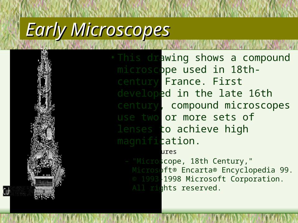
Early MicroscopesEarly Microscopes• This drawing shows a compound
microscope used in 18th-century France. First developed in the late 16th century, compound microscopes use two or more sets of lenses to achieve high magnification.
• Culver Pictures– "Microscope, 18th Century," Microsoft® Encarta®
Encyclopedia 99. © 1993-1998 Microsoft Corporation. All rights reserved.

Early MicroscopesEarly Microscopes
• English scientist Robert Hooke built this microscope in the 17th century and used it to conduct pioneering research. He discovered the cell structure of plants by observing a thin slice of cork under his microscope.
• Cecil Fox/Science Source/Photo Researchers, Inc.
– "Hooke’s Microscope," Microsoft® Encarta® Encyclopedia 99. © 1993-1998 Microsoft Corporation. All rights reserved.

Compound MicroscopeCompound Microscope
• The compound microscope uses two lenses, an objective lens and an ocular lens, mounted at opposite ends of a closed tube, to provide greater magnification than is possible with a single lens. The total magnification of a compound microscope is determined by the two lens systems and can be more than 2000 times.
• We will use compound microscopes.

Compound MicroscopeCompound Microscope
Objective LensesObjective Lens
Ocular Lens
(Eye Piece)

Compound MicroscopesCompound Microscopes
• Two key characteristics affect the quality of a microscope—magnification and resolving power.
• A compound microscope's magnification is determined by multiplication: it is equal to the magnification produced by the ocular times the magnification produced by the objective.
• Viewing an object with a 10X ocular lens (a lens that magnifies things ten times) and a 100X objective lens produces an enlarged image that is 1,000X—that is, it appears to be a thousand times larger.

Compound MicroscopesCompound Microscopes
• Today, a typical compound microscope can resolve better than 1 micrometer (one-thousandth of a millimeter; 1 m equals one-millionth of a meter, a meter being about 39.37 inches).
• Most bacteria are about 1 m long; by comparison, human hair is around 100 m thick. An atom is much smaller than that—about 0.1 nanometer (a nanometer is only a billionth of a meter).

MicroscopesMicroscopes
Bacteria

Compound MicroscopesCompound Microscopes
• Optical microscopes have a firm stand with a flat stage to hold the material examined and some means for moving the microscope tube toward and away from the specimen to bring it into focus: course and fine adjustment knobs.
• Ordinarily, specimens are transparent and are mounted on slides—thin, rectangular pieces of clear glass that are placed on the stage for viewing.

Compound MicroscopesCompound Microscopes
Stage
Base
Stage Clips
Course Adjustment
Fine Adjustment

Compound MicroscopeCompound Microscope
• The stage has a small hole through which light can pass from a light source mounted underneath the stage—either a mirror that reflects natural light or a special electric light that directs light through the specimen.
• A revolving diaphram regulates how much light is allowed to pass through to the slide.

Compound MicroscopeCompound Microscope
DiaphramLight Source

MicroscopesMicroscopes
• In photomicrography, the process of taking photographs through a microscope, a camera is mounted directly above the microscope's eyepiece. Normally the camera does not contain a lens because the microscope itself acts as the lens system.
• Microscopes used for research have a number of refinements to enable a complete study of the specimens. Because the image of a specimen is highly magnified and inverted, manipulating the specimen by hand is difficult.
• You will find this out for yourself, so be patient!

MicroscopesMicroscopes
• The stages of high-powered research microscopes can by moved by micrometer screws. You will use these if you take Med. Tech. or Bio II. In some microscopes, the stage can also be rotated.
• Research microscopes are also equipped with three or more objective lenses, mounted on a revolving head, so that the magnifying power of the microscope can be varied.
• Our microscopes are equipped with a revolving head and three objective lenses.

MicroscopesMicroscopes
Revolving HeadArm
MicroscopesMicroscopes
• In this class you will have hands-on experience with a microscope.
• A miniature world will be opened up as you slip a glass slide under the instrument and peer at all sorts of interesting things.These images will be exciting and fun!
• Sophisticated research microscopes can provide even more.

Seeing the Invisible: The New Microscope Seeing the Invisible: The New Microscope
• By Samuel H. Cohen
• Recent years have seen ingenious refinements of the traditional microscope that relies on visible light.
• Innovative types of electron microscopes—which rely on electrons—have appeared as well. It was the electron microscope (whose development in the 1930s eventually earned its inventor, German physicist Ernst Ruska, a Nobel Prize) that brought the human eye into contact with the world of molecules and atoms.

Seeing the Invisible: The New MicroscopeSeeing the Invisible: The New Microscope
• Within the last decade scientists have also devised new types of microscopes that can look even further into the hidden structure of reality.
• To get an idea of the smallness of the features that these microscopes can detect (smaller than an atom), think of it this way: about 100 billion atoms could be fitted into the period at the end of this sentence.

Electron MicroscopesElectron Microscopes
• The electron microscope, which works like a light microscope but uses a beam of electrons instead of a beam of light.
• The electron beam is deflected and focused by means of magnetic lenses.
• Using an electron microscope, scientists, for the first time, could peer into the structure of crystals and fibers and explore the world of viruses, DNA, and objects of like scale.

Electron MicroscopesElectron Microscopes
This false color image of the head of a fruit fly illustrates the level of detail that electron microscopes are capable of resolving. The magnification of this image is about 200 times. Electron microscopes achieve much greater magnifications than light microscopes by using electrons with wavelengths considerably shorter than those associated with visible light. Light microscopes are capable of maximum magnifications of about 2,000 times, whereas magnifications approaching 1,000,000 times are possible with electron microscopes.
Oliver Meckes/Photo Researchers, Inc.
"Image of a Fruit Fly," Microsoft® Encarta® Encyclopedia 99. © 1993-1998 Microsoft Corporation.
All rights reserved.