Embed Size (px)
Citation preview
RESEARCH POSTER PRESENTATION DESIGN © 2012
www.PosterPresentations.com
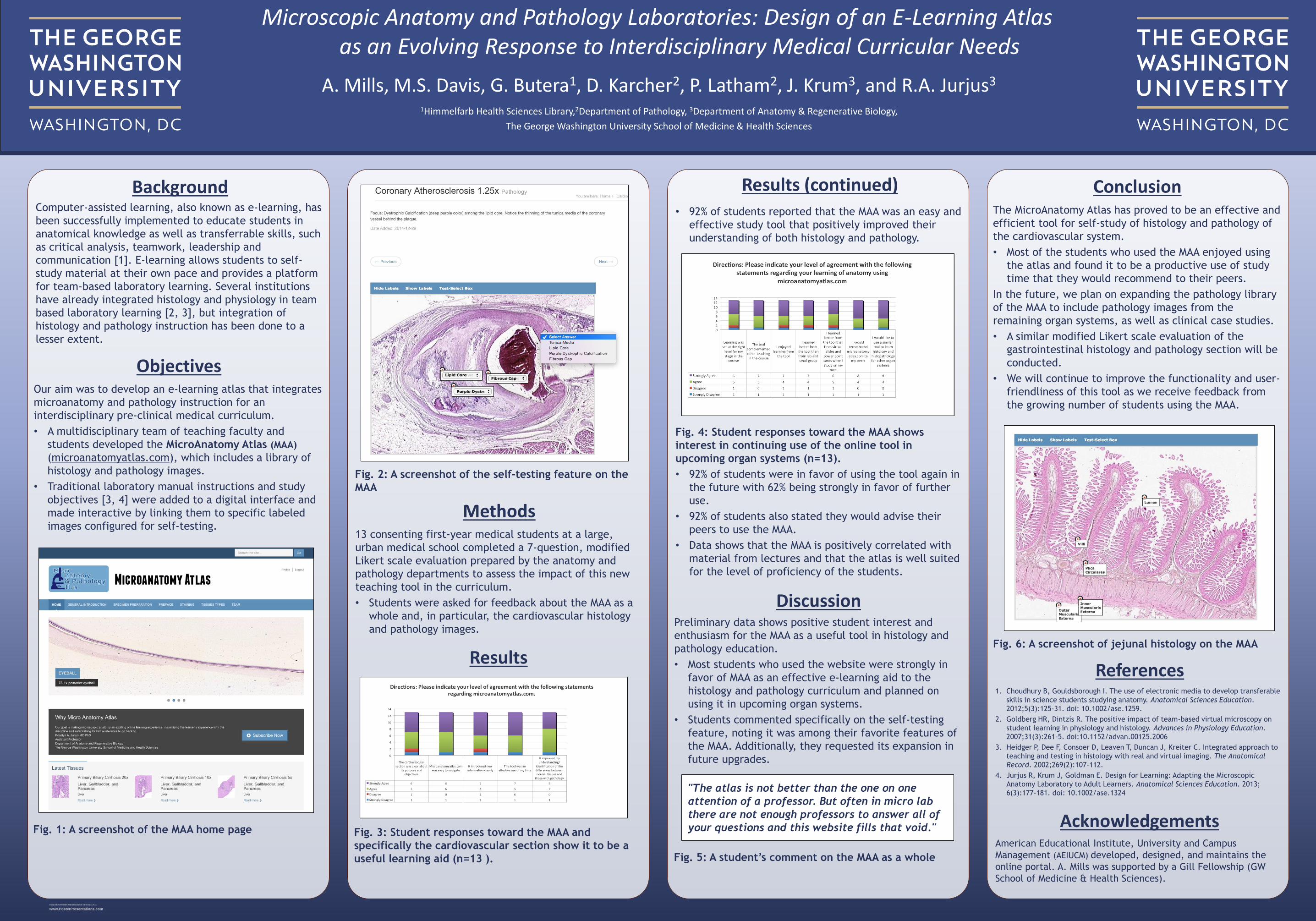
Objectives
13 consenting first-year medical students at a large,
urban medical school completed a 7-question, modified
Likert scale evaluation prepared by the anatomy and
pathology departments to assess the impact of this new
teaching tool in the curriculum.
• Students were asked for feedback about the MAA as a
whole and, in particular, the cardiovascular histology
and pathology images.
Methods
Fig. 3: Student responses toward the MAA and
specifically the cardiovascular section show it to be a
useful learning aid (n=13 ).
Results
ConclusionThe MicroAnatomy Atlas has proved to be an effective and
efficient tool for self-study of histology and pathology of
the cardiovascular system.
• Most of the students who used the MAA enjoyed using
the atlas and found it to be a productive use of study
time that they would recommend to their peers.
In the future, we plan on expanding the pathology library
of the MAA to include pathology images from the
remaining organ systems, as well as clinical case studies.
• A similar modified Likert scale evaluation of the
gastrointestinal histology and pathology section will be
conducted.
• We will continue to improve the functionality and user-
friendliness of this tool as we receive feedback from
the growing number of students using the MAA.
References1. Choudhury B, Gouldsborough I. The use of electronic media to develop transferable
skills in science students studying anatomy. Anatomical Sciences Education.
2012;5(3):125-31. doi: 10.1002/ase.1259.
2. Goldberg HR, Dintzis R. The positive impact of team-based virtual microscopy on
student learning in physiology and histology. Advances in Physiology Education.
2007;31(3):261-5. doi:10.1152/advan.00125.2006
3. Heidger P, Dee F, Consoer D, Leaven T, Duncan J, Kreiter C. Integrated approach to
teaching and testing in histology with real and virtual imaging. The Anatomical
Record. 2002;269(2):107-112.
4. Jurjus R, Krum J, Goldman E. Design for Learning: Adapting the Microscopic
Anatomy Laboratory to Adult Learners. Anatomical Sciences Education. 2013;
6(3):177-181. doi: 10.1002/ase.1324
AcknowledgementsAmerican Educational Institute, University and Campus
Management (AEIUCM) developed, designed, and maintains the
online portal. A. Mills was supported by a Gill Fellowship (GW
School of Medicine & Health Sciences).
Our aim was to develop an e-learning atlas that integrates
microanatomy and pathology instruction for an
interdisciplinary pre-clinical medical curriculum.
• A multidisciplinary team of teaching faculty and
students developed the MicroAnatomy Atlas (MAA)
(microanatomyatlas.com), which includes a library of
histology and pathology images.
• Traditional laboratory manual instructions and study
objectives [3, 4] were added to a digital interface and
made interactive by linking them to specific labeled
images configured for self-testing.
Fig. 5: A student’s comment on the MAA as a whole
• 92% of students reported that the MAA was an easy and
effective study tool that positively improved their
understanding of both histology and pathology.
Fig. 2: A screenshot of the self-testing feature on the
MAA
Preliminary data shows positive student interest and
enthusiasm for the MAA as a useful tool in histology and
pathology education.
• Most students who used the website were strongly in
favor of MAA as an effective e-learning aid to the
histology and pathology curriculum and planned on
using it in upcoming organ systems.
• Students commented specifically on the self-testing
feature, noting it was among their favorite features of
the MAA. Additionally, they requested its expansion in
future upgrades.
Fig. 4: Student responses toward the MAA shows
interest in continuing use of the online tool in
upcoming organ systems (n=13).
• 92% of students were in favor of using the tool again in
the future with 62% being strongly in favor of further
use.
• 92% of students also stated they would advise their
peers to use the MAA.
• Data shows that the MAA is positively correlated with
material from lectures and that the atlas is well suited
for the level of proficiency of the students.
Fig. 1: A screenshot of the MAA home page
Computer-assisted learning, also known as e-learning, has
been successfully implemented to educate students in
anatomical knowledge as well as transferrable skills, such
as critical analysis, teamwork, leadership and
communication [1]. E-learning allows students to self-
study material at their own pace and provides a platform
for team-based laboratory learning. Several institutions
have already integrated histology and physiology in team
based laboratory learning [2, 3], but integration of
histology and pathology instruction has been done to a
lesser extent.
A. Mills, M.S. Davis, G. Butera1, D. Karcher2, P. Latham2, J. Krum3, and R.A. Jurjus3
1Himmelfarb Health Sciences Library,2Department of Pathology, 3Department of Anatomy & Regenerative Biology,
The George Washington University School of Medicine & Health Sciences
Microscopic Anatomy and Pathology Laboratories: Design of an E-Learning Atlas as an Evolving Response to Interdisciplinary Medical Curricular Needs
Background
Discussion
Results (continued)
"The atlas is not better than the one on one
attention of a professor. But often in micro lab
there are not enough professors to answer all of
your questions and this website fills that void."
Fig. 6: A screenshot of jejunal histology on the MAA