Embed Size (px)
Citation preview
Midlands Critical Care & Trauma Networks
Trauma Handbook v5
December 2014
Midlands Trauma Networks - Major Trauma Handbook
Contents
Ref Title Introduction Network map Operational Delivery Networks structure
Office address and staff contact details 0 West Midlands Ambulance Service Regional Trauma Desk contact numbers 1 West Midlands Ambulance Service Major Trauma triage tool 2 North West Ambulance Service Adult Pathfinder 3 North West Ambulance Service Paediatric Pathfinder 4 East Midlands Ambulance Service Major Trauma Standard Operating Procedure for Primary
patient transfers 5 East Midlands Ambulance Service Major Trauma Standard Operating Procedure for Secondary
transfers 7 Standard operating procedure for on-call advice to regional pre-hospital trauma service 8 Ambulance criteria for diverting specialist trauma to Major Trauma Centres 9 Pre-hospital flow of paediatric patient
10 Pre-hospital flow of paediatric patient for Northampton General & Kettering General 11 ATMIST handover tool 12 Trauma team activation 13 Emergency management of traumatic cardiac arrest 14 Management of major haemorrhage protocol 15 The management of paediatric massive haemorrhage 16 Tranexamic Acid in paediatric major haemorrhage trauma 17 Intercostal chest drain 18 Management of pelvis fractures 19 Assessing of c-spines/spinal clearance (Adult) 20 Paediatric c-spine evaluation pathway 21 Radiology reporting standards 22 Pathway for patients with Spinal Cord Injuries 23 Pathway for paediatric patients with Spinal Cord Injuries 24 Guideline for the assessment, care & transfer of adult patients with potentially non-survivable
burn injuries in an ED 25 Midland Burn Network flow chart for adult burns 26 Midland Burn Network flow chart for paediatric burns 27 Management of paediatric burns 28 Central England Trauma Unit burns poster 29 Queen Elizabeth Hospital Burns flowchart 30 Prophylactic antibiotics in trauma surgery and open fractures 31 Limb compartment pressure measurement 32 Management of brachial plexus injuries 33 Thromboprophylaxis guidelines Queen Elizabeth Hospital trauma 34 Tetanus prophylaxis - adult 35 Tetanus prophylaxis - paediatric 36 Transfer of blood products with patients 37 Network transfer policy 38 Trauma Unit (LEH) to Major Trauma Centre hyper acute transfer policy & flow charts 39 Central England Trauma Network TU to MTC adult transfer protocol poster v3
40 Acute trauma admission pathway for Queen Elizabeth Hospital 41 Adult head injury pathway for Worcester Royal and Alexander Hospitals v3 42 Emergency referral for specialist advice 43 Bham, Black Country, Hereford & Worcester Trauma Network - care closer to home pathway 44 University Hospitals Coventry & Warwickshire Repatriation of Major Trauma patients back to
Trauma Units Northampton General Hospital & Kettering General Hospital 45 Midlands Trauma Networks original care closer to home pathway 46 Queen Elizabeth Hospital rehabilitation pathway 47 University Hospital North Staffordshire rehabilitation pathway 48 Birmingham Children’s Hospital rehabilitation pathway 49 Rehabilitation assessment and outcomes 50 Midlands rehabilitation prescription 51 Directory of rehabilitation services (DoRS) 52 Intensive Care Unit protocol for escalation and capacity management 53 Network TRIID (Trauma Risks, Incidents, Issues, Deaths) reporting framework 54 Network minimum and aspirational training standards 55 Morbidity and mortality reporting template 56 Trauma team roles poster 57 University Hospital North Staffordshire chart templates–Ortho spinal chart 58 University Hospital North Staffordshire chart template – Scribe chart 59 University Hospital North Staffordshire chart templates - Surgical specialty chart 60 University Hospital North Staffordshire chart templates - Tertiary survey 61 University Hospital North Staffordshire chart templates – Trauma Resuscitation Anaesthetist 62 UHNS chart templates – Trauma Team Leader chart 63 West Midlands protocol for severe hypothermia 64 65
West Midlands ECMO protocol V2 North Wales CCU weaning guidelines for spinal cord injured patients
N.B Should you wish to ‘localise’ any of the documents in this handbook please contact the Network office on 0121 454 2576 or 0121 454 7774 and we will send you the ‘word’ version.
Introduction The Trauma Handbook aims to provide all those involved in the care of major trauma patients in the Midlands Trauma Networks with additional information and guidance that aims to improve the care they provide. The Midlands Trauma Networks comprises:
Birmingham, Black Country, Hereford & Worcester Trauma Network
Central England including Northamptonshire Trauma Network
North West Midlands & North Wales Trauma Network
The Handbook is designed as a quick reference tool which we will develop and update throughout its lifetime. The information held in this Handbook has been developed by a number of clinicians and managers from various organisation including Pre-Hospital Providers, Major Trauma Centres, Trauma Units & Rehabilitation Providers including University establishments, and it is with our thanks to all those involved that helps us strive for better care for our major trauma patients.
Jeff Osborne Network Manager Midlands Critical Care and Trauma Networks
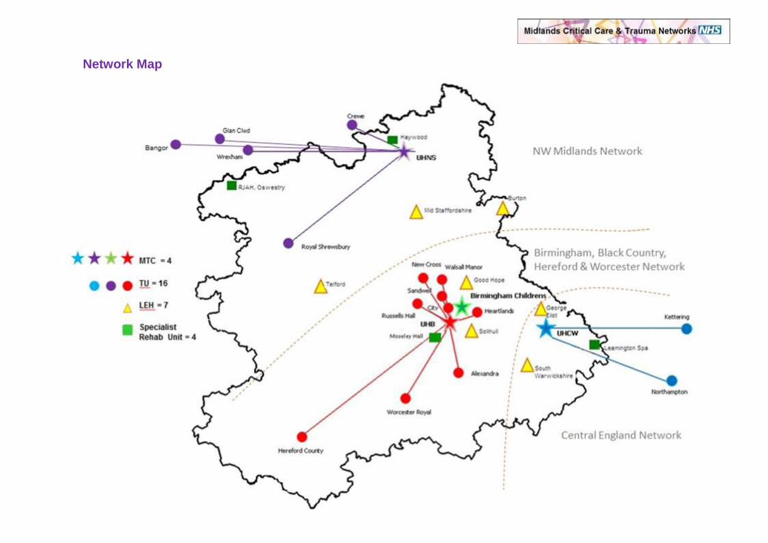
Network Map
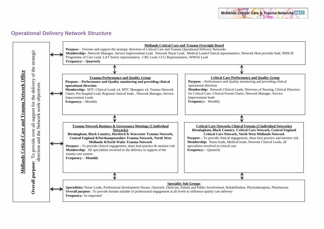
Operational Delivery Network Structure
Midlands Critical Care and Trauma Oversight Board
Purpose:- Oversee and support the strategic direction of Critical Care and Trauma Operational Delivery Networks
Membership:- Network Manager, Service Improvement Lead, Network Nurse Lead, Medical Leads/Clinical representative, Network Host provider lead, NHSCB
Programme of Care Lead, LAT Senior representative, CRG Lead, CCG Representative, WMAS Lead
Frequency: - Quarterly
Critical Care Performance and Quality Group
Purpose: - Performance and Quality monitoring and providing clinical
operational direction
Membership: Network Clinical Leads, Directors of Nursing, Clinical Directors
for Critical Care, Clinical Forum Chairs, Network Manager, Service
Improvement leads
Frequency:- Monthly
Trauma Network Business & Governance Meetings (3 Individual
Networks)
Birmingham, Black Country, Hereford & Worcester Trauma Network,
Central England &Northamptonshire Trauma Network, North West
Midlands &North Wales Trauma Network
Purpose: - To provide clinical engagement, share best practice & monitor risk
Membership: All specialities involved in the delivery or support of the
trauma care system
Frequency: - Monthly
Critical Care Networks Clinical Forums (3 Individual Networks)
Birmingham, Black Country, Critical Care Network, Central England
Critical Care Network, North West Midlands Network
Purpose: - To provide clinical engagement, share best practice and monitor risk
Membership: Nurse leads, Medical leads, Network Clinical Leads, all
specialities involved in critical care
Frequency: - Quarterly
Speciality Sub Groups
Specialities: Nurse Leads, Professional development Nurses, Outreach, Dietician, Patient and Public Involvement, Rehabilitation. Physiotherapists, Pharmacists
Overall purpose: To provide forums suitable of professional engagement at all levels to influence quality care delivery
Frequency: As requested
Mid
lan
ds
Cri
tica
l C
are
an
d T
rau
ma N
etw
ork
Off
ice
Over
all
pu
rpose
: T
o p
rovid
e over
all
support
for
the
del
iver
y o
f th
e st
rate
gic
dir
ecti
on a
nd t
he
Net
work
work
obje
ctiv
es
Trauma Performance and Quality Group
Purpose: - Performance and Quality monitoring and providing clinical
operational direction
Membership: MTC Clinical Leads x4, MTC Managers x4, Trauma Network
Chairs, Pre hospital Lead, Regional clinical leads , Network Manager, Service
Improvement Leads
Frequency: - Monthly
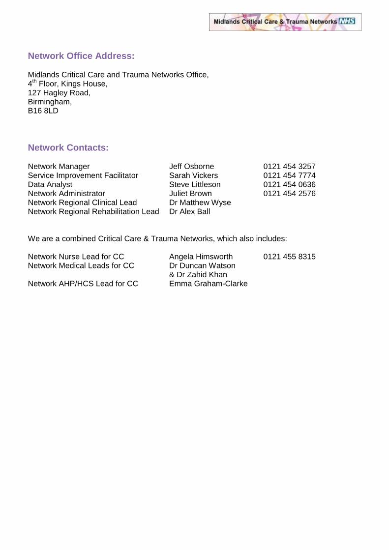
Network Office Address: Midlands Critical Care and Trauma Networks Office, 4th Floor, Kings House, 127 Hagley Road, Birmingham, B16 8LD
Network Contacts: Network Manager Jeff Osborne 0121 454 3257 Service Improvement Facilitator Sarah Vickers 0121 454 7774 Data Analyst Steve Littleson 0121 454 0636 Network Administrator Juliet Brown 0121 454 2576 Network Regional Clinical Lead Dr Matthew Wyse Network Regional Rehabilitation Lead Dr Alex Ball We are a combined Critical Care & Trauma Networks, which also includes: Network Nurse Lead for CC Angela Himsworth 0121 455 8315 Network Medical Leads for CC Dr Duncan Watson
& Dr Zahid Khan Network AHP/HCS Lead for CC Emma Graham-Clarke
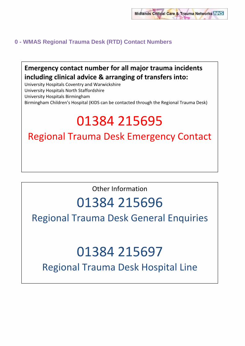
0 - WMAS Regional Trauma Desk (RTD) Contact Numbers
Emergency contact number for all major trauma incidents including clinical advice & arranging of transfers into: University Hospitals Coventry and Warwickshire University Hospitals North Staffordshire University Hospitals Birmingham Birmingham Children’s Hospital (KIDS can be contacted through the Regional Trauma Desk)
01384 215695 Regional Trauma Desk Emergency Contact
Other Information
01384 215696 Regional Trauma Desk General Enquiries
01384 215697
Regional Trauma Desk Hospital Line
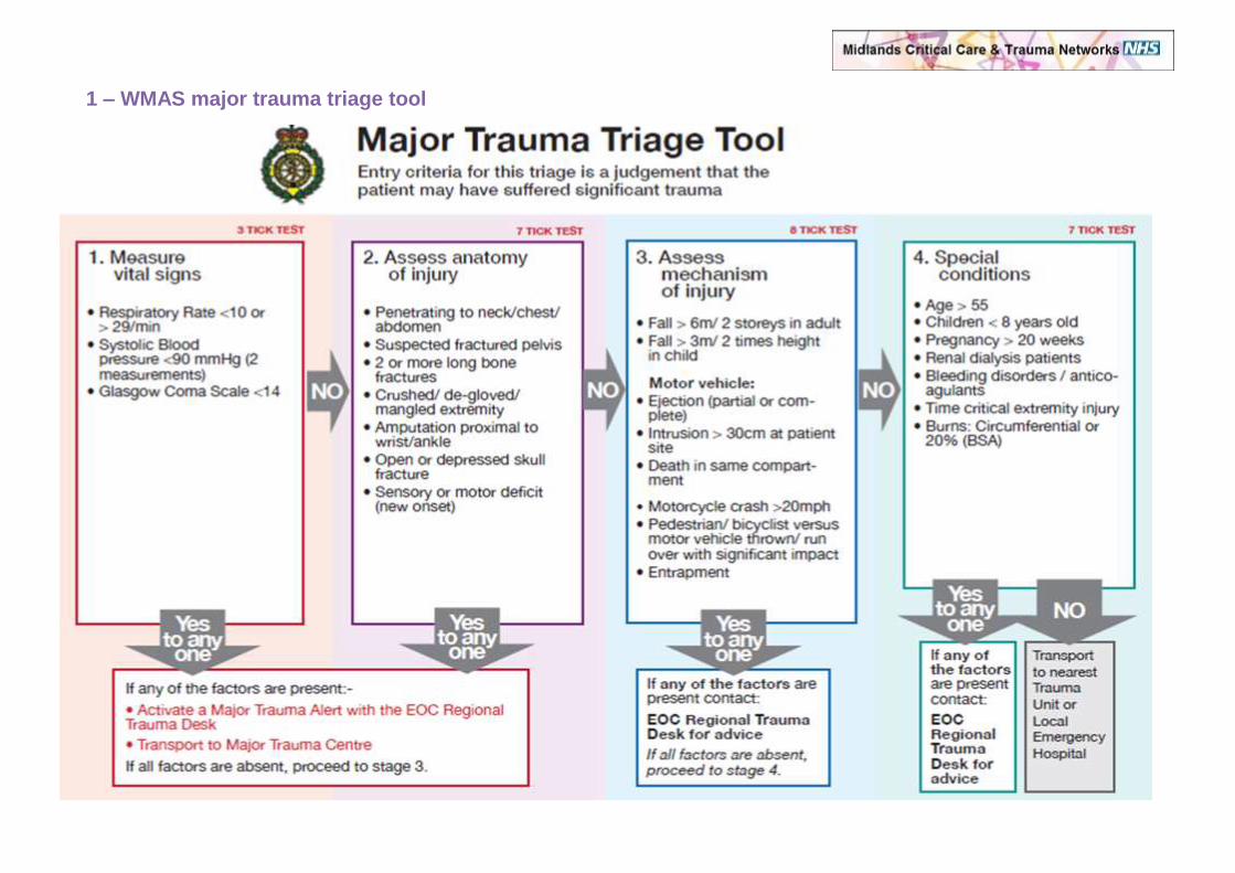
1 – WMAS major trauma triage tool
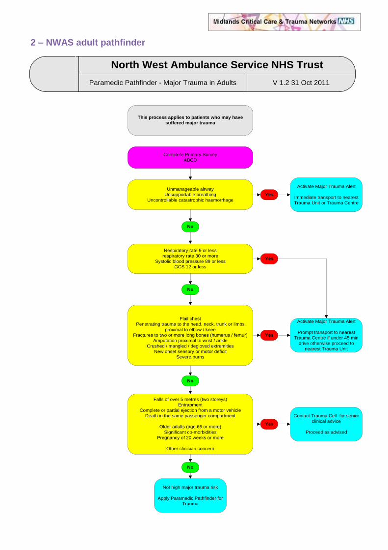
This process applies to patients who may have
suffered major trauma
Paramedic Pathfinder - Major Trauma in Adults V 1.2 31 Oct 2011
North West Ambulance Service NHS Trust
Complete Primary Survey
ABCD
Unmanageable airway
Unsupportable breathing
Uncontrollable catastrophic haemorrhage
Yes
No
Respiratory rate 9 or less
respiratory rate 30 or more
Systolic blood pressure 89 or less
GCS 12 or less
Flail chest
Penetrating trauma to the head, neck, trunk or limbs
proximal to elbow / knee
Fractures to two or more long bones (humerus / femur)
Amputation proximal to wrist / ankle
Crushed / mangled / degloved extremities
New onset sensory or motor deficit
Severe burns
No
Yes
Activate Major Trauma Alert
Immediate transport to nearest
Trauma Unit or Trauma Centre
Activate Major Trauma Alert
Prompt transport to nearest
Trauma Centre if under 45 min
drive otherwise proceed to
nearest Trauma Unit
Yes
Falls of over 5 metres (two storeys)
Entrapment
Complete or partial ejection from a motor vehicle
Death in the same passenger compartment
Older adults (age 65 or more)
Significant co-morbidities
Pregnancy of 20 weeks or more
Other clinician concern
No
Not high major trauma risk
Apply Paramedic Pathfinder for
Trauma
Yes
No
Contact Trauma Cell for senior
clinical advice
Proceed as advised
2 – NWAS adult pathfinder
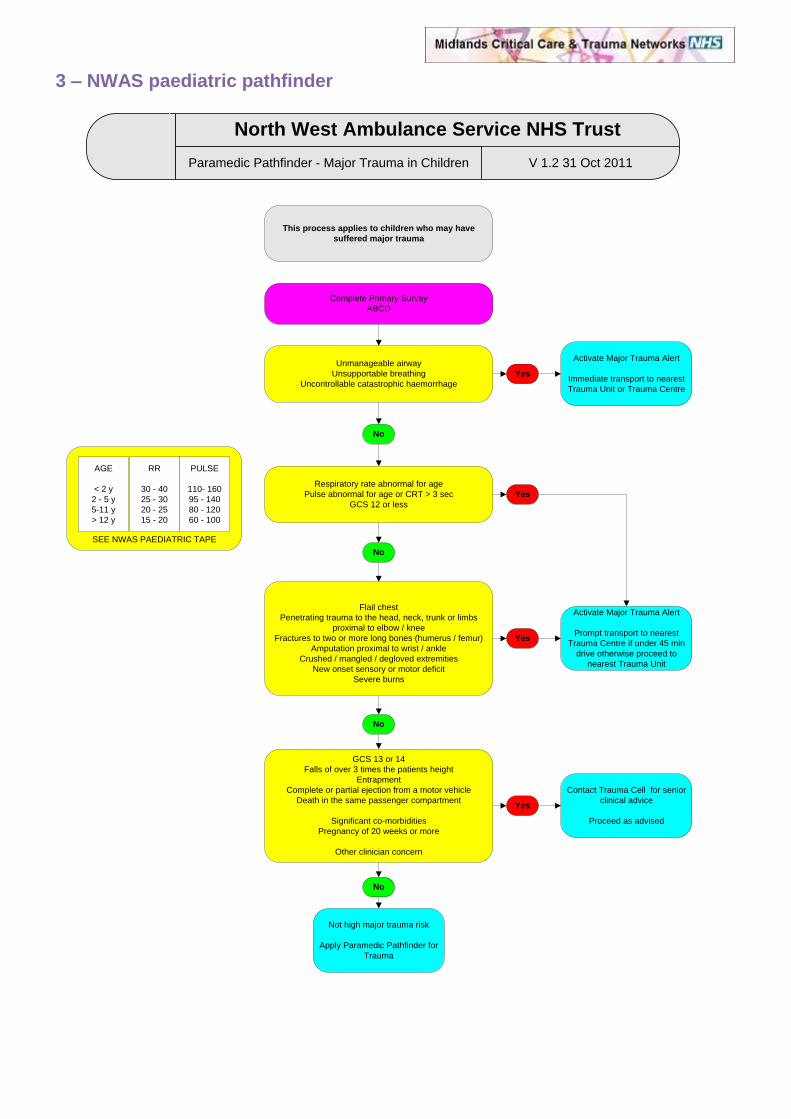
SEE NWAS PAEDIATRIC TAPE
This process applies to children who may have
suffered major trauma
Paramedic Pathfinder - Major Trauma in Children V 1.2 31 Oct 2011
North West Ambulance Service NHS Trust
Complete Primary Survey
ABCD
Unmanageable airway
Unsupportable breathing
Uncontrollable catastrophic haemorrhage
Yes
No
Respiratory rate abnormal for age
Pulse abnormal for age or CRT > 3 sec
GCS 12 or less
Flail chest
Penetrating trauma to the head, neck, trunk or limbs
proximal to elbow / knee
Fractures to two or more long bones (humerus / femur)
Amputation proximal to wrist / ankle
Crushed / mangled / degloved extremities
New onset sensory or motor deficit
Severe burns
No
Yes
Activate Major Trauma Alert
Immediate transport to nearest
Trauma Unit or Trauma Centre
Activate Major Trauma Alert
Prompt transport to nearest
Trauma Centre if under 45 min
drive otherwise proceed to
nearest Trauma Unit
Yes
GCS 13 or 14
Falls of over 3 times the patients height
Entrapment
Complete or partial ejection from a motor vehicle
Death in the same passenger compartment
Significant co-morbidities
Pregnancy of 20 weeks or more
Other clinician concern
No
Not high major trauma risk
Apply Paramedic Pathfinder for
Trauma
Yes
No
Contact Trauma Cell for senior
clinical advice
Proceed as advised
AGE
< 2 y
2 - 5 y
5-11 y
> 12 y
RR
30 - 40
25 - 30
20 - 25
15 - 20
PULSE
110- 160
95 - 140
80 - 120
60 - 100
3 – NWAS paediatric pathfinder
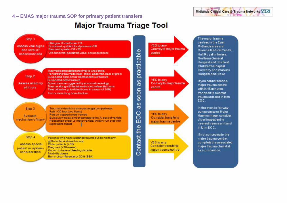
4 – EMAS major trauma SOP for primary patient transfers
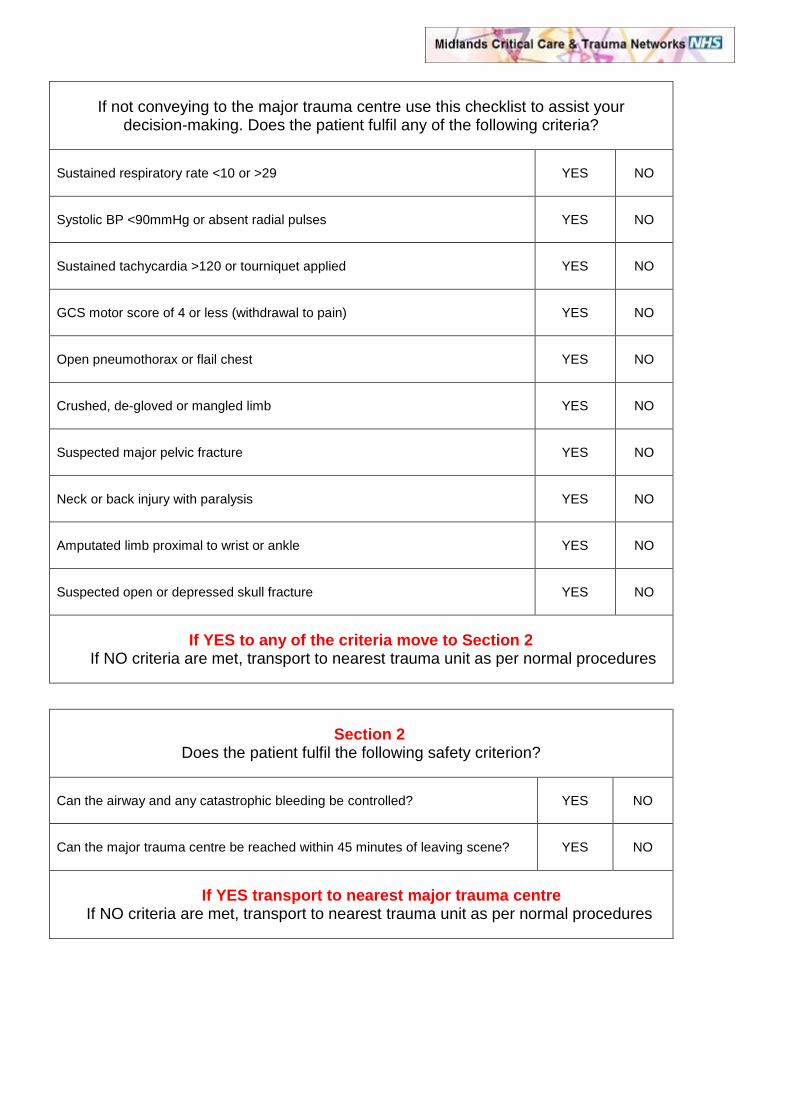
If not conveying to the major trauma centre use this checklist to assist your decision-making. Does the patient fulfil any of the following criteria?
Sustained respiratory rate <10 or >29
YES
NO
Systolic BP <90mmHg or absent radial pulses
YES
NO
Sustained tachycardia >120 or tourniquet applied
YES
NO
GCS motor score of 4 or less (withdrawal to pain)
YES
NO
Open pneumothorax or flail chest
YES
NO
Crushed, de-gloved or mangled limb
YES
NO
Suspected major pelvic fracture
YES
NO
Neck or back injury with paralysis
YES
NO
Amputated limb proximal to wrist or ankle
YES
NO
Suspected open or depressed skull fracture
YES
NO
If YES to any of the criteria move to Section 2 If NO criteria are met, transport to nearest trauma unit as per normal procedures
Section 2 Does the patient fulfil the following safety criterion?
Can the airway and any catastrophic bleeding be controlled?
YES
NO
Can the major trauma centre be reached within 45 minutes of leaving scene?
YES
NO
If YES transport to nearest major trauma centre If NO criteria are met, transport to nearest trauma unit as per normal procedures
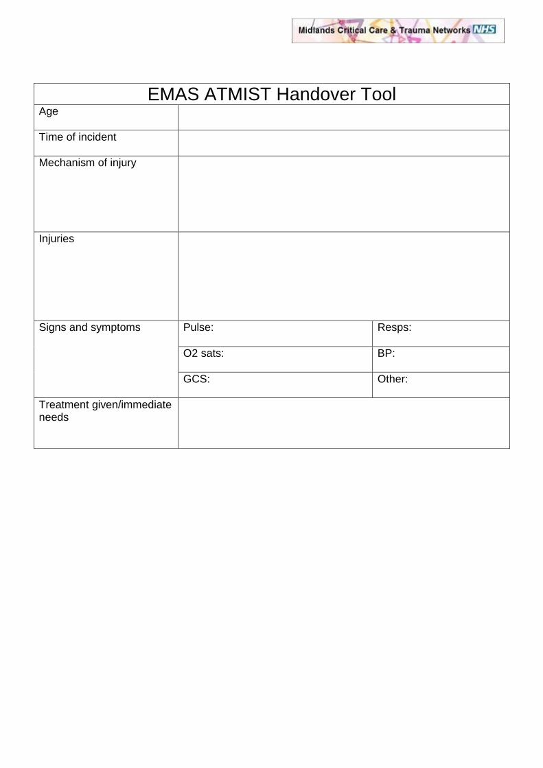
EMAS ATMIST Handover Tool Age
Time of incident
Mechanism of injury
Injuries
Signs and symptoms
Pulse:
Resps:
O2 sats:
BP:
GCS:
Other:
Treatment given/immediate needs
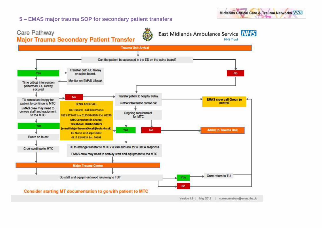
5 – EMAS major trauma SOP for secondary patient transfers
7 - SOP for the on-call advice to regional pre-hospital trauma service
Standard:
The 24/7/365 day availability of a consultant grade doctor with proven competencies in pre-hospital care to give remote advice to the control room paramedic and/or to the crews on scene or in transport.
Operational Model
All calls for advice to be routed through the regional trauma desk (RTD)
The clinician on the trauma desk to be the first provider of advice
Senior on call to be provided by the Level 8 practitioner on MERIT/MAA.
All calls to be conferenced through the RTD and recorded.
Where advice given to change receiving hospital or institute alternative course of treatment this to be noted specifically in the case log by the RTD clinician.
If advice is in relation to a child the conference call should include the consultant from the KIDS service. This is particularly important if the child is going to any MTC or TU unit other than BCH. (The KIDS on call team are not experts in pre-hospital care therefore the role is to give advice on paediatric treatment matters rather than the pre-hospital care)
Advice call criteria Calls for advice should be generated whenever an on scene clinician dealing with a major trauma case wishes to discuss care with another colleague. Typical scenarios are:
To discuss by pass of TU (LEH) to MTC
To discuss triage decision in patients who are in high risk category (Step 4 on triage tool)
To discuss triage to Birmingham Children’s Hospital
To discuss exceeding 45 minute transport time
To request or discuss need for an enhanced care team
For advice on use of new therapies in trauma care (e.g. tourniquet)
In most case the RTD clinician will have the experience and knowledge to support the on scene practitioner and will only utilise the on call service when the RTD clinician requires senior clinical input.
Communication pathway
Contacting the Regional Trauma Desk (RTD) The RTD can be contacted by changing channel to talk group 282 on the ARP radio. This is currently marked as “Air Ambulance 3” on the ARP radio folder display. To change to this talk group press the ‘mode’ button (pause for a couple of seconds) followed by 282 and then ‘transmit’ to confirm. Contacting by telephone The following telephone numbers can be used to contact the Regional Trauma Desk: 01384 215695 - RTD Emergency Contact 01384 215696 - RTD General Enquiries 01384 215697 - RTD Hospital Line
Governance arrangements The overarching governance of the regional trauma networks will sit with the Network board comprising commissioners, senior clinicians from across the network providers and patient representatives. WMAS and the providers of MERIT should ensure that all significant cases are formally reviewed and an occurrence report produced for the Regional Trauma Network board on a three monthly basis. Dispatch of MERIT or Enhanced care provider. As a result of a request for on call advice it may be appropriate to suggest the need for an enhanced care response to scene. The provider of advice should make this suggestion to the RTD clinician who will dispatch the closest response. The provider of advice should not self task to scene. Reviewed and amended by: Dr Nick Crombie: Midland Air Ambulance Shane Roberts: WMAS Dr Tina Newton: BCH
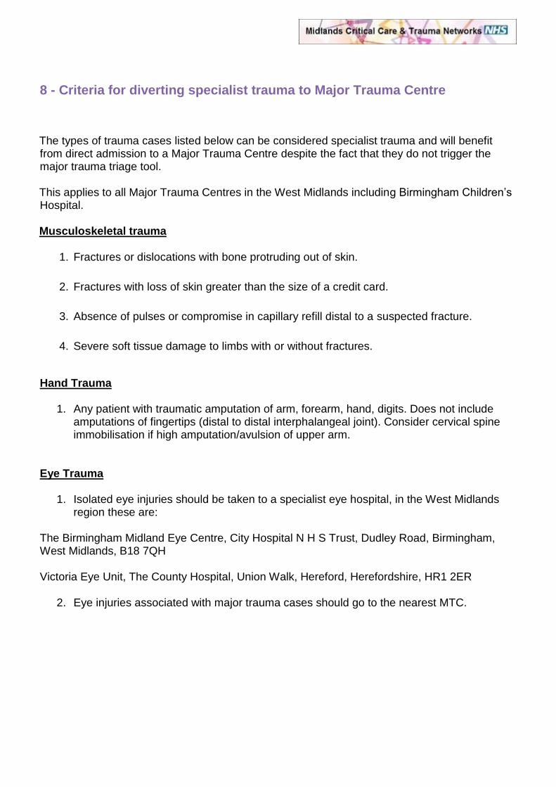
8 - Criteria for diverting specialist trauma to Major Trauma Centre
The types of trauma cases listed below can be considered specialist trauma and will benefit from direct admission to a Major Trauma Centre despite the fact that they do not trigger the major trauma triage tool. This applies to all Major Trauma Centres in the West Midlands including Birmingham Children’s Hospital. Musculoskeletal trauma
1. Fractures or dislocations with bone protruding out of skin.
2. Fractures with loss of skin greater than the size of a credit card.
3. Absence of pulses or compromise in capillary refill distal to a suspected fracture.
4. Severe soft tissue damage to limbs with or without fractures.
Hand Trauma
1. Any patient with traumatic amputation of arm, forearm, hand, digits. Does not include amputations of fingertips (distal to distal interphalangeal joint). Consider cervical spine immobilisation if high amputation/avulsion of upper arm.
Eye Trauma
1. Isolated eye injuries should be taken to a specialist eye hospital, in the West Midlands region these are:
The Birmingham Midland Eye Centre, City Hospital N H S Trust, Dudley Road, Birmingham, West Midlands, B18 7QH Victoria Eye Unit, The County Hospital, Union Walk, Hereford, Herefordshire, HR1 2ER
2. Eye injuries associated with major trauma cases should go to the nearest MTC.
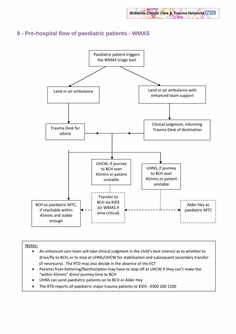
9 - Pre-hospital flow of paediatric patients - WMAS
Notes: An enhanced care team will take clinical judgment in the child’s best interest as to whether to
drive/fly to BCH, or to stop at UHNS/UHCW for stabilisation and subsequent secondary transfer
(if necessary). The RTD may also decide in the absence of the ECT
Patients from Kettering/Northampton may have to stop off at UHCW if they can’t make the “within 45mins” direct journey time to BCH
UHNS can send paediatric patients on to BCH or Alder Hey
The RTD reports all paediatric major trauma patients to KIDS - 0300 200 1100
BCH as paediatric MTC, if reachable within 45mins and stable
enough
UHCW, if journey to BCH over
45mins or patient unstable
UHNS, if journey to BCH over
45mins or patient unstable
Transfer to BCH via KIDS (or WMAS if time critical)
Paediatric patient triggers the WMAS triage tool
Land or air ambulance Land or air ambulance with enhanced team support
Trauma Desk for advice
Clinical judgment, informing Trauma Desk of destination
Alder Hey as paediatric MTC
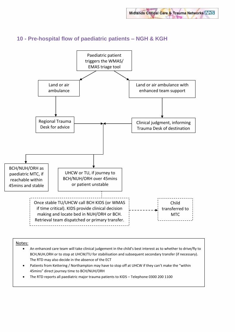
10 - Pre-hospital flow of paediatric patients – NGH & KGH
Land or
Paediatric patient triggers the WMAS/
EMAS triage tool
Clinical judgment, informing Trauma Desk of destination
Regional Trauma Desk for advice
Notes: An enhanced care team will take clinical judgement in the child’s best interest as to whether to drive/fly to
BCH,NUH,ORH or to stop at UHCW/TU for stabilisation and subsequent secondary transfer (if necessary).
The RTD may also decide in the absence of the ECT
Patients from Kettering / Northampton may have to stop off at UHCW if they can’t make the “within
45mins” direct journey time to BCH/NUH/ORH
The RTD reports all paediatric major trauma patients to KIDS – Telephone 0300 200 1100
BCH/NUH/ORH as paediatric MTC, if reachable within
45mins and stable enough
UHCW or TU, if journey to BCH/NUH/ORH over 45mins
or patient unstable
Child transferred to
MTC
Once stable TU/UHCW call BCH KIDS (or WMAS if time critical). KIDS provide clinical decision making and locate bed in NUH/ORH or BCH.
Retrieval team dispatched or primary transfer.
Land or air ambulance
Land or air ambulance with enhanced team support
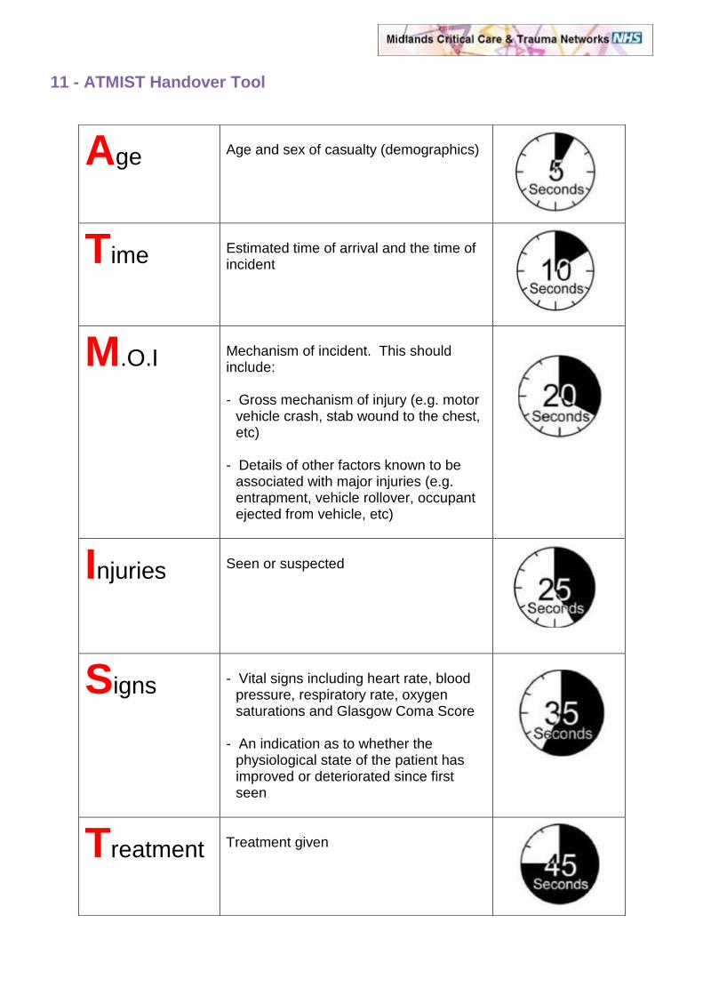
11 - ATMIST Handover Tool
Age Age and sex of casualty (demographics)
Time Estimated time of arrival and the time of incident
M.O.I Mechanism of incident. This should include: - Gross mechanism of injury (e.g. motor
vehicle crash, stab wound to the chest, etc)
- Details of other factors known to be
associated with major injuries (e.g. entrapment, vehicle rollover, occupant ejected from vehicle, etc)
Injuries Seen or suspected
Signs - Vital signs including heart rate, blood
pressure, respiratory rate, oxygen saturations and Glasgow Coma Score
- An indication as to whether the
physiological state of the patient has improved or deteriorated since first seen
Treatment Treatment given
12 - Trauma Team Activation
Scope
This policy should be used in conjunction with the West Midlands Pre-Hospital Triage tool to determine the hospital response to a trauma alert call. The Policy is applicable to all Major Trauma Centre’s and Trauma Units within the West Midlands.
Definitions
Triage tool: Pre-hospital decision making flowchart to determine the likelihood of major trauma. Regional Trauma Desk: 24/7 clinician staffed coordinating desk based in ambulance control. Trauma Team: Pre defined hospital response to a trauma case. Trauma Team Leader: Pre defined consultant, senior trainee or equivalent with training and responsibility to lead trauma team. Trauma Alert: A call from or via regional trauma desk notifying the receiving unit of an incoming patient.
Over-arching policy
All MTCs and TUs within the West Midlands must have an internal Trauma Team policy that
defines:
Method of activation and communication
Team membership
Trauma team leader
Whether full trauma team or limited trauma team is to be called.
Procedure to call in from home additional or extended team members as required.
Whether pre-hospital activation of the massive haemorrhage protocol is supported or not. All units should audit the trauma team activation process on a regular basis.
Staging activation
1. An individual unit may have an internal policy that provides a tiered response to a trauma
alert.
2. If the trauma alert from the regional trauma desk is based on step 1 or 2 of the triage tool a full team response is mandated.
3. If the call is based on step 3 or 4 a limited response may be used to undertake the primary
survey provided this is clearly documented in the unit’s policy and the MTC/ TU has the ability to rapidly upscale to a full trauma team if necessary.
4. Where a patient is being triaged in step 4 on the basis of >20 weeks gestation the team
must include a midwife and senior obstetrician (Consultant or ST4 or above) who is able to make rapid assessment and decisions about need for delivery. The procedure for activating obstetric support needs to be clearly documented.
13 - Emergency Management of Traumatic Cardiac Arrest
Introduction Traumatic cardiac arrest caused by trauma has a very high mortality, with an overall survival of just 5.6% (range 0–17%). For reasons that are unclear, reported survival rates in the last 5 years are better than reported previously. In those who survive (and where data are available) neurological outcome is good in only 1.6% of those sustaining traumatic cardiorespiratory arrest (TCRA). In a retrospective database review, the survival rate of patients whose cardiac arrest was the result of
hypoxemia (hanging, drowning, electrocution, burns, traumatic asphyxia) had a survival rate of 17%2. In
addition, patients who underwent out-of-hospital thoracotomy after penetrating trauma had a higher chance of survival (11.8%). The subgroup of patients who arrest after hypoxic Insults (box 1) have a significantly increased chance of survival. This group of patients should have their Hypoxic insult treated aggressively with full ALS protocols. The following protocol aims to maximise the chances of survival of this critically injured cohort. Management should proceed in a horizontal fashion according to the <C>ABC paradigm. <C> Catastrophic Haemorrhage • Consider activation of the Massive Transfusion Protocol (MTP). • Give 2 units O neg blood STAT, preferably via a level 1 infuser. • Catastrophic limb haemorrhage should be treated with the application of a CAT tourniquet above
the injury (over the femur or humerus) and tightened until the bleeding stops. • Haemostatic agents (CELOX-A or CELOX gauze) are indicated when:
o The patient has catastrophic life-threatening external haemorrhage which is not controllable by any other means (including direct pressure and elevation, wound packing, temporary sutures or tourniquet application)
o AND the patient will require emergency surgery for their injuries
<A> Airway • Secure the airway • Ventilate with 100% O2 • Consider a suxemethonium-only intubation if the patient has just arrested since airway reflexes
may still be present. • Look for airway obstruction or airway disruption.
Summary statement /scope of the guideline:
This document provides general guidance regarding the management of trauma patients in cardiac arrest in the emergency department.
Recommendations for guideline content: Clinical guidelines should detail clear and explicit recommendations for practice and in addition to providing general guidance should clearly detail behaviour specific instruction; what, who, when, where and how. This will increase the likelihood of guidance use.
Hanging Drowning
Burns Electrocution C-spine injury
Traumatic asphyxia Box 1
<B> Breathing • Perform bilateral thoracostomies to decompress the chest and thus exclude tension
pneumothoraces (may be bilateral) • In penetrating trauma, perform an emergency thoracotomy if there were vital signs <10 minutes
prior to cardiac arrest and there is no return of spontaneous circulation (ROSC). • Exclude life-threatening chest injuries
o Tension pneumothorax o Open pneumothorax o Massive haemothorax o Flail chest o Cardiac tamponade
<C> Circulation
• Ensure that there are two wide-bore IV cannulae in situ. • If IV access proves difficult, move immediately onto intraosseous access using EZ-IO. (Choose an
uninjured limb away from the side of a potential pelvic fracture). • Apply a pelvic splint and realign limb fractures. • The majority of external haemorrhage can be controlled by direct pressure and elevation. • Check the heart rhythm for shockable VT or VF. This is likely in the elderly patient in whom the
MOI suggests a relatively low energy transfer. CPR Chest compressions are unlikely to be effective in hypovolaemic cardiac arrest, however most survivors do not have hypovolaemia and in this subgroup standard advanced life support may be life-saving. Standard CPR should not delay the treatment of reversible causes (e.g., thoracotomy for cardiac tamponade Adrenaline should be used cautiously in traumatic cardiac arrest during CPR; it may worsen intracellular hypoxia and increase bleeding.
REMEMBER: In the majority of cases of traumatic cardiac arrest, the cause is tension physiology (pneumothorax or cardiac tamponade) or severe hypovolaemia (empty heart). A primary cardiac event must be excluded, but is uncommon. Therefore the team must focus on providing good oxygenation and ventilation, relieving pressure and filling the cardiovascular system with blood and blood products. Termination of Resuscitation If there is no response within 20 minutes, despite the above measures, the patient should be pronounced dead. Commotio Cordis This rare condition is actual or near cardiac arrest caused by a blunt impact to the chest wall over the heart. A blow to the chest during the vulnerable phase of the cardiac cycle may cause malignant arrhythmias (usually ventricular fibrillation). Commotio cordis occurs mostly during sports and victims are usually young males (mean age 14 years). In a series of 1866 cardiac arrests in athletes in Minneapolis, 65 (3%) were due to commotio cordis. The overall survival rate from commotio cordis is 15%, but 25% if resuscitation is started within 3 min.
Audit All cases of traumatic cardiac arrest will be audited by the Emergency Department and submitted to TARN.
14 - Management of Major Haemorrhage
Scope This document sets out the standards for all receiving units in the West Midlands Trauma service in respect of massive haemorrhage protocols. This is now the preferred protocol for all units. Introduction The timely provision of tranexamic acid and blood products to major trauma patients is associated with improved outcomes. Military experience shows that using a high ratio of Fresh Frozen Plasma (FFP) and platelets to packed cells reduces coagulopathy and overall blood use, although there is no absolute consensus on the exact ratio. Policy 1. Every receiving unit should have a clearly defined massive haemorrhage policy for trauma
approved by the local blood transfusion committee.
2. Within the policy there should be clear guidance on the following: a. Activation criteria and method of activation b. The roles and responsibilities of the personnel involved c. The ratio of packed cells to FFP which should be in the range 1:1 to 2:1 d. The ratio of packed cells to platelet transfusion e. The communication mechanism between clinicians and the labs f. The availability and method of communicating with the on call haematology consultant. g. The stand down criteria
3. Every unit must have facilities for in line warming of blood products immediately available within the resuscitation room
4. Every receiving unit should have evidence that the activations of the massive haemorrhage protocol are monitored and audited.
5. Every unit should have tranexamic Acid immediately available in the resuscitation room.
6. The time and dose of tranexamic acid administration must be recorded on the trauma chart.
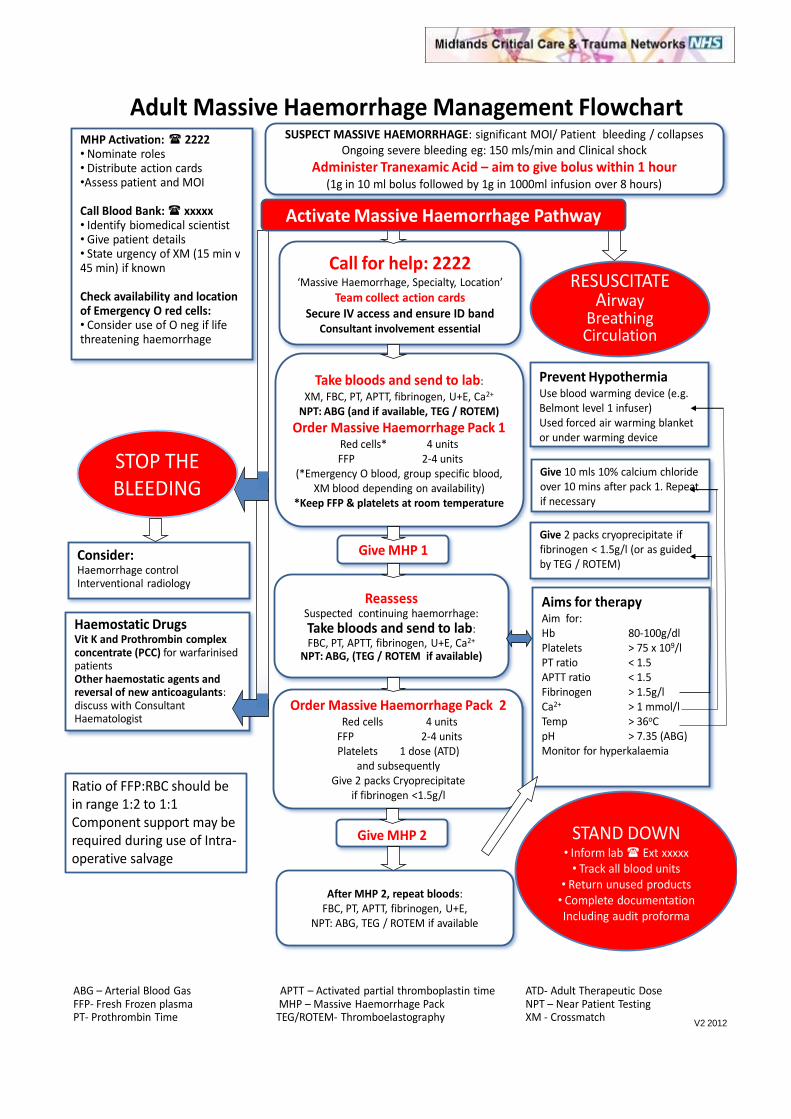
Adult Massive Haemorrhage Management FlowchartSUSPECT MASSIVE HAEMORRHAGE: significant MOI/ Patient bleeding / collapses
Ongoing severe bleeding eg: 150 mls/min and Clinical shock
Administer Tranexamic Acid – aim to give bolus within 1 hour(1g in 10 ml bolus followed by 1g in 1000ml infusion over 8 hours)
Call for help: 2222‘Massive Haemorrhage, Specialty, Location’
Team collect action cardsSecure IV access and ensure ID band
Consultant involvement essential
Take bloods and send to lab:
XM, FBC, PT, APTT, fibrinogen, U+E, Ca2+
NPT: ABG (and if available, TEG / ROTEM)
Order Massive Haemorrhage Pack 1Red cells* 4 unitsFFP 2-4 units
(*Emergency O blood, group specific blood, XM blood depending on availability)
*Keep FFP & platelets at room temperature
ReassessSuspected continuing haemorrhage:
Take bloods and send to lab:FBC, PT, APTT, fibrinogen, U+E, Ca2+
NPT: ABG, (TEG / ROTEM if available)
Give MHP 2
MHP Activation: 2222• Nominate roles• Distribute action cards•Assess patient and MOI
Call Blood Bank: xxxxx• Identify biomedical scientist• Give patient details• State urgency of XM (15 min v 45 min) if known
Check availability and location of Emergency O red cells:• Consider use of O neg if life threatening haemorrhage
STOP THE BLEEDING
RESUSCITATEAirway
BreathingCirculation
Consider:Haemorrhage controlInterventional radiology
Haemostatic DrugsVit K and Prothrombin complex concentrate (PCC) for warfarinisedpatientsOther haemostatic agents and reversal of new anticoagulants: discuss with Consultant Haematologist
Prevent HypothermiaUse blood warming device (e.g. Belmont level 1 infuser)Used forced air warming blanket or under warming device
Give 10 mls 10% calcium chloride over 10 mins after pack 1. Repeat if necessary
Give 2 packs cryoprecipitate if fibrinogen < 1.5g/l (or as guided by TEG / ROTEM)
Aims for therapyAim for:Hb 80-100g/dlPlatelets > 75 x 109/lPT ratio < 1.5APTT ratio < 1.5Fibrinogen > 1.5g/lCa2+ > 1 mmol/lTemp > 36oCpH > 7.35 (ABG) Monitor for hyperkalaemia
STAND DOWN• Inform lab Ext xxxxx
• Track all blood units• Return unused products
• Complete documentation Including audit proforma
Give MHP 1
ABG – Arterial Blood Gas APTT – Activated partial thromboplastin time ATD- Adult Therapeutic DoseFFP- Fresh Frozen plasma MHP – Massive Haemorrhage Pack NPT – Near Patient TestingPT- Prothrombin Time TEG/ROTEM- Thromboelastography XM - Crossmatch
Order Massive Haemorrhage Pack 2Red cells 4 units
FFP 2-4 unitsPlatelets 1 dose (ATD)
and subsequentlyGive 2 packs Cryoprecipitate
if fibrinogen <1.5g/l
After MHP 2, repeat bloods:FBC, PT, APTT, fibrinogen, U+E,
NPT: ABG, TEG / ROTEM if available
Activate Massive Haemorrhage Pathway
V2 2012
Ratio of FFP:RBC should be in range 1:2 to 1:1Component support may be required during use of Intra-operative salvage
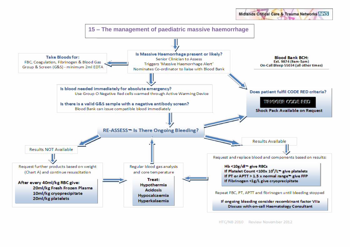
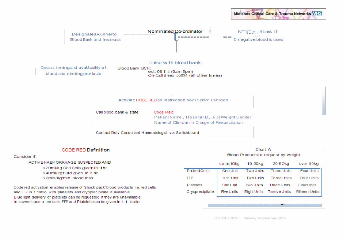
15 – The management of paediatric massive haemorrhage
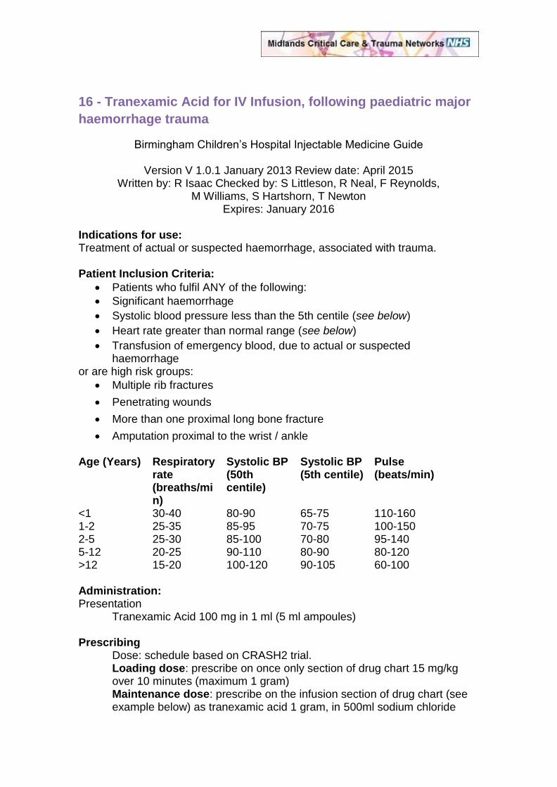
16 - Tranexamic Acid for IV Infusion, following paediatric major
haemorrhage trauma
Birmingham Children’s Hospital Injectable Medicine Guide
Version V 1.0.1 January 2013 Review date: April 2015 Written by: R Isaac Checked by: S Littleson, R Neal, F Reynolds,
M Williams, S Hartshorn, T Newton Expires: January 2016
Indications for use: Treatment of actual or suspected haemorrhage, associated with trauma. Patient Inclusion Criteria:
Patients who fulfil ANY of the following:
Significant haemorrhage
Systolic blood pressure less than the 5th centile (see below)
Heart rate greater than normal range (see below)
Transfusion of emergency blood, due to actual or suspected haemorrhage
or are high risk groups:
Multiple rib fractures
Penetrating wounds
More than one proximal long bone fracture
Amputation proximal to the wrist / ankle
Age (Years) Respiratory rate (breaths/min)
Systolic BP (50th centile)
Systolic BP (5th centile)
Pulse (beats/min)
<1 30-40 80-90 65-75 110-160 1-2 25-35 85-95 70-75 100-150 2-5 25-30 85-100 70-80 95-140 5-12 20-25 90-110 80-90 80-120 >12 15-20 100-120 90-105 60-100 Administration: Presentation
Tranexamic Acid 100 mg in 1 ml (5 ml ampoules) Prescribing
Dose: schedule based on CRASH2 trial. Loading dose: prescribe on once only section of drug chart 15 mg/kg over 10 minutes (maximum 1 gram) Maintenance dose: prescribe on the infusion section of drug chart (see example below) as tranexamic acid 1 gram, in 500ml sodium chloride
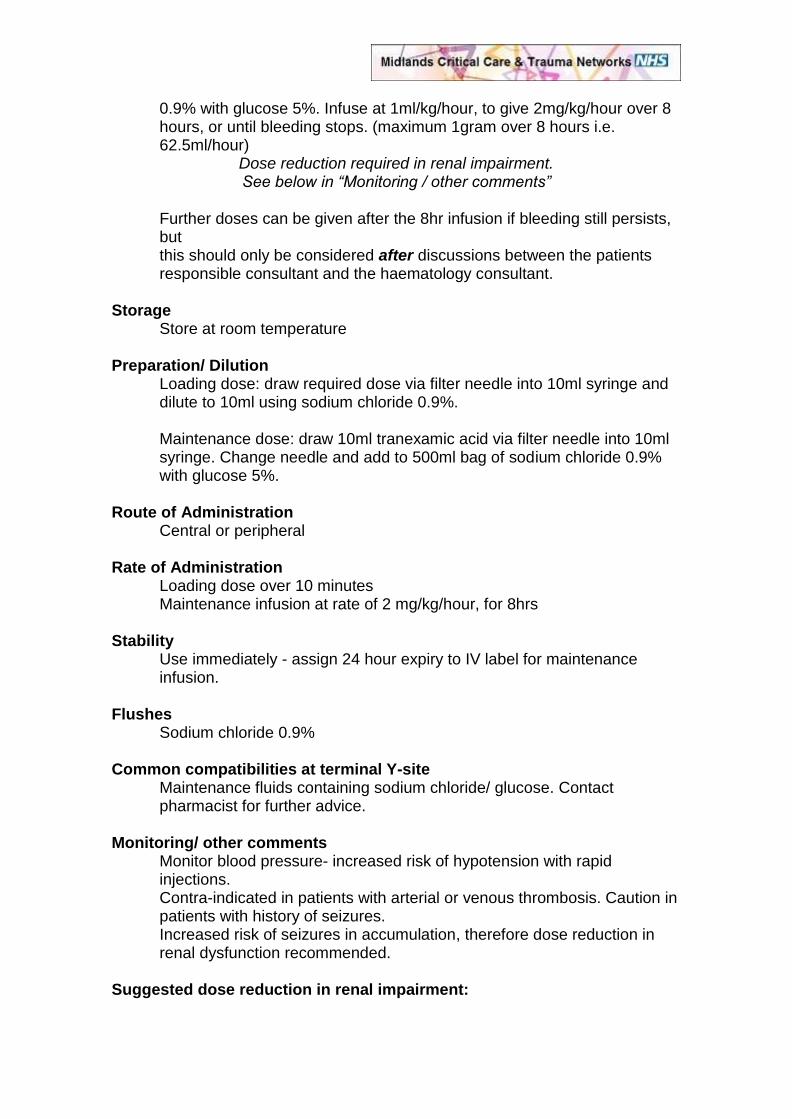
0.9% with glucose 5%. Infuse at 1ml/kg/hour, to give 2mg/kg/hour over 8 hours, or until bleeding stops. (maximum 1gram over 8 hours i.e. 62.5ml/hour)
Dose reduction required in renal impairment. See below in “Monitoring / other comments”
Further doses can be given after the 8hr infusion if bleeding still persists, but this should only be considered after discussions between the patients responsible consultant and the haematology consultant.
Storage
Store at room temperature Preparation/ Dilution
Loading dose: draw required dose via filter needle into 10ml syringe and dilute to 10ml using sodium chloride 0.9%.
Maintenance dose: draw 10ml tranexamic acid via filter needle into 10ml syringe. Change needle and add to 500ml bag of sodium chloride 0.9% with glucose 5%.
Route of Administration
Central or peripheral
Rate of Administration Loading dose over 10 minutes Maintenance infusion at rate of 2 mg/kg/hour, for 8hrs
Stability Use immediately - assign 24 hour expiry to IV label for maintenance infusion.
Flushes Sodium chloride 0.9%
Common compatibilities at terminal Y-site Maintenance fluids containing sodium chloride/ glucose. Contact pharmacist for further advice.
Monitoring/ other comments Monitor blood pressure- increased risk of hypotension with rapid injections. Contra-indicated in patients with arterial or venous thrombosis. Caution in patients with history of seizures. Increased risk of seizures in accumulation, therefore dose reduction in renal dysfunction recommended.
Suggested dose reduction in renal impairment:
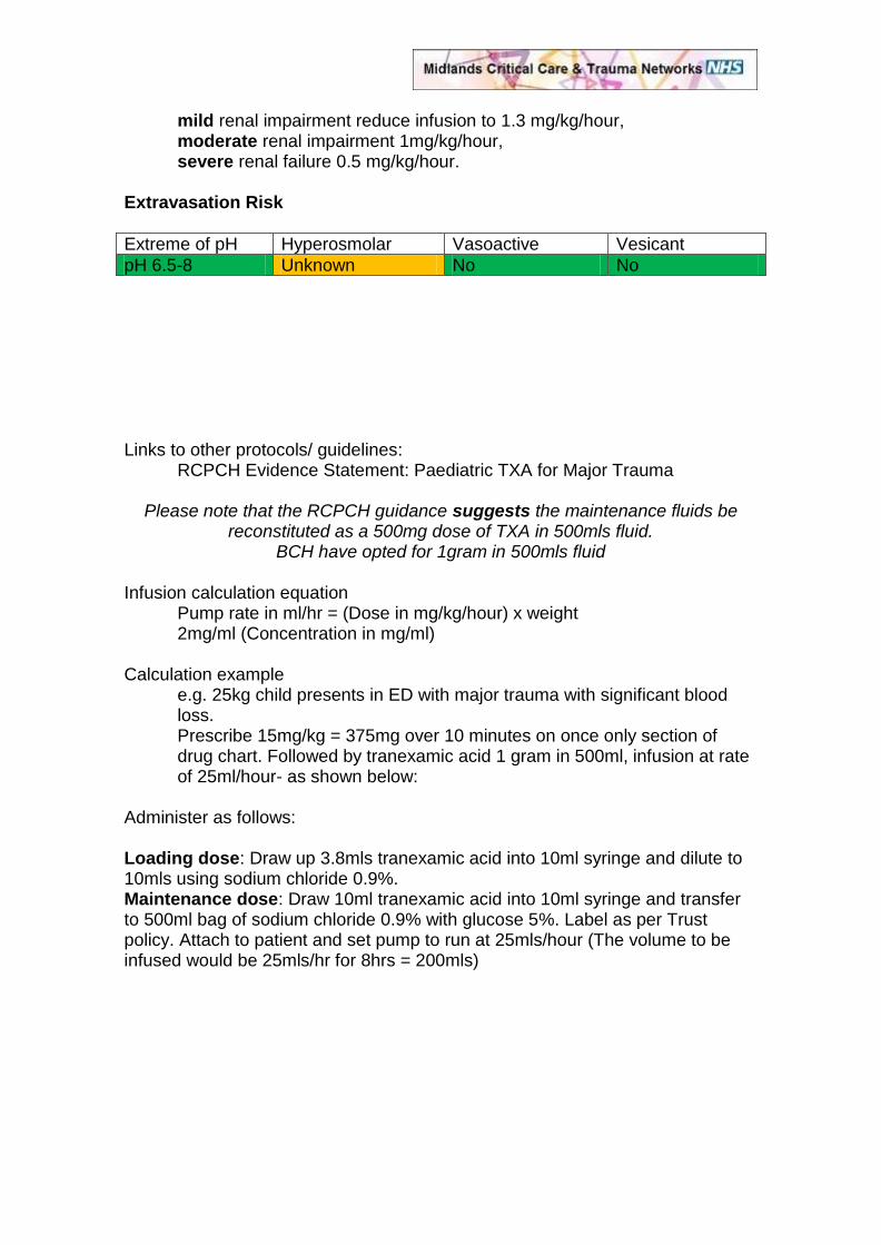
mild renal impairment reduce infusion to 1.3 mg/kg/hour, moderate renal impairment 1mg/kg/hour, severe renal failure 0.5 mg/kg/hour.
Extravasation Risk
Extreme of pH Hyperosmolar Vasoactive Vesicant
pH 6.5-8 Unknown No No
Links to other protocols/ guidelines:
RCPCH Evidence Statement: Paediatric TXA for Major Trauma
Please note that the RCPCH guidance suggests the maintenance fluids be reconstituted as a 500mg dose of TXA in 500mls fluid.
BCH have opted for 1gram in 500mls fluid Infusion calculation equation
Pump rate in ml/hr = (Dose in mg/kg/hour) x weight 2mg/ml (Concentration in mg/ml)
Calculation example e.g. 25kg child presents in ED with major trauma with significant blood loss. Prescribe 15mg/kg = 375mg over 10 minutes on once only section of drug chart. Followed by tranexamic acid 1 gram in 500ml, infusion at rate of 25ml/hour- as shown below:
Administer as follows: Loading dose: Draw up 3.8mls tranexamic acid into 10ml syringe and dilute to 10mls using sodium chloride 0.9%. Maintenance dose: Draw 10ml tranexamic acid into 10ml syringe and transfer to 500ml bag of sodium chloride 0.9% with glucose 5%. Label as per Trust policy. Attach to patient and set pump to run at 25mls/hour (The volume to be infused would be 25mls/hr for 8hrs = 200mls)
17 - Intercostal Chest Drains
1. Introduction
2. Purpose and Scope
3. Aims and Objectives
4. Accountabilities and Responsibilities
5. Training
6. Indications
7. Pre‐drainage risk assessment
8. Imaging
9. Consent
10. The Procedure
11. Drainage
12. Monitoring
13. Suction
14. Analgesia
15. Removal
16. Nursing Care
17. Monitoring of Compliance
18. Review
19. References
Appendix 1 – Equipment required for chest drain insertion
Appendix 2 – Chest drain observation chart
1. Introduction Chest drains are used to remove air, blood, pus or fluid from the pleural cavity. Their insertion is a common procedure but insertion is not without risk. In May 2008 the National Patient Safety Agency (NSPA) published a Rapid Response Alert (NPSA/2008/RRR003) following reports of 12 deaths and 15 cases of serious harm relating to chest drain insertion between January 2005 and March 2008. The Trust recognises the risk associated with chest drain insertion and this document is designed to address many of the issues raised in the NSPA report.
2. Purpose and Scope The standards in this policy aim to rationalise the use of chest drains and standardise care throughout the Trust.
This policy applies to all areas of the trust where chest drain insertion takes place with the exception of those taking place in theatre during a surgical procedure.
The policy applies to adult patients only.
The ongoing care of all chest drains should be carried out in line with this policy.
3.
a)
Aims and Objectives
To identify the need for a chest drain
b) c) d)
To identify the safe insertion and subsequent removal of a chest drain To ensure competency of the doctor performing the procedure To support the nursing care of a patient with a chest drain
4.
Accountabilities and Responsibilities
All staff undertaking the insertion of chest drains or providing care for patients with chest drains must ensure that they:
a) Are compliant with the standards set out in this document b) Work within their own competence c) Gain written consent from patients (except in the event of a life
threatening emergency) d) To report all incidents involving chest drains (including near miss events)
via the DATIX incident reporting system
5. Training and Dissemination It is the responsibility of the directorate or department to ensure that all staff who are involved in the management of chest drains are aware of these guidelines.
Within UHL chest drains will be inserted by medical staff only. Before insertion of a chest drain all operators1 should be adequately trained. Training opportunities available to operators include the Institute of Lung Health (ILH) chest drain course. In order to insert chest drains independently, junior operators should have attended the ILH chest drain course and be in possession of a satisfactory DOPS form. As a matter of good practice, operators should record chest drain procedures in their log book. Individual departments are recommended to keep records of operators deemed competent to insert chest drains independently.
POLICY STANDARDS 6. Indications for Chest Drain Insertion
Chest Drains rarely need to be inserted as an emergency and should be inserted within hours. The exceptions are:
e) Tension pneumothorax
f) A pneumothorax in a ventilated patient g) Traumatic haemopneumothorax (contact trauma team/thoracic
surgery)
Other indications for chest drain insertion include:
a) Malignant pleural effusions b) Empyema (these patients should be transferred to GGH urgently for
management by the respiratory team) c) Complicated parapneumonic effusions d) Pneumothorax which is persistent or recurrent after simple aspiration e) Large secondary spontaneous pneumothorax in patients older than 50yrs
For patients who require a chest drain for these non‐urgent conditions, the respiratory registrar on call should be contacted, who will arrange for transfer of the patient if appropriate. If a patient requires symptomatic relief for a large pleural effusion, a therapeutic tap (removing up to 1.5L of fluid) can be performed, and the patient referred to respiratory medicine the following morning.
7. Pre‐Drainage Risk Assessment
Any coagulaopathy or platelet defect should be corrected prior to chest drain insertion. (Routine measurement of platelet count or clotting screen is only recommended in patients with known risk factors). 1
Operator is used as a term for all grades of medical staff undertaking chest drain insertion
The differential diagnosis between bullous disease and pneumothorax requires careful radiological assessment. Similarly it is important to differentiate between the presence of collapse and pleural effusion when the CXR shows a unilateral “whiteout”.
8. Imaging A Chest X‐ray must be available and be reviewed at the time of the drain insertion except in the case of a tension pneumothorax.
Image guidance is highly desirable in all but the most acute emergencies. Where possible the site of chest drain insertion (for the removal of pleural fluid) should be marked using ultrasound. The marking should be done with the patient in the same position as the intercostal drain would be inserted i.e. semi‐recumbent for axillary approach. At the present time there is no national requirement to perform ultrasound prior to insertion of intercostal drains but it is likely that it will become mandatory in the future. The Trust should commit itself to investing in portable ultrasound machines for areas where chest drains are inserted and to adequately train staff (up to level 1 competency in pleural ultrasound – Royal College of Radiologists: Ultrasound Training Recommendations for Medical & Surgical Specialties).
9. Consent Prior to commencing chest drain insertion the procedure should be explained fully to the patient and written consent should be obtained. The current Trust consent form should be used. Risks should be quoted to include :
• Pain
• Bleeding
• Infection (empyema)
• Misplacement of drain
• Drain failure, dislodgement or blockage
10. The Procedure 10.1 Location Chest drains should be inserted in a clean treatment room wherever possible to reduce the complication of iatrogenic pleural infection.
10.2 Equipment Prior to commencing the procedure all equipment needed should be available. (See appendix 1).
10.3 Patient position The patient should be positioned appropriately; this will depend on the reason for insertion and the clinical condition of the patient. The most commonly used position
is with the patient lying with their arm raised behind the head to expose the axillary area. Insertion should be in the “safe triangle” (triangle bordered by the anterior border of latissimus dorsi, the lateral border of pectoralis major. A line superior to the horizontal level of the nipple and an apex below the axilla). A more posterior position may be chosen if suggested by the presence of a locule.
10.4 Premedication To reduce pain associated with chest drains, analgesia should be considered as pre‐ medication and should be prescribed for all patients with a chest drain in place
Chest drain insertion can be a painful procedure and therefore premedication should be considered. If formal sedation is used during the procedure, this should be given in line with the UHL policy for safety and sedation of Adult patients undergoing diagnostic and therapeutic procedures (intranet link). Doctors administering conscious sedation must ensure they have had sufficient training in line with this guidance. Pre‐medication could be an intravenous anxiolytic (eg Midazolam 1‐2 mg titrated to achieve adequate sedation) or analgesic 2.5mg eg iv morphine given immediately prior to the procedure or 10mg oromorph 1 hour prior to the procedure. No single technique has been shown to be clearly superior. Both these classes of drugs can cause respiratory depression and patients with underlying lung disease (e.g. COPD) should be observed, and reversal agents (e.g. Naloxone or flumazenil) must be immediately available if necessary. Intravenous access should be maintained throughout the procedure and oxygen saturation should be monitored continuously. Sedation should allow the patient to remain conversant throughout the procedure and should be combined with sensitive explanation throughout the procedure with reassurance.
10.5 Aseptic technique All drains should be inserted using aseptic technique. Gloves and a gown must be worn and the patient should be covered by a sterile drape. The skin should be sterilised with two applications of an alcohol based skin preparation (2% chlorhexidine, 70% isopropol alcohol).
10.6 Drain selection Small bore drains are recommended as they are more comfortable than larger bore tubes. Large bore tubes are recommended for drainage of acute haemothorax or empyema.
10.7 Anaesthesia Local anaesthetic (1% lignocaine – up to a maximum of 3mg/kg) should be infiltrated into the skin (using an orange needle) and more deeply to anaesthetise the intercostal muscles and the pleural surface (using a green needle). (The volume is considered to be more important than the dose, so it is possible to mix 10mls of 1% lignocaine with 10mls of normal saline in a 20ml syringe).
10.8 Insertion of chest tube Chest drain insertion should be performed without using substantial force. Never proceed if you cannot aspirate pleural fluid (or air in the case of a pneumothorax).
In the case of a seldinger chest drain, a needle and syringe are used to localise the position for insertion by identification of pleural fluid or air. A guide wire is then passed down the hub of the needle, the needle removed and the tract enlarged using a dilator. A small bore tube can be passed into the thoracic cavity along the wire. To insert a large bore tube an incision should be made which is similar to the diameter of the tube being inserted. Blunt dissection is used to enter the pleural cavity. For a large chest drain, similar in size to the finger, this tract should be explored with a finger to ensure that
there are no underlying organs which might be damaged by tube insertion. The chest tube should be inserted through the chest wall, with the trocal positioned a few centimetres from the tip of the tube, so as to help its positioning but avoid organ damage. The chest tube should be aimed apically for a pneumothorax and basally for fluid.
The chest tube should be attached to an underwater seal and secured to the skin using a 0 or 1/0 silk suture. In the case of large bore chest drains, 2 sutures are usually inserted, the first to close the wound after drain removal and the second to secure the drain. Purse string sutures should not be used. In the case of small bore tubes, one suture should be sufficient.
11. Drainage Drainage of a large pleural effusion should be controlled to prevent re‐expansion pulmonary oedema. The patient should be monitored closely for the first 30 minutes following insertion of the chest drain. Once 1 litre of fluid has drained, the drain should be clamped, (or the 3 way tap closed) for 1 hour. After 1 hour the drain should be unclamped and a further litre of fluid drained. This process should continue until all of the fluid has been drained.
When draining a pneumothorax, the chest drain should never be clamped as this could potentially cause a tension pneumothorax.
12. Monitoring The Trust chest drain observation chart should be used for every patient with a chest drain in situ (See appendix 2). The frequency of observations depends on clinical need. When a large amount of fluid is being drained, there is a potential risk that the patient could develop hypotension. Therefore, a full set of observations (P, BP, O2
sats) should be performed every 15 minutes for the first hour after insertion of a chest drain for a large pleural effusion.
13. Suction
If a pneumothorax fails to resolve following chest drain insertion, the drain can be placed on suction (10‐20cm H2O). When chest drain suction is required a high volume/low pressure system should be used. Chest drains must not be connected directly to the high negative pressure available from wall suction.
14. Analgesia Having a chest drain in place can be painful and adequate analgesia should be prescribed on a regular basis. 15. Removal The timing for removal of the chest drain is dependent upon the original reason for insertion. In the case of pneumothorax, the drain should not usually be removed until bubbling has ceased and the CXR demonstrates lung re‐inflation. The chest drain should not be clamped before removal.
Chest drain removal should be performed using aseptic technique. The chest drain should be removed while the patient performs the valsalva manoeuvre or during expiration with a brisk firm movement whilst an assistant ties the previously placed closure suture.
16. Nursing Care Prior to the procedure:
• Ensure consent obtained
• Check that the doctor has all the equipment required to undertake the procedure safely
• Check that an aseptic technique is maintained
• Ensure that the patient is comfortable and well positioned
• Make sure adequate analgesia has been given
During the procedure:
• Stay with the patient
• Monitor for signs of distress or pain
• Monitor pulse, BP, respiratory rate and O2 saturation
• Give prescribed oxygen
Following the procedure Patients should be managed on a ward familiar with chest tubes. Instruction to and appropriate training of the nursing staff is imperative.
• Stay with the patient for at least 20 minutes to monitor for signs of distress or pain.
• Monitor drainage and document on the chest drain observation chart.
• If an underwater seal is used, instructions must be given to the patient to keep the bottle below insertion site at all times and that it is kept upright. Patients should be encouraged to take responsibility for their chest tube and drainage system. They should be taught to keep the underwater seal bottle below the level of their chest and to report any problems such as pulling on the drain insertion site.
• Ensure the drain is patent and that there is adequate water in the system to cover the end of the tube – does the fluid in the tubing swing when the patient breathes in and out? If the fluid in the tubing is not swinging it may be blocked or have come out of position. Medical staff should be informed.
• If the drain has been put in for a pleural effusion there is a risk of low blood pressure and pulmonary oedema if too much fluid drains too quickly. Therefore, after 1 litre of fluid has been drained, the tube should be clamped for 1 hour. After 1 hour has passed, the tube should be unclamped and a further litre drained. The process of clamping and unclamping should continue until all the fluid has been drained.
• If the drain has been put in for a pneumothorax the bottle should be observed for bubbling and the presence of bubbling should be recorded on the observation chart. If bubbling is not seen, ask the patient to cough.
17. Monitoring of compliance Implementation and compliance of this policy will be monitored by an audit of the Policy Standards and by review of Datix incidents.
Both the Audit and Incidents Review will be lead by Respiratory Services.
The audit will be carried out on an annual basis and will include patients from all areas where chest drains have been inserted and results will be reported both at trust and departmental level.
Incidents will be reviewed quarterly and any identified themes will be fed back to the relevant areas for actioning.
18. Review This policy will be reviewed in 12 months’ time, in light of the audit results and incident reviews.
19. References This guidance has been based on the British Thoracic Society’s Guidelines for Insertion of a chest drain (Laws D, Duffy J et al. Thorax 2003;58 (Suppl II);ii53‐59.
Appendix 1 – Equipment required for chest drain insertion
Sterile gloves
Sterile gown
Skin antiseptic solution – i.e. 2% chlorhexidine, 70% isopropol alcohol
Sterile drapes
Sterile Gauze swabs
A selection of syringes and needles (21-25G)
Lignocaine 1%
Scalpel and blade
Sutures
Chest drain pack – containing an instrument for blunt dissection
Chest drain (either standard argyle tube or seldinger kit)
Connecting tubing
Closed drainage system and sterile water
Sterile Dressing
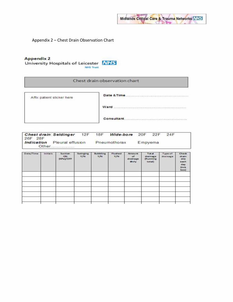
Appendix 2 – Chest Drain Observation Chart
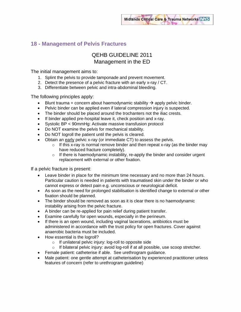
18 - Management of Pelvis Fractures
QEHB GUIDELINE 2011
Management in the ED
The initial management aims to:
1. Splint the pelvis to provide tamponade and prevent movement. 2. Detect the presence of a pelvic fracture with an early x-ray / CT. 3. Differentiate between pelvic and intra-abdominal bleeding.
The following principles apply:
Blunt trauma + concern about haemodynamic stability apply pelvic binder.
Pelvic binder can be applied even if lateral compression injury is suspected.
The binder should be placed around the trochanters not the iliac crests.
If binder applied pre-hospital leave it, check position and x-ray.
Systolic BP < 90mmHg: Activate massive transfusion protocol
Do NOT examine the pelvis for mechanical stability.
Do NOT logroll the patient until the pelvis is cleared.
Obtain an early pelvic x-ray (or immediate CT) to assess the pelvis. o If this x-ray is normal remove binder and then repeat x-ray (as the binder may
have reduced fracture completely). o If there is haemodynamic instability, re-apply the binder and consider urgent
replacement with external or other fixation.
If a pelvic fracture is present:
Leave binder in place for the minimum time necessary and no more than 24 hours.
Particular caution is needed in patients with traumatised skin under the binder or who
cannot express or detect pain e.g. unconscious or neurological deficit.
As soon as the need for prolonged stabilisation is identified change to external or other
fixation should be planned.
The binder should be removed as soon as it is clear there is no haemodynamic
instability arising from the pelvic fracture.
A binder can be re-applied for pain relief during patient transfer.
Examine carefully for open wounds, especially in the perineum. If there is an open wound, including vaginal lacerations, antibiotics must be
administered in accordance with the trust policy for open fractures. Cover against
anaerobic bacteria must be included.
How essential is the logroll? o If unilateral pelvic injury: log-roll to opposite side
o If bilateral pelvic injury: avoid log-roll if at all possible, use scoop stretcher.
Female patient: catheterise if able. See urethrogram guidance.
Male patient: one gentle attempt at catheterisation by experienced practitioner unless features of concern (refer to urethrogram guideline)
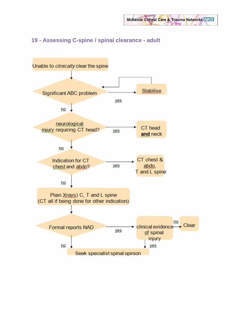
19 - Assessing C-spine / spinal clearance - adult
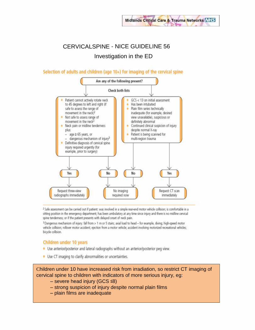
CERVICALSPINE - NICE GUIDELINE 56
Investigation in the ED
Children under 10 have increased risk from irradiation, so restrict CT imaging of
cervical spine to children with indicators of more serious injury, eg:
– severe head injury (GCS 8) – strong suspicion of injury despite normal plain films – plain films are inadequate
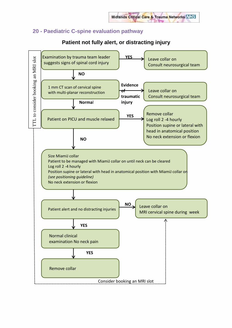
20 - Paediatric C-spine evaluation pathway
Patient not fully alert, or distracting injury
Examination by trauma team leader suggests signs of spinal cord injury
YES Leave collar on
Consult neurosurgical team
NO
1 mm CT scan of cervical spine with multi-planar reconstruction
Evidence of traumatic injury
Leave collar on
Consult neurosurgical team
Normal
Patient on PICU and muscle relaxed YES Remove collar
Log roll 2 -4 hourly
Position supine or lateral with head in anatomical position
No neck extension or flexion NO
Size MiamiJ collar Patient to be managed with MiamiJ collar on until neck can be cleared
Log roll 2 -4 hourly
Position supine or lateral with head in anatomical position with MiamiJ collar on
(see positioning guideline) No neck extension or flexion
Patient alert and no distracting injuries
YES
Normal clinical examination No neck pain
YES
Remove collar
NO Leave collar on
MRI cervical spine during week
Consider booking an MRI slot
TT
L t
o c
onsi
der
bookin
g a
n M
RI
slot
21 - Radiology reporting standards
Please refer to the Royal College of Radiologists (RCR). ‘Standards of practice and
guidance for trauma radiology in severely injured patients’. 2011. The Image Exchange Portal (IEP) will be the mechanism through which teleradiology will be transmitted for the Midlands Trauma Networks. This will be available 24/7 with linkage between all service providers and accessible for ‘home viewing’ for on-call Consultant Radiologists. The RCR standards, as contained in this policy, will be adopted across the Midlands Trauma Networks and will provide the framework for compliance monitoring and governance reporting by 31.12.2012.
Standard Guidance 1. The trauma team leader (TTL) is in overall
charge in acute care The acute trauma setting is not the place for
disagreements about the patients pathway, Immediate management decisions must be made by the designated TTL
Quality indicator – MTC’s & TU’s will have multidisciplinary debriefings about severely injured patients (SIPs) on a regular basis and adjust pathways if necessary. A radiologist involved in trauma management should attend such meetings. Individual cases should be considered in the radiology department on a regular basis.
2. Protocol-driven imaging and intervention must be available and delivered by experienced staff. Acute care for SIPs must be consultant delivered.
Just as the TTL must be an experienced consultant, there must also be a consultant-delivered input for imaging and intervention
3. MDCT should be adjacent to, or in, the emergency room.
If NOT then transfers should be rehearsed and performed according to protocol and organisations should plan to make this a future provision.
The less a patient is moved and the shorter the distance, the greater will be the chance of survival.
Imaging in SIPs more accurately delineates the extent of the injury.
For SIPs head-to-thigh contrast-enhanced multi-detector computed tomography (MDCT) is the most definitive imaging technique.
Definitive imaging should not be delayed by other, less accurate, investigations.
The imaging room requires the same life-support facilities available in ED. The room should accommodate visual and technical monitoring by anaesthetic staff.
4. Digital radiography must be available in the emergency room.
Digital radiology (DR0 should be available in the emergency room
A chest X-ray may precede a MDCT if there is doubt about a pneumothorax.
Life threatening injuries should be diagnosed and treated prior to extremity imaging.
CT should be used for C-spine clearance
5. If there is an early decision to request
MDCT, FAST and DR should not cause delay.
Quality indicator – Where FAST or plain films have been used in a SIP, their use and value in that case should be evaluated in a multidisciplinary debriefing.
FAST – Fast Abdominal Sonography in Trauma should not be preferred to CT.
FAST is valuable in diagnosing pericardial effusion and detecting free intra-abdominal fluid. It has an important role in triage when multiple SIP’s present.
6. Magnetic Resonance Imaging (MRI) must be available with safe access.
Quality indicator – Availability of clear protocols for the transfer of SIP’s to MRI facilities within 12 hours.
MRI must be available in MTC’s 24/hours/day, 7 days/week. In the same building as the ED or supported by SIP transfer protocols.
TU’s without 24/7 access to on site MRI should have transfer protocols in place.
7. A CT request in the trauma setting should comply with the Ionising Radiation (Medical Exposure) Regulations 2000 (IR[ME]R) justification regulations like any other request for imaging involving ionizing radiation.
Quality Indicator – An annual audit of justification in trauma imaging should be carried out in the radiology department.
The outcome from the REACT trial recruiting patients to a CT-first or resuscitation-first protocol might supersede these indications and major trauma may justify immediate MDCT.
8. There should be clear written protocols for MDCT preparation and transfer to the scan room.
Quality indicator – Such protocols should be written and available and the process should be a statutory evaluation at debriefing.
There should be local protocols clearly attributing responsibility at every stage including request for MDCT and transfer route to CT.
The need for IV access, urinary catheterisation, pelvic fracture stabilisation, limb fracture immobilisation, and pregnancy status should all be addressed.
9. Whole-body contrast-enhanced MDCT is the default imaging procedure of choice in SIP. Imaging protocols should be clearly defined and uniform across a regional trauma network.
Network policies should be agreed to avoid repeat scanning.
On-call interventional radiologist opinion should immediately be sought where contrast extravasation is seen and findings are equivocal.
10. Future planning and design of emergency rooms should concentrate on increasing the number of SIP’s stable enough for MDCT and intervention.
11. The primary survey report should be issued immediately to the TTL. It should be signed and designated and a copy should be retained in the CT department (or RIS)
Initial MDCT should be attended by appropriately trained on-call radiologist.
Reporting follows ATLS system with primary survey followed by secondary survey.
Clinical teams should fill in their contact details for future contact access.
12. On-call consultant radiologist should provide the final report on the SIP within one hour of MDCT image acquisition
Quality indicator – All imaging should be discussed at debriefing meetings and errors of protocols or fact discussed at discrepancy meetings.
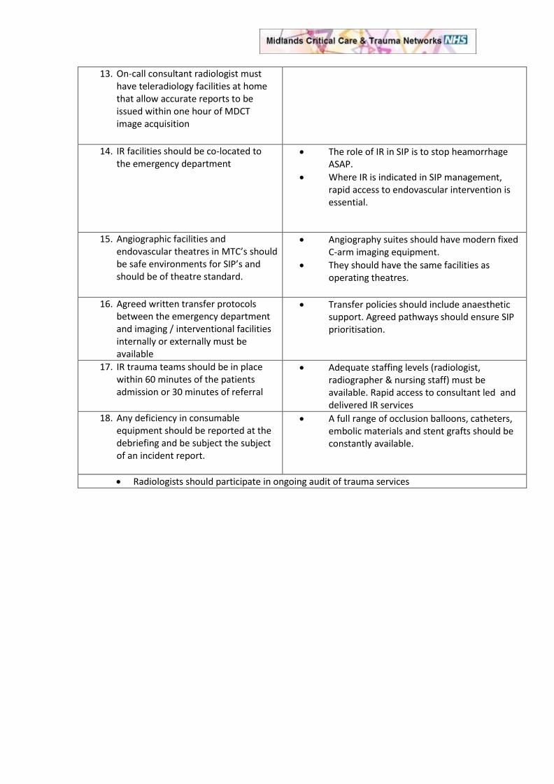
13. On-call consultant radiologist must have teleradiology facilities at home that allow accurate reports to be issued within one hour of MDCT image acquisition
14. IR facilities should be co-located to the emergency department
The role of IR in SIP is to stop heamorrhage ASAP.
Where IR is indicated in SIP management, rapid access to endovascular intervention is essential.
15. Angiographic facilities and endovascular theatres in MTC’s should be safe environments for SIP’s and should be of theatre standard.
Angiography suites should have modern fixed C-arm imaging equipment.
They should have the same facilities as operating theatres.
16. Agreed written transfer protocols between the emergency department and imaging / interventional facilities internally or externally must be available
Transfer policies should include anaesthetic support. Agreed pathways should ensure SIP prioritisation.
17. IR trauma teams should be in place within 60 minutes of the patients admission or 30 minutes of referral
Adequate staffing levels (radiologist, radiographer & nursing staff) must be available. Rapid access to consultant led and delivered IR services
18. Any deficiency in consumable equipment should be reported at the debriefing and be subject the subject of an incident report.
A full range of occlusion balloons, catheters, embolic materials and stent grafts should be constantly available.
Radiologists should participate in ongoing audit of trauma services
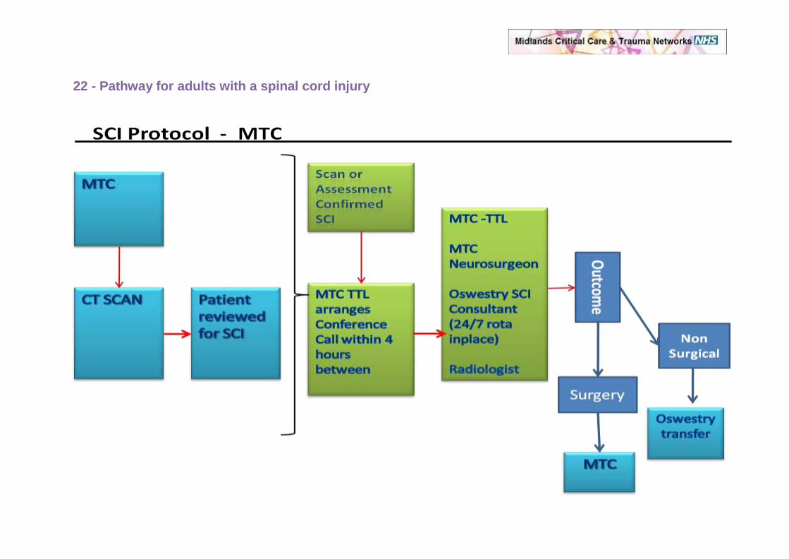
22 - Pathway for adults with a spinal cord injury
PATHWAY FOR PATIENTS WITH SCIs
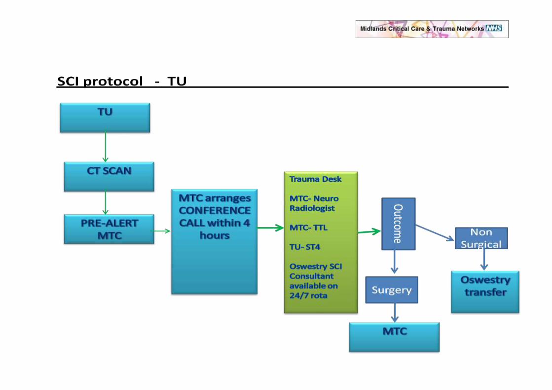
PATHWAY FOR PATIENTS WITH SCIs
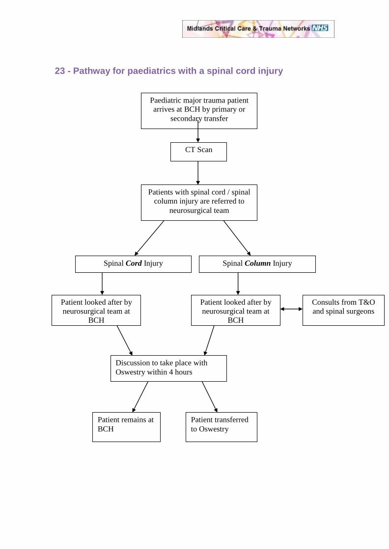
23 - Pathway for paediatrics with a spinal cord injury
Patient transferred
to Oswestry
Patient remains at
BCH
Discussion to take place with
Oswestry within 4hrs
Consults from
T&O and spinal
surgeons
Patient looked after by
neurosurgical team at
BCH
Patient looked after by
neurosurgical team at
BCH
Paediatric major trauma patient
arrives at BCH by primary or
secondary transfer
Patients with spinal cord / spinal
column injury are referred to
neurosurgical team
CT Scan
Spinal Cord Injury Spinal Column Injury
Consults from T&O
and spinal surgeons
Discussion to take place with
Oswestry within 4 hours
Patient remains at
BCH
Patient transferred
to Oswestry
24 - Guideline for the assessment, care & transfer of adult
patients with potentially non-survivable burn injuries in an ED
Decisions about End of Life Care for burn injured patients are only considered after the patient has been assessed by a senior and experienced Clinician. Decisions of this nature must be taken using a team approach and wherever possible must involve the patient, their family and carers. Clinical factors relevant to making these decisions include:
The size of the burn / percentage Total Body Surface Area %TBSA
The depth of the burn (Partial Thickness / Full Thickness (PTB and FTB)
The age of the patient *
Any co-morbidities present
The wishes of the patient and/or the family/carers
* Age and % TBSA have historically been used as indicators of the likelihood of Burn injury survival.
There are two possible scenarios when it is appropriate to consider end of life care as the most appropriate treatment plan for burn injured patients:
Where an injury is catastrophic and there is no feasible prospect of survival (comfort
care is regarded as the only realistic option)
Where a patient’s condition deteriorates and there is no prospect of recovery. In
these cases the damage is irreversible; this would be during treatment in a Burns
Service.
The first category (a catastrophic injury) is the most likely to present itself to the Clinicians working in an Emergency Department (ED). For Clinicians who do not regularly assess burn injuries these decisions can be difficult. The overriding principle should be that there is ALWAYS discussion between the medical team responsible for the initial treatment and the Consultant Burn Surgeon on call in either the local Burn Unit or Burn Centre. Any decision must only be made after the following has occurred
Patient assessment in ED by two consultants and after discussion with a consultant
Burn Surgeon at the local Burns Unit or Burn Centre.
The two Consultants must be in agreement (after discussion with a Consultant Burn
Surgeon at the local Burns Unit or Burn Centre), that the patient is considered to
have “non - survivable” injuries after taking into account % TBSA, depth, inhalation
injury and co-morbidities.
If a decision is made that the patient’s burn injuries are non-survivable, this should
be communicated to the patient (if appropriate) and the family/carers in an honest
but sensitive manner.
Where EDs are located on the same site as a 24 hour Burns or Plastic Surgery Services then the Burns / Plastic Surgery Service should be contacted for advice so that a member of the team can review the patient in the ED. This review should be undertaken in person by a Consultant, Registrar or equivalent.
Location of Patient care The local Burn Service should be contacted for all burns advice regarding the best location for the care, management and support of the patient and family/carers. The overall aims are to optimise quality of life, care and support in the end stages of life. If advice is required for nursing care at the ED then the nurse in charge at the local Burns Service should be contacted (e.g. wound care, family/carer physical contact with patient)
When deciding the best location and service to care for the patient with a burn injury
that is regarded as non survivable the needs and wishes of both the patient and
their family must be discussed with them and considered. Depending on the
circumstances this may be the local hospital or a specialised Burn Service
If it is expected that the patient will survive 24 hours then it would be best practice to
transfer them to the local Burn Service unless it is the patient’s or family/carer’s wish
not to transfer them
If the Burns Service or ED is in any doubt then the patient should be transferred.
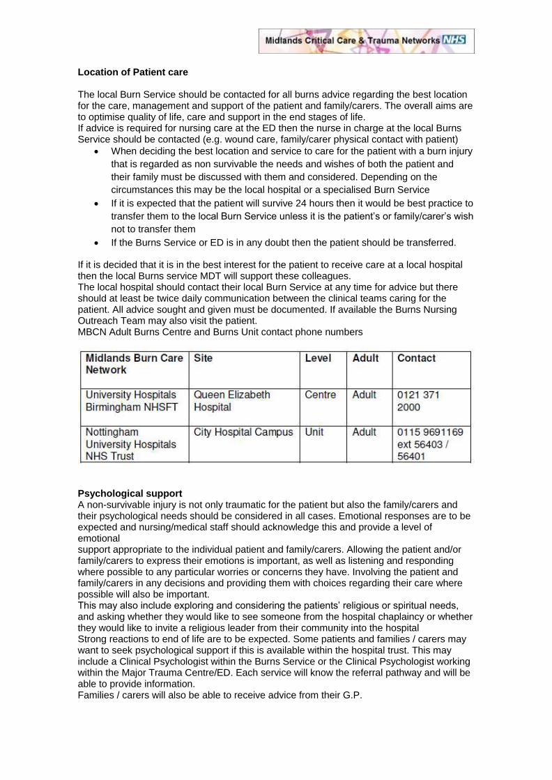
If it is decided that it is in the best interest for the patient to receive care at a local hospital then the local Burns service MDT will support these colleagues. The local hospital should contact their local Burn Service at any time for advice but there should at least be twice daily communication between the clinical teams caring for the patient. All advice sought and given must be documented. If available the Burns Nursing Outreach Team may also visit the patient. MBCN Adult Burns Centre and Burns Unit contact phone numbers
Psychological support A non-survivable injury is not only traumatic for the patient but also the family/carers and their psychological needs should be considered in all cases. Emotional responses are to be expected and nursing/medical staff should acknowledge this and provide a level of emotional support appropriate to the individual patient and family/carers. Allowing the patient and/or family/carers to express their emotions is important, as well as listening and responding where possible to any particular worries or concerns they have. Involving the patient and family/carers in any decisions and providing them with choices regarding their care where possible will also be important. This may also include exploring and considering the patients’ religious or spiritual needs, and asking whether they would like to see someone from the hospital chaplaincy or whether they would like to invite a religious leader from their community into the hospital Strong reactions to end of life are to be expected. Some patients and families / carers may want to seek psychological support if this is available within the hospital trust. This may include a Clinical Psychologist within the Burns Service or the Clinical Psychologist working within the Major Trauma Centre/ED. Each service will know the referral pathway and will be able to provide information. Families / carers will also be able to receive advice from their G.P.
25 - Midlands burn network flowchart - adult
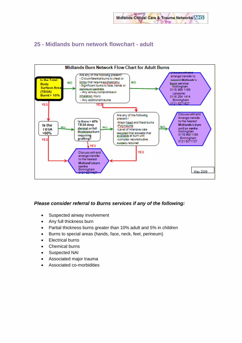
Please consider referral to Burns services if any of the following:
Suspected airway involvement
Any full thickness burn
Partial thickness burns greater than 10% adult and 5% in children
Burns to special areas (hands, face, neck, feet, perineum)
Electrical burns
Chemical burns
Suspected NAI
Associated major trauma
Associated co-morbidities
26 - Midlands burn network flowchart – paediatric
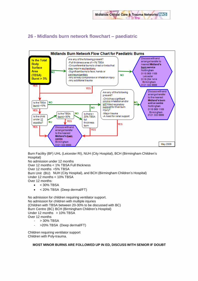
Burn Facility (BF) UHL (Leicester RI), NUH (City Hospital), BCH (Birmingham Children’s Hospital)
No admission under 12 months Over 12 months < 1% TBSA Full thickness Over 12 months <5% TBSA
Burn Unit (BU) NUH (City Hospital), and BCH (Birmingham Children’s Hospital)
Under 12 months < 10% TBSA Over 12 months:
< 30% TBSA
< 20% TBSA (Deep dermal/FT)
No admission for children requiring ventilator support. No admission for children with multiple injuries (Children with TBSA between 20-30% to be discussed with BC) Burn Centre (BC) BCH (Birmingham Children’s Hospital)
Under 12 months > 10% TBSA Over 12 months
- > 30% TBSA
- >20% TBSA (Deep dermal/FT)
Children requiring ventilator support Children with Poly-trauma.
MOST MINOR BURNS ARE FOLLOWED UP IN ED, DISCUSS WITH SENIOR IF DOUBT
27 - Management of paediatric burns
PRIMARY SURVEY In children who have been severely burned, it is all-to-easy to focus on the immediate problems of the burn and forget the possible presence of any other injuries. The approach to the child with burns should be along the exact same structure as any patient with major trauma. Airway and cervical spine The airway may be compromised either because of inhalational injury and oral scalds or because of severe burns to the face. Indications of inhalational injury:
History of exposure to smoke in a confined space
Deposits around the mouth and nose
Carbonaceous sputum
Oedema occurs following thermal injury, and the airway can deteriorate rapidly. Thus even suspicion of airway compromise should lead to immediate consideration of tracheal intubation. This procedure increases in difficulty as oedema progresses; it is therefore important to perform it as soon as possible. All but the most experienced should seek expert help urgently, unless apnoea requires immediate intervention. If there is any suspicion of cervical spine injury, or if the history is unobtainable, appropriate precautions should be taken until such injury is excluded.
Breathing Once the airway is secured, the adequacy of breathing should be assessed. Signs that should arouse suspicion of inadequacy include abnormal rate, abnormal chest movements and cyanosis. Circumferential burns to the chest or abdomen (the latter in infants) may cause breathing difficulty by mechanically restricting chest movement. All children who have suffered burns should be given high-flow oxygen. If there are signs of breathing problems then intubation and ventilation should be commenced Circulation In the first few hours following injury, signs of hypovolaemic shock are rarely attributable to burns, therefore any such signs should raise the suspicion of bleeding from elsewhere, and the source should be actively sought. Intravenous access should be established with two cannulae during resuscitation and fluids started. If possible, drips should be put up in unburnt areas - remember that the intraosseous route can be used in paediatrics. Disability Reduced conscious level following burns may be due to hypoxia (remember smoke-filled rooms may contain little oxygen), head injury or hypovolaemia. It is essential that a quick assessment is made during the primary, because this provides a baseline for later observations.
Exposure Burned children lose heat especially rapidly, and should be kept in a warm environment and be covered with blankets when not being examined
SECONDARY SURVEY As well as being burned, children may suffer the effects of blast, may be injured by falling objects, or may fall while trying to escape from the fire. Thus other injuries are not uncommon and a thorough head-to-toe secondary survey must be carried out. Any injuries discovered, including the burn, should be treated in order of priority. The severity of a burn depends on its relative surface area and depth. Burns to particular areas require special attention. Surface area It is particularly important to use a paediatric chart when assessing burn size in children (if possible), because the relative surface areas of the head and limbs change with age. A useful method of estimating relative surface area relies on the fact that the patient‘s palm and adducted fingers cover an area of approximately 1% of the body surface. This method can be used when charts are not immediately available. Note that the ‘rule of 9s’ cannot be applied to children who are less than 14 years old. Special areas Burns involving the childs hands or feet can cause severe functional loss if scarring occurs. Perineal burns are prone to infection and present particularly difficult management problems. Circumferential, full- or partial-thickness burns of the limbs or neck may require urgent incision to relieve distal ischaemia. Similarly, circumferential burns to the torso may restrict ventilation and also require urgent incision. An escharotomy usually needs to be done before transfer to a burns centre.
EMERGENCY MANAGEMENT
Analgesia Most burned children will be in severe pain, and this should be dealt with urgently. Some older children may manage to use Entonox, but most will not. Any child with burns that are anything other than minor should be given intravenous morphine as soon as possible. Further doses are often required but must be titrated against pain and sedation. There is no place for administration of intramuscular analgesia in burns because absorption is unreliable. Fluid therapy IV or IO access should have already been established during the primary survey and fluid resuscitation commenced for shock if indicated. Children with burns of 10% or more will require intravenous fluids as part of their burns care. This fluid is in addition to their normal fluid requirement.
The additional fluid (in ml) required per day to treat the burn can be estimated using the following formula:
Percentage burn × Weight (kg) × 4
Half of this should be given in the first 8 hours following the time of their burn. The fluid given is usually crystalloid. Remember that this is only an initial guide; subsequent therapy will be guided by urine output, which should be kept at 2 ml/kg/hour or more. Urethral catheterisation should therefore be performed as soon as is practicable Wound care Burns should be covered with sterile towels, and unnecessary re-examination should be avoided. Blisters should be left intact. Although cold compresses and irrigation with cold water may reduce pain, it should be remembered that burned children lose heat rapidly. These treatments should only be used for 10 minutes or less, and only in patients with superficial or partial-thickness burns totalling less than 10%. Children should never be transferred with cold soaks in place. Cling film is often used as a sterile dressing and can be applied loosely onto the burned area. No additional ointments or creams should be applied. Management of carbon monoxide poisoning Children who have been in house fires should have their blood carboxyhaemoglobin measured. (Note: Most pulse oximeters show the oxygen saturation, regardless of haemoglobin concentration i.e. normal SpO2 does not exclude carbon monoxide poisoning) Levels of 5–20% - treated with oxygen to speed up the removal of CO Levels over 20% - should prompt consideration of hyperbaric oxygen chamber treatment – discuss with the paediatric burns unit. In some environments the burning of plastics, wool and silk can produce cyanide. Assessment and treatment are complex. Be aware of the possibility of cyanide poisoning and consider it in a child from a house fire who is in a coma without apparent cause. In general, antidotes are used when blood levels of cyanide are greater than 3 mg/l. Discuss treatment with a poisons centre as other factors such as the concomitant presence of carboxyhaemoglobin are contraindications for some antidotes. Continuing stabilisation and transfer to definitive care Definitive care may require transfer to a paediatric burns facility - if in doubt discuss the
child with the paediatric burns unit.
Criteria for transfer include:
10% partial- and/or full-thickness burns
5% full-thickness burns
Burns to special areas: face, hands, feet or perineum.
Any circumferential burn
Significant inhalational burn (excluding pure carbon monoxide poisoning)
Chemical, radiation or high voltage electrical burns

As with any injury in childhood, consider the possibility of child abuse. Note the timeliness of presentation, and assess whether the history given to account for the burn or scald fits in with the clinical appearance of the injury in size, shape, age and location. Consider whether the injury is consistent with the child‘s developmental attainments. If concerned or in doubt, consult with a child protection specialist.
(Adapted from the Advanced Paediatric Life Support 5th
edition)
28 - Central England Trauma Network TU burns poster
Burns patients should go directly to a Burns facility / unit / centre from the Trauma Unit. If the patient has other Major Trauma injuries, they
should go to a Burns Centre with Major Trauma Status.
NB: If a patient is primarily Major Trauma with a burn requiring burns facility care, then UHCW can accept this as per the usual Hyperacute
Trauma Pathway.
Burns Referral Criteria:
• A child with a partial thickness burn greater than 2% TBSA • An adult with a partial thickness burn greater than 3% TBSA
In addition to the % TBSA thresholds described for children and adults any patient with a burn injury regardless of age or %TBSA that presents with any of the following should be discussed with the local burn service and consideration given for the need for referral:
• Inhalation injury is defined as visual evidence of suspected upper airway smoke inhalation, laryngoscopic and/or bronchoscopic evidence of tracheal or more distal contamination / injury or suspicion of inhalation of non soluble toxic gases.
• A full thickness burn greater than 1% TBSA • Burns to special areas (hands, face, neck, feet, perineum) • Burns to an area involving a joint which may adversely affect mobility and function • Electrical burns • Chemical burns • Suspected non-accidental injury (NAI). Any burn with suspicion of nonaccidental injury should be referred to a specialised burn service
for an expert assessment within 24 hours. • A burn associated with major trauma • A burn associated with significant co-morbidities • Circumferential burns to the trunk or limbs • Any burn not healed in 2 weeks
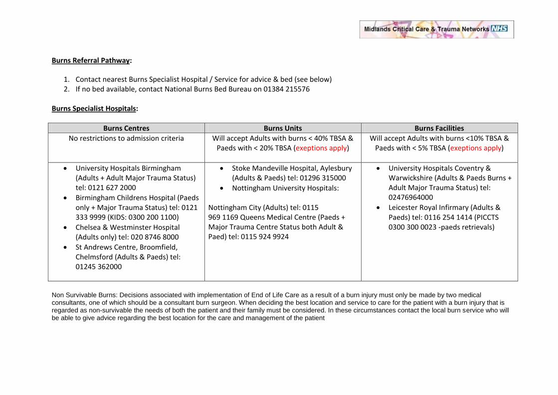
Burns Referral Pathway:
1. Contact nearest Burns Specialist Hospital / Service for advice & bed (see below) 2. If no bed available, contact National Burns Bed Bureau on 01384 215576
Burns Specialist Hospitals:
Burns Centres Burns Units Burns Facilities
No restrictions to admission criteria
Will accept Adults with burns < 40% TBSA & Paeds with < 20% TBSA (exeptions apply)
Will accept Adults with burns <10% TBSA & Paeds with < 5% TBSA (exeptions apply)
University Hospitals Birmingham (Adults + Adult Major Trauma Status) tel: 0121 627 2000
Birmingham Childrens Hospital (Paeds only + Major Trauma Status) tel: 0121 333 9999 (KIDS: 0300 200 1100)
Chelsea & Westminster Hospital (Adults only) tel: 020 8746 8000
St Andrews Centre, Broomfield, Chelmsford (Adults & Paeds) tel: 01245 362000
Stoke Mandeville Hospital, Aylesbury (Adults & Paeds) tel: 01296 315000
Nottingham University Hospitals: Nottingham City (Adults) tel: 0115 969 1169 Queens Medical Centre (Paeds + Major Trauma Centre Status both Adult & Paed) tel: 0115 924 9924
University Hospitals Coventry & Warwickshire (Adults & Paeds Burns + Adult Major Trauma Status) tel: 02476964000
Leicester Royal Infirmary (Adults & Paeds) tel: 0116 254 1414 (PICCTS 0300 300 0023 -paeds retrievals)
Non Survivable Burns: Decisions associated with implementation of End of Life Care as a result of a burn injury must only be made by two medical consultants, one of which should be a consultant burn surgeon. When deciding the best location and service to care for the patient with a burn injury that is regarded as non-survivable the needs of both the patient and their family must be considered. In these circumstances contact the local burn service who will be able to give advice regarding the best location for the care and management of the patient

29 - UHB burns flowchart We are currently awaiting the final version which will be posted when complete.
Thank you.
30 - Prophylactic antibiotics in trauma surgery and open fractures
Routine insertion of metalwork - adapted from the current BNF
Joint replacement including hip hemiarthroplasty
Single dose of intravenous flucloxacillin and intravenous gentamicin
If history of allergy to penicillins or if high risk of meticillin-resistant Staphylococcus aureus, use single dose 400mg of intravenous teicoplanin + 120mg intravenous gentamicin
Closed fractures
Single dose of 1g intravenous flucloxacillin up to 30 minutes before the start of the procedure. If history of allergy to penicillins or if high risk of meticillin-resistant Staphylococcus aureus, use single 400mg dose of intravenous teicoplanin. Prolonged/high risk procedures should add up to two subsequent doses.
Open fractures adapted from the BNF and BOA/BAPRAS guidelines
1.2g intravenous co-amoxiclav alone (or 600mg intravenous clindamycin alone if history of allergy to penicillins). Add 400mg intravenous teicoplanin if high risk of meticillin-resistant Staphylococcus aureus. Start prophylaxis within 3 hours of injury and continue until soft tissue closure (max. 72 hours); at first debridement also use a single dose of 1.2g intravenous co-amoxiclav + 120mg intravenous gentamicin (or intravenous clindamycin + intravenous gentamicin if history of allergy to penicillins). At time of skeletal stabilisation and definitive soft tissue closure use a single dose of 120mg intravenous gentamicin + 800mg intravenous teicoplanin up to 30 minutes before the start of the procedure.
31 - Limb compartment pressure measurement
1. Equipment for compartment pressure measurement is available in the theatre suite
(Stryker probe), or can be set up in theatre or ITU from a pressure transducer on the
monitors.
2. A high index of clinical suspicion for compartment syndrome should be maintained.
Compartment pressure measurement is only indicated where there is significant
clinical doubt or the patient is unable to give reliable information regarding pain.
3. Where pressures are measured they should be compared with the diastolic pressure.
If the difference is less than 30mmHg, this constitutes an indication for fasciotomy.
4. Training on compartment syndrome management is delivered at each local induction
of junior doctors.
32 - Management of brachial plexus injuries
Author: Mark R Foster, MD, PhD, FACS; Chief Editor: Mary Ann E Keenan, MD
High-energy trauma to the upper extremity and neck can cause a variety of lesions to the brachial plexus. Most common are traction injuries, in which the head and neck are moved away violently from the ipsilateral shoulder; injuries may also be caused by compression between the clavicle and first rib, penetrating injuries, or direct blows. Recognition may be delayed by other injuries, particularly to the spinal cord and head. The common mechanism for traction injuries of the brachial plexus is violent distraction of the entire forequarter from the rest of the body. These injuries usually result from a motorcycle accident or a high-speed motor vehicle accident. A fall from a significant height may also result in brachial plexus injury.
The patient may present with the following symptoms:
Pain, especially of the neck and shoulder. Pain over a nerve is common with rupture, as opposed to lack of percussion tenderness with avulsion
Paresthesias and dysesthesias Weakness or heaviness in the extremity Diminished pulses, as vascular injury may accompany traction injury.
Physical examination
The standard advanced trauma life support (ATLS) protocol should be followed. Abrasions to the head, helmet, or tip of the shoulder suggest supraclavicular injury. Ptosis (lid droop), enophthalmos (sinking of the eye into the orbit), anhydrosis (dry eye), and miosis (small pupil) or Horner syndrome suggest a complete lower plexus lesion, as the sympathic ganglion for T1 is in close proximity to the brachial plexus.
Swelling about the shoulder can be dramatic. Diminished or absent pulses suggest vascular injury, and special consideration should be given to rupture of the subclavian vessels. Clavicle fractures often are palpable. Careful inspection and palpation of the axial skeleton may reveal concomitant injuries. Examine each cervical root individually for motor and sensory function as soon as circumstances allow.
Some special considerations are warranted for the neurologic examination, as follows:
Sensory examination is extremely important. Deep pressure sensation may be the only clue to continuity in a nerve with no motor function or other sensation. Apply full pinch to the nail base and pull the patient's finger outward. Any burning suggests continuity of the tested nerve. When no burning is elicited, these examination findings are less helpful because a neuropraxia can persist for more than 6 months.
Deep Pressure Test
Location of deep pressure
test
Affected spinal nerve
Nerve Affected cord
Thumb C6 Median nerve Lateral cord
Middle Finger C7 Median nerve Lateral cord
Little finger C8 Ulnar nerve Medial cord
Examination of wrist and finger sensation and motion with respect to the median, ulnar, and radial nerves may help start to locate the lesion within the brachial plexus.
Guide to Motor Testing
Cervical root
Clinically Relevant Gross Motor Function
C5 Shoulder abduction, extension, and external rotation; some elbow flexion
C6 Elbow flexion, forearm pronation and supination, some wrist extension
C7 Diffuse loss of function in the extremity without complete paralysis of a specific muscle group, elbow extension, consistently supplies the latissimus dorsi
C8 Finger extensors, finger flexors, wrist flexors, hand intrinsics
T1 Hand intrinsics
Imaging Studies
Radiographic evaluation
In anteroposterior (AP) chest radiography, specific attention should be directed to the distance between the spinous processes of the thoracic spine and the scapula. If the radiograph is not malrotated, an increase in this distance compared with the contralateral side may indicate scapulothoracic dissociation.
AP and axillary lateral views of the shoulder reveal clavicle fractures, most scapular fractures, and most proximal humerus fractures.
Cervical spine series including AP, lateral, and odontoid views are useful.
Computed tomography (CT) scanning
Adequate plain radiographs may be difficult to obtain, especially of the odontoid and the cervicothoracic junction. A CT scan of the neck can often be obtained in conjunction with CT scanning that is a part of the evaluation of many trauma patients. Plain CT scanning is very helpful in evaluating any cervical fractures, and should be obtained if fractures are suspected based on plain radiographic findings. CT scanning of the chest may reveal subclavian vessel injuries, scapular fractures, humeral fractures, and thoracic spine fractures.
Myelography
The most reliable indicator of root avulsion is an absent root shadow on plain myelography. A common sign of a root avulsion is a meningocele at the affected level; hence, myelography may best be delayed for 4 weeks so that any blood clot will not be dislodged by the study and the meningocele can be allowed to form.
CT myelography (CTM)
The literature is still inconclusive regarding the sensitivity and specificity of CTM, but CTM is being performed more often. Lower concentrations of contrast medium can be detected by CTM than by standard myelography. Burge states that CTM may be better able to reveal small meningoceles, but artifact from surrounding soft tissues may be problematic at the lower cervical levels.
Magnetic resonance imaging (MRI)
MRI is the current criterion standard for visualizing spinal cord injuries, but reports of its utility in evaluating traumatic lesions of the brachial plexus are sparse. MRI is the only technique that can be used to visualize the postganglionic brachial plexus. While the impact of MRI on surgical decision-making is yet to be defined, it no doubt will play a larger role in the evaluation of the brachial plexus in the future.
Angiography
Both conventional angiography and magnetic resonance angiography (MRA) are valuable tools in evaluating any suspected vascular disruption.
Other Tests Sensory nerve action potentials (SNAPs): SNAPs are very helpful in differentiating preganglionic from postganglionic injuries. If the injury is proximal to the dorsal root ganglion (DRG), no Wallerian degeneration occurs because the sensory axon is intact. Thus, a SNAP observed in a nerve with an anesthetic dermatome confirms a preganglionic lesion. SNAPs are not useful for C5 evaluation because C5 does not provide a significant contribution to a major peripheral sensory nerve.
Electromyography (EMG): In the first week after injury, EMG cannot be used to exclude a complete nerve disruption unless voluntary motor unit action potentials are observed. If no signs of denervation are apparent in a paralyzed muscle by 3 weeks after injury, EMG can be used to confirm neuropraxia.
Somatosensory evoked potentials (SSEPs): Intraoperative SSEPs are useful in brachial plexus surgery. The presence of SSEPs suggests continuity between the peripheral nervous system and the CNS via the DRG. SSEPs are absent in postganglionic or combined pre- and postganglionic lesions.
Medical Therapy Nonoperative treatment of brachial plexus lesions is complex and may best be addressed by an integrated multidisciplinary team that includes a skilled orthotist, occupational therapists, physical therapists, and physicians. Bracing often plays a role in preventing contractures while waiting for recovery after surgery or while waiting for recovery from neurapraxia.
Surgical Therapy Operative care of the brachial plexus is a highly specialized field that is limited to relatively few tertiary care centers. Wide variation exists in how these injuries are addressed surgically. The availability of subspecialists with experience in the operative management of these lesions is critical if operative management is considered. In general, the surgical options consist of nerve transfers; nerve grafting, muscle transfers, free muscle transfers, and neurolysis of scar around the brachial plexus in incomplete lesions.
Open injuries, particularly high-velocity gunshot wounds, call for debridement and immediate repair when possible, or tagging of nerves for delayed repair. External neurolysis should be performed for intraoperative monitoring and electrical studies, or neurolysis alone for nerves in continuity that exhibit a nerve action potential (NAP). The NAP can demonstrate preserved axons or significant regeneration, and potential for further recovery; a neurapraxic lesion shows no NAP, as opposed to axonometric lesions (positive for NAP). Otherwise (no intraoperative NAP) nerve grafting can be done for postganglionic neuromas or neural ruptures
Contraindications to surgery include the following:
Joint contractures Severe edema Advanced patient age Lack of patient motivation or lack of patient understanding of surgical goals
33 - Thromboprophylaxis guidelines – QEHB trauma
ALL lower limb/pelvic/abdo/chest/spine/head admissions should receive antiembolism stockings (GECS) unless contra-indicated. These should be applied certainly to the "good" leg(s) and to the "bad" leg if the injuries permit this to be done safely. If contra-indicated or the patient refuses then this MUST be recorded in the notes.
ALL lower limb/pelvic/abdo/chest admissions should receive 40mg enoxaparin subcut once daily unless contra-indicated. If contra-indicated or the patient refuses then this MUST be recorded in the notes.
Spinal and head-injured patients will need special consideration, preferably in consultation with the neuro/spinal surgeons, but should receive 40mg enoxaparin subcut once daily unless contra-indicated. If contra-indicated or patient refuses then this MUST be recorded in the notes.
ALL upper limb patients will need at the least an assessment of risk. If in doubt they should ALL receive GECS. If there is particular risk, especially if they are immobile (e.g. following a flap reconstruction) they should receive 40mg enoxaparin subcut once daily unless contra-indicated. If contra-indicated or the patient refuses then this MUST be recorded in the notes.
Where enoxaparin is contra-indicated by active bleeding or a high risk of bleeding (e.g. conservatively managed solid organ injury) or another reversible reason the contra-indicating risk MUST be reviewed regularly and frequently so that enoxaparin can be started as soon as safely possible.
In some high-risk patients where enoxaparin is contra-indicated a caval filter may be appropriate. This is a decision that needs to be made at a relatively senior level. Make sure that VTE prohylaxis is something you discuss on your ward rounds.
Enoxaparin should be stopped 12 hours before surgery and restarted 6—12 hours post surgery. If a patient is postponed for theatre then this requires an active decision to give any dose that would otherwise have been omitted.
Hip fracture patients require their enoxaparin continued to 35 days post-surgery EVEN IF THEY GO HOME rather than to a rehab unit. Please ensure this is made to happen at discharge.
Make sure that, as with any decision about patient management, decisions about VTE prophylaxis are recorded in the notes.
No trauma/orthopaedic patients should receive 20mg enoxaparin (unless dose reduced for renal impairment), so if you see this change it. Almost all trauma patients should be wearing GECS until fully mobile, so if you see someone without GECS question it and prescribe them if appropriate.
34 - Tetanus prophylaxis - adult
A TETANUS PRONE WOUND IS:
• Any wound or burn that requires surgical intervention that is delayed for > 6 hours
• Any wound or burn at any interval after injury that shows one or more of the
following characteristics:
- A significant degree of devitalised tissue
- Puncture-type wound
- Contact with soil or manure likely to harbour tetanus organisms
• Compound fractures
• Any wound containing foreign bodies
• Wounds or burns in patients who have systemic sepsis.
Intravenous drug abusers are at greater risk of tetanus. Every opportunity
should be taken to ensure that they are fully protected against tetanus. Booster
doses should be given if there is any doubt about their immunisation status.
Immunosupressed patients may not be adequately protected against tetanus, despite having been fully immunised. They should be managed as if they were incompletely immunized.
MOST MINOR BURNS ARE FOLLOWED UP IN ED. DISCUSS WITH SENIOR IF DOUBT
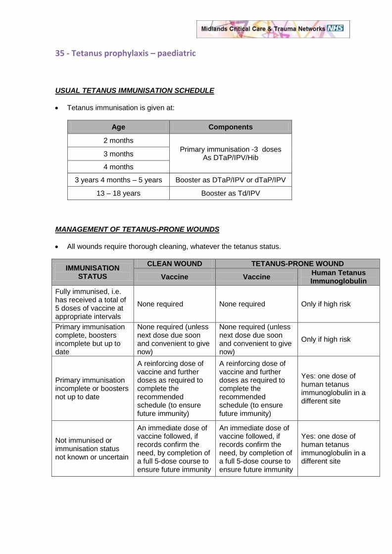
35 - Tetanus prophylaxis – paediatric
USUAL TETANUS IMMUNISATION SCHEDULE
Tetanus immunisation is given at:
Age Components
2 months
Primary immunisation -3 doses As DTaP/IPV/Hib
3 months
4 months
3 years 4 months – 5 years Booster as DTaP/IPV or dTaP/IPV
13 – 18 years Booster as Td/IPV
MANAGEMENT OF TETANUS-PRONE WOUNDS
All wounds require thorough cleaning, whatever the tetanus status.
IMMUNISATION STATUS
CLEAN WOUND TETANUS-PRONE WOUND
Vaccine Vaccine Human Tetanus Immunoglobulin
Fully immunised, i.e. has received a total of 5 doses of vaccine at appropriate intervals
None required None required Only if high risk
Primary immunisation complete, boosters incomplete but up to date
None required (unless next dose due soon and convenient to give now)
None required (unless next dose due soon and convenient to give now)
Only if high risk
Primary immunisation incomplete or boosters not up to date
A reinforcing dose of vaccine and further doses as required to complete the recommended schedule (to ensure future immunity)
A reinforcing dose of vaccine and further doses as required to complete the recommended schedule (to ensure future immunity)
Yes: one dose of human tetanus immunoglobulin in a different site
Not immunised or immunisation status not known or uncertain
An immediate dose of vaccine followed, if records confirm the need, by completion of a full 5-dose course to ensure future immunity
An immediate dose of vaccine followed, if records confirm the need, by completion of a full 5-dose course to ensure future immunity
Yes: one dose of human tetanus immunoglobulin in a different site
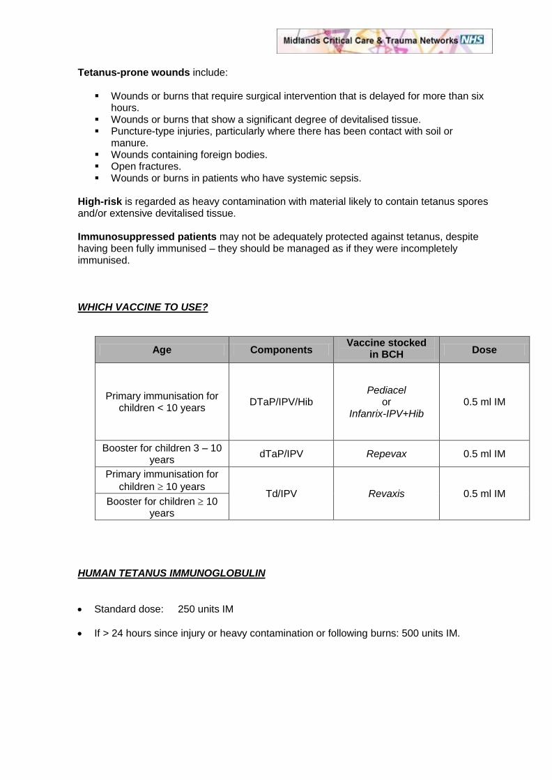
Tetanus-prone wounds include:
Wounds or burns that require surgical intervention that is delayed for more than six hours.
Wounds or burns that show a significant degree of devitalised tissue. Puncture-type injuries, particularly where there has been contact with soil or
manure. Wounds containing foreign bodies. Open fractures. Wounds or burns in patients who have systemic sepsis.
High-risk is regarded as heavy contamination with material likely to contain tetanus spores and/or extensive devitalised tissue. Immunosuppressed patients may not be adequately protected against tetanus, despite having been fully immunised – they should be managed as if they were incompletely immunised. WHICH VACCINE TO USE?
Age Components Vaccine stocked
in BCH Dose
Primary immunisation for children < 10 years
DTaP/IPV/Hib Pediacel
or Infanrix-IPV+Hib
0.5 ml IM
Booster for children 3 – 10 years
dTaP/IPV Repevax 0.5 ml IM
Primary immunisation for
children 10 years Td/IPV Revaxis 0.5 ml IM
Booster for children 10 years
HUMAN TETANUS IMMUNOGLOBULIN
Standard dose: 250 units IM
If > 24 hours since injury or heavy contamination or following burns: 500 units IM.
36 - Transfer of blood products with the patient
NHSBT Appropriate Use of Blood Group & National Laboratory Manager Group of the CMOs National Blood Transfusion Committee
Disclaimer
While the advice and information in these recommendations is believed to be true and accurate, neither the authors nor the two groups (NHSBT Appropriate Use of Blood Group and the National Laboratory Managers Group) accept any legal responsibility for the content of these recommendations.
Introduction This guidance for the emergency ad hoc transfer of blood with patients has been prompted by recent changes in medical practice, the organisation of health services and legal requirements, including:
The regulatory framework requires a vein-to-vein audit trail between donor and recipient (EU directive 2002/98/EC; Blood Safety and Quality Regulations 2005 (as amended).
Improvements in the transfusion process, especially in documentation and patient identification (Better Blood Transfusion initiatives).
Changes in the clinical management of patients with major bleeding and increased centralisation of health services in formal clinical networks ( hub and spoke ) have reduced the need for transfer of blood with patients.
The aim of this guidance is to standardise procedures for the emergency ad hoc transfer of blood and components between hospitals served by the NHS Blood and Transplant (NHSBT) and the Welsh Blood Service. Although it is intended as a guide that encompasses practices from all users, hospitals are encouraged, where appropriate, to add local protocols to the policy to complement the practices outlined in this document.
Blood and components are referred to in this document as blood unless specific components are discussed.
The committees concluded that a compelling need to transfer blood would be rare in modern practice though hospitals must undertake risk assessments to guide local practice. Two scenarios were considered to be an exception:
1. Blood allocated to a specific patient who was actively bleeding and in
whom the risk of transfer to a specialist unit was considered appropriate. Such patients would require a medical and/or nursing escort.
2. Patients being transferred who have special transfusion requirements such
as complex phenotyped blood, irradiated blood or HLA matched platelets.
However these blood components should be transferred directly to the laboratory in the receiving hospital (see page 5; point 2)
This document does not cover:
Agreed transfer of stock between hospital transfusion laboratories for optimal management of blood stocks.
Transfer of blood for a specific patient to a blood fridge located in a satellite hospital/unit of the dispatching hospital.
Contingency planning for a blood shortage. Purpose
The purpose of this guideline is to help ensure the following:
1. Blood is only transferred in the appropriate clinical scenario. 2. Blood is transported and packaged in accordance with validated
procedures to ensure product quality and safety. 3. The transfer of blood is correctly documented to maintain proof of the cold
chain of blood storage 4. Vein-to-vein traceability is maintained. 5. The roles and responsibility of the dispatching and receiving hospitals are clearly defined. 6. Transport of blood is optimally managed by transfer from one transfusion
laboratory to another transfusion laboratory. 7. Wastage of blood is minimised.
Clinical Guidelines
The following information has been provided by the surgical and anaesthetic representatives on the NHSBT Appropriate Use of Blood Group.
Changes in practice
There are several changes in clinical practice greatly reducing the need to transfuse patients during transfer.
Experience within vascular surgery networks has shown that survival following emergency surgery is improved by the transfer of patients to specialised units. The provision of specialist surgeons, anaesthetists, theatre teams and intensive care facilities outweigh early emergency surgery in peripheral hospitals1.
Recent changes in our knowledge of resuscitation favour permissive hypotension and rapid transfer, usually without medically qualified escorts. Blood transfusion is rarely used during transfer. Clear fluids are administered sparingly to maintain consciousness or a palpable radial pulse regardless of the blood pressure, which is kept low to prevent further bleeding2.
Blood transfusion and component therapy administered in the dispatching hospital aims to render the patient stable enough for transfer. Surgical first aid such as packing liver lacerations has the same aim; if the patient remains unstable they are usually unfit for transfer and have a very low chance of survival3.
Historically, the purpose of transferring blood with the patient was to provide an immediate supply of blood to use during the definitive operation in the receiving hospital. Advances in laboratory practice have made this unnecessary, except in rare situations.
Results of London and South East Regional Transfusion Committee Audit
Audit in the London and SE has shown that during a three month period, 425 units of blood were transferred in 113 patient episodes. Over 75% were not used for the intended patient and of these 56% were wasted, largely due to inadequate packaging or temperature control. Only 2.7% patients were transfused en route
(www.transfusionguidelines.co.uk).
Results of North East Region Transfusion Committee Audit
In terms of the fate of the units transferred only 5% were wasted. However, only 46% were transfused to the transferred patient with the balance of 49% being able to have the cold chain verified and subsequently accepted into hospital stock. Hospitals have reported that they would then need to re-crossmatch the units to allow issue by their own IT systems. 1. www.vascularsociety.org.uk/.../vascular.../33-the-provision-of-emergency-vascular-services-2007.html 2. Current trends in resuscitation strategy for the multiply injured patient Stahel PF, Smith WR, Moore EE. Injury. 2009;40 Suppl 4:S27-35. 3. Exsanguination in trauma: A review of diagnostics and treatment options. Geeraedts LM, Kaasjager HA, van Vugt AB, Frolke JP. Injury 2009 Jan;40(1):11-20.
Avoiding transfer of blood with patients The receiving hospital is, by definition, a specialist centre with up-to-date transfusion laboratory facilities. The transfusion laboratory in the dispatching hospital will have tested the patient sample and crossmatched blood. The blood group and results of antibody screening can be communicated by fax or telephone to the receiving hospital laboratory, to provide advance warning.
Preparation for anaesthesia and surgery in the receiving hospital provides a window of time for registration of the patient, fitting of identification wristbands and the provision of a blood sample for post transfer full blood count, biochemistry and cross-match.
If blood transfusion is required urgently in the receiving hospital, group O RhD negative or type specific blood can be issued immediately and transfused.
It is recommended that provision of facilities for cell salvage (equipment and trained personnel) should be considered in tertiary centres receiving such patients and, where appropriate, should be set up ready to receive the patient.
Recommendations Transfer of blood or components with a patient is required in exceptional circumstances only. This should be reserved for patients who will need transfusing during the journey. Two units of blood should be sufficient.
The transfusion laboratory should coordinate the transfer of blood and ideally this will occur from laboratory to laboratory. Blood should never be transferred without the knowledge of the transfusion laboratory.
Principles of laboratory guidance for the transfer of blood The cold chain is a temperature-controlled supply chain of storage and distribution activities which maintain a given temperature range. Insulated boxes containing cool packs, or other validated packaging materials, ensure that the optimum temperature is maintained for transport. Records are kept of transport of blood and components in order to maintain an audit trail of the cold chain. The fate of individual units must be recorded by both the receiving and dispatching hospitals.
Some hospitals do not generally transfer blood and may not have validated the packing of transport boxes. These hospitals should follow the appropriate local transfusion centre policy for packing and ensure they have the appropriate packaging and transport materials within the laboratory for use in the emergency situation. The temperature storage validation times from NHSBT are given in Appendix 1.
Transfusion laboratories that transport blood routinely to other hospitals, clinics and hospices should use their own validated method (in accordance with the BSQR 2005) to pack the blood for transportation for transfer.
Not all transfusion laboratories pack blood for transport in exactly the same way. Even if the dispatching hospital s method is different to that of the receiving hospital, the information on the received paperwork should be accepted as valid i.e. the expiry time of the cold chain of the packaged blood and the intact seal on the transport box.
Procedure for the dispatching hospital Prior to packaging the blood, ensure suitable transport arrangements are in place.
Blood Packaging and Final Documentation 1. Locate the blood to be sent.
2. Complete a transfer document (Appendix 2). The component detail
section can be computer generated and attached. Make a copy of this
documentation for your records and fax to the receiving hospital. Return the blood to suitable storage conditions whilst preparing the transport box, packing materials and labels. Blood which may have been difficult to source should not be transferred with the patient as the figures cited show that a large proportion of this would be wasted it is preferable to send the blood by taxi/courier directly to the receiving transfusion laboratory.
3. Immediately before sending, place the blood in the appropriate transport
box validated for the number of units being transferred. Follow the local validated procedure for packaging and transport or, if this is not normal practice, in an emergency refer to the local blood centre protocols.
4. Place all the appropriate documentation in the transport box, retaining
a copy of the transfer document. 5. Replace the transport box lid. Ensure label details are complete and label
attached to transport box (Appendix 3). 6. The transport box should be sealed by a method (e.g. cable tie)
that alerts the user/laboratory, if removed or broken, that the cold chain has been broken.
7. Staff accompanying patients with transport boxes should be advised
regarding the temperature control of blood and given a copy of Appendices 4 and 5.
Dispatch of Blood Components 1. On dispatch of the blood, immediately telephone the transfusion
laboratory of the receiving hospital to confirm dispatch and to check that their fax number is correct.
2. Confirm the following
Dispatching transfusion laboratory contact details. Time of dispatch. Mode of transport (courier or ambulance with the patient). Estimated time of arrival. Number and type of units. Patient identification details and the ward or department (if known) expected to receive the patient. Patient s blood group, any antibodies, special requirements and recent transfusion history. Complete and fax a Shared Care Document if appropriate. This is to communicate any special transfusion requirements.
3. Fax a copy of the transfer document (Appendix 2) to the receiving transfusion laboratory.
4. It is necessary for the dispatching hospital to record the final fate of the units. This may be:
Transfused to the patient.
Wasted due to breach of cold chain.
Not transfused but put into receiving hospital s stock. 5. The receiving hospital must ensure that they can make this information
available. The receiving hospital should record receipt, arrival time and final designation of the blood on the hospital LIMS or a paper record if the LIMS system does not allow for this.
Procedure for the receiving hospital The blood should be sent to the transfusion laboratory as soon as it arrives at the receiving hospital. The clinical area where the patient is being transferred to should be aware that they are required to send the transport box immediately on arrival to the transfusion laboratory to ensure proper process.
Local policies should be in place to ensure received blood is transferred to suitable storage facilities as soon as possible, taking note of the expiry time displayed on the transport box.
1. On arrival, transfusion laboratory staff should check the integrity of the
transport box, complete the transfer documentation and check the blood is still under correct storage conditions.
2. Blood samples must be taken from the patient immediately and sent to the blood transfusion laboratory for testing.
3. Blood received must be entered on the LIMS and the fate should be recorded as follows:
Transfused to the patient
Wasted due to breach of
cold chain. Not transfused
but entered into stock. 4. The receiving transfusion laboratory must ensure that all transferred units
are accounted for. 5. For blood transferred with a patient, the receiving transfusion laboratory
must inform the dispatching transfusion laboratory (preferably by fax) of the fate of the units to enable update of records as above. This ensures the correct fate of the units is recorded at both hospitals.
Wristbands Wristbands must be used to identify the patient during transfer. Most receiving hospitals will re- register the patient and issue a second set of wristbands. Communication between the clinical area and the transfusion laboratory is necessary to ensure that patient identification is managed in a safe and appropriate manner. A policy should be in place to minimise the risk of multiple hospital numbers and wherever possible the NHS number should be incorporated.
Writing Group
Shubha Allard, Chair NHSBT Appropriate Use of Blood Group
Anna Capps Jenner, Acting Chair National Lab Managers Group of the CMO s NBTransfusion
Committee
Joan Jones, Welsh Blood Service John Thompson, Consultant Surgeon, Royal Devon and Exeter NHS Foundation Trust Dafydd Thomas, Consultant Anaesthetist in Intensive Care Medicine, Swansea Tony Davies, Serious Hazards of Transfusion (SHOT) Rebecca Gerrard, Head of Better Blood Transfusion, NHSBT Sue Cotton, Blood Stocks Management Scheme Carol Cantwell, Transfusion Laboratory Manager, St Mary s Hospital, Imperial Healthcare Trusts
Acknowledgements Louise Meaney, Megan Rowley, Rachel Moss, Brian Robertson, Catherine Howell, Sarah Morley, Richard Gray, Shirley Hannam, Jot Hyare and all members of the NHSBT Appropriate Use Group and the National Laboratory Managers Group.
37 - Network transfer policy
Currently under review due March 2014
1. Introduction:
This policy relates to the transfer of Trauma patients, either as emergency or urgent transfers of critically ill patients (level 2 and 3))
or
non-urgent transfers for enhanced care or the repatriation of patients for continued care nearer to home.
Primary Transfers – from scene directly to appropriate level of care facility, usually to MTC or TU. Communication to the receiving hospital will be through the Regional Trauma Desk. Secondary transfer – from existing care provider to enhanced, specialised or step-down care closer to home / rehabilitation care provider. E.g. TU to MTC, MTC to Specialised Rehabilitation, MTC back to TU, Specialised Rehabilitation onto ‘continued care closer to home’.
No critically ill patient will be transferred without first being adequately resuscitated and stabilised. All relevant parties, including the relatives, must be fully informed that the transfer is taking place. The transfer of a patient for continued care closer to home should take place within 48 hours of referral therefore transport should be booked in a timely fashion. For secondary transfers, the patient is to be transferred in an appropriately equipped vehicle and accompanied by skilled and competent staff (Medical staff, Nurse Consultant, operating department practitioner, paramedic or Accident and Emergency nurse). All accompanying personnel should be familiar with the patient’s clinical condition, transfer procedure and associated equipment. A critically ill patient should be transferred in line with the Midlands Critical Care Networks Transfer policy. Longer and time critical journeys may require air transport. The decision to move a patient by air should take into consideration all the difficulties currently associated with this mode of transport. 2. Equipment: There should be a dedicated set of equipment available for transfer which should be stored near or on the critical care unit or Emergency Department. The staff accompanying the patient are responsible for checking the correct functioning of this equipment prior to departure. In particular, there should be sufficient battery power in any monitors and infusion pumps. Back-up equipment should be taken on longer journeys. A basic box of emergency drugs should also be available. The
accompanying doctor should decide what other drugs and fluids, e.g. sedation and inotropes, should be taken in addition
3. Preparation for Transfer:
Meticulous preparation, resuscitation and stabilisation of the patient before transfer is the key to avoiding complications during the journey. The transfer personnel should fully familiarise themselves with the patient’s history, present condition and treatment up to the point of departure. Prior to departure they should make a full clinical assessment to ensure that the patient is ready for transfer. In addition, the accompanying personnel should ensure that they are adequately prepared for the journey. Suitable clothing should be worn, refreshments must be available for longer journeys, mobile phones and money should be taken in case of emergency. They should also know the precise destination of the patient and have a named contact in the receiving unit. The team must contact the receiving hospital as they set out for confirmation that a bed is still available at the receiving unit. For enhanced care (TU to MTC) this should be communicated through the Trauma Desk. 4. Monitoring during Transfer: During transfer, the standard of monitoring should reflect the patient’s condition and for critically ill patients this should remain as high as in the Resuscitation room or Critical Care Unit. Non-invasive blood pressure measurement suffers from motion artifact and invasive blood pressure monitoring is preferable. End tidal carbon dioxide monitoring should be used with all ventilated patients. 5. Paediatric Patients: The trauma desk should ring KIDS 0300 200 1100 to arrange any paediatric transfers. The normal receiving area will be the ED for primary transfers and for children secondary transfers will be to the PICU Documentation: The network transfer form should always be used to record details of ALL transfers.
38 - TU or LEH to MTC hyper-acute transfer policy
Operating principle For a patient(s) in a TU or LEH who requires MTC level of care for immediate intervention there should be no delays to transfer. A principle of “call and send” will be used. The regional trauma coordinating desk (RTD) will be the hub for communication. The TU (LEH) will be responsible for ensuring that the patient(s) are safe to transfer. It will not be possible to ensure that all patients are completely stable as the intervention to achieve stability may also be the reason for the transfer. As a basic principle the TU receiving team should be satisfied that: The airway is safe for the duration of transfer or secured That life threatening chest injuries have been excluded or treated That appropriate hemorrhage control has been achieved That the cervical spine immobilisation is maintained. That an escort is provided who is clinically capable of dealing with the patient’s condition. That all relevant imaging is transferred electronically to the receiving MTC The selected MTC is responsible for ensuring that the patient is received in an appropriate clinical area (usually the ED resuscitation room) and that the trauma team is alerted to the arrival of the patient. The MTC trauma team leader should: Be available to offer advice to the TU Trauma Team Leader (TTL) if necessary or requested. Review the TU images on the Imaging Exchange Portal prior to patient arrival if possible. Notify relevant tertiary services as necessary. Assemble the trauma team The Regional Trauma desk is responsible for coordinating the communication between MTC, TU and transporting ambulance provider. Specifically the RTD will: Take the call from the TU and note basic details of transfer Set up “conference call” with MTC TTL and monitor the call. Task appropriate vehicle to TU. Update MTC on departure of transport vehicle from TU and expected time of arrival Coordinate calls between vehicle and MTC TTL when advice or updated information needs to be passed.
Standards for service. 1. That from call to RTD to transfer commencing should be less than 30 minutes 2. That 90% patients are transferred to nearest MTC 3. That all patients are received in an MTC by a consultant led trauma team. Pre Transfer Actions Trauma units should refer patients for hyper-acute transfer when the patient meets the criteria for needing immediate MTC level of care. This process should not be routinely used for logistical reasons such as lack of ITU beds. For a patient who has been assessed and had their initial treatment at a TU but for whom on going reconstructive care at a MTC or specialist unit (Oswestry) is required the urgent (48 hour) transfer pathway should be used. Pre transfer actions at TU 1. Undertake full primary survey. 2. Secure airway if necessary 3. CXR and Pelvic X Ray. Only go for CT if there is doubt about need for transfer 4. Decompress peumothoraces or haemothoraces. Ideally use transport type
drains not under water seal. 5. Control haemorrhage:
5.1. Stop external bleeding 5.2. Use haemostatic agents (e.g. CELOX®) if necessary 5.3. Activate massive transfusion protocol if required 5.4. GIVE INITIAL DOSE TRANEXAMIC ACID 5.5. Use minimal fluid resuscitation, ideally using balanced solutions such as
Hartmann’s solution. 5.6. Target Blood pressures should be 90mmHG in non head injured patients
and 110mmHg if isolated intracranial injury suspected. 5.7. If exsanguinating internal haemorrhage perform damage control laparotomy
or definitive care. 5.8. Apply pelvic binder (T Pod or SAM) if required
6. Splint femoral fractures with traction splint 7. Immobilise all other fractures with splints or plaster 8. Package patient on scoop stretcher, ideally Ferno (yellow) scoop 9. If transferring blood/ FFP with patient complete relevant forms. Do not delay transfer to insert invasive monitoring, use non-invasive methods.
.
Haemodynamically unstable patients It is widely accepted that these patients offer the most challenging decisions for the Network clinician to make. In the patient with suspected internal bleeding who is a non-responder or transient responder to fluid resuscitation the Trauma Team Leader in the TU will need to deicide if the patient will be safer undergoing hyper-acute transfer to the MTC or undergoing surgical intervention at the TU. Factors that will influence this decision may include:
On site presence of suitably experienced surgical & anaesthetic consultants
Availability of theatre
Time to CT scan (CT radiographer on site?)
Time of day The decision should be discussed and agreed with the TTL at the MTC. Escort The appropriate escort should be determined by the TU TTL:
For intubated and ventilated patients this will normally be an anaesthetist or ITU doctor however there may be some centres that have advanced nurse practitioners providing this level of care.
Where available a pre-hospital Enhanced Care Team (ECT) may be used however these services are limited and cannot be guaranteed available
For non-intubated patients the escort must be capable of dealing with the anticipated complications on route.
The ambulance service will not routinely return escorts to the referring TU. The MTC will arrange taxi transfers to return the escort and their equipment. Ambulance Transport West Midlands Ambulance Service or East Midlands Ambulance Service will be the provider for most hyper acute transfers. They will provide a double manned ambulance (DMA) from the emergency fleet. It will be equipped with a defibrillator and portable ventilator – Note this is not a suitable ventilator for most critically ill patient transfers (particularly thoracic injuries). If the transferring unit has a more appropriate transfer ventilator, that one should be used. The crew may not always contain a paramedic, if there is no paramedic the senior clinician on board will be an emergency medical technician (EMT). When a doctor escort is being provided by the TU it is not necessary to insist on a paramedic crew as the EMT will be more than capable of providing the support required.
CHECKLIST
Action Completed by Comments
Call RTD and speak to MTC team leader
Name of MTC TTL
Up load images to IEP/ PACS
Airway secured?
Chest decompressed?
Pelvis splinted?
Femurs splinted?
External bleeding stopped?
Tranexamic acid given?
Cx Spine immobilised?
Patient on Ferno scoop
Escort personal briefed?
Transfer bag checked?
Transfer drugs ready?
CCN transfer form available?
Copy of trauma chart and ambulance (e) PRF ready?
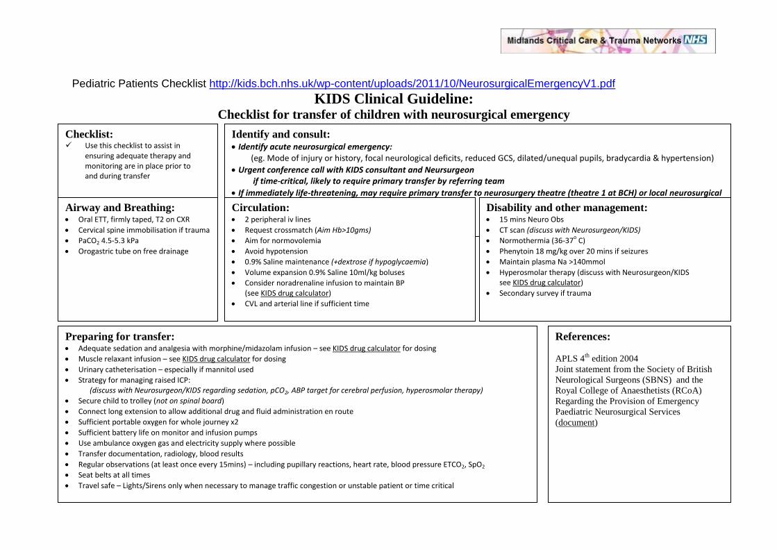
Pediatric Patients Checklist http://kids.bch.nhs.uk/wp-content/uploads/2011/10/NeurosurgicalEmergencyV1.pdf
KIDS Clinical Guideline: Checklist for transfer of children with neurosurgical emergency
Checklist: Use this checklist to assist in
ensuring adequate therapy and monitoring are in place prior to and during transfer
Identify and consult: Identify acute neurosurgical emergency:
(eg. Mode of injury or history, focal neurological deficits, reduced GCS, dilated/unequal pupils, bradycardia & hypertension)
Urgent conference call with KIDS consultant and Neursurgeon if time-critical, likely to require primary transfer by referring team
If immediately life-threatening, may require primary transfer to neurosurgery theatre (theatre 1 at BCH) or local neurosurgical intervention – discuss with neurosurgeon and KIDS consultant Airway and Breathing:
Oral ETT, firmly taped, T2 on CXR
Cervical spine immobilisation if trauma
PaCO2 4.5-5.3 kPa
Orogastric tube on free drainage
Circulation: 2 peripheral iv lines
Request crossmatch (Aim Hb>10gms)
Aim for normovolemia
Avoid hypotension
0.9% Saline maintenance (+dextrose if hypoglycaemia)
Volume expansion 0.9% Saline 10ml/kg boluses
Consider noradrenaline infusion to maintain BP (see KIDS drug calculator)
CVL and arterial line if sufficient time
Disability and other management: 15 mins Neuro Obs
CT scan (discuss with Neurosurgeon/KIDS)
Normothermia (36-37o C)
Phenytoin 18 mg/kg over 20 mins if seizures
Maintain plasma Na >140mmol
Hyperosmolar therapy (discuss with Neurosurgeon/KIDS see KIDS drug calculator)
Secondary survey if trauma
Preparing for transfer: Adequate sedation and analgesia with morphine/midazolam infusion – see KIDS drug calculator for dosing
Muscle relaxant infusion – see KIDS drug calculator for dosing
Urinary catheterisation – especially if mannitol used
Strategy for managing raised ICP: (discuss with Neurosurgeon/KIDS regarding sedation, pCO2, ABP target for cerebral perfusion, hyperosmolar therapy)
Secure child to trolley (not on spinal board)
Connect long extension to allow additional drug and fluid administration en route
Sufficient portable oxygen for whole journey x2
Sufficient battery life on monitor and infusion pumps
Use ambulance oxygen gas and electricity supply where possible
Transfer documentation, radiology, blood results
Regular observations (at least once every 15mins) – including pupillary reactions, heart rate, blood pressure ETCO2, SpO2
Seat belts at all times
Travel safe – Lights/Sirens only when necessary to manage traffic congestion or unstable patient or time critical
References:
APLS 4th
edition 2004
Joint statement from the Society of British
Neurological Surgeons (SBNS) and the
Royal College of Anaesthetists (RCoA)
Regarding the Provision of Emergency
Paediatric Neurosurgical Services
(document)
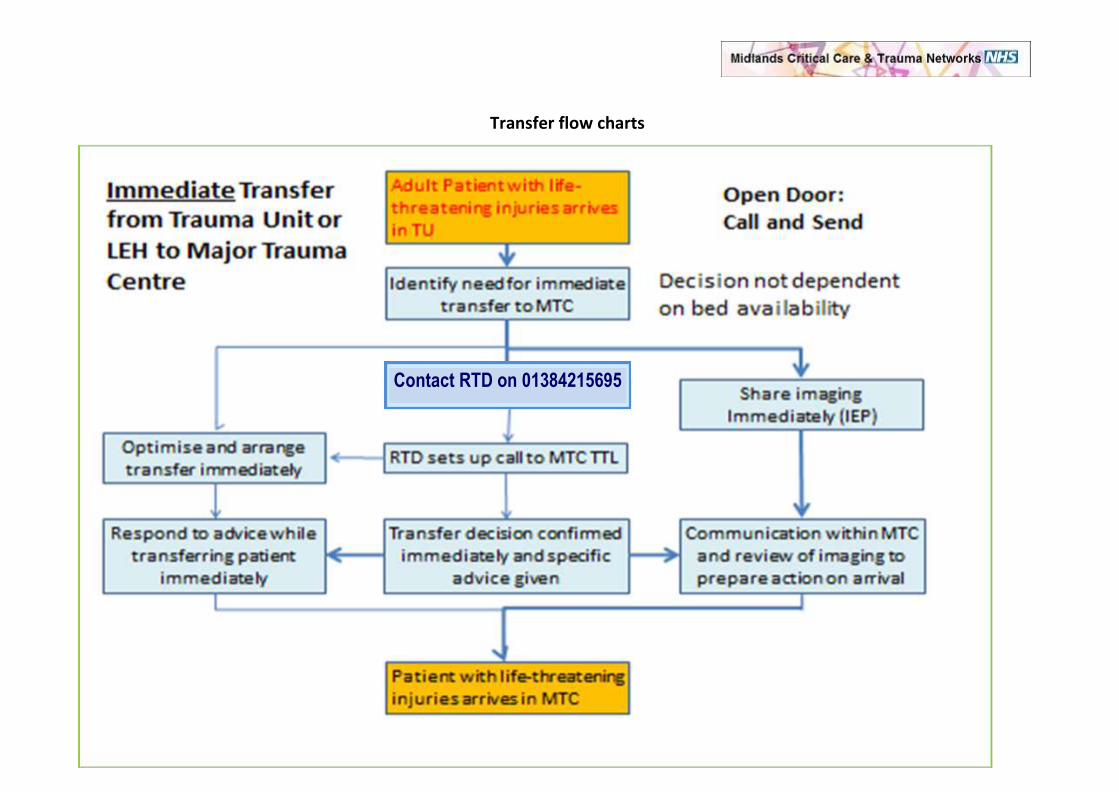
Transfer flow charts
Contact RTD on 01384215695
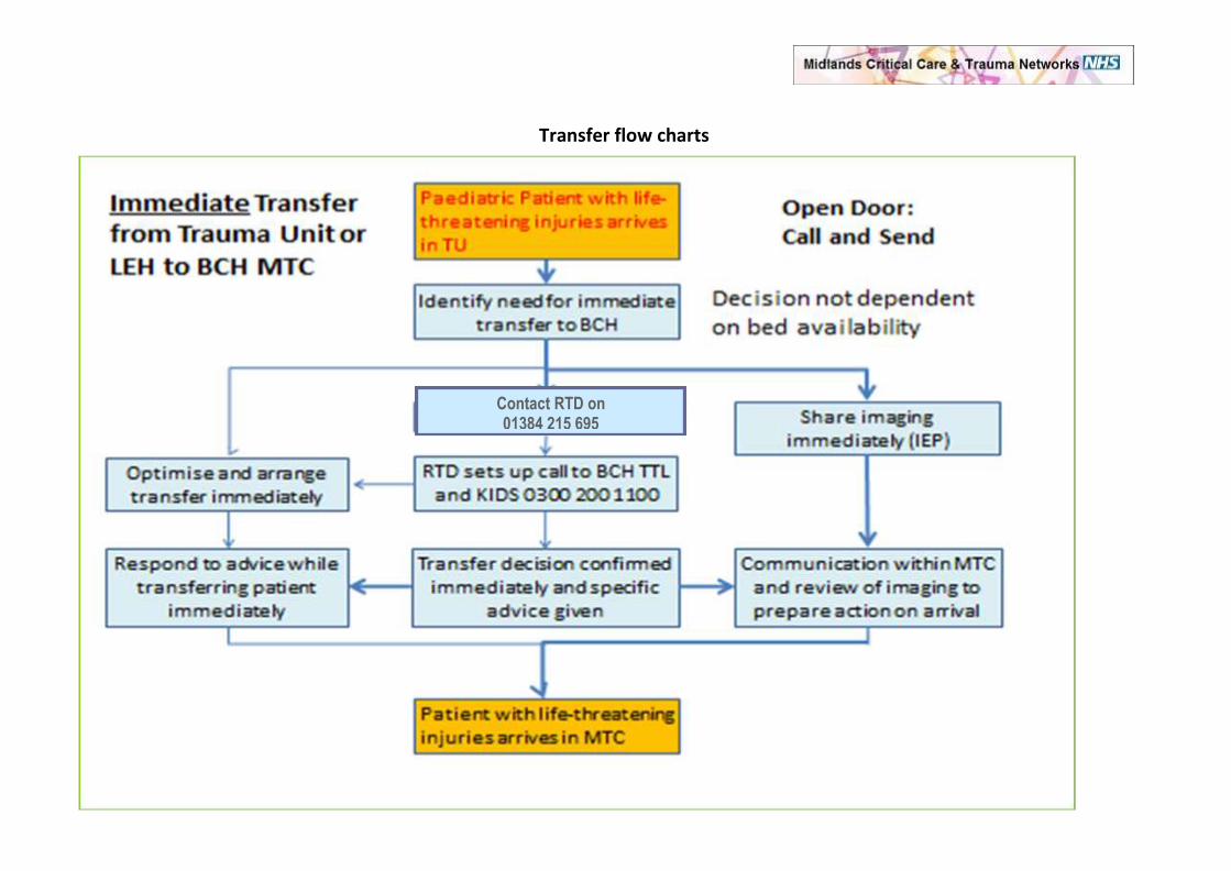
Transfer flow charts
Contact RTD on 01384 215 695
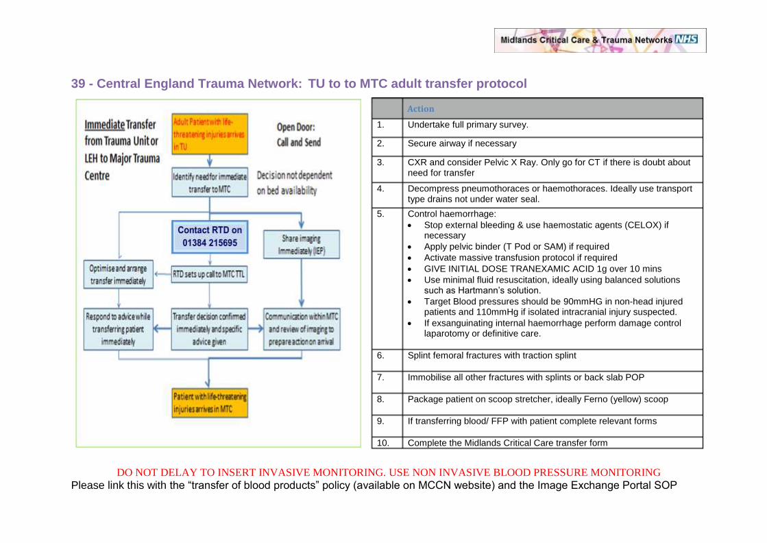
39 - Central England Trauma Network: TU to to MTC adult transfer protocol
DO NOT DELAY TO INSERT INVASIVE MONITORING. USE NON INVASIVE BLOOD PRESSURE MONITORING
Please link this with the “transfer of blood products” policy (available on MCCN website) and the Image Exchange Portal SOP
Action
1. Undertake full primary survey.
2. Secure airway if necessary
3. CXR and consider Pelvic X Ray. Only go for CT if there is doubt about need for transfer
4. Decompress pneumothoraces or haemothoraces. Ideally use transport type drains not under water seal.
5. Control haemorrhage:
Stop external bleeding & use haemostatic agents (CELOX) if necessary
Apply pelvic binder (T Pod or SAM) if required
Activate massive transfusion protocol if required
GIVE INITIAL DOSE TRANEXAMIC ACID 1g over 10 mins
Use minimal fluid resuscitation, ideally using balanced solutions such as Hartmann’s solution.
Target Blood pressures should be 90mmHG in non-head injured patients and 110mmHg if isolated intracranial injury suspected.
If exsanguinating internal haemorrhage perform damage control laparotomy or definitive care.
6. Splint femoral fractures with traction splint
7. Immobilise all other fractures with splints or back slab POP
8. Package patient on scoop stretcher, ideally Ferno (yellow) scoop
9. If transferring blood/ FFP with patient complete relevant forms
10. Complete the Midlands Critical Care transfer form
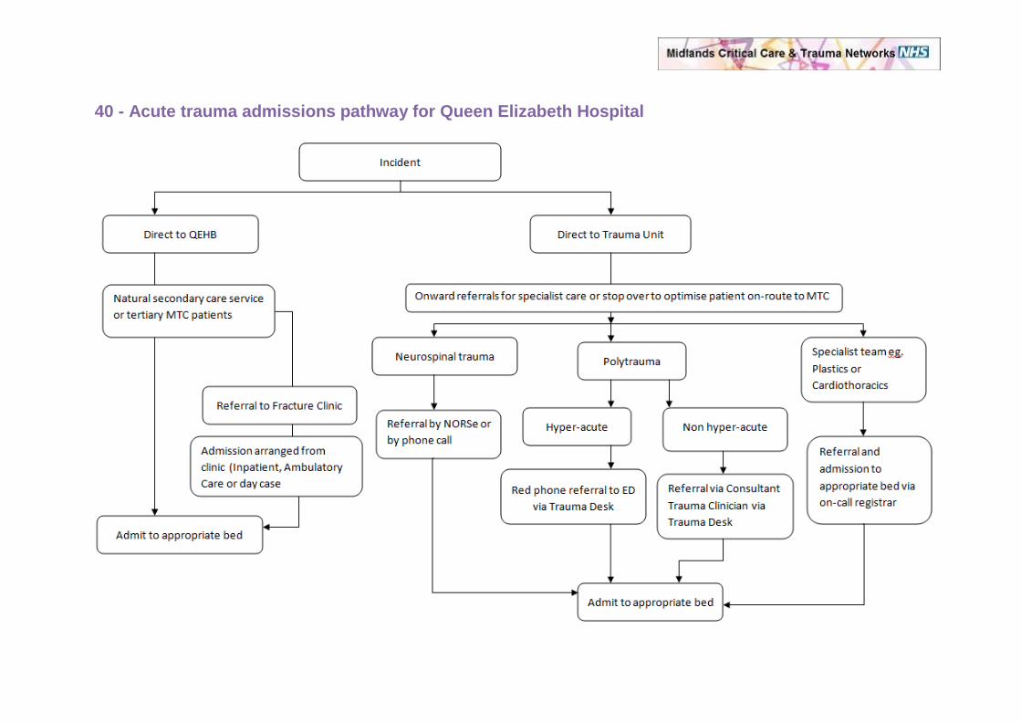
40 - Acute trauma admissions pathway for Queen Elizabeth Hospital
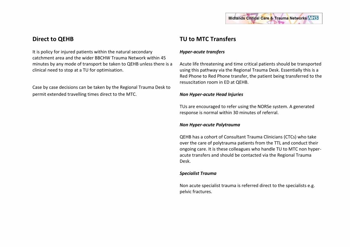
Direct to QEHB
It is policy for injured patients within the natural secondary catchment area and the wider BBCHW Trauma Network within 45 minutes by any mode of transport be taken to QEHB unless there is a clinical need to stop at a TU for optimisation.
Case by case decisions can be taken by the Regional Trauma Desk to
permit extended travelling times direct to the MTC.
TU to MTC Transfers
Hyper-acute transfers Acute life threatening and time critical patients should be transported using this pathway via the Regional Trauma Desk. Essentially this is a Red Phone to Red Phone transfer, the patient being transferred to the resuscitation room in ED at QEHB. Non Hyper-acute Head Injuries TUs are encouraged to refer using the NORSe system. A generated response is normal within 30 minutes of referral. Non Hyper-acute Polytrauma QEHB has a cohort of Consultant Trauma Clinicians (CTCs) who take over the care of polytrauma patients from the TTL and conduct their ongoing care. It is these colleagues who handle TU to MTC non hyper-acute transfers and should be contacted via the Regional Trauma Desk. Specialist Trauma Non acute specialist trauma is referred direct to the specialists e.g. pelvic fractures.
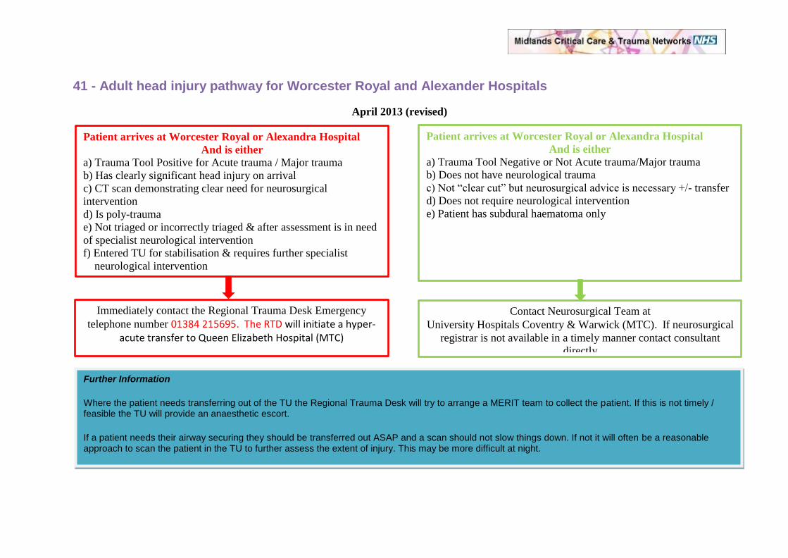
41 - Adult head injury pathway for Worcester Royal and Alexander Hospitals
April 2013 (revised)
Patient arrives at Worcester Royal or Alexandra Hospital
And is either
a) Trauma Tool Positive for Acute trauma / Major trauma
b) Has clearly significant head injury on arrival
c) CT scan demonstrating clear need for neurosurgical
intervention
d) Is poly-trauma
e) Not triaged or incorrectly triaged & after assessment is in need
of specialist neurological intervention
f) Entered TU for stabilisation & requires further specialist
neurological intervention
Patient arrives at Worcester Royal or Alexandra Hospital
And is either a) Trauma Tool Negative or Not Acute trauma/Major trauma
b) Does not have neurological trauma
c) Not “clear cut” but neurosurgical advice is necessary +/- transfer
d) Does not require neurological intervention
e) Patient has subdural haematoma only
Immediately contact the Regional Trauma Desk Emergency
telephone number 01384 215695. The RTD will initiate a hyper-acute transfer to Queen Elizabeth Hospital (MTC)
Contact Neurosurgical Team at
University Hospitals Coventry & Warwick (MTC). If neurosurgical
registrar is not available in a timely manner contact consultant
directly
Further Information
Where the patient needs transferring out of the TU the Regional Trauma Desk will try to arrange a MERIT team to collect the patient. If this is not timely / feasible the TU will provide an anaesthetic escort.
If a patient needs their airway securing they should be transferred out ASAP and a scan should not slow things down. If not it will often be a reasonable approach to scan the patient in the TU to further assess the extent of injury. This may be more difficult at night.
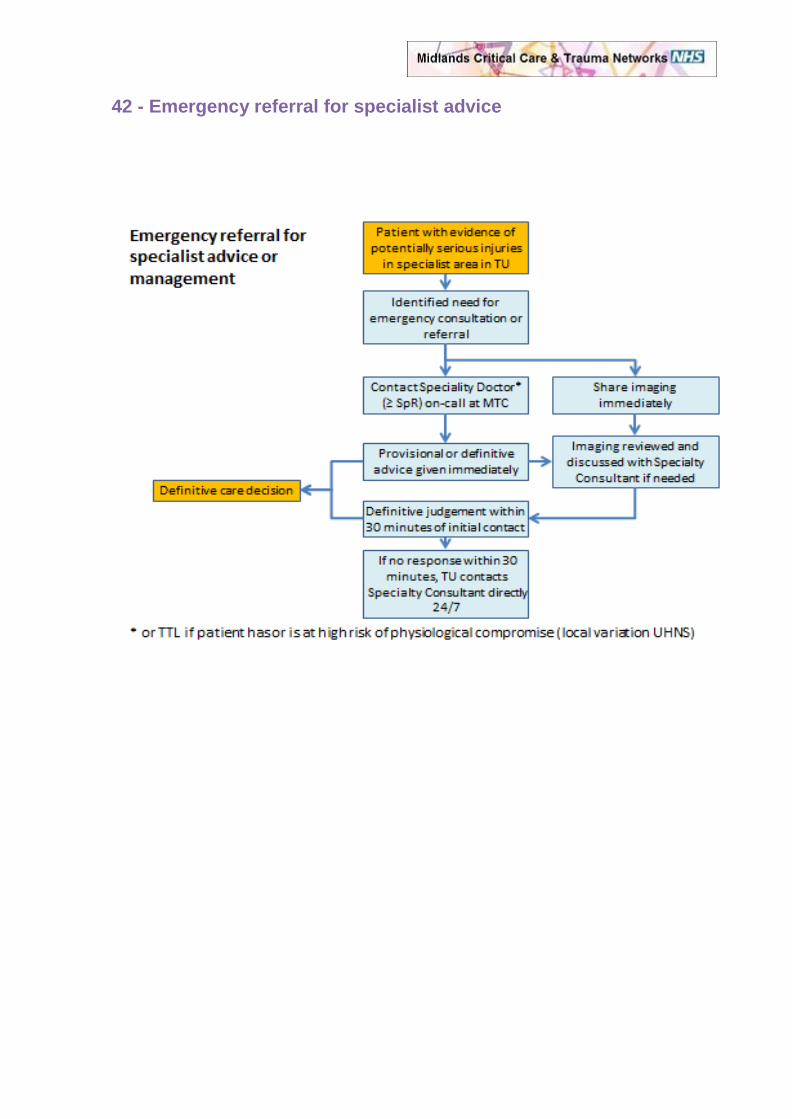
42 - Emergency referral for specialist advice
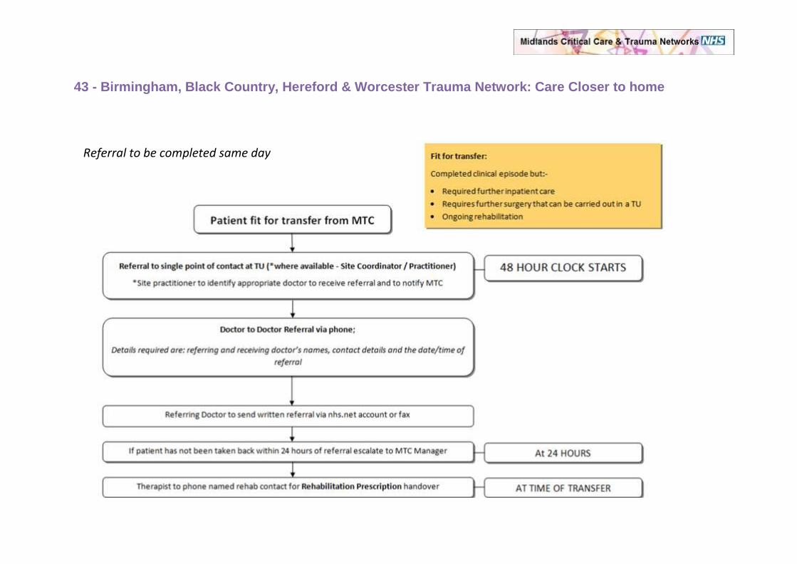
43 - Birmingham, Black Country, Hereford & Worcester Trauma Network: Care Closer to home
Referral to be completed same day
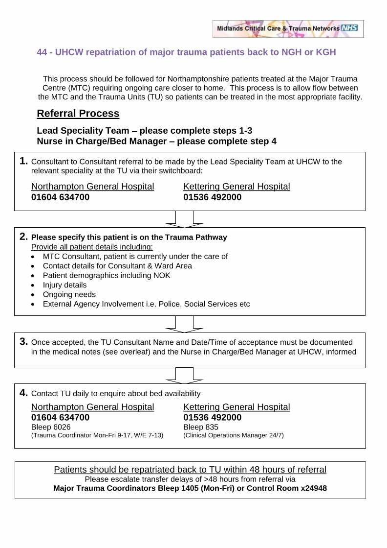
44 - UHCW repatriation of major trauma patients back to NGH or KGH
This process should be followed for Northamptonshire patients treated at the Major Trauma Centre (MTC) requiring ongoing care closer to home. This process is to allow flow between
the MTC and the Trauma Units (TU) so patients can be treated in the most appropriate facility.
Referral Process
Lead Speciality Team – please complete steps 1-3 Nurse in Charge/Bed Manager – please complete step 4
Care Closer to Home Record – Checklist for transfer
1. Consultant to Consultant referral to be made by the Lead Speciality Team at UHCW to the relevant speciality at the TU via their switchboard:
Northampton General Hospital Kettering General Hospital 01604 634700 01536 492000
2. Please specify this patient is on the Trauma Pathway
Provide all patient details including:
MTC Consultant, patient is currently under the care of
Contact details for Consultant & Ward Area
Patient demographics including NOK
Injury details
Ongoing needs
External Agency Involvement i.e. Police, Social Services etc
3. Once accepted, the TU Consultant Name and Date/Time of acceptance must be documented
in the medical notes (see overleaf) and the Nurse in Charge/Bed Manager at UHCW, informed
4. Contact TU daily to enquire about bed availability
Northampton General Hospital Kettering General Hospital 01604 634700 01536 492000
Bleep 6026 Bleep 835 (Trauma Coordinator Mon-Fri 9-17, W/E 7-13) (Clinical Operations Manager 24/7)
Patients should be repatriated back to TU within 48 hours of referral Please escalate transfer delays of >48 hours from referral via
Major Trauma Coordinators Bleep 1405 (Mon-Fri) or Control Room x24948
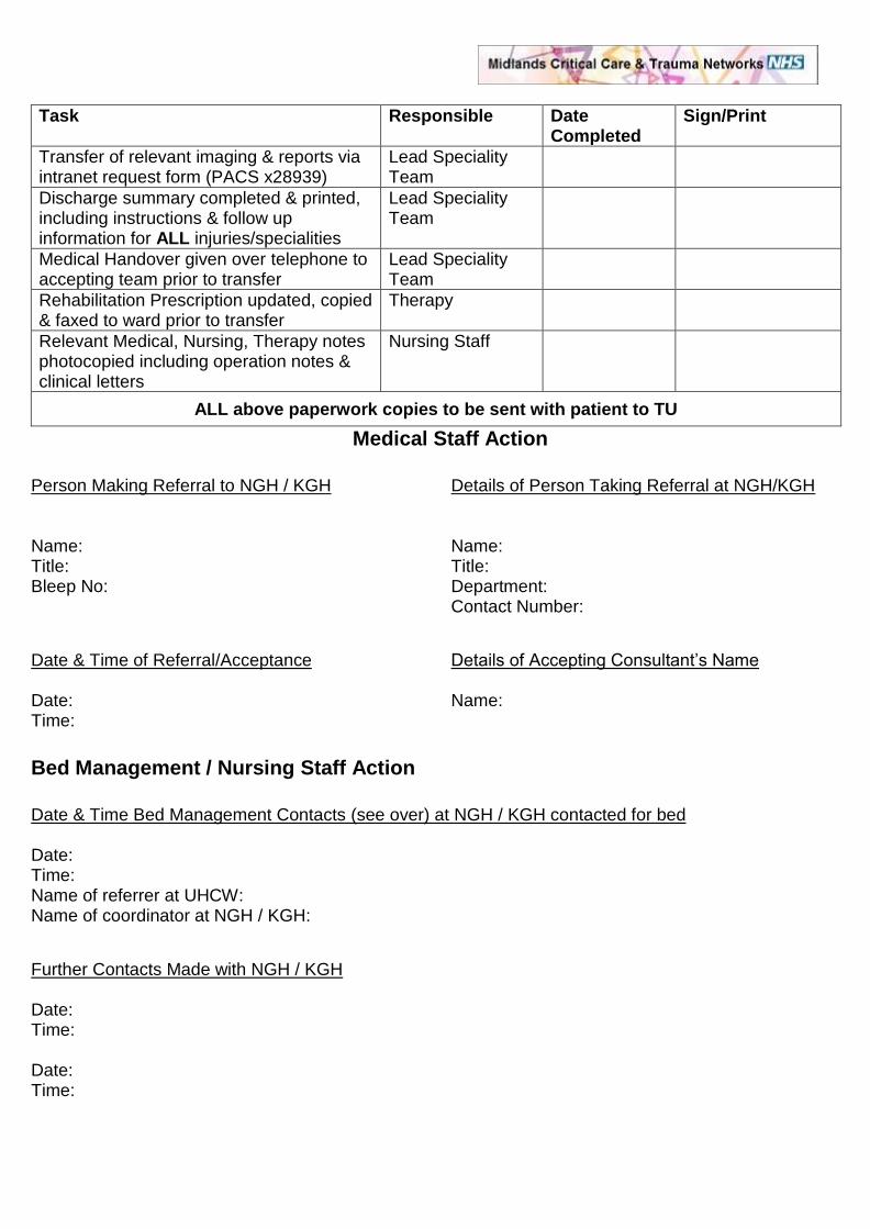
Task Responsible Date Completed
Sign/Print
Transfer of relevant imaging & reports via intranet request form (PACS x28939)
Lead Speciality Team
Discharge summary completed & printed, including instructions & follow up information for ALL injuries/specialities
Lead Speciality Team
Medical Handover given over telephone to accepting team prior to transfer
Lead Speciality Team
Rehabilitation Prescription updated, copied & faxed to ward prior to transfer
Therapy
Relevant Medical, Nursing, Therapy notes photocopied including operation notes & clinical letters
Nursing Staff
ALL above paperwork copies to be sent with patient to TU
Medical Staff Action
Person Making Referral to NGH / KGH Details of Person Taking Referral at NGH/KGH Name: Name: Title: Title: Bleep No: Department: Contact Number:
Date & Time of Referral/Acceptance Details of Accepting Consultant’s Name Date: Name: Time:
Bed Management / Nursing Staff Action
Date & Time Bed Management Contacts (see over) at NGH / KGH contacted for bed Date: Time: Name of referrer at UHCW: Name of coordinator at NGH / KGH:
Further Contacts Made with NGH / KGH Date: Time: Date: Time:
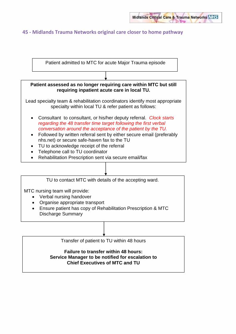
45 - Midlands Trauma Networks original care closer to home pathway
Patient admitted to MTC for acute Major Trauma episode
Patient assessed as no longer requiring care within MTC but still requiring inpatient acute care in local TU.
Lead specialty team & rehabilitation coordinators identify most appropriate
specialty within local TU & refer patient as follows:
Consultant to consultant, or his/her deputy referral. Clock starts regarding the 48 transfer time target following the first verbal conversation around the acceptance of the patient by the TU.
Followed by written referral sent by either secure email (preferably nhs.net) or secure safe-haven fax to the TU
TU to acknowledge receipt of the referral
Telephone call to TU coordinator
Rehabilitation Prescription sent via secure email/fax
TU to contact MTC with details of the accepting ward. MTC nursing team will provide:
Verbal nursing handover
Organise appropriate transport
Ensure patient has copy of Rehabilitation Prescription & MTC Discharge Summary
Transfer of patient to TU within 48 hours
Failure to transfer within 48 hours: Service Manager to be notified for escalation to
Chief Executives of MTC and TU
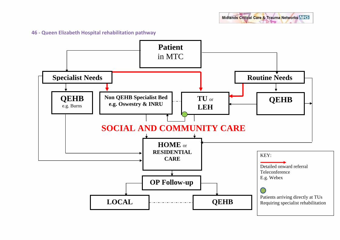
HOME or RESIDENTIAL
CARE
OP Follow-up
LOCAL QEHB
Patient
in MTC
46 - Queen Elizabeth Hospital rehabilitation pathway
QEHB e.g. Burns
Non QEHB Specialist Bed
e.g. Oswestry & INRU TU or
LEH QEHB
SOCIAL AND COMMUNITY CARE
Routine Needs
KEY:
Detailed onward referral
Teleconference
E.g. Webex
Patients arriving directly at TUs
Requiring specialist rehabilitation
Specialist Needs
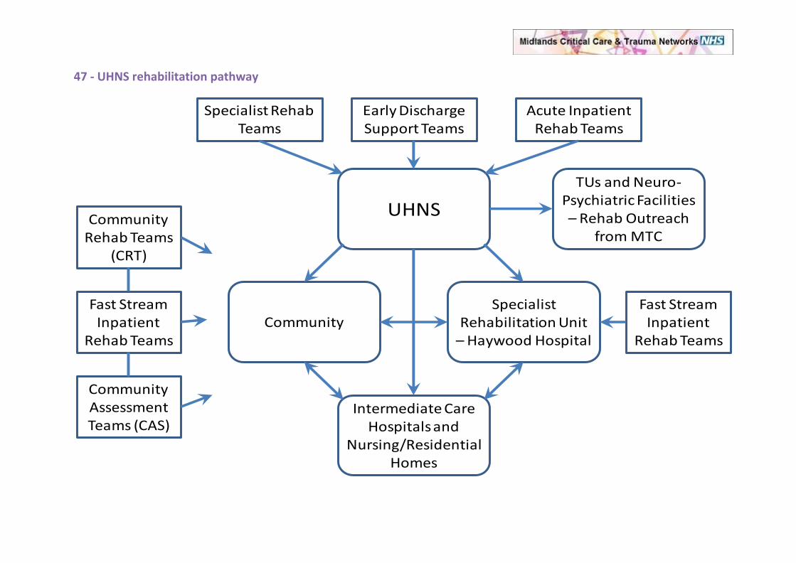
47 - UHNS rehabilitation pathway
Early Discharge Support Teams
TUs and Neuro-Psychiatric Facilities – Rehab Outreach
from MTC
UHNS
CommunitySpecialist
Rehabilitation Unit – Haywood Hospital
Intermediate Care Hospitals and
Nursing/Residential Homes
Specialist Rehab Teams
Acute Inpatient Rehab Teams
Fast Stream Inpatient
Rehab Teams
Fast Stream Inpatient
Rehab Teams
Community Rehab Teams
(CRT)
Community Assessment Teams (CAS)
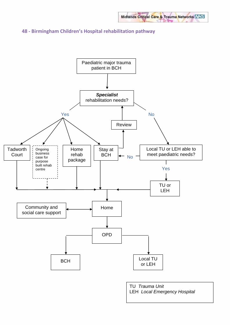
48 - Birmingham Children’s Hospital rehabilitation pathway
Paediatric major trauma patient in BCH
Specialist rehabilitation needs?
Tadworth Court
Stay at BCH
TU or LEH
Home
OPD
BCH Local TU or LEH
Local TU or LEH able to meet paediatric needs?
Yes No
Yes
No
Review
Community and social care support
TU Trauma Unit LEH Local Emergency Hospital
Home rehab
package
Ongoing business case for purpose built rehab centre
49 - Rehabilitation assessment and outcomes
The Rehabilitation Prescription provides a framework for the comprehensive rehabilitation assessment of all patients with major trauma and will ensure a minimum standard is maintained. The content of the Rehabilitation Prescription is described in Protocol 38. The information collected at discharge from the Major Trauma Centres and Trauma Units as part of the Rehabilitation Prescription should be aligned as closely as possible to the UKROC (UK Rehabilitation Outcomes Collaborative) national dataset. UKROC is the recognised national rehabilitation database and is to be linked to the TARN database. Data flow between these two national databases will ultimately enable tracking of patients as they move from acute care services through in-patient rehabilitation, to the community. The following measures should be considered as mandatory for inclusion:
Barthel Index (Appendix 1)– validated measure of functional status / activities of daily living, recognised limitations due to ‘floor’ and ‘ceiling’ effects, but in widespread use and included for its simplicity and reproducibility
Glasgow Outcome Scale- Extended (GOSe) (for traumatic brain injury only, at specific time points)
RCS (Rehabilitation Complexity Scale), part of the UKROC dataset, clinimetric analysis, discriminates between specialist level 1 and 2 services.
The following measures are desirable for inclusion and should be mandatory as part of specialist inpatient rehabilitation
FIM/FAM (Functional Independence Measure, Functional Assessment Measure) - validated comprehensive functional scoring systems, the most widely used internationally. FAM includes cognitive and psychosocial items.
Goal Attainment Scaling (GAS) – person-centred measure of the achievement of rehabilitation goals
Record of goal achievement
For patients with spinal cord injury, the ASIA (American Spinal Injury Association) and SCIM (Spinal Cord Independence Measure) are recommended in addition. It is essential to note that outcome measurement is a dynamic process and is time-point dependent. Thus individual scores will change over time and should be recorded at key stages in the patient journey, such as discharge from the Major Trauma Centre or discharge from the specialist rehabilitation unit. In addition, true ‘outcome’ of the rehabilitation process cannot reliably be determined within the acute stage of care following major trauma. Long term outcomes, such as return to work, independent living, ability to drive etc., more comprehensively and meaningfully reflect the impact of rehabilitation, but may take lengthy periods to reach a steady state. As the Rehabilitation subset of the TARN database is developed, it is anticipated that there will be modifications to the exact outcome measures recorded, which will be agreed across the region as well as nationally and the recommendations of this protocol updated accordingly. *Note that UKROC is currently focussed upon neurological rehabilitation and future modification to include musculoskeletal measures is required, as suggested.
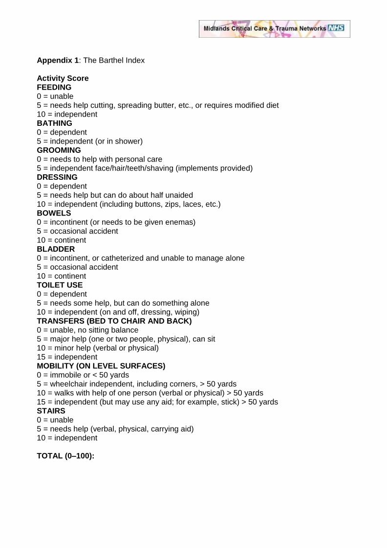
Appendix 1: The Barthel Index Activity Score FEEDING 0 = unable 5 = needs help cutting, spreading butter, etc., or requires modified diet 10 = independent BATHING 0 = dependent 5 = independent (or in shower) GROOMING 0 = needs to help with personal care 5 = independent face/hair/teeth/shaving (implements provided) DRESSING 0 = dependent 5 = needs help but can do about half unaided 10 = independent (including buttons, zips, laces, etc.) BOWELS 0 = incontinent (or needs to be given enemas) 5 = occasional accident 10 = continent BLADDER 0 = incontinent, or catheterized and unable to manage alone 5 = occasional accident 10 = continent TOILET USE 0 = dependent 5 = needs some help, but can do something alone 10 = independent (on and off, dressing, wiping) TRANSFERS (BED TO CHAIR AND BACK) 0 = unable, no sitting balance 5 = major help (one or two people, physical), can sit 10 = minor help (verbal or physical) 15 = independent MOBILITY (ON LEVEL SURFACES) 0 = immobile or < 50 yards 5 = wheelchair independent, including corners, > 50 yards 10 = walks with help of one person (verbal or physical) > 50 yards 15 = independent (but may use any aid; for example, stick) > 50 yards STAIRS 0 = unable 5 = needs help (verbal, physical, carrying aid) 10 = independent TOTAL (0–100):
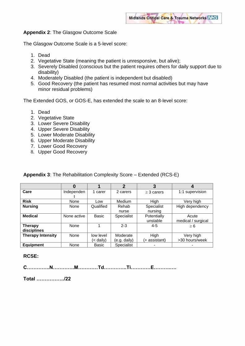
Appendix 2: The Glasgow Outcome Scale The Glasgow Outcome Scale is a 5-level score:
1. Dead 2. Vegetative State (meaning the patient is unresponsive, but alive); 3. Severely Disabled (conscious but the patient requires others for daily support due to
disability) 4. Moderately Disabled (the patient is independent but disabled) 5. Good Recovery (the patient has resumed most normal activities but may have
minor residual problems)
The Extended GOS, or GOS-E, has extended the scale to an 8-level score:
1. Dead 2. Vegetative State 3. Lower Severe Disability 4. Upper Severe Disability 5. Lower Moderate Disability 6. Upper Moderate Disability 7. Lower Good Recovery 8. Upper Good Recovery
Appendix 3: The Rehabilitation Complexity Score – Extended (RCS-E)
0 1 2 3 4 Care Independen
t 1 carer 2 carers 3 carers 1:1 supervision
Risk None Low Medium High Very high
Nursing None Qualified Rehab nurse
Specialist nursing
High dependency
Medical None active Basic Specialist Potentially unstable
Acute medical / surgical
Therapy disciplines
None 1 2-3 4-5 6
Therapy Intensity None low level (< daily)
Moderate (e.g. daily)
High (+ assistant)
Very high >30 hours/week
Equipment None Basic Specialist - -
RCSE: C…………..N………….M…………Td…………..Ti…………E……….…. Total ……………../22
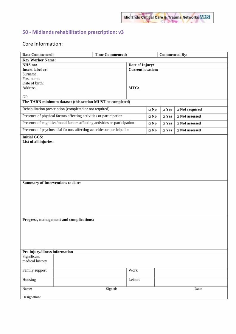
50 - Midlands rehabilitation prescription: v3
Core Information:
Date Commenced: Time Commenced: Commenced By:
Key Worker Name:
NHS no: Date of Injury:
Insert label or:
Surname:
First name:
Date of birth:
Address:
GP:
Current location:
MTC:
The TARN minimum dataset (this section MUST be completed)
Rehabilitation prescription (completed or not required) □ No □ Yes □ Not required
Presence of physical factors affecting activities or participation □ No □ Yes □ Not assessed
Presence of cognitive/mood factors affecting activities or participation □ No □ Yes □ Not assessed
Presence of psychosocial factors affecting activities or participation □ No □ Yes □ Not assessed
Initial GCS:
List of all injuries:
Summary of Interventions to date:
Progress, management and complications:
Pre-injury/illness information
Significant
medical history
Family support
Work
Housing
Leisure
Name: Signed: Date:
Designation:
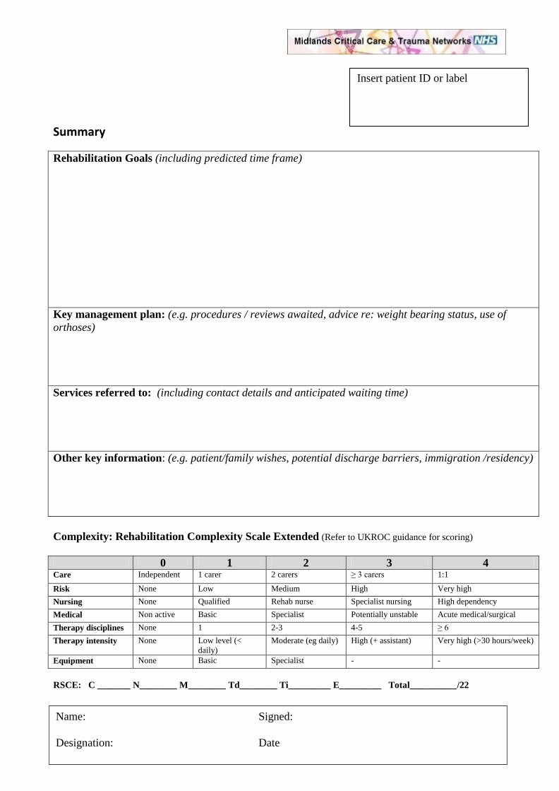
Summary
Rehabilitation Goals (including predicted time frame)
Key management plan: (e.g. procedures / reviews awaited, advice re: weight bearing status, use of
orthoses)
Services referred to: (including contact details and anticipated waiting time)
Other key information: (e.g. patient/family wishes, potential discharge barriers, immigration /residency)
Complexity: Rehabilitation Complexity Scale Extended (Refer to UKROC guidance for scoring)
0 1 2 3 4
Care Independent 1 carer 2 carers ≥ 3 carers 1:1
Risk None Low Medium High Very high
Nursing None Qualified Rehab nurse Specialist nursing High dependency
Medical Non active Basic Specialist Potentially unstable Acute medical/surgical
Therapy disciplines None 1 2-3 4-5 ≥ 6
Therapy intensity None Low level (<
daily)
Moderate (eg daily) High (+ assistant) Very high (>30 hours/week)
Equipment None Basic Specialist - -
RSCE: C _______ N________ M________ Td________ Ti_________ E_________ Total__________/22
Insert patient ID or label
Name: Signed:
Designation: Date
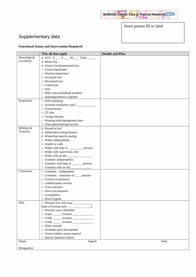
Supplementary data Functional Status and Intervention Required:
Tick all that apply Details and Plan Neurological/
Locomotor □ GCS: E____ V____ M____ Total ______
□ Motor loss
□ Sensory loss/hypersensitivity
□ Visual impairment
□ Hearing impairment
□ Increased tone
□ Decreased tone
□ Contracture
□ Pain
□ Other musculoskeletal problem
□ Splinting/orthotics required
Respiratory □ Self ventilating
□ Assisted ventilation: type? _____________
□ Tracheostomy
□ ET tube
□ Oxygen therapy
□ Weaning plan/management plan
□ Chest physiotherapy/suction
Mobility &
Transfers □ Nursed in bed
□ Independent sitting balance
□ Wheelchair/special seating
□ Walks independently
□ Unable to walk
□ Walks with help of __________ persons
□ Walks with supervision only
□ Walks with an aid _________________
□ Transfers independently
□ Transfers with help of _______ persons
□ Transfers with an aid ______________
Continence □ Continent – independent
□ Continent – assistance of ____ persons
□ Urinary incontinence
□ Catheter/pads/conveen
□ Urine retention
□ Faecal incontinence
□ Constipation
□ Bowel regime
Skin □ Pressure sore risk score _____________
(type of scoring used ________________)
□ Pressure sore/s identified
□ Grade ______ location ______________
□ Grade ______ location ______________
□ Grade ______ location ______________
□ Other wounds
□ Treatment plan documented
□ Tissue viability nurse required
□ Special mattress/cushion
Name: Signed: Date:
Designation:
Insert patient ID or label
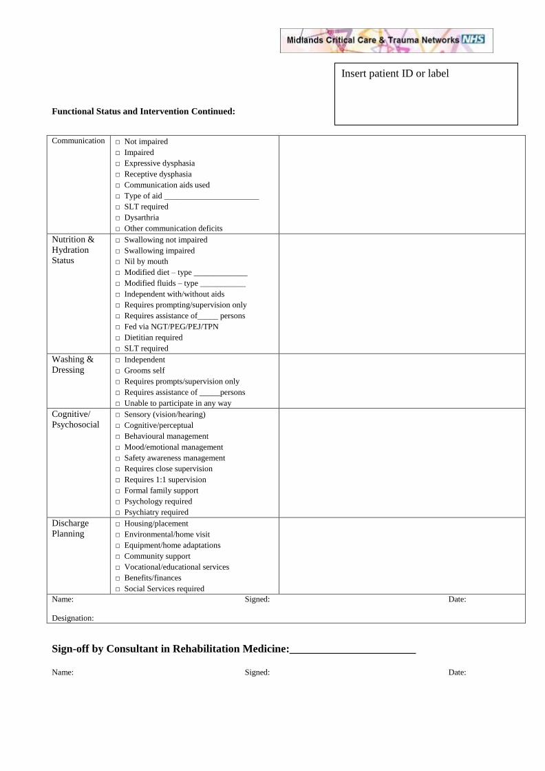
Functional Status and Intervention Continued:
Communication □ Not impaired
□ Impaired
□ Expressive dysphasia
□ Receptive dysphasia
□ Communication aids used
□ Type of aid _______________________
□ SLT required
□ Dysarthria
□ Other communication deficits
Nutrition &
Hydration
Status
□ Swallowing not impaired
□ Swallowing impaired
□ Nil by mouth
□ Modified diet – type _____________
□ Modified fluids – type ___________
□ Independent with/without aids
□ Requires prompting/supervision only
□ Requires assistance of_____ persons
□ Fed via NGT/PEG/PEJ/TPN
□ Dietitian required
□ SLT required
Washing &
Dressing
□ Independent
□ Grooms self
□ Requires prompts/supervision only
□ Requires assistance of _____persons
□ Unable to participate in any way
Cognitive/
Psychosocial
□ Sensory (vision/hearing)
□ Cognitive/perceptual
□ Behavioural management
□ Mood/emotional management
□ Safety awareness management
□ Requires close supervision
□ Requires 1:1 supervision
□ Formal family support
□ Psychology required
□ Psychiatry required
Discharge
Planning
□ Housing/placement
□ Environmental/home visit
□ Equipment/home adaptations
□ Community support
□ Vocational/educational services
□ Benefits/finances
□ Social Services required
Name: Signed: Date:
Designation:
Sign-off by Consultant in Rehabilitation Medicine:_______________________
Name: Signed: Date:
Insert patient ID or label
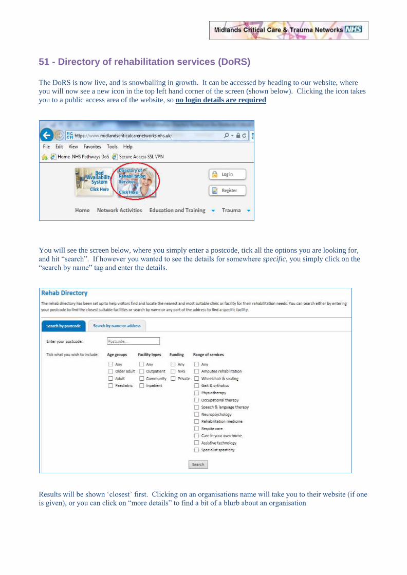
51 - Directory of rehabilitation services (DoRS) The DoRS is now live, and is snowballing in growth. It can be accessed by heading to our website, where
you will now see a new icon in the top left hand corner of the screen (shown below). Clicking the icon takes
you to a public access area of the website, so no login details are required
You will see the screen below, where you simply enter a postcode, tick all the options you are looking for,
and hit “search”. If however you wanted to see the details for somewhere specific, you simply click on the
“search by name” tag and enter the details.
Results will be shown ‘closest’ first. Clicking on an organisations name will take you to their website (if one
is given), or you can click on “more details” to find a bit of a blurb about an organisation

Clicking on the circled link above for example will bring up the following screen:
You can also access the organisations website, or email them, from this screen
Organisations are being encouraged to notify the network office of any ommissions they note, so that this
directory can be of real benefit to all
52 - ICU protocol for escalation and capacity management
1. INTRODUCTION
2. “NO NOTICE” INCIDENTS
3. CAPACITY MANAGEMENT, COMMAND CONTROL & COORDINATION
4. CRITICAL CARE ESCALATION PLAN
1.0 INTRODUCTION This critical care escalation plan has been produced by members of the Midlands Critical Care Network (MCCN) to support rapid escalation of critical care capacity in the face of a sudden requirement for critical care in a number of organisations with minimal to no notice of the event. It is accepted that sudden, significant increases in capacity can only be maintained for short periods of time and will, despite best efforts, inevitably lead to transient decreases in quality of overall care.
For most potential threats critical care capacity might need to be increased in parallel with other acute
services (i.e. conventional major incident, terrorist blast or ballistic incident). However there are some
circumstances where increases in critical care capacity may be disproportionate to other acute areas (i.e.
biological, chemical or radiological incident).
Critical care is a finite resource. Historically most West Midlands critical care units have run at high
occupancy rates which makes rapid expansion difficult to achieve. Therefore coordination of rapid
expansion is of paramount importance to ensure that not only does expansion occur but that patient flows
are managed to prevent (where possible) individual units being overwhelmed.
2.0 “No Notice” events
“No notice” events are significant health care threats occurring with no (or minimal) warning. They can
result in potentially large numbers of patients with significant injuries rapidly presenting to acute Trusts
allowing minimal time to establish a clinical response.
In conventional major incidents (i.e. plane, train or road accidents) there is usually some warning of
casualty numbers and severity from the major incident controller to allow initiation of escalation plans
before patients arrive at acute Trusts.
“No notice” incidents are frequently terrorist related. If this is the case some patients may leave the scene
and seek medical assistance before effective police cordons have been put in place – the first a Trust may
know of the incident is when injured patients arrive in their Emergency Department (ED). Once a police
cordon is in place patients will not leave the incident until the scene is safe. However from this point
patients may leave in large numbers having received only basic first aid interventions.
Critical care will clearly need to expand to manage these injured patients. Many Trusts have critical care
expansion plans that rely on utilising equipment and staff from operating theatres. It is imperative that
critical care expansion does not jeopardise ED expansion of theatre capacity. Potentially large numbers of
patients will require ED resuscitation followed by immediate life-saving surgery and critical care. Expansion
of all three clinical areas needs to be balanced to prevent bottle necks occurring and maximise patient
flows. Some Trusts have chosen to manage critical care and theatres as a single entity to help manage
patient flow.
3.0 CAPACITY MANAGEMENT, COMMAND CONTROL & COORDINATION The SHA have an integrated plan for managing major incidents. Dynamic capacity information is essential to effectively manage a rapidly changing significant incident. The following describes how critical care capacity data will be gathered, circulated and reported around the health system. 3.1 Capacity Management and Data Sources Critical care capacity is monitored using the MCCN capacity website. Each unit submits daily capacity data
which is supplemented by amendments whenever there is a capacity change.
During a major incident the same system will be used but individual units will be asked to submit data
more frequently. While it is recognised that this is a slightly onerous task it should be emphasised that
this data is vital to allow effective management of patients.
Critical care capacity is monitored by the MCCN. In the event of a sudden requirement for additional critical
care capacity First Response would be responsible for contacting each unit by phone to ask individual units
to:
1. Immediately update their capacity data 2. Update this every time their capacity changes (i.e. patients are admitted / discharged or extra beds
are opened)
Units will be notified as soon as frequent data updates are no longer required. 3.2 Medical coordination of critical care capacity Previously the MCCN devised a system of consultant critical care advisors to manage capacity issues associated with pandemic influenza. Like many of the threats requiring rapid capacity increase pandemic influenza placed a disproportionate burden on critical care services and it seems logical that a similar system of medically advised capacity management would be beneficial. A central, coordinated response would allow efficient use of the finite critical care resource and limit the chance individual units being unable to cope. The SHA have approached selected trusts who have recommended clinicians who may be able to assist in this role. These individuals have been contacted by the SHA.
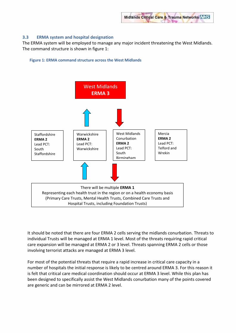
3.3 ERMA system and hospital designation The ERMA system will be employed to manage any major incident threatening the West Midlands. The command structure is shown in figure 1:
Figure 1: ERMA command structure across the West Midlands
There will be multiple ERMA 1 Representing each health trust in the region or on a health economy basis
(Primary Care Trusts, Mental Health Trusts, Combined Care Trusts and Hospital Trusts, including Foundation Trusts)
Staffordshire ERMA 2 Lead PCT: South Staffordshire
Warwickshire ERMA 2 Lead PCT: Warwickshire
West Midlands Conurbation ERMA 2 Lead PCT: South Birmingham
Mercia ERMA 2 Lead PCT: Telford and Wrekin
West Midlands ERMA 3
It should be noted that there are four ERMA 2 cells serving the midlands conurbation. Threats to individual Trusts will be managed at ERMA 1 level. Most of the threats requiring rapid critical care expansion will be managed at ERMA 2 or 3 level. Threats spanning ERMA 2 cells or those involving terrorist attacks are managed at ERMA 3 level. For most of the potential threats that require a rapid increase in critical care capacity in a number of hospitals the initial response is likely to be centred around ERMA 3. For this reason it is felt that critical care medical coordination should occur at ERMA 3 level. While this plan has been designed to specifically assist the West Midlands conurbation many of the points covered are generic and can be mirrored at ERMA 2 level.
For most “no notice” events a number of hospitals are immediately designated as receiving hospitals – they will receive the patients with significant injuries (P1 and P2). A zone of hospitals around these receiving hospitals will be designated a supporting role. This would primarily involve management of less injured patients (P3). However supporting hospitals may be required to activate their critical care expansion plans if the patient load on the receiving hospitals is excessive. In this situation supporting hospitals may be required to retrieve patients requiring critical care from receiving hospitals.
4.0 CRITICAL CARE ESCALATION PLAN
The proposed principles of a West Midlands conurbation approach are: a) An Integrated model. b) Rapid coordinated increases in capacity. c) Preservation of the ‘standard’ clinical pathway for critically ill patients if possible. d) Preservation of emergency, general and specialist services if possible. e) Paediatric patients will be admitted to PICU’s if possible, using the national PICU bed stock as a
resource.
On receiving a potential threat requiring increased critical care capacity the ERMA 3 commander will initiate the following plan:
1. Designate receiving and supporting hospitals 2. Designated hospitals will activate their major incident plans and manage patients according to
these plans 3. First Response will be contact all critical care units asking them to immediately update their
capacity and to ensure any changes are immediately recorded for the duration of the incident 4. If individual critical care units have capacity or clinical concerns these should be immediately
communicated to the hospital commander who will then liaise with the ERMA 3 commander 5. If required the major incident commander will contact a consultant critical care advisor. It is
likely that this will not require the consultant to relocate to the major incident command centre. If, however, the consultant is required to be physically present at ERMA control or will need to give significant time to coordinating the critical care response they will contact a consultant colleague to take over their trust clinical duties.
a. The consultant wills assess current capacity using the MCCN web dashboard. b. The consultant will liaise with the ERMA 3 commander to gauge the potential
requirement for critical care c. If this can be managed with current resources then the consultant will advise where
patients can be sent to effectively and evenly manage patient flows. d. If the incident overwhelms the current critical care capacity the consultant will advise
the ERMA 3 commander to activate enhanced capacity in selected supporting units e. Individual units will be contacted and informed of the requirement to increase capacity.
Increases in capacity will be recorded real-time using the MCCN dashboard. f. The consultant will continue to coordinate patient flows utilising real-time enhanced
capacity and advise the ERMA 3 commander regarding regional transfers to prevent individual units becoming overstretched
g. If required the consultant will contact individual units as required to verify capacity and clinical capability to manage patients
h. As the requirement for critical care plateaus the coordinating consultant will be responsible for ensuring effective patient flows to try and ensure that individual units do not shoulder a disproportionate clinical burden. It is likely that the MCCN will assist in this role.
i. As the incident decelerates the coordinating consultant will continue to support the ERMA 3 commander until the commander feels that specialist critical care advice is no longer needed
j. For incidents of extended duration the coordinating consultant will be need to be relieved on a regular (12 hourly) basis.
k. For incidents requiring activation of adjacent ERMA 3 cells the coordinating consultant may liaise directly with their opposite number to ensure a coordinated regional response
l. If required the consultant will contact the MCCN Network Manager for additional support.
6. If receiving hospital critical care patient load is excessive then stable patients may be transferred within or outside the region as soon as appropriate transport is available
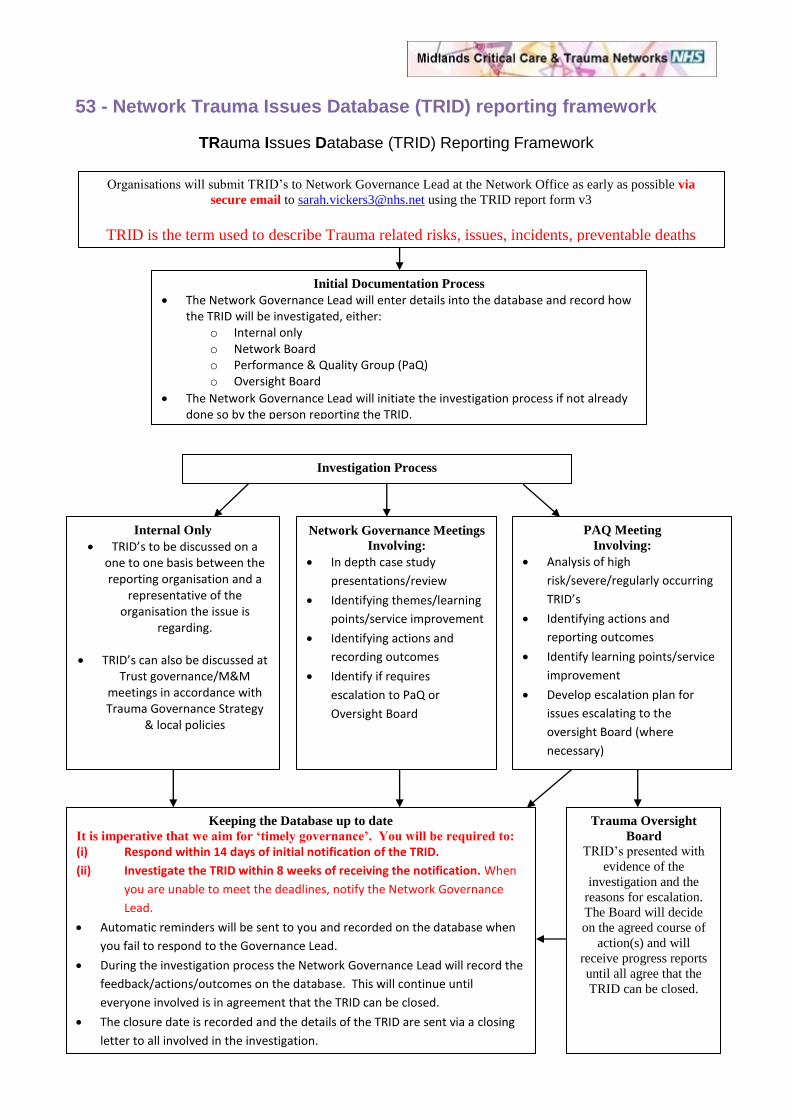
53 - Network Trauma Issues Database (TRID) reporting framework
TRauma Issues Database (TRID) Reporting Framework
Initial Documentation Process
The Network Governance Lead will enter details into the database and record how the TRID will be investigated, either:
o Internal only o Network Board o Performance & Quality Group (PaQ) o Oversight Board
The Network Governance Lead will initiate the investigation process if not already done so by the person reporting the TRID.
Organisations will submit TRID’s to Network Governance Lead at the Network Office as early as possible via
secure email to [email protected] using the TRID report form v3
TRID is the term used to describe Trauma related risks, issues, incidents, preventable deaths
Internal Only
TRID’s to be discussed on a one to one basis between the reporting organisation and a
representative of the organisation the issue is
regarding.
TRID’s can also be discussed at Trust governance/M&M
meetings in accordance with Trauma Governance Strategy
& local policies
Network Governance Meetings
Involving:
In depth case study
presentations/review
Identifying themes/learning
points/service improvement
Identifying actions and
recording outcomes
Identify if requires
escalation to PaQ or
Oversight Board
Investigation Process
PAQ Meeting
Involving:
Analysis of high
risk/severe/regularly occurring
TRID’s
Identifying actions and
reporting outcomes
Identify learning points/service
improvement
Develop escalation plan for
issues escalating to the
oversight Board (where
necessary)
Trauma Oversight
Board
TRID’s presented with
evidence of the
investigation and the
reasons for escalation.
The Board will decide
on the agreed course of
action(s) and will
receive progress reports
until all agree that the
TRID can be closed.
Keeping the Database up to date
It is imperative that we aim for ‘timely governance’. You will be required to:
(i) Respond within 14 days of initial notification of the TRID.
(ii) Investigate the TRID within 8 weeks of receiving the notification. When
you are unable to meet the deadlines, notify the Network Governance
Lead.
Automatic reminders will be sent to you and recorded on the database when
you fail to respond to the Governance Lead.
During the investigation process the Network Governance Lead will record the
feedback/actions/outcomes on the database. This will continue until
everyone involved is in agreement that the TRID can be closed.
The closure date is recorded and the details of the TRID are sent via a closing
letter to all involved in the investigation.
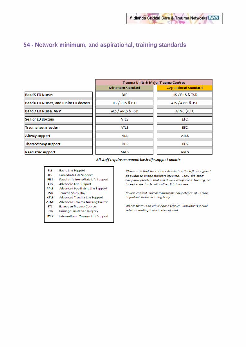
54 - Network minimum, and aspirational, training standards
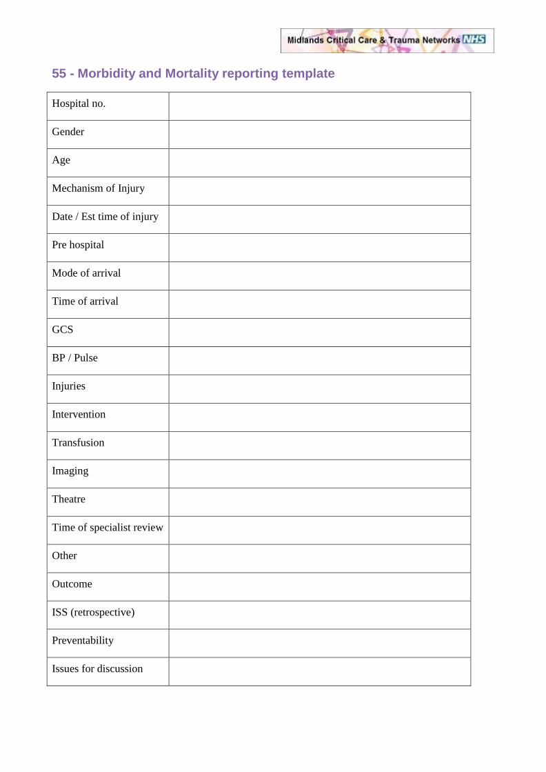
55 - Morbidity and Mortality reporting template
Hospital no.
Gender
Age
Mechanism of Injury
Date / Est time of injury
Pre hospital
Mode of arrival
Time of arrival
GCS
BP / Pulse
Injuries
Intervention
Transfusion
Imaging
Theatre
Time of specialist review
Other
Outcome
ISS (retrospective)
Preventability
Issues for discussion
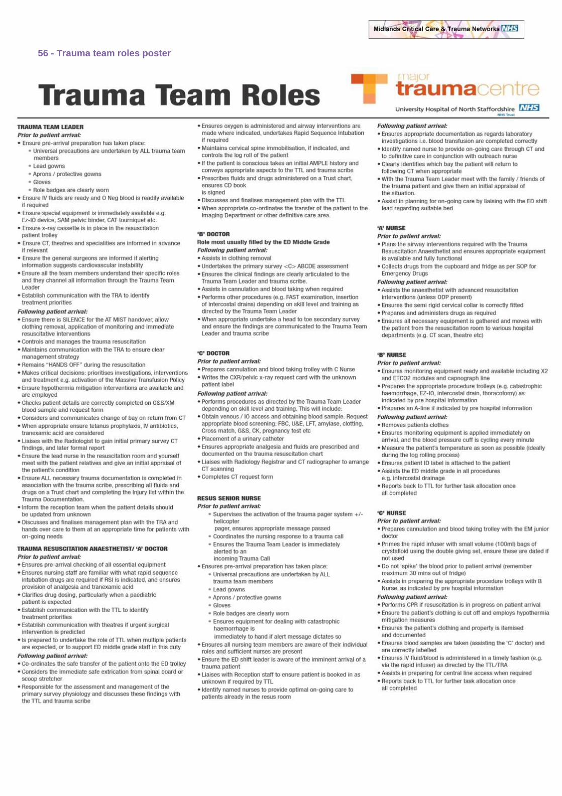
56 - Trauma team roles poster
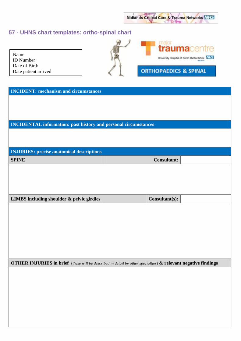
57 - UHNS chart templates: ortho-spinal chart
INCIDENT: mechanism and circumstances
INCIDENTAL information: past history and personal circumstances
INJURIES: precise anatomical descriptions
SPINE Consultant:
LIMBS including shoulder & pelvic girdles Consultant(s):
OTHER INJURIES in brief (these will be described in detail by other specialties) & relevant negative findings
Name
ID Number
Date of Birth
Date patient arrived
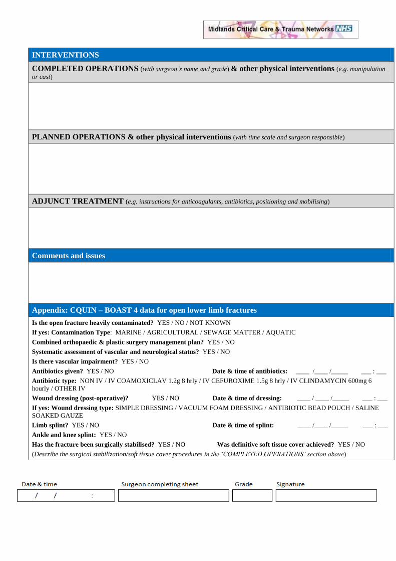
INTERVENTIONS
COMPLETED OPERATIONS (with surgeon’s name and grade) & other physical interventions (e.g. manipulation
or cast)
PLANNED OPERATIONS & other physical interventions (with time scale and surgeon responsible)
ADJUNCT TREATMENT (e.g. instructions for anticoagulants, antibiotics, positioning and mobilising)
Comments and issues
Appendix: CQUIN – BOAST 4 data for open lower limb fractures
Is the open fracture heavily contaminated? YES / NO / NOT KNOWN
If yes: Contamination Type: MARINE / AGRICULTURAL / SEWAGE MATTER / AQUATIC
Combined orthopaedic & plastic surgery management plan? YES / NO
Systematic assessment of vascular and neurological status? YES / NO
Is there vascular impairment? YES / NO
Antibiotics given? YES / NO Date & time of antibiotics: ____ /____ /_____ ___ : ___
Antibiotic type: NON IV / IV COAMOXICLAV 1.2g 8 hrly / IV CEFUROXIME 1.5g 8 hrly / IV CLINDAMYCIN 600mg 6
hourly / OTHER IV
Wound dressing (post-operative)? YES / NO Date & time of dressing: ____ / ____ /_____ ___ : ___
If yes: Wound dressing type: SIMPLE DRESSING / VACUUM FOAM DRESSING / ANTIBIOTIC BEAD POUCH / SALINE
SOAKED GAUZE
Limb splint? YES / NO Date & time of splint: ____ /____ /_____ ___ : ___
Ankle and knee splint: YES / NO
Has the fracture been surgically stabilised? YES / NO Was definitive soft tissue cover achieved? YES / NO
(Describe the surgical stabilization/soft tissue cover procedures in the ‘COMPLETED OPERATIONS’ section above)
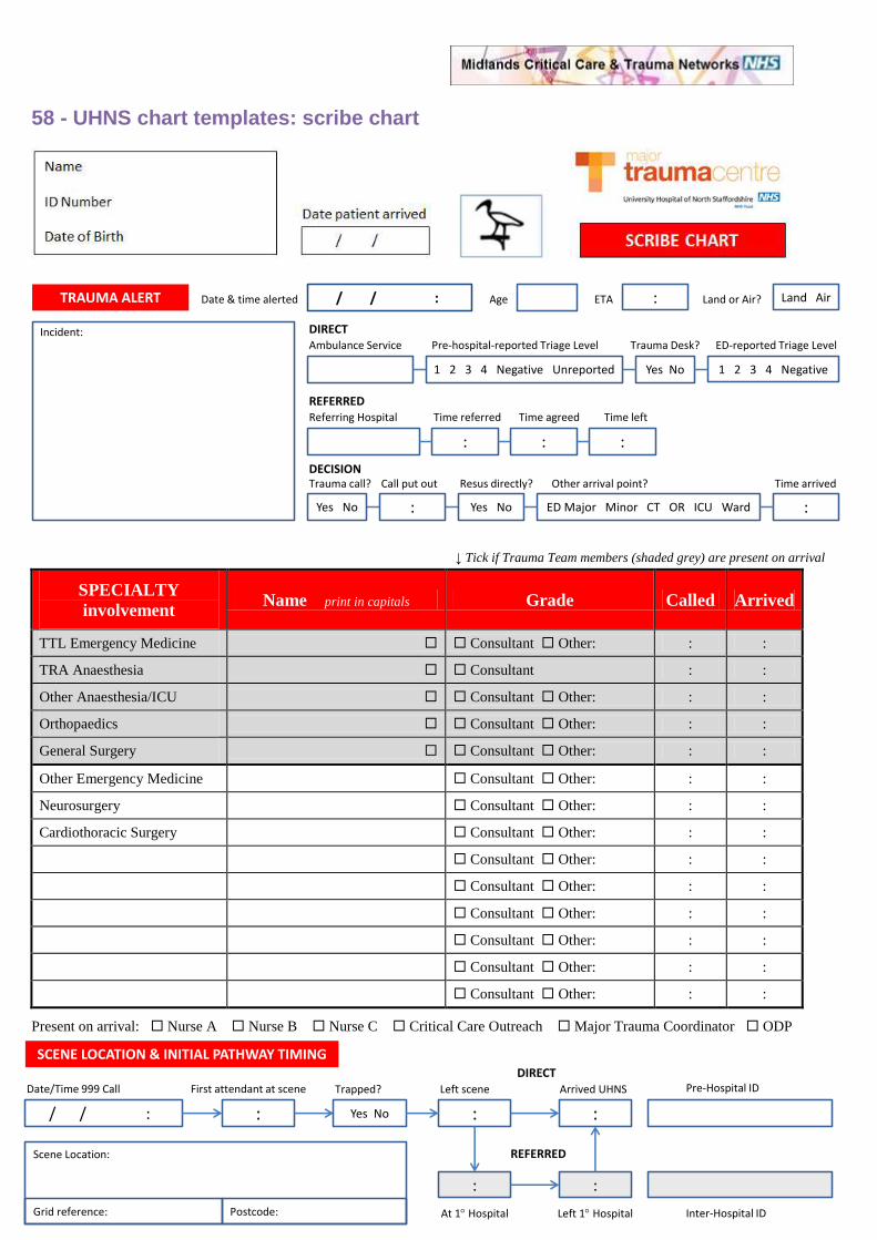
Date/Time 999 Call
/ / :
Left scene
:
:
At 1 Hospital
:
Left 1 Hospital
:
Arrived UHNS
Scene Location:
Pre-Hospital ID
Inter-Hospital ID
:
First attendant at scene
Yes No
Trapped?
SCENE LOCATION & INITIAL PATHWAY TIMINGDIRECT
REFERRED
Grid reference: Postcode:
58 - UHNS chart templates: scribe chart
SPECIALTY
involvement Name print in capitals Grade Called Arrived
TTL Emergency Medicine Consultant Other: : :
TRA Anaesthesia Consultant : :
Other Anaesthesia/ICU Consultant Other: : :
Orthopaedics Consultant Other: : :
General Surgery Consultant Other: : :
Other Emergency Medicine Consultant Other: : :
Neurosurgery Consultant Other: : :
Cardiothoracic Surgery Consultant Other: : :
Consultant Other: : :
Consultant Other: : :
Consultant Other: : :
Consultant Other: : :
Consultant Other: : :
Consultant Other: : :
Present on arrival: Nurse A Nurse B Nurse C Critical Care Outreach Major Trauma Coordinator ODP
DIRECT
REFERRED
Ambulance Service
Referring Hospital
ETA
:
Time referred
Call put out
Land or Air?
Trauma Desk?
TRAUMA ALERT / / :
1 2 3 4 NegativeYes No
Pre-hospital-reported Triage Level
1 2 3 4 Negative Unreported
: :
Trauma call?
Time agreed Time left
Resus directly?
:Yes No Yes No
ED-reported Triage LevelIncident:
Age : Land Air
Other arrival point?
ED Major Minor CT OR ICU Ward
Time arrived
:
Date & time alerted
DECISION
↓ Tick if Trauma Team members (shaded grey) are present on arrival
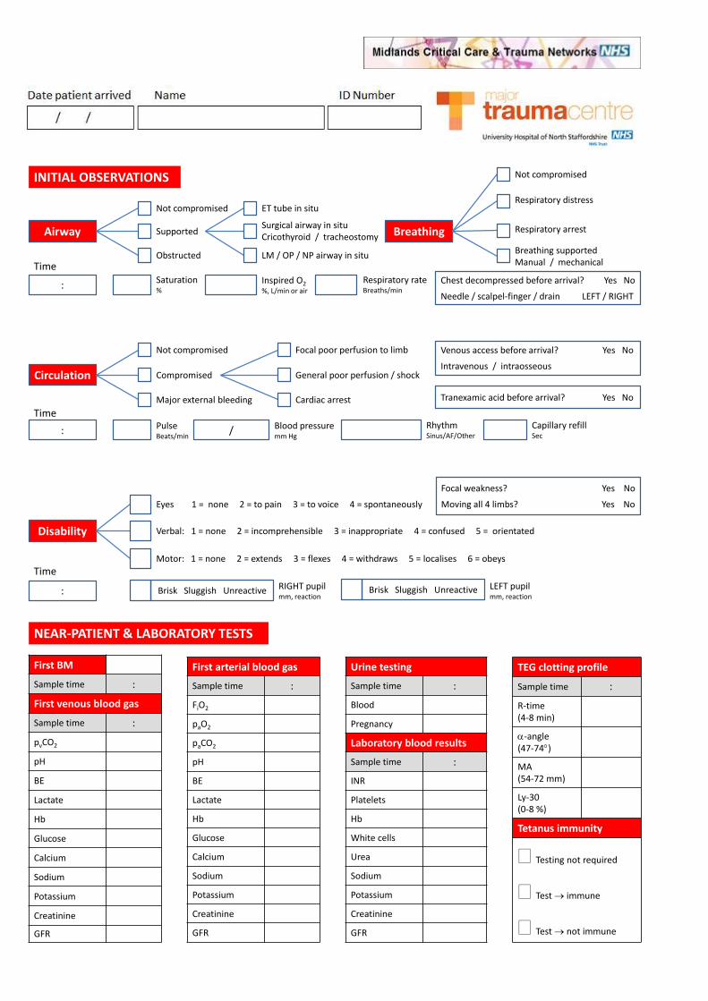
Airway Breathing
Obstructed
Not compromised
Supported
Respiratory distress
Respiratory arrest
Saturation%
Inspired O2%, L/min or air
Respiratory rateBreaths/min
Circulation Compromised
Not compromised
Major external bleeding
PulseBeats/min
/ Blood pressuremm Hg
RhythmSinus/AF/Other
Disability
Motor: 1 = none 2 = extends 3 = flexes 4 = withdraws 5 = localises 6 = obeys
Eyes 1 = none 2 = to pain 3 = to voice 4 = spontaneously
Verbal: 1 = none 2 = incomprehensible 3 = inappropriate 4 = confused 5 = orientated
RIGHT pupilmm, reaction
LEFT pupilmm, reaction
Time
:
Time
:
Time
:
Capillary refillSec
INITIAL OBSERVATIONS
ET tube in situ
Surgical airway in situ Cricothyroid / tracheostomy
LM / OP / NP airway in situ
Brisk Sluggish Unreactive Brisk Sluggish Unreactive
Cardiac arrest
Focal poor perfusion to limb
General poor perfusion / shock
Focal weakness? Yes No
Moving all 4 limbs? Yes No
Not compromised
Chest decompressed before arrival? Yes No
Needle / scalpel-finger / drain LEFT / RIGHT
Venous access before arrival? Yes No
Intravenous / intraosseous
Breathing supportedManual / mechanical
Tranexamic acid before arrival? Yes No
First BM
Sample time :
First venous blood gas
Sample time :
pvCO2
pH
BE
Lactate
Hb
Glucose
Calcium
Sodium
Potassium
Creatinine
GFR
First arterial blood gas
Sample time :
FiO2
paO2
paCO2
pH
BE
Lactate
Hb
Glucose
Calcium
Sodium
Potassium
Creatinine
GFR
Urine testing
Sample time :
Blood
Pregnancy
Laboratory blood results
Sample time :
INR
Platelets
Hb
White cells
Urea
Sodium
Potassium
Creatinine
GFR
NEAR-PATIENT & LABORATORY TESTS
TEG clotting profile
Sample time :
R-time(4-8 min)
-angle(47-74)
MA(54-72 mm)
Ly-30(0-8 %)
Tetanus immunity
Testing not required
Test immune
Test not immune
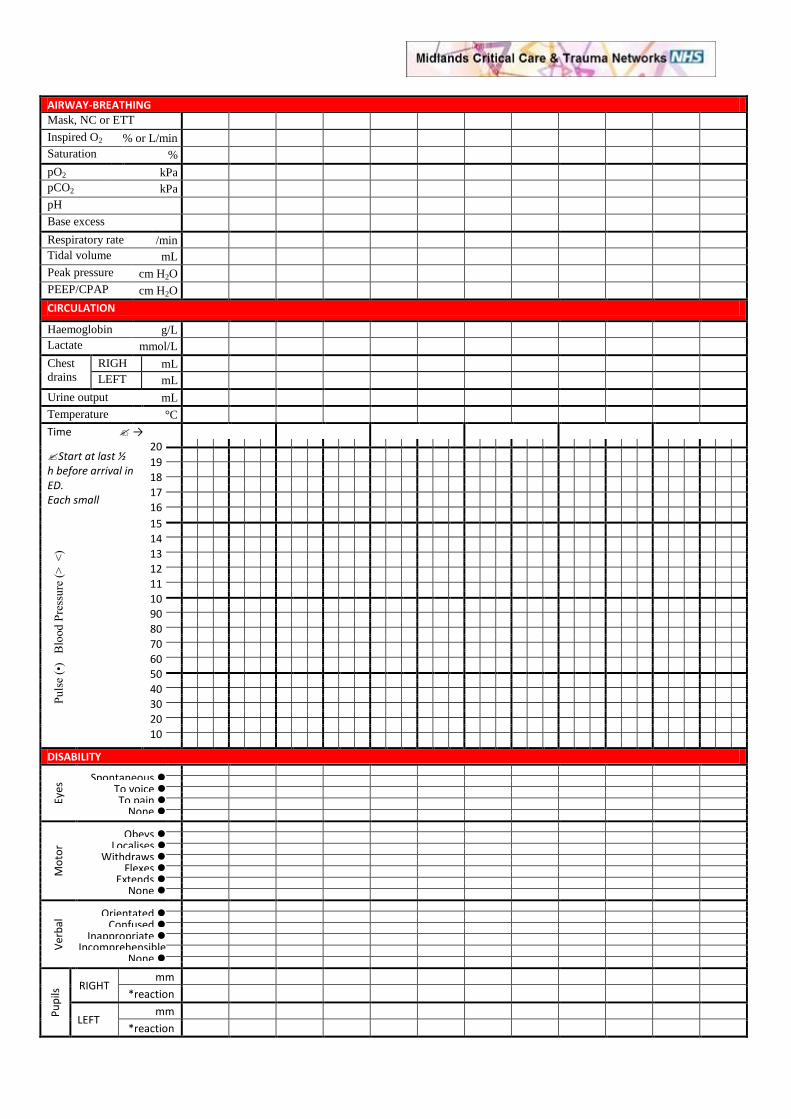
AIRWAY-BREATHING
Mask, NC or ETT
Inspired O2 % or L/min
Saturation %
pO2 kPa
pCO2 kPa
pH
Base excess
Respiratory rate /min
Tidal volume mL
Peak pressure cm H2O
PEEP/CPAP cm H2O
CIRCULATION
Haemoglobin g/L
Lactate mmol/L
Chest
drains
RIGH
T mL
LEFT mL
Urine output mL
Temperature °C
Time →
200
Start at last ½ h before arrival in ED. Each small square indicates 5 minutes
19
0
180
170
160
Pu
lse
(•)
B
lood
Pre
ssu
re (
>
<)
15
0
140
130
120
110
100
90
80
70
60
50
40
30
20
10
DISABILITY
Eyes
Spontaneous To voice To pain
None
Mo
tor
Obeys Localises
Withdraws Flexes
Extends None
Ver
bal
Orientated Confused
Inappropriate Incomprehensible
None
Pu
pils
RIGHT mm
*reaction
LEFT mm
*reaction
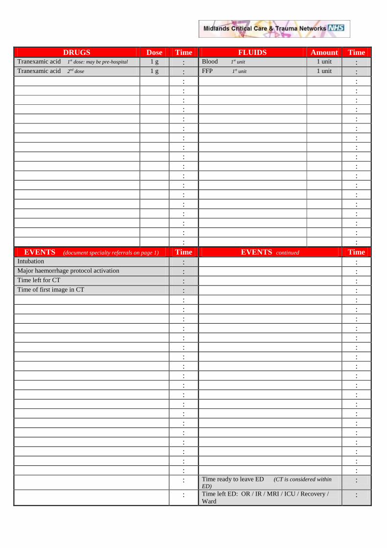
DRUGS Dose Time FLUIDS Amount Time Tranexamic acid 1st dose: may be pre-hospital 1 g : Blood 1st unit 1 unit : Tranexamic acid 2nd dose 1 g : FFP 1st unit 1 unit : : : : :
: :
: :
: :
: :
: :
: :
: :
: :
: :
: :
: :
: :
: :
: :
: :
: :
EVENTS (document specialty referrals on page 1) Time EVENTS continued Time Intubation : : Major haemorrhage protocol activation : : Time left for CT : : Time of first image in CT : :
: :
: :
: :
: :
: :
: :
: :
: :
: :
: :
: :
: :
: :
: :
: :
: :
: :
: :
: :
: Time ready to leave ED (CT is considered within
ED) :
: Time left ED: OR / IR / MRI / ICU / Recovery /
Ward :
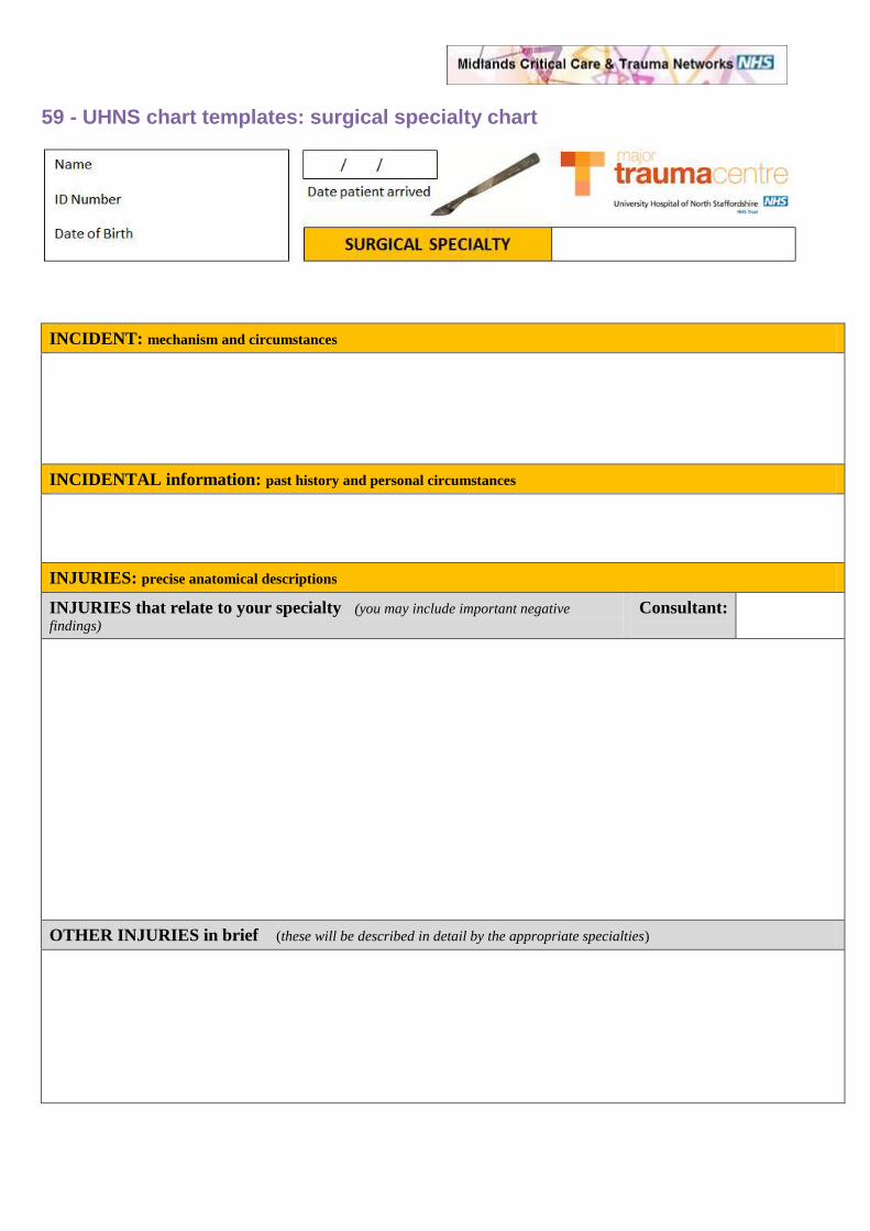
59 - UHNS chart templates: surgical specialty chart
INCIDENT: mechanism and circumstances
INCIDENTAL information: past history and personal circumstances
INJURIES: precise anatomical descriptions
INJURIES that relate to your specialty (you may include important negative
findings)
Consultant:
OTHER INJURIES in brief (these will be described in detail by the appropriate specialties)
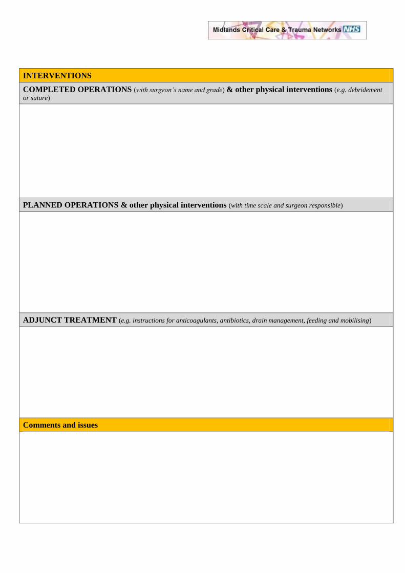
INTERVENTIONS
COMPLETED OPERATIONS (with surgeon’s name and grade) & other physical interventions (e.g. debridement
or suture)
PLANNED OPERATIONS & other physical interventions (with time scale and surgeon responsible)
ADJUNCT TREATMENT (e.g. instructions for anticoagulants, antibiotics, drain management, feeding and mobilising)
Comments and issues
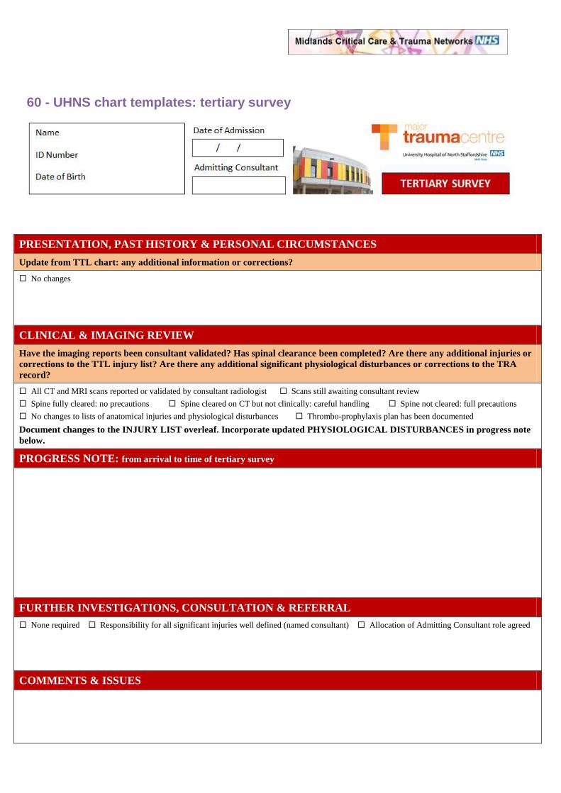
60 - UHNS chart templates: tertiary survey
PRESENTATION, PAST HISTORY & PERSONAL CIRCUMSTANCES
Update from TTL chart: any additional information or corrections?
No changes
CLINICAL & IMAGING REVIEW
Have the imaging reports been consultant validated? Has spinal clearance been completed? Are there any additional injuries or
corrections to the TTL injury list? Are there any additional significant physiological disturbances or corrections to the TRA
record?
All CT and MRI scans reported or validated by consultant radiologist Scans still awaiting consultant review
Spine fully cleared: no precautions Spine cleared on CT but not clinically: careful handling Spine not cleared: full precautions
No changes to lists of anatomical injuries and physiological disturbances Thrombo-prophylaxis plan has been documented
Document changes to the INJURY LIST overleaf. Incorporate updated PHYSIOLOGICAL DISTURBANCES in progress note
below.
PROGRESS NOTE: from arrival to time of tertiary survey
FURTHER INVESTIGATIONS, CONSULTATION & REFERRAL
None required Responsibility for all significant injuries well defined (named consultant) Allocation of Admitting Consultant role agreed
COMMENTS & ISSUES
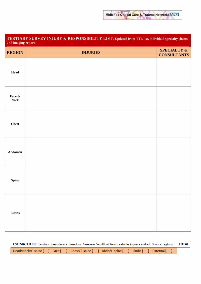
TERTIARY SURVEY INJURY & RESPONSIBILITY LIST: Updated from TTL list, individual specialty charts
and imaging reports
REGION INJURIES SPECIALTY &
CONSULTANTS
Head
Face &
Neck
Chest
Abdomen
Spine
Limbs
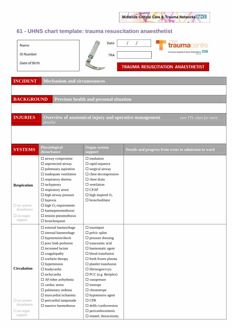
61 - UHNS chart template: trauma resuscitation anaesthetist
INCIDENT Mechanism and circumstances
BACKGROUND Previous health and personal situation
INJURIES Overview of anatomical injury and operative management (see TTL chart for more
details)
SYSTEMS Physiological
disturbance Organ system
support Details and progress from scene to admission to ward
Respiration
no system
disturbance
no organ
support
airway compromise
unprotected airway
pulmonary aspiration
inadequate ventilation
respiratory distress
tachypnoea
respiratory arrest
high airway pressure
hypoxia
high O2 requirements
haemopneumothorax
tension pneumothorax
bronchospasm
intubation
rapid sequence
surgical airway
chest decompression
chest drain
ventilation
CPAP
high inspired O2
bronchodilator
Circulation
no system
disturbance
no organ
support
external haemorrhage
internal haemorrhage
hypotension/shock
poor limb perfusion
increased lactate
coagulopathy
warfarin therapy
hypertension
bradycardia
tachycardia
AF/other arrhythmia
cardiac arrest
pulmonary oedema
myocardial ischaemia
pericardial tamponade
massive haemothorax
tourniquet
pelvic splint
pressure dressing
tranexamic acid
haemostatic agent
blood transfusion
fresh frozen plasma
platelet transfusion
fibrinogen/cryo.
PCC (e.g. Beriplex)
vasopressor
inotrope
chronotrope
hypotensive agent
CPR
defib./cardioversion
pericardiocentesis
immed. thoracotomy
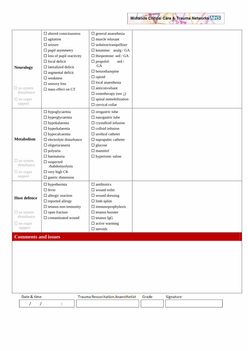
Neurology
no system
disturbance
no organ
support
altered consciousness
agitation
seizure
pupil asymmetry
loss of pupil reactivity
focal deficit
lateralized deficit
segmental deficit
weakness
sensory loss
mass effect on CT
general anaesthesia
muscle relaxant
sedation/tranquilliser
ketamine: analg / GA
thiopentone: sed / GA
propofol: sed /
GA
benzodiazepine
opioid
local anaesthesia
anticonvulsant
osmotherapy (see ↓)
spinal immobilization
cervical collar
Metabolism
no system
disturbance
no organ
support
hypoglycaemia
hyperglycaemia
hypokalaemia
hyperkalaemia
hypocalcaemia
electrolyte disturbance
oliguria/anuria
polyuria
haematuria
suspected
rhabdomyolysis
very high CK
gastric distension
orogastric tube
nasogastric tube
crystalloid infusion
colloid infusion
urethral catheter
suprapubic catheter
glucose
mannitol
hypertonic saline
Host defence
no system
disturbance
no organ
support
hypothermia
fever
allergic reaction
reported allergy
tetanus non-immunity
open fracture
contaminated wound
antibiotics
wound toilet
wound dressing
limb splint
immunoprophylaxis
tetanus booster
tetanus IgG
active warming
steroids
Comments and issues
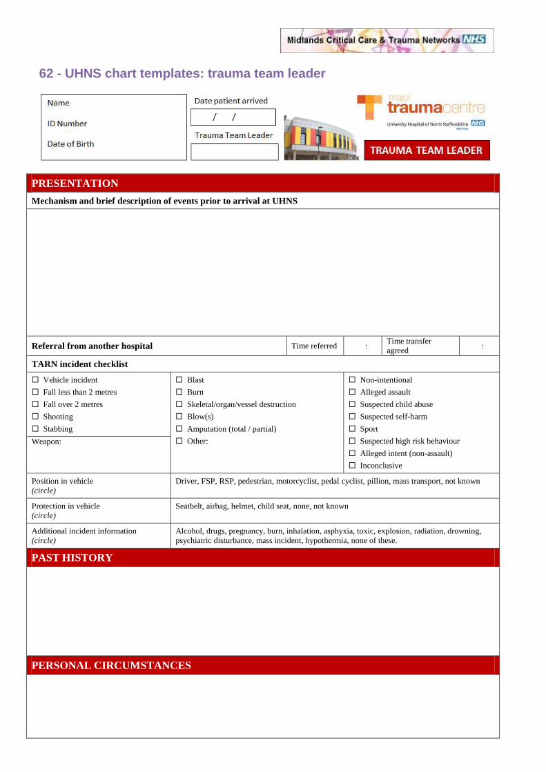
62 - UHNS chart templates: trauma team leader
PRESENTATION
Mechanism and brief description of events prior to arrival at UHNS
Referral from another hospital Time referred : Time transfer
agreed :
TARN incident checklist
Vehicle incident
Fall less than 2 metres
Fall over 2 metres
Shooting
Stabbing
Blast
Burn
Skeletal/organ/vessel destruction
Blow(s)
Amputation (total / partial)
Other:
Non-intentional
Alleged assault
Suspected child abuse
Suspected self-harm
Sport
Suspected high risk behaviour
Alleged intent (non-assault)
Inconclusive
Weapon:
Position in vehicle
(circle)
Driver, FSP, RSP, pedestrian, motorcyclist, pedal cyclist, pillion, mass transport, not known
Protection in vehicle
(circle)
Seatbelt, airbag, helmet, child seat, none, not known
Additional incident information
(circle)
Alcohol, drugs, pregnancy, burn, inhalation, asphyxia, toxic, explosion, radiation, drowning,
psychiatric disturbance, mass incident, hypothermia, none of these.
PAST HISTORY
PERSONAL CIRCUMSTANCES
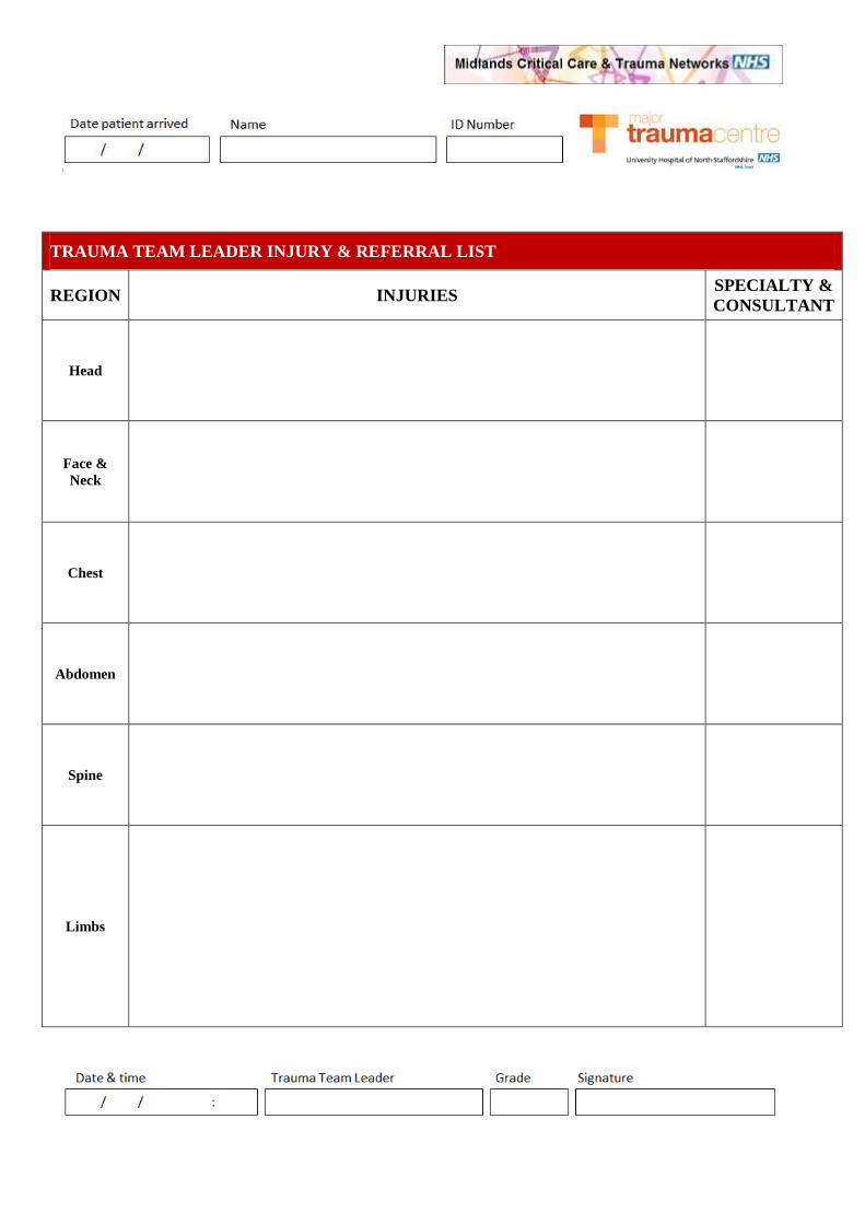
TRAUMA TEAM LEADER INJURY & REFERRAL LIST
REGION INJURIES SPECIALTY &
CONSULTANT
Head
Face &
Neck
Chest
Abdomen
Spine
Limbs
ASSESSMENT NOTES, COMMENTS & ISSUES
Date & time
/ / :
Trauma Team Leader Grade Signature
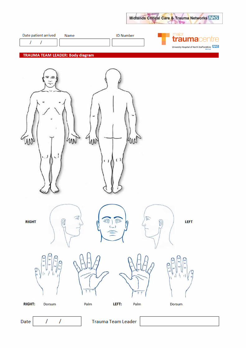
63 - West Midlands protocol for severe hypothermia
This protocol applies to anyone who is suspected either by environmental or clinical findings of having significant hypothermia. Significant European and Scandinavian experience has shown that good recoveries can be made from apparently hopeless situations. Patients have made full recoveries after hours of CPR but only with the appropriate care. This is deemed to be extra-corporal (circulation and warming of blood outside of the body) circulation, either with ECMO or cardio-pulmonary bypass. ECMO is the modality of choice causing less trauma to blood and requiring less anti-coagulation. Temperature measurement. Is vital to knowing how at risk the heart is of arrhythmia is, or if in cardiac arrest what the possible prognosis is. The rectal route is unreliable, oesophageal probes can trigger cardiac arrest . Oral or tympanic are the modalities of choice. ECG rhythm A normal looking rhythm compatible with a cardiac output is likely to suggest such but with a carotid pule too weak to feel. These patients should be treated as having an output and transferred as per protocol. Patient movement Patients must be treated with utmost care. The smallest movement can precipitate VF. Never raise the legs as the sudden return of cold blood to the core can have the same effect. Resistant VF At temperatures of less than 30
oC defibrillation is very unlikely to be successful. Repeated attempts will likely damage the
myocardium and should not be attempted. Once the patient is in asystole, an improvement in rhythm is very unlikely until the heart is rewarmed. Pre-hospital warming If mild, hypothermia should be treated aggressively with dry clothes/blankets, a warm environment and hot drinks. However once the temperature reaches 30 even the act of removing clothing could cause heart rhythm disturbance. Consideration should be made to moving patient as little as possible, leaving in wet clothing and expediting transfer to ECMO unit. CPR Should only be started when it can be continued. Mechanical devices are ideal if available. Hypothermia confers significant neuro-protection with brain metabolism falling by 6-10% every degree below 35
oC.
Complex heart operations of up to an hour are routinely carried out with no circulation at a temperature of 22oC.
Consequently delays in starting CPR are not as disastrous as imagined. Airway management The patient in cardiac arrest should have an ETT or LMA inserted for transfer. The decision in the severely hypothermic casualty not in cardiac arrest is more difficult. Airway manipulation may precipitate cardiac arrest, take great care with the use of airway devices. Discussion with ECMO centres This should be done at the earliest possible time via the ECMO co-ordinators at the Royal Leicester and Wytneshawe. Medical advice can be sought from the doctors overleaf in the interim. It may be possible for a mobile ECMO unit to be moved to the patient at a convenient DGH. Cardiopulmonary bypass is an alternative to ECMO and is available at all major trauma centres.
Severity of hypothermia by core body temp Mild 35 – 32
oC
Moderate 32-28oC
Severe <28oC
IKAR – MEDCOM on-site staging of hypothermia Stage 1 – Conscious, shivering. (35-32
oC)
Stage 2 – Impaired consciousness, no shivering. (32-28oC)
Stage 3 – Unconscious. (28-24oC)
Stage IV – Apparent death (24-13.7oC)
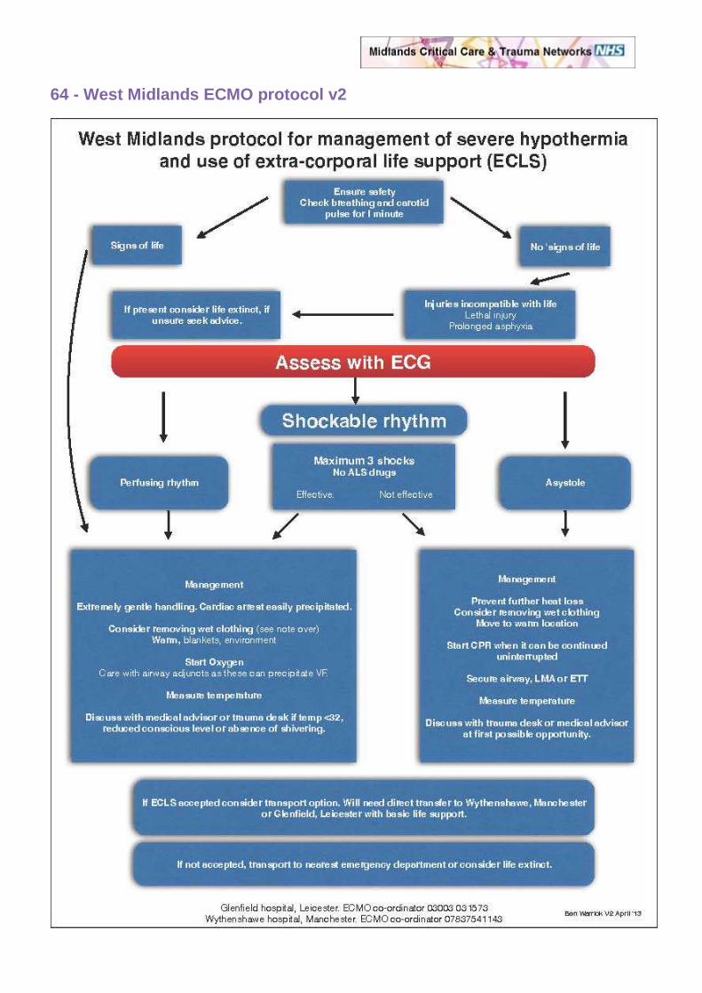
64 - West Midlands ECMO protocol v2
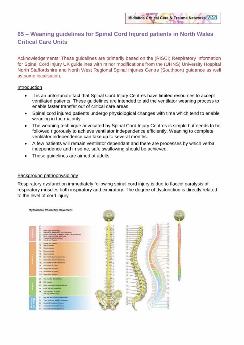
65 – Weaning guidelines for Spinal Cord Injured patients in North Wales
Critical Care Units
Acknowledgements: These guidelines are primarily based on the (RISCI) Respiratory Information
for Spinal Cord Injury UK guidelines with minor modifications from the (UHNS) University Hospital
North Staffordshire and North West Regional Spinal Injuries Centre (Southport) guidance as well
as some localisation.
Introduction
It is an unfortunate fact that Spinal Cord Injury Centres have limited resources to accept ventilated patients. These guidelines are intended to aid the ventilator weaning process to enable faster transfer out of critical care areas.
Spinal cord injured patients undergo physiological changes with time which tend to enable weaning in the majority.
The weaning technique advocated by Spinal Cord Injury Centres is simple but needs to be followed rigorously to achieve ventilator independence efficiently. Weaning to complete ventilator independence can take up to several months.
A few patients will remain ventilator dependant and there are processes by which verbal independence and in some, safe swallowing should be achieved.
These guidelines are aimed at adults.
Background pathophysiology
Respiratory dysfunction immediately following spinal cord injury is due to flaccid paralysis of
respiratory muscles both inspiratory and expiratory. The degree of dysfunction is directly related
to the level of cord injury
Lumbar cord injuries will lose some expiratory abdominal activity.
Thoracic cord injuries will additionally lose intercostal activity and will frequently be complicated
by rib fractures and pulmonary contusions. Haemothoraces may be present secondary to the
thoracic spine fractures.
Low cervical cord injuries will have lost all intercostal activity.
High cervical injuries may also lose diaphragmatic and scalene activity. Ventilatory failure is rapid
in these circumstances.
Autonomic disruption following on from cord injuries causes excessive bronchial secretions and a
tendency to bronchoconstriction.
Some respiratory afferent information is lost; patients may not feel dyspnoeic or become
tachypnoeic when failing.
Respiratory failure results from ineffective ventilation from compromised respiratory muscles
acting on a flaccid rib cage aggravated by intrapulmonary compliance changes and an inability to
spontaneously clear secretions.
It is occasionally possible using aggressive physiotherapy techniques and non invasive
ventilation to support patients until pulmonary compliance improves to the point that unsupported
ventilation is possible, but more commonly ventilatory failure occurs from minutes to days post
injury requiring intubation and ventilation.
The physiological processes by which weaning becomes feasible include:
Resolution of cord oedema. It is common for the neurological level to improve slightly with time which may allow use of previously paralysed respiratory muscles.
Resolution of pulmonary pathology. Pulmonary compliance needs to be as normal as possible for successful weaning.
Development of spasticity. Return of intercostal tone reduces chest wall compliance and improves ventilatory mechanics.
Retraining of remaining functioning respiratory muscles.
Tracheostomy
Once intubated we recommend early tracheostomy as successful early extubation is rare.
Tracheostomy simplifies weaning, abolishes the need for sedation, improves communication and
enables efficient secretion clearance.
There is no preference for percutaneous over surgical tracheostomy except with unstable cervical
fractures where a surgical technique may cause less vertebral movement.
Tube changes for those patients requiring long term tracheostomies may be easier following
surgical tracheostomy.
8 mm internal diameter tubes are optimal in adults.
Removable inner cannulae are recommended in the early stages.
Subglottic suction tubes may be of considerable benefit.
There is no evidence of benefit for fenestrated tubes but there is evidence that they are associated with overgranulation
Pre requisites for weaning:
Good pulmonary compliance : 50 ml/cm H2O or greater
FiO2 < 0.4
PEEP <10, preferably nearer 5 cmH2O
Awake and cooperative. Minimal opiates. Preferably no delirium
No active sepsis
Some evidence of spontaneous respiratory activity.
o Ventilator triggering does not necessarily imply useful activity.
o Many patients will appear to pass spontaneous breathing trials early following injury, but rapidly develop respiratory fatigue requiring re-ventilation.
Involved staff. Weaning proceeds more efficiently if a team of interested staff take control of the process.
Initial testing
The premise for weaning is that some respiratory activity is present but weak, and a degree of
respiratory muscle retraining is required.
The easiest and most reproducible measure of lung function for this is the vital capacity (VC). In
the presence of low flows and low volumes a mechanical Wrights spirometer tends to perform
better than electronic spirometers.
The vital capacity manoeuvre needs to be made by a cooperative patient completely free from
ventilatory support. If still on relatively high PEEP a few breaths before the measurement is
performed is advised.
A vital capacity as low as 150 mls is considered adequate to start weaning. A vital capacity
approaching 1000ml predicts straightforward weaning.
With cord injuries at C4 and above, if there is doubt as to whether diaphragm activity is present,
apnoea testing under sedation may be performed. This may show accessory muscle activity
(Nasalis, sternomastoid) when the PaCO2 rises above 6kPa without diaphragmatic activity if the
cord injury involves the phrenic nerves. This does not necessarily imply permanent ventilator
dependence but requires retesting at a later date.
Weaning principle
Based on the initial vital capacity measurement all ventilatory support is removed for a specified time and then re-instituted for a rest period. The common term for this is ventilator free breathing (VFB). In patients with a low initial VC or relatively high PEEP the ventilator may be allowed to deliver CPAP as long as no extra pressure/volume support is given.
Suggested VFB times based on VC are:
1. If VC is less than 250 mls, start with 5 mintues VFB.
2. If VC is less than 500 mls, start with 15 minutes VFB.
3. If VC is greater than 750 mls, start with 30 minutes VFB
4. If VC is greater than 1000mls, start with 60 minutes VFB (Southport SCI unit)
The on-ventilator rest period should be at least 1-2 hours. This should be repeated throughout the day.
Weaning progression is achieved by increasing VFB time by specified amounts dependant on the previous day’s results.
It is important that the patient is not fatigued which can be estimated by re-measuring the VC at the end of the VFB period. If it is less that 70% of the pre weaning VC then either the rest period should be extended or the VFB time reduced.
For Example: If a patient with a VC of 200 mls successfully achieves 3 episodes of 5 minutes VFB with 2
hour rest periods on day 1, with an end VFB VC of 180 mls, then increase the VFB time by 20% (to 6
mins) for day 2. If day 2 is satisfactory increase by 20% (8 mins) for day 3.
Utilisation of the weaning stickers (courtesy of UHNS) will help to ensure a consistent weaning
plan for the patients and communication for the critical care team.
WEANING PLAN FOR …………………… Signature:………………….
VC
VFB x ………………x Day .……………… Min
The initial aim is for VFB up to 18 hours during daytime, but for ventilation at night, as spinal cord
injured patients can have significant REM sleep hypoventilation. To asses safe VFB overnight
requires either PaCO2 or TcCO2 monitoring.
Adjuncts to weaning
Biochemistry and nutrition should be addressed. It is recommended that cervical cord injured patients and potential slow weaners have gastrostomies inserted.
Regular salbutamol nebulisation may improve respiratory muscle function.
VFB periods should be performed supine, not sitting. There is a drop of up to 20% in VC
from supine to sitting, so VFB periods will be better tolerated supine.
Secretion clearance should be performed prior to VFB periods. Tenacious sputum may be treated with oral/PEG carbocysteine or nebulised acetylcysteine.
There is some evidence that during rest ventilation periods, high tidal volume ventilation whilst maintaining normocarbia accelerates weaning as it may reduce atelectasis.
RR/Sats Time on Time off RR/Sats
Tracheostomy cuff deflation
For all spinal cord injured patients the ability to communicate is paramount to rehabilitation and
reintegration. Being in a critical care unit for considerable amounts of time without easy
communication is at best frustrating and can contribute to psychological morbidity.
Cuff deflation can be achieved either on or off ventilation. Not only does this enable speech but
also reduces microaspiration, restores laryngeal and pharyngeal reflexes leading to resumption of
safe swallowing. There is however a risk of passive aspiration especially if the patient has poor
laryngeal reflexes. To mitigate aspiration suction regularly while the cuff is down.
Off ventilator cuff deflation during VFB for fast weaners should be considered. If a subglottic
suction tracheostomy is in place then this should be aspirated, otherwise a tracheal suction
catheter placed to catch pooled saliva as it passes the deflating cuff. When deflated a speaking
valve should be used, (if there is sufficient insufflation leak – if not consider downsizing)
preferably a Passy Muir as they have favourable mechanics for spontaneously breathing low
volume patients.
The use of a speaking valve introduces an element of PEEP which may improve respiratory
mechanics and reduce the development of atelectasis.
On ventilator cuff deflation should be considered for slow weaners. Ventilator settings should be
adjusted to allow for the resultant leak, either increases in IPAP or inspiratory time. Many
ventilators will alarm continuously with this degree of leak so either change to the unit’s NIV
machine, use your invasive ventilators on an NIV setting or a simpler, domicillary type device can
be considered. It may be helpful to contact your Spinal Cord Injury Centre to ask what machine
they use.
Many patients develop increased leaks when asleep, requiring partial or full cuff inflation in order
to achieve adequate ventilation.
Optimal practice is to change cuffed for uncuffed tubes wherever possible when cuffs can be
deflated for 24 hours.
Swallowing
Attempts at swallowing with an inflated tracheostomy cuff are never safe. It is advisable to wait
until cuff deflation is achieved and enlist the advice of a speech and language therapist.
Post weaning maintenance
Patients who have successfully weaned or who are ventilator free during the day are still at risk of
respiratory decompensation. Functional residual capacity and inspiratory muscle strength
continue at a reduced level. Intermittent IPPB or manual hyperinflation are of benefit in reducing
atelectasis.
Further information
All UK Spinal Cord Injury Centres have someone with an interest in respiratory management. Contacts can be found at www.risci.org.uk