Embed Size (px)
Citation preview
MIF Contributes to Trypanosoma brucei AssociatedImmunopathogenicity DevelopmentBenoı̂t Stijlemans1,2*, Lin Leng3, Lea Brys1,2, Amanda Sparkes1,2, Liese Vansintjan1,2, Guy Caljon4,
Geert Raes1,2, Jan Van Den Abbeele4, Jo A. Van Ginderachter1,2, Alain Beschin1,2, Richard Bucala3.,
Patrick De Baetselier1,2.
1 Department of Cellular and Molecular Immunology, Vrije Universiteit Brussel (VUB), Brussels, Belgium, 2 Myeloid Cell Immunology Laboratory, Vlaams Instituut voor
Biotechnologie, Brussels, Belgium, 3 Department of Internal Medicine, Yale University School of Medicine, New Haven, Connecticut, United States of America, 4 Unit of
Veterinary Protozoology, Department of Biomedical Sciences, Institute of Tropical Medicine, Antwerp, Belgium
Abstract
African trypanosomiasis is a chronic debilitating disease affecting the health and economic well-being of many people indeveloping countries. The pathogenicity associated with this disease involves a persistent inflammatory response, wherebyM1-type myeloid cells, including Ly6Chigh inflammatory monocytes, are centrally implicated. A comparative gene analysisbetween trypanosusceptible and trypanotolerant animals identified MIF (macrophage migrating inhibitory factor) as animportant pathogenic candidate molecule. Using MIF-deficient mice and anti-MIF antibody treated mice, we show that MIFmediates the pathogenic inflammatory immune response and increases the recruitment of inflammatory monocytes andneutrophils to contribute to liver injury in Trypanosoma brucei infected mice. Moreover, neutrophil-derived MIF contributedmore significantly than monocyte-derived MIF to increased pathogenic liver TNF production and liver injury duringtrypanosome infection. MIF deficient animals also featured limited anemia, coinciding with increased iron bio-availability,improved erythropoiesis and reduced RBC clearance during the chronic phase of infection. Our data suggest that MIFpromotes the most prominent pathological features of experimental trypanosome infections (i.e. anemia and liver injury),and prompt considering MIF as a novel target for treatment of trypanosomiasis-associated immunopathogenicity.
Citation: Stijlemans B, Leng L, Brys L, Sparkes A, Vansintjan L, et al. (2014) MIF Contributes to Trypanosoma brucei Associated ImmunopathogenicityDevelopment. PLoS Pathog 10(9): e1004414. doi:10.1371/journal.ppat.1004414
Editor: Edward Mitre, Uniformed Services University of the Health Sciences, United States of America
Received April 27, 2014; Accepted August 21, 2014; Published September 25, 2014
Copyright: � 2014 Stijlemans et al. This is an open-access article distributed under the terms of the Creative Commons Attribution License, which permitsunrestricted use, distribution, and reproduction in any medium, provided the original author and source are credited.
Data Availability: The authors confirm that all data underlying the findings are fully available without restriction. All relevant data are within the paper and itsSupporting Information files.
Funding: This work, performed in frame of an Interuniversity Attraction Pole Program (PAI-IAP N. P7/41, http://www.belspo.be/belspo/iap/index_en.stm), wassupported by a grant from the FWO (KaN 1515813N) and NIH AI042310. The funders had no role in study design, data collection and analysis, decision to publish,or preparation of the manuscript.
Competing Interests: The authors have declared that no competing interests exist.
* Email: [email protected]
. These authors contributed equally to this work.
Introduction
African trypanosomiasis is a parasitic disease of medical and
veterinary importance that adversely affects the public health and
economic development of sub-Saharan Africa. The causative
agents, trypanosomes transmitted by the tsetse fly (Glossina spp),
are extracellular hemoflagellated protozoans that cause fatal
diseases in mammals, commonly called sleeping sickness in
humans (HAT, Human African Trypanosomiasis) or nagana in
domestic livestock [1]. In the case of bovine trypanosomiasis,
anemia is considered to be the most prominent pathogenicity
feature and the major cause of death associated with the disease
[2]. In fact, the main difference between trypanosusceptible and
trypanotolerant animals relies in their capacity to control anemia
development. The underlying mechanisms mediating trypano-
some-associated anemia have been scrutinized in murine models
[3]. The data collectively suggest that a strong pro-inflammatory/
type I immune response, involving classically activated myeloid
cells/macrophages (M1), is required for initial parasite growth
control. Yet, if maintained, this response contributes to
pathogenicity in general and anemia in particular in trypanosus-
ceptible mice, resulting in reduced survival of the host. Hereby,
myeloid cell hyperactivation was proposed to be involved in the
extravascular destruction of red blood cells (RBCs) due to
enhanced erythrophagocytosis by spleen and liver-associated M1
cells of the infected host [3,4] causing trypanosome-associated
anemia. Such type I immune response driven anemia resembles
anemia of inflammation, also termed anemia of chronic disease
(ACD), that is associated with chronic infections and sterile
inflammations [5,6].
Uncontrolled inflammation associated with persistence of M1
cells is also a major cause of liver injury and cachexia observed in
trypanosusceptible animals, whereby the anti-inflammatory cyto-
kine IL-10 was found to be detrimental to prevent these
pathogenic features [7]. Hence, therapies should aim at re-
establishing the balance between pro- and anti-inflammatory
signals during the disease to avoid tissue damage. In this context,
the glycosylphosphatidylinositol (GPI)-anchor of the Variant
Surface Glycoprotein (VSG) coat was identified as a major
parasite-derived molecule with a M1-activating potential [8].
PLOS Pathogens | www.plospathogens.org 1 September 2014 | Volume 10 | Issue 9 | e1004414
Interestingly, a GPI-based treatment was found to protect against
infection-associated cachexia, liver damage, anemia, and to
prolong survival by modulating the myeloid cell activation state,
i.e. forcing a transition from M1 to M2 (alternatively) activated
myeloid cells during the course of infection [9].
The possibility to render trypanosusceptible animals more
tolerant by modulating the activation state of myeloid cells offers
an attractive model to identify genes and gene-products involved
in the pathogenicity of African trypanosomiasis. In this context, a
comparative gene expression analysis revealed that the macro-
phage migration inhibitory factor (MIF) expression was signifi-
cantly reduced in mice rendered trypanotolerant upon GPI
treatment. This ‘‘early response’’ cytokine is expressed by
numerous cell types, including myeloid cells, and plays a key role
in innate and adaptive immunity [10,11]. MIF is a prominent
inducer of systemic inflammation in many inflammatory diseases
[12,13]. It functions by recruiting myeloid cells to the site of
inflammation [14], by inducing their differentiation towards M1
cells secreting TNF [15] and by suppressing p53-dependent
apoptosis of inflammatory cells [16]. Since African trypanosomes
trigger a persistent type I/M1 immune response in trypanosus-
ceptible (e.g. T. brucei brucei (T. brucei)) infected mice, we
evaluated the potential role of MIF in the development of
infection-associated pathogenicity. More specifically, the effect of
MIF on the infiltration of myeloid cells, liver damage and anemia
development was investigated.
Results
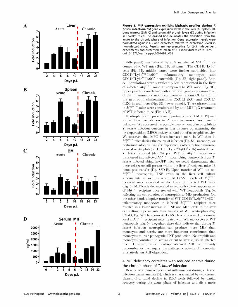
1. MIF expression levels show two distinct waves duringT. brucei infection
As a first step towards evaluating the potential role of MIF
during the course of T. brucei infection, we analysed its gene
expression in different organs. As shown in Fig. 1A-C, MIF gene
expression level in liver, spleen and bone marrow was character-
ized by two distinct phases, i.e. an initial increase during the acute
phase of infection that returns back to the level of non-infected
mice, followed by a second more progressive increase during the
chronic phase of infection. Serum MIF protein levels followed the
same kinetic as in the tested organs (Fig. 1D).
2. MIF deficiency correlates with reduced type Iinflammation during T. brucei infection
To evaluate the potential role of MIF in inflammation-
associated pathogenicity occurring during T. brucei infection,
two strategies targetting MIF production/activity were evaluated,
(i) a comparison between wild type (WT) and MIF-deficient
(Mif2/2) mice and (ii) a comparison between monoclonal anti-
MIF IgG or isotype control antibody treated WT mice. As shown
in Fig. 2A, first peak parasitemia and the further progression of
parasitemia development were similar in WT and Mif2/2 mice.
However, there was a small, yet significant prolongation in median
survival time in Mif2/2 mice (Fig. 2B).
Next, we investigated whether infected WT and Mif2/2 mice
exhibited different cytokine immune responses. The results shown
in Fig. 2C indicate that during the course of infection, both strains
of mice mounted a prominent pro-inflammatory immune response
as evidenced by the elevated serum levels of IFN-c, TNF and IL-6.
Yet, these cytokine levels were lower in Mif2/2 mice than in WT
mice, especially during the chronic stage of infection. Conversely,
serum IL-10 levels progressively increased during the chronic stage
of infection (day 18 p.i. till the end) in Mif2/2 mice whereas they
were low/marginal in WT mice.
Similar to Mif2/2 mice, anti-MIF IgG treatment did not affect
parasitemia development but increased the median survival time
compared to control antibody treated mice (Fig. S1A-B),
suggesting a role for extracellular MIF in disease pathogenesis.
As in Mif2/2 mice, the pro-inflammatory cytokine production was
decreased and IL-10 production was elevated during the chronic
stage of infection upon anti-MIF IgG treatment of WT mice (Fig.
S1C).
In a tsetse fly-mediated T. brucei infection model that mimics
the natural route of infection, Mif deficiency did not affect
parasitemia development but resulted in a prolonged survival (Fig.
S2A-B) and a reduced pro-inflammatory cytokine profile (mainly
IFN-c) together with an increased IL-10 production during the
chronic stage of infection (Fig. S2C).
3. Tissue pathogenicity and infiltration ofCD11b+Ly6chighLy6G2 and CD11b+Ly6cintLy6G+ myeloidcells are reduced in Mif2/2 mice during the chronicphase of T. brucei infection
A persistent pro-inflammatory immune response contributes to
liver damage in the chronic stage of T. brucei infection [7,17].
Interestingly, at this stage (day 25 p.i.), Mif2/2 mice exhibited
significantly reduced liver pathogenicity than WT mice, as
evidenced by lower hepatomegaly and reduced ALT (alanine
aminotransferase) levels (Fig. 3A left and middle panel, respec-
tively). The reduced serum AST (aspartate aminotransferase) levels
further confirmed lower tissue pathogenicity in infected Mif2/2
mice (Fig. 3A right panel).
We have documented that infiltration of CD11b+Ly6c+ myeloid
cells in the chronic stage of T. brucei infection contributes to liver
pathogenicity in WT mice [7,18]. Upon gating on CD45+ liver
non-parenchymal cells (see gating strategy Fig. S3A-C), we found
that the infiltration of CD11b+Ly6c+ myeloid cells (Fig. 3B,
Author Summary
Uncontrolled inflammation is a major contributor topathogenicity development during many chronic parasiticinfections, including African trypanosome infections.Hence, therapies should aim at re-establishing the balancebetween pro- and anti-inflammatory responses to reducetissue damage. Our experiments uncovered that macro-phage migration inhibitory factor (MIF) plays a pivotal rolein trypanosomiasis-associated pathogenicity development.Hereby, MIF-deficient and neutralizing anti-MIF antibody-treated wild type (WT) T. brucei-infected mice exhibiteddecreased inflammatory responses, reduced liver damageand anemia (i.e. the most prominent pathogenicityfeatures) compared to WT control mice. The reducedtissue damage coincided with reduced infiltration ofpathogenic monocytic cells and neutrophils, wherebyneutrophil-derived MIF contributed more significantly thanmonocyte-derived MIF to tissue damage. MIF also pro-moted anemia development by suppressing red blood cellproduction and enhancing their clearance. The clinicalsignificance of these findings follows from human geneticdata indicating that low-expression (protective) MIF allelesare enriched in Africans. The current findings thereforeoffer promise for human translation and open thepossibility of assessing MIF levels or MIF genotype as anindication of an individual’s risk for severe trypanosomiasis.Furthermore, given the unmet medical need of Africantrypanosomiasis affecting millions of people, these find-ings highlight MIF as a potential new therapeutic target fortreatment of trypanosomiasis-associated pathogenicity.
MIF, Liver Damage and Anemia
PLOS Pathogens | www.plospathogens.org 2 September 2014 | Volume 10 | Issue 9 | e1004414
middle panel) was reduced by 25% in infected Mif2/2 mice
compared to WT mice (Fig. 3B, left panel). The CD11b+Ly6c+
cells (Fig. 3B, middle panel) were further subdivided into
CD11b+Ly6chighLy6G2 inflammatory monocytes and
CD11b+Ly6cintLy6G+ neutrophils (Fig. 3B, right panel). Both
cell populations were significantly less represented in the liver
of infected Mif2/2 mice as compared to WT mice (Fig. 3C,
upper panels), correlating with a reduced gene expression level
of the inflammatory monocyte chemoattractant CCL2 and of
the neutrophil chemoattractants CXCL1 (KC) and CXCL5
(LIX) in total liver (Fig. 3C, lower panels). These observations
in Mif2/2 mice were corroborated by anti-MIF IgG treatment
of WT infected mice (Fig. 4A-B).
Neutrophils can represent an important source of MIF [19] and
so far their contribution to African trypanosomiasis remains
unknown. We addressed the possible involvement of neutrophils to
T. brucei infection outcome in first instance by measuring the
myeloperoxidase (MPO) activity as read-out of neutrophil activity.
We observed that MPO levels increased more in WT than in
Mif2/2 mice during the course of infection (Fig. S4). Secondly, we
performed adoptive transfer experiments whereby bone marrow-
derived neutrophils (i.e. CD11b+Ly6cintLy6G+ cells) isolated from
T. brucei infected (day 24 p.i.) WT or Mif2/2 mice were
transferred into infected Mif2/2 mice. Using neutrophils from T.brucei infected ubiquitin-GFP mice we could demonstrate that
these cells were still present within the liver of recipient mice 18
hours post-transfer (Fig. S3D-E). Upon transfer of WT but not
Mif2/2 neutrophils, TNF levels in the liver cell culture
supernatants as well as serum ALT/AST levels of Mif2/2
recipient mice increased to the levels of infected WT mice
(Fig. 5). MIF levels also increased in liver cells culture supernatants
of Mif2/2 recipient mice treated with WT neutrophils (Fig. 5),
reflecting the contribution of neutrophils to MIF production. On
the other hand, adoptive transfer of WT CD11b+Ly6chighLy6G2
inflammatory monocytes in infected Mif2/2 recipient mice
resulted in a lower increase in TNF and MIF levels in the liver
cell culture supernatants than transfer of WT neutrophils (Fig.
S3F-G; Fig. 5). The serum ALT/AST levels increased to a similar
level in Mif2/2 recipient mice treated with WT monocytes or WT
neutrophils (Fig. 5). Together, these data indicate that during T.brucei infection neutrophils can produce more MIF than
monocytes and hereby are more important contributors than
monocytes to liver pathogenic TNF production. Neutrophils and
monocytes contribute to similar extent to liver injury in infected
mice. However, while neutrophil-derived MIF is primarily
responsible for liver injury, the pathogenic activity of monocytes
is relatively less MIF-dependent.
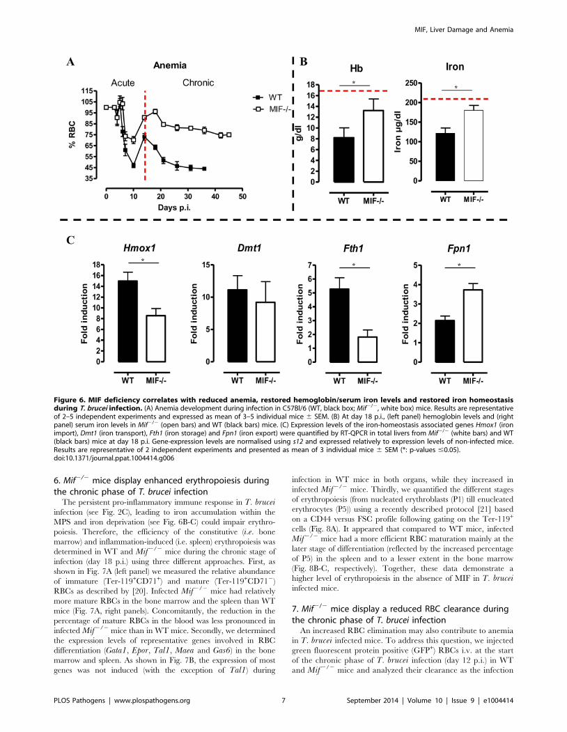
4. MIF deficiency correlates with reduced anemia duringthe chronic phase of T. brucei infection
Besides liver damage, persistent inflammation during T. bruceiinfection causes anemia [5], which is characterized by two distinct
phases; (i) a rapid decline in RBC levels followed by partial
recovery during the acute phase of infection and (ii) a more
Figure 1. MIF expression exhibits biphasic profiles during T.brucei infection. Mif gene expression levels in the liver (A), spleen (B),bone marrow (BM) (C) and serum MIF protein levels (D) during infectionin C57Bl/6 mice. The dashed line delineates the transition from theacute to the chronic phase of infection. Gene expression levels werenormalized against s12 and expressed relative to expression levels innon-infected mice. Results are representative for 2–3 independentexperiments and presented as mean of 2–3 individual mice 6 SEM.doi:10.1371/journal.ppat.1004414.g001
MIF, Liver Damage and Anemia
PLOS Pathogens | www.plospathogens.org 3 September 2014 | Volume 10 | Issue 9 | e1004414

progressive decline in RBC levels during the chronic phase of
infection (Fig. 6A). During the acute phase of infection, the initial
drop in RBC percentages starts at the same time (day 5–10 p.i.) in
both mouse strains but was more severe in WT than Mif2/2 mice.
Indeed, Mif2/2 mice lost about 30% of the total percentage of
RBCs while WT mice lost about 50% as compared to non-infected
mice (Fig. 6A). Subsequently, a partial RBC recovery phase
reaching about 70% of that of non-infected mice occurred (day
10–14 p.i.), whereas in Mif2/2 mice this recovery reached about
90% over a longer time period (day 10–18 p.i.). During the
chronic phase of infection (following recovery) the RBC levels
declined progressively and remained lower in WT than Mif2/2
mice. Furthermore, treatment of infected WT mice with anti-MIF
IgG resulted in a better recovery of RBCs similar as in infected
Figure 2. MIF deficiency confers protection and reduces inflammatory immune responses during T. brucei infection. Parasitemia (A)and survival (B) during infection in C57Bl/6 (WT, black box; Mif2/2, white box) Mice. Results are representative of 2–5 independent experiments andexpressed as mean of 5 individual mice 6 SEM. (C) Serum cytokine levels of IFN-c (upper left panel) (1 U = 100 pg), TNF (upper right panel), IL-6 (lowerleft panel) and IL-10 (lower right panel) in Mif2/2 (white bars) and WT (black bars) mice. The dashed line delineates the transition from the acute tothe chronic phase of infection. Results are representative of 3 independent experiments and presented as mean of 3 individual mice 6 SEM (*: p-values #0.05, **: p-values #0.01, p-value: ***#0.001).doi:10.1371/journal.ppat.1004414.g002
MIF, Liver Damage and Anemia
PLOS Pathogens | www.plospathogens.org 4 September 2014 | Volume 10 | Issue 9 | e1004414
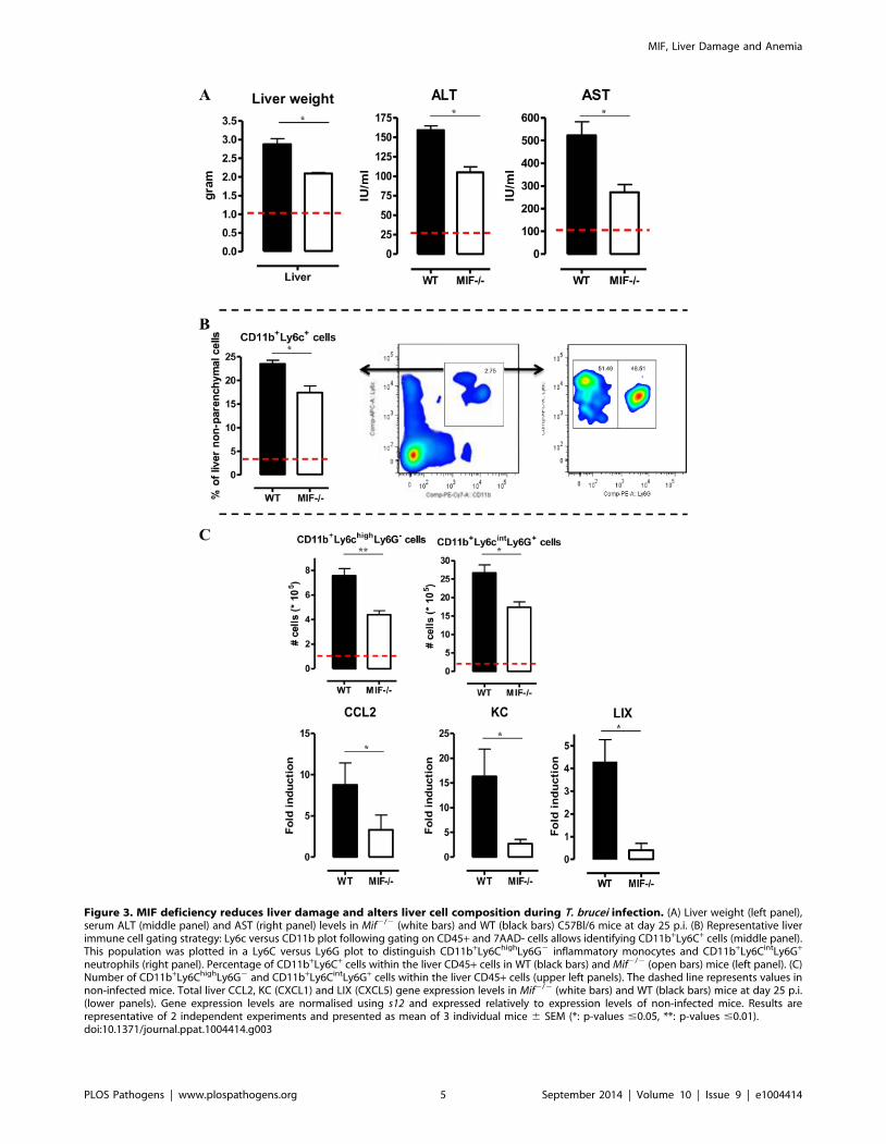
Figure 3. MIF deficiency reduces liver damage and alters liver cell composition during T. brucei infection. (A) Liver weight (left panel),serum ALT (middle panel) and AST (right panel) levels in Mif2/2 (white bars) and WT (black bars) C57Bl/6 mice at day 25 p.i. (B) Representative liverimmune cell gating strategy: Ly6c versus CD11b plot following gating on CD45+ and 7AAD- cells allows identifying CD11b+Ly6C+ cells (middle panel).This population was plotted in a Ly6C versus Ly6G plot to distinguish CD11b+Ly6ChighLy6G2 inflammatory monocytes and CD11b+Ly6CintLy6G+
neutrophils (right panel). Percentage of CD11b+Ly6C+ cells within the liver CD45+ cells in WT (black bars) and Mif2/2 (open bars) mice (left panel). (C)Number of CD11b+Ly6ChighLy6G2 and CD11b+Ly6CintLy6G+ cells within the liver CD45+ cells (upper left panels). The dashed line represents values innon-infected mice. Total liver CCL2, KC (CXCL1) and LIX (CXCL5) gene expression levels in Mif2/2 (white bars) and WT (black bars) mice at day 25 p.i.(lower panels). Gene expression levels are normalised using s12 and expressed relatively to expression levels of non-infected mice. Results arerepresentative of 2 independent experiments and presented as mean of 3 individual mice 6 SEM (*: p-values #0.05, **: p-values #0.01).doi:10.1371/journal.ppat.1004414.g003
MIF, Liver Damage and Anemia
PLOS Pathogens | www.plospathogens.org 5 September 2014 | Volume 10 | Issue 9 | e1004414
Mif2/2 mice (Fig. S5A). Finally, Mif2/2 mice also showed
reduced anemia upon tsetse fly-based infection (Fig. S5B).
5. Mif2/2 mice exhibit restored iron homeostasis duringthe chronic phase of T. brucei infection
Anemia development during persistent inflammation can
result from iron accumulation within the mononuclear phago-
cyte system (MPS) leading to iron deprivation from erythro-
poiesis. Hence, the reduced pro-inflammatory immune re-
sponse in Mif2/2 mice during the chronic stage of T. bruceiinfection as compared to WT mice might impact on iron-
homeostasis and hemoglobin levels. At day 18 p.i., the time-
point when the differences in anemia development and cytokine
levels between WT and Mif2/2 mice become apparent (see
Fig. 6A and Fig. 2C), hemoglobin and serum iron levels were
less reduced in Mif2/2 mice as compared to WT mice
(Fig. 6B). These differences were corroborated at the level of
expression of genes implicated in iron homeostasis in the liver.
In this context, it should be emphasized that under physiolog-
ical conditions, tissue-associated myeloid cells, in particular
liver myeloid cells, recover ferrous iron (Fe2+) via engulfment of
senescent RBCs and hemoglobin recycling. The Fe2+ iron from
hemoglobin is extracted by myeloid cells via heme oxygenase-1
(HO-1), then transported into the cytosol by the divalent metal
transporter 1 (DMT-1/Nramp2), from where it can be either
exported from or stored inside the myeloid cells via ferroportin-
1 (FPN-1) or ferritin (FHC), respectively, to prevent toxicity,
depending on the iron demand of the host [6]. As shown in
Fig. 6C, there was a lower liver HO-1 (Hmox1) gene expression
level in infected Mif2/2 than WT mice (suggesting lower iron
extraction), while the DMT-1 (Dmt1) gene expression levels
were similar in both mouse groups (suggesting similar iron
transport). In addition, the FHC (Fth1) gene expression levels
were lower in infected Mif2/2 mice (Fig. 6C), indicating
reduced iron accumulation in Mif2/2 than WT mice. Finally,
Fpn1 mRNA levels were higher in infected Mif2/2 mice
(Fig. 6C), suggesting that iron export in these mice is less
impaired as compared to WT mice. Together these data
indicate that there is less iron extracted and retained in the
MPS from infected Mif2/2 than WT mice, which in turn
allows increased iron availability for erythropoiesis.
Figure 4. Anti-MIF treatment reduces serum ALT/AST levelsand affects liver cell composition during T. brucei infection. (A)Serum ALT and AST levels of non-infected (dashed line), isotype controlantibody treated (black box) and anti-MIF IgG treated (white box) WTC57Bl/6 mice at day 25 p.i. (B) Total numbers of liver CD11b+Ly6Chigh-Ly6G2 and CD11b+Ly6CintLy6G+ cells in the chronic (day 25 p.i.) phaseof infection calculated using the gating strategy described in Fig. 3B.Results are representative of 2 independent experiments and presentedas mean of 3 individual mice 6 SEM (*: p-values #0.05, **: p-values #0.01).doi:10.1371/journal.ppat.1004414.g004
Figure 5. Neutrophil-derived MIF and monocyte-derived MIFcontribute to different extent to TNF production and liverinjury in T. brucei infected mice. TNF and MIF levels from liver cellcultures (A, B), serum ALT (C) and AST (D) levels of infected (day 24 p.i.)Mif2/2 mice (white box), of infected Mif2/2 mice treated withneutrophils from WT (dark grey box) or Mif2/2(white hatched box)infected mice, of infected Mif2/2 mice treated with monocytes from WT(light grey box) or Mif2/2 (darker grey hatched box) infected mice andof infected WT mice (black box). Results are representative of 3–4independent experiments and presented as mean of 3 individual mice6 SEM (*: p-values #0.05, **: p-values #0.01, ***: p-value #0.001).doi:10.1371/journal.ppat.1004414.g005
MIF, Liver Damage and Anemia
PLOS Pathogens | www.plospathogens.org 6 September 2014 | Volume 10 | Issue 9 | e1004414
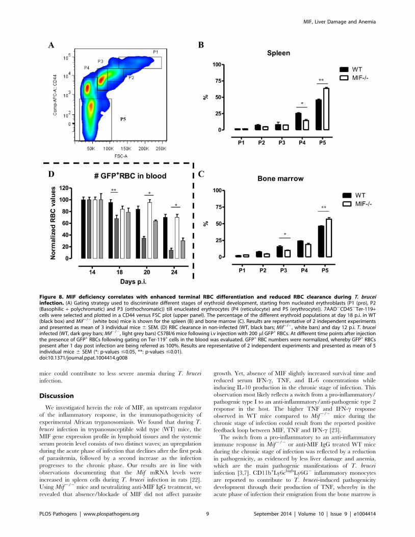
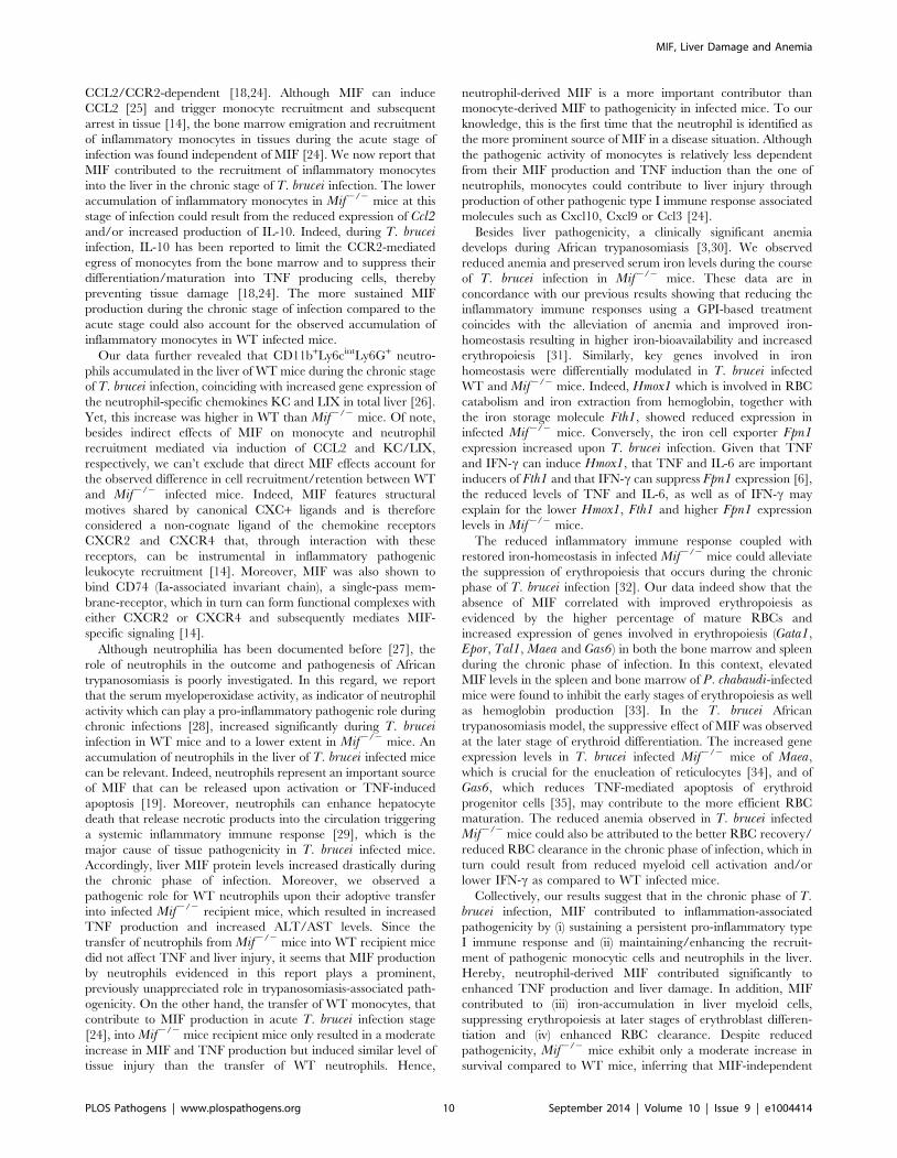
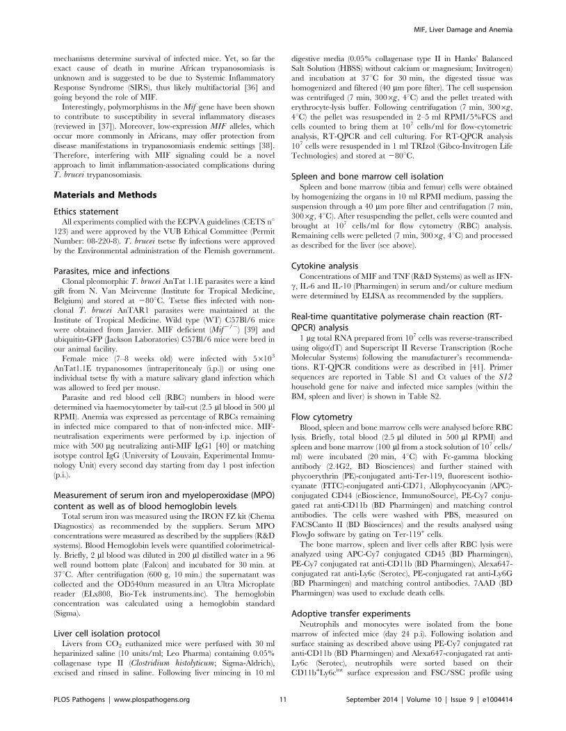
6. Mif2/2 mice display enhanced erythropoiesis duringthe chronic phase of T. brucei infection
The persistent pro-inflammatory immune response in T. bruceiinfection (see Fig. 2C), leading to iron accumulation within the
MPS and iron deprivation (see Fig. 6B-C) could impair erythro-
poiesis. Therefore, the efficiency of the constitutive (i.e. bone
marrow) and inflammation-induced (i.e. spleen) erythropoiesis was
determined in WT and Mif2/2 mice during the chronic stage of
infection (day 18 p.i.) using three different approaches. First, as
shown in Fig. 7A (left panel) we measured the relative abundance
of immature (Ter-119+CD71+) and mature (Ter-119+CD712)
RBCs as described by [20]. Infected Mif2/2 mice had relatively
more mature RBCs in the bone marrow and the spleen than WT
mice (Fig. 7A, right panels). Concomitantly, the reduction in the
percentage of mature RBCs in the blood was less pronounced in
infected Mif2/2 mice than in WT mice. Secondly, we determined
the expression levels of representative genes involved in RBC
differentiation (Gata1, Epor, Tal1, Maea and Gas6) in the bone
marrow and spleen. As shown in Fig. 7B, the expression of most
genes was not induced (with the exception of Tal1) during
infection in WT mice in both organs, while they increased in
infected Mif2/2 mice. Thirdly, we quantified the different stages
of erythropoiesis (from nucleated erythroblasts (P1) till enucleated
erythrocytes (P5)) using a recently described protocol [21] based
on a CD44 versus FSC profile following gating on the Ter-119+
cells (Fig. 8A). It appeared that compared to WT mice, infected
Mif2/2 mice had a more efficient RBC maturation mainly at the
later stage of differentiation (reflected by the increased percentage
of P5) in the spleen and to a lesser extent in the bone marrow
(Fig. 8B-C, respectively). Together, these data demonstrate a
higher level of erythropoiesis in the absence of MIF in T. bruceiinfected mice.
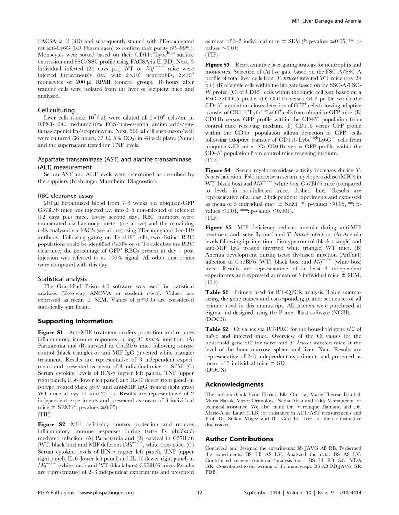
7. Mif2/2 mice display a reduced RBC clearance duringthe chronic phase of T. brucei infection
An increased RBC elimination may also contribute to anemia
in T. brucei infected mice. To address this question, we injected
green fluorescent protein positive (GFP+) RBCs i.v. at the start
of the chronic phase of T. brucei infection (day 12 p.i.) in WT
and Mif2/2 mice and analyzed their clearance as the infection
Figure 6. MIF deficiency correlates with reduced anemia, restored hemoglobin/serum iron levels and restored iron homeostasisduring T. brucei infection. (A) Anemia development during infection in C57Bl/6 (WT, black box; Mif2/2, white box) mice. Results are representativeof 2–5 independent experiments and expressed as mean of 3–5 individual mice 6 SEM. (B) At day 18 p.i., (left panel) hemoglobin levels and (rightpanel) serum iron levels in Mif2/2 (open bars) and WT (black bars) mice. (C) Expression levels of the iron-homeostasis associated genes Hmox1 (ironimport), Dmt1 (iron transport), Fth1 (iron storage) and Fpn1 (iron export) were quantified by RT-QPCR in total livers from Mif2/2 (white bars) and WT(black bars) mice at day 18 p.i. Gene-expression levels are normalised using s12 and expressed relatively to expression levels of non-infected mice.Results are representative of 2 independent experiments and presented as mean of 3 individual mice 6 SEM (*: p-values #0.05).doi:10.1371/journal.ppat.1004414.g006
MIF, Liver Damage and Anemia
PLOS Pathogens | www.plospathogens.org 7 September 2014 | Volume 10 | Issue 9 | e1004414
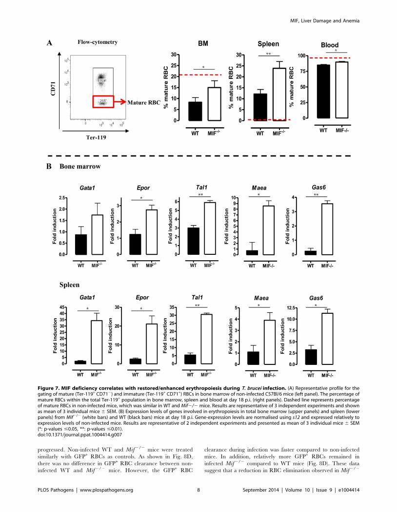
progressed. Non-infected WT and Mif2/2 mice were treated
similarly with GFP+ RBCs as controls. As shown in Fig. 8D,
there was no difference in GFP+ RBC clearance between non-
infected WT and Mif2/2 mice. However, the GFP+ RBC
clearance during infection was faster compared to non-infected
mice. In addition, relatively more GFP+ RBCs remained in
infected Mif2/2 compared to WT mice (Fig. 8D). These data
suggest that a reduction in RBC elimination observed in Mif2/2
Figure 7. MIF deficiency correlates with restored/enhanced erythropoiesis during T. brucei infection. (A) Representative profile for thegating of mature (Ter-119+ CD712) and immature (Ter-119+ CD71+) RBCs in bone marrow of non-infected C57Bl/6 mice (left panel). The percentage ofmature RBCs within the total Ter-119+ population in bone marrow, spleen and blood at day 18 p.i. (right panels). Dashed line represents percentageof mature RBCs in non-infected mice, which was similar in WT and Mif2/2 mice. Results are representative of 3 independent experiments and shownas mean of 3 individual mice 6 SEM. (B) Expression levels of genes involved in erythropoiesis in total bone marrow (upper panels) and spleen (lowerpanels) from Mif2/2 (white bars) and WT (black bars) mice at day 18 p.i. Gene-expression levels are normalised using s12 and expressed relatively toexpression levels of non-infected mice. Results are representative of 2 independent experiments and presented as mean of 3 individual mice 6 SEM(*: p-values #0.05, **: p-values #0.01).doi:10.1371/journal.ppat.1004414.g007
MIF, Liver Damage and Anemia
PLOS Pathogens | www.plospathogens.org 8 September 2014 | Volume 10 | Issue 9 | e1004414
mice could contribute to less severe anemia during T. bruceiinfection.
Discussion
We investigated herein the role of MIF, an upstream regulator
of the inflammatory response, in the immunopathogenicity of
experimental African trypanosomiasis. We found that during T.brucei infection in trypanosusceptible wild type (WT) mice, the
MIF gene expression profile in lymphoid tissues and the systemic
serum protein level consists of two distinct waves; an upregulation
during the acute phase of infection that declines after the first peak
of parasitemia, followed by a second increase as the infection
progresses to the chronic phase. Our results are in line with
observations documenting that the Mif mRNA levels were
increased in spleen cells during T. brucei infection in rats [22].
Using Mif2/2 mice and neutralizing anti-MIF IgG treatment, we
revealed that absence/blockade of MIF did not affect parasite
growth. Yet, absence of MIF slightly increased survival time and
reduced serum IFN-c, TNF, and IL-6 concentrations while
inducing IL-10 production in the chronic stage of infection. This
observation most likely reflects a switch from a pro-inflammatory/
pathogenic type I to an anti-inflammatory/anti-pathogenic type 2
response in the host. The higher TNF and IFN-c response
observed in WT mice compared to Mif2/2 mice during the
chronic stage of infection could result from the reported positive
feedback loop between MIF, TNF and IFN-c [23].
The switch from a pro-inflammatory to an anti-inflammatory
immune response in Mif2/2 or anti-MIF IgG treated WT mice
during the chronic stage of infection was reflected by a reduction
in pathogenicity, as evidenced by less liver damage and anemia,
which are the main pathogenic manifestations of T. bruceiinfection [3,7]. CD11b+Ly6chighLy6G2 inflammatory monocytes
are reported to contribute to T. brucei-induced pathogenicity
development through their production of TNF, whereby in the
acute phase of infection their emigration from the bone marrow is
Figure 8. MIF deficiency correlates with enhanced terminal RBC differentiation and reduced RBC clearance during T. bruceiinfection. (A) Gating strategy used to discriminate different stages of erythroid development, starting from nucleated erythroblasts (P1 (pro), P2(Basophilic + polychromatic) and P3 (orthochromatic)) till enucleated erythrocytes (P4 (reticulocyte) and P5 (erythrocyte)). 7AAD2CD452Ter-119+cells were selected and plotted in a CD44 versus FSC plot (upper panel). The percentage of the different erythroid populations at day 18 p.i. in WT(black box) and Mif2/2 (white box) mice is shown for the spleen (B) and bone marrow (C). Results are representative of 2 independent experimentsand presented as mean of 3 individual mice 6 SEM. (D) RBC clearance in non-infected (WT, black bars; Mif2/2, white bars) and day 12 p.i. T. bruceiinfected (WT, dark grey bars; Mif2/2, light grey bars) C57Bl/6 mice following i.v injection with 200 ml GFP+ RBCs. At different time points after injectionthe presence of GFP+ RBCs following gating on Ter-119+ cells in the blood was evaluated. GFP+ RBC numbers were normalized, whereby GFP+ RBCspresent after 1 day post infection are being referred as 100%. Results are representative of 2 independent experiments and presented as mean of 5individual mice 6 SEM (*: p-values #0.05, **: p-values #0.01).doi:10.1371/journal.ppat.1004414.g008
MIF, Liver Damage and Anemia
PLOS Pathogens | www.plospathogens.org 9 September 2014 | Volume 10 | Issue 9 | e1004414
CCL2/CCR2-dependent [18,24]. Although MIF can induce
CCL2 [25] and trigger monocyte recruitment and subsequent
arrest in tissue [14], the bone marrow emigration and recruitment
of inflammatory monocytes in tissues during the acute stage of
infection was found independent of MIF [24]. We now report that
MIF contributed to the recruitment of inflammatory monocytes
into the liver in the chronic stage of T. brucei infection. The lower
accumulation of inflammatory monocytes in Mif2/2 mice at this
stage of infection could result from the reduced expression of Ccl2and/or increased production of IL-10. Indeed, during T. bruceiinfection, IL-10 has been reported to limit the CCR2-mediated
egress of monocytes from the bone marrow and to suppress their
differentiation/maturation into TNF producing cells, thereby
preventing tissue damage [18,24]. The more sustained MIF
production during the chronic stage of infection compared to the
acute stage could also account for the observed accumulation of
inflammatory monocytes in WT infected mice.
Our data further revealed that CD11b+Ly6cintLy6G+ neutro-
phils accumulated in the liver of WT mice during the chronic stage
of T. brucei infection, coinciding with increased gene expression of
the neutrophil-specific chemokines KC and LIX in total liver [26].
Yet, this increase was higher in WT than Mif2/2 mice. Of note,
besides indirect effects of MIF on monocyte and neutrophil
recruitment mediated via induction of CCL2 and KC/LIX,
respectively, we can’t exclude that direct MIF effects account for
the observed difference in cell recruitment/retention between WT
and Mif2/2 infected mice. Indeed, MIF features structural
motives shared by canonical CXC+ ligands and is therefore
considered a non-cognate ligand of the chemokine receptors
CXCR2 and CXCR4 that, through interaction with these
receptors, can be instrumental in inflammatory pathogenic
leukocyte recruitment [14]. Moreover, MIF was also shown to
bind CD74 (Ia-associated invariant chain), a single-pass mem-
brane-receptor, which in turn can form functional complexes with
either CXCR2 or CXCR4 and subsequently mediates MIF-
specific signaling [14].
Although neutrophilia has been documented before [27], the
role of neutrophils in the outcome and pathogenesis of African
trypanosomiasis is poorly investigated. In this regard, we report
that the serum myeloperoxidase activity, as indicator of neutrophil
activity which can play a pro-inflammatory pathogenic role during
chronic infections [28], increased significantly during T. bruceiinfection in WT mice and to a lower extent in Mif2/2 mice. An
accumulation of neutrophils in the liver of T. brucei infected mice
can be relevant. Indeed, neutrophils represent an important source
of MIF that can be released upon activation or TNF-induced
apoptosis [19]. Moreover, neutrophils can enhance hepatocyte
death that release necrotic products into the circulation triggering
a systemic inflammatory immune response [29], which is the
major cause of tissue pathogenicity in T. brucei infected mice.
Accordingly, liver MIF protein levels increased drastically during
the chronic phase of infection. Moreover, we observed a
pathogenic role for WT neutrophils upon their adoptive transfer
into infected Mif2/2 recipient mice, which resulted in increased
TNF production and increased ALT/AST levels. Since the
transfer of neutrophils from Mif2/2 mice into WT recipient mice
did not affect TNF and liver injury, it seems that MIF production
by neutrophils evidenced in this report plays a prominent,
previously unappreciated role in trypanosomiasis-associated path-
ogenicity. On the other hand, the transfer of WT monocytes, that
contribute to MIF production in acute T. brucei infection stage
[24], into Mif2/2 mice recipient mice only resulted in a moderate
increase in MIF and TNF production but induced similar level of
tissue injury than the transfer of WT neutrophils. Hence,
neutrophil-derived MIF is a more important contributor than
monocyte-derived MIF to pathogenicity in infected mice. To our
knowledge, this is the first time that the neutrophil is identified as
the more prominent source of MIF in a disease situation. Although
the pathogenic activity of monocytes is relatively less dependent
from their MIF production and TNF induction than the one of
neutrophils, monocytes could contribute to liver injury through
production of other pathogenic type I immune response associated
molecules such as Cxcl10, Cxcl9 or Ccl3 [24].
Besides liver pathogenicity, a clinically significant anemia
develops during African trypanosomiasis [3,30]. We observed
reduced anemia and preserved serum iron levels during the course
of T. brucei infection in Mif2/2 mice. These data are in
concordance with our previous results showing that reducing the
inflammatory immune responses using a GPI-based treatment
coincides with the alleviation of anemia and improved iron-
homeostasis resulting in higher iron-bioavailability and increased
erythropoiesis [31]. Similarly, key genes involved in iron
homeostasis were differentially modulated in T. brucei infected
WT and Mif2/2 mice. Indeed, Hmox1 which is involved in RBC
catabolism and iron extraction from hemoglobin, together with
the iron storage molecule Fth1, showed reduced expression in
infected Mif2/2 mice. Conversely, the iron cell exporter Fpn1expression increased upon T. brucei infection. Given that TNF
and IFN-c can induce Hmox1, that TNF and IL-6 are important
inducers of Fth1 and that IFN-c can suppress Fpn1 expression [6],
the reduced levels of TNF and IL-6, as well as of IFN-c may
explain for the lower Hmox1, Fth1 and higher Fpn1 expression
levels in Mif2/2 mice.
The reduced inflammatory immune response coupled with
restored iron-homeostasis in infected Mif2/2 mice could alleviate
the suppression of erythropoiesis that occurs during the chronic
phase of T. brucei infection [32]. Our data indeed show that the
absence of MIF correlated with improved erythropoiesis as
evidenced by the higher percentage of mature RBCs and
increased expression of genes involved in erythropoiesis (Gata1,
Epor, Tal1, Maea and Gas6) in both the bone marrow and spleen
during the chronic phase of infection. In this context, elevated
MIF levels in the spleen and bone marrow of P. chabaudi-infected
mice were found to inhibit the early stages of erythropoiesis as well
as hemoglobin production [33]. In the T. brucei African
trypanosomiasis model, the suppressive effect of MIF was observed
at the later stage of erythroid differentiation. The increased gene
expression levels in T. brucei infected Mif2/2 mice of Maea,
which is crucial for the enucleation of reticulocytes [34], and of
Gas6, which reduces TNF-mediated apoptosis of erythroid
progenitor cells [35], may contribute to the more efficient RBC
maturation. The reduced anemia observed in T. brucei infected
Mif2/2 mice could also be attributed to the better RBC recovery/
reduced RBC clearance in the chronic phase of infection, which in
turn could result from reduced myeloid cell activation and/or
lower IFN-c as compared to WT infected mice.
Collectively, our results suggest that in the chronic phase of T.brucei infection, MIF contributed to inflammation-associated
pathogenicity by (i) sustaining a persistent pro-inflammatory type
I immune response and (ii) maintaining/enhancing the recruit-
ment of pathogenic monocytic cells and neutrophils in the liver.
Hereby, neutrophil-derived MIF contributed significantly to
enhanced TNF production and liver damage. In addition, MIF
contributed to (iii) iron-accumulation in liver myeloid cells,
suppressing erythropoiesis at later stages of erythroblast differen-
tiation and (iv) enhanced RBC clearance. Despite reduced
pathogenicity, Mif2/2 mice exhibit only a moderate increase in
survival compared to WT mice, inferring that MIF-independent
MIF, Liver Damage and Anemia
PLOS Pathogens | www.plospathogens.org 10 September 2014 | Volume 10 | Issue 9 | e1004414
mechanisms determine survival of infected mice. Yet, so far the
exact cause of death in murine African trypanosomiasis is
unknown and is suggested to be due to Systemic Inflammatory
Response Syndrome (SIRS), thus likely multifactorial [36] and
going beyond the role of MIF.
Interestingly, polymorphisms in the Mif gene have been shown
to contribute to susceptibility in several inflammatory diseases
(reviewed in [37]). Moreover, low-expression MIF alleles, which
occur more commonly in Africans, may offer protection from
disease manifestations in trypanosomiasis endemic settings [38].
Therefore, interfering with MIF signaling could be a novel
approach to limit inflammation-associated complications during
T. brucei trypanosomiasis.
Materials and Methods
Ethics statementAll experiments complied with the ECPVA guidelines (CETS nu
123) and were approved by the VUB Ethical Committee (Permit
Number: 08-220-8). T. brucei tsetse fly infections were approved
by the Environmental administration of the Flemish government.
Parasites, mice and infectionsClonal pleomorphic T. brucei AnTat 1.1E parasites were a kind
gift from N. Van Meirvenne (Institute for Tropical Medicine,
Belgium) and stored at 280uC. Tsetse flies infected with non-
clonal T. brucei AnTAR1 parasites were maintained at the
Institute of Tropical Medicine. Wild type (WT) C57Bl/6 mice
were obtained from Janvier. MIF deficient (Mif2/2) [39] and
ubiquitin-GFP (Jackson Laboratories) C57Bl/6 mice were bred in
our animal facility.
Female mice (7–8 weeks old) were infected with 56103
AnTat1.1E trypanosomes (intraperitonealy (i.p.)) or using one
individual tsetse fly with a mature salivary gland infection which
was allowed to feed per mouse.
Parasite and red blood cell (RBC) numbers in blood were
determined via haemocytometer by tail-cut (2.5 ml blood in 500 ml
RPMI). Anemia was expressed as percentage of RBCs remaining
in infected mice compared to that of non-infected mice. MIF-
neutralisation experiments were performed by i.p. injection of
mice with 500 mg neutralizing anti-MIF IgG1 [40] or matching
isotype control IgG (University of Louvain, Experimental Immu-
nology Unit) every second day starting from day 1 post infection
(p.i.).
Measurement of serum iron and myeloperoxidase (MPO)content as well as of blood hemoglobin levels
Total serum iron was measured using the IRON FZ kit (Chema
Diagnostics) as recommended by the suppliers. Serum MPO
concentrations were measured as described by the suppliers (R&D
systems). Blood Hemoglobin levels were quantified colorimetrical-
ly. Briefly, 2 ml blood was diluted in 200 ml distilled water in a 96
well round bottom plate (Falcon) and incubated for 30 min. at
37uC. After centrifugation (600 g, 10 min.) the supernatant was
collected and the OD540nm measured in an Ultra Microplate
reader (ELx808, Bio-Tek instruments.inc). The hemoglobin
concentration was calculated using a hemoglobin standard
(Sigma).
Liver cell isolation protocolLivers from CO2 euthanized mice were perfused with 30 ml
heparinized saline (10 units/ml; Leo Pharma) containing 0.05%
collagenase type II (Clostridium histolyticum; Sigma-Aldrich),
excised and rinsed in saline. Following liver mincing in 10 ml
digestive media (0.05% collagenase type II in Hanks’ Balanced
Salt Solution (HBSS) without calcium or magnesium; Invitrogen)
and incubation at 37uC for 30 min, the digested tissue was
homogenized and filtered (40 mm pore filter). The cell suspension
was centrifuged (7 min, 3006g, 4uC) and the pellet treated with
erythrocyte-lysis buffer. Following centrifugation (7 min, 3006g,
4uC) the pellet was resuspended in 2–5 ml RPMI/5%FCS and
cells counted to bring them at 107 cells/ml for flow-cytometric
analysis, RT-QPCR and cell culturing. For RT-QPCR analysis
107 cells were resuspended in 1 ml TRIzol (Gibco-Invitrogen Life
Technologies) and stored at 280uC.
Spleen and bone marrow cell isolationSpleen and bone marrow (tibia and femur) cells were obtained
by homogenizing the organs in 10 ml RPMI medium, passing the
suspension through a 40 mm pore filter and centrifugation (7 min,
3006g, 4uC). After resuspending the pellet, cells were counted and
brought at 107 cells/ml for flow cytometry (RBC) analysis.
Remaining cells were pelleted (7 min, 3006g, 4uC) and processed
as described for the liver (see above).
Cytokine analysisConcentrations of MIF and TNF (R&D Systems) as well as IFN-
c, IL-6 and IL-10 (Pharmingen) in serum and/or culture medium
were determined by ELISA as recommended by the suppliers.
Real-time quantitative polymerase chain reaction (RT-QPCR) analysis
1 mg total RNA prepared from 107 cells was reverse-transcribed
using oligo(dT) and Superscript II Reverse Transcription (Roche
Molecular Systems) following the manufacturer’s recommenda-
tions. RT-QPCR conditions were as described in [41]. Primer
sequences are reported in Table S1 and Ct values of the S12household gene for naive and infected mice samples (within the
BM, spleen and liver) is shown in Table S2.
Flow cytometryBlood, spleen and bone marrow cells were analysed before RBC
lysis. Briefly, total blood (2.5 ml diluted in 500 ml RPMI) and
spleen and bone marrow (100 ml from a stock solution of 107 cells/
ml) were incubated (20 min, 4uC) with Fc-gamma blocking
antibody (2.4G2, BD Biosciences) and further stained with
phycoerythrin (PE)-conjugated anti-Ter-119, fluorescent isothio-
cyanate (FITC)-conjugated anti-CD71, Allophycocyanin (APC)-
conjugated CD44 (eBioscience, ImmunoSource), PE-Cy7 conju-
gated rat anti-CD11b (BD Pharmingen) and matching control
antibodies. The cells were washed with PBS, measured on
FACSCanto II (BD Biosciences) and the results analysed using
FlowJo software by gating on Ter-119+ cells.
The bone marrow, spleen and liver cells after RBC lysis were
analyzed using APC-Cy7 conjugated CD45 (BD Pharmingen),
PE-Cy7 conjugated rat anti-CD11b (BD Pharmingen), Alexa647-
conjugated rat anti-Ly6c (Serotec), PE-conjugated rat anti-Ly6G
(BD Pharmingen) and matching control antibodies. 7AAD (BD
Pharmingen) was used to exclude death cells.
Adoptive transfer experimentsNeutrophils and monocytes were isolated from the bone
marrow of infected mice (day 24 p.i). Following isolation and
surface staining as described above using PE-Cy7 conjugated rat
anti-CD11b (BD Pharmingen) and Alexa647-conjugated rat anti-
Ly6c (Serotec), neutrophils were sorted based on their
CD11b+Ly6cint surface expression and FSC/SSC profile using
MIF, Liver Damage and Anemia
PLOS Pathogens | www.plospathogens.org 11 September 2014 | Volume 10 | Issue 9 | e1004414
FACSAria II (BD) and subsequently stained with PE-conjugated
rat anti-Ly6G (BD Pharmingen) to confirm their purity (95–99%).
Monocytes were sorted based on their CD11b+Ly6chigh surface
expression and FSC/SSC profile using FACSAria II (BD). Next, 3
individual infected (24 days p.i.) WT or Mif2/2 mice were
injected intravenously (i.v.) with 26106 neutrophils, 26106
monocytes or 200 ml RPMI (control group). 18 hours after
transfer cells were isolated from the liver of recipient mice and
analyzed.
Cell culturingLiver cells (stock: 107/ml) were diluted till 26106 cells/ml in
RPMI-1640 medium/10% FCS/non-essential amino acids/glu-
tamate/penicillin/streptomycin. Next, 500 ml cell suspension/well
were cultured (36 hours, 37uC, 5% CO2) in 48 well plates (Nunc)
and the supernatant tested for TNF levels.
Aspartate transaminase (AST) and alanine transaminase(ALT) measurement
Serum AST and ALT levels were determined as described by
the suppliers (Boehringer Mannheim Diagnostics).
RBC clearance assay200 ml heparinized blood from 7–8 weeks old ubiquitin-GFP
C57Bl/6 mice was injected i.v. into 3–5 non-infected or infected
(12 days p.i.) mice. Every second day, RBC numbers were
enumerated via haemocytometer (see above) and the remaining
cells analysed via FACS (see above) using PE-conjugated Ter-119
antibody. Following gating on Ter-119+ cells, two distinct RBC
populations could be identified (GFP+ or -). To calculate the RBC
clearance, the percentage of GFP+ RBCs present at day 1 post
injection was referred to as 100% signal. All other time-points
were compared with this day.
Statistical analysisThe GraphPad Prism 4.0 software was used for statistical
analyses (Two-way ANOVA or student t-test). Values are
expressed as mean 6 SEM. Values of p#0.05 are considered
statistically significant.
Supporting Information
Figure S1 Anti-MIF treatment confers protection and reduces
inflammatory immune responses during T. brucei infection. (A)
Parasitemia and (B) survival in C57Bl/6 mice following isotype
control (black triangle) or anti-MIF IgG (inverted white triangle)
treatment. Results are representative of 3 independent experi-
ments and presented as mean of 3 individual mice 6 SEM. (C)
Serum cytokine levels of IFN-c (upper left panel), TNF (upper
right panel), IL-6 (lower left panel) and IL-10 (lower right panel) in
isotype treated (dark grey) and anti-MIF IgG treated (light gray)
WT mice at day 11 and 25 p.i. Results are representative of 2
independent experiments and presented as mean of 3 individual
mice 6 SEM (*: p-values #0.05).
(TIF)
Figure S2 MIF deficiency confers protection and reduces
inflammatory immune responses during tsetse fly (AnTar1)
mediated infection. (A) Parasitemia and (B) survival in C57Bl/6
(WT, black box) and MIF deficient (Mif2/2, white box) mice. (C)
Serum cytokine levels of IFN-c (upper left panel), TNF (upper
right panel), IL-6 (lower left panel) and IL-10 (lower right panel) in
Mif2/2 (white bars) and WT (black bars) C57Bl/6 mice. Results
are representative of 2–3 independent experiments and presented
as mean of 3–5 individual mice 6 SEM (*: p-values #0.05, **: p-
values #0.01).
(TIF)
Figure S3 Representative liver gating strategy for neutrophils and
monocytes. Selection of (A) live gate based on the FSC-A/SSC-A
profile of total liver cells from T. brucei infected WT mice (day 24
p.i.); (B) of single cells within the life gate based on the SSC-A/FSC-
W profile; (C) of CD45+ cells within the single cell gate based on a
FSC-A/CD45 profile. (D) CD11b versus GFP profile within the
CD45+ population allows detection of GFP+ cells following adoptive
transfer of CD11b+Ly6cintLy6G+ cells from ubiquitin-GFP mice. (E)
CD11b versus GFP profile within the CD45+ population from
control mice receiving medium. (F) CD11b versus GFP profile
within the CD45+ population allows detection of GFP+ cells
following adoptive transfer of CD11b+Ly6chighLy6G2 cells from
ubiquitin-GFP mice. (G) CD11b versus GFP profile within the
CD45+ population from control mice receiving medium.
(TIF)
Figure S4 Serum myeloperoxidase activity increases during T.brucei infection. Fold increase in serum myeloperoxidase (MPO) in
WT (black box) and Mif2/2 (white box) C57Bl/6 mice (compared
to levels in non-infected mice, dashed line). Results are
representative of at least 2 independent experiments and expressed
as mean of 5 individual mice 6 SEM. (*: p-values #0.05, **: p-
values #0.01, ***: p-values #0.001).
(TIF)
Figure S5 MIF deficiency reduces anemia during anti-MIF
treatment and tsetse fly mediated T. brucei infection. (A) Anemia
levels following i.p. injection of isotype control (black triangle) and
anti-MIF IgG treated (inverted white triangle) WT mice. (B)
Anemia development during tsetse fly-based infection (AnTar1)
infection in C57Bl/6 (WT) (black box) and Mif2/2 (white box)
mice. Results are representative of at least 3 independent
experiments and expressed as mean of 5 individual mice 6 SEM.
(TIF)
Table S1 Primers used for RT-QPCR analysis. Table summa-
rizing the gene names and corresponding primer sequences of all
primers used in this manuscript. All primers were purchased at
Sigma and designed using the Primer-Blast software (NCBI).
(DOCX)
Table S2 Ct values via RT-PRC for the household gene s12 of
naı̈ve and infected mice. Overview of the Ct values for the
household gene s12 for naı̈ve and T. brucei infected mice at the
level of the bone marrow, spleen and liver. Note: Results are
representative of 2–3 independent experiments and presented as
mean of 3 individual mice 6 SD.
(DOCX)
Acknowledgments
The authors thank Yvon Elkrim, Ella Omasta, Marie-Therese Detobel,
Maria Slazak, Victor Orimoloye, Nadia Abou and Eddy Vercauteren for
technical assistance. We also thank Dr. Veronique Flamand and Dr.
Marie-Aline Laute (ULB) for assistance in ALT/AST measurements and
Prof. Dr. Stefan Magez and Dr. Carl De Trez for their constructive
discussions.
Author Contributions
Conceived and designed the experiments: BS JAVG AB RB. Performed
the experiments: BS LB AS LV. Analyzed the data: BS AS LV.
Contributed reagents/materials/analysis tools: BS LL RB GC JVDA
GR. Contributed to the writing of the manuscript: BS AB RB JAVG GR
PDB.
MIF, Liver Damage and Anemia
PLOS Pathogens | www.plospathogens.org 12 September 2014 | Volume 10 | Issue 9 | e1004414
References
1. Barrett MP, Burchmore RJ, Stich A, Lazzari JO, Frasch AC, et al. (2003) The
trypanosomiases. Lancet 362: 1469–1480.2. d’Ieteren GD, Authie E, Wissocq N, Murray M (1998) Trypanotolerance, an
option for sustainable livestock production in areas at risk from trypanosomosis.Rev Sci Tech 17: 154–175.
3. Stijlemans B, Vankrunkelsven A, Caljon G, Bockstal V, Guilliams M, et al.
(2010) The central role of macrophages in trypanosomiasis-associated anemia:rationale for therapeutical approaches. Endocr Metab Immune Disord Drug
Targets 10: 71–82.4. Naessens J (2006) Bovine trypanotolerance: A natural ability to prevent severe
anaemia and haemophagocytic syndrome? Int J Parasitol 36: 521–528.
5. Stijlemans B, Vankrunkelsven A, Brys L, Magez S, De Baetselier P (2008) Roleof iron homeostasis in trypanosomiasis-associated anemia. Immunobiology 213:
823–835.6. Weiss G, Goodnough LT (2005) Anemia of chronic disease. N Engl J Med 352:
1011–1023.7. Bosschaerts T, Guilliams M, Stijlemans B, De Baetselier P, Beschin A (2009)
Understanding the role of monocytic cells in liver inflammation using parasite
infection as a model. Immunobiology 214: 737–747.8. Magez S, Stijlemans B, Baral T, De Baetselier P (2002) VSG-GPI anchors of
African trypanosomes: their role in macrophage activation and induction ofinfection-associated immunopathology. Microbes Infect 4: 999–1006.
9. Stijlemans B, Baral TN, Guilliams M, Brys L, Korf J, et al. (2007) A
glycosylphosphatidylinositol-based treatment alleviates trypanosomiasis-associat-ed immunopathology. J Immunol 179: 4003–4014.
10. Calandra T, Roger T (2003) Macrophage migration inhibitory factor: aregulator of innate immunity. Nat Rev Immunol 3: 791–800.
11. Larson DF, Horak K (2006) Macrophage migration inhibitory factor: controllerof systemic inflammation. Crit Care 10: 138.
12. Ayoub S, Hickey MJ, Morand EF (2008) Mechanisms of disease: macrophage
migration inhibitory factor in SLE, RA and atherosclerosis. Nat Clin PractRheumatol 4: 98–105.
13. Rosado Jde D, Rodriguez-Sosa M (2011) Macrophage migration inhibitoryfactor (MIF): a key player in protozoan infections. Int J Biol Sci 7: 1239–1256.
14. Bernhagen J, Krohn R, Lue H, Gregory JL, Zernecke A, et al. (2007) MIF is a
noncognate ligand of CXC chemokine receptors in inflammatory andatherogenic cell recruitment. Nat Med 13: 587–596.
15. Calandra T, Bernhagen J, Metz CN, Spiegel LA, Bacher M, et al. (1995) MIF asa glucocorticoid-induced modulator of cytokine production. Nature 377: 68–71.
16. Mitchell RA, Liao H, Chesney J, Fingerle-Rowson G, Baugh J, et al. (2002)Macrophage migration inhibitory factor (MIF) sustains macrophage proin-
flammatory function by inhibiting p53: regulatory role in the innate immune
response. Proc Natl Acad Sci U S A 99: 345–350.17. Magez S, Truyens C, Merimi M, Radwanska M, Stijlemans B, et al. (2004) P75
tumor necrosis factor-receptor shedding occurs as a protective host responseduring African trypanosomiasis. J Infect Dis 189: 527–539.
18. Guilliams M, Movahedi K, Bosschaerts T, VandenDriessche T, Chuah MK, et
al. (2009) IL-10 dampens TNF/inducible nitric oxide synthase-producingdendritic cell-mediated pathogenicity during parasitic infection. J Immunol 182:
1107–1118.19. Daryadel A, Grifone RF, Simon HU, Yousefi S (2006) Apoptotic neutrophils
release macrophage migration inhibitory factor upon stimulation with tumornecrosis factor-alpha. J Biol Chem 281: 27653–27661.
20. Socolovsky M, Nam H, Fleming MD, Haase VH, Brugnara C, et al. (2001)
Ineffective erythropoiesis in Stat5a(2/2)5b(2/2) mice due to decreasedsurvival of early erythroblasts. Blood 98: 3261–3273.
21. Liu J, Zhang J, Ginzburg Y, Li H, Xue F, et al. (2013) Quantitative analysis ofmurine terminal erythroid differentiation in vivo: novel method to study normal
and disordered erythropoiesis. Blood 121: e43–49.
22. Nishimura K, Nakaya H, Nakagawa H, Matsuo S, Ohnishi Y, et al. (2011)Differential effects of Trypanosoma brucei gambiense and Trypanosoma brucei
brucei on rat macrophages. J Parasitol 97: 48–54.
23. Calandra T, Bernhagen J, Mitchell RA, Bucala R (1994) The macrophage is an
important and previously unrecognized source of macrophage migration
inhibitory factor. J Exp Med 179: 1895–1902.
24. Bosschaerts T, Guilliams M, Stijlemans B, Morias Y, Engel D, et al. (2010) Tip-
DC development during parasitic infection is regulated by IL-10 and requires
CCL2/CCR2, IFN-gamma and MyD88 signaling. PLoS Pathog 6: e1001045.
25. Gregory JL, Morand EF, McKeown SJ, Ralph JA, Hall P, et al. (2006)
Macrophage migration inhibitory factor induces macrophage recruitment via
CC chemokine ligand 2. J Immunol 177: 8072–8079.
26. Lin M, Carlson E, Diaconu E, Pearlman E (2007) CXCL1/KC and CXCL5/
LIX are selectively produced by corneal fibroblasts and mediate neutrophil
infiltration to the corneal stroma in LPS keratitis. J Leukoc Biol 81: 786–792.
27. Oka M, Nagasawa H, Ito Y, Himeno K (1989) Granulocyte-macrophage
colony-stimulating activity in the serum of mice stimulated with homogenates of
Trypanosoma gambiense. Clin Exp Immunol 78: 285–291.
28. Klebanoff SJ (2005) Myeloperoxidase: friend and foe. J Leukoc Biol 77: 598–
625.
29. Marques PE, Amaral SS, Pires DA, Nogueira LL, Soriani FM, et al. (2012)
Chemokines and mitochondrial products activate neutrophils to amplify organ
injury during mouse acute liver failure. Hepatology 56: 1971–1982.
30. Naessens J, Kitani H, Nakamura Y, Yagi Y, Sekikawa K, et al. (2005) TNF-
alpha mediates the development of anaemia in a murine Trypanosoma brucei
rhodesiense infection, but not the anaemia associated with a murine
Trypanosoma congolense infection. Clin Exp Immunol 139: 405–410.
31. Stijlemans B, Vankrunkelsven A, Brys L, Raes G, Magez S, et al. (2010)
Scrutinizing the mechanisms underlying the induction of anemia of inflamma-
tion through GPI-mediated modulation of macrophage activation in a model of
African trypanosomiasis. Microbes Infect 12: 389–399.
32. Nishimura K, Nakaya H, Nakagawa H, Matsuo S, Ohnishi Y, et al. (2011) Effect
of Trypanosoma brucei brucei on erythropoiesis in infected rats. J Parasitol 97:
88–93.
33. McDevitt MA, Xie J, Shanmugasundaram G, Griffith J, Liu A, et al. (2006) A
critical role for the host mediator macrophage migration inhibitory factor in the
pathogenesis of malarial anemia. J Exp Med 203: 1185–1196.
34. Soni S, Bala S, Gwynn B, Sahr KE, Peters LL, et al. (2006) Absence of
erythroblast macrophage protein (Emp) leads to failure of erythroblast nuclear
extrusion. J Biol Chem 281: 20181–20189.
35. Angelillo-Scherrer A, Burnier L, Lambrechts D, Fish RJ, Tjwa M, et al. (2008)
Role of Gas6 in erythropoiesis and anemia in mice. J Clin Invest 118: 583–596.
36. De Muylder G, Daulouede S, Lecordier L, Uzureau P, Morias Y, et al. (2013) A
Trypanosoma brucei kinesin heavy chain promotes parasite growth by triggering
host arginase activity. PLoS Pathog 9: e1003731.
37. Renner P, Roger T, Calandra T (2005) Macrophage migration inhibitory factor:
gene polymorphisms and susceptibility to inflammatory diseases. Clin Infect Dis
41 Suppl 7: S513–519.
38. Zhong XB, Leng L, Beitin A, Chen R, McDonald C, et al. (2005) Simultaneous
detection of microsatellite repeats and SNPs in the macrophage migration
inhibitory factor (MIF) gene by thin-film biosensor chips and application to rural
field studies. Nucleic Acids Res 33: e121.
39. Fingerle-Rowson G, Petrenko O, Metz CN, Forsthuber TG, Mitchell R, et al.
(2003) The p53-dependent effects of macrophage migration inhibitory factor
revealed by gene targeting. Proc Natl Acad Sci U S A 100: 9354–9359.
40. Calandra T, Echtenacher B, Roy DL, Pugin J, Metz CN, et al. (2000) Protection
from septic shock by neutralization of macrophage migration inhibitory factor.
Nat Med 6: 164–170.
41. Raes G, Brys L, Dahal BK, Brandt J, Grooten J, et al. (2005) Macrophage
galactose-type C-type lectins as novel markers for alternatively activated
macrophages elicited by parasitic infections and allergic airway inflammation.
J Leukoc Biol 77: 321–327.
MIF, Liver Damage and Anemia
PLOS Pathogens | www.plospathogens.org 13 September 2014 | Volume 10 | Issue 9 | e1004414


![Trypanosoma [1]](https://img.pdfslide.net/doc/110x75/58cedaba1a28abd4098b6285/trypanosoma-1.jpg)