Embed Size (px)
Citation preview
13
Cellular Degradation Machineries in Age-Related Loss of Muscle Mass (Sarcopenia)
Mikael Altun1, Max Grönholdt-Klein1, Lingzhan Wang1,2 and Brun Ulfhake1
1Department of Neuroscience, Karolinska Institutet, Stockholm, 2Department of Human Anatomy, School of Medicine,
Inner Mongolia University for Nationalities, Tongliao City, 1Sweden
2P. R. China
1. Introduction
One of the most characteristic features of the aged is a change in body composition and human cross-sectional and longitudinal studies consistently demonstrate gain in fat mass and decline in lean mass (Attaix et al., 2005). Skeletal muscle mass is gradually reduced through both atrophy and loss of myofibers, and along with this connective tissue and intramyocellular lipids increase (Fig. 1) (St-Onge, 2005). Although sarcopenia is widely recognized it remains poorly understood and has not received appropriate attention until quite recently. Sarcopenia is often assumed to have a multi-factorial background (Adamo and Farrar, 2006; Attaix et al., 2005; Paddon-Jones and Rasmussen, 2009; Roth et al., 2006; Solomon and Bouloux, 2006). A sedentary life-style combined with periods of prolonged bed-rest during illnesses has well-established detrimental effects on muscle mass and function (Janssen et al., 2002). Skeletal muscles do not only generate the power to let us move but collectively they are a highly significant component of the systemic metabolic homeostasis machinery. The progressive loss of muscle mass with advancing age is evident in both the ‘healthy’ aging population and specialist patient populations. The reduced functional muscle mass is associated with increased morbidity, frailty and reduced quality of life (Baumgartner et al., 1998a). The prevalence of sarcopenia increases with about 5% per year and typically begins in the fourth decade of life (Baumgartner et al., 1998b). After the age of 50 years 1-2 % of the muscle mass is lost annually. Individuals with clinically manifest sarcopenia have 4 times greater risk of disability, three times greater risk of balance impairment, and 3 times greater risk of falling (Baumgartner et al., 1998b). Sarcopenia is the single most common etiology to falls and fall-associated fractures in elderly.
1.1 Regulation of muscle mass and tentative mechanisms in sarcopenia
Myofibers are complex structures build-up by merger of aligned myocytes (myotubes as intermediates). Thus, the myofiber has a common plasma membrane confining multiple sub domains each supported by one myonucleus. According to morphometric analysis the ratio myofiber volume to a myonucleus remains fairly constant suggesting an optimal size of the
www.intechopen.com

Senescence 270
domain supported by the machinery of one myonucleus. The impressive increase in muscle mass during late fetal and postnatal development but also in conditions of repair in adult muscle across life-span is dependent on recruitment of myogenic progenitors from the resident satellite cells (SC) in the stem cell niche (Kadi et al., 2005). SCs remain quiescent until activation through trophic stimuli e.g. IGF-1 and Notch-related signaling (Carlson et al., 2008). The activity of the SC pool is under the influence of several signaling pathways,
enhancing or inhibiting SC proliferation, such as Wnt-Catenin, DeltaL/Jagged1-Notch or
Smad2/3- TGF-/Activin/Myostatin (Carlson et al., 2008; Zammit, 2008).
Fig. 1. Changes in fiber size and occurrence of nuclei with a central location in aged rat
soleus muscle. Eosin-Htx staining of soleus cross sections (8m) showing (A) young adult muscle where central nuclei (arrow) and very small fibers (double arrow) are infrequent. In early aging (B) fiber size becomes more irregular and the frequency of very small fibers increases (arrows). At advanced age (C) fiber size is highly irregular and centrally located nuclei are frequently occurring. Original micrographs shot with a x20 dry objective.
A B
C
www.intechopen.com
Cellular Degradation Machineries in Age-Related Loss of Muscle Mass (Sarcopenia) 271
The signaling events leading to myofiber atrophy converge onto members of the FOXO family of transcription factors, which in an active state induce atrophy by increased proteasome degradation of myofibrillar proteins (Bodine et al., 2001; Sacheck et al., 2007; Sacheck et al., 2004; Sandri et al., 2004; Stitt et al., 2004). Recently, it was discovered that the signaling pathway that activates FOXO also induces an increased degradation through autophagy and lysosomal degradation (Sandri et al., 2006; Solomon and Bouloux, 2006). This “atrophy program” is activated in a range of conditions such as disuse, denervation and systemic diseases (Solomon and Bouloux, 2006). Conversely, myofiber contraction and growth signals (e.g. IGF-1) acting via protein kinase B, mTOR, and pgc-1 induce synthesis of myofibrillar proteins, deactivation of FOXO and, in parallel, adaptation of the energy producing machinery (idem). The Activin A/myostatin pathway, acting on Act receptor IIB upstream of Smad, can activate myofibrillar proteolysis (i.e. induce muscle fiber atrophy) and depress SC proliferation (Zhou et al., 2010). This creates a regulatory link between cellular anabolism & catabolism and SC activation & deactivation which should work in concert in conditions with atrophy and hypertrophy, respectively.
A growing body of evidence suggests that loss of muscle mass in elderly occurs through mechanisms more complex than those involved in disuse and disease atrophy in younger individuals (Altun et al., 2010; Edstrom et al., 2007; Edstrom and Ulfhake, 2005). In humans and rodents alike, a number of mechanisms have been suggested to underpin sarcopenia: 1) loss of innervation 2) disuse 3) impaired maintenance and repair including decline in endo-, para- and autocrine signaling (e.g. IGF-1) 4) systemic inflammation 5) imbalance between protein synthesis and degradation and 6) poor nutrition (Adamo and Farrar, 2006; Attaix et al., 2005; Paddon-Jones and Rasmussen, 2009; Roth et al., 2006; Solomon and Bouloux, 2006). However, awaiting more definitive evidence it remains unclear if the mechanism behind loss of muscle mass in elderly is different from that operating in muscle atrophy in young individuals. It should be noted that the progress over the past decade in our understanding changes in muscle mass in health and disease stems mainly from work done in laboratory animals, in particular rodents, and highlights:
1. Impaired regeneration of myonuclei from SCs. The satellite cell pool has been reported to be reduced in aged individuals and the frequency of myonuclear apoptosis to increase, however, these changes have not been directly associated with the loss of muscle mass in sarcopenia. A reduced SC pool could be consequence of an imbalance of SC recruitment/activation and the replenishment of the SC pool (see above). Furthermore, reports suggest also that age-induced changes in the microenvironment influence utilization of the SC through negative effects on proliferation and differentiation of the SCs and that in the aged the SCs are not activated on demand as in adults (Carlson et al., 2009). Importantly, inhibition of, or failure, to activate SC may augment proliferation of other progenitor cells known to co-exist in the skeletal muscle (Christov et al., 2007). An interplay exists between myogenic cells and fibro/adipogenic cells, and myogenic cells are known to inhibit fibro/adipogenic cells in the SC niche (Rodeheffer, 2010). In animal models this has demonstrated to influence muscle regeneration i.e. the scar tissue formation and fat storage in the skeletal muscle (Fuso et al., 2010), i.e. well-established features of the sarcopenic muscle.
2. Loss of innervation. Observations in aged human skeletal muscle have shown loss of myofibers, myofiber-atrophy, a selective vulnerability of type IIa fibers and fiber-type grouping suggesting that an underlying mechanism is a progressive age-dependent denervation (Larsson, 1995). Direct observations in animal models have shown both a progressive denervation (Valdez et al., 2010a) and a dramatic increase in fibers re-
www.intechopen.com
Senescence 272
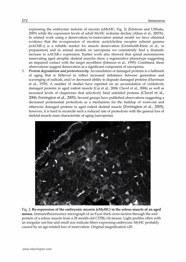
expressing the embryonic isoform of myosin (eMyHC; Fig. 2) (Edstrom and Ulfhake, 2005) while the expression levels of adult MyHC isoforms decline (Altun et al., 2007b). In related work using a denervation/re-innervation animal model we have obtained evidence that the re-expression of nicotinic acetylcholine receptor subunit gamma (nAChR-) is a reliable marker for muscle denervation (Grönholdt-Klein et al., in preparation) and in animal models on sarcopenia we consistently find a dramatic increase in nAChR- expression. Earlier work also showed that spinal motoneurons innervating aged atrophic skeletal muscles show a regenerative phenotype suggesting an impaired contact with the target myofibers (Johnson et al., 1995). Combined, these observations suggest denervation as a significant component of sarcopenia.
3. Protein degradation and proteotoxicity. Accumulation of damaged proteins is a hallmark of aging that is believed to reflect increased imbalance between generation and scavenging of radicals, and/or decreased ability to degrade damaged proteins (Harriman et al., 1970). A number of studies have reported on an accumulation of oxidatively damaged proteins in aged rodent muscle (Cai et al., 2004; Clavel et al., 2006) as well as
increased levels of chaperones that selectively bind unfolded proteins (Clavel et al., 2006; Ferrington et al., 2005). Several groups have published observations suggesting a
decreased proteasomal proteolysis as a mechanism for the buildup of worn-out and
otherwise damaged proteins in aged rodent skeletal muscle (Ferrington et al., 2005), however, it is hard to reconcile such a reduced rate of proteolysis with the general loss of skeletal muscle mass characteristic of aging (sarcopenia).
Fig. 2. Re-expression of the embryonic myosin (eMyHC) in the soleus muscle of an aged
mouse. Immunofluorescence micrograph of an 8 m thick cross-section through the mid portion of a soleus muscle from a 28 month-old C57BL/6J mouse. Light profiles often with an irregular out-line and small size indicate fibers expressing embryonic MyHC probably caused by an age-related loss of innervation. Original magnification x20.
www.intechopen.com
Cellular Degradation Machineries in Age-Related Loss of Muscle Mass (Sarcopenia) 273
This chapter will focus on the main cellular degradation machineries and our current understanding of how age-related changes in these systems impact skeletal muscle integrity.
2. Regulated proteolysis
Regulated proteolysis is instrumental for such diverse cellular processes as signaling, cell cycle progression, and apoptosis. Protein and organelle degradation enables recycling of the building blocks and is also an important survival response to starvation, whereby proteins are degraded to supply the organism with fuel for energy production. The accumulation of aggregates of misfolded proteins is a hallmark of cells and tissues of aged organisms, and protein aggregation occurs when damaged or partially unfolded proteins are not efficiently degraded. Disposal of proteins is in most conditions a selective and coordinated process, handled mainly by two cellular proteolytic systems, the ubiquitin-proteasomal system (UPS) and autophagy-lysosomal system (ALS).
2.1 The ubiquitin proteasomal system (UPS)
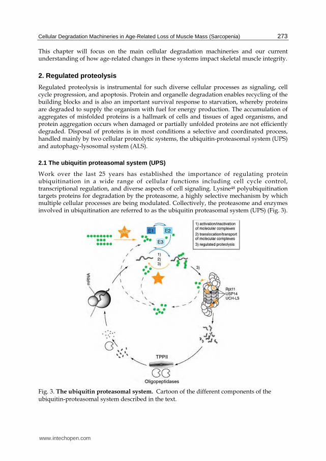
Work over the last 25 years has established the importance of regulating protein ubiquitination in a wide range of cellular functions including cell cycle control, transcriptional regulation, and diverse aspects of cell signaling. Lysine48 polyubiquitination targets proteins for degradation by the proteasome, a highly selective mechanism by which multiple cellular processes are being modulated. Collectively, the proteasome and enzymes involved in ubiquitination are referred to as the ubiquitin proteasomal system (UPS) (Fig. 3).
Fig. 3. The ubiquitin proteasomal system. Cartoon of the different components of the ubiquitin-proteasomal system described in the text.
www.intechopen.com

Senescence 274
However, ubiquitination does not only associate with proteasomal degradation, other forms of ubiquitination play roles in such diverse processes as transcriptional regulation, endocytosis, DNA modifications and stabilization of protein complexes (Harper and Schulman, 2006). Ubiquitin (Ub) is transcribed as precursor proteins from 4 genes and after cleavage to monomeric Ub, Ub-conjugation occurs through an enzymatic cascade where the E3-ligases (>600 E3 ligases have been identified so far) provide for target specificity. Prior to cleavage in the proteasome of a lys48 poly-Ub conjugated protein, the Ub-recognition signal is removed (Fig. 3). This serves to enhance passage of the targeted protein into the catalytic chamber and will also free ubiquitin for reuse and hinder the proteasomes to be preoccupied with destroying Ub [itself] (Hanna et al., 2007; Koulich et al., 2008). Hydrolysis of bound Ub and processing of Ub precursor proteins is accomplished by members of the large group of deubiquitinating enzymes (DUBs) (>90 members identified so far); DUBs are cysteine proteases that can be divided into distinct subfamilies based on sequence similarities and likely mechanism of action (reviewed in Nijman et al., 2005). Since DUBs can reshuffle ubiquitin from poly- to mono- or multi-ubiquitin chains, they are not only important for protein half-life but probably all cellular processes that involve ubiquitination
of proteins (Clague and Urbe, 2006).
2.2 Activation of the UPS in sarcopenia
Because available data on the integrity of the UPS in the elderly was controversial we examined in 30-month old Sprague-Dawley rats the effects of aging on the content and activity of 26S proteasomes, proteasome-associated regulatory proteins and various other components of the UPS, including multiple DUBs (Altun et al., 2010). Muscles of these aged animals undergo marked atrophy compared to muscles of young adult animals and this age-related atrophy could be impeded if the animals were maintained on a restricted diet (Altun et al., 2007a). Analysis of proteasome protein content and proteasome degradation capacity in these animals revealed a 2-3 fold increase in proteasomes in the aged muscle. The increase in 26S proteasomes was suppressed completely in aged animals maintained on a restricted diet (30% of the consumption recorded for rats having free access to food). Since muscle wasting was reduced in these animals, these findings suggest that the buildup of proteasomes contributes to the loss of muscle mass in aged animals. In support of this notion we found that the levels of proteasome subunits in aged skeletal muscle were inversely correlated with muscle weight (subunits 1: r=-0.71, p<0.05; 5: r=-0.69; p<0.05), and no such inverse relationship was found in muscles of adult rats or aged animals maintained on dietary restriction (Altun et al., 2010). However, the underlying mechanism of this accumulation remains unclear. In contrast to muscles atrophying due to disuse, fasting or various systemic diseases, the age-related accumulation of proteasomes occurred without any increase in corresponding mRNAs. Thus, the accumulation of proteasomes in aged muscle must be due to enhanced subunit translation, more efficient assembly of the 26S particles, or slower degradation of the 26S particles (see below under 2.4).
Previous reports suggested that age-related decrease in the proteasome’s peptidase activities were due to oxidative modifications (Bulteau et al., 2000; Conconi et al., 1996; Ferrington et al., 2005; Grune et al., 2001; Hayashi and Goto, 1998; Keller et al., 2000). However, we could not observe such defect in degrading capacity towards a range of substrates including ubiquitinated native proteins (Fig. 4). Instead, our data indicates that proteasome content increases during sarcopenia, and that these particles retain their full ability to function in
www.intechopen.com

Cellular Degradation Machineries in Age-Related Loss of Muscle Mass (Sarcopenia) 275
protein breakdown. Together these findings suggest that the enhanced capacity of the UPS may be a response to the increased generation of damaged polypeptides. Strong support for this conclusion is our finding of elevated levels of the ubiquitin ligases, CHIP and E6AP, in muscle of aged animals. CHIP ubiquitinates misfolded or mutated proteins bound to Hsp70 or Hsp90 (Connell et al., 2001), and recently a similar function in cellular quality control has been reported for E6AP (Mishra et al., 2009). Moreover, most of these conclusions about a negative impact of aging on proteasomal degradation in muscle were based exclusively on measurements of the 20S core particle. Although some degradation of unfolded or denatured proteins may occur by the free 20S (Jariel-Encontre et al., 2008), the bulk of proteasome-mediated degradation, even of oxidatively damaged proteins (Medicherla and Goldberg, 2008), seems to require the 26S proteasome and ubiquitylation of the substrate.
Fig. 4. Degradation capacity towards different substrates of proteasomes isolated from triceps surae muscle of adult rats (Ad-AL), aged (Ag-AL) and aged rats maintained on dietary restriction (Ag-DR) (original data reproduced from Altun et al., 2010). The higher content per unit muscle mass of proteasomes in aged (Ag-AL) muscles is paralleled by a corresponding increase in degradation of synthetic peptides designed for the chymotryptic and caspase sites of the proteasome (A) as well as casein (B). In (C) degradation of a native ubiquitinylated protein is shown illustrating that we could not detect any impairment of the degradation capacity of 26S proteasomes isolated from aged rats muscle. Further details on experimental design in Altun at al., 2010.
www.intechopen.com
Senescence 276
A decline in the cell’s pool of free ubiquitin can limit the rate of proteolysis by the proteasome (Hanna et al., 2003; Kimura et al., 2009) and free ubiquitin is in turn released during degradation of ubiquitylated proteins by the DUBs associated with the 26S proteasome. Thus, in order to achieve high degradation rates, the capacity for deubiquitylation probably needs to be increased too. Using mechanism-based probes to assess the state of DUB activity and expression (Altun et al., 2010), eleven DUB-enzymes were found to be strongly up-regulated in the muscles of aged rats; including USP14 and Uch37 which are known to be associated with the 26S proteasome. USP14 and UCH37 trim off ubiquitin from the polyubiquitin chain and release ubiquitin monomers for re-use. In addition, USP14 controls gate opening into the 20S proteasome and facilitates substrate degradation (Peth et al., 2009). On the other hand, the yeast homolog of USP14, Ubp6, regulates the overall rate of proteolysis and appears critical in replenishing the free ubiquitin pool in yeast (Hanna et al., 2003; Hanna et al., 2007). Similarly, USP5 hydrolyses anchorless ubiquitin chains (Reyes-Turcu et al., 2006; Reyes-Turcu et al., 2008) and appears to work downstream of Rpn11, an intrinsic DUB on the proteasome, to prevent the binding of free ubiquitin chains to the 19S, which would inhibit proteolysis. Together these data illustrate important roles for deubiquitylation in the aged muscle, probably ensuring a supply of ubiquitin for enhanced proteolysis but perhaps also serving additional regulatory functions.
Loss of muscle mass can occur through increased protein degradation but also decreased protein synthesis or through some combination of these responses. With aging, sarcopenia develops over months to years depending on the species, unlike the rapid loss of muscle weight induced by fasting, disuse, and in various catabolic diseases, where marked atrophy (20-50% loss) can occur in rodents in several days. In these latter types of rapid atrophy, as described above, there is a common program of changes in the transcription of a set of atrophy-related genes (Lecker et al., 2006; Lecker et al., 2004; Sacheck et al., 2007). Several of the biochemical changes observed in aged atrophic muscle clearly distinguish them from those undergoing rapid atrophy in adult animals. Upon denervation or fasting, the atrophy-specific ubiquitin ligases, Atrogin-1/MAFbx and MuRF1, are induced by members of the FOXO family of transcription factors, and this induction is essential for the rapid weight loss (Bodine et al., 2001; Lecker et al., 2006; Sandri et al., 2004). Inhibition of FOXOs prevents their induction and the loss of muscle mass upon denervation, fasting, or glucocorticoid treatment (Sandri et al., 2004). In contrast, in the aged muscles, mRNAs for Atrogin-1/MAFbx, MuRF1 and the Ub-conjugating enzyme, E2-14K, were unchanged or lower than adult levels (Edstrom et al., 2006). Also, treatment with the glucocorticoid dexamethasone failed to induce Atrogin-1/MAFbx or MuRF1 or to cause muscle wasting, as it does in adult animals (Bodine et al., 2001; Gomes et al., 2001). However, MuRF1 protein increased in aged muscle, while Atrogin-1/MAFbx protein decreased (in accord with the mRNA data). Another distinction between sarcopenia and rapid atrophy is that the ubiquitin ligases, CHIP and E6AP, increased markedly in the aged muscle, though they do not rise in rapidly atrophying muscles. Their induction may reflect adaptations in the aged muscle to eliminate more efficiently misfolded proteins.
In summary, the finding of increased content of proteasomes and other UPS components (e.g. MuRF1) argues that proteolysis also increases in these muscles and may contribute to muscle wasting in the aged rats. Dietary restriction decreased levels of proteasomes and several other UPS components toward the levels in adult animal and partially inhibited the development of sarcopenia.
www.intechopen.com
Cellular Degradation Machineries in Age-Related Loss of Muscle Mass (Sarcopenia) 277
2.3 Accumulation of ubiquitinated proteins in sarcopenic muscles
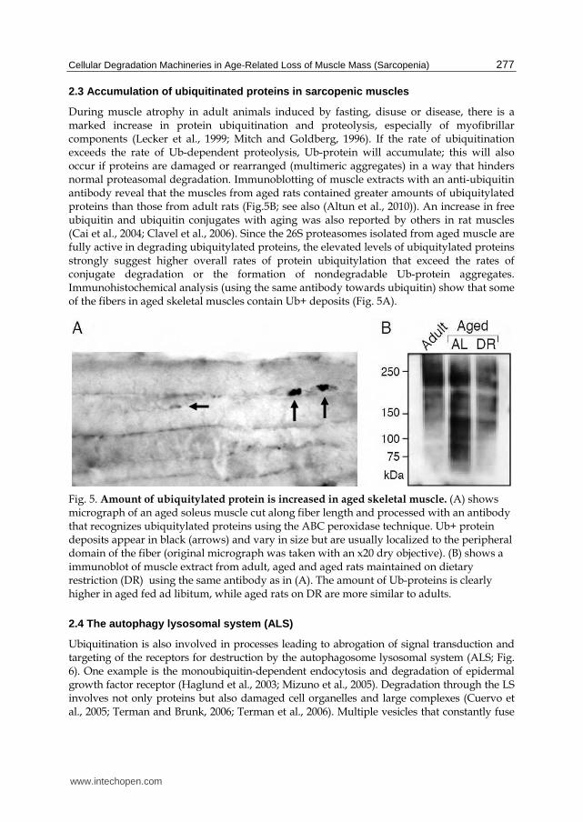
During muscle atrophy in adult animals induced by fasting, disuse or disease, there is a marked increase in protein ubiquitination and proteolysis, especially of myofibrillar components (Lecker et al., 1999; Mitch and Goldberg, 1996). If the rate of ubiquitination exceeds the rate of Ub-dependent proteolysis, Ub-protein will accumulate; this will also occur if proteins are damaged or rearranged (multimeric aggregates) in a way that hinders normal proteasomal degradation. Immunoblotting of muscle extracts with an anti-ubiquitin antibody reveal that the muscles from aged rats contained greater amounts of ubiquitylated proteins than those from adult rats (Fig.5B; see also (Altun et al., 2010)). An increase in free ubiquitin and ubiquitin conjugates with aging was also reported by others in rat muscles (Cai et al., 2004; Clavel et al., 2006). Since the 26S proteasomes isolated from aged muscle are fully active in degrading ubiquitylated proteins, the elevated levels of ubiquitylated proteins strongly suggest higher overall rates of protein ubiquitylation that exceed the rates of conjugate degradation or the formation of nondegradable Ub-protein aggregates. Immunohistochemical analysis (using the same antibody towards ubiquitin) show that some of the fibers in aged skeletal muscles contain Ub+ deposits (Fig. 5A).
Fig. 5. Amount of ubiquitylated protein is increased in aged skeletal muscle. (A) shows micrograph of an aged soleus muscle cut along fiber length and processed with an antibody that recognizes ubiquitylated proteins using the ABC peroxidase technique. Ub+ protein deposits appear in black (arrows) and vary in size but are usually localized to the peripheral domain of the fiber (original micrograph was taken with an x20 dry objective). (B) shows a immunoblot of muscle extract from adult, aged and aged rats maintained on dietary restriction (DR) using the same antibody as in (A). The amount of Ub-proteins is clearly higher in aged fed ad libitum, while aged rats on DR are more similar to adults.
2.4 The autophagy lysosomal system (ALS)
Ubiquitination is also involved in processes leading to abrogation of signal transduction and targeting of the receptors for destruction by the autophagosome lysosomal system (ALS; Fig. 6). One example is the monoubiquitin-dependent endocytosis and degradation of epidermal growth factor receptor (Haglund et al., 2003; Mizuno et al., 2005). Degradation through the LS involves not only proteins but also damaged cell organelles and large complexes (Cuervo et al., 2005; Terman and Brunk, 2006; Terman et al., 2006). Multiple vesicles that constantly fuse
www.intechopen.com

Senescence 278
and fission constitute the lysosomal system. Lysosomal degradation is initiated through several pathways based on the substrate delivering mechanism (Fig. 6): Endocytosis (clathrin mediated pinching-off of membrane patches with/without inclusion of extracellular material) occurs and the resulting particle is referred to as an endosomes (carrying rab5). Endosomes can then develop into late endosomes, which are equipped with enzymes delivered by secretory vesicles from the trans-Golgi-network and sorted by mannose-6-phosphate receptors, which are markers for late endosomes. Late endosomes may also fuse with lysosomes (then they lose the mannose-6-phosphate receptors).
A second route is chaperone mediated autophagy (CMA), which targets proteins carrying a pentapeptide (KFERQ) motif that is recognized by the chaperon Hsc73 and through binding to the LAMP-2A receptor (lysosome associated membrane protein 2A; a marker of the lysosome), the targeted protein is taken up and degraded in the lysosomal lumen (Fig. 6). The major route for organelles and proteins targeted for lysosomal degradation is via autophagy, a process controlled by the autophagy-conjugase complexes Atg12-Atg5 and Atg8-Atg3 (Atg, autophagy related genes) whereby in-bulk cytoplasm becomes entrapped in a double membrane forming an autophagosome (autophagic vacuole, AV) (Terman and Brunk, 2004).
Fig. 6. Schematic drawing of the autophagy-lysosomal system. The different cargo routes to lysosomal degradation are illustrated and described in the text.
www.intechopen.com
Cellular Degradation Machineries in Age-Related Loss of Muscle Mass (Sarcopenia) 279
Subsequently, the autophagosome fuses with a lysosome for degradation under the formation of an autophagolysosome (Fig. 6). With the formation of the AV, the Atg conjugase complexes disassociate, but the Atg 8 (the mammalian homologue is MAP-LC3) remains in the ALS until degradation. While, Atg8 may be a marker for AVs and [by its elimination also] the completion of this degradation route, the autophagy conjugase complexes indicate ongoing ALS formation/autophagy. The role of the autophagy-lysosomal system was not widely recognized until it was demonstrated that cathepsins (D and B+L, respectively; (Felbor et al., 2002)) and autophagy (Hara et al., 2006) were non-redundant for a normal development and that cells rely on a basal level of autophagy to keep them free of worn-out organelles and aberrant proteins.
2.5 Signs of distress of the ALS during aging
There is solid evidence for the engagement of autophagy in major human degenerative diseases and in normal aging. A decreased capacity of the lysosomes to degrade waste will cause a build-up of damaged organelles and proteins disturbing cell homeostasis; and e.g. accumulation of dysfunctional mitochondria in aging cardiomyocytes, myofibers and neurons has been reported. Recently, several studies have shown that the induction of autophagy, by intervention of the above discussed signaling pathways, increase cellular clearance of aberrant proteins also when the normal degradation route operates via the proteasome or through CMA. The load on the ALS in normal aging is evident from the dramatic accumulation of lipofuscin by time (Fig. 7 G-H) and the concomitant upregulation of markers of lysosomal proteolysis. Lipofuscin forms due to iron-catalyzed intralysosomal peroxidation and its accumulation in the lysosomal compartment seems to set down the capacity of autophagy (Brunk and Terman, 2002). Oxidative stress, even mild stress, accelerates the formation/build-up of lipofuscin (Kurz et al., 2008) and stress can cause lysosomes to become leaky due to intralysosomal reactive iron mediated Fenton- reactions with production of hydroxyl radicals and resulting labilization of the lysosomal membrane. Even though the pH is suboptimal, several of the lysosomal enzymes are apparently proteolytically active if released to the cytosol and may then trigger cell degeneration/death (Chu, 2006).
Analysis of atrophic skeletal muscle in aged rats (Altun et al., 2007b) revealed increased levels of molecules involved in iron transport (transferrin), binding (ferritin), as well as iron response element binding protein-1 activity; combined these observations suggest increased load on the iron-handling machinery. Measurements of iron levels revealed a significant accumulation in the aged skeletal muscle, providing further support for iron loading in senescence. Iron-loading increases the risk for common hallmarks of aging such as DNA damage, protein oxidation and misfolding, and lipid peroxidation and may accelerate the accumulation of lipofuscin (see above). Iron-load is not exclusive to aging-related muscle wasting, but is also evident in experimental disuse atrophy (Kondo et al., 1992) and iron-restriction has been shown to be beneficiary in certain inherited myopathies (Bornman et al., 1998).
3. Cross talk between the UPS and the ALS
Until recently the UPS and the ALS were considered as independent pathways for protein degradation. The UPS offers a fast and highly specific mechanism to remove selected proteins. However, the targeted protein must be a monomer and unfolded to be able to enter the proteolytic lumen of the proteasome. The ALS represents a partly overlapping,
www.intechopen.com
Senescence 280
partly complementary degradation route, taking care of folded and aggregated proteins as well as large complexes and organelles with heterogenic building blocks (carbohydrates, lipids and proteins). However, several lines of evidence indicate intersections between these two pathways. In neurons treated with proteasome inhibitors, aggregate-prone proteins normally degraded by the UPS are degraded by autophagy (Cuervo et al., 2004). Two recent papers also provide evidence that induction of the UPS and LS may occur via a common pathway in skeletal muscle (Mammucari et al., 2007; Zhou et al., 2010).
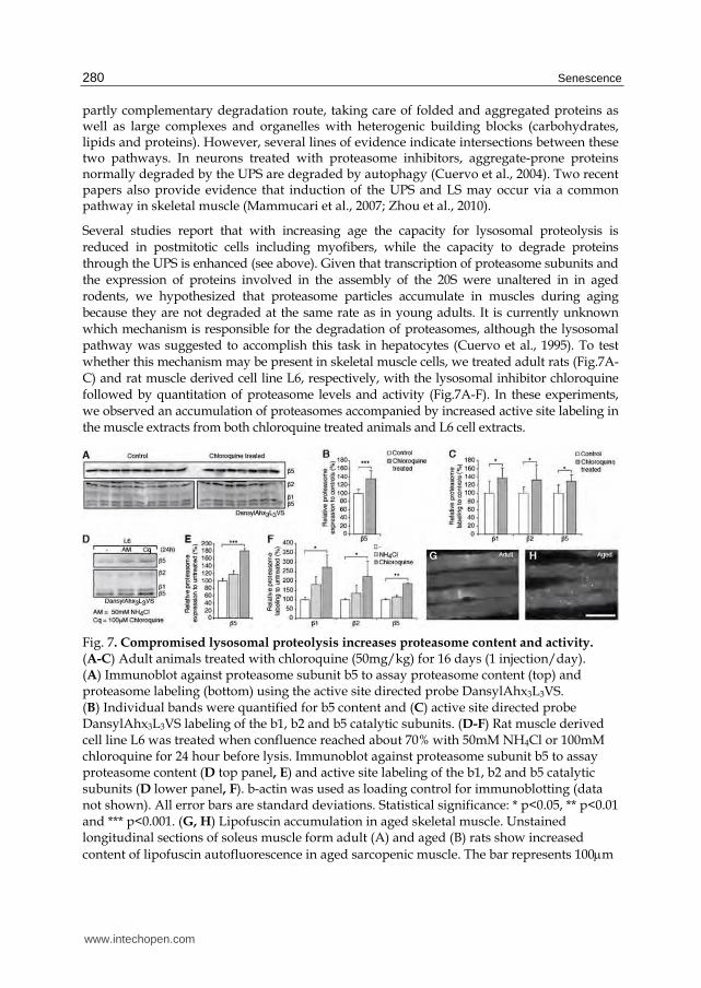
Several studies report that with increasing age the capacity for lysosomal proteolysis is reduced in postmitotic cells including myofibers, while the capacity to degrade proteins through the UPS is enhanced (see above). Given that transcription of proteasome subunits and the expression of proteins involved in the assembly of the 20S were unaltered in in aged rodents, we hypothesized that proteasome particles accumulate in muscles during aging because they are not degraded at the same rate as in young adults. It is currently unknown which mechanism is responsible for the degradation of proteasomes, although the lysosomal pathway was suggested to accomplish this task in hepatocytes (Cuervo et al., 1995). To test whether this mechanism may be present in skeletal muscle cells, we treated adult rats (Fig.7A-C) and rat muscle derived cell line L6, respectively, with the lysosomal inhibitor chloroquine followed by quantitation of proteasome levels and activity (Fig.7A-F). In these experiments, we observed an accumulation of proteasomes accompanied by increased active site labeling in the muscle extracts from both chloroquine treated animals and L6 cell extracts.
Fig. 7. Compromised lysosomal proteolysis increases proteasome content and activity. (A-C) Adult animals treated with chloroquine (50mg/kg) for 16 days (1 injection/day). (A) Immunoblot against proteasome subunit b5 to assay proteasome content (top) and proteasome labeling (bottom) using the active site directed probe DansylAhx3L3VS. (B) Individual bands were quantified for b5 content and (C) active site directed probe DansylAhx3L3VS labeling of the b1, b2 and b5 catalytic subunits. (D-F) Rat muscle derived cell line L6 was treated when confluence reached about 70% with 50mM NH4Cl or 100mM chloroquine for 24 hour before lysis. Immunoblot against proteasome subunit b5 to assay proteasome content (D top panel, E) and active site labeling of the b1, b2 and b5 catalytic subunits (D lower panel, F). b-actin was used as loading control for immunoblotting (data not shown). All error bars are standard deviations. Statistical significance: * p<0.05, ** p<0.01 and *** p<0.001. (G, H) Lipofuscin accumulation in aged skeletal muscle. Unstained longitudinal sections of soleus muscle form adult (A) and aged (B) rats show increased
content of lipofuscin autofluorescence in aged sarcopenic muscle. The bar represents 100m
www.intechopen.com
Cellular Degradation Machineries in Age-Related Loss of Muscle Mass (Sarcopenia) 281
As in the aged rat muscles, the accumulation of proteasomes following lysosomal inhibition was not induced by a transcriptional up-regulation (data not shown) and therefore results likely from a reduced clearance of proteasomes due to an impairment of lysosomal function. Consistent with this, we observed lipofuscin accumulation in the aged rat muscle tissue (Fig.7 G and H). In addition, Cuervo and colleagues reported that autophagy declines in liver with aging but this effect is reduced by dietary restriction (Hanna et al., 2003; Watts et al., 2004). Activation of autophagy in muscle by dietary restriction could explain our findings that this regime prevented the increase in proteasomes with age without any change in proteasome mRNA levels. Combined these observations suggest the ALS as a candidate pathway for proteasomal degradation awaiting more definitive evidence.
4. Concluding remarks
Human cross-sectional and longitudinal studies have consistently demonstrated a gain in fat mass and decline in lean mass during aging. Although aging-related muscle wasting, or sarcopenia, is widely recognized it still remains poorly understood. The reduced functional muscle mass is associated with increased morbidity and reduced quality of life. To maintain integrity muscle myofibers rely on degradation pathways to keep clean from worn-out organelles and damaged proteins. Our current understanding is that the autophagy-lysosomal system is in distress possibly driven by an age-dependent accumulation of reactive iron, while contrary to the widespread view, the complementary pathway for degradation of proteins, the UPS, is enhanced. The age-dependent increase in the muscle specific ubiquitin-ligase MuRF and ubiquitin-dependent proteasomal proteolysis is expected to occur in the progression of sarcopenia since this route is nonredundant for degradation of myofibrillar proteins. In addition, the UPS in aged skeletal muscle shows adaptations to an increased demand on degradation of aberrant proteins and an accumulation of ubiquitinated proteins. Combined these stigmata suggest that aged myofibers may be at risk to enter a state of proteotoxicity.
Normal skeletal muscles have a good capacity to regenerate following wasting conditions such as disuse. The muscle regenerative response relies on signaling that evokes satellite cell replication and asymmetric division generating offsprings that will differentiate to myocytes via the myoblast stage. Such cells are then incorporated into the myofiber allowing it to grow (for example in response to an exercise stimulus). Poor capacity to regenerate muscle tissue at advanced age may depend on impaired signaling, exhaustion of the SC pool or changes in the extracellular matrix or stem-cell niche impeding the regeneration and incorporation of myoblasts into existing/regenerating myofibers (for references see Introduction). Assessments of the regenerative drive in aged sarcopenic muscle have, however, shown that myogenic differentiation factors are upregulated and that there are overt signs of incorporation of new nuclei into existing fibers (Fig. 1B,C; (Edstrom and Ulfhake, 2005)). Still, regeneration fails and tissue atrophy progresses.
The triggering mechanism for the age-dependent fiber atrophy and fiber loss remains enigmatic. However, several lines of evidence converge towards support of the “neurogenic” theory (Gutman and Hanzlikova, 1972), which stipulates that sarcopenia is driven by a successive drop-out of motoneurons. Early evidence in favor of this theory was the observation of fiber-type grouping and also histological examination revealing regressive changes at the neuro-muscular junctions. The strongest argument against this theory is the absence of unbiased evidence of a significant age-dependent loss of
www.intechopen.com
Senescence 282
motoneurons. However, as discussed at length elsewhere (Johnson et al., 1995) the denervation may be a peripheral process primarily involving subdomains of the motor axon’s terminal arborization within a motor unit (see also (Valdez et al., 2010b)). Failure to maintain the distant axon arbor could cause branches and terminals to degenerate leaving myofibers vacated from innervation. As this process progresses more and more fibers will be denervated and read-outs validating this process are the increase in expression of the nAChR- subunit (Gronholdt-Klein et al., in preparation) and embryonic myosin (Edstrom and Ulfhake, 2005); and it should be noted that these proteins are re-expressed by young adult myofibers upon denervation. As denervation becomes significant, it will trigger the atrophy program by which MuRF1 and other enzymes will accelerate proteasomal myofibrillar protein degradation (see above). Recently, further direct histological evidence for this process was obtained by Valdez and coworkers (Valdez et al., 2010b). These authors also showed that exercise and dietary restriction slow-down the progression of age-dependent denervation, at least the latter observation is consistent with our biochemical data on the UPS (see above). It will be important to seek proof of principle for the neurogenic theory in experimental animal models followed by validation in humans since it will impact the development of strategies to impede sarcopenia in humans.
5. Acknowledgements
This work was supported by a grant from the Swedish research council (VR 10820, to B. Ulfhake), Swedish research council post doc grant to M. Altun and a guest researcher stipendship from the PR China government to L. Wang.
6. References
Adamo, M.L., and Farrar, R.P. (2006). Resistance training, and IGF involvement in the maintenance of muscle mass during the aging process. Ageing Res Rev 5, 310-331.
Altun, M., Bergman, E., Edstrom, E., Johnson, H., and Ulfhake, B. (2007a). Behavioral impairments of the aging rat. Physiol Behav 92, 911-923.
Altun, M., Besche, H.C., Overkleeft, H.S., Piccirillo, R., Edelmann, M.J., Kessler, B.M., Goldberg, A.L., and Ulfhake, B. (2010). Muscle wasting in aged, sarcopenic rats is associated with enhanced activity of the ubiquitin proteasome pathway. J Biol Chem 285, 39597-39608.
Altun, M., Edstrom, E., Spooner, E., Flores-Moralez, A., Bergman, E., Tollet-Egnell, P., Norstedt, G., Kessler, B.M., and Ulfhake, B. (2007b). Iron load and redox stress in skeletal muscle of aged rats. Muscle Nerve 36, 223-233.
Attaix, D., Mosoni, L., Dardevet, D., Combaret, L., Mirand, P.P., and Grizard, J. (2005). Altered responses in skeletal muscle protein turnover during aging in anabolic and catabolic periods. Int J Biochem Cell Biol 37, 1962-1973.
Baumgartner, R.N., Koehler, K.M., Gallagher, D., Romero, L., Heymsfield, S.B., Ross, R.R., Garry, P.J., and Lindeman, R.D. (1998a). Epidemiology of sarcopenia among the elderly in New Mexico. American Journal of Epidemiology 147, 755-763.
Baumgartner, R.N., Koehler, K.M., Gallagher, D., Romero, L., Heymsfield, S.B., Ross, R.R., Garry, P.J., and Lindeman, R.D. (1998b). Epidemiology of sarcopenia among the elderly in New Mexico. Am J Epidemiol 147, 755-763.
Bodine, S.C., Latres, E., Baumhueter, S., Lai, V.K., Nunez, L., Clarke, B.A., Poueymirou, W.T., Panaro, F.J., Na, E., Dharmarajan, K., et al. (2001). Identification of ubiquitin ligases required for skeletal muscle atrophy. Science 294, 1704-1708.
www.intechopen.com

Cellular Degradation Machineries in Age-Related Loss of Muscle Mass (Sarcopenia) 283
Bornman, L., Rossouw, H., Gericke, G.S., and Polla, B.S. (1998). Effects of iron deprivation on the pathology and stress protein expression in murine X-linked muscular dystrophy. Biochem Pharmacol 56, 751-757.
Brunk, U.T., and Terman, A. (2002). Lipofuscin: mechanisms of age-related accumulation and influence on cell function. Free Radic Biol Med 33, 611-619.
Bulteau, A.L., Petropoulos, I., and Friguet, B. (2000). Age-related alterations of proteasome structure and function in aging epidermis. Exp Gerontol 35, 767-777.
Cai, D., Lee, K.K., Li, M., Tang, M.K., and Chan, K.M. (2004). Ubiquitin expression is up-regulated in human and rat skeletal muscles during aging. Arch Biochem Biophys 425, 42-50.
Carlson, M.E., Silva, H.S., and Conboy, I.M. (2008). Aging of signal transduction pathways, and pathology. Exp Cell Res 314, 1951-1961.
Carlson, M.E., Suetta, C., Conboy, M.J., Aagaard, P., Mackey, A., Kjaer, M., and Conboy, I. (2009). Molecular aging and rejuvenation of human muscle stem cells. EMBO Mol Med 1, 381-391.
Christov, C., Chretien, F., Abou-Khalil, R., Bassez, G., Vallet, G., Authier, F.J., Bassaglia, Y., Shinin, V., Tajbakhsh, S., Chazaud, B., et al. (2007). Muscle satellite cells and endothelial cells: close neighbors and privileged partners. Mol Biol Cell 18, 1397-1409.
Chu, C.T. (2006). Autophagic stress in neuronal injury and disease. J Neuropathol Exp Neurol 65, 423-432.
Clague, M.J., and Urbe, S. (2006). Endocytosis: the DUB version. Trends Cell Biol 16, 551-559. Clavel, S., Coldefy, A.S., Kurkdjian, E., Salles, J., Margaritis, I., and Derijard, B. (2006).
Atrophy-related ubiquitin ligases, atrogin-1 and MuRF1 are up-regulated in aged rat Tibialis Anterior muscle. Mech Ageing Dev 127, 794-801.
Conconi, M., Szweda, L.I., Levine, R.L., Stadtman, E.R., and Friguet, B. (1996). Age-related decline of rat liver multicatalytic proteinase activity and protection from oxidative inactivation by heat-shock protein 90. Arch Biochem Biophys 331, 232-240.
Connell, P., Ballinger, C.A., Jiang, J., Wu, Y., Thompson, L.J., Hohfeld, J., and Patterson, C. (2001). The co-chaperone CHIP regulates protein triage decisions mediated by heat-shock proteins. Nat Cell Biol 3, 93-96.
Cuervo, A.M., Bergamini, E., Brunk, U.T., Droge, W., Ffrench, M., and Terman, A. (2005). Autophagy and aging: the importance of maintaining "clean" cells. Autophagy 1, 131-140.
Cuervo, A.M., Palmer, A., Rivett, A.J., and Knecht, E. (1995). Degradation of proteasomes by lysosomes in rat liver. Eur J Biochem 227, 792-800.
Cuervo, A.M., Stefanis, L., Fredenburg, R., Lansbury, P.T., and Sulzer, D. (2004). Impaired degradation of mutant alpha-synuclein by chaperone-mediated autophagy. Science 305, 1292-1295.
Edstrom, E., Altun, M., Bergman, E., Johnson, H., Kullberg, S., Ramirez-Leon, V., and Ulfhake, B. (2007). Factors contributing to neuromuscular impairment and sarcopenia during aging. Physiol Behav 92, 129-135.
Edstrom, E., Altun, M., Hagglund, M., and Ulfhake, B. (2006). Atrogin-1/MAFbx and MuRF1 are downregulated in aging-related loss of skeletal muscle. J Gerontol A Biol Sci Med Sci 61, 663-674.
Edstrom, E., and Ulfhake, B. (2005). Sarcopenia is not due to lack of regenerative drive in senescent skeletal muscle. Aging Cell 4, 65-77.
Felbor, U., Kessler, B., Mothes, W., Goebel, H.H., Ploegh, H.L., Bronson, R.T., and Olsen, B.R. (2002). Neuronal loss and brain atrophy in mice lacking cathepsins B and L. Proc Natl Acad Sci U S A 99, 7883-7888.
www.intechopen.com

Senescence 284
Ferrington, D.A., Husom, A.D., and Thompson, L.V. (2005). Altered proteasome structure, function, and oxidation in aged muscle. Faseb J 19, 644-646.
Fuso, A., Ferraguti, G., Grandoni, F., Ruggeri, R., Scarpa, S., Strom, R., and Lucarelli, M. (2010). Early demethylation of non-CpG, CpC-rich, elements in the myogenin 5'-flanking region: a priming effect on the spreading of active demethylation. Cell Cycle 9, 3965-3976.
Gomes, M.D., Lecker, S.H., Jagoe, R.T., Navon, A., and Goldberg, A.L. (2001). Atrogin-1, a muscle-specific F-box protein highly expressed during muscle atrophy. Proc Natl Acad Sci U S A 98, 14440-14445.
Grune, T., Shringarpure, R., Sitte, N., and Davies, K. (2001). Age-related changes in protein oxidation and proteolysis in mammalian cells. J Gerontol A Biol Sci Med Sci 56, B459-467.
Gutman, B., and Hanzlikova, V. (1972). Age changes in the neuromuscular system. Scientechnica Ltd, Bristol, 1-20.
Haglund, K., Sigismund, S., Polo, S., Szymkiewicz, I., Di Fiore, P.P., and Dikic, I. (2003). Multiple monoubiquitination of RTKs is sufficient for their endocytosis and degradation. Nat Cell Biol 5, 461-466.
Hanna, J., Leggett, D.S., and Finley, D. (2003). Ubiquitin depletion as a key mediator of toxicity by translational inhibitors. Mol Cell Biol 23, 9251-9261.
Hanna, J., Meides, A., Zhang, D.P., and Finley, D. (2007). A ubiquitin stress response induces altered proteasome composition. Cell 129, 747-759.
Hara, T., Nakamura, K., Matsui, M., Yamamoto, A., Nakahara, Y., Suzuki-Migishima, R., Yokoyama, M., Mishima, K., Saito, I., Okano, H., et al. (2006). Suppression of basal autophagy in neural cells causes neurodegenerative disease in mice. Nature 441, 885-889.
Harper, J.W., and Schulman, B.A. (2006). Structural complexity in ubiquitin recognition. Cell 124, 1133-1136.
Harriman, D.G., Taverner, D., and Woolf, A.L. (1970). Ekbom´s syndrome and burning paraesthesiae. A biopsy study by vital staining and electron microscopy of the intramuscular innervation with a note on age changes in motor nerve endings in distal muscles. Brain 93, 393-406.
Hayashi, T., and Goto, S. (1998). Age-related changes in the 20S and 26S proteasome activities in the liver of male F344 rats. Mech Ageing Dev 102, 55-66.
Janssen, I., Heymsfield, S.B., and Ross, R. (2002). Low relative skeletal muscle mass (sarcopenia) in older persons is associated with functional impairment and physical disability. J Am Geriatr Soc 50, 889-896.
Jariel-Encontre, I., Bossis, G., and Piechaczyk, M. (2008). Ubiquitin-independent degradation of proteins by the proteasome. Biochim Biophys Acta 1786, 153-177.
Johnson, H., Mossberg, K., Arvidsson, U., Piehl, F., Hökfelt, T., and Ulfhake, B. (1995). Increase in alpha-CGRP and GAP-43 in aged motoneurons: A study of peptides, growth factors, and ChAT mRNA in the lumbar spinal cord of senescent rats with symptoms of hindlimb incapacities. J Comp Neurol 359, 69-89.
Kadi, F., Charifi, N., Denis, C., Lexell, J., Andersen, J.L., Schjerling, P., Olsen, S., and Kjaer, M. (2005). The behaviour of satellite cells in response to exercise: what have we learned from human studies? Pflugers Arch 451, 319-327.
Keller, J.N., Huang, F.F., and Markesbery, W.R. (2000). Decreased levels of proteasome activity and proteasome expression in aging spinal cord. Neuroscience 98, 149-156.
Kimura, Y., Yashiroda, H., Kudo, T., Koitabashi, S., Murata, S., Kakizuka, A., and Tanaka, K. (2009). An inhibitor of a deubiquitinating enzyme regulates ubiquitin homeostasis. Cell 137, 549-559.
www.intechopen.com

Cellular Degradation Machineries in Age-Related Loss of Muscle Mass (Sarcopenia) 285
Kondo, H., Miura, M., Kodama, J., Ahmed, S.M., and Itokawa, Y. (1992). Role of iron in oxidative stress in skeletal muscle atrophied by immobilization. Pflugers Arch 421, 295-297.
Koulich, E., Li, X., and DeMartino, G.N. (2008). Relative structural and functional roles of multiple deubiquitylating proteins associated with mammalian 26S proteasome. Mol Biol Cell 19, 1072-1082.
Kurz, T., Terman, A., Gustafsson, B., and Brunk, U.T. (2008). Lysosomes in iron metabolism, ageing and apoptosis. Histochem Cell Biol 129, 389-406.
Larsson, L. (1995). Motor units: remodeling in aged animals. J Gerontol A Biol Sci Med Sci 50 Spec No, 91-95.
Lecker, S.H., Goldberg, A.L., and Mitch, W.E. (2006). Protein degradation by the ubiquitin-proteasome pathway in normal and disease states. J Am Soc Nephrol 17, 1807-1819.
Lecker, S.H., Jagoe, R.T., Gilbert, A., Gomes, M., Baracos, V., Bailey, J., Price, S.R., Mitch, W.E., and Goldberg, A.L. (2004). Multiple types of skeletal muscle atrophy involve a common program of changes in gene expression. Faseb J 18, 39-51.
Lecker, S.H., Solomon, V., Mitch, W.E., and Goldberg, A.L. (1999). Muscle protein breakdown and the critical role of the ubiquitin-proteasome pathway in normal and disease states. J Nutr 129, 227S-237S.
Mammucari, C., Milan, G., Romanello, V., Masiero, E., Rudolf, R., Del Piccolo, P., Burden, S.J., Di Lisi, R., Sandri, C., Zhao, J., et al. (2007). FoxO3 controls autophagy in skeletal muscle in vivo. Cell Metab 6, 458-471.
Medicherla, B., and Goldberg, A.L. (2008). Heat shock and oxygen radicals stimulate ubiquitin-dependent degradation mainly of newly synthesized proteins. J Cell Biol 182, 663-673.
Mishra, A., Godavarthi, S.K., Maheshwari, M., Goswami, A., and Jana, N.R. (2009). The ubiquitin ligase E6-AP is induced and recruited to aggresomes in response to proteasome inhibition and may be involved in the ubiquitination of Hsp70-bound misfolded proteins. J Biol Chem 284, 10537-10545.
Mitch, W.E., and Goldberg, A.L. (1996). Mechanisms of muscle wasting. The role of the ubiquitin-proteasome pathway. N Engl J Med 335, 1897-1905.
Mizuno, E., Iura, T., Mukai, A., Yoshimori, T., Kitamura, N., and Komada, M. (2005). Regulation of epidermal growth factor receptor down-regulation by UBPY-mediated deubiquitination at endosomes. Mol Biol Cell 16, 5163-5174.
Nijman, S.M., Luna-Vargas, M.P., Velds, A., Brummelkamp, T.R., Dirac, A.M., Sixma, T.K., and Bernards, R. (2005). A genomic and functional inventory of deubiquitinating enzymes. Cell 123, 773-786.
Paddon-Jones, D., and Rasmussen, B.B. (2009). Dietary protein recommendations and the prevention of sarcopenia. Curr Opin Clin Nutr Metab Care 12, 86-90.
Peth, A., Besche, H.C., and Goldberg, A.L. (2009). Ubiquitinated proteins activate the proteasome by binding to Usp14/Ubp6, which causes 20S gate opening. Molecular cell 36, 794-804.
Reyes-Turcu, F.E., Horton, J.R., Mullally, J.E., Heroux, A., Cheng, X., and Wilkinson, K.D. (2006). The ubiquitin binding domain ZnF UBP recognizes the C-terminal diglycine motif of unanchored ubiquitin. Cell 124, 1197-1208.
Reyes-Turcu, F.E., Shanks, J.R., Komander, D., and Wilkinson, K.D. (2008). Recognition of polyubiquitin isoforms by the multiple ubiquitin binding modules of isopeptidase T. J Biol Chem 283, 19581-19592.
Rodeheffer, M.S. (2010). Tipping the scale: muscle versus fat. Nat Cell Biol 12, 102-104.
www.intechopen.com

Senescence 286
Roth, S.M., Metter, E.J., Ling, S., and Ferrucci, L. (2006). Inflammatory factors in age-related muscle wasting. Curr Opin Rheumatol 18, 625-630.
Sacheck, J.M., Hyatt, J.P., Raffaello, A., Jagoe, R.T., Roy, R.R., Edgerton, V.R., Lecker, S.H., and Goldberg, A.L. (2007). Rapid disuse and denervation atrophy involve transcriptional changes similar to those of muscle wasting during systemic diseases. Faseb J 21, 140-155.
Sacheck, J.M., Ohtsuka, A., McLary, S.C., and Goldberg, A.L. (2004). IGF-I stimulates muscle growth by suppressing protein breakdown and expression of atrophy-related ubiquitin ligases, atrogin-1 and MuRF1. Am J Physiol Endocrinol Metab 287, E591-601.
Sandri, M., Lin, J., Handschin, C., Yang, W., Arany, Z.P., Lecker, S.H., Goldberg, A.L., and Spiegelman, B.M. (2006). PGC-1alpha protects skeletal muscle from atrophy by suppressing FoxO3 action and atrophy-specific gene transcription. Proc Natl Acad Sci U S A 103, 16260-16265.
Sandri, M., Sandri, C., Gilbert, A., Skurk, C., Calabria, E., Picard, A., Walsh, K., Schiaffino, S., Lecker, S.H., and Goldberg, A.L. (2004). Foxo transcription factors induce the atrophy-related ubiquitin ligase atrogin-1 and cause skeletal muscle atrophy. Cell 117, 399-412.
Solomon, A.M., and Bouloux, P.M. (2006). Modifying muscle mass - the endocrine perspective. J Endocrinol 191, 349-360.
St-Onge, M.P. (2005). Relationship between body composition changes and changes in physical function and metabolic risk factors in aging. Curr Opin Clin Nutr Metab Care 8, 523-528.
Stitt, T.N., Drujan, D., Clarke, B.A., Panaro, F., Timofeyva, Y., Kline, W.O., Gonzalez, M., Yancopoulos, G.D., and Glass, D.J. (2004). The IGF-1/PI3K/Akt pathway prevents expression of muscle atrophy-induced ubiquitin ligases by inhibiting FOXO transcription factors. Mol Cell 14, 395-403.
Terman, A., and Brunk, U.T. (2004). Myocyte aging and mitochondrial turnover. Exp Gerontol 39, 701-705.
Terman, A., and Brunk, U.T. (2006). Oxidative stress, accumulation of biological 'garbage', and aging. Antioxid Redox Signal 8, 197-204.
Terman, A., Gustafsson, B., and Brunk, U.T. (2006). The lysosomal-mitochondrial axis theory of postmitotic aging and cell death. Chem Biol Interact 163, 29-37.
Valdez, G., Tapia, J.C., Kang, H., Clemenson, G.D., Jr., Gage, F.H., Lichtman, J.W., and Sanes, J.R. (2010a). Attenuation of age-related changes in mouse neuromuscular synapses by caloric restriction and exercise. Proc Natl Acad Sci U S A 107, 14863-14868.
Valdez, G., Tapia, J.C., Kang, H., Clemenson, G.D., Jr., Gage, F.H., Lichtman, J.W., and Sanes, J.R. (2010b). Attenuation of age-related changes in mouse neuromuscular synapses by caloric restriction and exercise. Proc Natl Acad Sci U S A 107, 14863-14868.
Watts, G.D., Wymer, J., Kovach, M.J., Mehta, S.G., Mumm, S., Darvish, D., Pestronk, A., Whyte, M.P., and Kimonis, V.E. (2004). Inclusion body myopathy associated with Paget disease of bone and frontotemporal dementia is caused by mutant valosin-containing protein. Nat Genet 36, 377-381.
Zammit, P.S. (2008). All muscle satellite cells are equal, but are some more equal than others? J Cell Sci 121, 2975-2982.
Zhou, X., Wang, J.L., Lu, J., Song, Y., Kwak, K.S., Jiao, Q., Rosenfeld, R., Chen, Q., Boone, T., Simonet, W.S., et al. (2010). Reversal of cancer cachexia and muscle wasting by ActRIIB antagonism leads to prolonged survival. Cell 142, 531-543.
www.intechopen.com
SenescenceEdited by Dr. Tetsuji Nagata
ISBN 978-953-51-0144-4Hard cover, 850 pagesPublisher InTechPublished online 29, February, 2012Published in print edition February, 2012
InTech EuropeUniversity Campus STeP Ri Slavka Krautzeka 83/A 51000 Rijeka, Croatia Phone: +385 (51) 770 447 Fax: +385 (51) 686 166www.intechopen.com
InTech ChinaUnit 405, Office Block, Hotel Equatorial Shanghai No.65, Yan An Road (West), Shanghai, 200040, China
Phone: +86-21-62489820 Fax: +86-21-62489821
The book "Senescence" is aimed to describe all the phenomena related to aging and senescence of all formsof life on Earth, i.e. plants, animals and the human beings. The book contains 36 carefully reviewed chapterswritten by different authors, aiming to describe the aging and senescent changes of living creatures, i.e. plantsand animals.
How to referenceIn order to correctly reference this scholarly work, feel free to copy and paste the following:
Mikael Altun, Max Grönholdt-Klein, Lingzhan Wang and Brun Ulfhake (2012). Cellular DegradationMachineries in Age-Related Loss of Muscle Mass (Sarcopenia), Senescence, Dr. Tetsuji Nagata (Ed.), ISBN:978-953-51-0144-4, InTech, Available from: http://www.intechopen.com/books/senescence/cellular-degradation-machineries-in-age-related-loss-of-muscle-mass-sarcopenia-

© 2012 The Author(s). Licensee IntechOpen. This is an open access articledistributed under the terms of the Creative Commons Attribution 3.0License, which permits unrestricted use, distribution, and reproduction inany medium, provided the original work is properly cited.