Embed Size (px)
Citation preview

Yukari Endo MD PhDMingrui Dong MDSatoru Noguchi PhDMegumu Ogawa MSYukiko K Hayashi MD
PhDSatoshi Kuru MD PhDKenji Sugiyama MD
PhDShigehiro Nagai MDShiro Ozasa MD PhDIkuya Nonaka MD PhDIchizo Nishino MD
PhD
Correspondence toDr Noguchinoguchincnpgojp
See editorial
See related article
Supplemental dataat Neurologyorgng
Milder forms of muscular dystrophyassociated with POMGNT2 mutations
ABSTRACT
Objective To determine the genetic variants in patients with dystroglycanopathy (DGP) andassess the pathogenicity of these variants
Methods A total of 20 patients with DGP were identified by immunohistochemistry or Westernblot analysis Whole-exome sequencing (WES) was performed using patient samples The patho-genicity of the variants identified was evaluated on the basis of the phenotypic recovery in aknockout (KO) haploid human cell line by transfection with mutated POMGNT2 cDNA and onthe basis of the in vitro enzymatic activity of mutated proteins
Results WES identified homozygous and compound heterozygous missense variants inPOMGNT2 in 3 patients with the milder limb-girdle muscular dystrophy (LGMD) and intellectualdisability without brain malformation The 2 identified variants were located in the putative gly-cosyltransferase domain of POMGNT2 which affected its enzymatic activity MutatedPOMGNT2 cDNAs failed to rescue the phenotype of POMGNT2-KO cells
Conclusions Novel variants in POMGNT2 are associated with milder forms of LGMD The findingsof this study expand the clinical and pathologic spectrum of DGP associated with POMGNT2variants from the severest Walker-Warburg syndrome to the mildest LGMD phenotypes Thesimple method to verify pathogenesis of variants may allow researchers to evaluate any variantspresent in all of the known causative genes and the variants in novel candidate genes to detectDGPs particularly without using patientsrsquo specimens Neurol Genet 20151e33 doi 101212
NXG0000000000000033
GLOSSARYCK 5 creatine kinase DGP 5 dystroglycanopathy ER 5 endoplasmic reticulum HAP1 5 haploid human cell line HGVD 5Human Genetic Variation Database KO5 knockout LGMD5 limb-girdle muscular dystrophyWES5whole-exome sequenc-ing WWS 5 Walker-Warburg syndrome
Defects in the glycosylation of a-dystroglycan lead to a subgroup of muscular dystrophies andbrain and eye malformations known as dystroglycanopathies (DGPs)12 These diseases exhibit abroad spectrum of severity ranging fromWalker-Warburg syndrome (WWS) muscle-eye-braindisease and Fukuyama congenital muscular dystrophy to the milder forms of limb-girdle mus-cular dystrophy (LGMD) and asymptomatic hyperCKemia34 To date more than 17 causativegenes have been identified5ndash11 including GTDC2 identified as a cause of WWS7 which wasrenamed as POMGNT2 after the elucidation of its enzymatic properties12
POMGNT2 is an endoplasmic reticulum (ER)ndashresident protein that catalyzes the secondstep of the O-mannosyl glycosylation in the mucin-like domain of a-dystroglycan to produce
These authors contributed equally to this work
From the Department of Neuromuscular Research (YE MD S Noguchi MO YKH I Nonaka I Nishino) National Institute ofNeuroscience and Department of Genome Medicine Development (YE S Noguchi I Nishino) Medical Genome Center NCNP TokyoJapan Department of Neurology (MD) China-Japan Friendship Hospital Beijing China Department of Pathophysiology (YKH) TokyoMedical University National Hospital Organization Suzuka National Hospital (SK) Mie Japan Department of Pediatrics (KS) LocalIndependent Administrative Institution Mie Prefectural General Medical Center Department of Child Neurology (S Nagai) Shikoku MedicalCenter for Children and Adults Kagawa Japan and Department of Pediatrics (SO) Kumamoto University Kumamoto Japan
Funding information and disclosures are provided at the end of the article Go to Neurologyorgng for full disclosure forms The Article ProcessingCharge was paid by the authors
This is an open access article distributed under the terms of the Creative Commons Attribution-NonCommercial-NoDerivatives License 40 (CCBY-NC-ND) which permits downloading and sharing the work provided it is properly cited The work cannot be changed in any way or usedcommercially
Neurologyorgng copy 2015 American Academy of Neurology 1
ordf 2015 American Academy of Neurology Unauthorized reproduction of this article is prohibited
functional laminin-binding glycans1213 Thehigh expression levels of human POMGNT2in brain muscle heart and kidney in fetal aswell as adult tissues suggest the importance ofthis gene during development7 Three variants(pArg158His pTrp197 and pArg445)have been reported in patients with severeWWS714 The mildest forms of muscular dys-trophies have been reported in primary DGPswhich involves the mutated DAG1 and insecondary DGPs by mutations in FKRP andFKTN15ndash19 In this study we report 3 patientswith milder types of LGMD with or withoutintellectual disability We identified novelhomozygous or compound heterozygous mis-sense variants in POMGNT2 and demon-strated the pathogenicity of these variantsusing cell-rescue experiments and in vitroPOMGNT assays
METHODS Standard protocol approvals registrationsand patient consents This study was approved by the ethics
committee of the National Center of Neurology and Psychiatry
Japan All of the participants were enrolled after obtaining their
informed consent
Patient selection We selected a cohort of 20 unrelated individ-
uals who had been diagnosed with DGP based on their decreased
immunoreactivity to an antibody against the glycoepitope and lam-
inin binding according to Western blotting20 We confirmed that
all 20 patients had no 3-kb retrotransposal insertion in FKTN21
Whole-exome sequencing WES and mapping and alignment
of the data to the human genome chromosomal sequence were
performed as reported previously22 The data were filtered accord-
ing to the following conditions (1) mutation effect ie splicing
start lost exon deletion frame shift stop gained or lost non-
synonymous codon change codon insertion or deletion (2) var-
iation frequency 001 in any of HapMap ESP6500 1000
Genomes Project and Human Genetic Variation Database
(HGVD) and (3) inheritance mode ie homozygous variations
X-linked hemizygous variation or more than 2 variations in the
same gene Variants were confirmed by Sanger sequencing The
compound heterozygosity of variants in P1 was confirmed by
cloning and sequencing the PCR product which spanned
c268ndash1138 in the genomic DNA
Pathogenicity of the variants identified To examine the
pathogenicity of the variants identified we analyzed the func-
tional recovery of dystroglycan in a POMGNT2-knockout(KO) haploid human cell line (HAP1) by transfection with len-
tiviral vectors pLVSIN-IRES-ZsGreen (Clontech Mountain
View CA) which harbored the wild-type or mutated human
myc-tagged POMGNT2 cDNA as reported previously15 The
POMGNT2-KO HAP1 cells were provided by Thijn R
Brummelkamp PhD The Netherlands Cancer Institute and
cultured as reported previously23 For analyzing the recovery of
glycosylation in a-dystroglycan the POMGNT2-KO HAP1 cell
lines were transfected with myc-tagged wild-type or mutated
POMGNT2 constructs and pUCV-BSD and the transfected
cells were then selected with blasticidin S The glycosylation
in a-dystroglycan was analyzed by Western blotting and
laminin overlay after immunoprecipitation of the dystroglycan
complex20 The immunoprecipitation of the dystroglycan
complex from HAP1 cells was performed according to previous
reports with slight modifications in which 1 triton buffer and
polyclonal antibody against b-dystroglycan were used for protein
extraction and precipitation respectively24 The antibodies used
in this study are listed in table e-1 at Neurologyorgng The
GT20ADG antibody was provided by Kevin P Campbell
PhD University of Iowa Carver College of Medicine
POMGNT2 assay To analyze the localization of POMGNT2
myc-tagged wild-type or mutated human POMGNT2 cDNA was
transfected into HeLa cells To determine the POMNT2 activity
the lysates were prepared from the membrane fractions of
HEK293 cells and used on the POMGNT2 activity
measurement based on the catalysis of GlcNAc transfer to a
synthetic substrate 4-methylumbelliferyl-a-D-mannose as
described previously12 Statistical tests were performed using
GraphPad Prism (GraphPad Software La Jolla CA) Paired
data were analyzed using the Student t test Graphs were
plotted showing the mean 6 SD
RESULTS Identification of POMGNT2 variants by
WES After WES analysis in a cohort of 20 unrelatedpatients with DGP we identified 3 male patientswho harbored possible variants in POMGNT2 Wehypothesized that recessive mutations were present inthese patients and identified the following 20 32 and9 genes with homozygous variations 14 15 and 21genes with compound heterozygous variations and 86 and 4 genes with X-linked hemizygous variations inP1 P2 and P3 respectively In the known causativegene of DGP we identified 2 heterozygous variantsc494TC and c785CT in POMGNT2 in P1 andhomozygous variants of c785CT in P2 and P3 Inaddition we identified variations in genes involved inglycosylation and metabolism pathways ie com-pound heterozygous variations in MAN2B andB3GNTL1 in P2 (data not shown) Variations in mus-cle disease-causative genes were also identified iecompound heterozygous variations in TTN in P3(data not shown) However we did not analyze thepathogenicity of these variations
All of the POMGNT2 variants identified in thepatients were confirmed by Sanger sequencing (figure1A) After analyzing 24 clones of Escherichia colibased on PCR fragments spanning c268ndash1138 inP1 we found that 12 clones possessed only thec494TC variant and the other 12 had thec785CT variant indicating compound heterozy-gosity The variants c494TC and c785CT arepredicted to lead missense substitutions iepMet165Thr and pPro253Leu respectively In sil-ico functional predictions showed that both substitu-tions would be deleterious in Polyphen2 and SIFTBoth residues are located in the putative glycosyl-transferase domain and they have been highly con-served during evolution (figure 1 B and C)
2 Neurology Genetics
ordf 2015 American Academy of Neurology Unauthorized reproduction of this article is prohibited
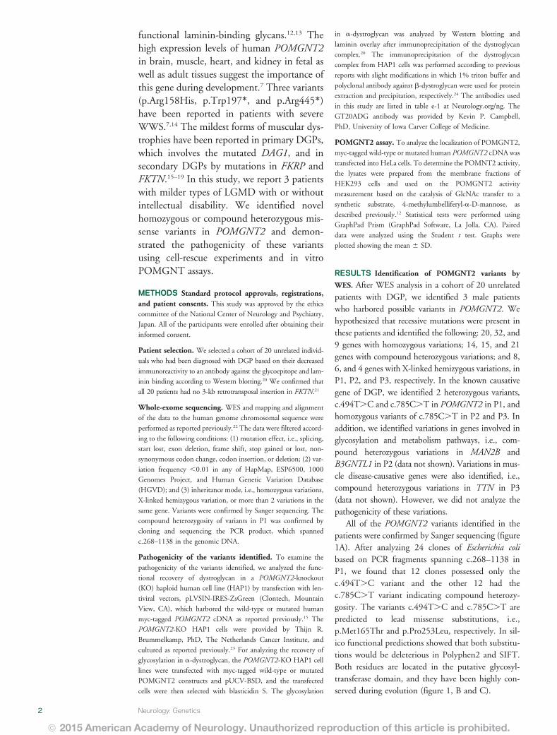
Clinical features The clinical information is summa-rized in table 1
Family history All 3 patients were from a noncon-sanguineous marriage and had no family history ofneuromuscular diseases or intellectual disabilities
Muscle defects P1 was noted to have become de-layed in his motor development immediately afterrecovering from a respiratory tract infection at 11months Myositis was diagnosed because of elevatedcreatine kinase (CK) levels of 3000 IUL (referencerange 20ndash145 IUL) and biopsy findings as describedbelow It was not certain whether a screening of anti-bodies was performed He was treated with steroidsand his motor development improved graduallyalthough the high CKemia continued He walked at2 years but did not run When he was a junior highschool student he could walk upstairs and squat fromthe sitting position When he was reexamined at age28 years he could not walk uphill or downstairs with-out handrails and had difficulty standing from a squatbecause of weakness in the proximal lower limbs Hiscalves were mildly enlarged which had not beennoted at onset He could raise his arms but not aheavy load There was no prominent facial weaknessor scapular winging P2 was first found to have aserum CK level of 13000 IUL at age 13 years on
a follow-up examination concerning congenital bili-ary atresia He underwent muscle biopsy although hehad no muscle symptoms and was diagnosed withDGP He showed no fatigue or weakness at 19 yearsbut calf hypertrophy was detected and high CKemiacontinued P3 had normal motor milestones and wasfound by chance to have high CKemia (3000ndash9000IUL) at age 4 years Muscle biopsy was performed atage 8 years and he was diagnosed with DGP At age14 years his calves were moderately enlarged but themuscle power in his limbs was very strong and hiswhole-body muscle volume was sufficient
Intellectual disabilities Intellectual development inP1 and P3 was slow P1 spoke his first few words at18 months and he began to communicate using sim-ple words before going to primary school He couldnot keep up with classwork so he went to a specialschool to support his learning His IQ was less than35 at age 28 years P3 spoke meaningful words withno sentence formation at age 6 years He had hyperac-tivity disorder and Wechsler Intelligence Scale forChildrenndashIII testing showed that his IQ was 60 atage 14 years
Ocular defects and others None of the 3 patients hadproblems with his eyes heart or kidneys P2 hadcongenital biliary atresia at birth and vitamin K
Figure 1 Genetic variations in POMGNT2
(A) Electropherograms around the mutation sites in POMGNT2 based on Sanger sequencing (B) Localizations of the iden-tified mutations (in red) and known mutations (in black) in the domain structures of the POMGNT2 protein This schema wasmodified from Ref 14 (C) Amino acid conservation in the mutation sites among species
Neurology Genetics 3
ordf 2015 American Academy of Neurology Unauthorized reproduction of this article is prohibited
deficiency which resulted in intracranial hemorrhageHe received a living liver transplantation at age 17 years
Muscle imagingMRI of P1 detected diffuse atrophyin the skeletal muscles especially in thigh musclesMuscle images of P2 and P3 detected no atrophy orfatty change in the skeletal muscles (figure 2A)
Brain imaging and EEG Brain MRI showed noabnormality in P1 or P3 (data not shown) CT scan-ning of P2 detected no anomalies except for afteref-fects of intracranial hemorrhage at birth EEG of P1at 11 months revealed asymmetric activity in sleepstage ie decreased spindles in the right occipitalregion There were no follow-up examination dataP2 had not been examined and P3 had normalEEG results
Histologic features based on muscle biopsy Biopsyof P1 at 11 months detected numerous necrotic
fibers some regenerating fibers marked cellularinfiltration in the endomysium perimysium andperivascular regions and moderate endomysialfibrosis The muscle histology in P2 at age 13 yearswas almost normal except for some fibers withinternal nuclei P3 at age 14 years exhibited a mus-cular dystrophyndashlike appearance includingnecrotic and regenerating fibers fibers with inter-nal nuclei and mild endomysial fibrosis Immuno-histochemistry was positive for b-dystroglycandystrophin merosin and sarcoglycans but nega-tive for the glycoepitope of a-dystroglycan (figure2B) in all 3 patients Immunostaining in P1 wasperformed at 23 years but not at onset Westernblotting with the glycoepitope antibody and thelaminin overlay assay of muscle proteins detectedspecific patterns with 2-weak reactive bands at 150and 120 kDa whereas 3 bands at 150 120 and 90
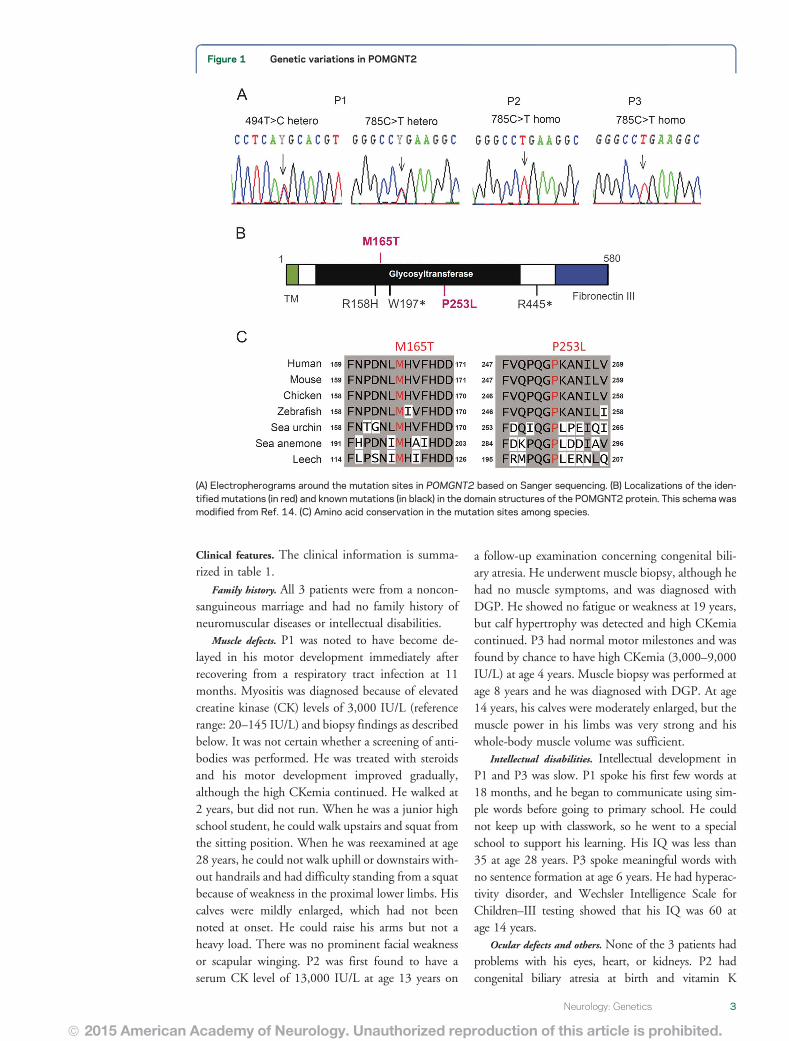
Table 1 Clinical laboratory and genetic findings
Patient
P1 P2 P3
Sex Male Male Male
Nucleotide change (amino acid change) c494TC (pMet165Thr) c758CT (pPro253Leu)homozygous
c758CT (pPro253Leu)homozygous
c758CT (pPro253Leu)
Onset
Age 11 mo 13 y 1 y
First symptom Fever regression High CK Speech delay high CK
Developmental delay
High CK
Biopsy y 2 13 8
Last examination
Age y 28 19 14
Muscle defects
Motor ability Ambulant Ambulant running Ambulant fast running
Muscle weakness Proximal No No
Muscle atrophy Limbs diffuse No No
Calf hypertrophy Mild Moderate Moderate
Facial involvement No No No
Gowers sign Positive No No
Serum CK IUL 1400 300ndash4000 1000ndash4000
Brain defects
Intelligence IQ 35 Normal IQ60
Cobblestone lissencephaly No No No
Enlarged ventricles No No No
Cerebellar hypoplasia No No No
Ocular defects No No No
Others mdash Congenital biliary atresia Hyperactivity disorder
Abbreviation CK 5 creatine kinase
4 Neurology Genetics
ordf 2015 American Academy of Neurology Unauthorized reproduction of this article is prohibited
Figure 2 Muscle imaging and hypoglycosylation of a-dystroglycan
(A) Images of the thigh and calf muscles of the patients P1 (at 28 years) exhibited atrophy and mild fatty infiltration in thighmuscles but the triceps surae muscles were rather normal P2 (at 19 years) and P3 (at 14 years) had sufficient skeletalmuscle volumes and no fatty change P1 and P3 T1-weighted image P2 CT image (B) Histology and immunostaining ofskeletal muscle from the patients The muscle histology in P1 exhibited a muscular dystrophyndashlike appearance includingnecrotic and regenerating fibers fibers with internal nuclei and mild endomysial fibrosis whereas those of P3were mild andof P2 were almost normal except for some fibers with internal nuclei according to hematoxylin and eosin staining (HE) Allmuscles were positive for antibodies to b-dystroglycan (b-DG) but negative for the glycoepitope antibody to a-dystroglycan(a-DG VIA4-1) (C) Western blotting with VIA4-1 antibody and the laminin overlay assay of muscle proteins detected 2reactive bands of a-DG at 150 and 120 kDa whereas the GT20ADG antibody against the core peptide detected anadditional band at 100 kDa b-DG staining was normal
Neurology Genetics 5
ordf 2015 American Academy of Neurology Unauthorized reproduction of this article is prohibited
kDa were detected with the GT20ADG antibodywhich recognizes the polypeptide of a-dystroglycan(figure 2C) b-Dystroglycan was normal
Subcellular localization of mutated POMGNT2 Thetransfected myc-tagged wild-type POMGNT2 proteincolocalized with ER proteins (figure 3A) Similarlythe myc-tagged pMet165Thr- and pPro253Leu-substituted POMGNT2 proteins were also localizedin the ER
POMGNT activities of missense-substituted proteins
The wild-type and missense-substituted POMGNT2
proteins had similar expression levels in humanembryonic kidney cells thereby indicating that thesevariations do not affect protein folding and stability(figure 3B) The specific POMGNT activities of boththe missense-substituted proteins (pMet165Thr andpPro253Leu) were greatly reduced to less than 10of that of the wild-type POMGNT2 (figure 3C)
Rescue of POMGNT2-KO HAP1 cells by wild-type and
missense POMGNT2 cDNA To determine the patho-genetic effects of the 2 missense variants identifiedin the patients we transfected lentiviral vectors
Figure 3 Subcellular localization and enzymatic activities of mutated POMGNT2
(A) Myc-tagged wild-type pMet165Thr-mutated and pPro253Leu-mutated POMGNT2 cDNAs were transfected into He-La cells Both the myc-tagged wild-type and missense mutants of POMGNT2 (c-myc red) were costained with endoplasmicreticulum (ER) protein protein disulfide isomerase (PDI green) Blue indicates the transfected cells with the marker proteinZsGreen The bar represents 20 mm (B) Expression levels of myc-tagged wild-type (wild) Met165Thr-mutated (M165T)and Pro253Leu-mutated (P253L) POMGNT2 in HEK293 cells (arrow) (C) Specific POMGNT activity of wild-type andmutated POMGNT2 expressed in HEK293 cells The relative POMGNT activity was calculated against that of wild-typePOMGNT2 Data obtained from 5 individual measurements are shown as the mean where the error bars indicate the SDp 001 vs wild type according to the Mann-Whitney U test
6 Neurology Genetics
ordf 2015 American Academy of Neurology Unauthorized reproduction of this article is prohibited
with wild-type or substituted POMGNT2 cDNAs (pMet165Thr and pPro253Leu) into POMGNT2-KOHAP1 cells which exhibited defects in their reactivityto the anti-a-dystroglycan antibody IIH6 ThePOMGNT2-KO HAP1 cells were rescued byintroducing the wild-type cDNA therebydemonstrating the recovery of the strongimmunoreactivity to IIH6 which was similar tothat in the wild-type HAP1 cells By contrastthe introduction of cDNAs with pMet165Thr andpPro253Leu failed to rescue POMGNT2-KO HAP1cells which did not stain with the IIH6 antibody(figure 4A)
The wild-type HAP1 cells produced 110-kDaglycosylated a-dystroglycan which reacts with
laminin By contrast the POMGNT2-KO HAP1cells expressed the 90-kDa a-dystroglycan which isrecognized by the peptide-core antibodyGT20DAG but it reacts negatively with IIH6 andlaminin The introduction of wild-type POMGNT2cDNAs into POMGNT2-KO HAP1 cells recoveredthe production of the 110-kDa glycosylated a-dys-troglycan which was able to bind to laminin whereasthe introduction of the pMet165Thr- or pPro253Leu-substituted POMGNT2 cDNAs failedto recover the glycosylation and laminin binding ofa-dystroglycan (figure 4B)
DISCUSSION In this study we identified 3 patientswith DGP 1 who had compound heterozygous and 2
Figure 4 Mutant POMGNT2 proteins do not rescue hypoglycosylation of a-dystroglycan in POMGNT2-knockout (KO) cells
(A) IIH6-4C2 staining of wild-type haploid human cell line (HAP1) cells POMGNT2-KO cells (HAP1-POMGNT2-KO) and POMGNT2-KO cells transfected withwild-type (1WTPOMGNT2) Met165Thr-mutated (1M165T POMGNT2) and Pro253Leu-mutated (1P253L POMGNT2) POMGNT2 cDNA (in red) Thetransfected cells were positive for ZsGreen expression (in green) HAP1 wild-type haploid cells HAP1-POMGNT2-KO POMGNT2-KO HAP1 cells The scalebar represents 20 mm (B) Recovery of the glycosylation of a-dystroglycan in POMGNT2-KO HAP1 cells a-Dystroglycan in POMGNT2-KO HAP1 cells waspositive for IIH6 antibody- and laminin-binding after transfection with wild-type (wild) POMGNT2 cDNA but not after transfection with M165T- and P253L-mutated POMGNT2 cDNA The 43-kDa aDG band and its degraded products are shown by the arrowhead and asterisk () respectively The immunoglobulinlight chain band is indicated by the double asterisk ()
Neurology Genetics 7
ordf 2015 American Academy of Neurology Unauthorized reproduction of this article is prohibited
others who had homozygous variants in POMGNT2According to the DGP classification25 P1 hadLGMD with mental retardation whereas P2 andP3 had high CKemia without muscle weakness withor without intellectual disability The clinical pheno-types of our patients were much milder comparedwith those of previously reported patients withWWS7 The pathologic findings were in proportionto the clinical severity Our findings expand the clin-ical and pathologic spectrum of DGP associated withPOMGNT2 variants from the severest WWS to themildest LGMD phenotypes
P1 was believed to have myositis for over 20 yearsbecause immunohistochemical analysis was not per-formed during the first pathologic analysis P2 wasdiagnosed as normal based only on a set of routine ex-aminations without immunohistochemistry Thussome patients with DGP associated with POMGNT2mutations with milder phenotypes may have escapeddiagnosis or they could have been diagnosed withmyositis or asymptomatic high CK Our patientsexperienced no disturbance to their cardiac respira-tory or renal functions whereas cardiac involvementhas been reported in ambulant patients with DGP
One of the identified variants c785CT wasfound in the ESP6500 database with an allele fre-quency of 0000077 but we could not find this var-iant in HGVD the genetic variation database for theJapanese population If we assume that the frequencyrate is the same in the Japanese population asESP6500 then the prevalence of homozygous pa-tients is expected to be 1 in 168 million individualsHowever we found 2 homozygous patients with thisvariant although the overall Japanese population is120 million so the allele frequency of this variant inthe Japanese population may be higher than that inwestern countries
As reported previously the hypoglycosylation lev-els of a-dystroglycan are not correlated consistentlywith the clinical severity26 Nevertheless our patientsexhibited a specific pattern of a-dystroglycan with 3bands of different sizes based on Western blottingwith a peptide-core recognition antibody Two largerisoforms also reacted with the glycoepitope antibodyand laminin whereas the smaller one did not therebysuggesting that there were at least 3 glycoforms ofa-dystroglycan in these patients The remaininglaminin-binding abilities of a-dystroglycan may berelated to the milder symptoms of the patients assuggested previously by a DGP model in mice wheresmaller amounts of the functional a-dystroglycanwere sufficient for maintaining muscle functionMoreover this specific pattern with 3 bands ofa-dystroglycan differs from those in milder forms ofDGP with FKTN or DAG1 mutations which wehave reported previously1516 Thus we hypothesize
that a reduction rather than the complete loss ofthe enzymatic activities due to missense mutationsin FKTN and POMGNT2may generate intermediateglycoforms of a-dystroglycan However it is impor-tant that the pattern of a-dystroglycan glycosylationin patients will be determined by the residualfunction of the affected enzyme POMGNT2functions in the ER where it transfers the secondGlcNAc residue during the synthesis of the coreM3 chain (GalNAc-b3-GlcNAc-b4-Man-aSerThr) which is believed to be a platform structure thatis essential for the initiation of heteropolymer ldquoLARGEglycanrdquo synthesis1227 whereas FKTN functions inLARGE glycan extension in the Golgi2829 Furtheranalyses should clarify the relationship betweengenetic variants and the glycosylation pattern ofresidual a-dystroglycan
Both of the variants that we identified(pMet165Thr and pPro253Leu) were located inthe putative glycosyltransferase domain and they werepredicted to be deleterious by in silico Our resultsshowed that these missense variants did not affectthe subcellular localization of protein products inthe ER14 and that the PONGNT2 enzymatic activ-ities were greatly reduced but not lost These resultsalso suggest that these missense variants may be thecause of milder symptoms in the patients because ofthe reduced activity of POMGNT2 According to aprevious report and the present study all of the iden-tified missense variants are located in the putativeglycosyltransferase domain Based on comparisonsof the patientsrsquo symptoms we suggest that thec758CT (pPro253Leu) mutation is associatedwith milder phenotypes in both the muscles andbrain compared with c494TC (pMet165Thr)and other reported mutations although we foundno differences in the in vitro enzymatic activitiesand glycosylation recovery in the complementationtests between 2 mutated POMGNT2 proteins Fur-thermore P2 and P3 had the same variants and ex-hibited similar phenotypes in their skeletal musclesbut there were slight differences in their clinical phe-notypes in other organs because P2 had congenitalbiliary atresia but had normal intelligence whereas P3exhibited intellectual disability and hyperactivityHowever the congenital biliary atresia in P2 andhyperactivity disorder in P3 may not have been dueto the variation in POMGNT2
In this study we also performed phenotypic rescueexperiments using POMGNT2-KOHAP1 cells basedon lentivirus-mediated expression of mutated cDNAin the same manner as DAG1 variants This methodwas sensitive in evaluating the pathogenic effects ofthe identified mutations rapidly and easily evenwhen the mutations were milder We extendedthis mutation-based assay to Western blotting and
8 Neurology Genetics
ordf 2015 American Academy of Neurology Unauthorized reproduction of this article is prohibited
laminin-overlay analyses to obtain objective and semi-quantitative results This method is technically assimple as immunostaining or Western blotting usingmuscle samples from patients therefore it may allowresearchers to evaluate any variants present in all ofthe known causative genes and the variants in novelcandidate genes to detect DGPs particularly withoutusing patientsrsquo specimens
AUTHOR CONTRIBUTIONSYE made WES pipeline analyzed and interpreted the data drafted
and edited the manuscript and performed statistical analysis MD
conducted acquisition analysis and interpretation of data drafted
and edited the manuscript S Noguchi supervised all aspects of this
study including study design and data interpretation drafted and edi-
ted the manuscript and performed statistical analysis MO contrib-
uted on technical aspects YKH selected patients and performed
WES SK KS S Nagai and SO collected clinical information of
the patient I Nonaka and I Nishino supervised manuscript prepara-
tion and edited the manuscript
ACKNOWLEDGMENTThe authors thank Nozomi Matsuyama and Kanako Goto for technical
support Thijn R Brummelkamp for supplying HAP1 cells and Kevin
P Campbell for supplying the GT20ADG antibody
STUDY FUNDINGThis study supported by Intramural Research Grant (23-5 25-5 and
26-8) for Neurological and Psychiatric Disorders of NCNP (to S Nogu-
chi YKH and I Nishino) grants from Research on Applying Health
Technology from the Ministry of Health Labor and Welfare (H23-
Jitsuyouka (Nanbyou)-Ippan-008) (to YKH and I Nishino) grants
from Research on rare and intractable diseases from the Ministry of
Health Labour and Welfare (H26-Itaku (Nan)-Ippan-081) (to S Nogu-
chi and I Nishino) and grants from MEXT-Supported Program
(S141101) for the Strategic Research Foundation at Private Universities
from The Ministry of Education Culture Sports Science and Technol-
ogy (MEXT) (to YKH)
DISCLOSUREDr Endo and Dr Dong report no disclosures Dr Noguchi holds patent
WO 2010131712A1 and has received research support from Novartis
Pharmaceuticals Japan the Ministry of Health Labour and Welfare
Ms Ogawa reports no disclosures Dr Hayashi has served on the edito-
rial board of Neuromuscular Disorders and has received grants from The
Ministry of Education Culture Sports Science and Technology
(MEXT) the Ministry of Health Labor and Welfare Japan Society
for the Promotion of Science (JSPS) and The Promotion and Mutual
Aid Corporation for Private Schools of Japan Dr Kuru Dr Sugiyama
Dr Nagai Dr Ozasa and Dr Nonaka report no disclosures Dr Nish-
ino has served on scientific advisory board(s) for Genzyme has served on
the editorial board of Neuromuscular Disorders Neurology and Clinical
Neuroscience Therapeutic Advances in Neurological Disorders the Journal
of the Neurological Sciences Skeletal Muscle and the Journal of Neuromus-
cular Diseases holds a patent for Efficacious agents to pathologic condi-
tion due to GNE protein dysfunction and has received research support
from Astellas Pharma Inc Genzyme Japan Japan Society for the Pro-
motion of Science (JSPS) and the Ministry of Health Labor and Wel-
fare Go to Neurologyorgng for full disclosure forms
Received September 16 2015 Accepted in final form September 24 2015
REFERENCES1 Hayashi YK Ogawa M Tagawa K et al Selective defi-
ciency of alpha-dystroglycan in Fukuyama-type congenital
muscular dystrophy Neurology 200157115ndash121
2 Muntoni F Brockington M Brown SC Glycosylation
eases muscular dystrophy Nat Med 200410676ndash677
3 Brown SC Torelli S Brockington M et al Abnormalities
in alpha-dystroglycan expression in MDC1C and
LGMD2I muscular dystrophies Am J Pathol 2004164
727ndash737
4 Mendell JR Boueacute DR Martin PT The congenital mus-
cular dystrophies recent advances and molecular insights
Pediatr Dev Pathol 20069427ndash443
5 Willer T Lee H Lommel M et al ISPD loss-of-function
mutations disrupt dystroglycan O-mannosylation and
cause Walker-Warburg syndrome Nat Genet 201244
575ndash580
6 Roscioli T Kamsteeg EJ Buysse K et al Mutations in
ISPD cause Walker-Warburg syndrome and defective
glycosylation of a-dystroglycan Nat Genet 201244
581ndash585
7 Manzini MC Tambunan DE Hill RS et al Exome
sequencing and functional validation in zebrafish identify
GTDC2 mutations as a cause of Walker-Warburg syn-
drome Am J Hum Genet 201291541ndash547
8 Carss KJ Stevens E Foley AR et al Mutations in GDP-
mannose pyrophosphorylase B cause congenital and limb-
girdle muscular dystrophies associated with hypoglycosylation
of a-dystroglycan Am J Hum Genet 20139329ndash41
9 Buysse K Riemersma M Powell G et al Missense mutations
in b-13-N-acetylglucosaminyltransferase 1 (B3GNT1) cause
Walker-Warburg syndrome Hum Mol Genet 201322
1746ndash1754
10 Shaheen R Faqeih E Ansari S Alkuraya FS A truncating
mutation in B3GNT1 causes severe Walker-Warburg syn-
drome Neurogenetics 201314243ndash245
11 Di Costanzo S Balasubramanian A Pond HL et al
POMK mutations disrupt muscle development leading
to a spectrum of neuromuscular presentations Hum
Mol Genet 2014235781ndash5792
12 Yoshida-Moriguchi T Willer T Anderson ME et al
SGK196 is a glycosylation-specific O-mannose kinase
required for dystroglycan function Science 2013341
896ndash899
13 Yoshida-Moriguchi T Yu L Stalnaker SH et al O-
mannosyl phosphorylation of alpha-dystroglycan is
required for laminin binding Science 201032788ndash92
14 Ogawa M Nakamura N Nakayama Y et al GTDC2
modifies O-mannosylated a-dystroglycan in the endoplas-
mic reticulum to generate N-acetyl glucosamine epitopes
reactive with CTD1106 antibody Biochem Biophys Res
Commun 201344088ndash93
15 Dong M Noguchi S Endo Y et al DAG1 mutations asso-
ciated with asymptomatic hyperCKemia and hypoglycosyla-
tion of a-dystroglycan Neurology 201584273ndash279
16 Murakami T Hayashi YK Noguchi S et al Fukutin gene
mutations cause dilated cardiomyopathy with minimal
muscle weakness Ann Neurol 200660597ndash602
17 de Paula F Vieira N Starling A et al Asymptomatic
carriers for homozygous novel mutations in the FKRP
gene the other end of the spectrum Eur J Hum Genet
200311923ndash930
18 Fernandez C de Paula AM Figarella-Branger D et al
Diagnostic evaluation of clinically normal subjects with
chronic hyperCKemia Neurology 2006661585ndash1587
19 Fiorillo C Moro F Astrea G et al Novel mutations in the
fukutin gene in a boy with asymptomatic hyperCKemia
Neuromuscul Disord 2013231010ndash1015
Neurology Genetics 9
ordf 2015 American Academy of Neurology Unauthorized reproduction of this article is prohibited
20 Michele DE Barresi R Kanagawa M et al Posttransla-
tional disruption of dystroglycanndashligand interactions
in congenital muscular dystrophies Nature 2002418
417ndash422
21 Kobayashi K Nakahori Y Miyake M et al An ancient
retrotransposal insertion causes Fukuyama-type congenital
muscular dystrophy Nature 1998394388ndash392
22 Endo Y Noguchi S Hara Y et al Dominant mutations in
ORAI1 cause tubular aggregate myopathy with hypocalce-
mia via constitutive activation of store-operated Ca21
channels Hum Mol Genet 201524637ndash648
23 Jae LT Raaben M Riemersma M et al Deciphering the
glycosylome of dystroglycanopathies using haploid screens
for lassa virus entry Science 2013340479ndash483
24 Noguchi S Wakabayashi E Imamura M Yoshida M
Ozawa E Formation of sarcoglycan complex with differ-
entiation in cultured myocyte Eur J Biochem 2000267
640ndash648
25 Godfrey C Clement E Mein R et al Refining geno-
type phenotype correlations in muscular dystrophies
with defective glycosylation of dystroglycan Brain
20071302725ndash2735
26 Jimenez-Mallebrera C Torelli S Feng L et al A compar-
ative study of alpha-dystroglycan glycosylation in dystro-
glycanopathies suggests that the hypoglycosylation of
alpha-dystroglycan does not consistently correlate with
clinical severity Brain Pathol 200919596ndash611
27 Hara Y Kanagawa M Kunz S et al Like-acetylgluco-
saminyltransferase (LARGE)-dependent modification
of dystroglycan at Thr-317319 is required for laminin
binding and arenavirus infection Proc Natl Acad Sci U
S A 201110817426ndash17431
28 Willer T Inamori K Venzke D et al The glucuronyl-
transferase B4GAT1 is required for initiation of LARGE-
mediated a-dystroglycan functional glycosylation Elife
20143 doi 107554eLife03941
29 Praissman JL Live DH Wang S et al B4GAT1 is the
priming enzyme for the LARGE-dependent functional gly-
cosylation of a-dystroglycan Elife 20143 doi 107554
eLife03943
10 Neurology Genetics
ordf 2015 American Academy of Neurology Unauthorized reproduction of this article is prohibited
DOI 101212NXG000000000000003320151 Neurol Genet
Yukari Endo Mingrui Dong Satoru Noguchi et al mutationsPOMGNT2Milder forms of muscular dystrophy associated with
This information is current as of December 10 2015
Neurology All rights reserved Online ISSN 2376-7839an open-access online-only continuous publication journal Copyright copy 2015 American Academy of
is an official journal of the American Academy of Neurology Published since April 2015 it isNeurol Genet
ServicesUpdated Information amp
httpngneurologyorgcontent14e33fullhtmlincluding high resolution figures can be found at
Supplementary Material httpngneurologyorgcontentsuppl2015121014e33DC1
Supplementary material can be found at
References httpngneurologyorgcontent14e33fullhtmlref-list-1
This article cites 27 articles 4 of which you can access for free at
Citations httpngneurologyorgcontent14e33fullhtmlotherarticles
This article has been cited by 1 HighWire-hosted articles
Subspecialty Collections
httpngneurologyorgcgicollectionmuscle_diseaseMuscle disease
httpngneurologyorgcgicollectionall_pediatricAll Pediatric
httpngneurologyorgcgicollectionall_neuromuscular_diseaseAll Neuromuscular Disease
httpngneurologyorgcgicollectionall_geneticsAll Geneticsfollowing collection(s) This article along with others on similar topics appears in the
Permissions amp Licensing
httpngneurologyorgmiscaboutxhtmlpermissionsits entirety can be found online atInformation about reproducing this article in parts (figurestables) or in
Reprints
httpngneurologyorgmiscaddirxhtmlreprintsusInformation about ordering reprints can be found online
Neurology All rights reserved Online ISSN 2376-7839an open-access online-only continuous publication journal Copyright copy 2015 American Academy of
is an official journal of the American Academy of Neurology Published since April 2015 it isNeurol Genet
functional laminin-binding glycans1213 Thehigh expression levels of human POMGNT2in brain muscle heart and kidney in fetal aswell as adult tissues suggest the importance ofthis gene during development7 Three variants(pArg158His pTrp197 and pArg445)have been reported in patients with severeWWS714 The mildest forms of muscular dys-trophies have been reported in primary DGPswhich involves the mutated DAG1 and insecondary DGPs by mutations in FKRP andFKTN15ndash19 In this study we report 3 patientswith milder types of LGMD with or withoutintellectual disability We identified novelhomozygous or compound heterozygous mis-sense variants in POMGNT2 and demon-strated the pathogenicity of these variantsusing cell-rescue experiments and in vitroPOMGNT assays
METHODS Standard protocol approvals registrationsand patient consents This study was approved by the ethics
committee of the National Center of Neurology and Psychiatry
Japan All of the participants were enrolled after obtaining their
informed consent
Patient selection We selected a cohort of 20 unrelated individ-
uals who had been diagnosed with DGP based on their decreased
immunoreactivity to an antibody against the glycoepitope and lam-
inin binding according to Western blotting20 We confirmed that
all 20 patients had no 3-kb retrotransposal insertion in FKTN21
Whole-exome sequencing WES and mapping and alignment
of the data to the human genome chromosomal sequence were
performed as reported previously22 The data were filtered accord-
ing to the following conditions (1) mutation effect ie splicing
start lost exon deletion frame shift stop gained or lost non-
synonymous codon change codon insertion or deletion (2) var-
iation frequency 001 in any of HapMap ESP6500 1000
Genomes Project and Human Genetic Variation Database
(HGVD) and (3) inheritance mode ie homozygous variations
X-linked hemizygous variation or more than 2 variations in the
same gene Variants were confirmed by Sanger sequencing The
compound heterozygosity of variants in P1 was confirmed by
cloning and sequencing the PCR product which spanned
c268ndash1138 in the genomic DNA
Pathogenicity of the variants identified To examine the
pathogenicity of the variants identified we analyzed the func-
tional recovery of dystroglycan in a POMGNT2-knockout(KO) haploid human cell line (HAP1) by transfection with len-
tiviral vectors pLVSIN-IRES-ZsGreen (Clontech Mountain
View CA) which harbored the wild-type or mutated human
myc-tagged POMGNT2 cDNA as reported previously15 The
POMGNT2-KO HAP1 cells were provided by Thijn R
Brummelkamp PhD The Netherlands Cancer Institute and
cultured as reported previously23 For analyzing the recovery of
glycosylation in a-dystroglycan the POMGNT2-KO HAP1 cell
lines were transfected with myc-tagged wild-type or mutated
POMGNT2 constructs and pUCV-BSD and the transfected
cells were then selected with blasticidin S The glycosylation
in a-dystroglycan was analyzed by Western blotting and
laminin overlay after immunoprecipitation of the dystroglycan
complex20 The immunoprecipitation of the dystroglycan
complex from HAP1 cells was performed according to previous
reports with slight modifications in which 1 triton buffer and
polyclonal antibody against b-dystroglycan were used for protein
extraction and precipitation respectively24 The antibodies used
in this study are listed in table e-1 at Neurologyorgng The
GT20ADG antibody was provided by Kevin P Campbell
PhD University of Iowa Carver College of Medicine
POMGNT2 assay To analyze the localization of POMGNT2
myc-tagged wild-type or mutated human POMGNT2 cDNA was
transfected into HeLa cells To determine the POMNT2 activity
the lysates were prepared from the membrane fractions of
HEK293 cells and used on the POMGNT2 activity
measurement based on the catalysis of GlcNAc transfer to a
synthetic substrate 4-methylumbelliferyl-a-D-mannose as
described previously12 Statistical tests were performed using
GraphPad Prism (GraphPad Software La Jolla CA) Paired
data were analyzed using the Student t test Graphs were
plotted showing the mean 6 SD
RESULTS Identification of POMGNT2 variants by
WES After WES analysis in a cohort of 20 unrelatedpatients with DGP we identified 3 male patientswho harbored possible variants in POMGNT2 Wehypothesized that recessive mutations were present inthese patients and identified the following 20 32 and9 genes with homozygous variations 14 15 and 21genes with compound heterozygous variations and 86 and 4 genes with X-linked hemizygous variations inP1 P2 and P3 respectively In the known causativegene of DGP we identified 2 heterozygous variantsc494TC and c785CT in POMGNT2 in P1 andhomozygous variants of c785CT in P2 and P3 Inaddition we identified variations in genes involved inglycosylation and metabolism pathways ie com-pound heterozygous variations in MAN2B andB3GNTL1 in P2 (data not shown) Variations in mus-cle disease-causative genes were also identified iecompound heterozygous variations in TTN in P3(data not shown) However we did not analyze thepathogenicity of these variations
All of the POMGNT2 variants identified in thepatients were confirmed by Sanger sequencing (figure1A) After analyzing 24 clones of Escherichia colibased on PCR fragments spanning c268ndash1138 inP1 we found that 12 clones possessed only thec494TC variant and the other 12 had thec785CT variant indicating compound heterozy-gosity The variants c494TC and c785CT arepredicted to lead missense substitutions iepMet165Thr and pPro253Leu respectively In sil-ico functional predictions showed that both substitu-tions would be deleterious in Polyphen2 and SIFTBoth residues are located in the putative glycosyl-transferase domain and they have been highly con-served during evolution (figure 1 B and C)
2 Neurology Genetics
ordf 2015 American Academy of Neurology Unauthorized reproduction of this article is prohibited
Clinical features The clinical information is summa-rized in table 1
Family history All 3 patients were from a noncon-sanguineous marriage and had no family history ofneuromuscular diseases or intellectual disabilities
Muscle defects P1 was noted to have become de-layed in his motor development immediately afterrecovering from a respiratory tract infection at 11months Myositis was diagnosed because of elevatedcreatine kinase (CK) levels of 3000 IUL (referencerange 20ndash145 IUL) and biopsy findings as describedbelow It was not certain whether a screening of anti-bodies was performed He was treated with steroidsand his motor development improved graduallyalthough the high CKemia continued He walked at2 years but did not run When he was a junior highschool student he could walk upstairs and squat fromthe sitting position When he was reexamined at age28 years he could not walk uphill or downstairs with-out handrails and had difficulty standing from a squatbecause of weakness in the proximal lower limbs Hiscalves were mildly enlarged which had not beennoted at onset He could raise his arms but not aheavy load There was no prominent facial weaknessor scapular winging P2 was first found to have aserum CK level of 13000 IUL at age 13 years on
a follow-up examination concerning congenital bili-ary atresia He underwent muscle biopsy although hehad no muscle symptoms and was diagnosed withDGP He showed no fatigue or weakness at 19 yearsbut calf hypertrophy was detected and high CKemiacontinued P3 had normal motor milestones and wasfound by chance to have high CKemia (3000ndash9000IUL) at age 4 years Muscle biopsy was performed atage 8 years and he was diagnosed with DGP At age14 years his calves were moderately enlarged but themuscle power in his limbs was very strong and hiswhole-body muscle volume was sufficient
Intellectual disabilities Intellectual development inP1 and P3 was slow P1 spoke his first few words at18 months and he began to communicate using sim-ple words before going to primary school He couldnot keep up with classwork so he went to a specialschool to support his learning His IQ was less than35 at age 28 years P3 spoke meaningful words withno sentence formation at age 6 years He had hyperac-tivity disorder and Wechsler Intelligence Scale forChildrenndashIII testing showed that his IQ was 60 atage 14 years
Ocular defects and others None of the 3 patients hadproblems with his eyes heart or kidneys P2 hadcongenital biliary atresia at birth and vitamin K
Figure 1 Genetic variations in POMGNT2
(A) Electropherograms around the mutation sites in POMGNT2 based on Sanger sequencing (B) Localizations of the iden-tified mutations (in red) and known mutations (in black) in the domain structures of the POMGNT2 protein This schema wasmodified from Ref 14 (C) Amino acid conservation in the mutation sites among species
Neurology Genetics 3
ordf 2015 American Academy of Neurology Unauthorized reproduction of this article is prohibited
deficiency which resulted in intracranial hemorrhageHe received a living liver transplantation at age 17 years
Muscle imagingMRI of P1 detected diffuse atrophyin the skeletal muscles especially in thigh musclesMuscle images of P2 and P3 detected no atrophy orfatty change in the skeletal muscles (figure 2A)
Brain imaging and EEG Brain MRI showed noabnormality in P1 or P3 (data not shown) CT scan-ning of P2 detected no anomalies except for afteref-fects of intracranial hemorrhage at birth EEG of P1at 11 months revealed asymmetric activity in sleepstage ie decreased spindles in the right occipitalregion There were no follow-up examination dataP2 had not been examined and P3 had normalEEG results
Histologic features based on muscle biopsy Biopsyof P1 at 11 months detected numerous necrotic
fibers some regenerating fibers marked cellularinfiltration in the endomysium perimysium andperivascular regions and moderate endomysialfibrosis The muscle histology in P2 at age 13 yearswas almost normal except for some fibers withinternal nuclei P3 at age 14 years exhibited a mus-cular dystrophyndashlike appearance includingnecrotic and regenerating fibers fibers with inter-nal nuclei and mild endomysial fibrosis Immuno-histochemistry was positive for b-dystroglycandystrophin merosin and sarcoglycans but nega-tive for the glycoepitope of a-dystroglycan (figure2B) in all 3 patients Immunostaining in P1 wasperformed at 23 years but not at onset Westernblotting with the glycoepitope antibody and thelaminin overlay assay of muscle proteins detectedspecific patterns with 2-weak reactive bands at 150and 120 kDa whereas 3 bands at 150 120 and 90
Table 1 Clinical laboratory and genetic findings
Patient
P1 P2 P3
Sex Male Male Male
Nucleotide change (amino acid change) c494TC (pMet165Thr) c758CT (pPro253Leu)homozygous
c758CT (pPro253Leu)homozygous
c758CT (pPro253Leu)
Onset
Age 11 mo 13 y 1 y
First symptom Fever regression High CK Speech delay high CK
Developmental delay
High CK
Biopsy y 2 13 8
Last examination
Age y 28 19 14
Muscle defects
Motor ability Ambulant Ambulant running Ambulant fast running
Muscle weakness Proximal No No
Muscle atrophy Limbs diffuse No No
Calf hypertrophy Mild Moderate Moderate
Facial involvement No No No
Gowers sign Positive No No
Serum CK IUL 1400 300ndash4000 1000ndash4000
Brain defects
Intelligence IQ 35 Normal IQ60
Cobblestone lissencephaly No No No
Enlarged ventricles No No No
Cerebellar hypoplasia No No No
Ocular defects No No No
Others mdash Congenital biliary atresia Hyperactivity disorder
Abbreviation CK 5 creatine kinase
4 Neurology Genetics
ordf 2015 American Academy of Neurology Unauthorized reproduction of this article is prohibited
Figure 2 Muscle imaging and hypoglycosylation of a-dystroglycan
(A) Images of the thigh and calf muscles of the patients P1 (at 28 years) exhibited atrophy and mild fatty infiltration in thighmuscles but the triceps surae muscles were rather normal P2 (at 19 years) and P3 (at 14 years) had sufficient skeletalmuscle volumes and no fatty change P1 and P3 T1-weighted image P2 CT image (B) Histology and immunostaining ofskeletal muscle from the patients The muscle histology in P1 exhibited a muscular dystrophyndashlike appearance includingnecrotic and regenerating fibers fibers with internal nuclei and mild endomysial fibrosis whereas those of P3were mild andof P2 were almost normal except for some fibers with internal nuclei according to hematoxylin and eosin staining (HE) Allmuscles were positive for antibodies to b-dystroglycan (b-DG) but negative for the glycoepitope antibody to a-dystroglycan(a-DG VIA4-1) (C) Western blotting with VIA4-1 antibody and the laminin overlay assay of muscle proteins detected 2reactive bands of a-DG at 150 and 120 kDa whereas the GT20ADG antibody against the core peptide detected anadditional band at 100 kDa b-DG staining was normal
Neurology Genetics 5
ordf 2015 American Academy of Neurology Unauthorized reproduction of this article is prohibited
kDa were detected with the GT20ADG antibodywhich recognizes the polypeptide of a-dystroglycan(figure 2C) b-Dystroglycan was normal
Subcellular localization of mutated POMGNT2 Thetransfected myc-tagged wild-type POMGNT2 proteincolocalized with ER proteins (figure 3A) Similarlythe myc-tagged pMet165Thr- and pPro253Leu-substituted POMGNT2 proteins were also localizedin the ER
POMGNT activities of missense-substituted proteins
The wild-type and missense-substituted POMGNT2
proteins had similar expression levels in humanembryonic kidney cells thereby indicating that thesevariations do not affect protein folding and stability(figure 3B) The specific POMGNT activities of boththe missense-substituted proteins (pMet165Thr andpPro253Leu) were greatly reduced to less than 10of that of the wild-type POMGNT2 (figure 3C)
Rescue of POMGNT2-KO HAP1 cells by wild-type and
missense POMGNT2 cDNA To determine the patho-genetic effects of the 2 missense variants identifiedin the patients we transfected lentiviral vectors
Figure 3 Subcellular localization and enzymatic activities of mutated POMGNT2
(A) Myc-tagged wild-type pMet165Thr-mutated and pPro253Leu-mutated POMGNT2 cDNAs were transfected into He-La cells Both the myc-tagged wild-type and missense mutants of POMGNT2 (c-myc red) were costained with endoplasmicreticulum (ER) protein protein disulfide isomerase (PDI green) Blue indicates the transfected cells with the marker proteinZsGreen The bar represents 20 mm (B) Expression levels of myc-tagged wild-type (wild) Met165Thr-mutated (M165T)and Pro253Leu-mutated (P253L) POMGNT2 in HEK293 cells (arrow) (C) Specific POMGNT activity of wild-type andmutated POMGNT2 expressed in HEK293 cells The relative POMGNT activity was calculated against that of wild-typePOMGNT2 Data obtained from 5 individual measurements are shown as the mean where the error bars indicate the SDp 001 vs wild type according to the Mann-Whitney U test
6 Neurology Genetics
ordf 2015 American Academy of Neurology Unauthorized reproduction of this article is prohibited
with wild-type or substituted POMGNT2 cDNAs (pMet165Thr and pPro253Leu) into POMGNT2-KOHAP1 cells which exhibited defects in their reactivityto the anti-a-dystroglycan antibody IIH6 ThePOMGNT2-KO HAP1 cells were rescued byintroducing the wild-type cDNA therebydemonstrating the recovery of the strongimmunoreactivity to IIH6 which was similar tothat in the wild-type HAP1 cells By contrastthe introduction of cDNAs with pMet165Thr andpPro253Leu failed to rescue POMGNT2-KO HAP1cells which did not stain with the IIH6 antibody(figure 4A)
The wild-type HAP1 cells produced 110-kDaglycosylated a-dystroglycan which reacts with
laminin By contrast the POMGNT2-KO HAP1cells expressed the 90-kDa a-dystroglycan which isrecognized by the peptide-core antibodyGT20DAG but it reacts negatively with IIH6 andlaminin The introduction of wild-type POMGNT2cDNAs into POMGNT2-KO HAP1 cells recoveredthe production of the 110-kDa glycosylated a-dys-troglycan which was able to bind to laminin whereasthe introduction of the pMet165Thr- or pPro253Leu-substituted POMGNT2 cDNAs failedto recover the glycosylation and laminin binding ofa-dystroglycan (figure 4B)
DISCUSSION In this study we identified 3 patientswith DGP 1 who had compound heterozygous and 2
Figure 4 Mutant POMGNT2 proteins do not rescue hypoglycosylation of a-dystroglycan in POMGNT2-knockout (KO) cells
(A) IIH6-4C2 staining of wild-type haploid human cell line (HAP1) cells POMGNT2-KO cells (HAP1-POMGNT2-KO) and POMGNT2-KO cells transfected withwild-type (1WTPOMGNT2) Met165Thr-mutated (1M165T POMGNT2) and Pro253Leu-mutated (1P253L POMGNT2) POMGNT2 cDNA (in red) Thetransfected cells were positive for ZsGreen expression (in green) HAP1 wild-type haploid cells HAP1-POMGNT2-KO POMGNT2-KO HAP1 cells The scalebar represents 20 mm (B) Recovery of the glycosylation of a-dystroglycan in POMGNT2-KO HAP1 cells a-Dystroglycan in POMGNT2-KO HAP1 cells waspositive for IIH6 antibody- and laminin-binding after transfection with wild-type (wild) POMGNT2 cDNA but not after transfection with M165T- and P253L-mutated POMGNT2 cDNA The 43-kDa aDG band and its degraded products are shown by the arrowhead and asterisk () respectively The immunoglobulinlight chain band is indicated by the double asterisk ()
Neurology Genetics 7
ordf 2015 American Academy of Neurology Unauthorized reproduction of this article is prohibited
others who had homozygous variants in POMGNT2According to the DGP classification25 P1 hadLGMD with mental retardation whereas P2 andP3 had high CKemia without muscle weakness withor without intellectual disability The clinical pheno-types of our patients were much milder comparedwith those of previously reported patients withWWS7 The pathologic findings were in proportionto the clinical severity Our findings expand the clin-ical and pathologic spectrum of DGP associated withPOMGNT2 variants from the severest WWS to themildest LGMD phenotypes
P1 was believed to have myositis for over 20 yearsbecause immunohistochemical analysis was not per-formed during the first pathologic analysis P2 wasdiagnosed as normal based only on a set of routine ex-aminations without immunohistochemistry Thussome patients with DGP associated with POMGNT2mutations with milder phenotypes may have escapeddiagnosis or they could have been diagnosed withmyositis or asymptomatic high CK Our patientsexperienced no disturbance to their cardiac respira-tory or renal functions whereas cardiac involvementhas been reported in ambulant patients with DGP
One of the identified variants c785CT wasfound in the ESP6500 database with an allele fre-quency of 0000077 but we could not find this var-iant in HGVD the genetic variation database for theJapanese population If we assume that the frequencyrate is the same in the Japanese population asESP6500 then the prevalence of homozygous pa-tients is expected to be 1 in 168 million individualsHowever we found 2 homozygous patients with thisvariant although the overall Japanese population is120 million so the allele frequency of this variant inthe Japanese population may be higher than that inwestern countries
As reported previously the hypoglycosylation lev-els of a-dystroglycan are not correlated consistentlywith the clinical severity26 Nevertheless our patientsexhibited a specific pattern of a-dystroglycan with 3bands of different sizes based on Western blottingwith a peptide-core recognition antibody Two largerisoforms also reacted with the glycoepitope antibodyand laminin whereas the smaller one did not therebysuggesting that there were at least 3 glycoforms ofa-dystroglycan in these patients The remaininglaminin-binding abilities of a-dystroglycan may berelated to the milder symptoms of the patients assuggested previously by a DGP model in mice wheresmaller amounts of the functional a-dystroglycanwere sufficient for maintaining muscle functionMoreover this specific pattern with 3 bands ofa-dystroglycan differs from those in milder forms ofDGP with FKTN or DAG1 mutations which wehave reported previously1516 Thus we hypothesize
that a reduction rather than the complete loss ofthe enzymatic activities due to missense mutationsin FKTN and POMGNT2may generate intermediateglycoforms of a-dystroglycan However it is impor-tant that the pattern of a-dystroglycan glycosylationin patients will be determined by the residualfunction of the affected enzyme POMGNT2functions in the ER where it transfers the secondGlcNAc residue during the synthesis of the coreM3 chain (GalNAc-b3-GlcNAc-b4-Man-aSerThr) which is believed to be a platform structure thatis essential for the initiation of heteropolymer ldquoLARGEglycanrdquo synthesis1227 whereas FKTN functions inLARGE glycan extension in the Golgi2829 Furtheranalyses should clarify the relationship betweengenetic variants and the glycosylation pattern ofresidual a-dystroglycan
Both of the variants that we identified(pMet165Thr and pPro253Leu) were located inthe putative glycosyltransferase domain and they werepredicted to be deleterious by in silico Our resultsshowed that these missense variants did not affectthe subcellular localization of protein products inthe ER14 and that the PONGNT2 enzymatic activ-ities were greatly reduced but not lost These resultsalso suggest that these missense variants may be thecause of milder symptoms in the patients because ofthe reduced activity of POMGNT2 According to aprevious report and the present study all of the iden-tified missense variants are located in the putativeglycosyltransferase domain Based on comparisonsof the patientsrsquo symptoms we suggest that thec758CT (pPro253Leu) mutation is associatedwith milder phenotypes in both the muscles andbrain compared with c494TC (pMet165Thr)and other reported mutations although we foundno differences in the in vitro enzymatic activitiesand glycosylation recovery in the complementationtests between 2 mutated POMGNT2 proteins Fur-thermore P2 and P3 had the same variants and ex-hibited similar phenotypes in their skeletal musclesbut there were slight differences in their clinical phe-notypes in other organs because P2 had congenitalbiliary atresia but had normal intelligence whereas P3exhibited intellectual disability and hyperactivityHowever the congenital biliary atresia in P2 andhyperactivity disorder in P3 may not have been dueto the variation in POMGNT2
In this study we also performed phenotypic rescueexperiments using POMGNT2-KOHAP1 cells basedon lentivirus-mediated expression of mutated cDNAin the same manner as DAG1 variants This methodwas sensitive in evaluating the pathogenic effects ofthe identified mutations rapidly and easily evenwhen the mutations were milder We extendedthis mutation-based assay to Western blotting and
8 Neurology Genetics
ordf 2015 American Academy of Neurology Unauthorized reproduction of this article is prohibited
laminin-overlay analyses to obtain objective and semi-quantitative results This method is technically assimple as immunostaining or Western blotting usingmuscle samples from patients therefore it may allowresearchers to evaluate any variants present in all ofthe known causative genes and the variants in novelcandidate genes to detect DGPs particularly withoutusing patientsrsquo specimens
AUTHOR CONTRIBUTIONSYE made WES pipeline analyzed and interpreted the data drafted
and edited the manuscript and performed statistical analysis MD
conducted acquisition analysis and interpretation of data drafted
and edited the manuscript S Noguchi supervised all aspects of this
study including study design and data interpretation drafted and edi-
ted the manuscript and performed statistical analysis MO contrib-
uted on technical aspects YKH selected patients and performed
WES SK KS S Nagai and SO collected clinical information of
the patient I Nonaka and I Nishino supervised manuscript prepara-
tion and edited the manuscript
ACKNOWLEDGMENTThe authors thank Nozomi Matsuyama and Kanako Goto for technical
support Thijn R Brummelkamp for supplying HAP1 cells and Kevin
P Campbell for supplying the GT20ADG antibody
STUDY FUNDINGThis study supported by Intramural Research Grant (23-5 25-5 and
26-8) for Neurological and Psychiatric Disorders of NCNP (to S Nogu-
chi YKH and I Nishino) grants from Research on Applying Health
Technology from the Ministry of Health Labor and Welfare (H23-
Jitsuyouka (Nanbyou)-Ippan-008) (to YKH and I Nishino) grants
from Research on rare and intractable diseases from the Ministry of
Health Labour and Welfare (H26-Itaku (Nan)-Ippan-081) (to S Nogu-
chi and I Nishino) and grants from MEXT-Supported Program
(S141101) for the Strategic Research Foundation at Private Universities
from The Ministry of Education Culture Sports Science and Technol-
ogy (MEXT) (to YKH)
DISCLOSUREDr Endo and Dr Dong report no disclosures Dr Noguchi holds patent
WO 2010131712A1 and has received research support from Novartis
Pharmaceuticals Japan the Ministry of Health Labour and Welfare
Ms Ogawa reports no disclosures Dr Hayashi has served on the edito-
rial board of Neuromuscular Disorders and has received grants from The
Ministry of Education Culture Sports Science and Technology
(MEXT) the Ministry of Health Labor and Welfare Japan Society
for the Promotion of Science (JSPS) and The Promotion and Mutual
Aid Corporation for Private Schools of Japan Dr Kuru Dr Sugiyama
Dr Nagai Dr Ozasa and Dr Nonaka report no disclosures Dr Nish-
ino has served on scientific advisory board(s) for Genzyme has served on
the editorial board of Neuromuscular Disorders Neurology and Clinical
Neuroscience Therapeutic Advances in Neurological Disorders the Journal
of the Neurological Sciences Skeletal Muscle and the Journal of Neuromus-
cular Diseases holds a patent for Efficacious agents to pathologic condi-
tion due to GNE protein dysfunction and has received research support
from Astellas Pharma Inc Genzyme Japan Japan Society for the Pro-
motion of Science (JSPS) and the Ministry of Health Labor and Wel-
fare Go to Neurologyorgng for full disclosure forms
Received September 16 2015 Accepted in final form September 24 2015
REFERENCES1 Hayashi YK Ogawa M Tagawa K et al Selective defi-
ciency of alpha-dystroglycan in Fukuyama-type congenital
muscular dystrophy Neurology 200157115ndash121
2 Muntoni F Brockington M Brown SC Glycosylation
eases muscular dystrophy Nat Med 200410676ndash677
3 Brown SC Torelli S Brockington M et al Abnormalities
in alpha-dystroglycan expression in MDC1C and
LGMD2I muscular dystrophies Am J Pathol 2004164
727ndash737
4 Mendell JR Boueacute DR Martin PT The congenital mus-
cular dystrophies recent advances and molecular insights
Pediatr Dev Pathol 20069427ndash443
5 Willer T Lee H Lommel M et al ISPD loss-of-function
mutations disrupt dystroglycan O-mannosylation and
cause Walker-Warburg syndrome Nat Genet 201244
575ndash580
6 Roscioli T Kamsteeg EJ Buysse K et al Mutations in
ISPD cause Walker-Warburg syndrome and defective
glycosylation of a-dystroglycan Nat Genet 201244
581ndash585
7 Manzini MC Tambunan DE Hill RS et al Exome
sequencing and functional validation in zebrafish identify
GTDC2 mutations as a cause of Walker-Warburg syn-
drome Am J Hum Genet 201291541ndash547
8 Carss KJ Stevens E Foley AR et al Mutations in GDP-
mannose pyrophosphorylase B cause congenital and limb-
girdle muscular dystrophies associated with hypoglycosylation
of a-dystroglycan Am J Hum Genet 20139329ndash41
9 Buysse K Riemersma M Powell G et al Missense mutations
in b-13-N-acetylglucosaminyltransferase 1 (B3GNT1) cause
Walker-Warburg syndrome Hum Mol Genet 201322
1746ndash1754
10 Shaheen R Faqeih E Ansari S Alkuraya FS A truncating
mutation in B3GNT1 causes severe Walker-Warburg syn-
drome Neurogenetics 201314243ndash245
11 Di Costanzo S Balasubramanian A Pond HL et al
POMK mutations disrupt muscle development leading
to a spectrum of neuromuscular presentations Hum
Mol Genet 2014235781ndash5792
12 Yoshida-Moriguchi T Willer T Anderson ME et al
SGK196 is a glycosylation-specific O-mannose kinase
required for dystroglycan function Science 2013341
896ndash899
13 Yoshida-Moriguchi T Yu L Stalnaker SH et al O-
mannosyl phosphorylation of alpha-dystroglycan is
required for laminin binding Science 201032788ndash92
14 Ogawa M Nakamura N Nakayama Y et al GTDC2
modifies O-mannosylated a-dystroglycan in the endoplas-
mic reticulum to generate N-acetyl glucosamine epitopes
reactive with CTD1106 antibody Biochem Biophys Res
Commun 201344088ndash93
15 Dong M Noguchi S Endo Y et al DAG1 mutations asso-
ciated with asymptomatic hyperCKemia and hypoglycosyla-
tion of a-dystroglycan Neurology 201584273ndash279
16 Murakami T Hayashi YK Noguchi S et al Fukutin gene
mutations cause dilated cardiomyopathy with minimal
muscle weakness Ann Neurol 200660597ndash602
17 de Paula F Vieira N Starling A et al Asymptomatic
carriers for homozygous novel mutations in the FKRP
gene the other end of the spectrum Eur J Hum Genet
200311923ndash930
18 Fernandez C de Paula AM Figarella-Branger D et al
Diagnostic evaluation of clinically normal subjects with
chronic hyperCKemia Neurology 2006661585ndash1587
19 Fiorillo C Moro F Astrea G et al Novel mutations in the
fukutin gene in a boy with asymptomatic hyperCKemia
Neuromuscul Disord 2013231010ndash1015
Neurology Genetics 9
ordf 2015 American Academy of Neurology Unauthorized reproduction of this article is prohibited
20 Michele DE Barresi R Kanagawa M et al Posttransla-
tional disruption of dystroglycanndashligand interactions
in congenital muscular dystrophies Nature 2002418
417ndash422
21 Kobayashi K Nakahori Y Miyake M et al An ancient
retrotransposal insertion causes Fukuyama-type congenital
muscular dystrophy Nature 1998394388ndash392
22 Endo Y Noguchi S Hara Y et al Dominant mutations in
ORAI1 cause tubular aggregate myopathy with hypocalce-
mia via constitutive activation of store-operated Ca21
channels Hum Mol Genet 201524637ndash648
23 Jae LT Raaben M Riemersma M et al Deciphering the
glycosylome of dystroglycanopathies using haploid screens
for lassa virus entry Science 2013340479ndash483
24 Noguchi S Wakabayashi E Imamura M Yoshida M
Ozawa E Formation of sarcoglycan complex with differ-
entiation in cultured myocyte Eur J Biochem 2000267
640ndash648
25 Godfrey C Clement E Mein R et al Refining geno-
type phenotype correlations in muscular dystrophies
with defective glycosylation of dystroglycan Brain
20071302725ndash2735
26 Jimenez-Mallebrera C Torelli S Feng L et al A compar-
ative study of alpha-dystroglycan glycosylation in dystro-
glycanopathies suggests that the hypoglycosylation of
alpha-dystroglycan does not consistently correlate with
clinical severity Brain Pathol 200919596ndash611
27 Hara Y Kanagawa M Kunz S et al Like-acetylgluco-
saminyltransferase (LARGE)-dependent modification
of dystroglycan at Thr-317319 is required for laminin
binding and arenavirus infection Proc Natl Acad Sci U
S A 201110817426ndash17431
28 Willer T Inamori K Venzke D et al The glucuronyl-
transferase B4GAT1 is required for initiation of LARGE-
mediated a-dystroglycan functional glycosylation Elife
20143 doi 107554eLife03941
29 Praissman JL Live DH Wang S et al B4GAT1 is the
priming enzyme for the LARGE-dependent functional gly-
cosylation of a-dystroglycan Elife 20143 doi 107554
eLife03943
10 Neurology Genetics
ordf 2015 American Academy of Neurology Unauthorized reproduction of this article is prohibited
DOI 101212NXG000000000000003320151 Neurol Genet
Yukari Endo Mingrui Dong Satoru Noguchi et al mutationsPOMGNT2Milder forms of muscular dystrophy associated with
This information is current as of December 10 2015
Neurology All rights reserved Online ISSN 2376-7839an open-access online-only continuous publication journal Copyright copy 2015 American Academy of
is an official journal of the American Academy of Neurology Published since April 2015 it isNeurol Genet
ServicesUpdated Information amp
httpngneurologyorgcontent14e33fullhtmlincluding high resolution figures can be found at
Supplementary Material httpngneurologyorgcontentsuppl2015121014e33DC1
Supplementary material can be found at
References httpngneurologyorgcontent14e33fullhtmlref-list-1
This article cites 27 articles 4 of which you can access for free at
Citations httpngneurologyorgcontent14e33fullhtmlotherarticles
This article has been cited by 1 HighWire-hosted articles
Subspecialty Collections
httpngneurologyorgcgicollectionmuscle_diseaseMuscle disease
httpngneurologyorgcgicollectionall_pediatricAll Pediatric
httpngneurologyorgcgicollectionall_neuromuscular_diseaseAll Neuromuscular Disease
httpngneurologyorgcgicollectionall_geneticsAll Geneticsfollowing collection(s) This article along with others on similar topics appears in the
Permissions amp Licensing
httpngneurologyorgmiscaboutxhtmlpermissionsits entirety can be found online atInformation about reproducing this article in parts (figurestables) or in
Reprints
httpngneurologyorgmiscaddirxhtmlreprintsusInformation about ordering reprints can be found online
Neurology All rights reserved Online ISSN 2376-7839an open-access online-only continuous publication journal Copyright copy 2015 American Academy of
is an official journal of the American Academy of Neurology Published since April 2015 it isNeurol Genet
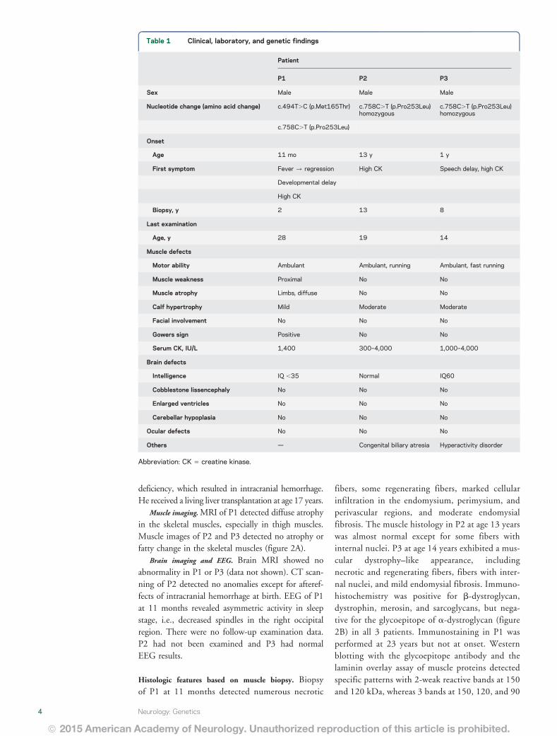
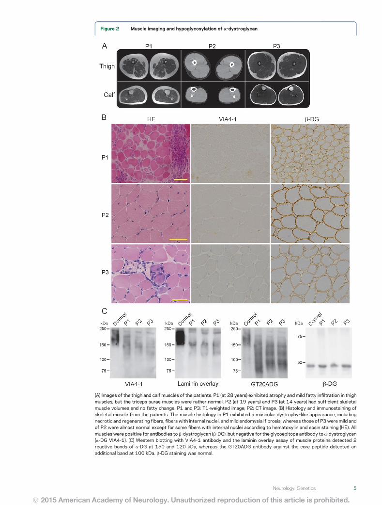
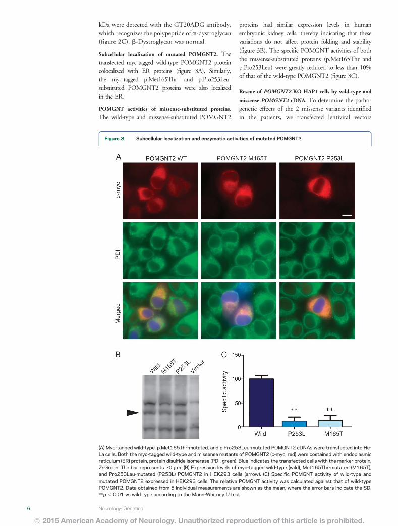