Embed Size (px)
Citation preview

Mineral pigments: the colourful palette of nature
Ina REICHE
National Museum, Berlin and CNRS, Paris
e-mail: [email protected]
The use of minerals as pigments in art and on archaeological objects, from the use ofochre in prehistoric caves to the elaborate transformation and use in ancient and modernartist palettes, is reviewed in this chapter.
Starting from the purposes of the study of pigments, the chapter presents currenttrends in the study of coloured minerals in cultural heritage science. It emphasizesthrough the use of case studies the potential of these minerals in terms of informationabout former ways of life and especially the artistic techniques employed in ancienttimes.
This information is gained through knowledge of geological and physicochemicalprocesses acting on minerals and on artefacts produced by human activities. Some newtrends are presented as the state of the art of how to master most of the methods andtechniques useful for investigating our common cultural heritage.
1. Introduction
Besides ancient glasses, ceramics and metals, mineral pigments are among the most
studied materials in cultural heritage sciences. This is due to the fact that pigments play a
crucial role in cultural heritage because of the wide colour palette they can provide.
Beautiful colours are very decorative and persist over very long time scales so that they
can even be preserved on objects dating back to the Stone Age. The available paint palette
for artists evolved continuously from the use of coloured minerals or other naturally
available materials such as charcoal to more elaborated paint palettes including synthetic
pigments and colourants in modern and contemporaneous times. Today pigments and
colourants, or mixtures of them, of nearly every perceptible hue are available.
Pigments have been used in paintings and drawings as well as to decorate various
decorative objects, walls and outdoor architectural elements. From prehistoric times
there are polychrome painted caves, painted stone or osseous tools. From later on,
coloured glasses and glazed ceramics, illuminated manuscripts, drawings on parchment
or paper as well as paintings on wooden supports such as the beautiful Fayoum portraits
(Fig, 1; Rohrs et al., 2017) are found. Today many different canvases are used for
paintings with pigments in contemporaneous art.
Coloured decorations are often associated with strong symbolic meanings as humans
make them on purpose. Therefore, the pigments and colourants used can be regarded as
indicators of human thinking and technical choices. The choice of materials used for
decorating art works is driven largely by the availability but also by the price of the raw
EMU Notes in Mineralogy, Vol. 20 (2019), Chapter 7, 283–322
#Copyright 2019 the European Mineralogical Union and the Mineralogical Society of Great Britain & Ireland
DOI: 10.1180/EMU-notes.20.7

materials. Some pigments, such as lapis lazuli and purple were highly valued in ancient
times because natural blue and purple pigments were rare. Their use in art objects is
thus an indicator of the social status of the commissioner and/or owner. All these
reasons explain why the study of ancient pigments is of fundamental interest when
studying ancient societies. A wealth of information can be gained from their study.
Pigments hold in their chemical and mineralogical characteristics information that can
be used to improve understanding of the artistic and archaeological purposes, socio-
economical changes and societal status of objects because it is possible, for instance, to
trace the materials chosen and the trading of particular pigments.
Figure 1. Mummyportrait of a youngwoman,with an enlarged section and locations of the analysis (ANT
Iinv. No. 31161,48).# Rathgen-Forschungslabor/Antikensammlung, Staatliche Museen zu Berlin.
284 I. Reiche
In the study of synthetic pigments, technological know-how and transfer can be
obtained. Their date of production and introduction to the artistic market can be used as
a terminus ante quem, meaning an indirect dating criterion for objects that contain a
particular pigment and thus providing possible hints for confirming or denying the
authenticity of objects. Alteration phenomena leading, for example, to fading or
discolouration of sensitive pigments can be studied as well as strategies for
conservation and restoration.
In the current chapter, the use of natural pigments in artistic and archaeological
contexts from prehistory to contemporary times and their chemistry and mineralogy
will be reviewed. Chemical and mineralogical analyses of pigments allow classification
as a function of their properties, period of use, local knowledge, or geographical origin.
The investigative methods range from classical to cutting-edge techniques: techniques
based on sampling, techniques used in the field or on museum collections, techniques
providing information at the micro and nanoscales as well as those allowing chemical
imaging in the laboratory. Imaging devices used in the field are reviewed briefly. For
more detailed information about analytical techniques the reader is referred to
appropriate references such as e.g. Calligaro et al., 2004; Dik et al., 2008; Cotte et al.,
2009a, 2010; Bertrand et al., 2012; Alfeld and Broekaert 2013; Reiche and Chalmin
2014; Alfeld and de Viguerie 2017.
Some case studies are discussed in order to illustrate the potential information gained
from the scientific investigation of mineral pigments in the art-historical and
archaeological contexts. As an example the composition and properties of
Palaeolithic drawings in cave sites of southern France are presented. Furthermore,
the study of art technology and the investigation of alteration phenomena of central
Asian wall paintings in grottoes are quoted. Investigation and imaging of the chemical
composition and layered structure of oil paintings are highlighted in a study of a
Courbet painting with two hidden compositions.
2. Terminological definitions
2.1. Definition of minerals, pigments and colourants
Mineral pigments are referred to as naturally occurring coloured minerals.
Mineralogically they are defined by their chemical, structural and physical properties.
In the context of cultural heritage science the characteristic of outstanding importance
is colour. That is the obvious reason why such minerals have been used for decorating
various objects. They are generally crystalline and of inorganic origin although
coloured biominerals such as corals, shells or ivory do exist. To name a few examples of
typical minerals from heritage contexts, there are yellow orpiment, yellow goethite, red
hematite, blue lapis lazuli, green malachite and black manganese oxides. In a wider
sense synthetic coloured compounds can also be considered as ‘mineral’ pigments
because they are all composed of inorganic constituents, although they are not minerals
sensu stricto. Examples of synthetic inorganic pigments are Egyptian blue and Han
purple or pigments composed of different constituents such as cobalt violet (CoO6
Mineral pigments 285

Al2O3) introduced in 1775. There are also a few composite (hybrid) pigments such as
Maya blue that are formed by a mineral (palygorskite) and an organic constituent
(indigo) trapped in the clay crystal structure. In this chapter the focus is on the
presentation of natural pigments. The reader is referred to the following references for
more information on ancient and new synthetic pigments: Berke (2002, 2004, 2007),
Hahn et al. (2006), Dejoie et al. (2010).
Colourants are defined here as coloured organic compounds that can be crystalline or
amorphous, e.g. indigo or carmine.
2.2. Definition of paints
Polychrome surfaces or paintings can be created by dispersing pigments in a liquid
binding medium on the surface forming so-called paint layers after drying. The layers
vary in thickness, composition and properties as a function of the application
technique, the binding medium and the type of support onto which they are applied.
Liquid binding media, for example, may be drying oil, Arabic gum or egg white. Fillers
may be added to paint; such fillers produce special drying or optical effects.
Generally, several superimposed paint layers create a ‘stratigraphy’ of polychrome
surfaces and painting. Only particular mural paintings called frescos or prehistoric rock
art figures are painted in one layer without binder as the lime mortar or the calcareous
rock hold the pigments after drying. The nature and thickness of the ‘stratigraphic’
system can be very variable and range from a few tens of microns up to 1 mm,
depending on the application technique and on the cultural and regional contexts.
2.3. Definition of colour
Because there are various ways of describing colours and coloured materials it is
important to clarify the vocabulary used in this chapter. This is crucial because
standardization of colour terminology did not exist in ancient times, meaning for
instance that a mineral used to produce a coloured surface could show different hues
and be named differently as a function of the context. Therefore, mineral colour alone is
not a diagnostic feature, i.e. different minerals or mixtures may show almost the same
visible colour, and the same mineral can yield very different colours depending on
chemistry, structure, defects or particle size. In addition, the physical effects that
produce a colour are numerous and depend on the texture and nature of the material to
which the colour is applied or by which the pigments are applied. Thus, the same
pigment or colourant can produce slightly different hues. Some secondary compounds
can also nuance the hue of a colour.
Because of terminological complexity, the use of defined standards and
internationally recognized systems as objective guidelines is crucial when approaching
coloured materials of our cultural heritage. We can cite systems such as the NCS �Natural Colour System21# of the Scandinavian Colour Institute AB or the Munsell
colour system. The Munsell colour system was created by Albert H. Munsell (Munsell,
1912) and corresponds to a colour space based on three colour dimensions: hue, value
286 I. Reiche

(lightness) and chroma (colour purity). Hunter introduced another mathematical
terminology in 1948, the Lab colour space (Seve, 2009). It describes all perceivable
colours in three dimensions. L in Lab stands for lightness as well as the letters a and b
for the colour extremes green�red and blue�yellow. Hunter Lab coordinates are the
result of a square root transformation of colour data. In 1976 the CIEL*a*b* or
CIELAB (Seve, 2009) system was created by a cube root transformation of the CIE
XYZ colour data and represents an objective way of characterizing colours (Schelper et
al., 2016). The Lab colour space exceeds the complete subset of colours of the red-
green-blue (RGB) model based on the human trichromacy and on the three cone cell
types that respond to three bands of light: red, green and blue. It also exceeds the CMYK
colour model, which is a subtractive colour system based on four colour dimensions
often used in colour reproduction. In contrast to the later models the Lab model is
device independent. Such a scientific approach enables all parameters which play a role
in colour creation (such as the nature of colour origin, the presence of one or several
colouring agents or other optical effects of the object surface) to be taken into account
correctly. Here in this chapter, the term colour is used as a definition of a hue observed
on objects.
2.4. Relevant archaeological and historical mineral pigments
Mineral pigments are well known in art-historical and archaeological contexts, as they
are, with a few exceptions, very stable against alteration due to temperature, climatic
changes, light and other environmental factors such as air pollution, so that they can be
recognised even on very ancient objects.
A palette of a hundred different types of mineral, inorganic or hybrid pigments can be
defined. They can be characterized by their colour, structure, grain size, geographical
origin and region of use as well as by their type of use and their natural or synthetic
origin (Eastaugh, 2004).
Minerals are divided into ten major chemical classes. Only a few coloured pigments
such as lapis lazuli belong to the silicate class that represents >90% of minerals. Most
pigments are oxides or mixed (hybrid) mineral compounds. This is linked to the fact
that transition metal ions form coloured materials with oxides through optical
absorption effects explained by crystal or ligand field splitting theory (Burns, 1993).
Selected common mineral pigments with their chemical composition or crystallo-
graphic phase may be identified unambiguously (Table 1). Indeed, some pigments
show the same or a very similar chemical composition and can only be differentiated
through identification of their mineralogical phases.
3. A general approach for analysing mineral pigments
Many archaeometric and art technological studies emphasize the importance of
establishing analytical strategies appropriately adapted to the study of cultural heritage
materials. The analysis of mineral pigments follows the same integrated reasoning. It is
crucial to start the investigation with the question to be answered and to adapt the
Mineral pigments 287

Table1.Selectedmineralpigmentsofarchaeologicalandart-historicalrelevance
withthechem
icalform
ulaofthecolouringcompoundand,in
thecase
of
synthetic
pigments,thedateofproduction.
Colo
ur
and
min
eral
classes
with
chem
ical
for-
mula
and
first
dateofuse
or
invention
in
parentheses
White
Yellow
Orange
Red
Violet
Green
Blue
Brown/
black
Silicates
Egyptian
Green
Cu-glass
Lazurite/Lapis
Lazuli
Na 8
-10Al 6Si 6O24S2-4fff
Cuprorivaite/Egyptian
blueCaC
uSi 4O10
SmaltCo-glass
Chrysocolla
Cu2�xAl x
(H2�xSi 2O5)(OH) 4)·nH2O
(x<1)
Aerinite
Sulfides
Orpim
entAs 2S3
Cadmium
Yellow
(1825)cadmium
sulfideCdS,C.I.
PigmentYellow
37
Greenokite(Ca 4(A
l,-
Fe,Mg) 10-
Si 12O35(O
H) 12CO3·-
12H2O)
Cadmium
sulfoselenide
CdS+CdSe.Depend-
ingontheS/Seratio,
C.I.PigmentOrange
20
Realgar
As 2S3Cinna-
bar
HgS
Cadmium
red
(Cadmium
sulfoselenide)
(1910)CdS+
CdSeDe-
pendingon
theS/Sera-
tio,C.I.Pig-
mentRed
108
Zinccadmium
sulfide,
CdS+
ZnS,C.I.Pig-
mentYellow
35
288 I. Reiche

oxides
and
hydroxides
Anastase
(1920)TiO
2
Rutile
(1938)TiO
2
Antimonywhite(1920)
senarmontite
Sb2O3
Zincwhite(1835)
zincite
ZnO
Goethite/yellow
ochre
a-FeO
OH
Lead-tin
yellow
Pb2SnO4
(typeI)PbSnO3
(typeII)
Massicot
PbO
Haematite/
redochre
Fe 2O3
Minium
Pb3O4
Viridian
Cr 2O3.nH2O
Paolo
Veronese
Green,Chrome
green(1809)
Atacamite/
Paratacam
ite
Cu2(O
H) 3Cl
Magnetite
(Prehistory?
/1920?)
Fe 3O4
Maghemiteg-Fe 2O3
Manganeseoxides
and
hydroxides
(bixbyite,
braunite,
hausm
annite,
hollandite,
polianite,
pyrolusite,romane-
chite...)MnO2+Mn2O3
Jacobsite,MnFe 2O4
Antimonyblack
Sb2O3
Manganeseandiron
oxides(M
n,Fe)
3O4
CobaltoxideCo2O3
carbonates
Chalk
CaC
O3
Leadwhite
2PbCO3Pb(O
H) 2
Huntite
Mg3Ca(CO3) 4
Malachite
CuCO3(O
H) 2
Azurite
Cu(C
O3) 2Cu(O
H) 2
nitrates
Cobaltyellow
(1848)
K3[Co(N
O2) 6]6
nH2O
Cobaltnitrate
Co(N
O3) 26
6H2O
sulfates
Gypsum
CaSO46
2H2O
Hem
ihydrate/bassa-
nite/
plaster
ofParis
CaSO46
0.5H2O
Anhydrite
CaSO4
Permanentwhite
(1830)baryte
BaSO4
Posnjakite
Cu(SO4)
Phosphates
apatite
ivory
orbonewhite
Ca 5(PO4) 3OH
zincphosphate
Zn3(PO4) 2
Boneturquoise
orodontolite
Mn5+dopedapa-
tite
Ca 5(PO4) 3OH
Vivianite
Fe 3(PO4)8(H
2O)
Mineral pigments 289

arsenite
Scheele’sGreen
copper
hydrogen
arsenite
CuHAsO
3(1775)
organic
com-
pounds
Verdigris
Cu(C
H3COO) 26
nCu(O
H) 2
Prussianblue(1704)Fe 4[-
Fe(CN) 6] 3
IndigoIndigotine
C16H10N2O2
CarbonGraphiteC
mixed
/hybrid
compounds
Lithopone(1853)
ZnO
+BaSO4
NaplesYellow
Pb(SbO3) 2/Pb3(SbO4) 2
Chromeyellow
(1797)
2PbSO46
PbCrO
4
MayaYellow
dehydro-
indigo(indigoides)as-
sociated
withpalygors-
kite(M
g,Al)2-
Si 4O10(O
H)·4(H
2O)
crocoite
chromered
(1809)
PbCrO
46
Pb(O
H) 2
Cobalt
violet
(1775)
CoO6
Al 2O3
Cobaltgreen
(1780)
CoO
65ZnO
Emeraldgreen
(1814)
Cu(C
H3COO) 26
3Cu(A
sO2) 2
Cadmium
green
(Cadmium
yel-
low
CdS+viri-
dianCr 2O3.nH2O
ManganeseblueBaSO46
Ba(MnO4) 2
MayaBlueindigo-paly-
gorskite(M
g,Al)2-
Si 4O10(O
H)·4(H
2O)
Ivory
orboneblack
C
+Ca 5(PO4) 3OH
(apa-
tite)
290 I. Reiche

analytical strategy to the type of object, the sampling options and the specificity and
rarity of the object. In addition, the heterogeneity and state of preservation of cultural
heritage materials need to be taken into account. Therefore, it is also of utmost
importance to study these materials within their context so as to be able to select
representative material and to obtain conclusive results from the selected analytical
methods.
Recent technical developments allow more and more non-invasive analyses or
chemical imaging in situ, thus keeping the object or sample intact and in its place during
the analysis.
3.1. Laboratory investigation based on samples3.1.1. Study of loose pigments
When a sample can be removed from a polychrome object, study of the pigment grains
directly is possible. In this case the investigation should start with optical microscopy.
Pigments are characterized on the one hand by their colour but on the other hand by:
their particle size and size distribution (fine or coarse-grained pigments); particle shape
(spherical, acicular, fibrous, bladed, lamellar, tabular, columnar, dendritic, pennate);
the possible aggregation of several crystals (botryoidal habit, framboidal,
cryptocrystalline); particle surface properties; fracture and cleavage properties; and
degree of translucency. These properties are diagnostic and can be used for pigment
identification through microscopic observation. Some of these characteristics can be
observed using an optical microscope; other properties require the use of a polarized
light microscope. If a crystal is pleochroic, change of colour is observed while rotating
under plane-polarized light. The refractive index and relief are characteristic as well.
Observation between crossed polars provides information about degree of anisotropy
and extinction phenomena.
Optical micrographs of different pigments show characteristic mineralogical
features of the crystal grains (Fig. 2).
Mineral pigments can also be studied by various techniques such as vibrational
spectroscopies such as Fourier transform infrared spectroscopy (FT-IR) or Raman
spectroscopy, X-ray diffraction, near infrared, visible and ultraviolet reflectance
spectroscopy as well as X-ray fluorescence analysis (XRF).
FT-IR spectra of Prussian or Berlin blue and lead white enable pigment identification
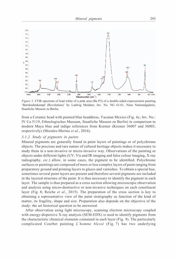
through characteristic absorption bands (Fig. 3). Lead white could be identified by FT-
IR in the case of the pink area (Ba P3) of a double sided expressionist painting
‘Barrikadenkampf (Revolution)’ by Ludwig Meidner (Inv. No. NG 61/61, Neue
Nationalgalerie, Staatliche Museen zu Berlin) but not vermilion, which was identified
by micro-Raman spectroscopy (Fig. 4b). Figure 5 shows: (a) an XRF spectrum with its
measurement point on a light blue area (b) of the Ludwig Meidner painting. Elements
such as Fe, Ca, Pb, Si, Al, K, Ti, Zn, (Cu) may be detected indicating the presence of a
mixture of Prussian blue, ultramarine, lead white and calcite (Rohrs et al., 2014).
Figure 6a shows an XRD pattern of modern Maya blue, indicating the presence of
palygorskite, and Fig. 6b shows a Vis spectrum of an archaeological Maya blue sample
Mineral pigments 291
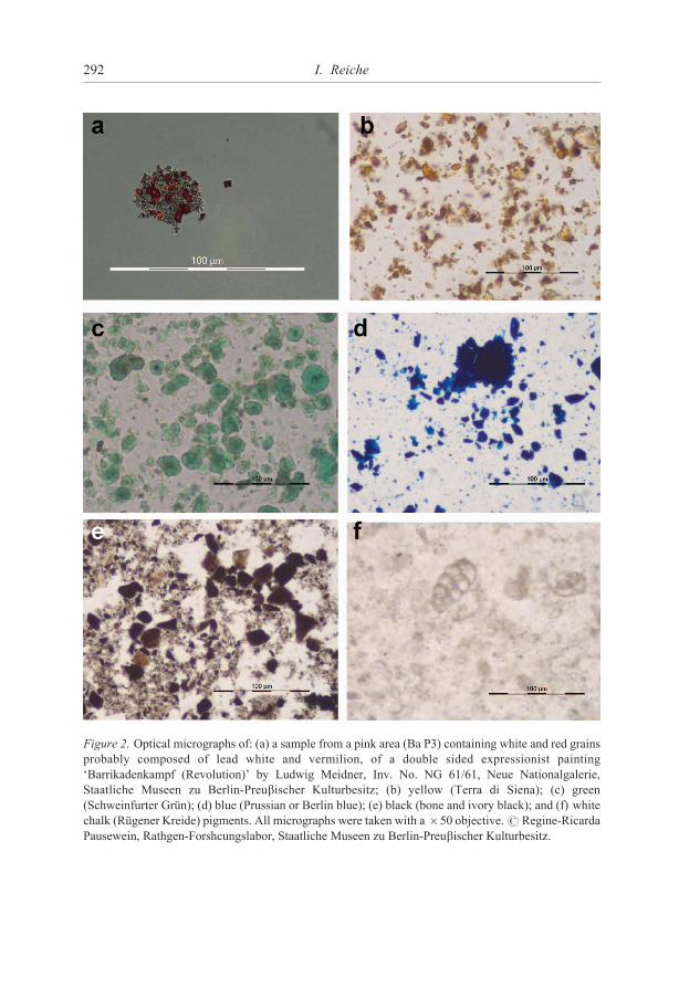
Figure 2. Optical micrographs of: (a) a sample from a pink area (Ba P3) containing white and red grains
probably composed of lead white and vermilion, of a double sided expressionist painting
‘Barrikadenkampf (Revolution)’ by Ludwig Meidner, Inv. No. NG 61/61, Neue Nationalgalerie,
Staatliche Museen zu Berlin-Preubischer Kulturbesitz; (b) yellow (Terra di Siena); (c) green
(Schweinfurter Grun); (d) blue (Prussian or Berlin blue); (e) black (bone and ivory black); and (f) white
chalk (Rugener Kreide) pigments. All micrographs were taken with a650 objective.# Regine-Ricarda
Pausewein, Rathgen-Forshcungslabor, Staatliche Museen zu Berlin-Preubischer Kulturbesitz.
292 I. Reiche
from a Ceramic head with painted blue headdress, Yucatan Mexico (Fig. 6c, Inv. No.:
IV Ca 5119, Ethnologisches Museum, Staatliche Museen zu Berlin) in comparison to
modern Maya blue and indigo references from Kremer (Kremer 36007 and 36003,
respectively) (Morales-Merino et al., 2016).
3.1.2. Study of pigments in paints
Mineral pigments are generally found in paint layers of paintings or of polychrome
objects. The precious and rare nature of cultural heritage objects makes it necessary to
study them in a non-invasive or micro-invasive way. Observations of the painting or
objects under different lights (UV, Vis and IR imaging and false colour imaging, X-ray
radiography, etc.) allow, in some cases, the pigment to be identified. Polychrome
surfaces or paintings are composed of more or less complex layers of paint ranging from
preparatory ground and priming layers to glazes and varnishes. To obtain a special hue,
sometimes several paint layers are present and therefore several pigments are included
in the layered structure of the paint. It is thus necessary to identify the pigment in each
layer. The sample is thus prepared as a cross section allowing microscopic observation
and analysis using micro-destructive or non-invasive techniques on each constituent
layer (Fig. 8; Reiche et al., 2015). The preparation of the cross section is key to
obtaining a representative view of the paint stratigraphy as function of the kind of
matter, its fragility, shape and size. Preparation also depends on the objective of the
study: the art historical question to be answered.
After observation using light microscopy, scanning electron microscopy coupled
with energy-dispersive X-ray analysis (SEM-EDX) is used to identify pigments from
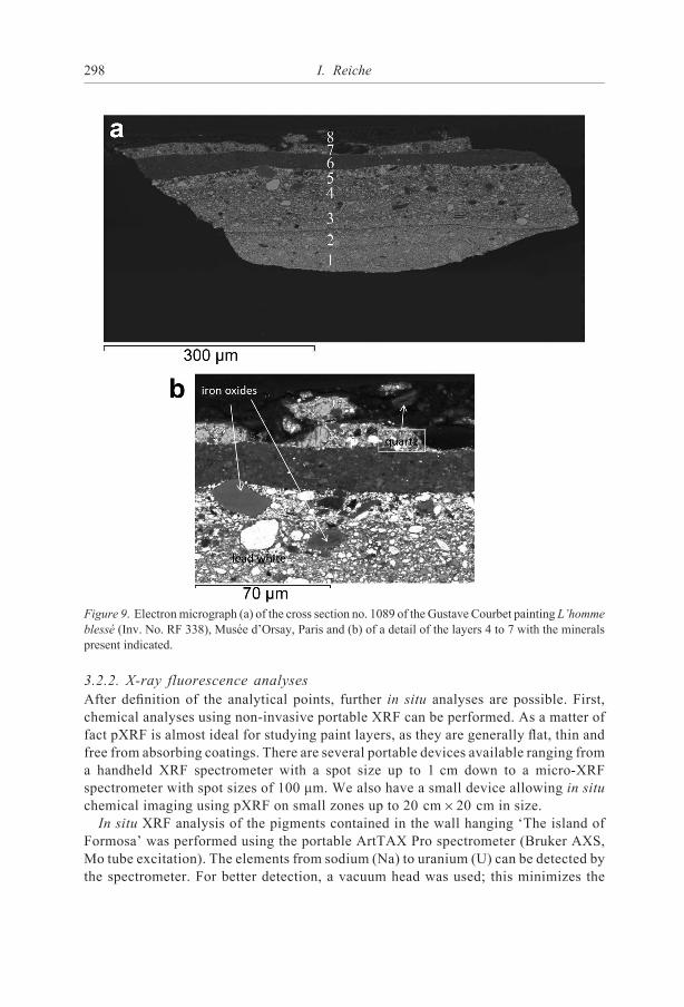
the characteristic chemical elements contained in each layer (Fig. 9). The particularly
complicated Courbet painting L’homme blesse (Fig. 7) has two underlying
Figure 3. FTIR spectrum of lead white of a pink area (Ba P3) of a double-sided expressionist painting
‘Barrikadenkampf (Revolution)’ by Ludwig Meidner, Inv. No. NG 61/61, Neue Nationalgalerie,
Staatliche Museen zu Berlin.
Mineral pigments 293

compositions: layer 1 of cross section 1089 contains lead white, iron oxide (orange) and
very small amounts of barium sulfate (with Sr); layer 2 contains lead white, quartz,
carbon-based black or brown and unidentified orange particles; layer 3 contains lead
white, vermilion, Prussian blue, iron oxides (orange and red) with Al and Si and carbon-
based black/brown; layer 4 contains lead white, iron oxides (orange and red) with As
and Si, vermilion, cobalt blue (with Al) and organic red on an alum substrate (small Sn
component); layer 5 contains lead white, iron oxides (orange) some with As and some
Figure 4. Micro-Raman spectra of (a) a blue area containing ultramarine (Ba P4) and (b) of a pink area
(Ba P3) containing lead white and vermilion of a double-sided expressionist painting ‘Barrikadenkampf
(Revolution)’ by Ludwig Meidner, Inv. No. NG 61/61, Neue Nationalgalerie, Staatliche Museen zu
Berlin.
294 I. Reiche

with Al, feldspar (K Al silicate), bone black (with Mg) and carbon-based black/brown;
layer 6 contains bone black, umber, Mn in clay and umber: layer 7 contains lead white,
barium sulfate (with Sr), bone black (withMg), carbon-based black. The ground layer is
missing in this cross section and layer 8 corresponds to the varnish of the actual
composition (Reiche et al., 2015).
The identity of pigments in the paint layers can only be inferred from EDX analysis.
A precise identification of each pigment needs further phase analyses like FTIR in ATR
mode (Fig. 10) or optical spectroscopy such as fibre optic reflectance spectroscopy
(Rohrs and Stehr, 2014).
3.2. In situ pigment analyses using portable equipment3.2.1. Digital microscopy
As for investigations in the laboratory, in situ analyses should start with microscopic
observations. New digital devices are available that allow the study of the objects in the
museum or at the archaeological site. Such investigations are of utmost importance for
choosing the appropriate analysis zones for further in situ pigment analyses. It allows
the definition of the different pigment hues present, the zones with overpaints, former
reparation or restorations. In the case of the leather wall hanging ‘The Island of
Formosa’ composed of ten different painted leather parts, twelve different hues can be
distinguished (Fig. 11a). Patches sewn on the object and inlays glued to the surface can
also be seen. Some areas show better protected colour and leather. Two areas are clearly
Figure 5. (a) Micro-XRF spectrum of a light blue area (b) containing Prussian Blue, ultramarine, lead
white and calcite of a double-sided expressionist painting ‘Barrikadenkampf (Revolution)’ by Ludwig
Meidner, Inv. No. NG 61/61, Neue Nationalgalerie, Staatliche Museen zu Berlin.
Mineral pigments 295

lighter than the others. The microscopic analysis also confirmed that the blue corners
are painted over the frame decorations, which might mean that they were added later.
Some parts of the painting layer are missing. Further missing parts are holes, seam yarn
and other details in the leather itself (Fig. 11b) (Aibeo et al., 2016).
Figure 6. (a) XRD diagram of modernMaya blue (Kremer 36007) and (b) Vis spectrum of archaeological
Maya blue of the object depicted in (c) ceramic headwith painted headdress, YucatanMexico (Inv. No. IV
Ca 5119, Ethnologisches Museum Berlin) with respect to a modern Maya blue and indigo reference.
296 I. Reiche

Figure. 7. ( a) Courbet painting L’homme blesse (81.5 cm697.5 cm, Inv. No. RF 338, Musee d’Orsay,
Paris) with a detail (b) of the photograph (and XRR superimposed) of the centre of the Courbet painting
showing the two hidden compositions
Figure 8. (a) Cross section of sample P3 showing up to 14 paint layers of the Caspar David Friedrich
painting ‘Monch am Meer’ (Inv. No. NG 9/85), Alte Nationalgalerie, Staatliche Museen zu Berlin and
(b) Cross section no. 1089 showing up to ten paint layers of the Gustave Courbet painting ‘L’homme
blesse’ (Inv. No. RF 338), Musee d’Orsay, Paris.
Mineral pigments 297
3.2.2. X-ray fluorescence analyses
After definition of the analytical points, further in situ analyses are possible. First,
chemical analyses using non-invasive portable XRF can be performed. As a matter of
fact pXRF is almost ideal for studying paint layers, as they are generally flat, thin and
free from absorbing coatings. There are several portable devices available ranging from
a handheld XRF spectrometer with a spot size up to 1 cm down to a micro-XRF
spectrometer with spot sizes of 100 mm. We also have a small device allowing in situ
chemical imaging using pXRF on small zones up to 20 cm620 cm in size.
In situ XRF analysis of the pigments contained in the wall hanging ‘The island of
Formosa’ was performed using the portable ArtTAX Pro spectrometer (Bruker AXS,
Mo tube excitation). The elements from sodium (Na) to uranium (U) can be detected by
the spectrometer. For better detection, a vacuum head was used; this minimizes the
Figure 9. Electronmicrograph (a) of the cross section no. 1089 of the Gustave Courbet painting L’homme
blesse (Inv. No. RF 338), Musee d’Orsay, Paris and (b) of a detail of the layers 4 to 7 with the minerals
present indicated.
298 I. Reiche

Figure 10. ATR FTIR point analyses for identification of the constituents of paint cross sections.
Examples of several measured spectra on the cross section P3 of the Caspar David Friedrich painting
‘Monch amMeer’, ANG showing the presence of smalt in layers 5 and 6. Characteristic absorption bands
of the Co-glass smalt are at ~1080 cm�1.
Mineral pigments 299

amount of air through which the X-rays must pass and therefore increases its sensitivity
to the lighter elements. A constant flow of He gas is used to minimize adsorption of the
fluorescent X-rays by air. The area analysed has a diameter of ~0.1 mm (or 100 mm)
thanks to focusing X-ray optics. This helps to study small details on the object. The
detailed experimental conditions were 45 kV, 100 mA, 100 s life time, 0�50 keV
Figure 11. (a) Leather wall hanging ‘The Island of Formosa’, 17th c. kept in the Ethnologisches Museum
in Berlin, Inv. No. I D 37597, digital micrographs of: (b) a detail (65 objective) showing different hues
and (c) a repaired area.
300 I. Reiche

spectral range and background suppression by 60 smoothing cycles. These conditions
allowed us to infer a large range of pigments used for the decoration of the leather wall
hanging including cinnabar and sometimes traces of minium, a mixture of minium and
lead white, lead white alone, a mixture of cinnabar and lead white, goethite and a
combination of azurite and lazurite. In the blue corners a special blue was found as it
contains Co, Ni, As and Pb. It is probably the synthetic pigment cobalt blue (Fig. 12).
Green, yellow and black pigments could not be identified using micro-XRF as they are
mainly or partly composed of organic constituents (Aibeo et al., 2016).
Even depth-resolved confocal microXRF analyses are possible in situ nowadays.
They allow the investigation of the chemical composition of different paint layers
without sampling. However, these analyses are restricted to the upper paint layers
because of the absorption of the emitted X-rays by upper layers. The stability of the
positioning of the depth-resolved analyses is still an issue (Muller et al., 2017).
3.2.3. Micro-Raman analyses
For the direct identification of pigments, complementary in situ analyses are necessary
using portable micro-Raman instruments. As for portable XRF devices, there is a
wealth of modern Raman spectrometers that allow in situ pigment study. They are
optimized to work in a horizontal set-up to work directly on the exhibited object. A
Horiba XploRa micro-Raman system was used for the analyses of the leather wall
Figure 12. Micro XRF spectrum of ‘The Island of Formosa’ leather wall hanging kept in the
Ethnologisches Museum in Berlin, Inv. No. (I D 37597), spot 43_16_139_blue in the blue corners,
showing the presence of K, Fe, Co, Ni, As, Pb and traces of Ca, Ba, Cu and Zn.
Mineral pigments 301
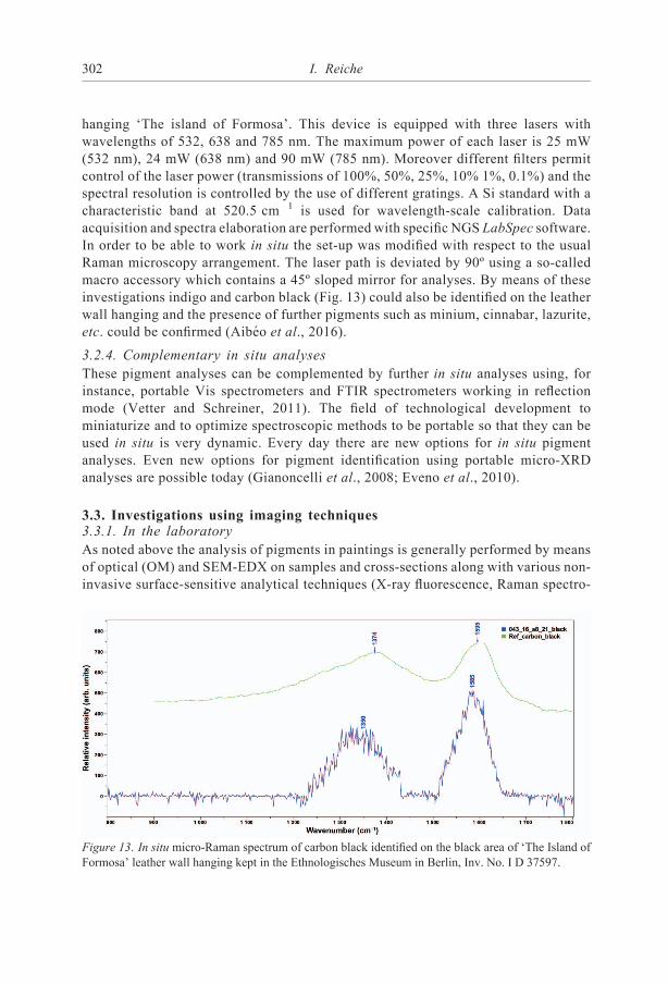
hanging ‘The island of Formosa’. This device is equipped with three lasers with
wavelengths of 532, 638 and 785 nm. The maximum power of each laser is 25 mW
(532 nm), 24 mW (638 nm) and 90 mW (785 nm). Moreover different filters permit
control of the laser power (transmissions of 100%, 50%, 25%, 10% 1%, 0.1%) and the
spectral resolution is controlled by the use of different gratings. A Si standard with a
characteristic band at 520.5 cm�1 is used for wavelength-scale calibration. Data
acquisition and spectra elaboration are performed with specific NGS LabSpec software.
In order to be able to work in situ the set-up was modified with respect to the usual
Raman microscopy arrangement. The laser path is deviated by 90º using a so-called
macro accessory which contains a 45º sloped mirror for analyses. By means of these
investigations indigo and carbon black (Fig. 13) could also be identified on the leather
wall hanging and the presence of further pigments such as minium, cinnabar, lazurite,
etc. could be confirmed (Aibeo et al., 2016).
3.2.4. Complementary in situ analyses
These pigment analyses can be complemented by further in situ analyses using, for
instance, portable Vis spectrometers and FTIR spectrometers working in reflection
mode (Vetter and Schreiner, 2011). The field of technological development to
miniaturize and to optimize spectroscopic methods to be portable so that they can be
used in situ is very dynamic. Every day there are new options for in situ pigment
analyses. Even new options for pigment identification using portable micro-XRD
analyses are possible today (Gianoncelli et al., 2008; Eveno et al., 2010).
3.3. Investigations using imaging techniques3.3.1. In the laboratory
As noted above the analysis of pigments in paintings is generally performed by means
of optical (OM) and SEM-EDX on samples and cross-sections along with various non-
invasive surface-sensitive analytical techniques (X-ray fluorescence, Raman spectro-
Figure 13. In situ micro-Raman spectrum of carbon black identified on the black area of ‘The Island of
Formosa’ leather wall hanging kept in the Ethnologisches Museum in Berlin, Inv. No. I D 37597.
302 I. Reiche

scopy or X-ray diffraction). This analysis is restricted to tiny zones. Therefore, the need
arose to use imaging techniques to get a more representative view of the pigment
distribution in paintings and polychrome objects. Classical painting imaging methods
are UV imaging, infrared reflectography or X-ray radiography.
These imaging techniques can today be complemented by new chemical imaging
techniques such as Raman and FTIR imaging, macro-XRF scanning (MA-XRF) and
hyperspectral imaging techniques (Dik et al., 2008; Janssens et al., 2010; Alfeld et al.,
2011, 2013; Alfeld and de Viguerie, 2017). All these techniques are available as
stationary laboratory equipment. Non-invasive two-dimensional scanning X-ray
fluorescence (MA-XRF) spectroscopy provides the imaging of elements present in
paintings down to trace concentrations. This is achieved by scanning the work area with
an X-ray beam of a small diameter and recording spectra point by point. Numerous
applications of this technique � even using mobile scanners as reviewed by Alfeld and
de Viguerie (2017) have highlighted the potential of MA-XRF for scanning precious
paintings from different periods revealing overpaints, ancient restorations, etc. Such
set-ups have already demonstrated attractive potentials and capabilities on numerous
art objects and especially paintings. The main advantage of this technique is that the
chemical XRF image can be superimposed on the optical image. Therefore, MA-XRF
imaging of paintings is today a well established method using different sources and also
using mobile equipment. The MA-XRF imaging of the Courbet painting L’homme
blesse shows the possibility of chemical imaging of paintings in the laboratory. The
investigation was carried out with a prototype designed at the C2RMF. The detailed
experimental description of the instrument is reported elsewhere (Ravaud et al., 2016).
Maps of 100 pixels6100 pixels were acquired with a step size of 1.4 mm
corresponding to a 140 mm6140 mm area. Eleven areas could be scanned on the
painting. They were selected according to the most interesting zones identified on the
X-radiograph. The selected areas correspond to the zones with the most important
changes in the three compositions of the painting. The data were processed with the
open source program PyMCA (Sole et al., 2007) which allows the generation of fitted
peak area maps for various elements from elemental maps (in PNG format) derived
using a program developed at the C2RMF calledDataImaging 3D (Pichon et al., 2015).
Detailed descriptions of the chemical maps obtained of the paintings were presented by
Reiche et al. (2016). As an example, the Hg L map of the case study presented in detail
in Section 4.3 is shown indicating two hidden underlying painting compositions
(Fig. 14). Besides the head of a male, visible today, two female heads are visible in the
Hg L distribution map. The Hg L map shows deeper paint layers as the Hg L
fluorescence line has a relatively high energy and thus a greater depth of information.
3.3.2. In situ MA-XRF scanning analyses
Chemical imaging studies are also possible today using portable XRF scanning devices
such as the portable ELIO XRF device (XGlab1). The in situ XRF map of one area of
the leather wall hanging ‘The Island of Formosa’ is shown as an example. The area
analysed is at the heart of the wall hanging in the so-called area 2 on a selected boat. The
area scanned is 2 cm64 cm (Fig. 15). The corresponding chemical maps of the Hg La
Mineral pigments 303

Figure 14. Macro-XRF Hg L map of eleven scanned zones superimposing the X-ray radiography of the
Gustave Courbet painting ‘L’homme blesse’, Musee d’Orsay, Inv. No. RF 338 and the two hidden
compositions indicated in yellow (first one) and red (second composition)
Figure 15. Photograph of the area of the XRF mapping in area 2 of the wall hanging ‘The Island of
Formosa’.
304 I. Reiche

and the Pb La fluorescence lines (Fig. 16) show that the red area is rich in Hg,
indicating vermilion and light yellow, red and pink zones contain significant amounts
of lead indicating the presence of lead-containing pigments such as lead white and
massicot. The dark yellow does not contain much Pb and is therefore likely to be
composed of an organic yellow colourant.
3.3.3. On the micro- and nanoscale, combined imaging methods at large-scale facilities
For the very special purposes of pigment identification and analysis, imaging methods
such as ion beam facilities or synchrotron sources are required. Such methods are
necessary because of the intrinsic complexity and multi-scalar heterogeneity of
painting materials, combined with the relative abundance of their individual
components. In addition, the analysis of chemical elements at the trace and ultra-
trace level, as well as determination of crystallographic phases at the micro- or even
nanoscale is often necessary in order to answer questions related to provenance,
production techniques, alterations and conservation of pigments. Several methods may
need to be combined on a small scale in order to characterize individual constituents.
This methodological challenge can only be met at large-scale facilities. In particular,
synchrotron radiation sources offer polychromaticity, high flux, low divergence, small
source size, stability, ‘calculability’ of the source, and polarization; these
characteristics are well adapted to meet the aforementioned challenges. After
preliminary laboratory investigations, such approaches using high-tech equipment
can solve very specific problems in cultural heritage science.
It is not possible to cover exhaustively all significant on-going imaging
developments at large-scale facilities. This chapter is, thus, restricted to the main
methods used for high-definition imaging in cultural heritage.
Synchrotron-based XRF or accelerator-based PIXE imaging are, without doubt, the
most commonly used methods at large-scale facilities. They offer non-invasive
analysis of the chemical compositions of pigments from the macro- to the nanoscale,
while being sensitive to major, minor and trace elements. Synchrotron XRF is very
often combined with X-ray absorption spectroscopy and X-ray diffraction, because
these methods can be performed simultaneously using the same X-ray source by
Figure 16. XRF-maps for (a) Hg La and (b) Pb La in area 2 of the wall hanging ‘The Island of Formosa’.
The intensity bar ranges from blue (low intensity) to red (high intensity) of the X-ray line.
Mineral pigments 305

adopting different imaging detectors. Further combinations are possible with other
X-ray techniques such as small-angle X-ray scattering (SAXS) and computed
tomography (CT) but molecular spectroscopic methods can also be performed
simultaneously with X-ray imaging such as Raman and FTIR imaging. For more
detailed information, the reader is referred to further literature (Dik et al., 2008; Cotte
et al., 2009a,b, 2010; Janssens et al., 2010; Bertrand et al., 2012; Reiche and Chalmin,
2014; Alberic et al., 2015; Gay et al., 2015; Alfeld and de Viguerie 2017)[i6] which
presents various possible combinations of large-scale facility imaging methods used in
cultural heritage studies.
4. Examples highlighting the information obtained when studying mineralpigments in historical and archaeological contexts
The pigments and colourants selected and the complexity of the paint palette employed
reflect differences in the choice of artistic technique and in the symbolic meaning or
function of works of art. They can also be used as indicators of the social rank of the
commissioner or owner or be specific to a social group.
Therefore, the technical study of pigments and colourants used in archaeological
artefacts and in works of art allows us insights into the nature, genesis and origin of
artefacts, into the production or transformation techniques used, and indirectly into the
use and exchange of knowledge in past societies. Such techniques may also solve
problems of authenticity.
Other important issues concerning the technical study of pigments and colourants in
cultural heritage are the understanding of their alteration over time, the set-up of
adequate conservation measures and the establishment of the best possible restoration
strategies as well as in the detection of forgeries.
The following examples are intended to illustrate the analytical approach and the
variety of questions that can be addressed while studying ancient mineral pigments.
4.1. Non-invasive in situ characterization of manganese oxides used forPalaeolithic cave art in the Rouffignac cave, Dordogne, south-westernFrance. Differentiation of artists’ contributions
Since its recognition as prehistoric art at the end of the 19th century, rock art preserved
in limestone caves in southern France and in northern Spain have been subjected to
extensive research that has been performed to study the style and technique of the
paintings and drawings, to date the figures, and to determine their possible overlap and
sequence of execution. Chemical analysis allows the identification of the constituent
pigments as well as the ‘paint pots’ obtained by mixtures of different minerals and
possibly also organic compounds that are today degraded. Paints or crayons used for
cave art generally contain red hematite or red ochre (a-Fe2O3), yellow goethite
(a-FeOOH) or yellow ochre; whereas black can contain either charcoal or various
manganese oxides/hydroxides. In the very fortunate case of the use of charcoal or other
C-based compounds the decoration of cave walls can be dated directly using the 14C
306 I. Reiche

method (Valladas et al., 2001; Beck et al., 2013; Quiles et al., 2016). In the absence of
black C-based pigments other indirect means have to be employed in order to gain more
insights into the chronological order of prehistoric representations. Therefore,
archaeologists seek the presence of C-based paints and pigments in caves. Further
studies are needed of the artistic techniques to differentiate between various artists
involved in painting caves to establish the chronological order for the representations.
Iron oxides, various Mn oxides and hydroxides and C-based compounds can be studied
using in situ techniques, which make sampling obsolete. On-site XRF analyses, XRD
and Raman analyses after microscopic observations of the walls are possible today
thanks to mobile spectrometers (Lahlil et al., 2012; Beck et al., 2014; Gay et al., 2015,
2016). From the variety of paint and drawing composition and application techniques,
it can be assumed that it is possible to distinguish several artists involved in the
execution of such rock art. The XRF analysis of Fe-based red pigments is difficult
because the rock substrate generally also contains Fe-bearing minerals. Concerning Mn
oxides/hydroxides, their differentiation from the rock substrate is more straightforward
because they are sufficiently different in composition. A differentiation or a
classification into different pigment groups is possible by means of XRF analysis.
However, there is a large variety of different mineralogical Mn oxides and hydroxides
with similar chemical compositions so that in situ phase analysis is required to separate
pigments used for different prehistoric representations. The challenge of pigment
identification is enhanced by the fact that Mn oxides and hydroxides are generally
poorly crystalline so that they show diffraction patterns with a small peak-to-noise
ratio. However, several studies in the Rouffignac cave showed the feasibility of a
combination of XRF/XRD and Raman analyses of the prehistoric figures in order to
distinguish pigment groups and to identify different mineral phases as a function of the
pigment group identified.
The Rouffignac cave situated in the Dordogne region in southern France is one of the
key sites of Upper Palaeolithic art. It shows ~240 animal drawings and engravings on
the walls attributed to the Magdalenian period (Barriere, 1982; Plassard, 1999; Plassard
and Plassard, 2016). Because of the predominance of representations of mammoths,
this animal is the distinctive element of the cave. It is also referred to as ‘the hundred
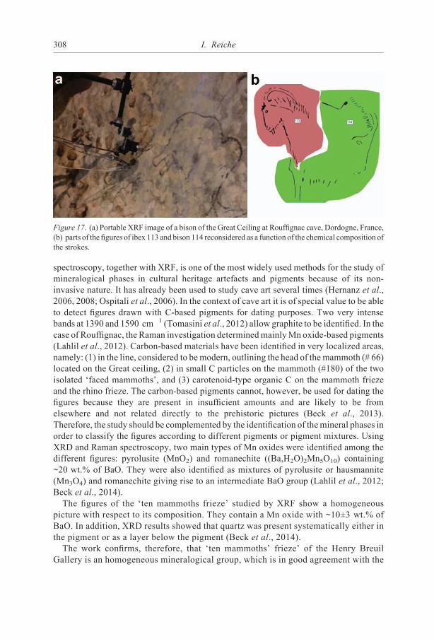
mammoths’ cave’. Its largest drawing composition, the ‘Grand Plafond’, comprises 65
black animals corresponding to one third of the cave’s rock art (Fig. 17a). On the
‘Grand Plafond’ (Great ceiling) the figures are arranged without any striking global
organization, whereas in the ‘Henri Breuil gallery’ with ‘the ten mammoths’ frieze’ and
the ‘rhino frieze’ as well as the ‘panel of the large Patriarche’, although engraved, are
all well aligned.
Four of the ten mammoths and one rhinoceros of the ‘three rhinoceros’ frieze’ of the
‘Henri Breuil gallery’ and the two isolated ‘faced mammoths’ as well as 45 figures of
the ‘Great Ceiling’ (Grand Plafond) have been studied systematically.
The methods used were necessarily non-invasive, allowing mineralogical and chemical
study of the figures without sampling or touching the figures. The techniques used were
portable micro-Raman spectroscopy and XRD as well as XRF analyses. Raman
Mineral pigments 307
spectroscopy, together with XRF, is one of the most widely used methods for the study of
mineralogical phases in cultural heritage artefacts and pigments because of its non-
invasive nature. It has already been used to study cave art several times (Hernanz et al.,
2006, 2008; Ospitali et al., 2006). In the context of cave art it is of special value to be able
to detect figures drawn with C-based pigments for dating purposes. Two very intense
bands at 1390 and 1590 cm�1 (Tomasini et al., 2012) allow graphite to be identified. In the
case of Rouffignac, the Raman investigation determinedmainlyMn oxide-based pigments
(Lahlil et al., 2012). Carbon-based materials have been identified in very localized areas,
namely: (1) in the line, considered to be modern, outlining the head of the mammoth (# 66)
located on the Great ceiling, (2) in small C particles on the mammoth (#180) of the two
isolated ‘faced mammoths’, and (3) carotenoid-type organic C on the mammoth frieze
and the rhino frieze. The carbon-based pigments cannot, however, be used for dating the
figures because they are present in insufficient amounts and are likely to be from
elsewhere and not related directly to the prehistoric pictures (Beck et al., 2013).
Therefore, the study should be complemented by the identification of themineral phases in
order to classify the figures according to different pigments or pigment mixtures. Using
XRD and Raman spectroscopy, two main types of Mn oxides were identified among the
different figures: pyrolusite (MnO2) and romanechite ((Ba,H2O)2Mn5O10) containing
~20 wt.% of BaO. They were also identified as mixtures of pyrolusite or hausmannite
(Mn3O4) and romanechite giving rise to an intermediate BaO group (Lahlil et al., 2012;
Beck et al., 2014).
The figures of the ‘ten mammoths frieze’ studied by XRF show a homogeneous
picture with respect to its composition. They contain a Mn oxide with ~10�3 wt.% of
BaO. In addition, XRD results showed that quartz was present systematically either in
the pigment or as a layer below the pigment (Beck et al., 2014).
The work confirms, therefore, that ‘ten mammoths’ frieze’ of the Henry Breuil
Gallery is an homogeneous mineralogical group, which is in good agreement with the
Figure 17. (a) Portable XRF image of a bison of the Great Ceiling at Rouffignac cave, Dordogne, France,
(b) parts of the figures of ibex 113 and bison 114 reconsidered as a function of the chemical composition of
the strokes.
308 I. Reiche
stylistic homogeneity of the drawings. Mineralogical results confirm the hypothesis of
a unity of conception and realization. Despite the prevailing content of one type of Mn
oxide, the black pigments used to draw the mammoth frieze seem to be a mixture of
several mineralogical phases, an observation that is compatible with the use of
untransformed raw materials from natural sources. Furthermore, romanechite is a
natural Mn oxide, which cannot be obtained by transformation of other Mn compounds.
This result confirms that the stylistic coherence of the ‘ten mammoth frieze’ is the result
of one artist or group that worked over a relatively small period of time with the same
rawmaterial. In contrast, the figures in the ‘rhinoceros frieze’ are different from the ‘ten
mammoth frieze’ and similar to some of the Great Ceiling. In the various animals
(horse, mammoth, ibex and bison) of the Great Ceiling, all three types of Mn oxide-
bearing pigments were identified in the different figures. These pigment groups cannot
be matched to a type of animal representation or to stylistic groups. Research is
currently under way to relate stylistic observations and superimposition studies to the
chemical fingerprint of the studied figures. It seems obvious that several hands created
the prehistoric artworks of the Great Ceiling in a relatively short time leading to a panel
lacking an evident overall organization (Gay et al. submitted). The first results of some
subsets show the value of combing different types of approaches, allowing the
rereading of figures and subsets of figures of the Great Ceiling. As an example, two
neighbouring drawings, the ibex 113 and the bison 114 of the panel are considered
(Fig. 17b). The front legs of the ibex 113 show pigment compositions of ~7% BaO,
corresponding to the intermediate BaO-containing MnO group. This composition is not
in agreement with that obtained for the ibex muzzle, the back of its head and its horn
(~3% BaO) corresponding to the very-low-BaO-containing MnO group. Interestingly,
the chemical signature of the ibex legs matches perfectly that of the head of the
neighbouring bison 114. The chemical characteristics, therefore, allow a new reading
of these two figures and indicate that the strokes, initially interpreted as front legs of
ibex 113, actually represent the front leg of bison 114 (Fig. 17b). As another example,
different chemical signatures were found for several parts attributed to mammoth 122.
Drawing strokes inside the mammoth and those on its legs illustrating the animal’s fur
differ from one of the contour strokes of the drawing (6% and 3%, respectively). It is
probably a stylistic addition by the artist him/herself or, more likely, was added by
another artist possibly at a later date.
Other examples discussed by Gay et al. (submitted) show the alternative use of
different crayons to create more or less complex compositions of figures. This
reinforces the idea that the ‘Grand Plafond’ was probably created in successive subsets
of figures by small teams of artists without taking into account the general organization
of the panel. The drawing of these subsets can be imagined the same way as the creation
of figures of the mammoth or the rhinoceros friezes in the Henry Breuil Gallery drawn
over a relatively short time period by a very small team. The superimposition of these
different subsets of the great ceiling leads to its actual presentation.
Mineral pigments 309

4.2. Alteration mechanisms of pigments on central Asian wall paintings fromthe Northern Silk road, kept at the Asian Art Museum, Berlin
Land and maritime routes, linking East and West for thousands of years and known
today under the terms ‘Silk Roads’ and ‘Spice Routes’ enabled not only the trade of
merchandise such as silk, wool, gold, metals, ivory, spices and other precious items but
also the exchange of cultures, ideas, knowledge and religious beliefs. Through these
ancient routes, Buddhism could spread from India to China so that important Buddhist
monasteries flourished over centuries in Central Asia, including the famous sites of
Turfan and Kucha, situated on the northern edge of the Taklamakan desert or Khotan
and Miran, south of the desert. Grottoes and temples, richly decorated with wall
paintings and painted sculptures, served the Buddhist monks as lodging and centres of
worship. The ‘Silk Roads’ inspired in the 19th and 20th century AD European
archaeologists, such as Aurel Stein (British: 1862�1943), Paul Peillot (French:
1879�1945) or Albert Grunwedel (German: 1856�1935) and Albert von Le Coq
(German: 1860�1930), who conducted expeditions around the Taklamakan desert
(today: Xinjiang Province, China) (Grunwedel, 1912; Von Le Coq, 1922�26). Thefindings of the four German exploration voyages between 1902 and 1914 include
amongst other artifacts mural paintings, painted sculptures and painted wooden
structures which are today stored in the Asian Art Museum, Berlin (Ulrich, 1964;
Riederer, 1977; Russell-Smith, 2005; Gabsch, 2012).
These mural paintings can be attributed artistically to four distinguishable styles:
(1) Indo-Iranian style (4th to 6th c. AD), (2) Indo-Iranian style (7th to 8th c. AD although
both styles could be contemporary), (3) Chinese style (8th to the 9th c. AD) and (4) the
Uygur style (10th to 13th c. AD, Fig. 18). The region of interest named here as the
Northern Silk Road represents the route between Kashgar and Turfan (Xinjiang,
China).
Once the grottoes were carved into the cliff of the soft sandstone mountains, two layers
of mud plaster (coarse underlayer and a fine upper layer) were applied to the walls,
consisting of clay mixed with sand and straw or other fibres such as cotton, wool or hemp.
A fine and smooth white preparation layer was then applied to the earthen plaster on which
the paintings were depicted using inorganic pigments, organic binding media and organic
colourants. The paintings were made using the a secco technique, in which an organic
binder is required to fix the pigments onto the painting surface. This technique has been
applied widely along the Silk Road; examples can be found in Bamiyan (Afghanistan) or
in the Mogao grottoes at Dunhuang (China). In the case of the detached mural paintings,
which are today kept at the Asian Art Museum in Berlin, the different conservation
measures as well as the damage caused by inappropriate storage during World Wars
affected the actual state of conservation of the mural paintings.
4.2.1. Identification of different mineral pigments
Pigments of 76 mural painting fragments from different Buddhist sites along the
Northern Silk Road (from west to east: Tumshuk, Kizil, Kumtura, Shortshuk, Bezeklik,
Turfan, Chotscho and Murtuk) have been studied by means of polarization light
310 I. Reiche

microscopy (PLM), IR spectroscopy, emission spectrum analysis and XRD (Riederer,
1977) (Fig. 19). The analyses were performed on the bulk material. Only a few cross-
sections, considered to be essetial nowadays for the investigation of paintings, were
performed. More recent works by Egel and Simon (2011a,b, 2013), Gabsch (2012),
Egel et al. (2015) and Schmidt et al. (2016) concentrate on fragments of three particular
caves (Cave 8, Kizil; Cave 40, Sim-sim; and Temple a, Chotscho). Although based on asmall series of tiny samples, the study performed by Riederer in the 1970s gives the
most global overview to date of the pigments used along the Northern Silk Road
through several centuries (6th�10th c. AD).White pigments: Gypsum, anhydrite and more rarely lead white (basic lead
carbonate) are the three pigments that were identified. Lead white was characterized
in only five of 76 fragments analysed and was found predominantly in the Eastern
branch of the Northern Silk Road (region of Turfan) rather than in the western region of
Kucha. When found in the western part, e.g. on a fragment from Kumtura, then the
mural paintings originated from a later period (8th�9th c., Fig. 19). The geographical
tendency to find lead white at sites located in the east coupled with the fact that deposits
of natural basic lead carbonate (hydrocerussite) are rare, suggest a synthetic origin for
this pigment originating in China. But results reported by other authors do not support
Figure 18. Part of a mural painting from cave 8 (Chinese numeration, Inv. No. MIK III 8426), Asian Art
Museum, Staatliche Museen zu Berlin, Stiftung Preubischer Kulturbesitz.
Mineral pigments 311

this assumption. Indeed, lead white or lead-based white pigments were characterized
from the west as well as from the east from the sites analysed by Riederer. At Buddhist
sites to the west of those analysed by Riederer, lead white was found on mural painting
from Afrasiab in Usbekistan (5th�8th c.; Kossolapov and Kalinina (2007)) and a white
lead-based pigment was characterized in Bamiyan (Afghanistan) on mural paintings
created no later than the 5th c. AD (Shoten, 2006). At Buddhist sites to the east, lead
white was identified on mural paintings from the late Tang Dynasty (618�907 AD) in
Cave 85 from the Mogao Grottoes site at Dunhuang (Schilling et al., 2010) and several
lead-based white pigments were identified in the Tiantishan Grottoes, situated between
Dunhuang and Xi’an. Lead white, lead chloride, lead sulfate and the mineral leadhillite
were found on samples assigned to the Tang period.
Yellow pigments: While yellow ochre was found frequently, the much brighter
coloured orpiment was identified on only two fragments (Bezeklik and Kizil). Its use as
a pigment is also reported for other Buddhist sites as in Penjikent (Tadjikistan), in
Afrasiab (Usbekistan) and Dunhuang in China (Kossolapov and Kalinina, 2007;
Schilling et al., 2010. Important natural deposits of orpiment, already known in
antiquity, were located in the Balkan basin, in Turkey and Persia, but also in China
(Yunnan province). The orpiment used on the Northern Silk Road could thus originate
from either eastern or western natural deposits. The rare identification of orpiment
(As2S3) by Riederer could be due to its transformation into the white arsenic trioxide
(As2O3) under certain circumstances, such as exposure to light or ozone. A
transformation mechanism into a white compound with unknown structure, containing
arsenic but no sulfur, also seems to be possible through the action of microorganisms
(Schilling et al., 2010).
Orange and red pigments: The orange pigment minium and red ochre were found
throughout the geographical range studied. On the contrary, the red mineral cinnabar
Figure 19. Pigments found by Riederer (1977) as a function of the location of the Buddhist site and the
time of origin of the paintings. Adapted from Egel et al. (2015).
312 I. Reiche

was only found on mural paintings from the east: between Shortshuk and Murtuk.
Meanwhile, cinnabar was also found on wall paintings from Buddhist sites situated
further to the west, e.g. in Sim-Sim (Schmidt et al., 2016), in Afrasiab (Kossolapov and
Kalinina, 2007) and Bamiyan (Shoten, 2006). The blackening of both pigments,
minium and cinnabar, is known from the literature (Cotte et al., 2006; Noller and
Helman-Wazny, 2013; Radepont et al., 2015). If the darkening of minium is commonly
observed in the mural paintings from the Asian Art Museum in Berlin, cinnabar was
found intact, the quality of the pigment being decisive for the alteration reaction (Noller
and Helman-Wazny, 2013).
Blue pigments: Lapis lazuli, and more rarely azurite, were used as blue pigments.
Lapis lazuli was found exclusively on earlier wall paintings (6th�7th c.), whereas bothpigments were characterized on the latest paintings (8th�10th c.). The intensity of the
blue colour can vary a lot from one fragment to the other (Fig. 20), which is probably
due to the quality of the pigment.
Green pigments: Atacamite was the most widespread green pigment and was used to
paint whole surfaces. Contours were accentuated with lines painted with chrysocolla.
Malachite could be identified on only two fragments from Kumtura. More recent
studies, performed on cross-sections, show that different green shades were also
obtained mixing malachite with atacamite in a gypsum matrix or adding indigo and
lapis lazuli to atacamite (Egel et al., 2018).
Black pigments: Vine black, obtained from the carbonization of wood, and soot
(lamp black) were the black pigments found, the latter probably being imported from
China, where it was used as ink.
4.2.2. Alteration phenomena
If inorganic pigments are relatively stable in comparison to the organic binders and
colourants, certain pigments still undergo alteration through oxidation or through other
degradation mechanisms over long periods. The brown/red-violet colour found today
on the mural paintings, the transformation of the blue pigment lapis lazuli into a greyish
colour and the presence of oxalates are typical alteration phenomena observed.
Figure 20. The quality of the lapis lazuli used can vary significantly resulting in variable colour intensity.
Mineral pigments 313

Lead oxide phases today show a brown-violet colour. Different formation processes
of minium, plattnerite and massicot are observed (Riederer, 1977, pers. comm.).
Plattnerite can be formed through oxidation of both massicot and minium. Riederer
made the assumption that the brown-violet alteration product was originally the yellow
pigment massicot.
For the blue colour made from lapis lazuli, a transformation into a greyish colour is
observed, the underlying alteration process of which is not fully understood.
Ultramarine is known to be sensitive to acids. Possible sources of acid could be:
(1) oxalic acid (bio-colonization); (2) release of acetic acid in cases where the
fragments consolidated with polyvinyl acetate (PVAc); or (3) colourant, which led to
an acidic environment because of its degradation. Two alteration phenomena have been
observed on carvings produced with lapis lazuli: first the formation of goethite,
hematite and jarosite because of the oxidation of pyrite contained in the lapis lazuli and
secondly the formation of a white mineral, enriched in aluminium and silicon, not
further identified (Calligaro, 2014).
Oxalates were detected in numerous paint layers without restriction to any specific
colour. The identification of metal oxalates has been reported many times in the
literature over recent decades (Egel and Simon, 2011b; Egel et al., 2018). Their
formation could be the result of attack on sensitive pigments such as calcium carbonate,
by oxalic acid. For the presence of oxalic acid, several sources are conceivable: it is a
metabolism product of lichens and fungi, it can be produced by many plants and the
photo-oxidation of organic binding media can lead to the formation of low molecular-
weight dicarboxylic acids, which can again convert to the lowest unit of oxalate acid
(Bordignon, 2008). The origin of oxalates in the paint layers from Central Asia is
unclear. Even if mould growth on several pigments bound in animal glue has been
proven (Stephan et al., 2012), the formation of oxalates due to fungal infestation or due
to the degradation of the binding medium on wall painting mock-ups has not.
In order to produce a rich palette of colours, painters have the the option to use large
numbers of materials (inorganic pigments and organic colourants) as a mixture or as an
overlay of several paint layers (Palitza, 2017). Organic colourants, prepared by
extraction from a variety of biological sources (vegetable, animal), have been used to
dye textiles or to prepare paints by precipitating the colourant on an inorganic substrate.
Only indigo has been identified (Riederer, 1977) also mixed with ultramarine and
atacamite, resulting in a deeper green (Egel, pers. comm.).
With respect to the question of colourants, violet-red tones are particularly
interesting. A white surface layer made of gypsum and/or silicates on the top of a
dark painting layer (hematite or minium partly oxidized) is often observed. An example
is given in Fig. 21: the red-violet colour of the hair (1) is due to hematite (Fe2O3) and
the decoration lines (2), showing today a brownish colour, are made of a white matrix
(silicate) with few inclusions of orange particles (minium). The white mineral could
have been used intentionally in the process as a ground for the dyestuffs (Palitza, 2017).
314 I. Reiche

4.3. Revealing the two underlying compositions of the Gustave Courbetpainting ‘L’Homme blesse’ using a combined non-invasive approach ofMA-XRF imaging and CXRF
The study of paintings using non-invasive analytical imaging methods allows in-depth
insights onto the chemical composition of paint layers, changes in composition,
overpaints and former restorations. Under favourable circumstances, when paint layers
of different chemical compositions are present, possible underlying paint layers and
compositions can be identified. A well established method revealing the chemistry of
paintings is two-dimensional (2D) macro-X-ray fluorescence imaging (MA-XRF). This
method gives an averaged image of the chemical composition of all detectable paint
layers. Only partial information on the paint layer stratigraphy can be gained by
combining information from different non-invasive examinations and observations
including X-ray radiography (XRR) and multispectral imaging methods (Fig. 7b).
However, revealing distinct paint layers, their composition, thickness and succession is
crucial to a better understanding of the genesis of the art work, the artist’s process and in
general the historical context of the painting, especially if several painting
compositions are present on one canvas. To overcome this shortcoming, new
approaches have been developed that allow non-invasive three-dimensional (3D)
analyses of the chemical composition of the paint stratigraphy including depth-
resolved analysis with confocal XRF set-ups (CXRF). The example presented here is
Figure 21. Red-violet area of the hair on the head of the figure depicted on the wall painting fragment III
4458b, Temple a, Khocho, Museum fur Asiatische Kunst, Staatliche Museen zu Berlin).
Mineral pigments 315

the first comprehensive combined use of MA-XRF with CXRF applied to the
investigation of a famous painting by Gustave Courbet ‘L’Homme blesse’ kept in the
Musee d’Orsay in Paris. According to former detailed studies, two other hidden
compositions are present on the painting (Reiche et al., 2016). By means of the
examination by XRR and of a few cross-sections taken in the 1970s by microscopical
methods, several hints of these previous compositions of the painting could be gained,
including the painting palette used (see Section 3.1.2). The actual composition presents
the painter itself; one hidden composition shows him with a woman, probably Virginie
Binet, his wife at this time; and another hidden composition shows an unknown woman
alone. Up to ten paint layers belonging to the three compositions are present on the
canvas prepared with a calcium-based ground and two layers of lead-white mixed with
iron oxides and quartz. Blue pigments are composed of Cobalt blue, Prussian blue and
ultramarine. Red and orange hues contain iron oxides, vermilion, and an organic lake
on alum substrate. Brown is obtained by iron oxides and umbers. Chalk, lead white,
baryte and clay are found in white. Black is made of bone and carbon-based black.
Green contains emerald or Scheele’s green. As the different painting compositions
were created around the 1840�50s, the artist’s pigment palette changed for the final
composition. At this time, artists began to use new synthetic pigments, thus allowing
more detailed insights into the details of the hidden compositions by chemical imaging
and depth profiling. The sequence of the three successive painting compositions needed
to be clarified. In addition, it was not certain that the first composition was a completed
version of the painting as the first composition is scarcely visible on the XRR (Fig. 7b).
Therefore, additional insights into the hidden compositions were required.
Macro-XRF maps of relevant chemical elements were prepared in combination with
depth profiles at strategic points crossing the three compositions. The chemical maps
provided more details of the two hidden compositions. The Hg L map is very informative
as the high X-ray energy of the Hg L fluorescence lines allows us to obtain information
about the deepest layers of the painting. Mercury is the constituent element of the red
pigment, vermilion (mercury sulfide, HgS). This pigment is found beside white pigments
in the flesh colours and in the red wound of Courbet in the actual composition. Therefore,
it reveals the hidden faces. In the composition of the man with the woman it shows the lips,
the nose and also the eyelid of the woman’s face. The hand embracing the young woman is
also visible. It is surrounded by a cuff, which appears dark because it does not contain
vermilion. The lips and the nose of the face of the woman alone, supposed to be the very
first composition, is also visible in the Hg Lmap superimposed with the hair of the woman
of the composition together with the man (inset in Fig. 14). Depth profiling using CXRF
was performed on a point where both compositions overlap and show mercury-containing
areas, such as parts of the faces indicating which of the two hidden compositions
corresponds to the first and which to the second. Figure 22 shows the depth profile at
location 11 of the painting (trunk of the background of the actual composition). Here, the
deepest mercury-containing paint layer revealed probably corresponds to the first
composition and thus to the woman alone because the XRR revealed her lips in this area.
The Pb profile does not allow us to differentiate between the two compositions. Therefore,
316 I. Reiche

it can be shown that the woman alone was painted first before Courbet painted himself
with Virginie Binet in the second composition.
This study revealed the order of three successive compositions of this painting. The
first composition was clearly unfinished as discussed in more detail by Reiche et al.
(2016). It also highlights the importance of employing complementary imaging
methods in order to obtain a complete three-dimensional vision of the chemistry and
stratigraphy of paintings.
5. Conclusions and perspectives
This chapter reviews the current state of study of coloured minerals of our cultural
heritage. This includes mineral pigments such as ochre or manganese oxides used in
prehistoric contexts, pigments from historical periods to modern times. The range of
pigments corresponding to minerals is emphasized. They are part of the increasingly
diverse artistic pigment palette. Special terms used in this chapter are defined so that a
common understanding of the chapter is possible.
The reasons for studying archaeological and historical pigments of a mineralogical
origin and the information potential they bear were described here.
Figure 22. Depth profile at analysis location no. 11 on the Courbet painting ‘L’homme blesse’
corresponding to the lips of the woman in one of the hidden compositions (graph modified from Reiche et
al., 2016).
Mineral pigments 317

The methods that allow the study of the different aspects of the pigments are
presented as well as current analytical developments from sample-taking techniques to
cutting-edge, complementary, non-invasive imaging methods.
Three case studies highlight the information potential of studying mineral pigments
in order to obtain new information not visible by optical means. Aspects such as the
differentiation of several artists who created the artifacts, the evolution of the paint
palette over time and by region along the Silk Road as well as alteration phenomena.
Last, but not least, insights were provided into the genesis of a painting by revealing
details and the sequence of two hidden painting compositions on a 19th century French
painting by Gustave Courbet by new 3D imaging methods.
Some new trends are presented of how to master most of the methods and techniques
useful for investigating our painted cultural heritage. These new trends focus in
particular on the combination of molecular, structural and chemical investigations
possible in imaging modes, such as macro-XRF scanning and hyperspectral imaging.
They produce chemical maps with complementary information that can be super-
imposed on optical images and can be understood by non-specialists. This makes them
much used and very useful techniques that can help to advance interdisciplinary
research such as required for the in-depth study of painting and polychrome objects.
Nevertheless, the basis for gaining new and precise insights into the history of our
cultural heritage, into their meaning and the ways of life in ancient societies is in accord
with the research questions addressed.
References
Aibeo, C.L., Sauvetre, A. and Reiche, I. (2016) In situ scientific analyses of the leather wall hanging ‘‘IlhaFormosa". Berliner Beitrage zur Archaometrie, Kunsttechnologie und Konservierungswissenschaft, 24,
55�68.Alberic, M., Muller, K., Pichon, L., Moignard, B., Lemasson, Q., Pacheco, C., Fontan, E. and Reiche, I. (2015)
Non-invasive quantitative micro-PIXE-RBS/EBS imaging reveals the lost polychromy and gilding of the
Neo-Assyrian ivories from the Louvre collection. Talanta, 137, 100�108.Alfeld, M. and Broekaert, J.A.C. (2013) Mobile depth profiling and sub-surface imaging techniques for
historical paintings u A review. Spectrochimica Acta Part B: Atomic Spectroscopy, 88, 211�230.Alfeld, M. and de Viguerie, L. (2017) Recent developments in spectroscopic imaging techniques for historical
paintings – A review. Spectrochimica Acta Part B: Atomic Spectroscopy, 136, 81�105.Alfeld, M., Janssens, K. Dik, J., de Nolf, W. and van der Snickt, G. (2011) Optimization of mobile scanning
macro-XRF systems for the in situ investigation of historical paintings. Journal of Analytical Atomic
Spectrometry, 26, 899�909.Alfeld, M., Pedroso, J.V., van Eikema Hommes, M., Van der Snickt, G., Tauber, G., Blaas, J., Haschke, M.,
Erler, K., Dik, J. and Janssens, K. (2013) A mobile instrument for in situ scanning macro-XRF investigation
of historical paintings. Journal of Analytical Atomic Spectrometry, 28, 760�767.Barriere, C. (1982) L’art parietal de Rouffignac. Picard, Fondation Singer-Polignac, Paris.
Beck, L., Genty, D., Lahlil, S. and Lebon, M. (2013) Non-destructive portable analytical techniques for carbon
in situ screening before sampling for dating prehistoric rock paintings. Radiocarbon, 55, 436�444.Beck, L., Rousseliere, H., Castaing, J., Duran, A., Lebon, M., Moignard, B. and Plassard, F. (2014) First use of a
portable system coupling X-ray diffraction and X-ray fluorescence for in-situ analysis of prehistoric art.
Talanta, 129, 459�464.Berke, H. (2002) Chemistry in ancient times: The development of blue and purple pigments. Angewandte
318 I. Reiche

Chemie International Edition, 41, 2483�2487.Berke, H. (2004) Blau und Purpur. Die Erfindung von Farbpigmenten im Altertum. Restauro, 6, 401�404.Berke, H. (2007) The invention of blue and purple pigments in ancient times. Chemical Society Reviews, 36,
15�30.Bertrand, L., Cotte, M., Stampanoni, M., Thoury, M., Marone, F. and Schoder, S. (2012) Development and
trends in synchrotron studies of ancient and historical materials. Physics Reports, 519, 51�96.Bordignon, F. (2008) The formation of metal oxalates in the painted layers of a medieval polychrome on stone,
as revealed by micro-Raman spectroscopy. Studies in Conservation, 53, 158�169.Burns, R.G. (1993) Mineralogical Applications of Crystal Field Theory. Cambridge University Press,
Cambridge, UK.
Calligaro, T. (2014) "Characterization of the lapis lazuli from the Egyptian treasure of Tod and its alteration
using external m-PIXE and m-IBIL. Nuclear Instruments and Methods in Physics Research B, 318:
139�144.Calligaro, T., Dran, J.-C., Salomon, J.and Walter, Ph. (2004) Review of accelerator gadgets for art and
archaeology. Nuclear Instruments and Methods in Physics Research Section B, 226, 29�37.Cotte, M., Susini, J., Metrich, N., Moscato, A., Gratziu, C., Bertagnini, A. and Pagano, M. (2006) Blackening of
Pompeian cinnabar paintings: X-ray microspectroscopy analysis. Analytical Chemistry, 78, 7484�7492.Cotte, M., Checroun, E., Mazel, V., Sole, V.A., Richardin, P., Taniguchi, Y., Walter, P. and Susini, J. (2009a).
Combination of FT-IR and X-rays synchrotron-based micro-imaging techniques for the study of ancient
paintings. a practical point of view. ePreservation Science, 6, 1�9.Cotte, M., P. Dumas, Taniguchi, Y., Checroun, E., Walter, P. and Susini, J. (2009b). Recent applications and
current trends in cultural heritage science using synchrotron-based Fourier transform infrared micro-
spectroscopy. Comptes Rendus Physique, 10, 590�600.Cotte, M., Susini, A., Dik, J. and Janssens, K. (2010) Synchrotron-based X-ray absorption spectroscopy for art
conservation: Looking back and looking forward. Accounts of Chemical Research, 43, 705�714.Dejoie, C., Martinetto, P., Dooryhee, E., Strobel, P., Blanc, S., Bordat, P., Brown, R., Porcher, F., Sanchez del
Rio, M. and Anne, M. (2010) Indigo@Silicalite: a New organic–inorganic hybrid pigment. ACS Applied
Materials & Interfaces, 2, 2308�2316.Dik, J., Janssens, K., Van Der Snickt, G., van der Loeff, L., Rickers, K. and Cotte, M. (2008) Visualization of a
lost painting by Vincent van Gogh using synchrotron radiation based X-ray fluorescence elemental
mapping. Analytical Chemistry, 80, 6436�6442.Eastaugh, N. (editor) (2004) Pigment Compendium, A Dictionary of Historical Pigments. Elsevier Butterworth-
Heinemann, Amsterdam.
Egel, E. and Simon, S. (2011a) The graying phenomenon on mural paintings from Cave 8, Kizil, Xinjiang,
China. (International Conference on the Research and Conservation of the Kucha Caves, Kizil), Science
Press, Beijing.
Egel, E. and Simon, S. (2011b). Technological studies of copper pigments and degradation products in Cave 40,
Simsim, Xinjiang, China. (International Conference on the Research and Conservation of the Kucha Caves,
Kizil). Science Press, Beijing.
Egel, E. and Simon, S. (2013) Investigation of the painting materials in Zhongshan Grottoes (Shaanxi, China).
Heritage Science, 1.
Egel, E., Mitschke, A., Gabsch, T. and Reiche, I. (2015) Untersuchung der Maltechnik und der
Alterungsphanomene einer buddhistischen Wandmalerei aus der Tempelruine Alpha (10./11.
Jahrhundert, Chotscho, Xinjiang, China) vom Museum fur Asiatische Kunst, Staatliche Museen zu
Berlin. Berliner Beitrage zur Archaometrie, Kunsttechnologie und Konservierungswissenschaft, 23, 41�52.Egel, E., Simon, S., Gabsch, T. and Reiche, I. (2018) A summary on material research on Buddhist mural
paintings from the Northern Silk Road – Pigment degradation and laboratory simulation of their alteration
mechanisms. Pp. 109�122 in: Archaeology and Conservation along the Silk Road (G. Krist and L. Zhang,
editors). Bohlau Verlag, Vienna, Cologne, Weimar.
Eveno, M., Duran, A. and Castaing, J. (2010) A portable X-ray diffraction apparatus for in situ analyses of
masters’ paintings. Applied Physics A, 100, 577�584.Gabsch, T. e. (2012) Auf Grunwedels Spuren, Restaurierung und Forschung an zentralasiatischen
Mineral pigments 319

Wandmalereien. Koehler & Amelang GmbH, Leipzig, Germany.
Gay, M., Alfeld, M., Menu, M., Laval, E., Arias, P., Ontaon R. and Reiche, I. (2015) Palaeolithic paint palettes
used at La Garma Cave (Cantabria, Spain) investigated by means of combined in situ and synchrotron X-ray
analytical methods. Journal of Analytical Atomic Spectrometry, 30, 767�776.Gay, M., Muller, K., Plassard, F., Cleyet-Merle, J.-J., Arias, P., Ontaon, R. and Reiche, I. (2016) Efficient
quantification procedures for data evaluation of portable X-ray fluorescence � Potential improvements for
Palaeolithic cave art knowledge. Journal of Archaeological Science: Reports, 10, 878�886.Gay, M., Plassard, F., Muller, K. and Reiche, I. (submitted) Relative chronology of Palaeolithic drawings of the
Great Ceiling, Rouffignac cave, by chemical, stylistic and superimposition studies. Journal of
Archaeological Science-Reports.
Gianoncelli, A., Castaing, J., Ortega, L., Dooryhee, E., Salomon, J., Walter, P., Hodeau, J.L. and Bordet, P.
(2008) A portable instrument for in situ determination of the chemical and phase compositions of cultural
heritage objects. X-ray Spectrometry, 37, 418�423.Grunwedel, A. (1912) Altbuddhistische Kultstatten in ChinesischTurkistan: Bericht uber archaologische
Arbeiten von 1906 bis 1907 bei Kuca, Qarasar und in der Oase Turfan. Reimer, Berlin.
Hahn, O., Reiche, I. and Stege, H. (2006) Applications in arts and archaeology. Pp. 687�700 in: Handbook of
Practical X-Ray Fluorescence Analysis (B. Beckhoff, B. Kanngieber, N. Langhoff, R.Wedell and H.Wolff,
editors). Springer-Verlag, Berlin, Heidelberg, New York.
Hernanz, A., Gavira-Vallejo, J.M. and Ruiz-Lopez, J.F. (2006) Introduction to Raman microscopy of prehistoric
rock paintings from the Sierra de las Cuerdas, Cuenca, Spain. Journal of Raman Spectroscopy, 37,
1054�1062.Hernanz, A., Gavira-Vallejo, J.M., Ruiz, J.F. and Edwards, H.G.M. (2008) "A comprehensive micro-Raman
spectroscopic study of prehistoric rock paintings from the Sierra de las Cuerdas, Cuenca, Spain." Journal of
Raman Spectroscopy, 39, 972�984.Janssens, K., Dik, J., Cotte, M. and Susini, J. (2010) Photon-based techniques for nondestructive subsurface
analysis of painted cultural heritage artifacts. Accounts of Chemical Research, 43, 814�825.Kossolapov, A. and Kalinina, K. (2007) The Scientific Study of Binding Media and Pigments of Mural Paintings
from Central Asia. Mural Paintings of the Silk Road � Cultural Exchanges between East and West. 29th
Annual International Symposium on the Conservation and Restoration of Cultural Property, National
Research Institute for Cultural Properties, Tokyo, Archetype, London.
Lahlil, S., Lebon, M., Beck,L., Rousseliere, H., Vignaud,C., Reiche, I., Menu, M., Paillet, P. and Plassard, F.
(2012) The first in situ micro-Raman spectroscopic analysis of prehistoric cave art of Rouffignac St-Cernin,
France. Journal of Raman Spectroscopy, 43, 1637�1643.Morales Merino, C., Rohrs, S., Marten, S. and Reiche, I. (2016) Characterizing Mesoamerican blue pigments by
non-invasive techniques. Tagungsband der Jahrestagung fur Archaometrie und Denkmalpflege, 28,
September � 1. Oktober 2016, Gottingen, Germany.
Muller, K., Hartwieg, B., Stelzig, S. and Reiche, I. (2017) In situ non-invasive study of paint layers on Early
Netherlandish paintings: new insights into the paintings’ history by portable CXRF. ICOM-CC,
Kopenhagen, Denmark, ICOM�CC.Munsell, A.H. (1912) A pigment color system and notation. The American Journal of Psychology, 23, 236�244.Noller, R. and Helman-Wazny, A. (2013) The materials of Turfan and Dunhuang manuscripts: Analysis of
paper, pigments and dyes. IDP Newsletter, 41, 6�7.Ospitali, F., Smith, D.C. and Lorblanchet, M. (2006) Preliminary investigations by Raman microscopy of
prehistoric pigments in the wall-painted cave at Roucadour, Quercy, France. Journal of Raman
Spectroscopy, 37, 1063�1071.Palitza, U. (2017) Mit Plinius auf der Seidenstrabe. Studien zu Farbenfabrikation und Maltechnik der Antike,
E.A. Semann, Leipzig, Germany, 144 pp.
Pichon, L., Calligaro, T., Lemasson, Q., Moignard, B. and Pacheco, C. (2015) Programs for visualization,
handling and quantification of PIXE maps at the AGLAE facility. Nuclear Instruments and Methods in
Physics B, 363, 48�54.Plassard, J. (1999) Rouffignac, le sanctuaire des mammouths. Le Seuil, Paris.
Plassard, F. and Plassard, J. (2016) Le grand plafond de Rouffignac. De nouveaux indices sur l’organisation des
320 I. Reiche
images. PALEO Numero special (Hommage a Norbert Aujoulat): 143�151.Quiles, A., Valladas, H., Bocherens, H., Delque-Koli, E., Kaltnecker, E., van der Plicht, J., Delannoy, J.-J.,
Feruglio, V., Fritz, C., Monney, J., Philippe, M., Tosello, G., Clottes, J. and Geneste, J.-M. (2016) First high-
precision chronological model for the decorated Upper Paleolithic cave of Chauvet-Pont d’Arc, Ardeche,
France. Proceedings of the National Academy of Sciences of the USA, 113, 4670�4675.Radepont, M., Coquinot, Y., Janssens, K., Ezrati, J.-J., de Nolf, W. and Cotte, M. (2015) Thermodynamic and
experimental study of the degradation of the red pigment mercury sulfide. Journal of Analytical Atomic
Spectrometry, 30, 599�612.Ravaud, E., Pichon, L., Laval, E., Gonzalez, V., Gonzalez, V., Eveno, M. and Calligaro, T. (2016) Development
of a versatile XRF scanner for the elemental imaging of paintworks. Applied Physics, A 122, 17.
Reiche, I. and Chalmin, E. (2014) Synchrotron methods: Color in paints and minerals. Pp. 209�239 in: Treatiseon Geochemistry, Second Edition vol. 12 (H.D. Holland and K.K. Turekian, editors). Elsevier, Oxford, UK.
Reiche, I., Muller, K., Mysak, E., Eveno, M. and Mottin, B. (2015) Towards a three-dimensional vision of the
different compositions and the stratigraphy of the painting L’Homme blesse by Gustave Courbet: Coupling
SEM-EDX and confocal micro-XRF. Applied Physics A, 121, 903�913.Reiche, I., Eveno, M., Muller, K., Calligaro, T., Pichon, L., Laval, E., Mysak, E. and Mottin, B. (2016) New
insights into the painting stratigraphy of L’Homme blesse by Gustave Courbet combining scanning macro-
XRF and confocal micro-XRF. Applied Physics A, 122, 947.
Riederer, J. (1977) Technik und Farbstoffe der fruhmittelalterlichen Wandmalereien Ostturkistans. Beitrage zur
Indienforschung, 4, 353�423.Rohrs, S., Pausewein, R.-R., Schwerdtfeger, S., Simon, S. and Reiche, I. (2014) RF Report No. 7_013114:
Doppelgemalde, Apokalyptische Landschaft, Barrikadenkampf von Ludwig Meidner (1884 � 1966), Inv.
Nr. NG 61/61, datiert 1912/13.
Rohrs, S. and Stehr, U. (2014) Vis diffuse reflectance spectroscopy coupled to a zoom stereo microscope � a
new tool for the analysis and documentation of miniature paintings. Berliner Beitrage zur Archaometrie,
Kunsttechnologie und Konservierungswissenschaft, 22, 47�56.Rohrs, S., Schwerdtfeger, S., Schwarzmaier, A. and Reiche, I. (2017) Non-invasive micro-analytical study of
five mummy portraits and the Severan Tondo of the Antikensammlung, Berlin. Berliner Beitrage zur
Archaometrie, Kunsttechnologie und Konservierungswissenschaft, 25, 137�148.Russell-Smith, L. (2005)Uygur Patronage in Dunhuang: Regional Art Centres on the Northern Silk Road in the
Tenth Century. Brill, Leiden and Bosten.
Schelper, T., Egel, E., Gassmann, J., Seyfried, F. and Reiche, I. (2016) Vom Original zum Malmodell: die
Bemalung der neuen Replik der Nofretete-Buste der Gipsformerei, Staatliche Museen zu Berlin. Berliner
Beitrage zur Archaometrie, Kunsttechnologie und Konservierungswissenschaft, 24, 10�20.Schilling, M.R., Mazurek, J., Carson, D., Bonin, S., Fan, Y. and Ma, Z. (2010) Analytical research in Cave 85.
Pp. 438�448 in: Conservation of Ancient Sites on the Silk Road. Proceedings of the Second International
Conference of the Grotto Sites, Mogao Grottoes, Dunhuang, People’s Republic of China, June 28th�July3rd, 2004 (N. Agnew, editor). Getty Conservation Institute, Los Angeles, California, USA.
Schmidt, B.A., Ziemann, M.A., Pentzien, S., Gabsch, T., Koch, W. and Kruger, J. (2016) Technical analysis of a
Central Asian wall painting detached from a Buddhist cave temple on the Northern Silk Road. Studies in
Conservation, 61, 113�122.Seve, R. (2009) Science de la couleur: Aspects physiques et perceptifs. Chalagam, Marseille, France, pp.
139�146.Shoten, A. (2006) Study of Afghanistan’s Displaced Cultural Properties, Materials and Techniques of the
Bamiyan Mural Paintings. Tokyo, Japan Center for International Cooperation in Conservation, National
Research Institute for Cultural Properties, Tokyo.
Sole, V.A., Papillon, E., Cotte, M., Walter, P. and Susini, J. (2007) A multiplatform code for the analysis of
energy-dispersive X-ray fluorescence spectra. Spectrochimica Acta Part B: Atomic Spectroscopy, 62,
63�68.Stephan, I., Gabsch, T., Dimke, T. and Egel, E. (2012) Schimmelpilzbewuchs auf Materialien in Anlehnung an
zentralasiatische Wandmalereien aus dem 5./6. Jh. Auf Grunwedels Spuren, Restaurierung und Forschung
an zentralasiatischen Wandmalereien, 12, 164�171.
Mineral pigments 321

Tomasini, E.P., Halac, E.B., Reinoso, M., Di Liscia, E.J. and Maier, M.S. (2012) Micro-Raman spectroscopy of
carbon-based black pigments. Journal of Raman Spectroscopy, 43, 1671�1675.Ulrich, F. (1964) Erfahrung bei der Wiederherstellung von Fresken und Lehmplastiken aus Ost-Turkistan.
Zeitschrift fur Museumstechnik, 10, 73�78.Valladas, H., Tisnerat-Laborde, N., Cachier, H. and Arnold, M. (2001) Radiocarbon AMS dates for Paleolithic
cave paintings. Radiocarbon, 43, 977�986.Vetter, W. and Schreiner, M. (2011) Characterization of pigment-binding media systems � comparison of non-
invasive in-situ reflection FTIR with transmission FTIR microscopy. e-Preservation Science, 8, 2.
Von Le Coq, A.D. (1922�26) Die buddhistische Spatantike in Mittelasien � Ergebnisse der Kgl. preußischen
Turfan-Expeditionen, Berlin.
322 I. Reiche