Embed Size (px)
Citation preview
Induction of apoptosis using tissue specific promoter guided suicide gene transfection in non invasive retinoblastoma cell line
Sandhya Varma S.1 and Krishnakumar S.2
1.Department of Genetic Engineering, SRM University, Chennai 6030203, India.2. Department of Nanobiotechnology, Vision Research Foundation, Sankara Nethralaya, Chennai
Abstract
Suicide gene therapy is a promising therapeutic approach for cancer. Targeted expression of desired therapeutic proteins within the tumour is the best approach to reduce toxicity and improve survival. The use of viral vectors for the delivery of the gene of interest is practised worldwide. This arises the question of safety concern. Recently, viral vectors are substituted by non-viral vectors and is proven to be an efficient means of gene delivery. The most common promoter used in gene therapy is cytomegalo virus (CMV) promoter. Though CMV promoter has high efficiency, it is universal promoter and results in the delivery of the gene of interest in all tissues. This drawback is countered with the help of tissue specific promoters. In this study, we deliveredsuicide gene using a tissue specific promoter in Retinoblastoma cell line to induce apoptosis guided suicide gene in a non viral vector to study the efficiency of the suicide gene in induction of apoptosis in non-invasive retinoblastma cell line.
Introduction
The main aim of gene therapy is the development of efficient, non toxic gene carriers that can encapsulate and deliver foreign genetic materials into specific cell types such as cancerous cells. During the past two decades, enormous research in the area of gene delivery has been conducted worldwide, in particular for cancer gene therapy application. Viral vectors are biological systems derived from naturally evolved viruses capable of transferring their genetic materials into host cells. Many viruses including retrovirus, adenovirus, herpes simplex virus (HSV), adeno-associates virus and pox virus have been modified to eliminate their toxicity and maintain their high gene transfer capability1 . The limitations associated with viral vectors, however in terms of their safety, particularly immunogenicity and in terms of their limited capacity of transgenic materials have encouraged researchers to increasingly focus on non viral vectors. Non-viral vectors are generally cationic in nature. They include cationic polymers, cationic peptides and cationic liposomes. Although non viral vectors are less efficient than viral vectors, they have the advantage of safety, simplicity of preparation andhigh gene encapsulation capability. The failure of clinical trials 2 is due to the vehicles that were used to deliver the therapeutic genes to the target tissue. The early recombinant viral vectors were
insufficient, failed to persist in host cells and transgene expression was typically short lived. In recent years, intense efforts has been concentrated on the understanding the molecular basis of how viruses and viral vectors interact with the host. The major problem, however with the viral vectors are hampered by safety concern and limitations of efficacy. This is overcome with the help of non-viral vectors. Tumour specific promoters (TSP) have emerged as highly tumour specific and effective in selectively allowing expression of thegene of interest in cancer cells. The up regulation of TSPs in a variety of tumours make it an ideal candidate for tumour targeted gene therapy. Though, TSPs offer a tumour specific targeting, the expression level of the gene of interest from the promoters are sub-optimal for apoptosis induction. But in turn may sensitize cells to other apoptosis inducing agents and chemotherapeutics. This can be overcome by increasing the activity of the proapoptotic gene by making dominant active forms.The objective of this study is to induce apoptosis using tissue specific promoter guided suicide gene in non-invasive retinoblastoma cell line. For this purpose, we tested the apoptotic activity of the suicide gene under a tissue specific promoter using a non-viral vector pUC18. There was a significant increase in apoptosis (~60%) in non-invasive retinoblastoma cells transfected with the suicide
gene plasmid when compared to untransfected non-invasive retinoblastoma cells.
Materials and method
The plasmid consisted of a tumor specific promoter region 1.1 kb, suicide gene 6.9 kb, a BGH polyadenylation signal at the 3’ end in pUC18 vector. Non invasive retinoblastoma cell line were cultured in RPMI medium.
Fig 1: Figure showing the map of the suicide gene plasmid
Cell suspension: just prior to preparing complexes, 4-8x10⁵cells were plated in 800 µl of growth medium (RPMI) without antibiotics. The non-invasive retinoblastoma cells were transfected with suicide gene using lipofectamine 2000 (Invitrogen, USA) and incubated in CO2 incubator for 48 hours at 37 ºC. After 48 hours of incubation in CO2
incubator, total RNA was isolated using trizol reagent (Invitrogen, USA). The RNA was converted into cDNA using (QIAGEN SensiscriptRT kit, USA) The cDNA was amplified by PCR using thermal cycler (Applied Biosystems, USA) at 37 ºC for 1 hour. The suicide gene was amplified using specific primers and conditions of amplifications as follows. The initial denaturation was set at 94 ºC for 5 min, denaturation at 94 ºC for 45s, annealing temperature at 60 ºC for 60s, extension at 72 ºC for 45s, final extension at 4 0C. Amplification and detection of mRNA by Real Time PCR were performed with 7500 Real Time PCR (Applied Biosystems, USA) using optical grade 96 well plate. PCR was performed in a total volume of 10 µl using SYBR green master mix (Qiagen, USA) containing 10 picomoles of forward and reverse primers. The reaction conditions for the amplification of the cDNA were, 10 min of pre incubation at 95 ºC followed by 40 cycles for 15 seconds at 95 ºC and 1min at 60 ºC. The non invasive cells were seeded on coverslip coated with poly-L-lysine and transfected with the suicide gene plasmid and incubated for 48 hours. The cells were treated with 4% para formaldehyde and incubated
for 25 min. Primary antibody (1µg/ml) was added and incubated overnight. Secondary antibody (1:250) tagged with fluorescein isothiocyanate (FITC) was added to the cells after washing with1X PBS. The cells were incubated in dark for 2 hours. Mountant containing 4’6’ diamidino-2 phenylindole (DAPI) was added to the cells and viewed under fluorescent microscope. The non invasive cells were seeded in 24 well plates at 105cells per plate. The subsequent day, the cells were transfected with 1µg/well of plasmid using lipofectamine 2000 kit. 1x10⁶ cells were washed with PBS and centrifuged at 200 g for 5 min. The cell pellet was resuspended in 100 µl of annexin V-fluos labelling solution and incubated at 15-25 ºC for 10-15 min. For fluorescence activated cell sorter (FACS) analysis, cells were stained with FITC labelled annexin V and propidium idodide (PI) and analysed by flow cytometry (BD Bioscences, USA)
Results and discussion
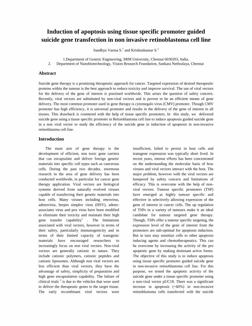
The over expression of the suicide gene was confirmed at mRNA level and a 10 fold increase in the expression of the suicide gene was detected in non-invasive retinoblastoma cells after 48 hours of transfection.
Suicide gene transfection in non invasive Retinoblastoma cell line
Fig 2: Graph showig the over expression of suicide gene in transfected non-invasive retinoblastomacell line(~10 fold increase).
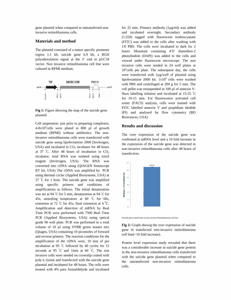
Protein level expression study revealed that there was a considerable increase in suicide gene protein in the non-invasive retinoblastoma cells transfected with the suicide gene plasmid when compared to the untransfected non-invasive retinoblastoma cells.
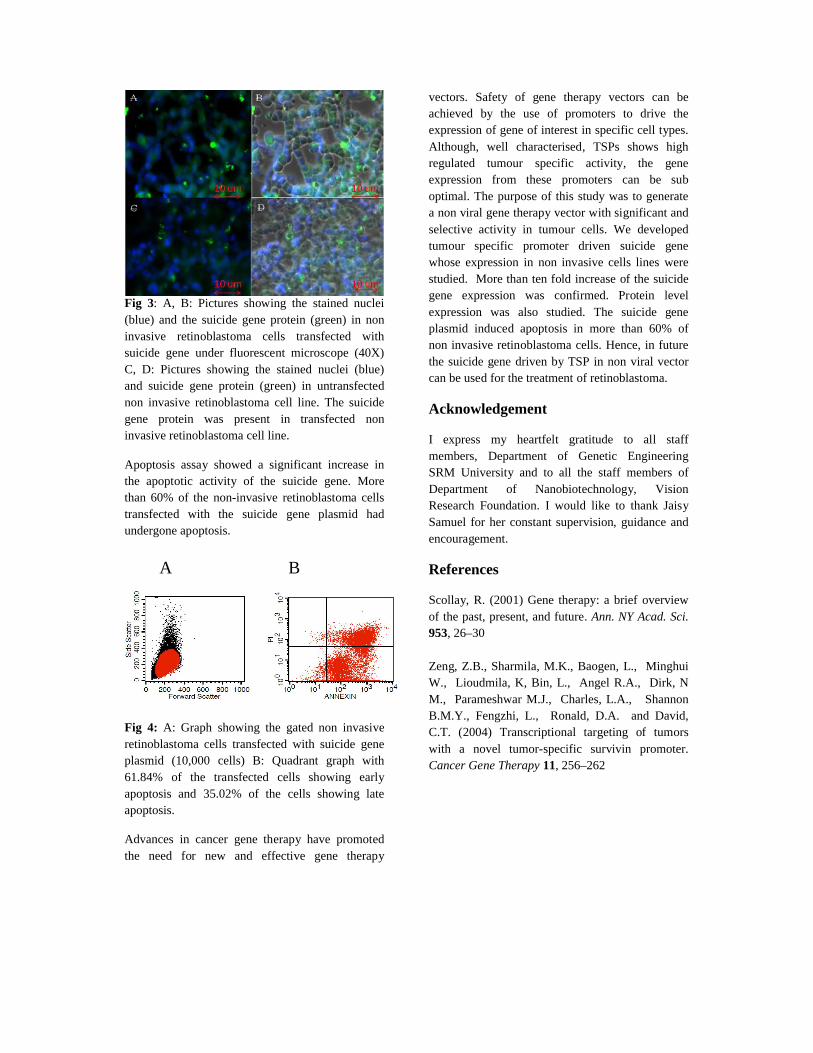
Fig 3: A, B: Pictures showing the stained nuclei (blue) and the suicide gene protein (green) in non invasive retinoblastoma cells transfected with suicide gene under fluorescent microscope (40X)C, D: Pictures showing the stained nuclei (blue) and suicide gene protein (green) in untransfected non invasive retinoblastoma cell line. The suicide gene protein was present in transfected non invasive retinoblastoma cell line.
Apoptosis assay showed a significant increase in the apoptotic activity of the suicide gene. More than 60% of the non-invasive retinoblastoma cells transfected with the suicide gene plasmid had undergone apoptosis.
Fig 4: A: Graph showing the gated non invasive retinoblastoma cells transfected with suicide gene plasmid (10,000 cells) B: Quadrant graph with 61.84% of the transfected cells showing early apoptosis and 35.02% of the cells showing late apoptosis.
Advances in cancer gene therapy have promoted the need for new and effective gene therapy
vectors. Safety of gene therapy vectors can be achieved by the use of promoters to drive the expression of gene of interest in specific cell types. Although, well characterised, TSPs shows high regulated tumour specific activity, the gene expression from these promoters can be sub optimal. The purpose of this study was to generate a non viral gene therapy vector with significant and selective activity in tumour cells. We developed tumour specific promoter driven suicide gene whose expression in non invasive cells lines were studied. More than ten fold increase of the suicide gene expression was confirmed. Protein level expression was also studied. The suicide gene plasmid induced apoptosis in more than 60% of non invasive retinoblastoma cells. Hence, in future the suicide gene driven by TSP in non viral vector can be used for the treatment of retinoblastoma.
Acknowledgement
I express my heartfelt gratitude to all staff members, Department of Genetic Engineering SRM University and to all the staff members of Department of Nanobiotechnology, Vision Research Foundation. I would like to thank Jaisy Samuel for her constant supervision, guidance and encouragement.
References
Scollay, R. (2001) Gene therapy: a brief overview of the past, present, and future. Ann. NY Acad. Sci. 953, 26–30
Zeng, Z.B., Sharmila, M.K., Baogen, L., Minghui W., Lioudmila, K, Bin, L., Angel R.A., Dirk, NM., Parameshwar M.J., Charles, L.A., Shannon B.M.Y., Fengzhi, L., Ronald, D.A. and David,C.T. (2004) Transcriptional targeting of tumors with a novel tumor-specific survivin promoter. Cancer Gene Therapy 11, 256–262
A B


![Sandhya Tattva Subhodini [1978]](https://img.pdfslide.net/doc/110x75/553028f2550346a10b8b468d/sandhya-tattva-subhodini-1978.jpg)