Embed Size (px)
Citation preview

MOL #79335 1
Mini-review for Molecular Pharmacology
New insights for drug design from the X-ray crystallographic structures of GPCRs
Kenneth A. Jacobson and Stefano Costanzi
Molecular Recognition Section, Laboratory of Bioorganic Chemistry (KAJ), and Laboratory of
Biological Modeling (SC), National Institutes of Diabetes and Digestive and Kidney Diseases,
National Institutes of Health, Bethesda, Maryland 20892.
Molecular Pharmacology Fast Forward. Published on June 13, 2012 as doi:10.1124/mol.112.079335
Copyright 2012 by the American Society for Pharmacology and Experimental Therapeutics.
This article has not been copyedited and formatted. The final version may differ from this version.Molecular Pharmacology Fast Forward. Published on June 13, 2012 as DOI: 10.1124/mol.112.079335
at ASPE
T Journals on M
ay 24, 2020m
olpharm.aspetjournals.org
Dow
nloaded from

MOL #79335 2
Running title: Drug design from GPCR crystallographic structures
Corresponding author:
Kenneth A. Jacobson, Ph.D., Laboratory of Bioorganic Chemistry, National Institutes of
Diabetes and Digestive and Kidney Diseases, NIH, Bldg. 8A, Rm. B1A-19, Bethesda, MD
20892-0810, USA
Email: [email protected].
Phone: 301-496-9024.
Fax: 301-480-8422.
Text pages: 11 Number of tables: 1 Number of figures: 6 Number of references: 92 Number of words in Abstract: 227 Number of words in Introduction: 632 Number of words in Discussion: n/a Abbreviations: BPM, biophysical mapping; BRIL, thermostabilized apocytochrome b562RIL; CVX15, cyclic disulfide of H-Arg-Arg-Nal-Cys-Tye-Gln-Lys-D-Pro-Pro-Tyr-Arg-Cit-Cys-Arg-Gly- D-Pro-OH; DREADD, designer receptors exclusively activated by designer drugs; EL, extracellular loop; IL, intracellular loop; IT1t, 6-dimethyl-5H,6H-imidazo[2,1-b][1,3]thiazol-3-yl}methyl)sulfanyl]methanimidamide; ML056, (R)-3-amino-(3-hexylphenylamino)-4-oxobutylphosphonic acid; SPR, surface plasmon resonance; StaR, thermostabilized form of a GPCR, TM, transmembrane helix; RASSL, receptor activated solely by synthetic ligands; UK-432097, 2-(3-[1-(pyridin-2-yl)piperidin-4-yl]ureido)ethyl-6-N-(2,2-diphenylethyl)-5′-N-ethylcarboxamidoadenosine-2-carboxamide; ZM241385, 4-(2-(7-amino-2-(furan-2-yl)-[1,2,4]triazolo[1,5-a][1,3,5]triazin-5-ylamino)ethyl)phenol.
This article has not been copyedited and formatted. The final version may differ from this version.Molecular Pharmacology Fast Forward. Published on June 13, 2012 as DOI: 10.1124/mol.112.079335
at ASPE
T Journals on M
ay 24, 2020m
olpharm.aspetjournals.org
Dow
nloaded from

MOL #79335 3
Abstract
Methodological advances in X-ray crystallography have made possible the recent
solution of X-ray structures of pharmaceutically important G protein-coupled receptors
(GPCRs), including receptors for biogenic amines, peptides, a nucleoside and a sphingolipid.
These high-resolution structures have greatly increased our understanding of ligand recognition
and receptor activation. Conformational changes associated with activation common to several
receptors entail outward movements of the intracellular side of transmembrane helix 6 (TM6)
and movements of TM5 toward TM6. Movements associated with specific agonists or receptors
have also been described, e.g. extracellular loop 3 (EL3) in the A2A adenosine receptor. The
binding sites of different receptors partly overlap but differ significantly in ligand orientation,
depth and breadth of contact areas in TM regions, and the involvement of the ELs. A current
challenge is how to utilize this structural information for the rational design of novel potent and
selective ligands. For example, new chemotypes were discovered as antagonists of various
GPCRs by subjecting chemical libraries to in silico docking in the X-ray structures. The vast
majority of GPCR structures and their ligand complexes are still unsolved, and no structures are
known outside of Family A GPCRs. Molecular modeling, informed by supporting information
from site-directed mutagenesis and structure activity relationships, has been validated as a useful
tool to extend structural insights to related GPCRs and to analyze docking of other ligands in
already crystallized GPCRs.
This article has not been copyedited and formatted. The final version may differ from this version.Molecular Pharmacology Fast Forward. Published on June 13, 2012 as DOI: 10.1124/mol.112.079335
at ASPE
T Journals on M
ay 24, 2020m
olpharm.aspetjournals.org
Dow
nloaded from
MOL #79335 4
Introduction
In the past five years, progress in the structure-based design of ligands for G protein-
coupled receptors (GPCRs) has greatly accelerated. The major contributing factor has been the
elucidation of X-ray crystallographic structures of high resolution for various drug-relevant
GPCRs, initially in the inactive antagonist-bound forms and more recently in agonist-bound
forms. The initial breakthrough were the reports in 2007 by Kobilka (Stanford Univ.), Stevens
(Scripps Research Inst.), Schertler and Tate (Medical Research Council Laboratory of Molecular
Biology in Cambridge, UK) and colleagues of the first non-rhodopsin GPCR structure, e.g. the
inactive human β2 adrenergic receptor in complex with the inverse agonist carazolol (Cherezov
et al., 2007; Rasmussen et al., 2007; Rosenbaum et al., 2007). These landmark studies were
followed by the determination of other GPCRs (Table 1), and the rapid pace of these reports is
continuing. Biogenic amine receptor complexes (epinephrine, dopamine, histamine, muscarinic),
nucleoside (adenosine) receptor complexes, sphingolipid (S1P1) and peptide (CXCR4, opioid)
receptor complexes have been reported. All of the crystallized receptors belong to the GPCR
family known with the names of Class A, Family 1, or Rhodopsin Family, which in humans
comprises over 80% of all GPCRs (Costanzi, 2012; Krishnan et al., 2012). As shown in Figure
1, most of these receptors belong to a branch of Class A that comprises receptors for biogenic
amines and MECA (melanocortin, endothelial differentiation sphingolipids, cannabinoid and
adenosine) receptors. The only exceptions are the chemokine CXCR4 and opioid receptors
(κ and µ), which are found in a branch of Class A predominantly populated by peptide receptors,
and rhodopsin, which is found in a small branch of Class A populated by opsins. Moreover, the
solution of the nociceptin/orphanin FQ peptide receptor has recently been announced. A large
portion of the dendrogram of Class A GPCRs, including a branch that comprises mostly
receptors for nucleotides and lipids, is still unexplored. There is reason to expect that many other
structures will be solved in the near future to shed light onto a still uncharted region of the GPCR
phylogenetic dendrogram. Furthermore, the solution of receptors belonging to families beyond
Class A is expected.
The technical advances that led to this dramatic progress include: 1) Fusion of the
receptor with the T4-lysozyme, which increases the tendency to form crystals – the T4-lysozyme
is usually inserted in lieu of the intracellular loop 3 (IL3) (Cherezov et al., 2007; Rosenbaum et
al., 2007), but has also been fused with the N-terminus, to facilitate the co-crystallization of a
This article has not been copyedited and formatted. The final version may differ from this version.Molecular Pharmacology Fast Forward. Published on June 13, 2012 as DOI: 10.1124/mol.112.079335
at ASPE
T Journals on M
ay 24, 2020m
olpharm.aspetjournals.org
Dow
nloaded from

MOL #79335 5
receptor-G protein complex (Rasmussen et al., 2011b); 2) Structurally-stabilizing point
mutations, either in the ligand-binding regions or more remotely – this approach has been
introduced by the groups of Schertler and Tate in Cambridge and the associated company
Heptares (Warne et al., 2008). These thermostabilizing mutations can be made to favor either an
antagonist-bound conformation or agonist-bound conformation (although poorly activatable due
to the energetic stabilization) of the same GPCR. The stabilization is so effective that the GPCR
protein can be captured on a Biacore chip to allow characterization of small molecule binding by
measuring surface plasmon resonance (SPR) (Zhukov et al., 2011); 3) Stabilization of the
receptors through antibodies (Rasmussen et al., 2007) – recently, nanobodies generated in llamas
inoculated with receptors as well as receptors cross-linked with G protein heterotrimers have
been used to solve crystal structures of the activates state of the β2 adrenergic receptor
(Rasmussen et al., 2011a; Rasmussen et al., 2011b) ; 4) Specialized agonists, such as irreversibly
binding agonists (Rosenbaum et al., 2011) or an agonist that has multiple arms extending from
the core pharmacophore structure (Xu et al., 2011). 5) Specialized techniques for producing
crystals of membrane-bound proteins, including adaptation of the lipidic cubic phase, which
forms a single lipid-aqueous bilayer that allows ordered protein molecules to make contacts in
their hydrophobic as well as hydrophilic portions (Cherezov, 2011).
The ligand binding cavity
All GPCRs are constituted by a single polypeptide chain that spans the plasma membrane
seven times with seven alpha helical structures (Costanzi et al., 2009). As the crystal structures
revealed, the helical bundle of most Class A GPCRs hosts a ligand binding cavity opened toward
the extracellular milieu, which provides access to diffusible ligands. Alternately, the cavities of
rhodopsin and S1P1 receptor are sealed from the extracellular space by the second extracellular
loop (EL2) and the N-terminus, respectively. It is likely that the hydrophobic ligands of these
receptors make their way into the binding cavity through the transmembrane domains. For most
GPCRs this ligand binding cavity is lined by transmembrane domains (TMs) 2, 3, 5, 6, and 7 and
is deeper in proximity to TMs 5 and 6 while shallower in proximity to TMs 2, and 7 (Figure 2).
Nevertheless, in some cases TMs 1 and 4 also form the pocket and can impact ligand binding.
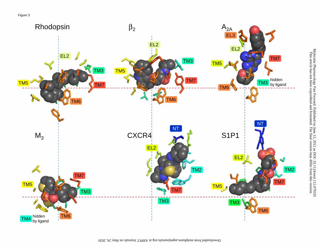
The various ligands co-crystallized with their receptors in the currently solved GPCR structures
variously occupy different regions of this cavity. This is evident from Figure 3, which shows a
This article has not been copyedited and formatted. The final version may differ from this version.Molecular Pharmacology Fast Forward. Published on June 13, 2012 as DOI: 10.1124/mol.112.079335
at ASPE
T Journals on M
ay 24, 2020m
olpharm.aspetjournals.org
Dow
nloaded from

MOL #79335 6
side by side comparison of six representative receptors featuring the bound ligands and the
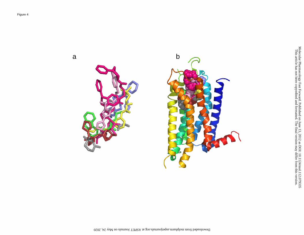
residues that surround them, as well as Figure 4, which provides an overlay of the same ligands
of these receptors resulting from a structural superposition of the receptors. Notably, different
ligands that bind to the same receptor may occupy different regions of the binding cavity. This
situation is particularly evident from Figure 5A, which compares the binding to the CXCR4
receptor of a small molecule antagonist and a cyclic peptide antagonist (Wu et al., 2010b): there
is virtually no overlap between the two molecules. Less extreme is the case illustrated in Figure
5B, which compares the binding to the adenosine receptor of a non-purine antagonist and an
adenosine-based agonist UK-432097 bearing large substituents at the C2 and N6 positions of the
purine ring (Xu et al., 2011): although there is substantial overlap between the two molecules,
the larger agonist occupies regions of the receptor unexploited by the antagonist. The ribose
moiety, which is an essential component of nearly all adenosine receptor agonists, confers to the
ligand its agonistic properties. This moiety is accommodated in a space of the binding cavity
close to TM3 that in the antagonist-bound state is unoccupied by a ligand and is partially filled
by water molecules. The hydroxyl and other H-bonding groups of agonists displace these water
molecules, thereby gaining an entropic advantage in the binding process. This is consistent with
the typically nanomolar affinities of agonists at 3 of the 4 subtypes of adenosine receptors. The
other large substituents present on the agonist UK-432097 co-crystallized with the A2A receptor
further enhance the binding affinity of the agonist by establishing contacts with the receptor.
The outer regions of a GPCR can serve as meta-binding sites for a ligand on its path to
the principle orthosteric binding site and can also contribute to the lining of the orthosteric
binding site. In particular, the X-ray structures have confirmed the hypothesis, based on
mutagenesis as well as molecular modeling (Moro et al., 1999; Olah et al., 1994; Peeters et al.,
2011), that parts of the ELs are intimately involved in recognition of ligands for both agonists
and antagonists. Particularly, we now know that the C-terminal part of EL2 tends to drop more or
less deeply, depending on the receptor, into the ligand-binding region to establish contacts with
the ligands (Figure 3), as predicted using site-directed mutagenesis. Peculiar are the cases of
rhodopsin and the S1P1 receptor, in which the ligand-binding cavity is substantially more
enclosed than in other receptors, thanks to a singular conformation of EL2 that occludes the
entrance of the cleft (Palczewski et al., 2000). Conversely, the conformation that EL2 assumes in
the chemokine CXCR4 receptor renders the binding cavity particularly open to the extracellular
This article has not been copyedited and formatted. The final version may differ from this version.Molecular Pharmacology Fast Forward. Published on June 13, 2012 as DOI: 10.1124/mol.112.079335
at ASPE
T Journals on M
ay 24, 2020m
olpharm.aspetjournals.org
Dow
nloaded from
MOL #79335 7
space, facilitating the binding of large peptides (Wu et al., 2010b). However, the role of ELs in
recognition of and activation by large peptide ligands will require further studies (Dong et al.,
2011).
In general, the extracellular loops are typically more varied between subtypes than the
TMs, which could facilitate the rational design of more selective orthosteric and allosteric
ligands, as guided by the 3D structures of the receptor complexes. For instance, the M2 (Haga et
al., 2012) and M3 (Kruse et al., 2012) muscarinic acetylcholine receptors contain a vestibule in
their outer portions, which corresponds to the regions of the receptor associated with the binding
of allosteric modulators. The fact that the orthosteric site of the muscarinic receptors and the
specific residues involved in ligand recognition are identical across the family has impeded the
medicinal chemical effort to design selective competitive ligands. The possibility of targeting the
vestibule, which is more divergent in sequence, offers now further opportunities for drug design.
However, for those receptors for which a crystallographically solved structure is not yet
available, the high variability of the extracellular regions makes their modeling subject to greater
uncertainty when compared to the modeling of the 7 TMs (Goldfeld et al., 2011).
Inactive and activated structures
The determination of both agonist- and antagonist-bound states of the same receptor was
accomplished for two classes, i.e. β adrenergic and adenosine receptors, in addition to visual
pigment receptors with the structures of opsin and rhodopsin (Choe et al., 2011; Park et al., 2008;
Rasmussen et al., 2011a; Rasmussen et al., 2011b; Scheerer et al., 2008; Standfuss et al., 2011;
Xu et al., 2011). This major step forward allowed a deeper understanding of the activation
processes operating in GPCRs. Thus, the details of agonist binding and activation, i.e.
characteristic agonist-induced movement of helices and key residues, are beginning to be
understood. The comparison of the structures of inactive rhodopsin with those of the meta II state
of rhodopsin as well as the unliganded opsin revealed several conformational changes associated
with the activation of the receptor. The most notable of these conformational changes entail
outward movements of the intracellular side of TM6 and movements of TM5 toward TM6
(Figures 6A and B). These conformational changes were also found in the agonist-bound β2
adrenergic and A2A adenosine receptors, although the displacement of TM6 is substantially less
pronounced in the A2A receptor as a consequence of the T4-lysozyme fused between the
This article has not been copyedited and formatted. The final version may differ from this version.Molecular Pharmacology Fast Forward. Published on June 13, 2012 as DOI: 10.1124/mol.112.079335
at ASPE
T Journals on M
ay 24, 2020m
olpharm.aspetjournals.org
Dow
nloaded from

MOL #79335 8
cytosolic ends of TM5 and TM6 (Figure 6C and D). Also, the distance between oppositely
charged amino acid side chains near the cytosolic side, e.g. an Arg and an Asp residue
respectively found in TMs 3 and 6 and predicted to be involved in a putative ionic lock
characteristic of the inactive state, increased upon binding of the agonist in each case. However,
it should be noted that this ionic lock was not fully closed in the inactive states of the β2
adrenergic and A2A adenosine receptors. Furthermore, some movements that were not predicted
in the opsin structure were seen in other receptors, such as a see-saw movement of TM7 in the
A2A adenosine receptor by which the intracellular end moves inward (Figure 5C and D).
Movements in the extracellular loop (EL) regions were also noted. These movements appear to
be more ligand-specific than the movements in the TM regions. For example, the outward
displacement of EL3 of the A2A adenosine receptor was considerably greater for an agonist
having bulky substitutions of the adenine ring than in the unsubstituted cases. Notably,
increasing evidence demonstrated that the stimulation of one GPCR can trigger different
signaling cascades in a ligand-dependent manner, through a phenomenon known as biased
agonism (Kahsai et al., 2011). As a result, the same receptor can be selectively induced to
activate a variety of pathways mediated by G proteins as well as β-arrestins. Most likely, these
different signaling states are due to distinct conformations of the same receptor that individual
ligands can induce or stabilize. Further light will be shed on the correlation between
conformation and signaling state of GPCRs once multiple structures of the same receptor with a
variety of biased ligands are solved. In this respect some progress has been made for the β1 and
β2 adrenergic receptors through crystallographic (Warne et al., 2012) and NMR studies (Liu et
al., 2012), respectively. For the β1 adrenergic receptor, the crystal structures revealed that the
biased agonists bicindolol and carvedilol, which stimulate β-arrestin-mediated signaling but act
as inverse agonists or partial agonists of G protein-dependent pathways, interact with additional
residues located in EL2 as well as TM7 when compared to unbiased β blockers. Moreover, the
abovementioned NMR spectroscopic analyses of the β2 adrenergic receptor suggest that ligands,
including biased ligands, do not “induce” states but shift equilibrium between preexisting states.
Specifically, the study indicates that unbiased ligands impact mostly the conformational state of
TM6, while biased agonists shift primarily the conformational state of TM7 (Liu et al., 2012).
This article has not been copyedited and formatted. The final version may differ from this version.Molecular Pharmacology Fast Forward. Published on June 13, 2012 as DOI: 10.1124/mol.112.079335
at ASPE
T Journals on M
ay 24, 2020m
olpharm.aspetjournals.org
Dow
nloaded from

MOL #79335 9
Molecular docking at GPCR homology models to predict the structure of receptor ligand
complexes
Until recently, the three-dimensional study of the interactions of GPCRs with their
ligands was limited to molecular modeling. The modeling efforts began more than two decades
ago, using as a crude template the structure of bacteriorhodopsin and progressed with the major
advance of the high resolution structure of bovine rhodopsin more than a decade ago (Ballesteros
et al., 2001; Costanzi et al., 2007; Costanzi et al., 2009). Although there has been much
variability in the confidence in the modeling results (papers were published with diametrically
opposed modes of ligand binding for the same receptor ligand-complex), there were examples of
well-supported modeling that was later validated crystallographically (among others, see: Ivanov
et al., 2009; Michino et al., 2009; Kufareva, 2011). Comparisons of crystal structures and
homology models revealed that, for some GPCRs, reasonably accurate receptor-ligand
complexes can be constructed through homology modeling followed by molecular docking
(Costanzi, 2008; Costanzi, 2010; Costanzi, 2012; Reynolds et al., 2009). However, accurate
complexes cannot always be obtained through the sole use of specialized software. Purely
computational docking of ligands at GPCR homology models could lead to substantially
inaccurate results if the contributions of specific residues to ligand biding or, even worse, the
location of the binding cavity are incorrectly recognized. A community wide challenge to
molecular modelers to predict the docking mode of antagonist ZM241385 to the A2A adenosine
receptor prior to the release of the X-ray structure showed how the ligand could assume almost a
random placement around the receptor (Michino et al., 2009). However, when informed with
accessory information about the molecular recognition elements, the placement of the ligand in
various docking models was shown to be within a reasonable range of accuracy (Michino et al.,
2009). In many cases, the identification of the correct receptor-ligand interactions is strictly
dependent on an expert selection of the docking poses based on insights derived from site-
directed mutagenesis, comparisons of SAR within the same chemical series, and bioinformatics
studies within a receptor family.
A further controlled assessment clarified that, when the target receptor shares a
significant sequence similarity with one of the available templates, the models are particularly
accurate. Conversely, predictions are more challenging for receptors that are more distant from
the available templates, in which case the modeling strategies need to be more closely guided
This article has not been copyedited and formatted. The final version may differ from this version.Molecular Pharmacology Fast Forward. Published on June 13, 2012 as DOI: 10.1124/mol.112.079335
at ASPE
T Journals on M
ay 24, 2020m
olpharm.aspetjournals.org
Dow
nloaded from
MOL #79335 10
through the incorporation of the abovementioned external information (Kufareva et al., 2011).
Since, as we pointed out, there are not yet X-ray structures representative of all major branches
of the GPCR dendrogram (Figure 1), the modeling of receptors that are more distant from the
available templates is still challenging. Moreover, the modeling of GPCRs belonging to classes
B and C (Hu et al., 2006; Wheatley et al., 2012) remains more uncertain, since none of the
structures of the members of these families have been solved crystallographically.
As mentioned, particularly important is the use of data gathered from site-directed
mutagenesis. In this regards, one variation of the use of site-directed mutagenesis that has been
particularly informative with respect to probing molecular recognition among GPCRs has been
that of reengineering the binding site to accept agonists that have been chemically modified.
These approaches, with some important differences, are known by various terms introduced by
different reseach groups: RASSLs, neoceptors, and DREADDs (Conklin et al., 2008). Different
degrees of design insight vs. empirical screening have been used to match a given mutant
receptor with an orthogonally activating agonist analogue. The neoceptor approach, in particular,
has focused on the adenosine receptor family to accurately predict the placement of the ribose
moiety of nucleoside agonists, using the three-way integrated combination of mutagenesis,
modeling, and chemical modification. Complementary changes in the structures of the ligand and
receptor that lead to enhanced affinity can help establish the orientation of a ligand within the
binding site.
Receptor-ligand interactions can also be studied through a systematic approach that was
recently introduced, termed Biophysical Mapping (BPM) (Zhukov et al., 2011). This method is
based on the characterization of the functional contour of the binding pocket of a given GPCR
using a thermostabilized form of the receptor (StaR). The effects of site-directed mutagenesis
within the binding site are correlated with binding data for diverse ligands obtained through SPR
measurements. Then, molecular modeling and docking, is used to map the small molecule
binding site with respect to each chemical class of ligands. This approach was used to identify
novel chemotypes (later to be optimized by chemical modification), such as chromones and
triazines, binding to the A2A adenosine receptor (Congreve et al., 2012; Langmead et al., 2012).
Structure-based discovery of GPCR ligands is increasingly more practical
This article has not been copyedited and formatted. The final version may differ from this version.Molecular Pharmacology Fast Forward. Published on June 13, 2012 as DOI: 10.1124/mol.112.079335
at ASPE
T Journals on M
ay 24, 2020m
olpharm.aspetjournals.org
Dow
nloaded from

MOL #79335 11
Looking ahead, it will be increasingly feasible to tap the potential of structure-based
design for GPCRs, initially for class A (Congreve et al., 2011; Salon et al., 2011) and then,
hopefully, for the other classes as well. For now, the newly revealed detailed knowledge of
GPCR structures has already facilitated a recent flurry of studies directed toward ligand
discovery.
The careful stepwise modification of known classes of agonist or antagonist for a given
receptor has been for years a major successful approach of medicinal chemists when applied
empirically. This study can now be expedited using accurate 3D knowledge of receptor-ligand
recognition. For instance, it is now feasible to target specific amino acid residues in the vicinity
of the bound pharmacophore that might confer enhanced affinity or receptor subtype selectivity
to the modified ligands. For example, recent reports have shown that A2A adenosine receptor
agonists and antagonists can be modified in this manner (Congreve et al., 2012; Deflorian et al.,
2012). Moreover, the interaction of small fragments with regions of the binding cavity proximal
to the ligand, to be considered as candidate substituents to enhance the receptor-ligand
interactions, can also be studied. In this context, in recent study the structure of a known GPCR
agonist was systematically varied using the ICM software (Internal Coordinate Mechanics,
Molsoft LLC). A library of 2000 small fragments was screened in silico for fit within a small
pocket, to successfully predict those favoring adenosine receptor affinity when linked to the 5’-
carbonyl group of modified adenosine (Tosh et al., 2012).
For the identification of ligands based on novel chemotypes, a technique that has proven
its value is the virtual screening through molecular docking of chemically diverse libraries for
the discovery of novel chemotypes that bind to various GPCRs (Costanzi, 2011). A number of
controlled experiments targeting the β adrenergic and adenosine receptors, conducted by
subjecting to molecular docking known agonists and blockers together with a larger number of
non-binders, clearly illustrated that such virtual screening campaigns are most effective when
applied to X-ray structures (Costanzi and Vilar, 2012; Reynolds et al., 2009; Vilar et al., 2011a;
Vilar et al., 2010). This observation is consistent with the successful identification of novel
structurally diverse ligands on the basis of virtual screenings conducted by targeting the crystal
structures of β-adrenergic, adenosine, dopamine, and histamine receptors (Carlsson et al., 2011;
Carlsson et al., 2010; de Graaf et al., 2011; Katritch et al., 2010a; Kolb et al., 2009; Langmead et
al., 2012; Sabio et al., 2008; van der Horst et al., 2011). The abovementioned controlled
This article has not been copyedited and formatted. The final version may differ from this version.Molecular Pharmacology Fast Forward. Published on June 13, 2012 as DOI: 10.1124/mol.112.079335
at ASPE
T Journals on M
ay 24, 2020m
olpharm.aspetjournals.org
Dow
nloaded from

MOL #79335 12
experiments also demonstrated that virtual screening campaigns, although not as effective as
when applied to a crystal structure, are also useful when applied to accurate homology models
(Cavasotto, 2011; Cavasotto et al., 2008; Katritch et al., 2010b; Phatak et al., 2010; Vilar et al.,
2011a; Vilar et al., 2010). This observation is in line with the results of virtual screening
campaigns through which, prior to the explosion of GPCR crystallography, novel GPCR ligands
were identified using rhodopsin-based homology models (Engel et al., 2008; Tikhonova et al.,
2008). More recently, Shoichet and coworkers conducted a virtual screening campaign by
targeting the crystal structure of the dopamine D3 receptor as well as a model of the same
receptor based on the β2 adrenergic homologue, with which it shares a relatively high sequence
identity (38% within the TMs and 61% within the binding cavity, defined as the residues found
within a 4 Å radius from the bound ligands) (Carlsson et al., 2011). Notably, each of these two
parallel campaigns yielded two overlapping sets of novel ligands suggesting that models based
on a relatively close template are indeed useful to ligand discovery.
Controlled experiments demonstrated also that not only blockers but agonists too are
substantially prioritized over non-binders in docking experiments (Costanzi and Vilar, 2012). In
particular, such controlled virtual screening campaigns revealed that the activated structure of the
β2 adrenergic receptor is significantly biased toward the preferential recognition of agonists over
blockers. Moreover, they also showed that structures of receptors crystallized in the inactive state
can be modified in silico by modeling the shape of the binding cavity around a docked agonist,
thus being turned into structures that preferentially recognize agonists rather than blockers
(Costanzi and Vilar, 2012; Vilar et al., 2011b). However, the identification of agonists based on
novel chemotypes through the screening of diverse libraries may prove particularly challenging,
in light of the likely stricter structural requirements for agonism than for blockade. Moreover, as
mentioned, it is increasingly evident that the same GPCR may trigger a variety of different
signaling cascades (Kahsai et al., 2011). The basis for distinguishing ligands needed for selective
effector pathway activation (i.e. biased ligands) is an important area for future investigation.
Several examples of ligand-specific interactions of the same receptor have been reported, but the
implications of these differences for signaling are still largely unknown.
Undoubtedly, docking-based virtual screening campaigns will become increasingly more
feasible with the experimental determination of new GPCR structures. Moreover, they will also
benefit from the fast pace at which supercomputing is progressing as well as the continuous
This article has not been copyedited and formatted. The final version may differ from this version.Molecular Pharmacology Fast Forward. Published on June 13, 2012 as DOI: 10.1124/mol.112.079335
at ASPE
T Journals on M
ay 24, 2020m
olpharm.aspetjournals.org
Dow
nloaded from
MOL #79335 13
improvement of computational algorithms. In particular, the scoring functions that are currently
utilized to estimate the likelihood of binding of large sets of screened compounds represent a
compromise between accuracy and rapidity. Large computer clusters as well as specialized
purpose-built supercomputers will increasingly allow the development and application of more
complex methods for the calculation of free binding energies (Mitchell, 2011). Moreover, an
increasingly higher number of alternative receptor conformations, either solved experimentally
or generated computationally from a single experimental structure, will be applicable in parallel
to the screening campaigns. This practice, known as receptor-ensemble docking, was already
demonstrated to significantly improve virtual screening yields by providing a means to account
for receptor flexibility (Bottegoni et al., 2011; Costanzi, 2011; Costanzi and Vilar, 2012; Vilar et
al., 2011a).
Conclusions
Based on methodological advances in X-ray crystallography, the structural elucidation of
GPCRs has begun a revolution in the medicinal chemical approaches applied to the discovery of
new GPCR ligands. The binding sites of different receptors partly overlap but differ significantly
in ligand orientation, depth and breadth of contact areas in TM regions, and the involvement of
the extracellular loops (ELs). Conformational changes associated with activation have been
analyzed for several receptors. However, there are still large areas where knowledge is lacking.
For example there still is an uncharacterized large portion of the GPCR phylogenetic
dendrogram, the interaction of large peptide ligands with their receptors are unclear, and the
structural basis for functional selectivity, i.e. the reasons behind which agonists display different
spectra of activation properties through the same GPCR, are poorly understood. Molecular
modeling, informed by supporting information from site-directed mutagenesis and structure
activity relationships, has been validated as a useful tool to extend structural insights to related
GPCRs and to analyze docking of other ligands in already crystallized GPCRs. Undoubtedly
further exploration of the interactions of GPCRs with their G protein and non G-protein
intracellular targets, through medicinal chemistry as well as techniques of structural biology, will
be required.
This article has not been copyedited and formatted. The final version may differ from this version.Molecular Pharmacology Fast Forward. Published on June 13, 2012 as DOI: 10.1124/mol.112.079335
at ASPE
T Journals on M
ay 24, 2020m
olpharm.aspetjournals.org
Dow
nloaded from
MOL #79335 14
Authorship contributions:
Performed data analysis: Costanzi.
Wrote or contributed to the writing of the manuscript: Jacobson, Costanzi.
This article has not been copyedited and formatted. The final version may differ from this version.Molecular Pharmacology Fast Forward. Published on June 13, 2012 as DOI: 10.1124/mol.112.079335
at ASPE
T Journals on M
ay 24, 2020m
olpharm.aspetjournals.org
Dow
nloaded from
MOL #79335 15
References
Ballesteros JA, Shi L and Javitch JA (2001) Structural mimicry in G protein-coupled receptors: implications of the high-resolution structure of rhodopsin for structure-function analysis of rhodopsin-like receptors. Mol Pharmacol 60: 1-19.
Bokoch M, Zou Y, Rasmussen S, Liu C, Nygaard R, Rosenbaum D, Fung J, Choi H, Thian F, Kobilka T, Puglisi J, Weis W, Pardo L, Prosser R, Mueller L and Kobilka B (2010) Ligand-specific regulation of the extracellular surface of a G-protein-coupled receptor. Nature 463: 108-112.
Bottegoni G, Rocchia W, Rueda M, Abagyan R, Cavalli A (2011) Systematic exploitation of multiple receptor conformations for virtual ligand screening. PLoS One 6: e18845
Carlsson J, Coleman RG, Setola V, Irwin JJ, Fan H, Schlessinger A, Sali A, Roth BL and Shoichet BK (2011) Ligand discovery from a dopamine D3 receptor homology model and crystal structure. Nat Chem Biol 7: 769-778.
Carlsson J, Yoo L, Gao Z, Irwin J, Shoichet B and Jacobson K (2010) Structure-based discovery of A2A adenosine receptor ligands. J Med Chem 53: 3748-3755.
Cavasotto CN (2011) Homology models in docking and high-throughput docking. Curr Top Med Chem 11: 1528-1534.
Cavasotto CN, Orry AJ, Murgolo NJ, Czarniecki MF, Kocsi SA, Hawes BE, O'Neill KA, Hine H, Burton MS, Voigt JH, Abagyan RA, Bayne ML and Monsma FJ (2008) Discovery of novel chemotypes to a G-protein-coupled receptor through ligand-steered homology modeling and structure-based virtual screening. J Med Chem 51: 581-588.
Cherezov V (2011) Lipidic cubic phase technologies for membrane protein structural studies. Curr Opin Struct Biol 21: 559-566.
Cherezov V, Rosenbaum D, Hanson M, Rasmussen S, Thian F, Kobilka T, Choi H, Kuhn P, Weis W, Kobilka B and Stevens R (2007) High-resolution crystal structure of an engineered human β2 adrenergic G protein-coupled receptor. Science 318: 1258-1265.
Chien EY, Liu W, Zhao Q, Katritch V, Han GW, Hanson MA, Shi L, Newman AH, Javitch JA, Cherezov V and Stevens RC (2010) Structure of the human dopamine D3 receptor in complex with a D2/D3 selective antagonist. Science 330: 1091-1095.
Choe HW, Kim YJ, Park JH, Morizumi T, Pai EF, Krauss N, Hofmann KP, Scheerer P and Ernst OP (2011) Crystal structure of metarhodopsin II. Nature 471: 651-655.
Congreve M, Andrews SP, Dore AS, Hollenstein K, Hurrell E, Langmead CJ, Mason JS, Ng IW, Tehan B, Zhukov A, Weir M and Marshall FH (2012) Discovery of 1,2,4-Triazine Derivatives as Adenosine A2A Antagonists using Structure Based Drug Design. J Med Chem 55: 1898-1903.
Congreve M, Langmead CJ, Mason JS and Marshall FH (2011) Progress in Structure Based Drug Design for G Protein-Coupled Receptors. J Med Chem 54: 4283-4311.
Conklin BR, Hsiao EC, Claeysen S, Dumuis A, Srinivasan S, Forsayeth JR, Guettier JM, Chang WC, Pei Y, McCarthy KD, Nissenson RA, Wess J, Bockaert J and Roth BL (2008) Engineering GPCR signaling pathways with RASSLs. Nat Methods 5: 673-678.
Costanzi S (2008) On the applicability of GPCR homology models to computer-aided drug discovery: a comparison between in silico and crystal structures of the β2 adrenergic receptor. J Med Chem 51: 2907-2914.
Costanzi S (2010) Modelling G protein-coupled receptors: a concrete possibility. Chim Oggi 28: 26-30.
This article has not been copyedited and formatted. The final version may differ from this version.Molecular Pharmacology Fast Forward. Published on June 13, 2012 as DOI: 10.1124/mol.112.079335
at ASPE
T Journals on M
ay 24, 2020m
olpharm.aspetjournals.org
Dow
nloaded from

MOL #79335 16
Costanzi S (2011) Chapter 18 Structure-based Virtual Screening for Ligands of G Protein-coupled Receptors, in G Protein-Coupled Receptors: From Structure to Function (Giraldo J and Pin J-P eds) pp 359-374, The Royal Society of Chemistry.
Costanzi S (2012) Homology modeling of class a g protein-coupled receptors. Methods Mol Biol 857: 259-279.
Costanzi S, Ivanov A, Tikhonova I and Jacobson K (2007) Structure and function of G protein-coupled receptors studied through using sequence analysis, molecular modeling and receptor engineering: adenosine receptors. Frontiers in Drug Design and Discovery, Bentham 3: 63-69.
Costanzi S, Siegel J, Tikhonova I and Jacobson K (2009) Rhodopsin and the others: a historical perspective on structural studies of G protein-coupled receptors. Curr Pharm Des 15: 3994-4002.
Costanzi S and Vilar S (2012) In Silico screening for agonists and blockers of the β2 adrenergic receptor: Implications of inactive and activated state structures. J Comput Chem 33: 561-572.
de Graaf C, Kooistra AJ, Vischer HF, Katritch V, Kuijer M, Shiroishi M, Iwata S, Shimamura T, Stevens RC, de Esch IJ and Leurs R (2011) Crystal structure-based virtual screening for fragment-like ligands of the human histamine H1 receptor. J Med Chem 54: 8195-8206.
Deflorian F, Kumar TS, Phan K, Gao ZG, Xu F, Wu H, Katritch V, Stevens RC and Jacobson KA (2012) Evaluation of Molecular Modeling of Agonist Binding in Light of the Crystallographic Structure of an Agonist-Bound A2A Adenosine Receptor. J Med Chem 55: 538-552.
Dong M, Lam PC, Pinon DI, Hosohata K, Orry A, Sexton PM, Abagyan R and Miller LJ (2011) Molecular basis of secretin docking to its intact receptor using multiple photolabile probes distributed throughout the pharmacophore. J Biol Chem 286: 23888-23899.
Dore AS, Robertson N, Errey JC, Ng I, Hollenstein K, Tehan B, Hurrell E, Bennett K, Congreve M, Magnani F, Tate CG, Weir M and Marshall FH (2011) Structure of the Adenosine A2A Receptor in Complex with ZM241385 and the Xanthines XAC and Caffeine. Structure 19: 1283-1293.
Engel S, Skoumbourdis A, Childress J, Neumann S, Deschamps J, Thomas C, Colson A, Costanzi S and Gershengorn M (2008) A virtual screen for diverse ligands: discovery of selective G protein-coupled receptor antagonists. J Am Chem Soc 130: 5115-5123.
Goldfeld DA, Zhu K, Beuming T and Friesner RA (2011) Successful prediction of the intra- and extracellular loops of four G-protein-coupled receptors. Proc Natl Acad Sci U S A 108: 8275-8280.
Granier S, Manglik A, Kruse AC, Kobilka TS, Thian FS, Weis WI and Kobilka BK (2012) Structure of the δ-opioid receptor bound to naltrindole. Nature 485(7398):400-404.
Haga K, Kruse AC, Asada H, Yurugi-Kobayashi T, Shiroishi M, Zhang C, Weis WI, Okada T, Kobilka BK, Haga T and Kobayashi T (2012) Structure of the human M2 muscarinic acetylcholine receptor bound to an antagonist. Nature 482: 547-551.
Hanson M, Cherezov V, Griffith M, Roth C, Jaakola V, Chien E, Velasquez J, Kuhn P and Stevens R (2008) A specific cholesterol binding site is established by the 2.8 A structure of the human β2 adrenergic receptor. Structure 16: 897-905.
Hanson MA, Roth CB, Jo E, Griffith MT, Scott FL, Reinhart G, Desale H, Clemons B, Cahalan SM, Schuerer SC, Sanna MG, Han GW, Kuhn P, Rosen H and Stevens RC (2012) Crystal structure of a lipid G protein-coupled receptor. Science 335: 851-855.
This article has not been copyedited and formatted. The final version may differ from this version.Molecular Pharmacology Fast Forward. Published on June 13, 2012 as DOI: 10.1124/mol.112.079335
at ASPE
T Journals on M
ay 24, 2020m
olpharm.aspetjournals.org
Dow
nloaded from

MOL #79335 17
Hino T, Arakawa T, Iwanari H, Yurugi-Kobayashi T, Ikeda-Suno C, Nakada-Nakura Y, Kusano-Arai O, Weyand S, Shimamura T, Nomura N, Cameron AD, Kobayashi T, Hamakubo T, Iwata S and Murata T (2012) G-protein-coupled receptor inactivation by an allosteric inverse-agonist antibody. Nature 482: 237-240.
Hu J, Jiang J, Costanzi S, Thomas C, Yang W, Feyen J, Jacobson K and Spiegel A (2006) A missense mutation in the seven-transmembrane domain of the human Ca2+ receptor converts a negative allosteric modulator into a positive allosteric modulator. J Biol Chem 281: 21558-21565.
Ivanov A, Barak D and Jacobson K (2009) Evaluation of homology modeling of G-protein-coupled receptors in light of the A2A adenosine receptor crystallographic structure. J Med Chem 52: 3284-3292.
Jaakola V, Griffith M, Hanson M, Cherezov V, Chien E, Lane J, IJzerman A and Stevens R (2008) The 2.6 angstrom crystal structure of a human A2A adenosine receptor bound to an antagonist. Science 322: 1211-1217.
Kahsai AW, Xiao K, Rajagopal S, Ahn S, Shukla AK, Sun J, Oas TG and Lefkowitz RJ (2011) Multiple ligand-specific conformations of the β2 adrenergic receptor. Nat Chem Biol 7: 692-700.
Katritch V, Jaakola V, Lane J, Lin J, IJzerman A, Yeager M, Kufareva I, Stevens R and Abagyan R (2010a) Structure-based discovery of novel chemotypes for adenosine A2A receptor antagonists. J Med Chem 53: 1799-1809.
Katritch V, Rueda M, Lam P, Yeager M and Abagyan R (2010b) GPCR 3D homology models for ligand screening: lessons learned from blind predictions of adenosine A2A receptor complex. Proteins 78: 197-211.
Kolb P, Rosenbaum D, Irwin J, Fung J, Kobilka B and Shoichet B (2009) Structure-based discovery of β2 adrenergic receptor ligands. Proc Natl Acad Sci U S A 106: 6843-6848.
Krishnan A, Almen MS, Fredriksson R and Schioth HB (2012) The origin of GPCRs: identification of mammalian like Rhodopsin, Adhesion, Glutamate and Frizzled GPCRs in fungi. PLoS One 7: e29817.
Kruse AC, Hu J, Pan AC, Arlow DH, Rosenbaum DM, Rosemond E, Green HF, Liu T, Chae PS, Dror RO, Shaw DE, Weis WI, Wess J and Kobilka BK (2012) Structure and dynamics of the M3 muscarinic acetylcholine receptor. Nature 482: 552-556.
Kufareva I, Rueda M, Katritch V, Stevens RC and Abagyan R (2011) Status of GPCR modeling and docking as reflected by community-wide GPCR Dock 2010 assessment. Structure 19: 1108-1126.
Langmead CJ, Andrews SP, Congreve M, Errey JC, Hurrell E, Marshall FH, Mason JS, Richardson CM, Robertson N, Zhukov A and Weir M (2012) Identification of Novel Adenosine A2A Receptor Antagonists by Virtual Screening. J Med Chem 55: 1904-1909.
Lebon G, Warne T, Edwards PC, Bennett K, Langmead CJ, Leslie AG and Tate CG (2011) Agonist-bound adenosine A2A receptor structures reveal common features of GPCR activation. Nature 474: 521-555.
Li J, Edwards PC, Burghammer M, Villa C and Schertler GF (2004) Structure of bovine rhodopsin in a trigonal crystal form. J Mol Biol 343: 1409-1438.
Liu JJ, Horst R, Katritch V, Stevens RC and Wüthrich (2012) Biased signaling pathways in β2-adrenergic receptor characterized by 19F-NMR. Science 335: 1106-1110.
Makino CL, Riley CK, Looney J, Crouch RK and Okada T (2010) Binding of more than one retinoid to visual opsins. Biophys J 99: 2366-2373.
This article has not been copyedited and formatted. The final version may differ from this version.Molecular Pharmacology Fast Forward. Published on June 13, 2012 as DOI: 10.1124/mol.112.079335
at ASPE
T Journals on M
ay 24, 2020m
olpharm.aspetjournals.org
Dow
nloaded from

MOL #79335 18
Manglik A, Kruse AC, Kobilka TS, Thian FS, Mathiesen JM, Sunahara RK, Pardo L, Weis WI, Kobilka BK and Granier S (2012) Crystal structure of the mu-opioid receptor bound to a morphinan antagonist. Nature 485: 321-326.
Michino M, Abola E, 2008 Participants G, Brooks Cr, Dixon J, Moult J and Stevens R (2009) Community-wide assessment of GPCR structure modelling and ligand docking: GPCR Dock 2008. Nat Rev Drug Discov 8: 455-463.
Mitchell W, Matsumoto S (2011) Large-scale integrated super-computing platform for next generation virtual drug discovery. Curr Opin Chem Biol 15: 553-559.
Moro S, Hoffmann C and Jacobson KA (1999) Role of the extracellular loops of G protein-coupled receptors in ligand recognition: a molecular modeling study of the human P2Y1 receptor. Biochemistry 38: 3498-3507.
Moukhametzianov R, Warne T, Edwards PC, Serrano-Vega MJ, Leslie AG, Tate CG and Schertler GF (2011) Two distinct conformations of helix 6 observed in antagonist-bound structures of a β1 adrenergic receptor. Proc Natl Acad Sci U S A 108: 8228-8232.
Murakami M and Kouyama T (2008) Crystal structure of squid rhodopsin. Nature 453: 363-367. Murakami M and Kouyama T (2011) Crystallographic analysis of the primary photochemical
reaction of squid rhodopsin. J Mol Biol 413: 615-627. Nakamichi H, Buss V and Okada T (2007) Photoisomerization mechanism of rhodopsin and 9-
cis-rhodopsin revealed by x-ray crystallography. Biophys J 92: L106-108. Nakamichi H and Okada T (2006a) Crystallographic analysis of primary visual photochemistry.
Angew Chem Int Ed Engl 45: 4270-4273. Nakamichi H and Okada T (2006b) Local peptide movement in the photoreaction intermediate of
rhodopsin. Proc Natl Acad Sci U S A 103: 12729-12734. Okada T, Fujiyoshi Y, Silow M, Navarro J, Landau EM and Shichida Y (2002) Functional role
of internal water molecules in rhodopsin revealed by X-ray crystallography. Proc Natl Acad Sci U S A 99: 5982-5987.
Okada T, Sugihara M, Bondar AN, Elstner M, Entel P and Buss V (2004) The retinal conformation and its environment in rhodopsin in light of a new 2.2 A crystal structure. J Mol Biol 342: 571-583.
Olah M, Jacobson K and Stiles G (1994) Role of the second extracellular loop of adenosine receptors in agonist and antagonist binding. Analysis of chimeric A1/A3 adenosine receptors. J Biol Chem 269: 24692-24698.
Palczewski K, Kumasaka T, Hori T, Behnke CA, Motoshima H, Fox BA, Le Trong I, Teller DC, Okada T, Stenkamp RE, Yamamoto M and Miyano M (2000) Crystal structure of rhodopsin: A G protein-coupled receptor. Science 289: 739-745.
Park JH, Scheerer P, Hofmann KP, Choe HW and Ernst OP (2008) Crystal structure of the ligand-free G-protein-coupled receptor opsin. Nature 454: 183-187.
Peeters MC, van Westen GJ, Li Q and IJzerman AP (2011) Importance of the extracellular loops in G protein-coupled receptors for ligand recognition and receptor activation. Trends Pharmacol Sci 32: 35-42.
Phatak SS, Gatica EA and Cavasotto CN (2010) Ligand-Steered Modeling and Docking: A Benchmarking Study in Class A G-Protein-Coupled Receptors. J Chem Inf Model 50: 2119-2128.
Rasmussen S, Choi H, Rosenbaum D, Kobilka T, Thian F, Edwards P, Burghammer M, Ratnala V, Sanishvili R, Fischetti R, Schertler G, Weis W and Kobilka B (2007) Crystal structure of the human β2 adrenergic G-protein-coupled receptor. Nature 450: 383-387.
This article has not been copyedited and formatted. The final version may differ from this version.Molecular Pharmacology Fast Forward. Published on June 13, 2012 as DOI: 10.1124/mol.112.079335
at ASPE
T Journals on M
ay 24, 2020m
olpharm.aspetjournals.org
Dow
nloaded from

MOL #79335 19
Rasmussen SG, Choi HJ, Fung JJ, Pardon E, Casarosa P, Chae PS, Devree BT, Rosenbaum DM, Thian FS, Kobilka TS, Schnapp A, Konetzki I, Sunahara RK, Gellman SH, Pautsch A, Steyaert J, Weis WI and Kobilka BK (2011a) Structure of a nanobody-stabilized active state of the β2 adrenoceptor. Nature 469: 175-180.
Rasmussen SG, DeVree BT, Zou Y, Kruse AC, Chung KY, Kobilka TS, Thian FS, Chae PS, Pardon E, Calinski D, Mathiesen JM, Shah ST, Lyons JA, Caffrey M, Gellman SH, Steyaert J, Skiniotis G, Weis WI, Sunahara RK and Kobilka BK (2011b) Crystal structure of the β2 adrenergic receptor-Gs protein complex. Nature 477: 549-555.
Reynolds K, Katritch V and Abagyan R (2009) Identifying conformational changes of the β2 adrenoceptor that enable accurate prediction of ligand/receptor interactions and screening for GPCR modulators. J Comput Aided Mol Des 23: 273-288.
Rosenbaum D, Cherezov V, Hanson M, Rasmussen S, Thian F, Kobilka T, Choi H, Yao X, Weis W, Stevens R and Kobilka B (2007) GPCR engineering yields high-resolution structural insights into β2 adrenergic receptor function. Science 318: 1266-1273.
Rosenbaum DM, Zhang C, Lyons JA, Holl R, Aragao D, Arlow DH, Rasmussen SG, Choi HJ, Devree BT, Sunahara RK, Chae PS, Gellman SH, Dror RO, Shaw DE, Weis WI, Caffrey M, Gmeiner P and Kobilka BK (2011) Structure and function of an irreversible agonist-β2 adrenoceptor complex. Nature 469: 236-240.
Sabio M, Jones K and Topiol S (2008) Use of the X-ray structure of the β2 adrenergic receptor for drug discovery. Part 2: Identification of active compounds. Bioorg Med Chem Lett 18: 5391-5395.
Salom D, Lodowski D, Stenkamp R, Le Trong I, Golczak M, Jastrzebska B, Harris T, Ballesteros J and Palczewski K (2006) Crystal structure of a photoactivated deprotonated intermediate of rhodopsin. Proc Natl Acad Sci U S A 103: 16123-16128.
Salon JA, Lodowski DT and Palczewski K (2011) The significance of G protein-coupled receptor crystallography for drug discovery. Pharmacol Rev 63: 901-937.
Scheerer P, Park JH, Hildebrand PW, Kim YJ, Krauss N, Choe HW, Hofmann KP and Ernst OP (2008) Crystal structure of opsin in its G-protein-interacting conformation. Nature 455: 497-502.
Shimamura T, Hiraki K, Takahashi N, Hori T, Ago H, Masuda K, Takio K, Ishiguro M and Miyano M (2008) Crystal structure of squid rhodopsin with intracellularly extended cytoplasmic region. J Biol Chem 283: 17753-17756.
Shimamura T, Shiroishi M, Weyand S, Tsujimoto H, Winter G, Katritch V, Abagyan R, Cherezov V, Liu W, Han GW, Kobayashi T, Stevens RC and Iwata S (2011) Structure of the human histamine H1 receptor complex with doxepin. Nature 475: 65-70.
Standfuss J, Edwards PC, D'Antona A, Fransen M, Xie G, Oprian DD and Schertler GF (2011) The structural basis of agonist-induced activation in constitutively active rhodopsin. Nature 471: 656-660.
Standfuss J, Xie G, Edwards PC, Burghammer M, Oprian DD and Schertler GF (2007) Crystal structure of a thermally stable rhodopsin mutant. J Mol Biol 372: 1179-1188.
Stenkamp RE (2008) Alternative models for two crystal structures of bovine rhodopsin. Acta Crystallogr D Biol Crystallogr D64: 902-904.
Teller DC, Okada T, Behnke CA, Palczewski K and Stenkamp RE (2001) Advances in determination of a high-resolution three-dimensional structure of rhodopsin, a model of G-protein-coupled receptors (GPCRs). Biochemistry 40: 7761-7772.
This article has not been copyedited and formatted. The final version may differ from this version.Molecular Pharmacology Fast Forward. Published on June 13, 2012 as DOI: 10.1124/mol.112.079335
at ASPE
T Journals on M
ay 24, 2020m
olpharm.aspetjournals.org
Dow
nloaded from

MOL #79335 20
Tikhonova I, Sum C, Neumann S, Engel S, Raaka B, Costanzi S and Gershengorn M (2008) Discovery of novel agonists and antagonists of the free fatty acid receptor 1 (FFAR1) using virtual screening. J Med Chem 51: 625-633.
Thompson AA, Liu W, Chun E, Katritch V, Wu H, Vardy E, Huang X-P, Trapella C, Guerrini R, Calo G, Roth BL, Cherezov V and Stevens RC (2012) Structure of the nociceptin/orphanin FQ receptor in complex with a peptide mimetic. Nature 485: 395-399.
Tosh DK, Phan K, Gao ZG, Gakh A, Xu F, Deflorian F, Abagyan R, Stevens RC, Jacobson KA and Katritch V (2012) Optimization of adenosine 5’-carboxamide derivatives as adenosine receptor agonists using structure-based ligand design and fragment-based searching. J Med Chem 55: 4297-4308.
van der Horst E, van der Pijl R, Mulder-Krieger T, Bender A and IJzerman AP (2011) Substructure-based virtual screening for adenosine A2A receptor ligands. ChemMedChem 6: 2302-2311.
Vilar S, Ferino G, Phatak SS, Berk B, Cavasotto CN and Costanzi S (2011a) Docking-based virtual screening for ligands of G protein-coupled receptors: not only crystal structures but also in silico models. J Mol Graph Model 29: 614-623.
Vilar S, Karpiak J, Berk B and Costanzi S (2011b) In silico analysis of the binding of agonists and blockers to the β2 adrenergic receptor. J Mol Graph Model 29: 809-817.
Vilar S, Karpiak J and Costanzi S (2010) Ligand and structure-based models for the prediction of ligand-receptor affinities and virtual screenings: Development and application to the β2 adrenergic receptor. J Comput Chem 31: 707-720.
Wacker D, Fenalti G, Brown MA, Katritch V, Abagyan R, Cherezov V and Stevens RC (2010) Conserved binding mode of human β2 adrenergic receptor inverse agonists and antagonist revealed by X-ray crystallography. J Am Chem Soc 132: 11443-11445.
Warne T, Edwards PC, Leslie AG and Tate CG (2012) Crystal structures of a stabilized β1 adrenoceptor bound to the biased agonists bucindolol and carvedilol. Structure 20: 841-849.
Warne T, Moukhametzianov R, Baker JG, Nehme R, Edwards PC, Leslie AG, Schertler GF and Tate CG (2011) The structural basis for agonist and partial agonist action on a β1 adrenergic receptor. Nature 469: 241-244.
Warne T, Serrano-Vega M, Baker J, Moukhametzianov R, Edwards P, Henderson R, Leslie A, Tate C and Schertler G (2008) Structure of a β1 adrenergic G-protein-coupled receptor. Nature 454: 486-491.
Wheatley M, Wootten D, Conner MT, Simms J, Kendrick R, Logan RT, Poyner DR and Barwell J (2012) Lifting the lid on GPCRs: the role of extracellular loops. Br J Pharmacol 165: 1688-1703.
Wu B, Chien EY, Mol CD, Fenalti G, Liu W, Katritch V, Abagyan R, Brooun A, Wells P, Bi FC, Hamel DJ, Kuhn P, Handel TM, Cherezov V and Stevens RC (2010) Structures of the CXCR4 chemokine GPCR with small-molecule and cyclic peptide antagonists. Science 330: 1066-1071.
Wu H, Wacker D, Mileni M, Katritch V, Han GW, Vardy E, Liu W, Thompson AA, Huang XP, Carroll FI, Mascarella SW, Westkaemper RB, Mosier PD, Roth BL, Cherezov V and Stevens RC (2012) Structure of the human kappa-opioid receptor in complex with JDTic. Nature 485: 327-332.
This article has not been copyedited and formatted. The final version may differ from this version.Molecular Pharmacology Fast Forward. Published on June 13, 2012 as DOI: 10.1124/mol.112.079335
at ASPE
T Journals on M
ay 24, 2020m
olpharm.aspetjournals.org
Dow
nloaded from
MOL #79335 21
Xu F, Wu H, Katritch V, Han GW, Jacobson KA, Gao ZG, Cherezov V and Stevens RC (2011) Structure of an Agonist-Bound Human A2A Adenosine Receptor. Science 332: 322-327.
Zhukov A, Andrews SP, Errey JC, Robertson N, Tehan B, Mason JS, Marshall FH, Weir M and Congreve M (2011) Biophysical mapping of the adenosine A2A receptor. J Med Chem 54: 4312-4323.
This article has not been copyedited and formatted. The final version may differ from this version.Molecular Pharmacology Fast Forward. Published on June 13, 2012 as DOI: 10.1124/mol.112.079335
at ASPE
T Journals on M
ay 24, 2020m
olpharm.aspetjournals.org
Dow
nloaded from

MOL #79335 22
Footnote:
This work was supported by the National Institutes of Health, National Institute of Diabetes and
Digestive and Kidney Diseases [Grant Z01 DK031126-08].
This article has not been copyedited and formatted. The final version may differ from this version.Molecular Pharmacology Fast Forward. Published on June 13, 2012 as DOI: 10.1124/mol.112.079335
at ASPE
T Journals on M
ay 24, 2020m
olpharm.aspetjournals.org
Dow
nloaded from

MOL #79335 23
Figure legends
Figure 1. Phylogenetic dendrogram of family A GPCRs based on aligned sequences. All the
family members with solved crystal structures, with the exception of rhodopsin, the CXCR4
chemokine receptor, and the δ, κ and μ opioid receptors and the nociceptin/orphanin FQ
(NOP) receptor, belong to a cluster of receptors for biogenic amines and MECA
(melanocortin, endothelial differentiation sphingolipids, cannabinoid and adenosine)
receptors.
Figure 2. Side-by-side comparison of the crystal structures of six representative receptors. All
receptors show a common topology composed of seven transmembrane α helices connected
by three extracellular and three intracellular loops. The N-terminus is in the extracellular
space while the C-terminus is in the cytosol. The co-crystallized ligands and are found within
an interhelical cavity open toward the extracellular milieu. The backbone of the receptors is
schematically represented as a cartoon, with a color gradient ranging from blue at the N-
terminus to red at the C-terminus (TM1: dark blue, TM2: pale blue; TM3: blue/green; TM4
green; TM5: yellow; TM6: yellow/orange; TM7: orange/red). The co-crystallized ligands are
represented as van der Waals spheres, with the carbon atoms colored in charcoal grey,
oxygen atoms in red, nitrogen atoms in blue and sulfur atoms in yellow.
Figure 3. The shown co-crystallized ligands – all antagonists or inverse agonists – are retinal for
rhodopsin, carazolol (1-(9H-carbazol-4-yloxy)-3-(propan-2-ylamino)propan-2-ol) for the β2
adrenergic receptor, ZM241385 (4-(2-(7-amino-2-(furan-2-yl)-[1,2,4]triazolo[1,5-
a][1,3,5]triazin-5-ylamino)ethyl)phenol) for the A2A adenosine receptor, tiotropium
((1α,2β,4β,7β)-7-[(hydroxidi-2-thienylacetyl)oxy]-9,9-dimethyl-3-oxa-9-
azoniatricyclo[3.3.1.02,4]nonane bromide) for the muscarinic M3 receptor, the small molecule
antagonist IT1t (6-dimethyl-5H,6H-imidazo[2,1-b][1,3]thiazol-3-
yl}methyl)sulfanyl]methanimidamide) for the CXCR4 receptor and ML056 ((R)-3-amino-(3-
hexylphenylamino)-4-oxobutylphosphonic acid) for the S1P1 receptor. Color-coded labels
indicate the 7 TMs, while, for selected residues, black labels indicate the GPCR residue
index. The alignment of the 6 panels derives from a superposition of the receptors, which are
This article has not been copyedited and formatted. The final version may differ from this version.Molecular Pharmacology Fast Forward. Published on June 13, 2012 as DOI: 10.1124/mol.112.079335
at ASPE
T Journals on M
ay 24, 2020m
olpharm.aspetjournals.org
Dow
nloaded from

MOL #79335 24
oriented with their axis perpendicular to the plane of the membrane as in Figure 2. To
facilitate a comparison of the structural alignment of the binding cavities, dashed lines are
drawn that intersect in correspondence to the conserved proline residue found in TM6 (P6.50
according to the GPCR residue indexing system). The red lines are parallel to the plane of the
membrane, while the blue lines are perpendicular to it. As is evident, some ligands bind more
deeply than others. Moreover, some ligands bind more toward TM5 (to the left of the blue
line), while others bind more in the direction of TM2 (the right of the blue line).
Figure 4. Panel a shows an overlay of the six ligands of the six representative receptors shown in
Figures 1 and 2, resulting from a structural superposition of the receptors – retinal in gray,
carazolol in green, ZM241385 in pink, tiotropium in dark red, IT1t in blue/purple, ML056 in
yellow. For the A2A adenosine receptor the agonist UK-432097 (2-(3-[1-(pyridin-2-
yl)piperidin-4-yl]ureido)ethyl-6-N-(2,2-diphenylethyl)-5′-N-ethylcarboxamidoadenosine-2-
carboxamide), in magenta, is also shown. In panel b, the same ligands are shown within the
backbone of the A2A receptor. For further explanation on the representation of the receptor,
see the legend of Figure 2.
Figure 5. Comparison of the binding mode of different ligands to the CXCR4 receptor (a) and
the A2A adenosine receptor (b). In the case of the CXCR4 receptor, there is virtually no
overlap between the bound small molecule antagonist IT1t (pale blue) and the cyclic peptide
antagonist CVX15 (cyclic disulfide of H-Arg-Arg-Nal-Cys-Tye-Gln-Lys-D-Pro-Pro-Tyr-
Arg-Cit-Cys-Arg-Gly- D-Pro-OH, pale pink). In the case of the adenosine receptor there is
more commonality between the binding mode of the two ligands, but the larger agonist (pale
blue) touches areas of the receptor that do not interact with the antagonist (pale pink). The
ligands are represented as van der Waals spheres. For further explanation on the
representation of the receptor, see the legend of Figure 2.
Figure 6. Comparison of inactive and activated structures for rhodopsin (panels a and b) and for
the A2A adenosine receptor (panels c and d). A seesaw movement of TM7 that is shifted
toward the core of the receptor in the agonist bound structure is more evident in the A2A
receptor than in rhodopsin (yellow arrows in panels a and c). Conversely, an outward
This article has not been copyedited and formatted. The final version may differ from this version.Molecular Pharmacology Fast Forward. Published on June 13, 2012 as DOI: 10.1124/mol.112.079335
at ASPE
T Journals on M
ay 24, 2020m
olpharm.aspetjournals.org
Dow
nloaded from

MOL #79335 25
movement of TM6 (yellow arrows in panels b and d) is more evident in rhodopsin than the
adenosine A2A receptor, where the conformational change might have been hindered by the
presence of a T4-lysozyme fused between TMs 5 and 6. The cartoon representations of the
receptors are colored in green for the activated receptors according to the scheme outlined in
the legend of Figure 2 for the inactive receptors. The ligands are represented as van der
Waals spheres, with the agonists colored in pale blue and the blockers colored in pale pink.
This article has not been copyedited and formatted. The final version may differ from this version.Molecular Pharmacology Fast Forward. Published on June 13, 2012 as DOI: 10.1124/mol.112.079335
at ASPE
T Journals on M
ay 24, 2020m
olpharm.aspetjournals.org
Dow
nloaded from
MOL #79335 26
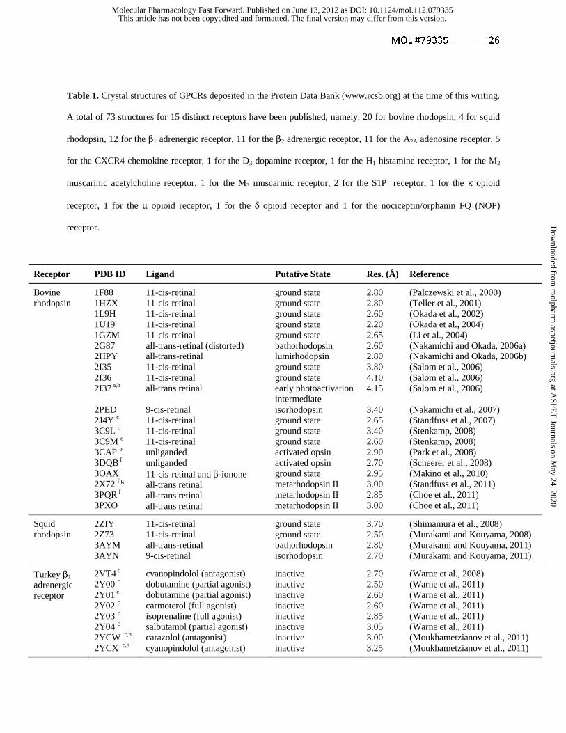
Table 1. Crystal structures of GPCRs deposited in the Protein Data Bank (www.rcsb.org) at the time of this writing.
A total of 73 structures for 15 distinct receptors have been published, namely: 20 for bovine rhodopsin, 4 for squid
rhodopsin, 12 for the β1 adrenergic receptor, 11 for the β2 adrenergic receptor, 11 for the A2A adenosine receptor, 5
for the CXCR4 chemokine receptor, 1 for the D3 dopamine receptor, 1 for the H1 histamine receptor, 1 for the M2
muscarinic acetylcholine receptor, 1 for the M3 muscarinic receptor, 2 for the S1P1 receptor, 1 for the κ opioid
receptor, 1 for the μ opioid receptor, 1 for the δ opioid receptor and 1 for the nociceptin/orphanin FQ (NOP)
receptor.
Receptor PDB ID Ligand Putative State Res. (Å) Reference
Bovine rhodopsin
1F88 1HZX 1L9H 1U19 1GZM 2G87 2HPY 2I35 2I36 2I37 a,b 2PED 2J4Y c 3C9L d 3C9M e 3CAP b 3DQB f 3OAX
2X72 f,g
3PQR f 3PXO
11-cis-retinal 11-cis-retinal 11-cis-retinal 11-cis-retinal 11-cis-retinal all-trans-retinal (distorted) all-trans-retinal 11-cis-retinal 11-cis-retinal all-trans retinal 9-cis-retinal 11-cis-retinal 11-cis-retinal 11-cis-retinal unliganded unliganded 11-cis-retinal and β-ionone all-trans retinal all-trans retinal all-trans retinal
ground state ground state ground state ground state ground state bathorhodopsin lumirhodopsin ground state ground state early photoactivation intermediate isorhodopsin ground state ground state ground state activated opsin activated opsin ground state metarhodopsin II metarhodopsin II metarhodopsin II
2.80 2.80 2.60 2.20 2.65 2.60 2.80 3.80 4.10 4.15 3.40 2.65 3.40 2.60 2.90 2.70 2.95 3.00 2.85 3.00
(Palczewski et al., 2000) (Teller et al., 2001) (Okada et al., 2002) (Okada et al., 2004) (Li et al., 2004) (Nakamichi and Okada, 2006a) (Nakamichi and Okada, 2006b) (Salom et al., 2006) (Salom et al., 2006) (Salom et al., 2006) (Nakamichi et al., 2007) (Standfuss et al., 2007) (Stenkamp, 2008) (Stenkamp, 2008) (Park et al., 2008) (Scheerer et al., 2008) (Makino et al., 2010) (Standfuss et al., 2011) (Choe et al., 2011) (Choe et al., 2011)
Squid rhodopsin
2ZIY 2Z73 3AYM 3AYN
11-cis-retinal 11-cis-retinal all-trans-retinal 9-cis-retinal
ground state ground state bathorhodopsin isorhodopsin
3.70 2.50 2.80 2.70
(Shimamura et al., 2008) (Murakami and Kouyama, 2008) (Murakami and Kouyama, 2011) (Murakami and Kouyama, 2011)
Turkey β1 adrenergic receptor
2VT4 c 2Y00 c 2Y01 c 2Y02 c 2Y03 c
2Y04 c 2YCW c,h 2YCX c,h
cyanopindolol (antagonist) dobutamine (partial agonist) dobutamine (partial agonist) carmoterol (full agonist) isoprenaline (full agonist) salbutamol (partial agonist) carazolol (antagonist) cyanopindolol (antagonist)
inactive inactive inactive inactive inactive inactive inactive inactive
2.70 2.50 2.60 2.60 2.85 3.05 3.00 3.25
(Warne et al., 2008) (Warne et al., 2011) (Warne et al., 2011) (Warne et al., 2011) (Warne et al., 2011) (Warne et al., 2011) (Moukhametzianov et al., 2011) (Moukhametzianov et al., 2011)
This article has not been copyedited and formatted. The final version may differ from this version.Molecular Pharmacology Fast Forward. Published on June 13, 2012 as DOI: 10.1124/mol.112.079335
at ASPE
T Journals on M
ay 24, 2020m
olpharm.aspetjournals.org
Dow
nloaded from

MOL #79335 27
2YCY c 2YCZ c
4AMI c
4AMJ c
cyanopindolol (antagonist) iodocyanopindolol (antagonist) bucindolol (biased agonist) carvedilol (biased agonist)
inactive inactive inactive inactive
3.15 3.65 3.20 2.30
(Moukhametzianov et al., 2011) (Moukhametzianov et al., 2011) (Warne et al., 2012) (Warne et al., 2012)
Human β2 adrenergic receptor
2R4R i,a 2R4S i,a 2RH1 j,b
3D4S j 3KJ6 i,a 3NY8 j 3NY9 j 3NYA j 3PDS j 3P0G j,k
3SN6 j,k,l
carazolol (inverse agonist) l carazolol (inverse agonist) l carazolol (inverse agonist) timolol (inverse agonist) carazolol (inverse agonist) ICI 118551 (inverse agonist) recent comp. (inverse-agonist) alprenolol (antagonist) FAUC50 (irreversible agonist) BI-167107 (agonist) BI-167107 (agonist)
inactive inactive inactive inactive inactive inactive inactive inactive inactive activated activated
3.40 3.40 2.40 2.80 3.40 2.84 2.84 3.16 3.50 3.50 3.20
(Rasmussen et al., 2007) (Rasmussen et al., 2007) (Cherezov et al., 2007; Rosenbaum et al., 2007) (Hanson et al., 2008) (Bokoch et al., 2010) (Wacker et al., 2010) (Wacker et al., 2010) (Wacker et al., 2010) (Rosenbaum et al., 2011) (Rasmussen et al., 2011a) (Rasmussen et al., 2011b)
Human A2A adenosine receptor
3EML j 2YDO c 2YDV c
3QAK j
3PWH c 3REY c 3RFM c
3VG9 m 3VGAm
3UZA c 3UZC c
ZM241385 (antagonist) adenosine (agonist) NECA (agonist) UK-432097 ZM241385 (antagonist) XAC (antagonist) caffeine (antagonist) ZM241385 (antagonist) ZM241385 (antagonist) 1,2,4-triazine 4e (antagonist) 1,2,4-triazine 4g (antagonist)
inactive inactive inactive activated inactive inactive inactive inactive inactive inactive inactive
2.60 3.00 2.60 2.71 3.30 3.31 3.60 2.70 3.10 3.27 3.24
(Jaakola et al., 2008) (Lebon et al., 2011) (Lebon et al., 2011) (Xu et al., 2011) (Dore et al., 2011) (Dore et al., 2011) (Dore et al., 2011) (Hino et al., 2012) (Hino et al., 2012) (Congreve et al., 2012) (Congreve et al., 2012)
Human CXCR4 chemokine receptor
3ODU j,b 3OE9 j,b 3OE8 j,b 3OE6 j,b 3OE0 j,b
IT1t (small mol. antagonists) IT1t (small mol. antagonists) IT1t (small mol. antagonists) IT1t (small mol. antagonists) CVX15 (peptide antagonist)
Inactive inactive inactive inactive inactive
2.50 3.10 3.10 3.20 2.90
(Wu et al., 2010) (Wu et al., 2010) (Wu et al., 2010) (Wu et al., 2010) (Wu et al., 2010)
Human D3
dopamine receptor
3PBLj eticlopride (antagonist) inactive 2.89 (Chien et al., 2010)
Human H1 histamine receptor
3RZE j doxepin (antagonist) inactive 3.10 (Shimamura et al., 2011)
Human M2 Muscarinic receptor
3UON j 3-quinuclidinyl-benzilate (antagonist)
inactive 3.00 (Haga et al., 2012)
Rat M3 Muscarinic receptor
4DAJ j Tiotropium (inverse agonist) inactive 3.40 (Kruse et al., 2012)
Human S1P1 sphingosine 1-phosphate receptor
3V2Y j,n 3V2W j
ML056 (antagonist) ML056 (antagonist)
inactive inactive
2.80 3.35
(Hanson et al., 2012) (Hanson et al., 2012)
Human κ 4DJH h,k JDTic (antagonist) inactive 2.90 (Wu et al., 2012)
This article has not been copyedited and formatted. The final version may differ from this version.Molecular Pharmacology Fast Forward. Published on June 13, 2012 as DOI: 10.1124/mol.112.079335
at ASPE
T Journals on M
ay 24, 2020m
olpharm.aspetjournals.org
Dow
nloaded from

MOL #79335 28
a Ligand not visible. b Potentially biologically relevant dimer observed in the structure c Thermally stable mutant receptor. d Alternative model of 1GZM. e Alternative model of 2J4Y. f In complex with a C-terminal peptide of the α-subunit of transducin. g Constitutively active mutant
h Showing an intact salt bridge linking the cytoplasmic ends of TMs 3 and 6 i In complex with a Fab. j T4-lysozyme fusion protein. k In complex with a camelid antibody fragment l In complex with a G protein (Gs) heterotrimer m In complex with a Fab that prevents agonist binding. n Processed with a microdiffraction data assembly method. o Fusion protein with thermostabilized apocytochrome b562RIL (BRIL).
opioid receptor
Mouse μ opioid receptor
4DKL j,b β-funaltrexamine (irreversible
antagonist) inactive 2.80 (Manglik et al., 2012)
Mouse δ opioid receptor
4EJ4 j,b Naltrindole (antagonist) inactive 3.40 (Granier et al., 2012)
Human nociceptin/ orphanin FQ (NOP) receptor
4EA3 o Peptide mimetic c-24 (antagonist)
inactive 3.01 (Thompson et al., 2012)
This article has not been copyedited and formatted. The final version may differ from this version.Molecular Pharmacology Fast Forward. Published on June 13, 2012 as DOI: 10.1124/mol.112.079335
at ASPE
T Journals on M
ay 24, 2020m
olpharm.aspetjournals.org
Dow
nloaded from

Nucleotideand lipid receptors
Biogenic aminesand MECA receptors
Chemokinereceptors
Opsins
A2A
S1P1
Rhodopsin
β2
β1
D3
CXCR4
H1
M3 M2
κ
Neuropeptides
μδNOP
Figure 1
This article has not been copyedited and form
atted. The final version m
ay differ from this version.
Molecular Pharm
acology Fast Forward. Published on June 13, 2012 as D
OI: 10.1124/m
ol.112.079335 at ASPET Journals on May 24, 2020 molpharm.aspetjournals.org Downloaded from

Rhodopsin β2 A2A
M3 CXCR4 S1P1
Figure 2
This article has not been copyedited and form
atted. The final version m
ay differ from this version.
Molecular Pharm
acology Fast Forward. Published on June 13, 2012 as D
OI: 10.1124/m
ol.112.079335 at ASPET Journals on May 24, 2020 molpharm.aspetjournals.org Downloaded from
Rhodopsin β2 A2A
M3 S1P1CXCR4
TM5
EL2
TM6
TM7
TM3 TM5
EL2
TM6
TM7
TM3
TM6
TM7
EL2
EL3
TM5
TM5
TM6
TM7
TM3
EL2
TM2
TM3
NTNT
TM2
TM3
TM6
EL2
TM5
hiddenby ligand
hiddenby ligand
TM7
TM3
TM4
TM7
Figure 3
This article has not been copyedited and form
atted. The final version m
ay differ from this version.
Molecular Pharm
acology Fast Forward. Published on June 13, 2012 as D
OI: 10.1124/m
ol.112.079335 at ASPET Journals on May 24, 2020 molpharm.aspetjournals.org Downloaded from
a b
Figure 4
This article has not been copyedited and form
atted. The final version m
ay differ from this version.
Molecular Pharm
acology Fast Forward. Published on June 13, 2012 as D
OI: 10.1124/m
ol.112.079335 at ASPET Journals on May 24, 2020 molpharm.aspetjournals.org Downloaded from

a b
Figure 5
This article has not been copyedited and form
atted. The final version m
ay differ from this version.
Molecular Pharm
acology Fast Forward. Published on June 13, 2012 as D
OI: 10.1124/m
ol.112.079335 at ASPET Journals on May 24, 2020 molpharm.aspetjournals.org Downloaded from

a b
c d
Figure 6
This article has not been copyedited and form
atted. The final version m
ay differ from this version.
Molecular Pharm
acology Fast Forward. Published on June 13, 2012 as D
OI: 10.1124/m
ol.112.079335 at ASPET Journals on May 24, 2020 molpharm.aspetjournals.org Downloaded from