Embed Size (px)
Citation preview

Medical Journal of Babylon-Vol. 11- No. 3 -2014 مجلة بابل الطبیة- المجلد الحادي عشر - العدد الثالث- ٢٠١٤
567
Received 8 December 2013 Accepted 13 April 2014 Abstract The carpal tunnel syndrome (CTS) is one of the most common peripheral neuropathies and is caused by the compression of the median nerve at the wrist region. The aim of this study was to analyze the results of patients who were operated by using 1.5 cm palmer skin incision. (228) carpal tunnel release operations were performed on (181) patients. These patients were assessed pre and postoperatively using Visual Analog Scale (VAS) and Visual Analog Patient Satisfaction Scale (VAPSS) [1]. The (1.5) cm palmer skin incision technique for carpal tunnel release is a safe and effective surgical procedure. It can be used in the surgical treatment of CTS to achieve better cosmetic results and to reduce the complications of other standard techniques. Keyword: carpal tunnel syndrome (CTS), peripheral neuropathies Visual Analog Scale (VAS) and Visual Analog Patient Satisfaction Scale (VAPSS), Erbil.
:ةالخالصتهدف هذه الدراسه الى . انضغاط العصب الوسطي خالل مروره في النفق الرسغي یعتبر من اكثر المتالزمات العصبیة المحیطیة شیوعا
تبین بعد تحلیل . بیان مدى اهمیه طریقة تحریر العصب الوسطي من خالل عملیة جراحیة بطول واحد ونصف سنتیمتر في كف الیدمائتي وواحد وثمانون مریض بان هذه الطریقة هي طریقه امینة ولها تتائج مرضیه للمرضى من نواحي الشفاء نتائج هذه العملیة ل
.السریري وجمالیه اثر العملیة .اربیل. تحریر العصب الوسطي. المتالزمات العصبیة المحیطیةانضغاط العصب الوسطي :الكلمات المفتاحیة
ـ ـــــ ـــــ ـ ــ ـــ ـــــــ ــــ ــــــ ــــ ــــــ ـــــ ــــــ ـــــ ــ ــــ ـــــ ــ ــ ــ ــــــ ــ ـــ ــــــ ـــــ ــــــ ــــــ ـ ــ ــ ــــــ ـ ــ ـــ ـــــ ــ ــــ ــــــ ـــــ ــــــ ــــــ ــ ــــــ ـــ ــ ــــــ ــ ــ ــ ــــــ ـــــ ــــــ ـــــ ـــ ـــ ـــــ ـــ ــ ـــــــ ـ ــ ــ ــــــ ـــــ ــــــ ــــــ ــ ــ ـــــــ ــ ــ ــ ـــــ ـ ــ ــــ ــــــ ــ ــــــ ــــــ ـــــ ــــــ ــــ ـــــــ ــــ ــ ـــــ ـــ ــ ــــــ ــــــ ـــــ ــــــ ــــ ــ ـــــ ــــ ــ ــــــ ــ ــ ـــــــ ـــــ ــــــ ـــــــــ ــ ـــ ــــــ ــ ــــ ــــــ ـــــ ــــــ ـــــ ــــــ ـــــ ــــــ ـــــ ــ ــــ ــــــ ـــــ ــــــ ـــــ ــــــ ـــــ ــ ــــ ـــــ ــ ـــــــ ــــــ ــــ ــــ ــــــ ــــIntroduction
he carpal tunnel syndrome (CTS) is the most common peripheral neuropathies and is
caused by the compression of the median nerve at the wrist region as it passes beneath the transverse carpal ligament. Symptoms of CTS include paresthesia (numbness, tingling and burning) in the median nerve distribution (radial 3 and half digits) along with deep aching pain in the hand and wrist[2]. CTS affect 1% and 5% of general population and working
population using their hands and wrists in daily living [3]. CTS mainly affects females aged between 30 and 50 years [4 -6]. Diagnosis of CTS needs interpretation of the history, physical examination and electrophysiological results [7,4]. Patients with mild symptoms of CTS can be managed with conservative treatment including non-steroid anti-inflammatory drugs, vitamin B6, local steroid injections or hand braces [7,8]. While patients with moderate and severe symptoms,
T
Minimal Invasive Surgery for Carpal Tunnel Syndrome using 1.5 cm Palmer Skin Incision
Sherwan Hamawandi
Collage of Medicine, Hawler Medical University, Erbil, IRAQ. E-mail: [email protected]

Medical Journal of Babylon-Vol. 11- No. 3 -2014 مجلة بابل الطبیة- المجلد الحادي عشر - العدد الثالث- ٢٠١٤
568
surgical treatment is generally needed [4, 8]. The first open carpal tunnel release was performed in 1924 by Mackinnon et al. in Mayo Clinic and popularized later by Phalen et al.[9]. Standard open carpal tunnel release with a long palmer curvilinear incision still remains to be the preferred surgical procedure for many departments and orthopedic surgeons [10,11], but this procedure has many complications including pillar pain, scar tenderness, cosmetic problems, loss of grip and pinch strength or time losses due to inability to work [11-13]. Endoscopic techniques and different mini skin incisions are described in the literature to decrease the risk of such complications [12,14-16]. Although endoscopic surgery tries to decrease complications related to the incision, it has risk of injury to the superficial palmar arc, median nerve, digital nerves, vessels and flexor tendons and insufficient release of the carpal tunnel. Thus, the advantages of endoscopic carpal tunnel release must be balanced against potential injury to adjacent neurovascular structures. In addition, the endoscopic technique has a long learning curve, and it is costy due to the special equipment required for this procedure [17]. Aims of study: The aim of this study was to analyze the results of patients who were operated by using 1.5 cm palmer skin incision. Materials and Methods Between October (2010) and December (2012), we performed (228) carpal tunnel releasing procedures on (181) consecutive patients. There were 171 (94 %) female and 10 (6 %) male patients with a mean age of (49) (ranging from 22 to 75). 132 operations were performed on the right hand and (96) were performed on the left. The
mean follow-up period was (17) months (ranging from 4 to 31 months). The operations were done in Erbil teaching hospital, Zheen International Hospital and Hawler Private Hospital. Each patient was evaluated with his/her history, physical examination and electromyelography (EMG). Night pain and numbness were observed in all patients. EMG showed moderate and severe CTS in 212 hands (93%). The pain status of the patients was pre and postoperatively assessed with the Visual Analog Scale (VAS). Incision scar hypersensitivity and cosmetic results were evaluated with the Visual Analog Patient Satisfaction Scale (VAPSS) postoperatively. Informed consent was obtained from each patient. Surgical Technique All the patients and their families were informed about the operative technique (length of incision) and postoperative care before the operation. All the operations were performed in the operating room under sterile conditions in the supine position. Before surgery, the affected hand, wrist and forearm were cleaned with povidone iodine solution. The area to be operated was covered with a sterile compress. Pneumatic tourniquet was used. The hand should be in an extended position. After these routine operation preparations, a longitudinal (1.5) cm long incision was performed at the wrist region, distal to the distal flexion crease, between the third and fourth finger as showed in Figure (1). The incised skin was retracted with the help of a mini retractor and subcutaneous fat tissue was dissected laterally. A small opening done in the carpal ligament with a fine scissors or surgical blade and a dissector was introduced beneath the carpal ligament and then the ligament was cut with surgical blade and scissors as showed in Figure (2). After the homeostasis,

Medical Journal of Babylon-Vol. 11- No. 3 -2014 مجلة بابل الطبیة- المجلد الحادي عشر - العدد الثالث- ٢٠١٤
569
the skin was sutured with 4/0 sutures mattress half buried as showed in Figure (3). The mean operation time was (15) minutes (ranging between 10-20 minutes). The mean hospital stay was 3 hours (2-4 hours). Results In this study, (228) carpal tunnel release operations were performed on (181) patients. There were no complications during the operations such as bleeding or nerve injury. The mean follow up period was (17) months and no procedure related complication was observed such as skin infection and palmar tenderness. The mean pre-operative VAS score for pain was 8.5, which decreased to (2.1) postoperatively. We used the Visual Analog Patient Satisfaction Scale described by Kılıncer and Zileli in 2006 [1] to evaluate the patients for cosmetic results, return to daily routine activities, palmar tenderness and scar sensitivity. Mean VAPSS score was (8.7) during the follow up period. In this study, (7) patients had temporary paraesthesia and (1) patient was repeated because of pain recurrence and paraesthesia. For such a patient we started a limited palmer skin incision, which was later extended to (3) cm because of the scar tissue. No problem occurred during the follow-up period (mean 4 months). (3) patients were developed scar tenderness which was treated by conservative measures. Discussion In this study, just like in the literatures, 94% of the patients were female with a mean age of 49 [4, 7,18]. Until recent years, standard incision with a long curvilinear incision was the most performed technique for CTS by many orthopedic and neurosurgeons. This technique is safe and effective as reported by authors, but it has some complications [4, 7, 10].
The complications of the standard incision including incomplete release of carpal ligament, injury to the palmar cutaneous and recurrent motor branch of median nerve or injury to the superficial palmar arch and ulnar artery are rare because the operation is performed under direct vision [7]. Some complications, on the other hand, have a relatively high incidence. These are hypertrophic scar formation, scar tenderness, pillar pain, loss of grip strength and sympathetic dystrophy resulting in the delay of returning to daily activities or work and emotional distress [7,12]. To reduce these complications, various limited incisions or endoscopic techniques are described by authors [4,14,19-24]. Some researchers have claimed that the endoscopic carpal tunnel release (ECTR) decreased the postoperative morbidity of standard open carpal tunnel release.[25-27] In previous studies, patients who underwent ECTR had less pillar pain, faster recovery of grip and pinch strength, and earlier return to daily activities and work than those who underwent nonendoscopic treatments In spite of the many advantages of endoscopic techniques, there are also some disadvantages; the difficulty of inserting a relatively large device through a narrow tunnel, nerve ischemia due to the use of tourniquet for a long time, performing transverse incision that might damage the superficial palmar arch and the experience needed for such an operation [28]. Also it is reported that the most common complications of these techniques are paresthesia of the median and ulnar nerves, tendon lacerations and injury to the arteries [7,13]. Some authors have also reported multiple limited mini open incision techniques to decrease the postoperative morbidity observed in standard open techniques [2,12, 11, 16,
Medical Journal of Babylon-Vol. 11- No. 3 -2014 مجلة بابل الطبیة- المجلد الحادي عشر - العدد الثالث- ٢٠١٤
570
19,22-24]. Mini open procedures have been performed using either a longitudinal incision on the wrist and/or palmar surface, or a transverse wrist incision. These mini open incision techniques represent the midway between the standard open incision and endoscopic technique taking the advantages of both with less disadvantages. In this study, we aimed to analyze the outcome of patients operated for carpal tunnel syndrome using (1.5) cm palmer skin incision. The incision is performed at the palmer region distally to the flexion crease; therefore, we thought that the complications including cosmetic problems, palmer tenderness and scar sensitivity would be less, as showed in Figure (4). In this study, we saw few complications caused by the type of incision, and the mean VAPSS score was (8.7) when the patients were evaluated for cosmetic results, return to daily routine activities, palmer tenderness and scar sensitivity. In this study, 132 (92 %) of patients have complete remission of symptoms which is near the results of literatures of mini open incisions [4,28,29]. In this study, there were (1) reoperation because of the recurrence of symptoms and (8) patients had temporary paresthesia; such patients gave history of excessive hand working after surgery. The palmar arteries, and particularly the superficial palmar arch, also have a potential risk due to the difficulties in visualization with these limited incisions or endoscopic techniques. However, just like the studies [4,12,18,28,29] we also did not experience any artery, nerve or tendon injury. Conclusion The (1.5) cm palmer skin incision technique for carpal tunnel release is a safe and effective surgical procedure. It can be used in the surgical treatment of
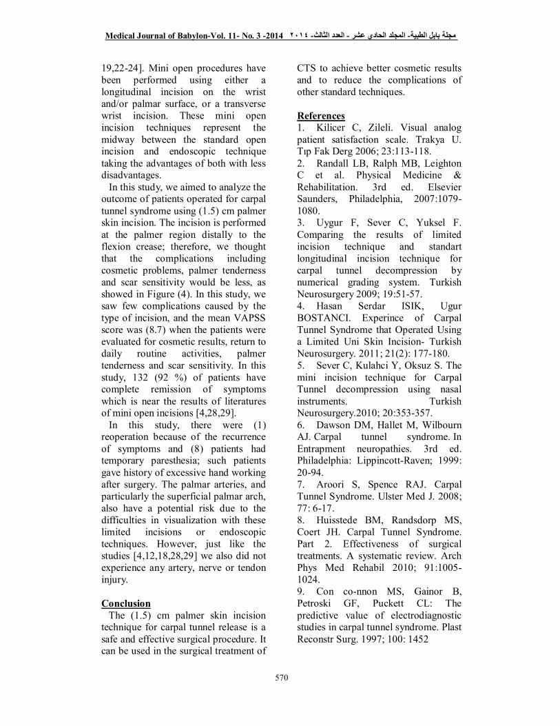
CTS to achieve better cosmetic results and to reduce the complications of other standard techniques. References 1. Kilicer C, Zileli. Visual analog patient satisfaction scale. Trakya U. Tıp Fak Derg 2006; 23:113-118. 2. Randall LB, Ralph MB, Leighton C et al. Physical Medicine & Rehabilitation. 3rd ed. Elsevier Saunders, Philadelphia, 2007:1079-1080. 3. Uygur F, Sever C, Yuksel F. Comparing the results of limited incision technique and standart longitudinal incision technique for carpal tunnel decompression by numerical grading system. Turkish Neurosurgery 2009; 19:51-57. 4. Hasan Serdar ISIK, Ugur BOSTANCI. Experince of Carpal Tunnel Syndrome that Operated Using a Limited Uni Skin Incision- Turkish Neurosurgery. 2011; 21(2): 177-180. 5. Sever C, Kulahci Y, Oksuz S. The mini incision technique for Carpal Tunnel decompression using nasal instruments. Turkish Neurosurgery.2010; 20:353-357. 6. Dawson DM, Hallet M, Wilbourn AJ. Carpal tunnel syndrome. In Entrapment neuropathies. 3rd ed. Philadelphia: Lippincott-Raven; 1999: 20-94. 7. Aroori S, Spence RAJ. Carpal Tunnel Syndrome. Ulster Med J. 2008; 77: 6-17. 8. Huisstede BM, Randsdorp MS, Coert JH. Carpal Tunnel Syndrome. Part 2. Effectiveness of surgical treatments. A systematic review. Arch Phys Med Rehabil 2010; 91:1005-1024. 9. Con co-nnon MS, Gainor B, Petroski GF, Puckett CL: The predictive value of electrodiagnostic studies in carpal tunnel syndrome. Plast Reconstr Surg. 1997; 100: 1452
Medical Journal of Babylon-Vol. 11- No. 3 -2014 مجلة بابل الطبیة- المجلد الحادي عشر - العدد الثالث- ٢٠١٤
571
10. Badger SA, O’Donnel ME, Sherigor JM, Conolly P, Spence RA. Open Carpal Tunnel release, still safe and effective operation. Ulster Med J. 2008; 77:22-24. 11. Lida J, Hirabayashi H, Nakase H, Sakaki T. Carpal Tunnel Syndrome: Electrophysiological grading and surgical results by minimum incision open carpal tunnel release. Neurol Med Chir (Tokyo). 2008; 48:554-559. 12. Aydın K, Cokluk C, Cengiz N, Bilgici A. (2006). Microsurgical open mini uniskin incision technique in the surgical treatment of Carpal Tunnel Syndrome. Neurology India. 2006; 54: 64-67. 13. Einhorn N, Leddy JP: Pitfalls of endoscopic carpal tunnel release. Orthop Clin North Am. 1996; 27:373 14. Agee JM, Mc Caroll HR, North ER. Endoscopic Carpal Tunnel release using the single proximal incision technique. Hand Clin. 1994;10: 647-659. 15. Agee JM, Peimer CA, Pyret CD. Endoscopic Carpal Tunnel release: A prospective study of complications and surgical experience. J Hand Surg Am. 1995; 20:165-171. 16. Cellocco P, Rossi C, Bizzarri F. Mini-open blind procedure versus limited open technique for Carpal Tunnel release: A 30-month follow-up study. Journal Hand Surgery. 2005; 30:493-499. 17. Okada M, Tsubata O, Yasumoto S, Toda N, Matsumoto T: Clinical study of surgical treatment of carpal tunnel syndrome: Open versus endoscopic technique. Journal of Orthopaedic Surgery. 2000; 8(2) : 19-25 18. Aydın K, Cokluk C, Piskin A, Kocabıcak E. Ultrasonographically checking the sectioning of the transverse carpal ligament during Carpal Tunnel Surgery with limited uni skin incisions. Turkish Neurosurgery. 2007; 17:219-223.
19. Avci S, Sayli U. Carpal Tunnel release using a short palmar incision and a new knife. J Hand Surg Br. 2000; 25:357-360. 20. Brown RA, Helbernan RH, Seiler JG, Abrahamsson SO, Weiland AJ, Urbaniak JR, Schoenfeld DA, Furcolo D. Carpal Tunnel release: a prospective randomized assessment of open and endoscopic methods. J Bone Joint Surgery. 1993; 75:1265. 21. Okada M, Tsubata O, Yasumoto S. Clinical study of surgical treatment of Carpal Tunnel Syndrome: Open versus endoscopic technique. Journal of Orthopaedic Surgery. 2000; 8:19-25. 22. Teh KK, Ng ES, Choon SK. Mini Open Carpal Tunnel release using knifelight: Evaluation of the safety and effectiveness of using a single wrist incision (cadaveric study). J Hand Surg Eur.2009; 34E: 506-510. 23. Trumble TE, Diao E, Abrams RA. Single-portal endoscopic Carpal Tunnel release compared with open release: A prospective randomized trial. J Bone Joint Surg Am. 2002; 84:1107-1115. 24. Wongsiri S, Suwanno P, Tangtrakulwanich B. (2008). A new tool for mini Open Carpal Tunnel release - The PSU retractor. BMC Musculoskeletal Disorders. 2008; 9: 126. 25. Agee JM, McCarroll HR, Tortosa RD, Berry DA, Szabo RM, Peimer CA. Endoscopic release of the carpal tunnel: a randomised prospective multicenter study. J Hand Surg 1992; 17A:987-995. 26. Trumble TE, Gilbert M, McCallister WV. Endoscopic versus open surgical treatment of carpal tunnel syndrome. Neurosurg Clin North Am 2001; 12:255-266. 27. Ferdinand RD, MacLean JG. Endoscopic versus open carpal tunnel release in bilateral carpal tunnel syndrome: a prospective, randomised,

Medical Journal of Babylon-Vol. 11- No. 3 -2014 مجلة بابل الطبیة- المجلد الحادي عشر - العدد الثالث- ٢٠١٤
572
blinded assessment. J Bone Joint Surg Br. 2002; 84:375-379. 28. Nazzi V, Franzini A, Messina G, Broggi G: Carpal Tunnel Syndrome. Matching minimally invasive surgical
techniques, Technical note. J Neurosurgery.2008; 108:1033-1036. 29. Wen-Ching Tzaan, et al. Accurate midpalmar carpal tunnel release. Chang Gung Med J. 2005; 28:97-103
Figure 1 showed the length of skin incision
Figure 2 showed the median nerve after incising the flexor retinaculum
Medical Journal of Babylon-Vol. 11- No. 3 -2014 مجلة بابل الطبیة- المجلد الحادي عشر - العدد الثالث- ٢٠١٤
573
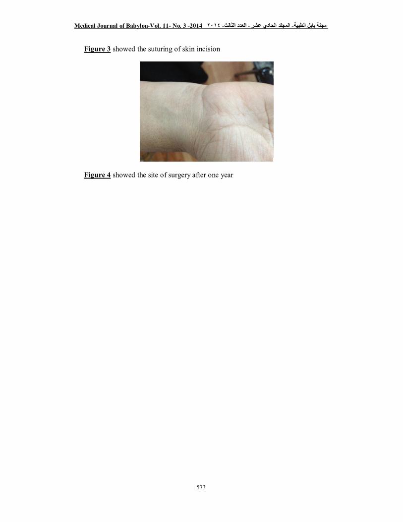
Figure 3 showed the suturing of skin incision
Figure 4 showed the site of surgery after one year