Embed Size (px)
Citation preview
Minimally Invasive Cancer Therapies in Interventional Radiology
Chief, Vascular and Interventional Radiology
Lancaster Radiology AssociatesCo-Director, Interventional Vascular Unit
Objectives
• 1- Identify currently available IR procedures related to cancer care at LGH
• 2- Enhance medical staff knowledge of such procedures
• 3- Discuss current IR cancer treatments
Palliative and curative therapies
• Diagnosis• Lung• Genitourinary• Gastrointestinal
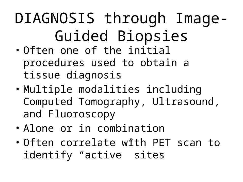
DIAGNOSIS through Image-Guided Biopsies
• Often one of the initial procedures used to obtain a tissue diagnosis
• Multiple modalities including Computed Tomography, Ultrasound, and Fluoroscopy
• Alone or in combination• Often correlate with PET scan to identify
“active” sites
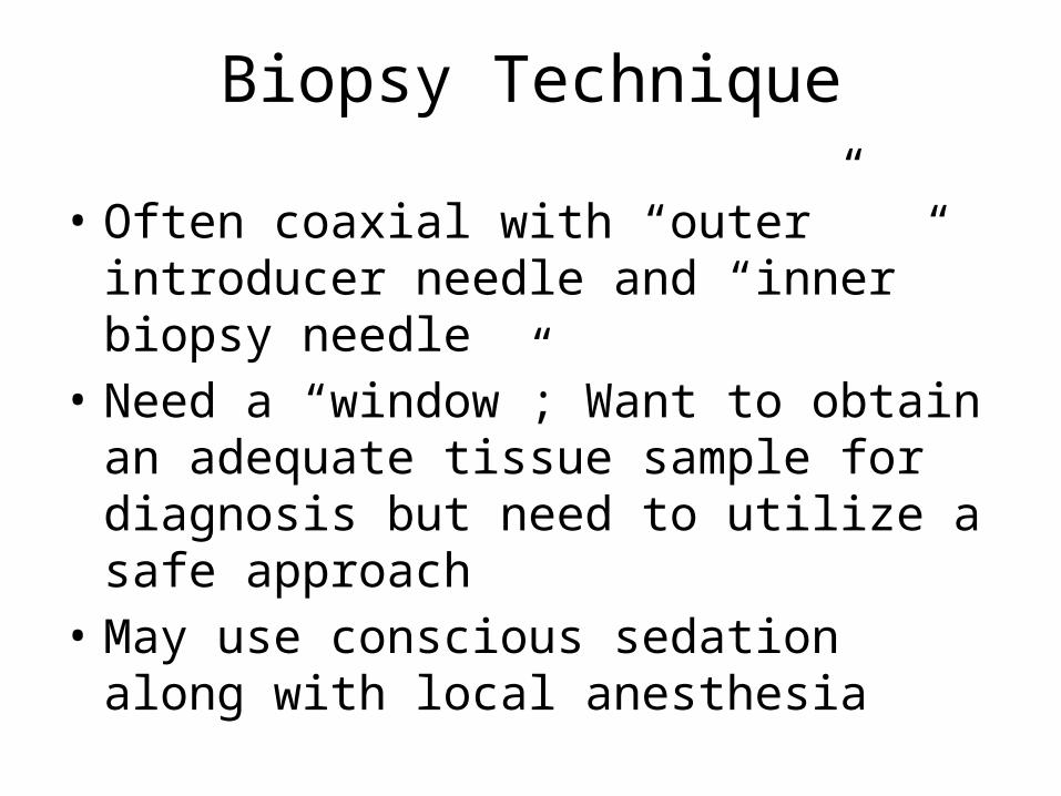
Biopsy Technique
• Often coaxial with “outer” introducer needle and “inner” biopsy needle
• Need a “window”; Want to obtain an adequate tissue sample for diagnosis but need to utilize a safe approach
• May use conscious sedation along with local anesthesia
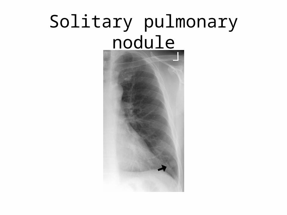
Solitary pulmonary nodule
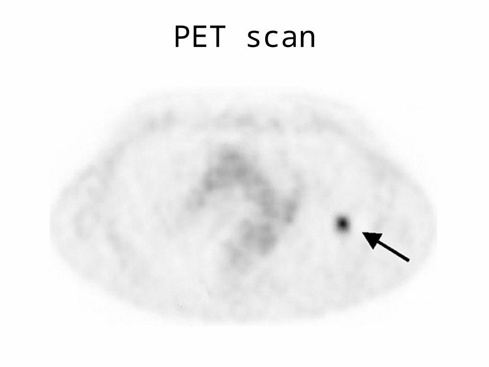
PET scan
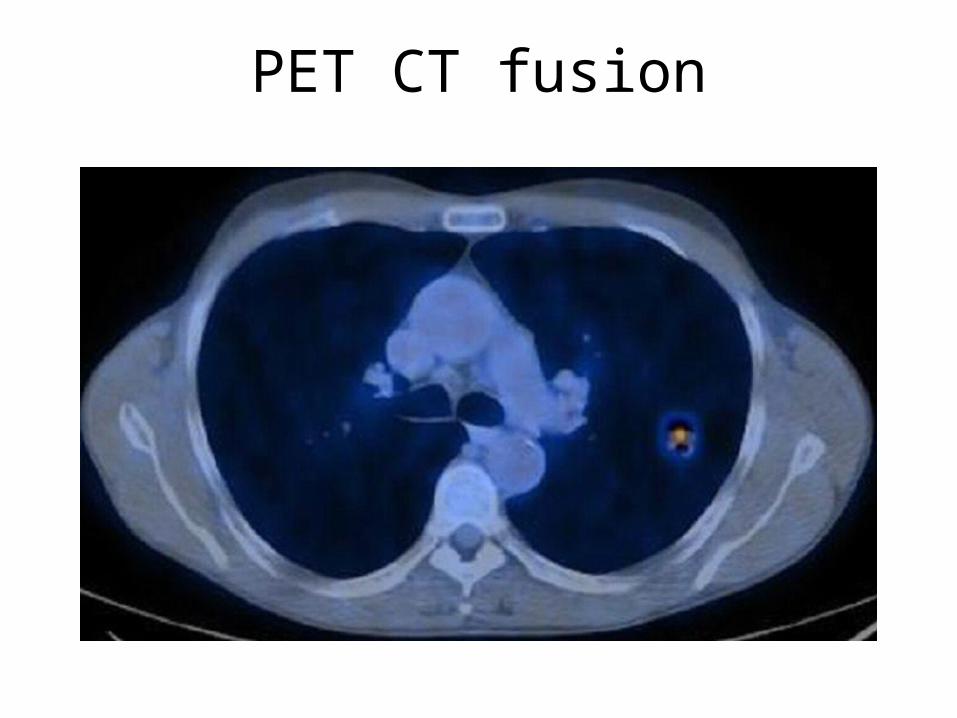
PET CT fusion
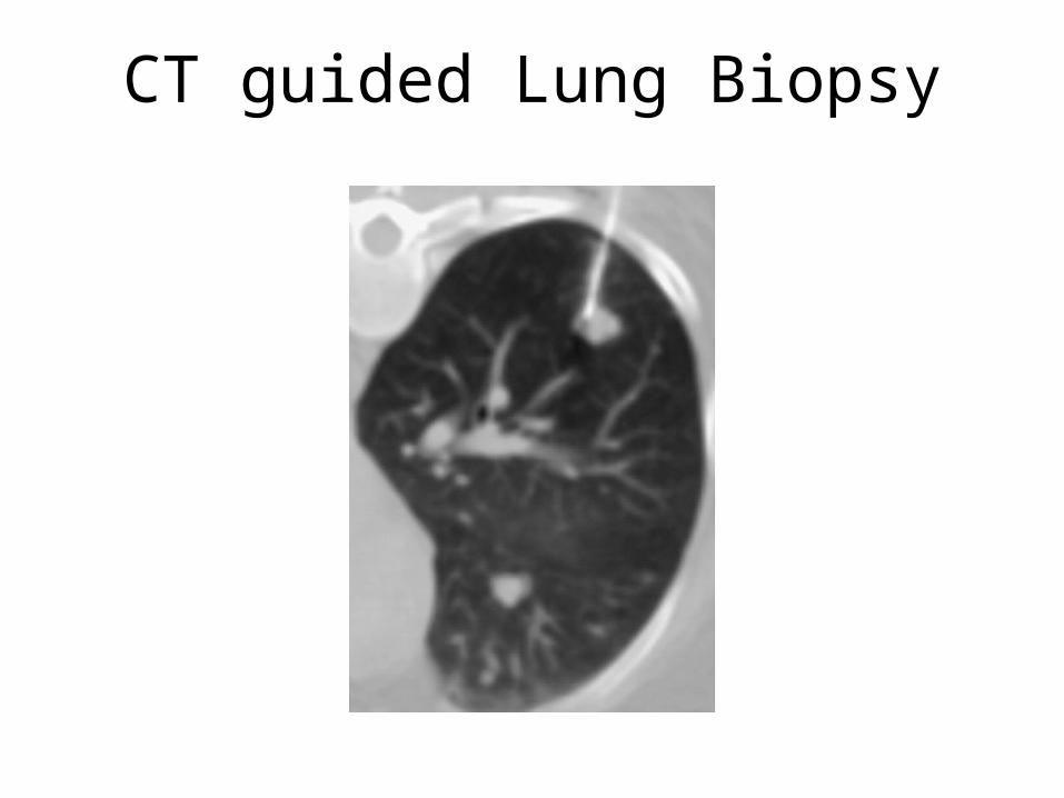
CT guided Lung Biopsy
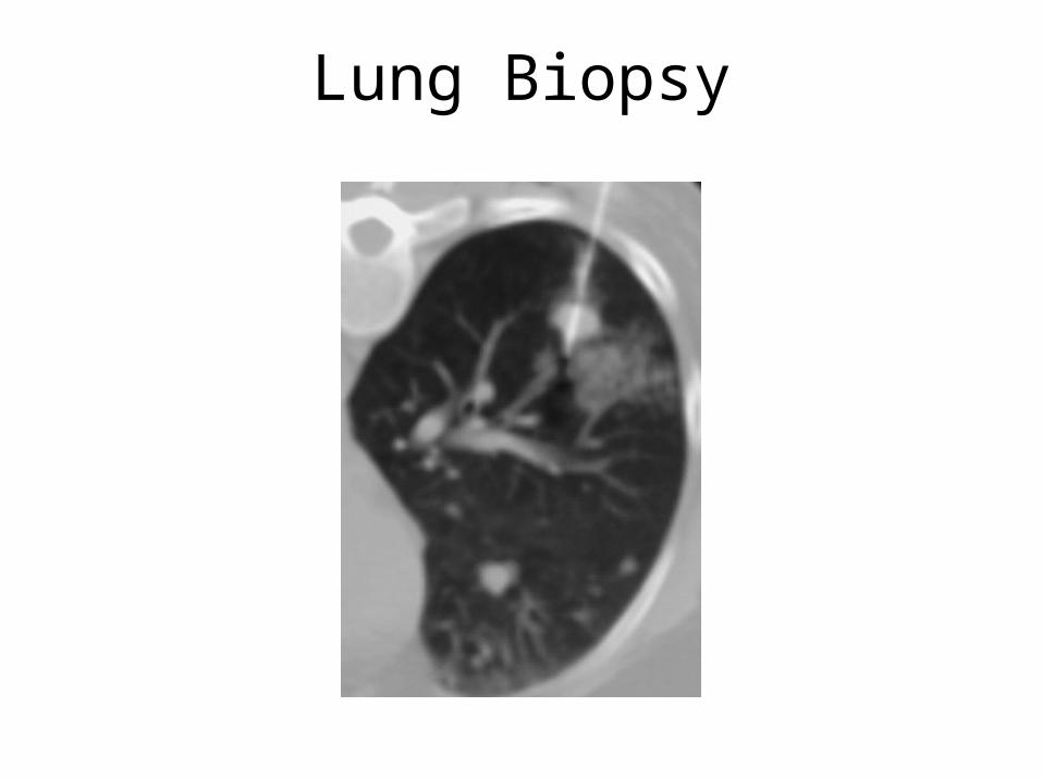
Lung Biopsy
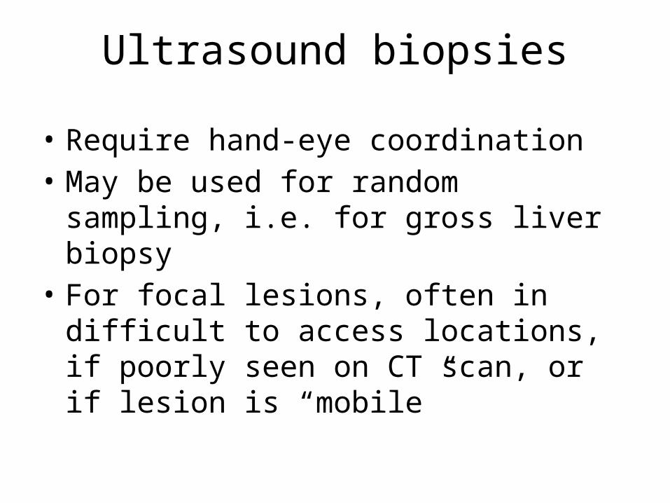
Ultrasound biopsies
• Require hand-eye coordination• May be used for random sampling, i.e. for
gross liver biopsy• For focal lesions, often in difficult to access
locations, if poorly seen on CT scan, or if lesion is “mobile”
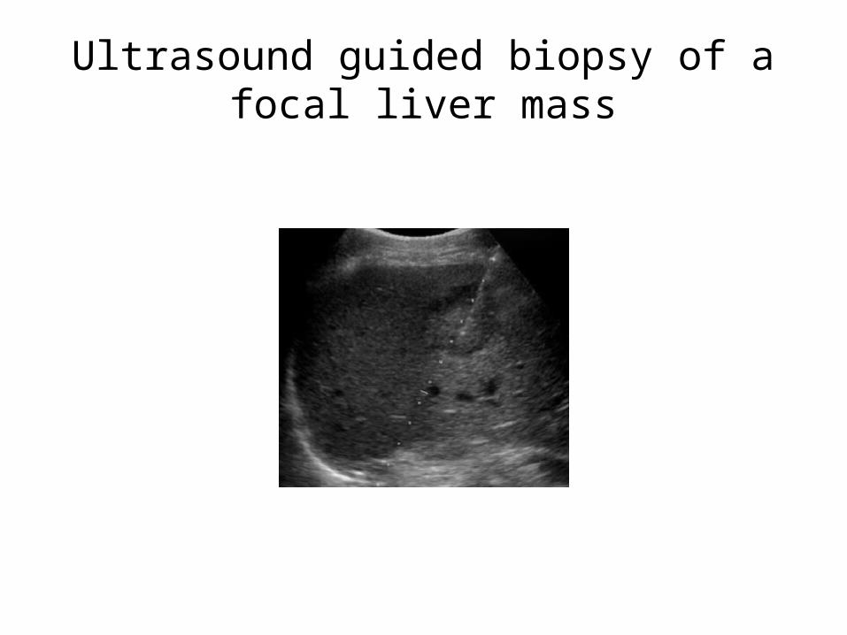
Ultrasound guided biopsy of a focal liver mass
X-ray guided biopsy
• Especially useful when patient positioning is limited; can rotate and angle the tube to obtain an approach for lesion access
• Advantage of real time imaging
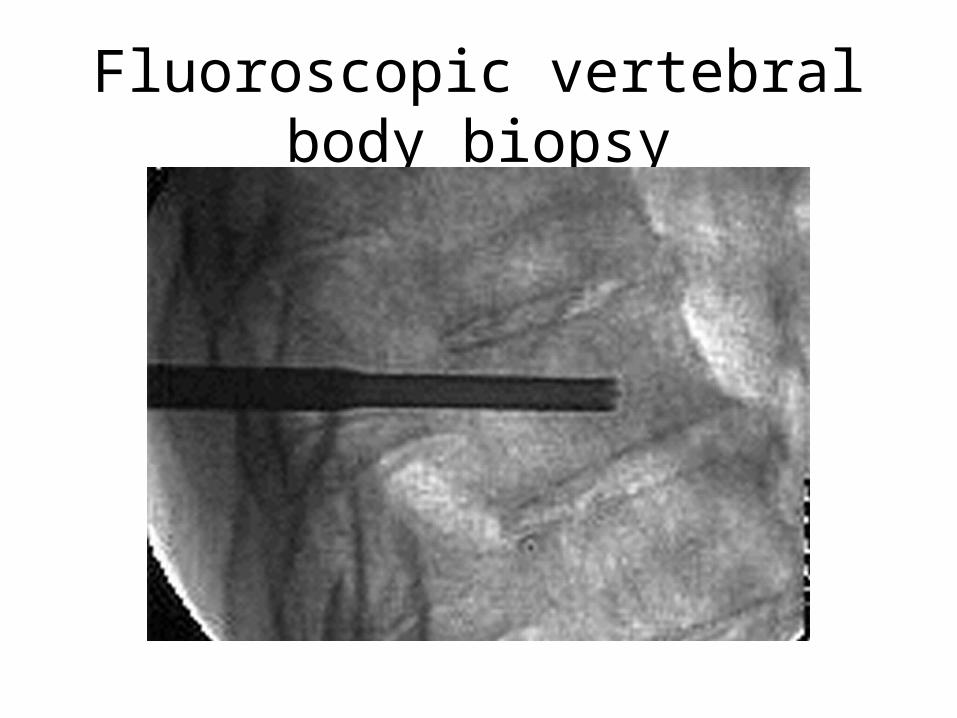
Fluoroscopic vertebral body biopsy
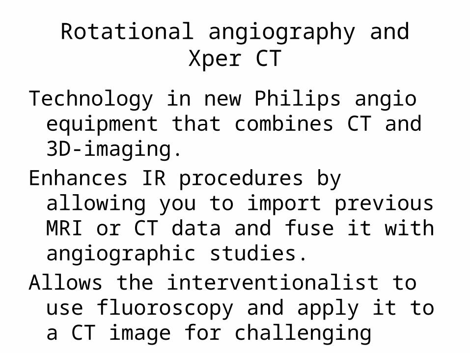
Rotational angiography and Xper CT
Technology in new Philips angio equipment that combines CT and 3D-imaging.
Enhances IR procedures by allowing you to import previous MRI or CT data and fuse it with angiographic studies.
Allows the interventionalist to use fluoroscopy and apply it to a CT image for challenging access.
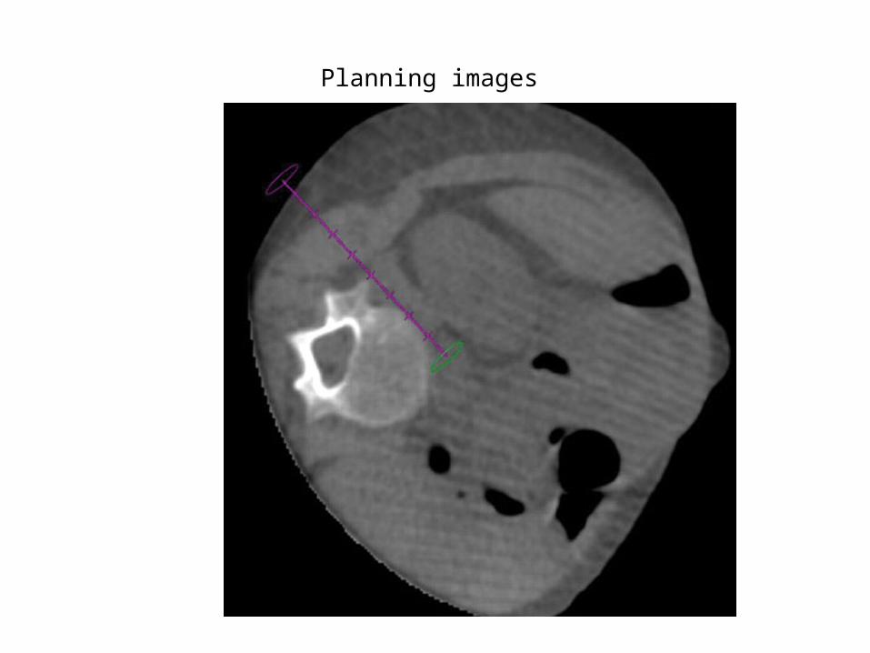
Planning images
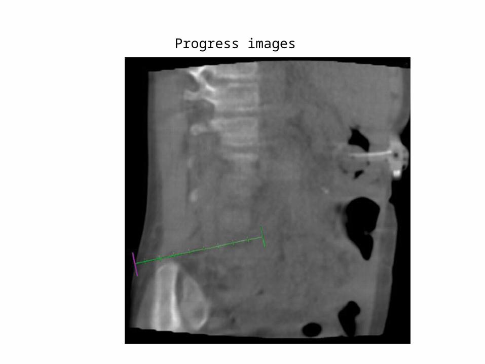
Progress images
Lung
PalliativeTunneled pleural cathetersThermal ablation of destructive chest wall lesions
CurativeRFA of unresectable lung cancers or lung metastases
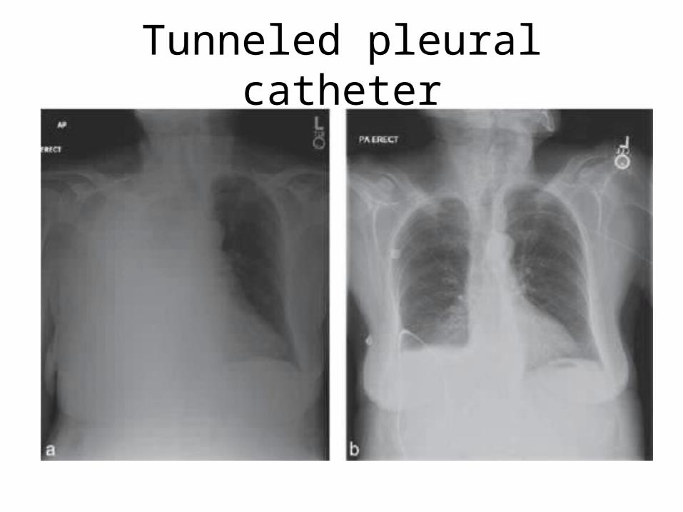
Tunneled pleural catheter
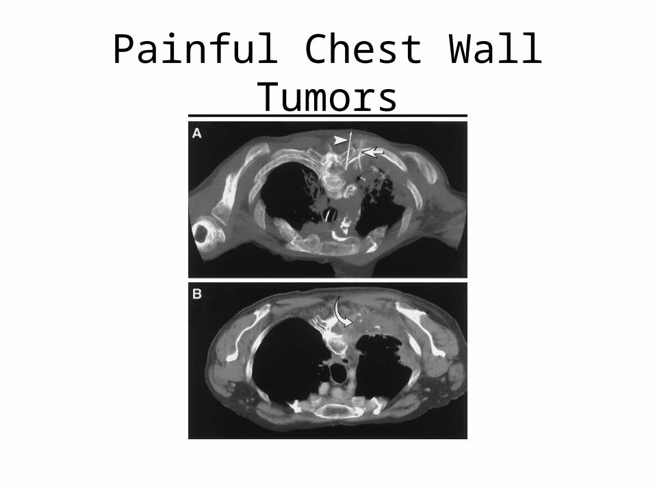
Painful Chest Wall Tumors
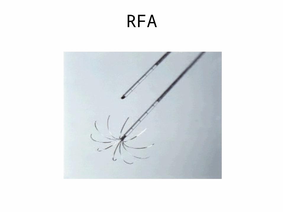
RFA
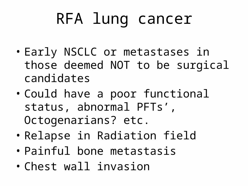
RFA lung cancer
• Early NSCLC or metastases in those deemed NOT to be surgical candidates
• Could have a poor functional status, abnormal PFTs’, Octogenarians? etc.
• Relapse in Radiation field• Painful bone metastasis• Chest wall invasion
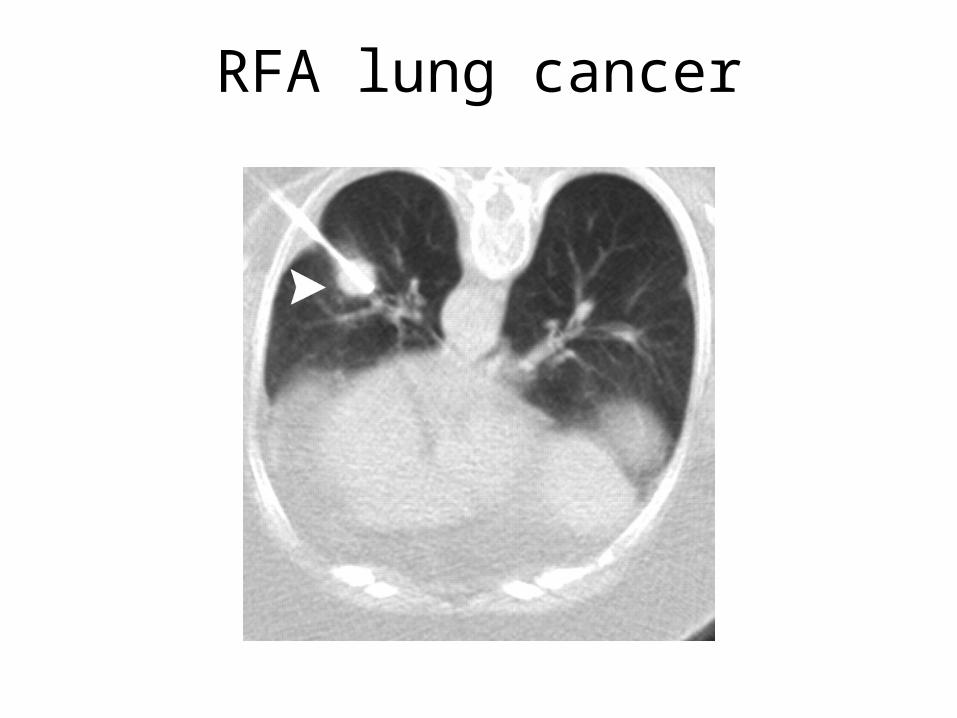
RFA lung cancer
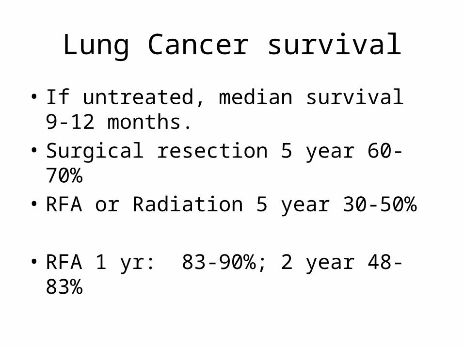
Lung Cancer survival
• If untreated, median survival 9-12 months.• Surgical resection 5 year 60-70%• RFA or Radiation 5 year 30-50%
• RFA 1 yr: 83-90%; 2 year 48-83%
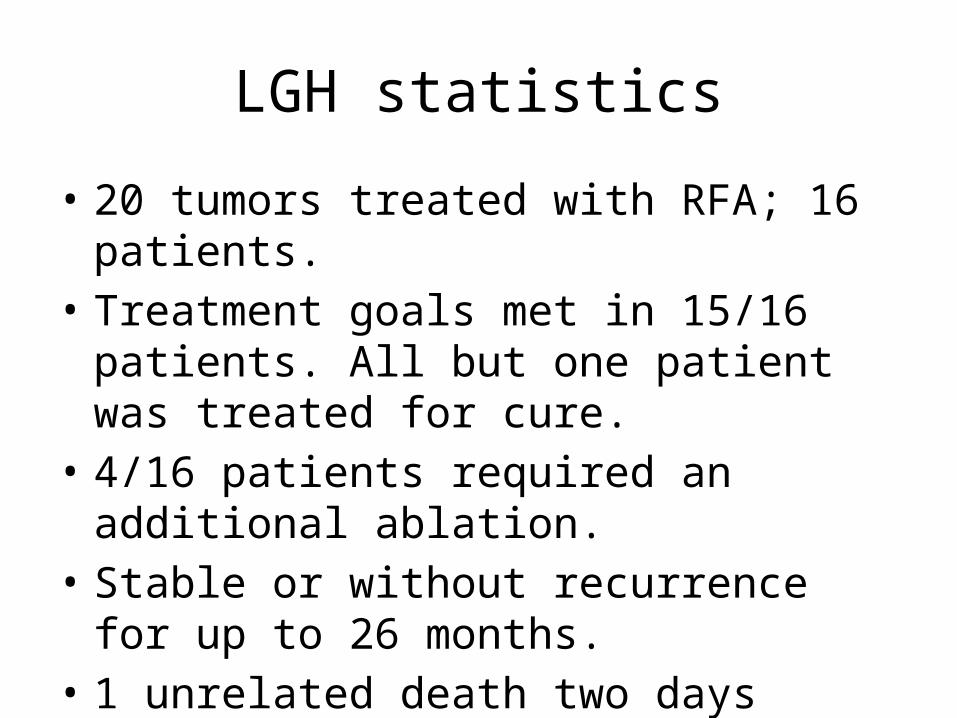
LGH statistics
• 20 tumors treated with RFA; 16 patients.• Treatment goals met in 15/16 patients. All but
one patient was treated for cure. • 4/16 patients required an additional ablation.• Stable or without recurrence for up to 26
months.• 1 unrelated death two days after treatment.
Cardiac arrest.
Genitourinary (GU)
• Palliative– Percutanous nephrostomy– Dialysis catheters– Fistula or hemodialysis access maintenance
• Curative– Thermal ablation of renal cell cancer
GU procedures
• Percutaneous access to the collecting system for benign or malignant obstructions, stone disease, or urosepsis
• Can place internal double J ureteral stents from percutaneous access
• Can provide access for future stone removal and/or manipulation
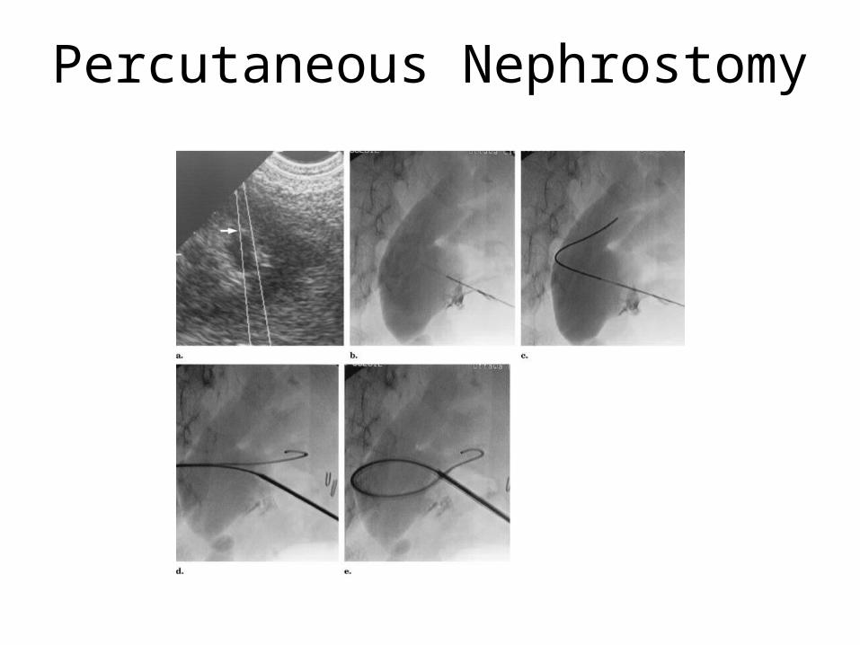
Percutaneous Nephrostomy
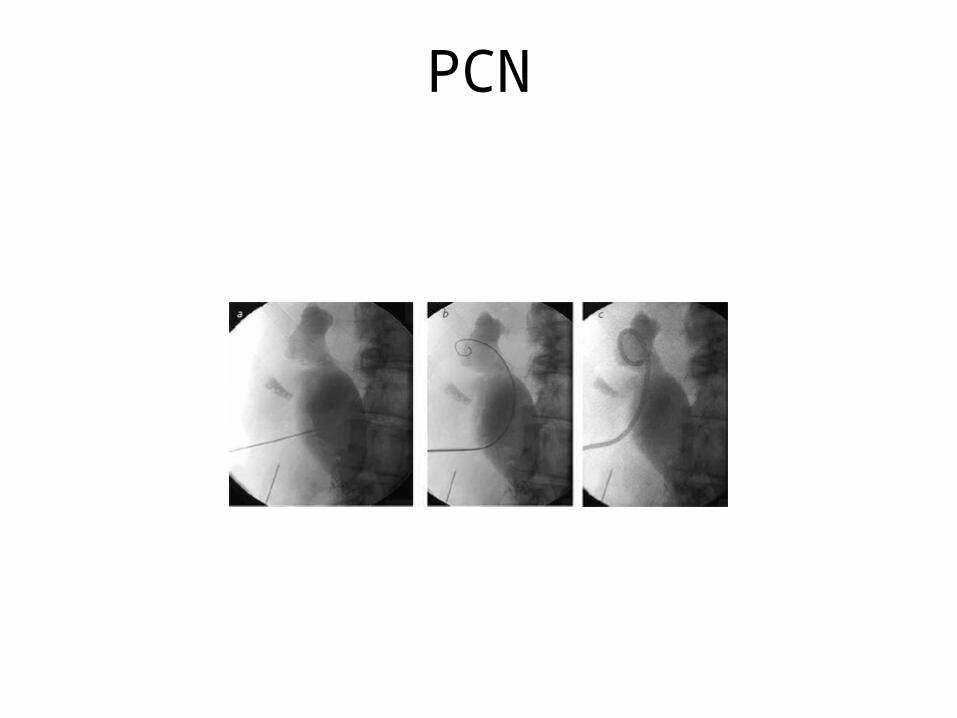
PCN
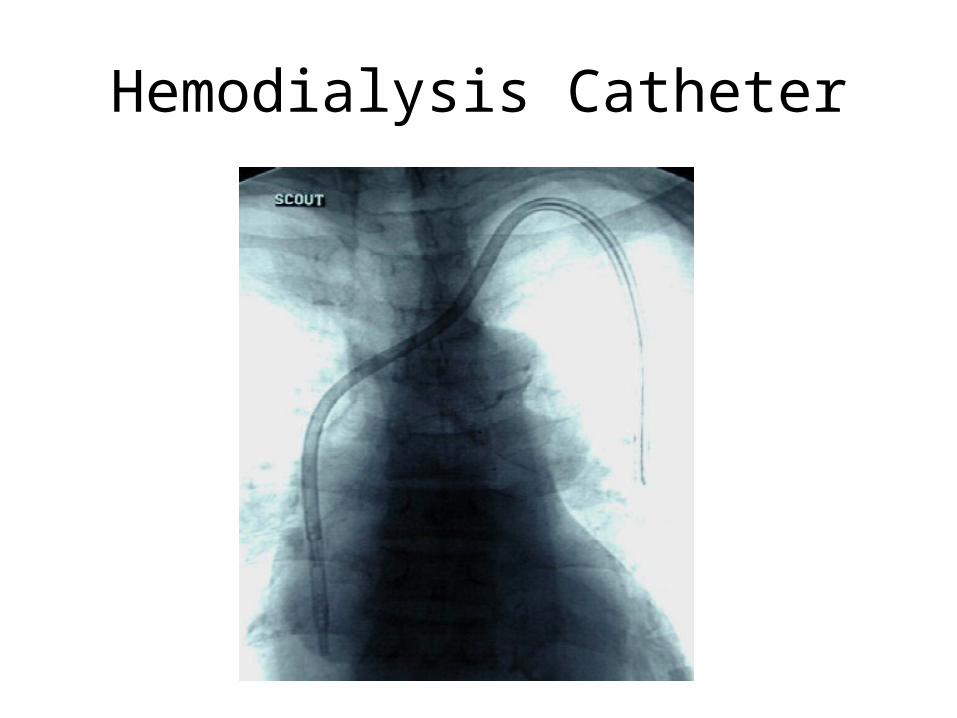
Hemodialysis Catheter
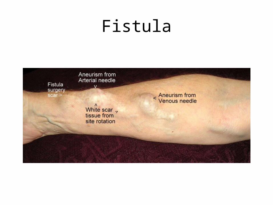
Fistula
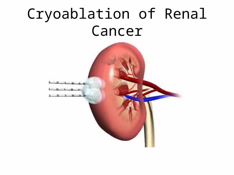
Cryoablation of Renal Cancer
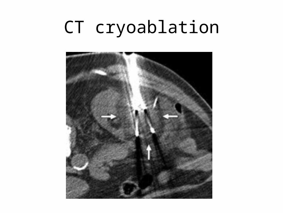
CT cryoablation
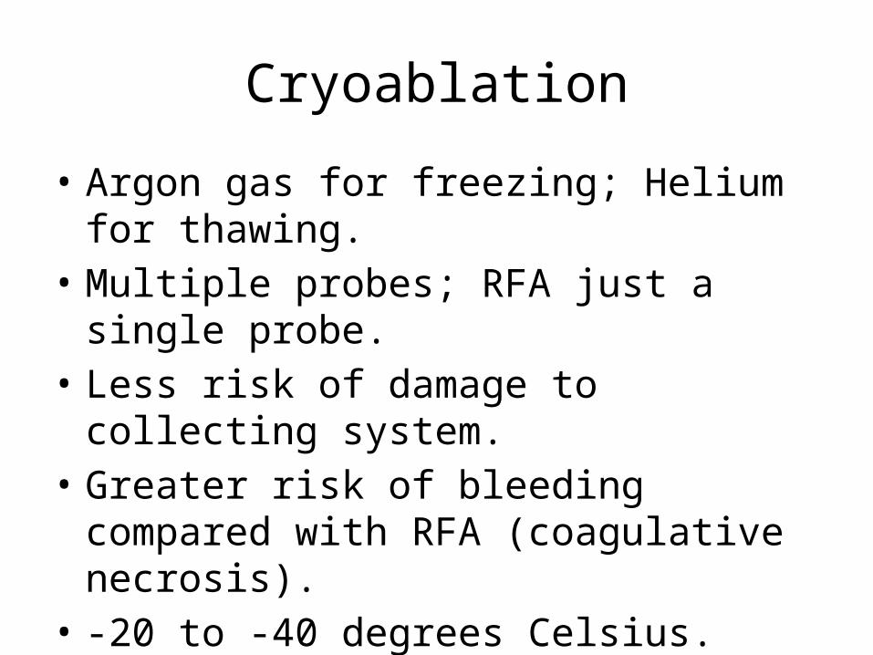
Cryoablation
• Argon gas for freezing; Helium for thawing.• Multiple probes; RFA just a single probe.• Less risk of damage to collecting system.• Greater risk of bleeding compared with RFA
(coagulative necrosis).• -20 to -40 degrees Celsius. Cell death.• Can better identify treated zone.
Survival
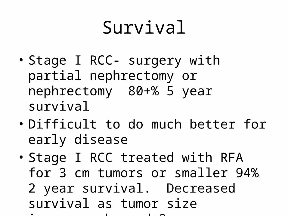
• Stage I RCC- surgery with partial nephrectomy or nephrectomy 80+% 5 year survival
• Difficult to do much better for early disease• Stage I RCC treated with RFA for 3 cm tumors
or smaller 94% 2 year survival. Decreased survival as tumor size increases beyond 3 cm.
36
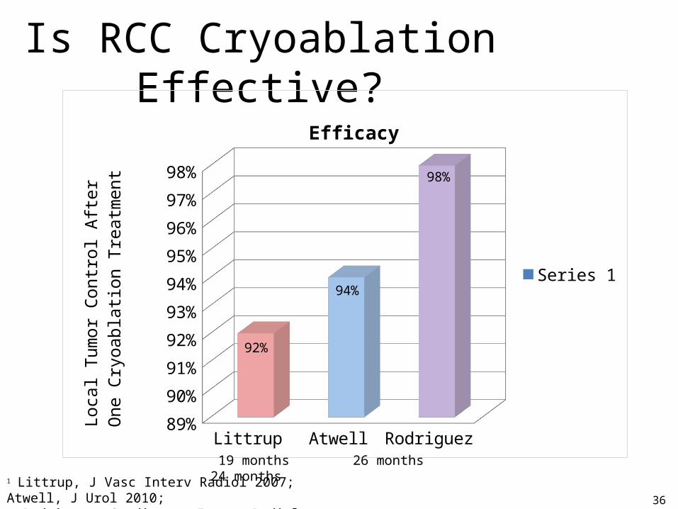
Is RCC Cryoablation Effective?
1 Littrup, J Vasc Interv Radiol 2007; Atwell, J Urol 2010; Rodriguez, Cardiovasc Interv Radiol 2011
Littrup Atwell Rodriguez89%
90%
91%
92%
93%
94%
95%
96%
97%
98%
92%
94%
98%
Efficacy
Series 1
Loca
l Tum
or C
ontr
ol A
fter
O
ne C
ryoa
blati
on T
reat
men
t
19 months 26 months 24 months
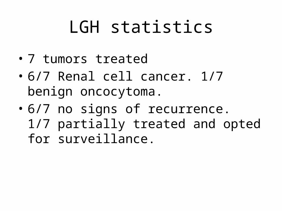
LGH statistics
• 7 tumors treated• 6/7 Renal cell cancer. 1/7 benign oncocytoma.• 6/7 no signs of recurrence. 1/7 partially
treated and opted for surveillance.
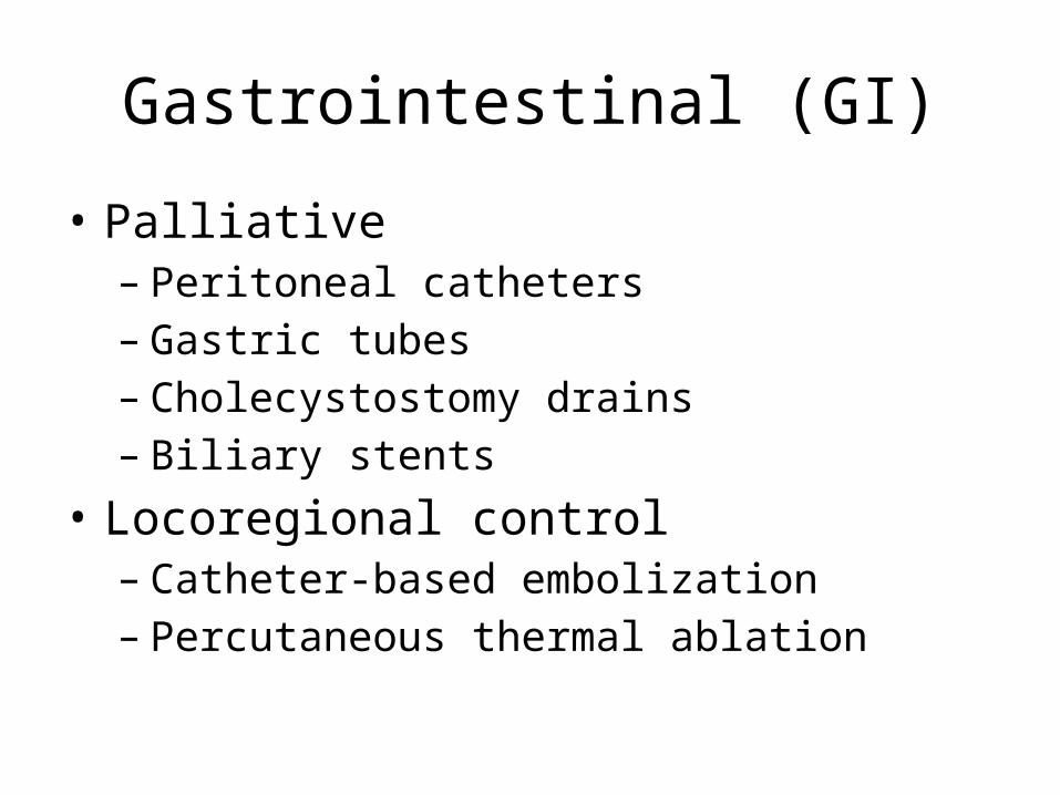
Gastrointestinal (GI)
• Palliative– Peritoneal catheters– Gastric tubes– Cholecystostomy drains– Biliary stents
• Locoregional control– Catheter-based embolization– Percutaneous thermal ablation
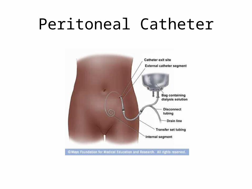
Peritoneal Catheter
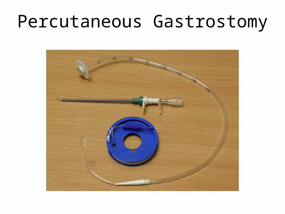
Percutaneous Gastrostomy
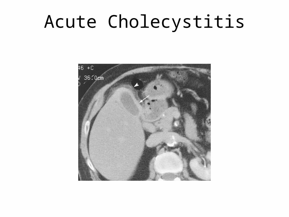
Acute Cholecystitis
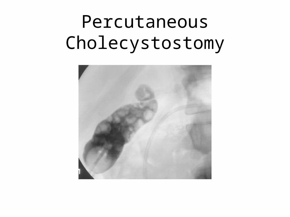
Percutaneous Cholecystostomy
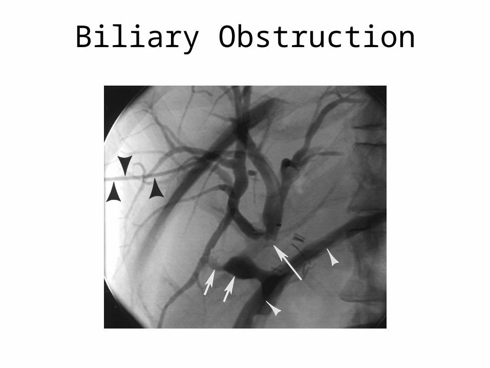
Biliary Obstruction
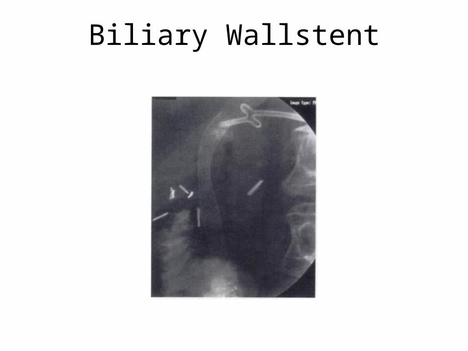
Biliary Wallstent
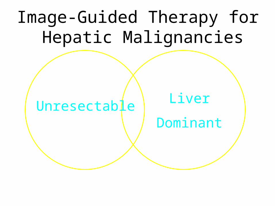
UnresectableLiver
Dominant
Image-Guided Therapy for Hepatic Malignancies
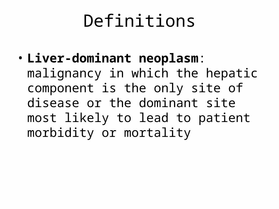
Definitions
• Liver-dominant neoplasm: malignancy in which the hepatic component is the only site of disease or the dominant site most likely to lead to patient morbidity or mortality
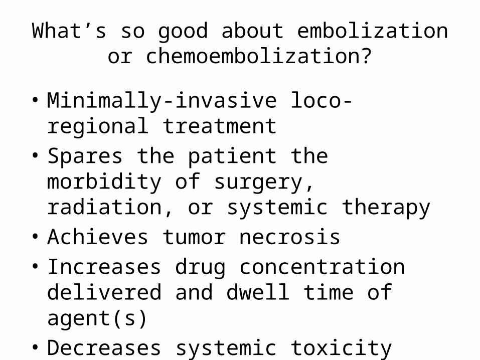
What’s so good about embolization or chemoembolization?
• Minimally-invasive loco-regional treatment• Spares the patient the morbidity of surgery,
radiation, or systemic therapy• Achieves tumor necrosis• Increases drug concentration delivered and
dwell time of agent(s)• Decreases systemic toxicity
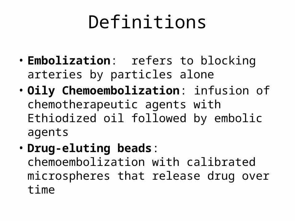
Definitions
• Embolization: refers to blocking arteries by particles alone
• Oily Chemoembolization: infusion of chemotherapeutic agents with Ethiodized oil followed by embolic agents
• Drug-eluting beads: chemoembolization with calibrated microspheres that release drug over time
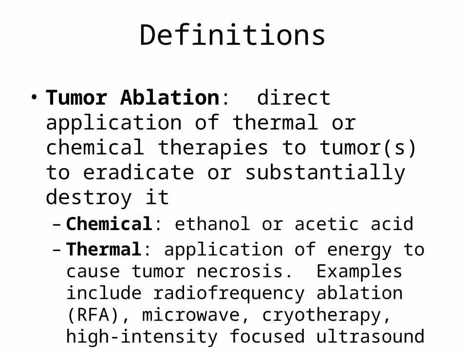
Definitions
• Tumor Ablation: direct application of thermal or chemical therapies to tumor(s) to eradicate or substantially destroy it– Chemical: ethanol or acetic acid– Thermal: application of energy to cause tumor
necrosis. Examples include radiofrequency ablation (RFA), microwave, cryotherapy, high-intensity focused ultrasound (HIFU)
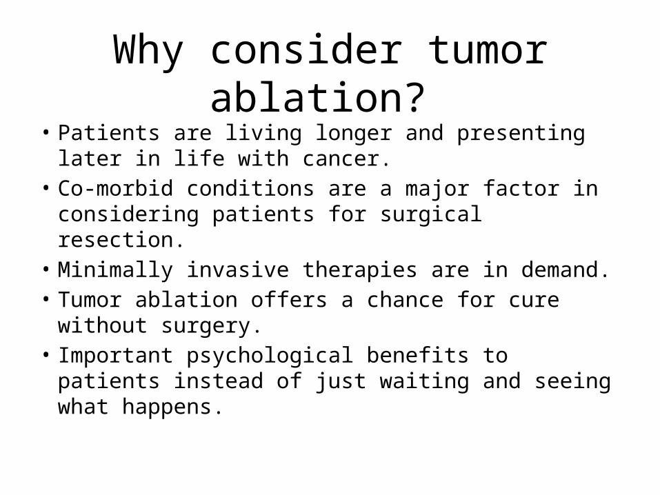
Why consider tumor ablation?
• Patients are living longer and presenting later in life with cancer.
• Co-morbid conditions are a major factor in considering patients for surgical resection.
• Minimally invasive therapies are in demand.• Tumor ablation offers a chance for cure without
surgery.• Important psychological benefits to patients
instead of just waiting and seeing what happens.
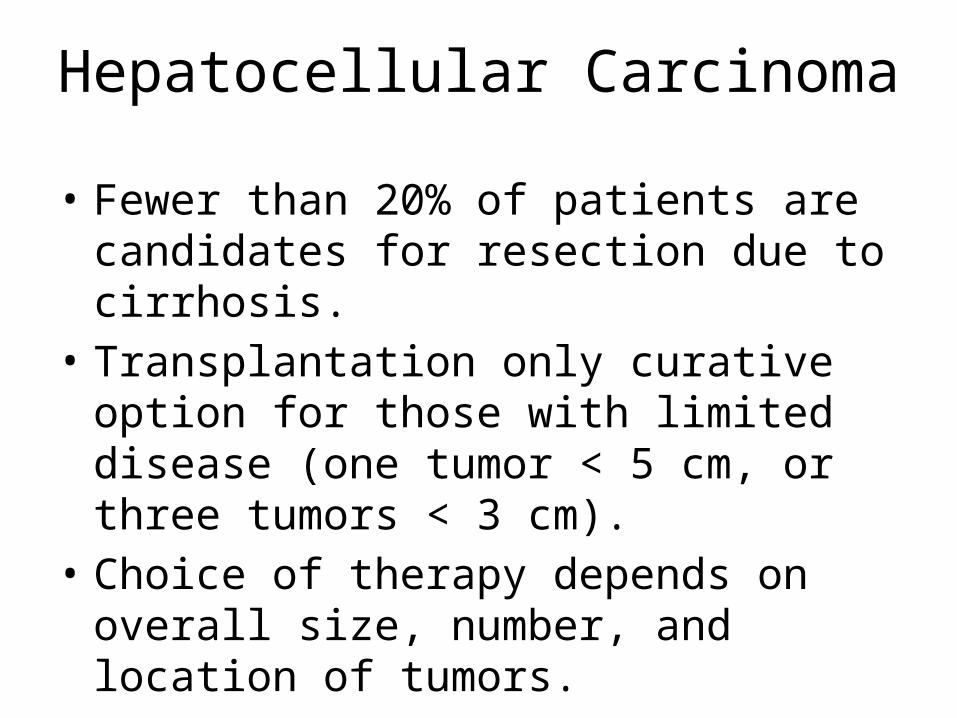
Hepatocellular Carcinoma
• Fewer than 20% of patients are candidates for resection due to cirrhosis.
• Transplantation only curative option for those with limited disease (one tumor < 5 cm, or three tumors < 3 cm).
• Choice of therapy depends on overall size, number, and location of tumors.
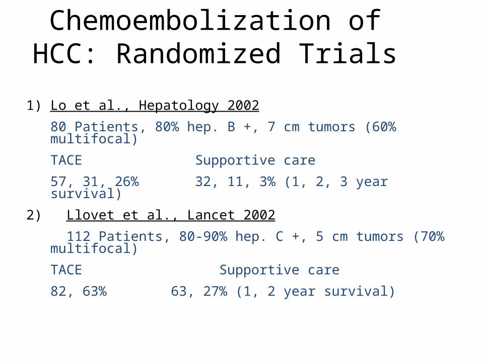
Chemoembolization of HCC: Randomized Trials
1) Lo et al., Hepatology 2002
80 Patients, 80% hep. B +, 7 cm tumors (60% multifocal)
TACE Supportive care
57, 31, 26% 32, 11, 3% (1, 2, 3 year survival)
2) Llovet et al., Lancet 2002
112 Patients, 80-90% hep. C +, 5 cm tumors (70% multifocal)
TACE Supportive care
82, 63% 63, 27% (1, 2 year survival)
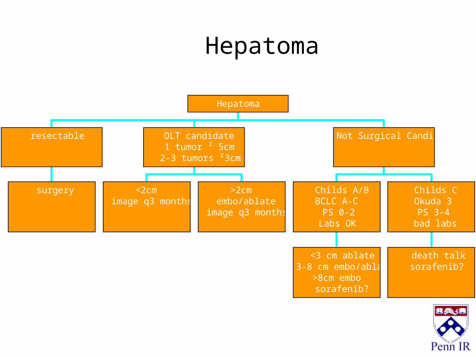
surgery
resectable
<2cmimage q3 months
>2cmembo/ablate
image q3 months
OLT candidate1 tumor ² 5cm
2-3 tumors ²3cm
<3 cm ablate3-8 cm embo/ablate
>8cm embosorafenib?
Childs A/BBCLC A-C
PS 0-2Labs OK
death talksorafenib?
Childs COkuda 3PS 3-4
bad labs
Not Surgical Candidate
Hepatoma
Hepatoma
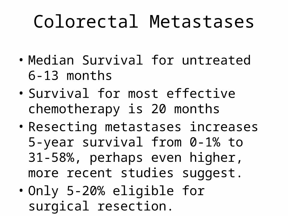
Colorectal Metastases
• Median Survival for untreated 6-13 months• Survival for most effective chemotherapy is 20
months• Resecting metastases increases 5-year survival
from 0-1% to 31-58%, perhaps even higher, more recent studies suggest.
• Only 5-20% eligible for surgical resection.
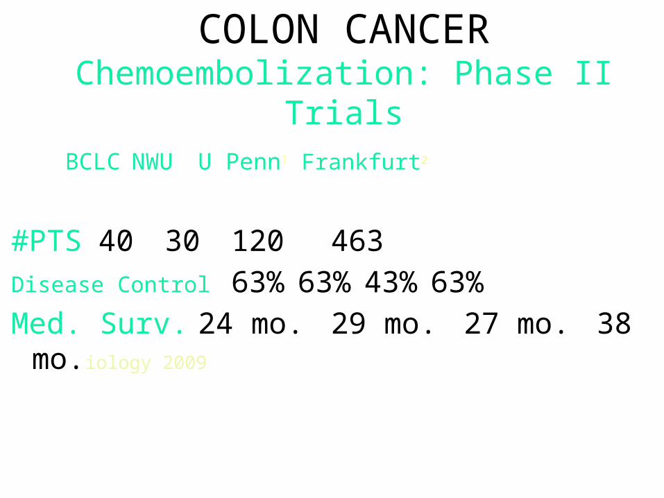
COLON CANCERChemoembolization: Phase II Trials
BCLC NWU U Penn1 Frankfurt2
#PTS 40 30 120 463
Disease Control 63% 63% 43% 63%
Med. Surv. 24 mo. 29 mo. 27 mo. 38 mo.iology 2009
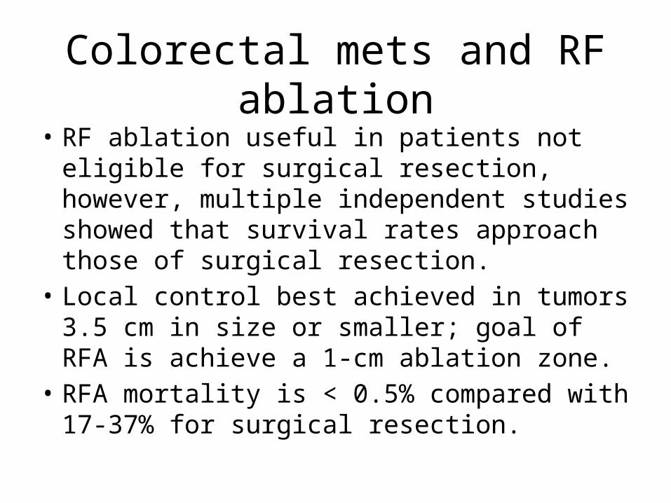
Colorectal mets and RF ablation
• RF ablation useful in patients not eligible for surgical resection, however, multiple independent studies showed that survival rates approach those of surgical resection.
• Local control best achieved in tumors 3.5 cm in size or smaller; goal of RFA is achieve a 1-cm ablation zone.
• RFA mortality is < 0.5% compared with 17-37% for surgical resection.
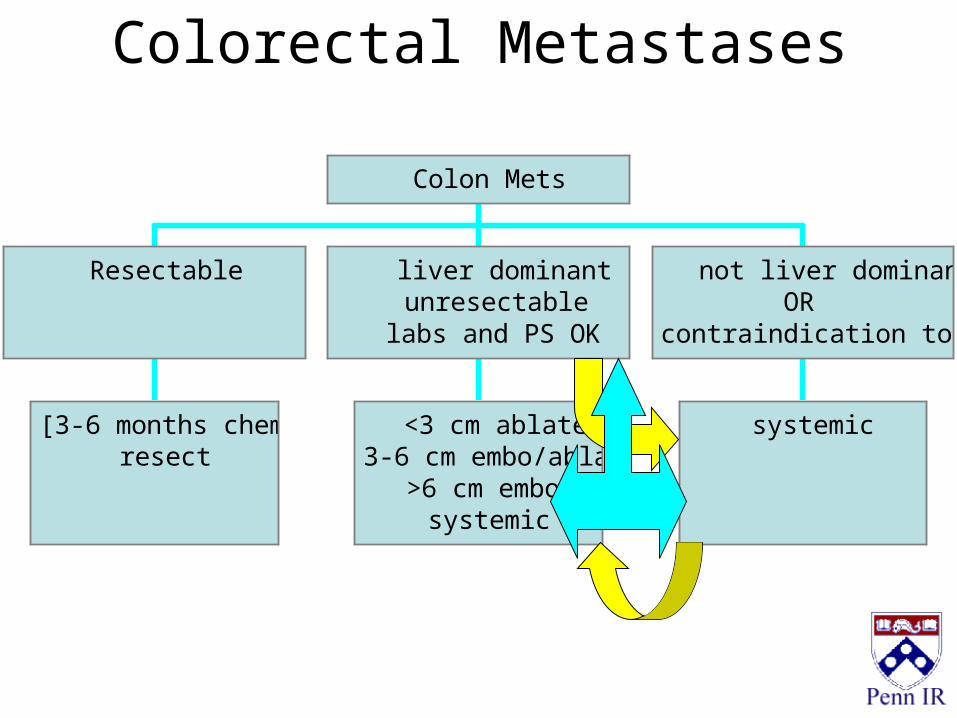
Colorectal Metastases
[3-6 months chemo]resect
Resectable
<3 cm ablate3-6 cm embo/ablate
>6 cm embosystemic
liver dominantunresectable
labs and PS OK
systemic
not liver dominantOR
contraindication to embo
Colon Mets
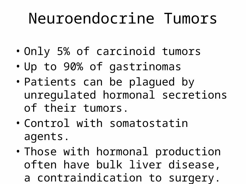
Neuroendocrine Tumors
• Only 5% of carcinoid tumors• Up to 90% of gastrinomas• Patients can be plagued by unregulated
hormonal secretions of their tumors.• Control with somatostatin agents.• Those with hormonal production often have
bulk liver disease, a contraindication to surgery.
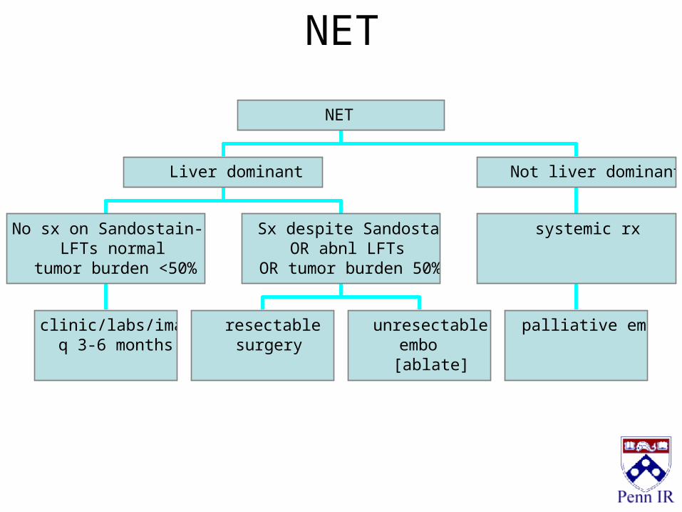
NET
clinic/labs/imagingq 3-6 months
No sx on Sandostain-LARLFTs normal
tumor burden <50%
resectablesurgery
unresectableembo
[ablate]
Sx despite SandostatinOR abnl LFTs
OR tumor burden 50%
Liver dominant
palliative embo
systemic rx
Not liver dominant
NET
Summary
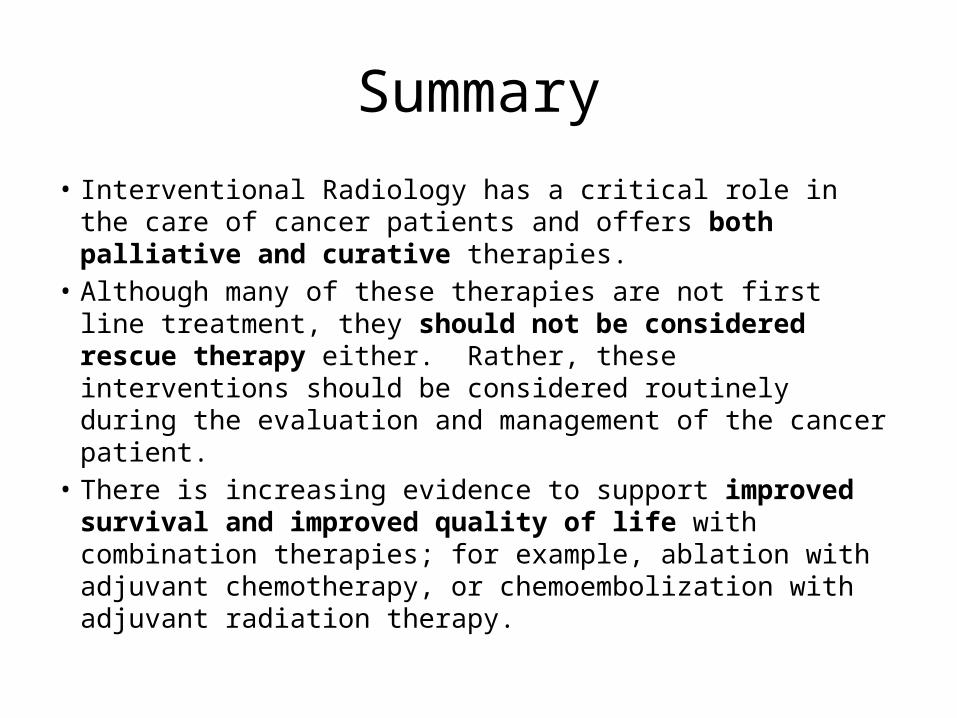
• Interventional Radiology has a critical role in the care of cancer patients and offers both palliative and curative therapies.
• Although many of these therapies are not first line treatment, they should not be considered rescue therapy either. Rather, these interventions should be considered routinely during the evaluation and management of the cancer patient.
• There is increasing evidence to support improved survival and improved quality of life with combination therapies; for example, ablation with adjuvant chemotherapy, or chemoembolization with adjuvant radiation therapy.
Thank you
• Lancaster Radiology Associates 299-4173• Interventional Radiology 544-4929• Consultations through Centralized Scheduling
at 544-5941.