Embed Size (px)
Citation preview

MINIMALLY INVASIVE SURGICAL PROCEDURES IN THE TREATMENT OF
BARRETT’S ESOPHAGUS
Zsolt Simonka M D
Ph D Thesis
UNIVERSITY OF SZEGED FACULTY OF MEDICINE
DEPARTMENT OF SURGERY
DOCTORAL SCHOOL OF CLINICAL MEDICINE
SUPERVISOR
Prof. György Lázár M D, Ph D, D Sc
2016
Szeged

1
CONTENTS
LIST OF FULL PAPERS RELATED TO THE SUBJECT OF THE THESIS ......................... 2
LIST OF ABSTRACTS RELATED TO THE SUBJECT OF THE THESIS ............................ 2
ABBREVIATIONS .................................................................................................................... 5
1. INTRODUCTION .................................................................................................................. 6
1.1. Barrett’s esophagus (BE) ................................................................................................. 6
1.2. Diagnostics of GERD and BE ......................................................................................... 7
1.3. Treatment of GERD and Barrets’ Esophagus.................................................................. 7
1.3.1. Surgical Treatment ........................................................................................................... 8
2. PROBLEM AND OBJECTIVES ........................................................................................... 9
3. PATIENTS AND METHODS ............................................................................................. 10
3.1. Comparison of patients subjected to surgery because of GERD or BE (Study 1) ........ 10
3.2. Clinical Risk Factors of the Development of BE .......................................................... 10
3.3. Components of the Preoperative Assessment ................................................................ 10
3.3.1. Endoscopy ...................................................................................................................... 10
3.3.2. Histological Examination ............................................................................................... 11
3.3.3. Functional Examinations ................................................................................................ 11
3.4. Surgical Treatment, Surgical Technique ....................................................................... 12
3.5. Postoperative Follow-up (Study 2) ................................................................................ 13
3.6. Endoscopic and Histological Follow-up of patients subjected to laparoscopic antireflux
procedure because of BE (Study 3) ...................................................................................... 13
3.7. Study of the Complications of the Antireflux Procedure (Study 4) .............................. 13
3.8. Complications of BE (Study 5) ..................................................................................... 14
3.9. Statistical Analysis ........................................................................................................ 14
4. RESULTS ............................................................................................................................. 15
4.1. Comparison of Patients Subjected to Surgery Beacause of GERD or BE (Study 1) .... 15
4.1.1. Preoperative Functional Results ..................................................................................... 16
4.2. Postoperative Results (Study 2) ..................................................................................... 19
4.2.1. Symptomatic Outcome ................................................................................................... 19
4.2.2. Postoperative Functional Results.................................................................................... 19
4.2.3. Postoperative Results by the Subgroup (Study 3) .......................................................... 20
4.3. Results of the Endoscopic Follow-up of the Subgroups (Endoscopic Surveillance) .... 21
4.4. Complications of Minimally Invasive Antireflux Procedures and their Treatment Study
(Study 4) ............................................................................................................................... 23
4.5. Endoscopic Treatment of Spontaneous Esophageal Rupture, a Complication Developed
on the Basis of BE ................................................................................................................ 23
5. DISCUSSION ....................................................................................................................... 25
SUMMARY, OUR MOST IMPORTANT RESULTS ............................................................ 33
ACKNOWLEDGEMENTS ..................................................................................................... 34
_Toc465066400

2
LIST OF FULL PAPERS RELATED TO THE SUBJECT OF THE THESIS
I. Simonka Zs, Paszt A, Ábrahám Sz, Pieler J, Tajti J, Tiszlavicz L, Németh I,
Izbéki F, Rosztóczy A, Wittmann T, Rárosi F, Lázár Gy. The effects of
laparoscopic Nissen fundoplication on Barrett's esophagus: long-term results.
Scand J Gastroenterol 47:13-21, 2012. IF: 2,019
II. Simonka Zs, Paszt A, Géczi T. Ábrahám Sz, Tóth I, Horváth Z, Pieler J,
Tajti J, Varga Á, Tiszlavicz L, Németh I, Izbéki F, Rosztóczy A, Wittmann T,
Lázár Gy. Refluxbetegség és Barrett-nyelőcső miatt műtétre kerülő betegek
összehasonlító vizsgálata. Comparison of surgical patients with gastroesophageal
reflux disease and Barrett’s esophagus. Magy Seb 67:287–296, 2014.
III. Rokszin R, Simonka Z, Paszt A, Szepes A, Kucsa K, Lazar G. Successful
endoscopic clipping in the early treatment of spontaneous esophageal perforation.
Surg Lapar Percutan Tech 21: e311-e31, 2011. IF: 1,227
IV. Lázár G Jr, Paszt A, Simonka Z, Bársony A, Abrahám S, Horváth G.
A successful strategy for surgical treatment of Boerhaave's syndrome. Surg
Endosc 25:3613-9, doi: 10.1007/s00464-011-1767-1. Epub Jun 11, 2011.
IF: 4,013
LIST OF ABSTRACTS RELATED TO THE SUBJECT OF THE THESIS
I. Simonka Zs, Paszt A, Géczi T, Ábrahám Sz, Tóth I, Horváth Z, Pieler J,
Tajti J, Varga Á, Lup M, Tiszlavicz L, Németh I, Izbéki F, Rosztóczy A,
Wittmann T, Lázár Gy. Barrett nyelőcső miatt végzett antireflux műtét hosszú
távú eredményei. In: Magyar Sebész Társaság Sebészeti Endoszkópos Szekció XVI.
Kongresszusa Sebészeti Továbbképző Tanfolyam. Eger, 2015. november 12 -14.
Eger: p. 100.

3
II. G Lazar, Zs Simonka, A Paszt, Sz Abraham, J Pieler, J Tajti, L Tiszlavicz, I
Nemeth, F Izbeki, A Rosztoczy, F Rarosi. The effects of laparoscopic Nissen
fundoplication on Barret's esophagus: longterm results. Surg Endosc 26:(Suppl. 1.)
p. S38, 2012.
III. Lázár Gy, Paszt A, Ábrahám Sz, Pieler J, Tajti J, Tiszlavicz L, Németh I,
Izbéki F, Rosztóczy A, Wittmann T, Rárosi F, Simonka Zs. The effects of
laparoscopic Nissen fundoplication on Barrett’s esophagus:: long-term results. Dis
Esophagus 25:(S1) Paper P01.07, 2012.
IV. Rokszin R, Simonka Zs, Paszt A, Szepes A, Kucsa K, Lázár Gy. A spontán
nyelőcső-perforatio korai ellátása endoscopos klippeléssel. Magy Seb 65:314-315,
2012.
V. Simonka Zs, Paszt A, Ábrahám Sz, Bársony A, Horváth G, Lázár Gy.
A Boerhaave-syndroma sebészi kezelésének stratégiája. Magy Seb 65:270-271,
2012.
VI. Simonka Zs, Paszt A, Géczi T, Ábrahám Sz, Pieler J, Tajti J, Izbéki F,
Rosztóczy A, Wittmann T, Tiszlavicz L, Németh I, Lázár Gy. A laparoscopos
antireflux műtét szerepe a Barrett-nyelőcső sebészi kezelésében. Magy Seb 64:163,
2011.
VII. Lázár Gy, Paszt A, Simonka Zs, Tiszlavicz L, Németh I, Izbéki F, Rosztóczy
A, Wittmann T. The effect of laparoscopic Nissen fundoplication on Barrett’s
esophagus and GERD. Dis Esophagus 23:(Suppl.1) 59A, 2010.
VIII. Simonka Zs, Paszt A, Géczi T, Ábrahám Sz, Pieler J, Tajti J, Izbéki F,
Rosztóczy A, Wittmann T, Tiszlavicz L, Németh I, Lázár Gy. A laparoscopos
antireflux műtét szerepe a Barrett nyelőcső sebészi kezelésében. Magy Seb 63:237-
238, 2010.
IX. Simonka Zs, Paszt A, Géczi T, Ábrahám Sz, Pieler J, Tiszlavicz L, Németh I,
Izbéki F, Rosztóczy A, Wittmann T, ifj Lázár Gy. The role of antireflux surgery
in the treatment of Barrett’s esophagus. Z Gastroenterol 48:615, 2010.
X. Simonka Zs, Paszt A, Ábrahám Sz, Géczi T, Pieler J, Tajti J, Mészáros P,
Dargai S, Horváth Z, Tiszlavicz L, Németh I, Izbéki F, Rosztóczy A,
Wittmann T, Lázár Gy. Efficiency of laparoscopic procedure in Barrett’s
esophagus. Nihon Geka Gakkai Zasshi 111:(suppl.2) p. 263, 2010.

4
XI. Simonka Zs, Paszt A, Ábrahám Sz, Géczi T, Tóth I, Horváth Z, Pethő I,
Tiszlavicz L, Németh I, Izbéki F, Rosztóczy A, Wittmann T, Lázár Gy.
Comparison of efficiency of laparoscopic procedures in GERD and Barrett’s
esophagus (short term functional and histological results). Br J Surg 96:(Suppl.5)
55-56, 2009.
XII. Simonka Z, Paszt A, Géczi T, Ábrahám S, Tóth I, Horváth Z, Pethő I,
Tiszlavicz L, Németh I, Izbéki F, Rosztóczy A, Wittmann T, Lázár G.
Comparison of efficiency of laparoscopic procedures in GERD and Barrett’s
esophagus. Z Gastroenterol 46:510-511, 2008.
XIII. Simonka Zs, Paszt A, Ábrahám Sz, Géczi T, Tóth I, Horváth Z, Pethő I,
Tiszlavicz L, Németh I, Izbéki F, Rosztóczy A, Wittmann T, Lázár Gy.
Comparison of efficiency of laparoscopic procedures in GERD and Barrett’s
esophagus (short term functional and histological results). Hepatogastroenterology
55:(Suppl.1) A186, 2008.
XIV. Simonka Zs, Paszt A, Ábrahám Sz, Pethő I, Tiszlavicz L, Németh I, Izbéki F,
Rocztóczy A, Wittmann T, Lázár Gy. Laparoscopos antireflux műtét szerepe a
Barrett-nyelőcső kezelésében. Magy Seb 61:187, 2008.
XV. Simonka Zs, Paszt A, Ábrahám Sz, Pethő I, Tiszlavicz L, Németh I, Izbéki F,
Rocztóczy A, Wittmann T, Lázár Gy. Laparoscopos antireflux műtét szerepe a
Barrett-nyelőcső kezelésében. In: A Magyar Sebész Társaság 59. Kongresszusa:
Sebészeti Továbbképző Tanfolyam. Debrecen, 2008. június 18-20. Paper 143.

5
ABBREVIATIONS
BE, Barrett’s esophagus
BMI, body mass index
CM, cardiac metaplasia
COX-2, cyclooxygenase-2
D, dysplastic (group)
ERD, erosive reflux disease
FM, fundic metaplasia
GERD, gastroesophageal reflux disease
GEJ, gastroesophageal junction
HGD, high-grade dysplasia
I, intestinal (group)
IH, immunohistochemistry
IM, intestinal metaplasia
LES, lower esophageal sphincter
LGD, low-grade dysplasia
lLES, length of lower esophageal sphincter
LSBE, long-segment Barrett’s esophagus
NBI, narrow band imaging
NERD, non-erosive reflux disease
NI, non-intestinal (group)
pLES, pressure of lower esophageal sphincter
PPI, proton-pump inhibitor
PTFE, polytetrafluoroethylene
PTX, pneumothorax
rLES, relaxation time of lower esophageal sphincter
SD, standard deviation
SSBE, short-segment Barrett’s esophagus
pRBC, packed red blood cells
UES, upper esophageal sphincter

6
1. INTRODUCTION
Gastroesophageal reflux disease (GERD) can be summarized as mucosal irritation,
inflammation and consequential symptoms caused by the reflux of gastric contents into the
esophagus due to the impaired function of the gastroesophageal junction (GEJ). It may be
accompanied by a wide range of symptoms. Most often, the main symptom is heartburn, a
retrosternal pain which may be caused by the regurgitation of gastric acid into the esophagus.
In certain cases, it may also be accompanied by dysphagia. Extraesophageal symptoms may
often be misleading: in case of proximal (high) reflux, airway symptoms may often be
expected; hoarseness, cough, asthma-like episodes, sinusitis or otitis media may also occur. A
chest pain of non-cardiac origin or dental caries may also raise the possibility of GERD.
The incidence of GERD shows a significant increase in developed Western countries. The
traditional classification of reflux disease (the Los Angeles Classification)[6]
was based on the
endoscopic picture, differentiating between cases without signs of inflammation (non-erosive
reflux disease, NERD) and those with erosive esophagitis (ERD) or with complications of
severe erosion. The Montreal Classification[7]
, besides the esophageal symptoms, also takes
the extraesophageal symptoms, i.e., the complaints of the patients, into account. In parallel
with the severity and duration of GERD, the risk of possible complications also increases.
Ulcers and/or strictures developed on the basis of inflammation may lead to severe dysphagia.
The condition of acute abdomen/acute chest due to the perforation of an ulcer or stricture may
require an urgent (surgical) intervention. Additional possible complications are Barrett’s
esophagus (BE) and, ultimately, adenocarcinoma developed based on this in the lower third of
the esophagus.
1.1. BARRETT’S ESOPHAGUS (BE)
The definition of BE can be understood based on its pathogenesis. As “regeneration” of the
mucosal inflammation and, later, mucosal damage developed due to the persistent acid and/or
mixed reflux, a columnar epithelium that is more resistant to the acidic environment appears
next to or replaces the squamous epithelium in the lower third of the esophagus. The
polymorphism of Barrett’s metaplasia is reflected by the fact that the regeneration starting
from the esophageal Schaffer glands may have varying histological appearance, possibly with
multiple histological entities next to each other. Besides intestinal metaplasia (which is

7
considered to be the classic form of BE), numerous other forms may appear, including fundic
or cardiac columnar epithelium, ciliary columnar epithelium, or even pancreatic acinar or
tubular metaplasia.[8]
Although the literature describes the possibility of dysplastic
transformation in the case of intestinal metaplasia, non-intestinal forms should not be
disregarded either because of the heterogeneity of the condition (histological forms present
next to each other or that may transform into each other).
The endoscopic appearance of the metaplasia can be described most accurately with the
“Prague C and M” classification[9]
, which also gives the extension and distance of the lesion
from the gastroesophageal junction. An extensive BE longer than 3 cm is a so-called long-
segment BE (LSBE), whereas a condition shorter than this is a short-segment BE (SSBE).
1.2. DIAGNOSTICS OF GERD AND BE
Besides the proper evaluation of the patient’s symptoms, many instrumental examination
methods are available to clarify the diagnosis. Flexible endoscopy, with its advance and
widespread use, has clearly become the gold standard in the assessment of the esophagus.
Without a proper endoscopic background, a thorough investigation of neither GERD, nor BE
can be performed. The new endoscopic examination methods (such as chromoendoscopy,
NBI, etc.) may help map GERD and BE more precisely. The need for proper professional
experience and technical equipment warrants the investigations to be performed in centers.
The objective confirmation and description of reflux and, ultimately, making the indication
for surgery are also inconceivable without proper functional examinations. The
gastroenterologist may decide about the correct treatment strategy in the knowledge of the
presence of acid or bile reflux obtained with pH-metry, impedance monitoring and Bilitec
monitoring.[24]
The information about the function, motility disorders and impairment of the
esophageal body and sphincters gained during manometry may influence the choice of
surgical procedure. [25]
A proper biopsy sampling procedure (Seattle protocol) [26]
and a well-
prepared pathological background are also indispensable for detecting the presence of BE.
1.3. TREATMENT OF GERD AND BARRETT’S ESOPHAGUS
The conservative (non-surgical) treatment is extremely complex and involves losing weight
(besides other lifestyle advices), increasing stomach emptying (with prokinetics), protecting
the esophageal mucosa, and decreasing the acid content of the refluxate.
8
Nowadays, the medical treatment is primarily based on effective gastric acid-reducing
therapies. The symptoms of reflux can be considerably improved with proton-pump inhibitor
therapy, the use of which may mean an adequate long-term control of reflux. Similarly, they
can be effective in the treatment of some patients with BE (but there is only indirect evidence
of their efficacy in the treatment of metaplasia and dysplasia).[27]
In case of BE, aspirin in
increased dose may be added to antacids as chemoprevention, which may reduce the risk of
transition from metaplasia to dysplasia through the inhibition of COX-2.
The purpose of the endoscopic treatment of BE is to remove or destroy the affected mucosa
when dysplasia appears, after which the regeneration restores the squamous epithelium or
results in a dysplasia-free columnar epithelium if the treatment is combined with successful
acid inhibitor therapy. Numerous procedures are known from endoscopic mucosal resection to
submucosal dissection. Ablation may be achieved with radiofrequency therapy but laser
therapy, argon plasma coagulation, cryotherapy and photodynamic therapy may also be used.
Out of these methods, however, the depth of the dissection can be reconstructed and an actual
histological examination can be performed only in the case of endoscopic resections. In the
rest of the procedures, there is less control of the ablation depth, and the rate of potential
complications (stricture and, more rarely, perforation) is higher. The indication for endoscopic
procedures is not clear in case of LGD. A special issue is recurrence in the submucosal
glandular structures after the endoscopic treatment. In case of LGD, the rate of this may be up
to 10% in the year following the procedure. However, an advantage of the endoscopic
procedure is that it can be repeated. [28]
In case of HGD, the higher relapse rate after endoscopic procedures or the appearance of
carcinoma may require surgical intervention. The presence of in situ carcinoma is almost
certain in the case of HGD; however, neither this, nor an early carcinoma means a
contraindication to endoscopic procedures. [28]
1.3.1. Surgical Treatment
The various antireflux procedures have long been accepted in the surgical treatment of GERD
and BE. Nowadays, with the spread of minimally invasive surgical methods, laparoscopic
antireflux procedures that have low mortality rates have become equivalent alternatives to
conservative treatment. In case of proper indication, the correctly performed procedure
successfully decreases the acid and bile reflux, and also restores the function of the lower
esophageal sphincter. In case of BE, with the reflux gone, we may suppose that the

9
progression of metaplastic and dysplastic processes is stopped and that regression is achieved.
The long-term success of antireflux procedures, similarly to that of medical treatment, is
contradictory, as well as their role in the prevention of adenocarcinoma. In cases of BE with
LGD, better results may be expected from the combination of mucosal ablation and medical
or surgical treatment. In cases of HGD, in situ carcinoma and early cancer, distal esophageal
and cardiac resection may be considered an oncologically adequate treatment from a surgical
point of view. In case of invasive adenocarcinoma, esophageal resection may be performed.
2. Problem and objectives
The increase in the incidence of esophageal adenocarcinoma calls the importance of this
condition to our attention. The key for the successful treatment of esophageal adenocarcinoma
is the prevention of its development or its early recognition. The only precancerous condition
of esophageal adenocarcinoma confirmed to date is BE. In our clinical study, we intended to
establish the clinical risk factors of the development of BE, the prevention of its development,
and the possible strategy for its surgical treatment.
1. An objective of our work was to understand the potential clinical risk factors and
relationships playing a role in the development of BE and the process of the Barrett’s
metaplasia—dysplasia—carcinoma transformation through the study of patients with GERD
and BE (Study 1).
2. A further objective was to study the efficacy of surgical treatment (laparoscopic antireflux
procedure) among patients subjected to surgery because of either GERD or BE (Study 2).
3. During our short-term and long-term follow-up, the effect of laparoscopic antireflux
procedure on the histological changes of Barrett’s esophagus, as well as its possible
preventive effect in the process of carcinogenesis were studied (Study 3).
4. The early and late complications of the antireflux procedure were studied (Study 4).
5. The successful endoscopic treatment of spontaneous esophageal perforation, a rare
complication of BE, is presented through one of our cases (Study 5).

10
3. PATIENTS AND METHODS
3.1. COMPARISON OF PATIENTS SUBJECTED TO SURGERY BECAUSE OF
GERD OR BE (STUDY 1)
Our retrospective clinical study was based on patients subjected to surgery because of GERD
or reflux disease accompanied by Barrett’s esophagus at the Department of Surgery of the
Faculty of Medicine of the University of Szeged between January 1, 2001 and December 31,
2008. Nissen’s laparoscopic antireflux procedure was performed in 176 cases because of
GERD (Group 1) and in 78 cases because of BE (Group 2).
In Study 1, the results of the preoperative assessment were compared between the above two
patient groups.
3.2. CLINICAL RISK FACTORS OF THE DEVELOPMENT OF BE
As a continuation of Study 1, patients in the BE group (78 patients) were divided into three
further groups on the basis of the histological results of their preoperative endoscopic
biopsies: a non-intestinal group (NI, 53 patients) with fundic (FM) and cardiac metaplasia
(CM), an intestinal group (I, 18 patients) with intestinal metaplasia (IM), and a dysplastic
group (D, 7 patients) with LGD. BE involved a short segment (< 3 cm, SSBE) in 67 cases
(85.9%) and a long segment (> 3 cm, LSBE) in 11 patients (14.1%). We compared the results
of the preoperative assessment between these three groups.
3.3. COMPONENTS OF THE PREOPERATIVE ASSESSMENT
3.3.1. Endoscopy
The esophagogastroscopy performed as part of the preoperative gastroenterological
assessment to confirm the reflux disease was the first step of the standard examination, during
which the gastroesophageal junction was examined. Hiatal hernia is diagnosed if the
impression of the diaphragmatic crura is widened by more than 2 cm. Its size is given in
centimeters. The reflux disease was described based on the Los Angeles Classification. [6]
Biopsy was performed (in all four quadrants, with 2-cm intervals) to confirm or rule out
Barrett’s metaplasia (Seattle protocol). [26]
Barrett’s esophagus was characterized based on the
Prague C & M Criteria. [9]

11
3.3.2. HISTOLOGICAL EXAMINATION
The formalin-fixed, paraffin-embedded histological samples were assessed for Barrett’s
metaplasia or dysplasia after hematoxylin-eosin and immunohistochemistry staining. In our
study, samples with either fundic or cardiac or intestinal metaplasia were included. In case of
BE with LGD described during the histological examination, two experienced pathologists
also examined the slides.
3.3.3. Functional Examinations
Esophageal pH-metry
Of the functional examinations, 24-hour pH-metry was performed in each case. The acid
reflux was measured with a pH electrode inserted nasally and secured 5 cm above the upper
margin of the lower esophageal sphincter (LES). The probe recorded the acid reflux episodes
for 24 hours—esophageal pH decrease below 4, number of reflux episodes longer than
5 minutes, duration of the longest reflux episode, percentage of time of exposure to pH below
4, and the DeMeester composite score, out of which the DeMeester score and the percentage
of time of exposure to pH below 4 have the highest sensitivity (96%) and specificity (100%).
Their normal value, based on studies conducted with health volunteers, is 14.7 and 4.2%,
respectively.[29,30,31,32,33]
Esophageal manometry
During the manometry, the motor function of the esophagus was examined with a catheter
inserted nasally into the esophagus, using the standard distilled water perfusion method, and
mapping the function of the entire esophageal body, the pharyngoesophageal junction and the
LES: the length of the sphincter (lLES), its mean pressure (pLES), its relaxation (rLES), the
amplitude and duration of the contractions of the esophageal body, and (optionally)
pharyngeal motility. The generally accepted values for the UES are a length of 2 to 5 cm and
a mean pressure of 40 to 100 mmHg; and for the LES, a length of 2 to 4 cm and a mean
pressure of 10 to 40 mmHg. The pressure in the lower third of the esophagus during
swallowing is 20 to 170 mmHg.[29,34,35,36]

12
Bilitec
In both groups, Bilitec monitoring was performed only in cases where bile regurgitation was
suspected during endoscopy. The nasally inserted catheter was positioned 5 cm above the
LES, and the photoabsorption (at 450 nm) of bile acids reaching the esophagus was detected
with a fiber optic spectrophotometer.[29, 30,31,32]
3.4. SURGICAL TREATMENT, SURGICAL TECHNIQUE
After the gastroenterological assessment, patients with GERD and BE underwent elective
surgery. Laparoscopic antireflux procedure with Nissen’s 360-degree fundoplication was
performed on the patients of both groups. During the standardized steps of the procedure, the
flaccid part of the lesser omentum was opened while retracting the left lobe of the liver, and
then the diaphragmatic crura were prepared (first the right, and then the left one), going
around the entire circumference of the esophagus, mobilizing this way its abdominal segment.
The fundus was mobilized by transecting the gastrosplenic ligament and the short arteries and
veins running within it. The posterior reconstruction of the diaphragmatic crura was
performed with interrupted stitches (using non-absorbable suture). After this, 360-degree
(Nissen’s) fundoplication was performed in each case. Partial (Toupet) and anterior (Dor)
fundoplications were excluded from the study.
During the surgeries, a mesh was placed because of the large hiatal hernia in 14 cases. The
indication for this was the unsuccessful tension-free closure of the diaphragmatic crura. A
PTFE mesh was used for the reconstruction, which was secured with a spiral clamp.
Gastropexy was performed in 6 cases in the GERD group, and a mesh was also placed in each
of these cases because of a large hiatal hernia. In 26 cases, cholecystectomy was also
performed in the same session because of the accompanying cholelithiasis.

13
3.5. POSTOPERATIVE FOLLOW-UP (STUDY 2)
During Study 2, the efficacy of procedures performed because of GERD or BE were
compared based on subjective measures (Visick score) and the results of early functional
examinations.
Assessment of symptoms and objective measures of outcome
Visick grading was used to assess the effect of surgery on the symptoms: complete resolution
(Grade I); an improvement (Grade II); no effect of surgery (Grade III); or deterioration
relative to the preoperative state (Grade IV). This scoring system was devised to give an
overall impression of the benefits of antireflux surgery because it exhibits good correlation
with heartburn, the most prominent symptom of GERD.[37]
In the postoperative period, patients were subjected to follow-up surgical and medical
examinations. Postoperative functional examinations, such as esophageal manometry, 24-hour
pH-metry and, in reasonable cases, bile exposure (Bilitec) monitoring and endoscopy were
performed in the early postoperative period with an average follow-up period of 13.8 ±
19.31 months in the GERD group and 16.7 + 17.00 months (range: 3–23) in the BE group.
3.6. ENDOSCOPIC AND HISTOLOGICAL FOLLOW-UP OF PATIENTS
SUBJECTED TO LAPAROSCOPIC ANTIREFLUX PROCEDURE BECAUSE OF BE
(STUDY 3)
During the study, in the later postoperative period, an additional upper endoscopy with biopsy
was carried out in the BE subgroups to assess the changes in BE. The overall average follow-
up time was 42 + 16.19 months (range: 3–61).
3.7. STUDY OF THE COMPLICATIONS OF THE ANTIREFLUX PROCEDURE
(STUDY 4)
Complications of the antireflux procedures
The early complications of laparoscopic antireflux procedure are well known. Bleeding may
start from the vessels that supply the stomach (left gastric artery and vein and short gastric

14
arteries and veins). Spleen injury during the mobilization of the fundus may be a severe
condition that often requires conversion. Injury to the hepatic capsule and bleeding of the liver
are rarely severe complications. Rarely, the iatrogenic or ischemic perforation of a hollow
organ (esophagus, stomach) may require an intraoperative solution or early reoperation.
Subcutaneous emphysema due to the insufflation, which involves less risk, rarely requires
surgical intervention. Pneumothorax accompanying pleural injuries in the posterior
mediastinum, however, may require pleural drainage in certain cases. Impaired gastric
emptying as a consequence of injury to the vagus nerve fibers may cause complaints in the
long term.
Among the late complications of the surgical treatment, persistent cases of dysphagia must be
mentioned first, the treatment of which, similarly to making the indication for surgery,
requires a close cooperation between the gastroenterologist and the surgeon. In case of an
unsuccessful dilation with bougies, balloon dilation may be required. In case of large hiatal
hernias, the placement of a mesh exposes the patient to special hazards. Erosion of the
esophageal wall may be a severe or even life-threatening condition, and it is to be treated
surgically with relative urgency: often, cardiac resection and jejunal interposition
(Merendino’s procedure) are required.
3.8. COMPLICATIONS OF BE (STUDY 5)
Stricture, bleeding, perforation and, ultimately, malignant transformation on the basis of
Barrett’s ulcer are well-known complications. Perforation as a consequence of Barrett’s ulcer
or a stricture is most often iatrogenic. Spontaneous perforation is an extremely rare, life-
threatening condition. The successful endoscopic treatment of this rare complication will be
presented through a case report.
3.9. STATISTICAL ANALYSIS
The values measured and summarized during the clinical study of the GERD and BE groups
were evaluated using SigmaStat®
3.1 Further statistical calculations were performed with
SPSS 17.0 for Windows, whereas the special Poisson-distributed ANOVA method was
performed with SAS for Windows 9.1.[38]
To compare changes in the patients’ parameters
before and after the operation in the three BE groups, a generalized mixed model repeated

15
measurements ANOVA method was applied (multivariate analysis), using the GLIMMIX
procedure of SAS 9.1.
4. RESULTS
4.1. COMPARISON OF PATIENTS SUBJECTED TO SURGERY BECAUSE OF
GERD OR BE (STUDY 1)
According to the results of Study 1, the gender distribution in the two patient groups showed a
predominance of women (112 women, 64 men), with a more balanced ratio in cases
complicated with Barrett’s esophagus (40 women, 38 men) but, contrary to literature data,
there was no male predominance in this group either (Table 1). There was no difference
between the two groups in mean patient age (Group 1: 53.87 ± 12.04 years vs. Group 2: 53 ±
12.7 years, p=0.495) or mean BMI (Group 1: 26.91 ± 4.54 vs. Group2: 28.31 ± 5.46,
p=0.451). It must be noted, however, that the majority of patients in both groups were
overweight, which is a known potential risk factor of reflux disease.
As to the results of the preoperative assessment, contrary to our hypothesis, there was no
difference between the two groups in the mean time from the onset of symptoms to the
surgery (p=0.653). A relatively long history was observed in both patient groups (68.86 ±
32.63 months in Group 1 and 68.98 ± 60.89 months in Group 2) (Table 1). In both groups, the
complaints of the patients mostly included heartburn, epigastric or chest pain, acid or bile
belching, dysphagia, loss of appetite, nausea, and vomiting.
In all of the cases, surgery was performed after an unsuccessful acid-reducing medical therapy
(mean duration of treatment: 19.87 ± 25.17 months in Group 1 and 19.20 ± 27.31 months in
Group 2). As expected, hiatal hernia was common among the patients (in 75.42% in Group 1
[133 cases] and in 64.10% in Group 2 [50 cases]). Its mean size, however, was almost the
same in the two groups (3.48 ± 1.59 vs. 3.73 ± 1.71 cm [p=0.296]) (Table 1). A mesh was
placed because of a large hiatal hernia in 10 cases (5.7%) in Group 1 and in 4 cases (5.1%) in
Group 2.
The theory according to which bacterial colonization, and metabolites produced during
bacterial metabolism, are potentially carcinogenic, was not supported in the case of BE by the
Helicobacter pylori infection observed in the stomach (confirmed in 27% in Group 1 and in

16
only 22% in Group 2). Besides the reflux, the accompanying gastritis was also often observed
during the endoscopic examination.
4.1.1. Preoperative Functional Results
Manometry
Although it can be established that the LES function measured with manometry was impaired
in both groups, thus allowing abnormal acid and/or bile reflux, there was no difference
between the two groups in the mean values of LES pressure (12.10 ± 7.93 mmHg vs. 12.57 ±
9.03 mmHg [p=0.892]), relaxation time (10.39 ± 2.99 sec vs. 10.36 ± 2.81 sec [p=0.773]) and
length (3.30 ± 1.84 cm vs. 3.17 ± 1.45 cm [p=0.377]) (Table 1).
pH-metry
Based on the results of pH-metry—in accordance with the pathomechanism of the disease—
the acid reflux was more severe in patients subjected to surgery because of Barrett’s
esophagus than in those with reflux disease alone. During the pH-metry performed 5 cm
above the cardia, the total number of reflux episodes (measured over 24 hours), the number of
upright episodes, the number of supine episodes, the number of postprandial episodes, the
number of episodes longer than 5 minutes, and the value of the longest episode were all
significantly higher in Group 2 (p<0.001). The DeMeester score was also higher in Group 2
(18.9 vs. 41.9, p<0.001) (Table 1).
Bilitec
Bilitec monitoring was performed only in a limited number of GERD cases, primarily if bile
reflux was suspected during endoscopy. Because of the selected cases, a statistical conclusion
cannot be drawn; however, in view of the Bilitec results, it can be established that the values
of the total time of bile reflux and the number of bile reflux episodes longer than 5 minutes
were higher, and the longest bile reflux episode was longer in the case of patients subjected to
surgery because of Barrett’s esophagus (Table 1).

17
PREOPERATIVE FUNCTIONAL RESULTS GERD BE p value
LES mean±SD mean±SD
PRESSURE (mmHg) 12.10±7.93 12.58±9.03 NS
RELAXATION TIME (sec) 10.39±2.99 10.37±2.81 NS
LENGTH (cm) 3.02±1.84 3.17±1.45 NS
pH-METRY
Total time of acid exposure <pH 4 65.62±69.39 123.11±134.71 <0.001
Upright acid exposure <pH 4 55.37±58.21 97.84±112.53 <0.001
Supine acid exposure <pH 4 11.00±23.21 25.49±36.68 <0.001
Postprandial acid exposure <pH 4 31.34±34.42 55.55±57.77 <0.001
>5 min acid exposure <pH 4 2.20±3.64 4.55±6.72 <0.001
Longest acid exposure <pH 4 12.04±17.50 25.96±49.80 <0.001
DeMeester score 18.85±21.39 41.93±51.15 <0.001
Bilitec
Total time of bile exposure 10.50±17.72 26.97±28.79 <0.001
Upright bile exposure 10.33±17.79 17.41±19.70 NS
Supine bile exposure 0.17±0.39 9.80±15.17 NS
Postprandial bile exposure 5.58±10.01 8.30±9.84 NS
>5 min bile exposure 2.00±4.24 6.69±9.53 <0.001
Longest bile exposure 16.17±20.44 82.96±105.14 <0.001
Table 1. Results of preoperative functional examinations in patients with GERD and BE.
4.1.2. Comparison of the functional examinations of patients subjected to surgery
because of BE based on the histological severity of BE
Preoperative characteristics of the BE patient population (NI, I and D groups)
Contrary to our expectations, IM and LGD did not show a longer history of reflux disease
when compared with the NI group, and history was longer in the NI group than in the I group
(p=0.057) (Table 1). The duration of medical treatment showed no difference either. Although
patients were overweight in all 3 groups, there was no difference in mean BMI. Hiatal hernia
was present with the same incidence in cases of more severe BM and LGD, but it was not
significantly higher than in the NI group. No statistical difference was detected between the
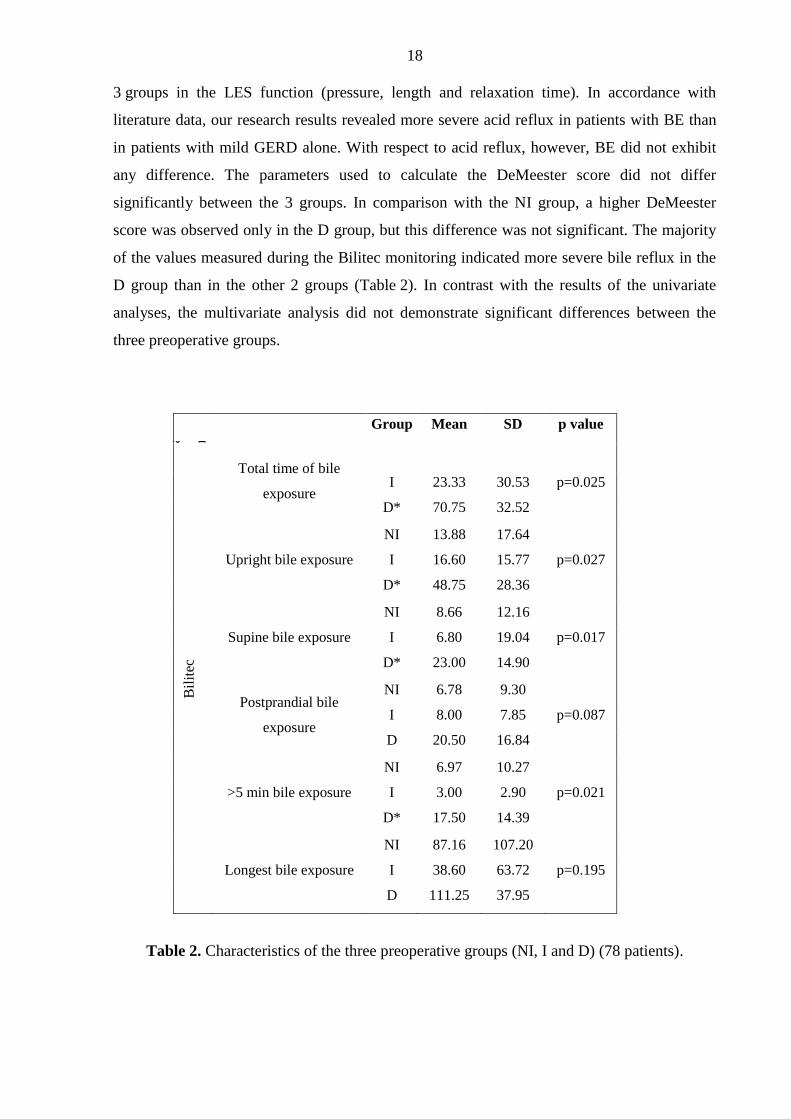
18
3 groups in the LES function (pressure, length and relaxation time). In accordance with
literature data, our research results revealed more severe acid reflux in patients with BE than
in patients with mild GERD alone. With respect to acid reflux, however, BE did not exhibit
any difference. The parameters used to calculate the DeMeester score did not differ
significantly between the 3 groups. In comparison with the NI group, a higher DeMeester
score was observed only in the D group, but this difference was not significant. The majority
of the values measured during the Bilitec monitoring indicated more severe bile reflux in the
D group than in the other 2 groups (Table 2). In contrast with the results of the univariate
analyses, the multivariate analysis did not demonstrate significant differences between the
three preoperative groups.
Group Mean SD p value
P a t i e n t s ’ c h a r a c t e r i s t i c s
Bil
itec
Total time of bile
exposure
I 23.33 30.53 p=0.025
D* 70.75 32.52
Upright bile exposure
NI 13.88 17.64
I 16.60 15.77 p=0.027
D* 48.75 28.36
Supine bile exposure
NI 8.66 12.16
I 6.80 19.04 p=0.017
D* 23.00 14.90
Postprandial bile
exposure
NI 6.78 9.30
I 8.00 7.85 p=0.087
D 20.50 16.84
>5 min bile exposure
NI 6.97 10.27
I 3.00 2.90 p=0.021
D* 17.50 14.39
Longest bile exposure
NI 87.16 107.20
I 38.60 63.72 p=0.195
D 111.25 37.95
Table 2. Characteristics of the three preoperative groups (NI, I and D) (78 patients).

19
4.2. POSTOPERATIVE RESULTS (STUDY 2)
In part 2 of the study, the efficacy of the antireflux surgery was assessed in view of the
postoperative results.
4.2.1. Symptomatic Outcome
Based on the Visick score[17]
determined in Group 1 after the surgery, at the early surgical
follow-up visit (at 3 months), complaints were gone or improved in 73% of the patients, they
were unchanged in 15%, and 12% of the patients reported worsening, primarily with a leading
symptom of dysphagia. In Group 2, 81% of the patients were complaint-free or reported
improved symptoms, 15% had unchanged complaints, and worsening was observed in 4%.
Dysphagia was the predominant symptom also in this group.
4.2.2. Postoperative Functional Results
After the surgery, patients were subjected to follow-up functional examinations and
endoscopy; the mean follow-up time was 13.8 ± 19.31 months in Group 1 and 16.7 ±
17.00 months in Group 2. The mean LES pressure was significantly increased compared with
the preoperative value in both groups (17.58 ± 7.60 mmHg in Group 1 and 18.70 ±
6.74 mmHg in Group 2). After the surgery, the LES length and relaxation time did not show a
statistically significant difference compared with the preoperative values (Table 4).
Based on the follow-up pH-metry, the number and duration of acid reflux episodes
significantly decreased in both groups. The postoperative DeMeester scores returned to the
normal range: they decreased to a mean score of 7.7 ± 17.41 in Group 1 and 12.7 ± 30.74 in
Group 2. When following the patients subjected to Bilitec monitoring before the surgery, a
decrease in the occurrence of bile reflux was also observed (Table 3).

20
FUNCTIONAL RESULTS GERD BE
Preop Postop p value Preop Postop p value
LES mean ± SD mean ± SD mean ± SD mean ± SD
PRESSURE (mmHg) 12.10±7.93 17.58±7.60 <0.001 12.58±9.03 18.70±6.74 <0.001
pH-METRY
DeMeester score 18.85±21.39 7.73±17.41 <0.001 41.93±51.15 12.72±30.74 <0.001
Bilitec
Total time of bile exposure 10.50±17.72 17.00±22.24 NS 26.97±28.79 22.08±30.57 <0.001
Table 3. Results of postoperative functional examinations in patients with GERD and BE.
4.2.3. Postoperative Results by BE Subgroup (Study 3)
In part 3 of the study, the rate of BE regression was studied in function of the laparoscopic
antireflux surgery. The early quality of life measures, the results of the early postoperative
functional examinations and the long-term endoscopic follow-up results were summarized for
the three BE subgroups.
4.2.3.1. Early postoperative results
4.2.3.1.1. Symptomatic outcome
The Visick score varied somewhat within the groups—in patients with intestinal BE and also
in those with LGD, complaints were alleviated relative to those with NI metaplasia. The
assessment of the changes in both the subjective and objective complaints demonstrated that
the symptoms recorded during the preoperative period tended to be relieved after laparoscopic
Nissen fundoplication. In accordance with our expectations, dysphagia increased.
4.2.3.1.2. Postoperative functional examinations (manomentry, 24-hour pH studies and
Bilitec)
Postoperative manometry, pH-metry and Bilitec monitoring did not reveal statistically
significant differences between the three groups. Changes in the LES function, which also
indicate the efficacy of the surgery, demonstrated that the postoperative pressure in the lower
esophagus was significantly increased relative to that measured preoperatively, whereas the

21
relaxation time remained unchanged. As a consequence of the surgical technique (a loose and
narrow Nissen floppy), the length of the LES was unchanged after fundoplication, but its
function (pressure) was restored, thus preventing acid and bile reflux. Comparison of the
results of pH-metry before and after the procedure between the three groups confirmed the
above findings, as mean DeMeester scores were clearly decreased after the surgery.
Accordingly, the incidence and severity of bile reflux were reduced, or this symptom was
eliminated. The multivariate analysis confirmed significant changes only in LES pressure and
the results of pH-metry between the preoperative and postoperative groups.
4.3. RESULTS OF THE ENDOSCOPIC FOLLOW-UP OF THE BE SUBGROUPS
(ENDOSCOPIC SURVEILLANCE)—LONG-TERM ENDOSCOPIC
SURVEILLANCE
The mean duration of endoscopic follow-up was 42 + 16.19 months. Postoperative endoscopy
was performed only in 64 patients (82%, 64/78). 14 patients, who were not subjected to upper
gastrointestinal endoscopy, were excluded from the long-term analysis.
Complete regression Partial regression No change Progression
Overall group
SSBE (n=56) 10 (17.9%) 5 (8.9%) 30 (53.6%) 11 (19.6%)
LSBE (n=8) 0 4 (50%) 4 (50%) 0
NI (n=44) 6 (13.6%) 4 (9.1%) 23 (52.3%) 11 (25%)
IM (n=15) 3 (20%) 3 (20%) 9 (60%) 0
LGD (n=5) 1 (20%) 2 (40%) 2 (40%) 0
Total (n=64) 10 (15.6%) 9 (14.1%) 34 (53.1%) 11 (17.2%)
Table 4. Endoscopic and histopathological changes of BE after laparoscopic fundoplication
(64 patients)
Before the antireflux surgery, SSBE was present in 56 patients and LSBE in 8 patients.
Preoperative histological examinations indicated FM in 11, CM in 33, IM in 15 and LGD in
5 patients. The postoperative check-up demonstrated a total regression of BE in 10 patients
(15.6%). Partial regression was seen in 9 cases (14.1%), no further progression in 34 patients
(53.1%), and progression from FM to CM in 4 patients (6.2%) or from CM to IM in
7 patients (11%), but no cases of dysplastic or malignant transformation were recorded.
There was no further progression in the patients with LGD, and in 3 of these 5 patients, LGD
disappeared, leaving only residual IM (Table 4).

22
There was no difference in the length of the follow-up period between the total regression
group and the other groups (partial, no change and progression). Where the regression of BE
was observed, the postoperative functional examination results of manometry (pLES) and
pH-metry were significantly better compared with those measured in the groups where no
changes in BE occurred, or progression of BE was found. We did not find differences
between the groups in the results of postoperative Bilitec monitoring, except for the longest
exposure values (Table 5).
Groups
Regression (SD) No change (SD) Progression (SD) p value
Ma
nom
etr
y pLES (mmHg) 18.04 (±6.405) 9 (±7.735) 11.02 (±7.815) 0.003
rLES (s) 10.04 (±1.613) 10.03 (±2.831) 9.89 (±4.285) 0.988
lLES (cm) 3.21 (±0.699 3.14 (±1.424) 2.89 (±1.269) 0.571
pH
-metr
y
Total time of acid exposure
<pH 4 23.77 (±25.21) 105.29 (±89.191) 112.2 (±82.974) <0.001
Upright acid exposure <pH 4 21.23 (±24.1229 79.79 (±67.776 87.9 (±74.929) 0.002
Supine acid exposure <pH 4 2.62 (±3.595 25.75 (±33.216) 24.6 (±21.798) 0.002
Postprandial acid exposure
<pH 4 12.42 (±16.649) 48.63 (±46.04) 61.3 (±53.506) 0.009
>5 min acid exposure <pH 4 0 (±0) 5.46 (±8.495) 5.1 (±5.607) <0.001
Longest acid exposure <pH 4 1.38 (±1.557) 19.33 (±27.223) 19.6 (±15.82) <0.001
DeMeester score 3.52 (±3.617) 40.88 (±51.37) 43.089 (±6.094) <0.001
Bil
itec
Total time of bile exposure 4.75 (±6.292) 32.05 (±34.861) 23 (±28.605) 0.097
Upright bile exposure 4 (±4.83) 19.21 (±22.062) 15.89 (±18.395) 0.143
Supine bile exposure 0.75 (±1.5) 13.05 (±19.478) 7.44 (±12.69) 0.295
Postprandial bile exposure 1 (±1.414) 8.11 (±10.954) 7.89 (±10.55) 0.117
>5 min bile exposure 0.75 (±1.5) 9.05 (±13.206) 6.33 (±9.206) 0.138
Longest bile exposure 3.25 (±5.188) 81.72 (±99.8) 72.78 (±93.641) 0.050
Table 5. Comparison of the postoperative functional examinations and the changes in BE
(between the three groups: regression, no change, progression (64 patients).

23
4.4. COMPLICATIONS OF MINIMALLY INVASIVE ANTIREFLUX PROCEDURES
AND THEIR TREATMENT (STUDY 4)
In part 4 of our study, the risk of the surgical treatment was evaluated. We processed the data
on surgical complications of patients subjected to surgery because of GERD or BE between
2001 and 2008. Intraoperative complications, as well as early (within 30 days) and late
complications were analyzed in detail.
Conversion was required in 1 case in the GERD group because of adhesions. Open
splenectomy (requiring postoperative transfusion) was performed in 1 case in the BE group
because of spleen bleeding. Intraoperative chest tube insertion was required in 1 case in the
GERD group because of left-sided pneumothorax. Reoperation was not performed in either
group in the early postoperative period. In the GERD group, 2 patients were given a total of
6 units of pRBC because of bleeding, and subcutaneous emphysema was detected in 1 case,
which did not require further treatment.
During the early and late follow-up in the GERD group, observation at the institution was
required in 3 cases because of dysphagia and stenosis: in 1 case, the complaints of the patient
resolved spontaneously, in 1 case, endoscopic foreign body retrieval was performed because
of food bolus obstruction (in the early period), and in 1 case, endoscopic balloon dilation was
performed (in the late period). Usually, a satisfactory result was achieved with dilation, and
reoperation has not been required in our practice to date. Endoscopic follow-up described the
appearance of ulcer in 2 cases in the GERD group, and in 2 cases, BE was developed. In
1 case, cardiac resection was needed because of erosion due to the implanted mesh 7 months
after the surgery. In the BE group, esophageal dilation was performed because of dysphagia in
2 cases. There were no mortalities in either group.
4.5. ENDOSCOPIC TREATMENT OF SPONTANEOUS ESOPHAGEAL RUPTURE,
A COMPLICATION DEVELOPED ON THE BASIS OF BE
In this section, we present the minimally invasive treatment strategy of spontaneous
esophageal rupture (Boerhaave’s syndrome), a rare condition associated with BE. A 53-year-
old male patient with lower-third rectal adenocarcinoma (T2N1) was admitted. He was known
to have gastroesophageal reflux disease complicated with Barrett’s esophagus (intestinal
metaplasia with low-grade dysplasia). On the second postoperative day after a low anterior
rectal resection, forceful vomiting occurred and was followed by chest pain without clinical
signs of esophageal perforation. The immediate chest x-ray revealed only a small amount of

24
pleural effusion on the right. The follow-up chest x-ray (acquired 12 hours later), however,
demonstrated an increase in the amount of the pleural effusion, and hydropneumothorax was
developed. A contrast swallow with a water-soluble contrast agent confirmed the presence of
a transpleural esophageal rupture (Figure 1). The immediate upper gastrointestinal endoscopy
showed a mucosal rupture of 5 to 7 mm in length on the posterior wall of the esophagus, 4 cm
above the gastroesophageal junction. The mucosal tear was successfully closed with 3
endoscopic clips (Olympus Quick Clip 2) (Figure 2). The endoscopic closure was
supplemented with right thoracic drainage, gastrostomy and catheter feeding jejunostomy.
Eight days after the endoclip application, esophagography demonstrated no further leakage,
and oral feeding could be resumed. There were no complications and the patient was
eventually discharged 14 days after the endoscopic intervention. Control endoscopy showed
only scar tissue at the site of the closed perforation with LGD of BE.

25
5. DISCUSSION
Reflux disease affects more than one third of the population, and it may range from an
asymptomatic condition through a condition without inflammatory signs (non-erosive reflux
disease) to a symptomatic condition accompanied by severe erosion and its complications that
considerably worsen the quality of life. Parallel to this, the incidence of esophageal
adenocarcinoma shows an increase in the developed world. In the presence of Barrett’s
metaplasia developed on the basis of reflux, the risk of developing esophageal
adenocarcinoma is 30 to 125 times higher than in the normal population.[39,40]
It has been known for a long time that in some cases, depending on the severity of the reflux,
the acidic gastric contents regurgitating into the esophagus may cause symptoms only,
whereas in other cases, they may damage the squamous epithelium lining the esophagus,
resulting in erosion, inflammation and, later, ulceration and stricture.[5]
In the development of reflux esophagitis, at the microscopic level, the opening and widening
of the gap between mucosal cells play a role. It was successfully triggered under experimental
conditions with both mild and severe acid or mixed reflux.[41]
At the site of the epithelial
defect that is developed in case of persistent reflux, the regeneration starting from the
submucosal pluripotent Schaffer glands may result in that columnar epithelium, which is more
resistant to the acidic environment, replaces the squamous epithelium, i.e., Barrett’s
esophagus is developed. Its predictive factors are the total time of acid reflux, its severity and
the consequentially worsening lower esophageal sphincter (LES) function.[13,42,43,44,45,46,47,48]
Our study supports the observation that the exposure to reflux is longer and more severe in the
group of patients with Barrett’s metaplasia than in the group of patients with reflux alone. In
our sample, however, no difference was found between the two groups in the duration of
complaints and the LES impairment. Nevertheless, the mean history of 5.6 years is in
accordance with the observation of Öberg who found that approximately the same time
(6.2 years) is required for the columnar epithelial metaplasia to appear as a result of the
increased exposure to acid. [49]
It can be established, based on the above, that one of the most
important factors in the squamous–columnar transformation is the appearance of acid in the
esophagus.

26
The driving force behind the transition within the columnar epithelium and the appearance of
intestinal metaplasia and dysplasia is the bile that is mixed with acid reflux.
In the acidic–biliary environment, several “evolutionary responses” may appear often parallel
to each other (fundic, cardiac and intestinal or even respiratory ciliary columnar epithelium,
pancreatic acinar or ductal metaplasia, or low-grade, high-grade dysplasia or even in situ
carcinoma). It is supposed that Schaffer glands have an important role in the development of
this diverse picture, because their pluripotent germ cells may be responsible for the
heterogeneous responses to the inflammatory damage. [8]
Besides the simultaneous presence
of metaplasias, transformation into each other may also be supposed. Nevertheless, many of
the above forms of metaplasia and dysplasia may be present in the mucosa even at the same
time. [8]
A true risk of potential malignancy is associated with the appearance of IM.[28, 52]
However, it is a known fact that the malignant transformation of non-intestinal epithelium
cannot be excluded either, although its estimated risk is considerably lower (0.07% annually)
than in the concomitant presence of intestinal metaplasia (0.38% annually).[28, 52]
In other
publications, the rate of dysplasia and carcinoma was almost the same in case of non-
intestinal metaplasia than in IM. It must be added, however, that during the 5-year follow-up
of this group, IM appeared in more than 50% of the cases, and this ratio reached 90% after
10 years.[53]
It appears to support our conclusions below about the possible limitations of
biopsy and the ability of metaplastic processes to transform into each other.
Although multiple biopsy samples may help assess the precise status of BE, only the
momentary status of a small area can be assessed this way, which makes it hard to evaluate
the efficacy of the treatment and the change in the condition. During the endoscopic
examinations, multiple sampling is performed as per the Seattle protocol: on the one hand,
from the visible Barrett lesions themselves, and on the other hand, from each esophageal
quadrant with 2 cm intervals[26]
. Increasing the number of biopsy samples clearly improves
the ability to detect IM. According to the results of Harrison et al., with only 4 samples taken
from the same patients, IM could be detected in only half of the IM cases previously
confirmed with samples taken from 8 biopsy sites.[54]
In view of the above, we still consider it important, when Barrett’s esophagus is developed,
not to focus only on intestinal metaplasias (that are confirmed to carry the potential for
malignant transformation) but to follow non-intestinal (fundic and cardiac) metaplasias and
other histological phenotypes as well.

27
We suppose that a more severe acid-bile reflux has a pathogenic role in the development of
dysplasia. Because of this, patients with BE in our study were assigned to three different
groups based on the presence of conditions indicating carcinogenesis or its risk, depending on
whether the histological sample taken during the endoscopy showed non-intestinal (i.e.,
fundic or cardiac) metaplasia (Group 1), intestinal metaplasia (Group 2) or low-grade
dysplasia on the basis of the latter (Group 3). Taking into account that parallel metastatic and
dysplastic conditions were likely in the biopsy samples, patients were always sorted based on
the most severe condition found.
Patients with BE reported a mean duration of complaints of 5.6 years before the surgery. It
was not different from the length of history reported by patients who underwent surgery
because of GERD. There was no correlation between the severity of Barrett metaplasia
(intestinal metaplasia and low-grade dysplasia) and the duration of complaints either and,
paradoxically, reflux complaints of longer duration were observed in the non-intestinal group,
compared to the intestinal one. There were no differences in patient demographics (age, BMI,
etc.) in the case of BE or, within this group, LGD.
When comparing patients who suffer from GERD with those who have BE, the more severe
acid reflux confirmed in the BE group may cause metaplasia but a further role of acid reflux
in the metaplasia–dysplasia transition in the BE subgroups could not be confirmed in the
second half of our study. Nevertheless, our study showed that bile reflux was significantly
more common and more severe in the low-grade dysplasia (LGD) group of BE patients than
in the groups of patients with non-dysplastic metaplasia.[58]
Our hypothesis that changes in the anatomy of the gastroesophageal junction, a larger hiatal
hernia or decreased LES pressure, decreased LES relaxation time, or a shorter LES, are more
common in case of LGD, was not confirmed.
Conservative and surgical therapies are both accepted in the treatment of reflux, which has a
central role in the development of the above mucosal transformation. With minimally invasive
surgery becoming part of everyday practice, we can say that the morbidity risk of the
laparoscopic antireflux surgery is low compared to its possible benefits to the patients.[59]
The
advantage of laparoscopic antireflux surgery over conservative therapy is that it attempts to
restore the previous anatomical situation, i.e., it eliminates hiatal hernia by reconstructing the
posterior diaphragmatic crus, restores the angle of His by retracting the lower portion of the
esophagus into the abdomen, and restores the LES function with fundoplication. Unlike

28
proton-pump inhibitors, it may eliminate not only acid reflux but also bile reflux. Compared
with the permanent, lifelong medical treatment, it may be considered cost effective. However,
opinions are divided on its long-term efficacy.[60, 61]
A long-term complaint-free status can be
expected in case of a correctly performed antireflux surgery. It can be established based in the
LOTUS trial that, regarding long-term efficacy, laparoscopic antireflux procedure is as
effective as medical treatment.[62]
Our patients, therefore, had heterogeneous indications for surgery but the procedure was
mostly performed after an unsuccessful conservative therapy. In our study, the patients in both
groups underwent surgery after a mean 19 to 20 months of unsuccessful medical treatment.
Our early postoperative functional examinations confirmed that laparoscopic antireflux
surgery could achieve improvement, i.e., good reflux control, even in this presumably selected
patient population with poorer prognosis and poorer response to conservative therapy
(unsuccessful after a mean duration of one year and a half). LES pressure returned to the
physiological range and the DeMeester score, which best describes acid reflux, decreased to
normal values in both groups. Surgical treatment, therefore, may have additional advantages
over medical treatment. Our results, however, must be considered to be of limited value
because of the short follow-up of the functional examinations. Its mean duration did not
exceed eighteen months in either group.
A further advantage of laparoscopic antireflux surgery is that it can be standardized, and
therefore the steps of the procedure can be reconstructed later at any time, and the results from
different institutions are comparable.[62]
Nissen’s fundoplication creates a relatively stronger
reflux barrier than partial fundoplication.[67]
Dysphagia and also later dilation are more
common in case of a 360-degree fundoplication but only among patients with decreased
esophageal motility. However Nissen’s fundoplication may provide the best reflux control in
the long term.[67, 68, 69, 70]
Since the first observation made by Brand et al.[73]
(1980), it has become a generally known
fact that antireflux surgery also creates an opportunity for the regression of BE. Based on the
results of randomized and retrospective studies conducted to date, it can be established that
antireflux surgery is more effective in preventing the progression of BE than medical
treatment. [74, 75, 76, 77, 78, 79, 80, 81]
According to the most recent meta-analysis, antireflux surgery
clearly has a beneficial effect on the regression of BE and dysplasia.[82]
It has been found in
some studies that surgery does not lead to an obvious decrease in the occurrence of
adenocarcinoma, despite the excellent results published by numerous institutions about the

29
beneficial effect of laparoscopic fundoplication in the treatment of reflux disease. [83, 84]
According to a recent Swedish study, antireflux surgery does not prevent the development of
esophageal or cardiac adenocarcinoma in some of the patients with GERD. [85]
The views on the role of antireflux surgery in prevention are quite contradictory in the
literature. To date, no meta-analysis confirming or refuting a preventive effect with clear
evidence has been published, and no large, prospective studies are expected in the near future
either because of the special and small patient population.
This assumption seems to be confirmed by the fact that during the 42-month endoscopic
follow-up of our patients subjected to surgery because of Barrett’s esophagus, an unchanged
status was observed in 53% of the cases, and regression was detected in a further 30%.
Progression occurred in only 17%, and all of these cases were observed in the non-intestinal
metaplasia group. Dysplasia was not developed in the group of patients with intestinal
metaplasia, and no further progression (to high-grade dysplasia or carcinoma) occurred in the
low-grade dysplasia group during the study period.
In the patient group showing regression, the postoperative functional results were
significantly better than in the groups that did not show regression. However, we consider it
important that the majority of our cases were short-segment BE. Based on the above, it can be
established that in certain (presumably early) cases of BE, a laparoscopic antireflux surgery
that provides effective reflux control may achieve regression even in patients not responding
to medical treatment. Csendes et al.[81]
have reached a similar conclusion. According to our
observations, laparoscopic antireflux surgery is associated with a low morbidity rate, and may
decrease the subjective complaints of short-segment BE patients in the long term (Visick
Grade I and Grade II in 86.3 to 100% of the patients), it leads to the regression of intestinal
metaplasia (IM) in two-thirds of the patients, and the IM does not progress to LGD, HGD or
adenocarcinoma. In case of BE, surgical treatment should be considered also according to
DeMeester et al., because they often observed the regression of LGD after antireflux
surgery.[13]
The regression of IM is more common in the group of patients treated surgically
than in those who receive medical treatment.[82]
A randomized study comparing the medical
and surgical treatment has confirmed that a correctly performed antireflux surgery
significantly decreases the rate of reflux esophagitis and stricture, and the segmental length of
BE also significantly decreases after the surgery.[80]
The rate of new-onset dysplasia was
statistically different between the surgery group and the medically treated group (2% vs. 20%,
respectively).[75, 86, 87, 88, 89]
At the same time, the risk of malignant transformation was not

30
lower than in the medically treated group. However, the same incidence of developing cancer
in the two groups may be influenced by the fact that surgical treatment is performed in
patients with more severe reflux - after medical treatment has failed.[86, 87, 89, 90]
A preventive
effect of laparoscopic antireflux surgery on the development of adenocarcinoma was not
confirmed by the Swedish cohort study published by Lagergren et al. either.[91, 92]
It must be
noted, however, that this study compared the rate of adenocarcinoma with that in the healthy
population and not in patients with reflux.[93]
A “new-onset” BE developed after the antireflux
surgery and the progression of an already present BE raise many concerns against the surgical
treatment.
In the GERD patient group, the occurrence of metaplasia during our postoperative observation
may have two explanations. The first is unsuccessful surgery. De novo BE may be expected in
case of inadequate reflux control. It is contradicted by the fact that the results of our
functional examinations did not differ from those observed in patients without progression.
The second, more likely explanation lies in the limitation of biopsy already mentioned, that is,
that the quadrant biopsy samples “taken blindly” in case of GERD provide a histological
picture of a small area only, which does not exclude the prior presence of Barrett metaplasia
in other areas, recognized only at the time of the second biopsy. Nevertheless, our opinion is
that the clinical manifestation of BE should not be considered a uniform condition.
Although surgery that provides adequate reflux control can lead to regression (primary
prevention) in a certain group of patients with BE, Barrett metaplasia was nonetheless
observed after surgery in another patient group and, in a smaller portion of the patients,
progression is not excluded either. Recognizing this patient group and following it more
closely are indispensable for secondary prevention, i.e., the early recognition and successful
treatment of cancer.
The conclusion of DeMeester et al. may possibly explain progression - if carcinogenesis
already started before the surgery because of the mixed reflux, and the meta- and dysplastic
cells already got out of autoregulation control due to the genetic damages, the antireflux
surgery, obviously, does not provide protection against advanced dysplastic processes, and in
this case, adenocarcinoma may appear within 5 years after the surgery. In case of cancers
developed later than this, they confirmed the recurrence of reflux.[13]
Other studies[94, 95, 96]
also point out that late adenocarcinoma following antireflux surgery can be explained by
postoperative reflux due to an unsuccessful surgery or by recurrent reflux, and they emphasize
the importance of pH-metry during follow-up.
31
It is also possible, however, that BE does not expose to a higher risk of developing
adenocarcinoma but appears only as a coincidence. The fact that the histological examination
confirmed BE next to adenocarcinoma in only half of the resections performed because of
tumor may support this theory.[97, 98]
Jamieson hypothesized that adenocarcinoma may be
developed not or not only from the Barrett epithelium during tumorigenesis but the
transformation of a pluripotent germ cell starts in response to inflammation and epithelial
damage due to the reflux.[92]
In this case, although the reflux-induced inflammation is also
responsible for the development of BE, BE should be considered an indicator of the severity
of reflux rather than a premalignant condition. The most likely case is that carcinogenesis
does not occur in one way only, and dysplasia and cancer developed on the basis of BE is just
one possible way in this process. Going ahead with this hypothesis, from the perspective of
carcinogenesis, therefore, a successful antireflux surgery performed in time may be of
preventive effect in certain patients with BE. Nevertheless, taking the slow progression of the
condition and the great heterogeneity of BE into account, regular long-term endoscopic
follow-up and biopsies are indispensable for the successful treatment of reflux disease and
Barrett’s esophagus. To confirm whether laparoscopic antireflux surgery can prevent the
progression of Barrett’s esophagus in the long term, repeat functional examinations to verify
the effective functioning of the antireflux barrier are required besides endoscopic follow-up.
The minimally invasive surgical treatment performed at our center was an effective treatment
alternative to unsuccessful medical treatment, without mortality and with a low morbidity
rate. The conversion rate of 0.6 to 1.3% is considered low. As to late complications, the most
common is dysphagia that requires hospitalization (1.7 to 2.6%), which can be considered an
“efficacy indicator” of Nissen’s fundoplication. The most severe late complication is erosion
of the esophageal wall after mesh placement (1.3%). Although it appears to be rare, its rate of
7.1% in patients with mesh placement only is high. Therefore, in our practice, besides the
choice of material (Teflon or composite mesh), we avoid wrapping the esophagus around
completely.
A similarly rare severe complication is spontaneous perforation or Boerhaave’s syndrome
developed on the basis of BE ulcer. According to our knowledge, the closure of the
perforation opening with endoscopic clips that we performed in our patient is the first
documented successful case in Hungary. The condition is the result of a pressure increase in
the lumen of the esophagus, which is primarily caused by voluminous vomiting. Since our
patient had known reflux disease complicated by BE with LGD, and esophagitis, a weakening

32
of the distal esophageal wall could be assumed as well. The condition was considered fatal
until the first successful surgical treatment performed by Barrett in 1947.[100]
Today, despite
the more effective treatment options, mediastinitis and the rapidly developed septic condition
are often irreversible. In cases where surgery is performed after more than 24 hours, the
mortality rate exceeds 20 to 30%.[101]
In the past years, several cases of successful use of
endoscopic clips and self-expanding stents in the treatment of esophageal rupture have been
reported.[105, 106]
Closure of an esophageal injury with endoscopically placed clips was first
performed in 1995 (the injury occurred during pneumatic dilation in a patient with
achalasia).[107]
In our case, the complete esophageal perforation (with a rupture on the
esophageal wall and also the mediastinal pleura) was detected within 24 hours. Since the
visible rupture on the esophagus was only 5 to 7 mm, it could be successfully closed with
endoscopic clips. Endoscopic stents have been successfully used in the treatment of different
types of esophageal perforation, including Boerhaave’s syndrome [115, 116, 117, 118, 119, 120]
To
summarize, we can establish based on the available experience that endoscopic and minimally
invasive surgical methods, if proper conditions are met, may be therapeutic alternatives in the
treatment of Boerhaave’s syndrome developed on the basis of BE.

33
SUMMARY, OUR MOST IMPORTANT RESULTS
1. The severity of the pathological acid reflux developed parallel to the incompetent
functioning of the impaired lower esophageal sphincter potentiates, besides the
inflammation in the lower third of the esophagus, the start of metaplastic processes
and, ultimately, the development of Barrett’s esophagus.
2. In response to the bile reflux accompanying the acid reflux, dysplastic changes may
start in the metaplastic columnar epithelium (that appeared due to acid reflux).
3. In selected GERD and BE patients resistant to medical treatment, Nissen’s correctly
performed laparoscopic surgery can successfully control (eliminate or decrease)
gastroesophageal reflux and is associated with a low morbidity rate.
4. The antireflux surgery may stop the progression of Barrett’s esophagus and result in
regression in some patients. Nevertheless, further long-term follow-up is required to
confirm the assumed preventive effect of antireflux surgery in the process of
carcinogenesis.
5. Endoscopic methods, if proper conditions are met, may be therapeutic alternatives in
the treatment of esophageal perforation.

34
ACKNOWLEDGEMENTS
First and foremost I wish to express my gratitude to my consultant, Prof. György Lázár for
having been given the opportunity and for the ongoing support and tireless inspiration in my
thesis.
I am particularly grateful to all members of the South Hungarian Regional Surveillance Group
for the Study of Barrett’s Esophagus for their constant help and support in the exemplary
gastroenterologist-surgeon-pathologist cooperation.
I would like to offer my special thanks for his assistance in doing the statistics to Dr. Krisztina
Boda and Ferenc Rárosi.
And I am obliged to all my young colleagues and medical students in the Department of
Surgery without whom this work would have never been completed.
At last, but not at least I would like to express my gratitude for my family for their support
and understanding.
Our study was sponsored by the Hungarian Research Fund, OTKA (grant K 73141), 340/09-
ETT from the Hungarian Ministry of Social Welfare and the TÁMOP-4.2.2.A-11/1/KONV-
2012-0035 projects.


![Minimally invasive non-surgical vs. surgical approach for ...dictable [12]. More recently, minimally invasive surgical therapy (MIST), modified minimally invasive surgical therapy](https://img.pdfslide.net/doc/110x75/5eddda76ad6a402d6669115c/minimally-invasive-non-surgical-vs-surgical-approach-for-dictable-12-more.jpg)