Embed Size (px)
Citation preview
e at SciVerse ScienceDirect
Toxicon 60 (2012) 851–863
Contents lists availabl
Toxicon
journal homepage: www.elsevier .com/locate/ toxicon
Mipartoxin-I, a novel three-finger toxin, is the major neurotoxiccomponent in the venom of the redtail coral snake Micrurus mipartitus(Elapidae)
Paola Rey-Suárez a, Rafael Stuani Floriano b, Sandro Rostelato-Ferreira b,Mónica Saldarriaga-Córdoba c, Vitelbina Núñez a,d, Léa Rodrigues-Simioni b, Bruno Lomonte e,*
a Programa de Ofidismo y Escorpionismo, Universidad de Antioquia, Medellín, ColombiabDepartamento de Farmacologia, Faculdade de Ciências Médicas, Universidade Estadual de Campinas, SP, Brazilc Facultad de Ciencias, Universidad Iberoamericana de Ciencias y Tecnología, Chiled Escuela de Microbiología, Universidad de Antioquia, Medellín, Colombiae Instituto Clodomiro Picado, Facultad de Microbiología, Universidad de Costa Rica, San José, Costa Rica
a r t i c l e i n f o
Article history:Received 1 March 2012Received in revised form 4 May 2012Accepted 24 May 2012Available online 4 June 2012
Keywords:Mipartoxin-IThree-finger toxinMicrurus mipartitusSnake venomNeurotoxicity
* Corresponding author. University of Costa RicaPicado, San Jose, Costa Rica. Tel.: þ506 2229 0344; f
E-mail address: [email protected] (B. Lom
0041-0101/$ – see front matter � 2012 Elsevier Ltd10.1016/j.toxicon.2012.05.023
a b s t r a c t
The major venom component of Micrurus mipartitus, a coral snake distributed fromNicaragua to northern South America, was characterized biochemically and functionally.This protein, named mipartoxin-I, is a novel member of the three-finger toxin superfamily,presenting the characteristic cysteine signature and amino acid sequence length of theshort-chain, type-I, a-neurotoxins. Nevertheless, it varies considerably from related toxins,with a sequence identity not higher than 70% in a multiple alignment of 67 proteins withinthis family. Its observed molecular mass (7030.0) matches the value predicted by its aminoacid sequence, indicating lack of post-translational modifications. Mipartoxin-I showeda potent lethal effect in mice (intraperitoneal median lethal dose: 0.06 mg/g body weight),and caused a clear neuromuscular blockade on both avian and mouse nerve-musclepreparations, presenting a post-synaptic action through the cholinergic nicotinic receptor.Since mipartoxin-I is the most abundant (28%) protein in M. mipartitus venom, it shouldplay a major role in its toxicity, and therefore represents an important target for developinga therapeutic antivenom, which is very scarce or even unavailable in the regions where thissnake inhabits. The structural information here provided might help in the preparation ofa synthetic or recombinant immunogen to overcome the limited venom availability.
� 2012 Elsevier Ltd. All rights reserved.
1. Introduction
Snake venoms are recognized as a vast, yet largelyunexplored source of bioactive proteins with potent toxiceffects. Acquisition of such toxic proteins was accom-plished through the recruitment of a small number of’non-toxic’ genes for expression in specialized oral glands,followed by an accelerated evolution process of divergence
, Instituto Clodomiroax: þ505 2292 0485.onte).
. All rights reserved.
and neofunctionalization under strong positive selection(Nakashima et al., 1993; Fry et al., 2006). This evolutionarypathway allowed the advanced snakes (Caenophidia) toundergo a striking change in their mode of subduingprey, shifting from a mechanical (based on constriction)to a biochemical (based on venom injection) strategy(Calvete et al., 2009).
Highly venomous snakes of medical relevance areclassified within the families Atractaspidae, Viperidae, andElapidae (Fry et al., 2003a,b). The latter contains over 60genera and nearly 300 species, one-third of which belongto ‘coral snakes’, a major radiation encompassing six genera
P. Rey-Suárez et al. / Toxicon 60 (2012) 851–863852
distributed in Asia and the Americas. The New World coralsnakes are currently classified within the genera Micrurus,Micruroides, and Leptomicrurus, the former being the mostdiverse, including over 70 species (Castoe et al., 2007).Is spite of the extraordinary biological diversity andcomplexity of coral snakes, distribued from Southern USAto Argentina, factors such as low venom yields, and theirpoor survival in captivity, pose important limitations forthe study of their venoms, as well as for the productionof therapeutic antivenoms. Nevertheless, newer sensitivetechniques for protein analysis have opened possibilitiesto characterize such scarce biological materials, andaccordingly, a few proteomes of Micrurus venoms havebeen described recently (Olamendi-Portugal et al., 2008;Fernández et al., 2011; Corrêa-Netto et al., 2011; Rey-Suárezet al., 2011; Ciscotto et al., 2011).
Micrurus mipartitus, commonly known as ‘redtail coralsnake’, ‘rabo de ají’, or ‘gargantilla’, inhabits the Caribbeanversants of Nicaragua, Costa Rica, the central Panamáisthmus, and large areas of Ecuador, Colombia, andVenezuela. Although this snake causes few human enve-nomings in comparison to pitvipers (family Viperidae),accidents may be fatal if unattended. Patients presentperipheral neurotoxic manifestations such as bilateralptosis and progressive respiratory paralysis which mayrequire ventilatory support (Otero, 1994). A major medicaldifficulty in dealing with envenomings by M. mipartitus isthe scarcity, or even complete unavailability in most of itsgeographic range, of a specific therapeutic antivenom. Thisproblem is aggravated by the lack of cross-protection bymore readily available antivenoms prepared against speciessuch as the Central American coral snake Micrurus nigro-cinctus, due to their antigenic differences (Cohen et al.,1971; Bolaños et al., 1975, 1978; Rey-Suárez et al., 2011).
Proteomic analyses of the venoms of M. mipartitusfrom Colombia and Costa Rica were recently reported(Rey-Suárez et al., 2011), aiming to provide a basic platformthat could be useful in the design of an antivenom.The study identified a prominent venom componentbelonging to the three-finger toxin (3FTx) family whichcaused lethality in mice. In the present work, this proteinwas isolated and characterized biochemically and func-tionally as a potent short-chain neurotoxin. The protein,named mipartoxin-I, is a new member of the 3FTx super-family which shows considerable amino acid sequencedifferences with other short-chain neurotoxins describedin the venoms of Elapidae snakes, including other Micrurusspecies. Due to its abundance in the venom ofM.mipartitus,and its high toxicity, the primary structure and three-dimensional modeling of mipartoxin-I here reportedmight be useful for the preparation of an immunogen,recombinant or synthetic, to elicit neutralizing antibodiesfor the treatment of envenomings by this snake species.
2. Materials and methods
2.1. Venom and toxin isolation
Venom was collected from five adult specimens ofM. mipartitus from Antioquia, Colombia. The venom waspooled, lyophilized, and stored at �20 �C. Fractionation of
venom (2 mg in 200 mL of water containing 0.1% trifluoro-acetic acid; TFA) was performed by RP-HPLC on a C18column (4.6 � 250 mm, 5 mm particle; Teknokroma) usingan Agilent 1200 chromatograph. Elution was performedat 1 ml/min by applying a gradient toward acetonitrilecontaining 0.1% TFA (solution B), as follows: 5% B for 5 min,5–15% B over 10 min, 15–45% B over 60 min, and 45–70% Bover 12 min, as previously described (Rey-Suárez et al.,2011). Absorbance was monitored at 215 nm, and themajor peak eluting atw26.5 min, named mipartoxin-I, wasmanually collected, dried in a vacuum centrifuge (Savant),and stored at �20 �C.
2.2. MALDI-TOF mass spectrometry (MS)
Mipartoxin-I was diluted in water containing 0.1% TFA,and dilutions ranging from 1000 to 10 ng weremixed at 1:1with either saturated sinapinic acid or a-cyano-hydrox-ycinnamic acid in 50% acetonitrile, 0.1% TFA, and spotted(1 mL) onto an OptiToF-384 plate for MALDI-TOF MS anal-ysis. Spectra were acquired on an Applied Biosystems4800-Plus instrument, in linear positive mode, using 500shots/spectrum and a laser intensity of 4200, over the m/zrange 4000–40,000. External calibration was performedwith CalMix-5 standards (ABSciex) spotted on the sameplate.
2.3. Electrospray ionization (ESI) MS
Mipartoxin-I, dissolved in 10 mL of 0.1% formic acid, wasloaded in a capillary tip (Proxeon) for direct infusion ina nano-ESI source coupled to an Applied Biosystems Q-Trap3200 mass spectrometer. Ionization was performed at1200 V and spectra were acquired in positive EnhancedMulti-Charge mode, in the m/z range 400–1600. Charge-state and deconvolution of the ion series were analyzedwith the aid of the Bayesian protein reconstruction toolof BioAnalyst v.1.5 software (ABSciex), and confirmed bymanual calculation.
2.4. Amino acid sequencing
Mipartoxin-I (250 mg) was dissolved in 50 mM ammo-nium bicarbonate and subjected to reduction with dithio-threitol (10 mM) and alkylation with iodoacetamide(50 mM). An aliquot of the reduced-alkylated protein wassubjected to N-terminal sequencing on a Shimadzu PPSQ-33A Protein Sequencer, according to the manufacturer. Therest of thismaterial was digested overnight with sequencinggrade bovine trypsin (in 25 mM ammonium bicarbonate,10% acetonitrile) at 37 �C. The resulting tryptic peptideswere separated by RP-HPLC on a C4 column (4.6� 150 mm;Vydac), eluted at 1 ml/min with a 0–70% acetonitrilegradient over 40 min, and manually collected. Each peakwas subjected to MS/MS fragmentation and de novo aminoacid sequencing, using the above-mentioned mass spec-trometers. For MALDI-TOF-TOF analyses, fragmentationspectra were acquired at 2 kV in positive reflectron mode,using a-cyano-hydroxycinnamic acid as matrix, 500 shots/spectrum, and a laser intensity of 3000. Spectra weresearched using the Paragon� algorithm of ProteinPilot 4.0
P. Rey-Suárez et al. / Toxicon 60 (2012) 851–863 853
(ABSciex) and the UniProt/SwissProt database (20100622),or interpreted manually. Peptides that did not producewell-resolved fragmentation spectra using MALDI werefurther subjected to nESI-MS/MS. Selected doubly- or triply-charged peptide ions were analyzed in Enhanced Resolutionmode (250 amu/s), and fragmented using the EnhancedProduct Ion tool, with Q0 trapping. Settings were: Q1, unitresolution; collision energy, 25–45 eV; linear ion trap Q3 filltime, 250 ms; and Q3 scan rate, 1000 amu/s. The resultingspectra were manually interpreted with the aid of theBioAnalyst 1.5 Manual Sequencing tool.
2.5. Homology modeling
The Swiss-Model automated protein structurehomology-modeling server (http://swissmodel.expasy.org/;Kiefer et al., 2009) was used tomodel the three-dimensionalstructure of mipartoxin-I. Proteins with highest sequenceidentity to this toxin were searched with this server andtested as templates. The resulting models were super-imposed and examined using Swiss-PDB Viewer v.4.0(http://www.expasy.org/spdbv/; Guex and Peitsch, 1997) orDS Viewer Pro v.6.0 (Accelrys). The best model was obtainedwhen using as template the crystal structure coordinates ofCTXA5, a non-cytolytic cardiotoxin variant from the venomof the cobra Naja atra (PDB code 1KXI), having 38% aminoacid sequence identity. The resulting model of mipartoxin-Iwas evaluated with ProCheck (Laskowski et al., 1993).
2.6. Phylogenetic relationships
Phylogenetic relationships of mipartoxin-I to othermembers of the 3FTx superfamily were analyzed using 67related amino acid sequences from venom proteins isolatedfrom the genera Micrurus, Pseudonaja, Ophiophagus, Oxy-uranus, Bungarus, Naja, Dendroaspis, Haemachatus, and Aspi-delaps, selected by a standard protein BLAST search (Altschulet al.,1990). A hypothetical uncharacterized protein from the
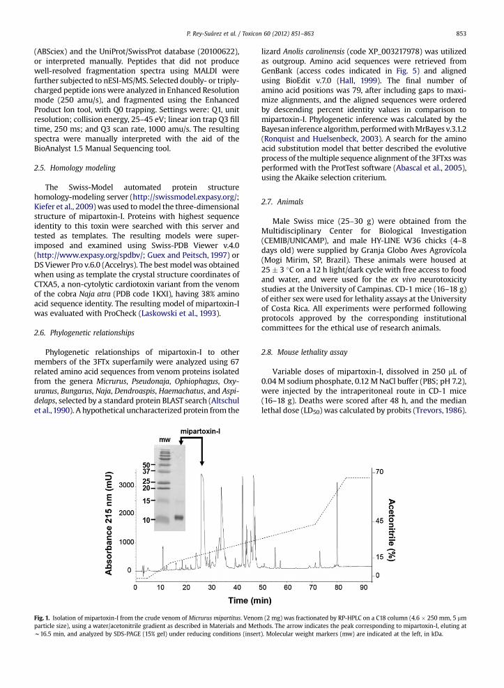
Fig. 1. Isolation of mipartoxin-I from the crude venom of Micrurus mipartitus. Venomparticle size), using a water/acetonitrile gradient as described in Materials and Metw16.5 min, and analyzed by SDS-PAGE (15% gel) under reducing conditions (insert
lizard Anolis carolinensis (code XP_003217978) was utilizedas outgroup. Amino acid sequences were retrieved fromGenBank (access codes indicated in Fig. 5) and alignedusing BioEdit v.7.0 (Hall, 1999). The final number ofamino acid positions was 79, after including gaps to maxi-mize alignments, and the aligned sequences were orderedby descending percent identity values in comparison tomipartoxin-I. Phylogenetic inference was calculated by theBayesan inference algorithm,performedwithMrBayes v.3.1.2(Ronquist and Huelsenbeck, 2003). A search for the aminoacid substitution model that better described the evolutiveprocess of themultiple sequence alignment of the 3FTxswasperformed with the ProtTest software (Abascal et al., 2005),using the Akaike selection criterium.
2.7. Animals
Male Swiss mice (25–30 g) were obtained from theMultidisciplinary Center for Biological Investigation(CEMIB/UNICAMP), and male HY-LINE W36 chicks (4–8days old) were supplied by Granja Globo Aves Agrovícola(Mogi Mirim, SP, Brazil). These animals were housed at25 � 3 �C on a 12 h light/dark cycle with free access to foodand water, and were used for the ex vivo neurotoxicitystudies at the University of Campinas. CD-1 mice (16–18 g)of either sex were used for lethality assays at the Universityof Costa Rica. All experiments were performed followingprotocols approved by the corresponding institutionalcommittees for the ethical use of research animals.
2.8. Mouse lethality assay
Variable doses of mipartoxin-I, dissolved in 250 mL of0.04 M sodium phosphate, 0.12 MNaCl buffer (PBS; pH 7.2),were injected by the intraperitoneal route in CD-1 mice(16–18 g). Deaths were scored after 48 h, and the medianlethal dose (LD50) was calculated by probits (Trevors, 1986).
(2 mg) was fractionated by RP-HPLC on a C18 column (4.6 � 250 mm, 5 mmhods. The arrow indicates the peak corresponding to mipartoxin-I, eluting at). Molecular weight markers (mw) are indicated at the left, in kDa.
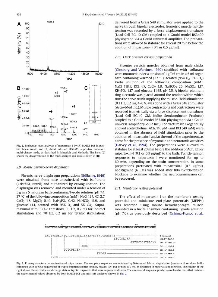
Fig. 2. Molecular mass analyses of mipartoxin-I by (A) MALDI-TOF in posi-tive linear mode, and (B) direct infusion nESI-MS in positive enhancedmulti-charge mode, as described in Materials and Methods. The inset (C)shows the deconvolution of the multi-charged ion series shown in (B).
P. Rey-Suárez et al. / Toxicon 60 (2012) 851–863854
2.9. Mouse phrenic-nerve diaphragm
Phrenic nerve-diaphragm preparations (Bülbring, 1946)were obtained from mice anesthetized with isoflurane(Cristália, Brazil) and euthanized by exsanguination. Thediaphragm was removed and mounted under a tension of5 g in a 5 ml organ bath containing Tyrode solution (pH 7.4,37 �C) of the following composition (mM): NaCl 137, KCl 2.7,CaCl2 1.8, MgCl2 0.49, NaH2PO4 0.42, NaHCO3 11.9, andglucose 11.1, aerated with 95% O2 and 5% CO2. Supra-maximal stimuli (4� threshold, 0.1 Hz, 0.2 ms for indirectstimulation and 70 Hz, 0.2 ms for tetanic stimulation)
Fig. 3. Primary structure determination of mipartoxin-I. The complete sequence wcombined with de novo sequencing of tryptic fragments of the toxin by MALDI-TOF-Tright shows the m/z values and charge state of tryptic fragments that were sequencthe experimental values observed by both MALDI-TOF and nESI-MS analyses, show
delivered from a Grass S48 stimulator were applied to thenerve through bipolar electrodes. Isometric muscle twitch-tension was recorded by a force-displacement transducer(Load Cell BG-10 GM) coupled to a Gould model RS3400physiograph via a Gould universal amplifier. The prepara-tions were allowed to stabilize for at least 20min before theaddition of mipartoxin-I (0.1 or 0.5 mg/ml).
2.10. Chick biventer cervicis preparation
Biventer cervicis muscles obtained from male chicks(Ginsborg and Warriner, 1960) sacrificed with isofluranewere mounted under a tension of 1 g/0.5 cm in a 5 ml organbath containing warmed (37 �C), aerated (95% O2, 5% CO2)Krebs solution of the following composition (mM):NaCl 118.7, KCl 4.7, CaCl2 1.8, NaHCO3 25, MgSO4 1.17,KH2PO4 1.17, and glucose 11.65, pH 7.5. A bipolar platinumring electrode was placed around the tendon within whichruns the nerve trunk supplying themuscle. Field stimulation(0.1 Hz, 0.2ms, 4–6 V) was donewith a Grass S48 stimulator(Astro-Med Inc.).Muscle contractions and contractureswererecorded isometrically via a force-displacement transducer(Load Cell BG-10 GM, Kulite Semiconductor Products)coupled to a Gould model RS3400 physiograph via a Goulduniversal amplifier (Gould Inc.). Contractures to exogenouslyapplied acetylcholine (ACh, 110 mM) and KCl (40 mM) wereobtained in the absence of field stimulation prior to theaddition ofmipartoxin-I and at the end of the experiment, asa test for the presence of myotoxic and neurotoxic activities(Harvey et al., 1994). The preparations were allowed tostabilize for at least 20min before the addition of ACh, KCl ormipartoxin-I (0.1 or 0.5 mg/ml) to the bath. Twitch-tensionresponses to mipartoxin-I were monitored for up to60 min, depending on the toxin concentration. In somepreparations pretreated with mipartoxin-I (0.5 mg/ml),neostigmine (6 mM) was added after 80% twitch-tensionblockade to examine whether the neurotransmission canbe recovered.
2.11. Membrane resting potential
The effect of mipartoxin-I on the membrane restingpotential and miniature end-plate potentials (MEPPs)was recorded using mouse hemidiaphragm musclemounted in a lucite chamber containing Tyrode solution(pH 7.0), as previously described (Oshima-Franco et al.,
as obtained by N-terminal Edman degradation (amino acid residues 1–38)OF or nESI-MS-MS, as described in Materials and Methods. The column at theed de novo. The amino acid sequence predicts a molecular mass that matchesn in Fig. 2.
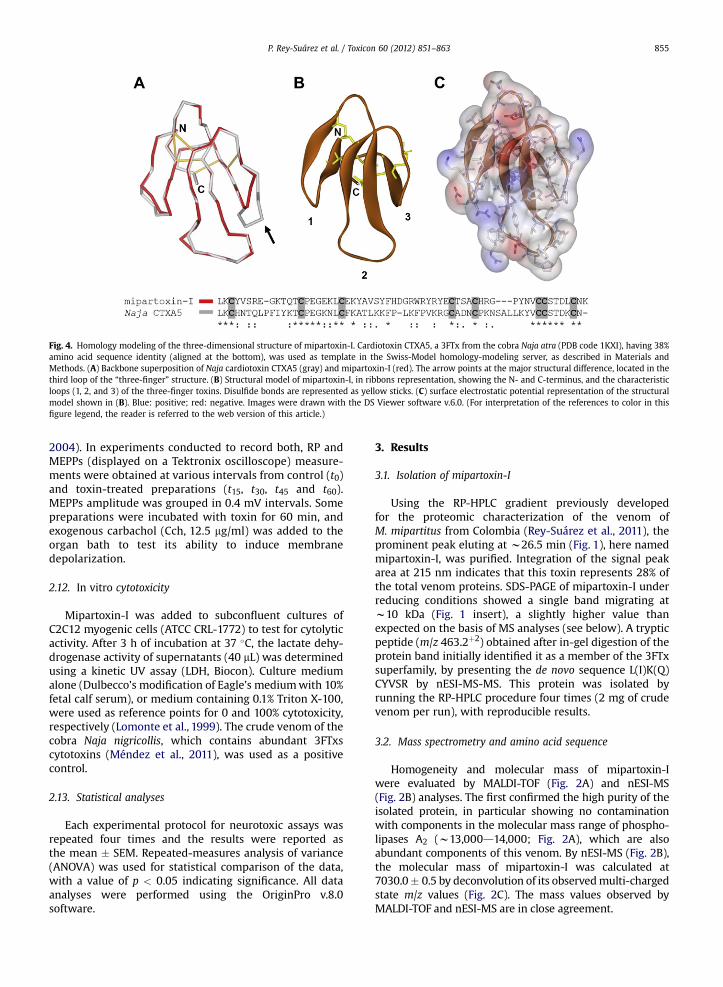
Fig. 4. Homology modeling of the three-dimensional structure of mipartoxin-I. Cardiotoxin CTXA5, a 3FTx from the cobra Naja atra (PDB code 1KXI), having 38%amino acid sequence identity (aligned at the bottom), was used as template in the Swiss-Model homology-modeling server, as described in Materials andMethods. (A) Backbone superposition of Naja cardiotoxin CTXA5 (gray) and mipartoxin-I (red). The arrow points at the major structural difference, located in thethird loop of the “three-finger” structure. (B) Structural model of mipartoxin-I, in ribbons representation, showing the N- and C-terminus, and the characteristicloops (1, 2, and 3) of the three-finger toxins. Disulfide bonds are represented as yellow sticks. (C) surface electrostatic potential representation of the structuralmodel shown in (B). Blue: positive; red: negative. Images were drawn with the DS Viewer software v.6.0. (For interpretation of the references to color in thisfigure legend, the reader is referred to the web version of this article.)
P. Rey-Suárez et al. / Toxicon 60 (2012) 851–863 855
2004). In experiments conducted to record both, RP andMEPPs (displayed on a Tektronix oscilloscope) measure-ments were obtained at various intervals from control (t0)and toxin-treated preparations (t15, t30, t45 and t60).MEPPs amplitude was grouped in 0.4 mV intervals. Somepreparations were incubated with toxin for 60 min, andexogenous carbachol (Cch, 12.5 mg/ml) was added to theorgan bath to test its ability to induce membranedepolarization.
2.12. In vitro cytotoxicity
Mipartoxin-I was added to subconfluent cultures ofC2C12 myogenic cells (ATCC CRL-1772) to test for cytolyticactivity. After 3 h of incubation at 37 �C, the lactate dehy-drogenase activity of supernatants (40 mL) was determinedusing a kinetic UV assay (LDH, Biocon). Culture mediumalone (Dulbecco’s modification of Eagle’s mediumwith 10%fetal calf serum), or medium containing 0.1% Triton X-100,were used as reference points for 0 and 100% cytotoxicity,respectively (Lomonte et al., 1999). The crude venom of thecobra Naja nigricollis, which contains abundant 3FTxscytotoxins (Méndez et al., 2011), was used as a positivecontrol.
2.13. Statistical analyses
Each experimental protocol for neurotoxic assays wasrepeated four times and the results were reported asthe mean � SEM. Repeated-measures analysis of variance(ANOVA) was used for statistical comparison of the data,with a value of p < 0.05 indicating significance. All dataanalyses were performed using the OriginPro v.8.0software.
3. Results
3.1. Isolation of mipartoxin-I
Using the RP-HPLC gradient previously developedfor the proteomic characterization of the venom ofM. mipartitus from Colombia (Rey-Suárez et al., 2011), theprominent peak eluting at w26.5 min (Fig. 1), here namedmipartoxin-I, was purified. Integration of the signal peakarea at 215 nm indicates that this toxin represents 28% ofthe total venom proteins. SDS-PAGE of mipartoxin-I underreducing conditions showed a single band migrating atw10 kDa (Fig. 1 insert), a slightly higher value thanexpected on the basis of MS analyses (see below). A trypticpeptide (m/z 463.2þ2) obtained after in-gel digestion of theprotein band initially identified it as a member of the 3FTxsuperfamily, by presenting the de novo sequence L(I)K(Q)CYVSR by nESI-MS-MS. This protein was isolated byrunning the RP-HPLC procedure four times (2 mg of crudevenom per run), with reproducible results.
3.2. Mass spectrometry and amino acid sequence
Homogeneity and molecular mass of mipartoxin-Iwere evaluated by MALDI-TOF (Fig. 2A) and nESI-MS(Fig. 2B) analyses. The first confirmed the high purity of theisolated protein, in particular showing no contaminationwith components in the molecular mass range of phospho-lipases A2 (w13,000d14,000; Fig. 2A), which are alsoabundant components of this venom. By nESI-MS (Fig. 2B),the molecular mass of mipartoxin-I was calculated at7030.0� 0.5 by deconvolution of its observedmulti-chargedstate m/z values (Fig. 2C). The mass values observed byMALDI-TOF and nESI-MS are in close agreement.
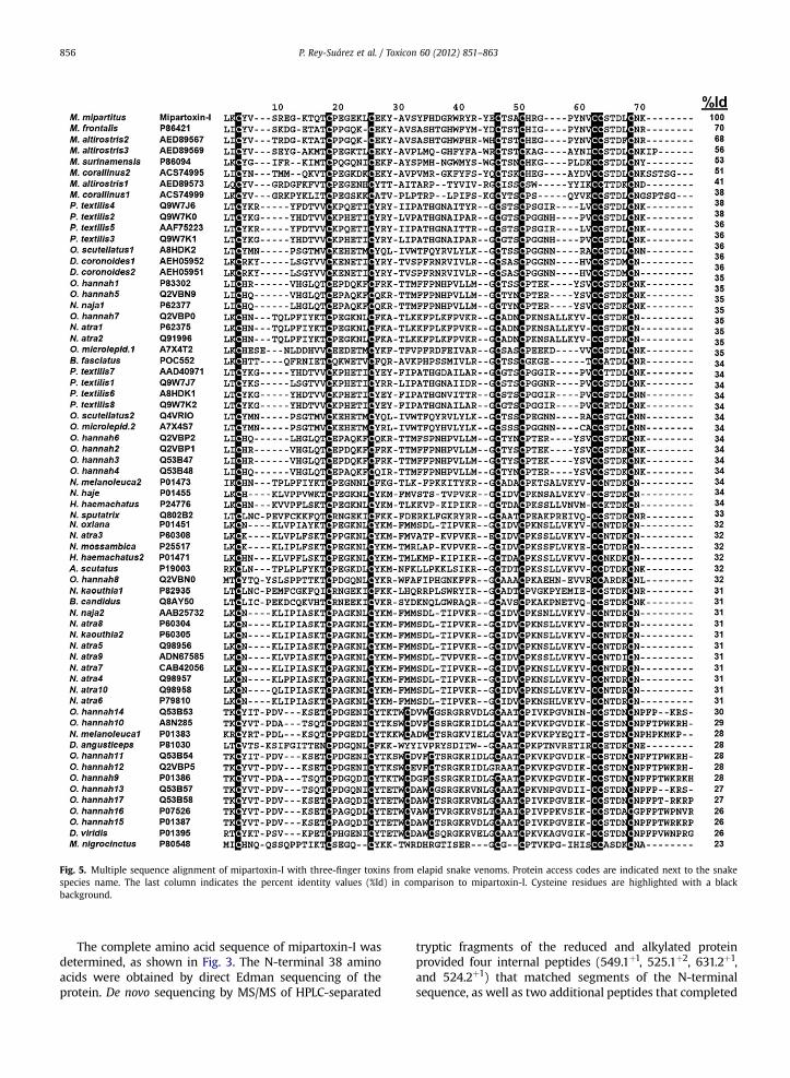
Fig. 5. Multiple sequence alignment of mipartoxin-I with three-finger toxins from elapid snake venoms. Protein access codes are indicated next to the snakespecies name. The last column indicates the percent identity values (%Id) in comparison to mipartoxin-I. Cysteine residues are highlighted with a blackbackground.
P. Rey-Suárez et al. / Toxicon 60 (2012) 851–863856
The complete amino acid sequence of mipartoxin-I wasdetermined, as shown in Fig. 3. The N-terminal 38 aminoacids were obtained by direct Edman sequencing of theprotein. De novo sequencing by MS/MS of HPLC-separated
tryptic fragments of the reduced and alkylated proteinprovided four internal peptides (549.1þ1, 525.1þ2, 631.2þ1,and 524.2þ1) that matched segments of the N-terminalsequence, as well as two additional peptides that completed
P. Rey-Suárez et al. / Toxicon 60 (2012) 851–863 857
the sequence (592.2þ2 and 844.3þ2). The predicted theo-retical mass of the sixty-amino acid sequence is 7037.98(average mass), which is in agreement with the observedmass of 7030.0 � 0.5, considering the presence of eightcysteines engaged in four disulfide bonds (loss of 8 Hþ),characteristic of the short-chain 3FTx superfamily. Further,the obtained sequence predicts mipartoxin-I to be a basicprotein, with a calculated pI of 8.5, according to the ComputeMw/pI tool at http://web.expasy.org/compute_pI/. Theamino acid sequence of mipartoxin-I was submitted to theSwissProt/UniProt database, and assigned the access codeB3EWF8.
3.3. Homology modeling
3FTxs for which three-dimensional structures areavailable at the RCSB Protein Data Bank showed highestsequence identity values with mipartoxin-I within therange of 27–39%. After testing these different 3FTxs astemplates in the Swiss-Model server, the homology modelobtained using as template a non-cytolytic cardiotoxinisoform from N. atra (1KXI; 38% amino acid sequenceidentity), provided the best results, whereas the use ofother 3FTxs as templates provided poor models (data notshown). Evaluation of the best model (vs. 1KXI) with Pro-Check indicated 98% of residues in allowed regions (74.5%in most favored regions, 19.6% in additional allowedregions, and 3.9% in generously allowed regions). The pre-dicted mipartoxin-I model superimposed over the back-bone residues of the Naja 3FTx is shown in Fig. 4A. Theirstructural comparison results in a r.m.s. value of 1.51 Å fora-carbon atoms and 1.59 Å for backbone. The predictedcore region of mipartoxin-I is virtually identical to thetemplate, showing full spatial conservation of the disulfidebonds. Slight spatial differences are predicted at the tips ofloops 1 and 2, whereas loop 3 presents a significant devi-ation (Fig. 4A) which is consistent with the presence of twoadditional amino acids in the Naja toxin (formed by 62residues).
3.4. Phylogenetic relationships
Sequence alignments of mipartoxin-I with 67 related3FTx proteins fromnine elapid snake genera (Fig. 5) revealedidentity values ranging from 70% to 38% for proteins found inother Micrurus venoms, with the exception of M. nigro-cinctus, whose toxin presented only 23% sequence identitycompared tomipartoxin-I. Identity values dropped to 38% orlower for proteins of elapid genera other than Micrurus,when compared to mipartoxin-I. The number and positionsof cysteine residues of mipartoxin-I adhere to the conservedscheme of the short-chain 3FTxs, presenting 8 cysteines(Fig. 5).
A phylogeny of the elapid 3FTx sequences aligned inFig. 5 was reconstructed by Bayesian inference, using theAkaike selection criterion and the WAG þ G amino acidsubstitution model. The resulting phylogenetic tree pre-sented a trichotomy, in which two groups are formed byshort-chain neurotoxins, while a third includes both short-chain and long-chain neurotoxins, as well as short-chaincytotoxins (Fig. 6). The first group (“a” in Fig. 6) is well
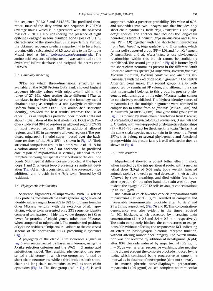
supported, with a posterior probability (PP) value of 0.95,and subdivides into two lineages: one that includes onlyshort-chain cytotoxins, from Naja, Hemachatus, and Aspi-delaps species, and another that includes the long-chainneurotoxins from O. hannah, Naja melanoleuca and D. vir-idis (PP ¼ 1.0) together with the short-chain neurotoxinsfrom Naja kaouthia, Naja sputatrix and B. candidus, whichform awell-supported group (PP¼ 1.0), and fromO. hannah,D. angusticeps and M. nigrocinctus, whose phylogeneticrelationships within this branch cannot be confidentlyestablished. The second group (“b” in Fig. 6) is formed by allthe short-chain neurotoxins present in the different SouthAmericanMicrurus species (M. mipartitus,Micrurus frontalis,Micrurus altirostris, Micrurus corallinus and Micrurus sur-inamensis), with the exception ofM. nigrocinctus, the CentralAmerican coral snake. This second group is also well-supported by significant PP values, and although it is clearthat mipartoxin-I belongs to this group, its precise phylo-genetic relationships with the other Micrurus toxins cannotbe conclusively established. The highest identity scores ofmipartoxin-I in the multiple alignment were obtained incomparison to toxins from M. frontalis (P86421, 70%) andM. altirostris (AED89567, 68%). Finally, the third group (“c” inFig. 6) is formed by short-chain neurotoxins from P. textilis,O. scutellatus, O. microlepidotus, D. coronoides, O. hannah andB. fasciatus, with well-supported phylogenetic relationships(PP¼ 0.95–1.0), except for the B. fasciatus toxin. The fact thatthe same snake species may contain in its venom different3FTxs that belong to several phylogenetic and functionalgroups within this protein family is well reflected in the treeshown in Fig. 6.
3.5. Toxic activities
Mipartoxin-I showed a potent lethal effect in mice,when injected by the intraperitoneal route, with a medianlethal dose (LD50) of 0.06 mg/g body weight. Injectedanimals rapidly showed a general decrease in their activityfollowed by slow breathing, and died within few hoursafter injection. On the other hand, the toxin was not cyto-toxic to the myogenic C2C12 cells in vitro, at concentrationsup to 180 mg/ml.
Incubation of chick biventer cervicis preparations withmipartoxin-I (0.1 or 0.5 mg/ml) resulted in complete andirreversible neuromuscular blockade after 46 � 2 and21�2min, respectively (Fig. 7A and B). This concentration-dependence was also evident in the times requiredfor 50% blockade, which decreased by increasing toxinconcentration (21 � 0.8 and 8.4 � 0.7 min, respectively).The toxin completely blocked the contractures to exoge-nous ACh without affecting the responses to KCl, indicatingan effect on post-synaptic nicotinic receptor function,without altering muscle fiber integrity. The twitch inhibi-tion was not reverted by addition of neostigmine (6 mM)after 80% blockade induced by mipartoxin-I (0.5 mg/ml,n ¼ 3), as well as after successive washings; also neostig-mine did not prevent the complete blockade induced by thetoxin, which continued being progressive at same timeinterval as in absence of neostigmine (data not shown).
In mouse phrenic nerve-diaphragm preparations,mipartoxin-I (0.5 mg/ml) caused complete neuromuscular
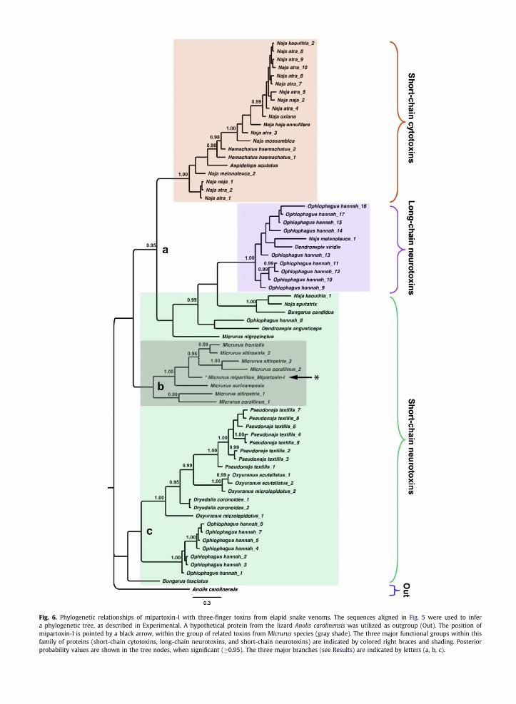
Fig. 6. Phylogenetic relationships of mipartoxin-I with three-finger toxins from elapid snake venoms. The sequences aligned in Fig. 5 were used to infera phylogenetic tree, as described in Experimental. A hypothetical protein from the lizard Anolis carolinensis was utilized as outgroup (Out). The position ofmipartoxin-I is pointed by a black arrow, within the group of related toxins from Micrurus species (gray shade). The three major functional groups within thisfamily of proteins (short-chain cytotoxins, long-chain neurotoxins, and short-chain neurotoxins) are indicated by colored right braces and shading. Posteriorprobability values are shown in the tree nodes, when significant (�0.95). The three major branches (see Results) are indicated by letters (a, b, c).
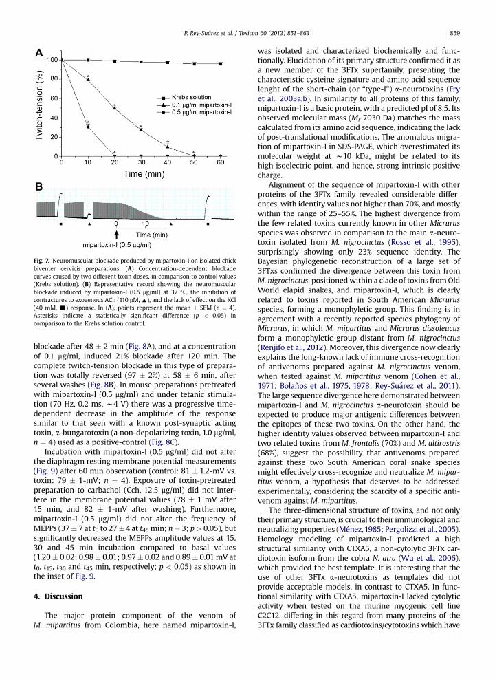
Fig. 7. Neuromuscular blockade produced by mipartoxin-I on isolated chickbiventer cervicis preparations. (A) Concentration-dependent blockadecurves caused by two different toxin doses, in comparison to control values(Krebs solution). (B) Representative record showing the neuromuscularblockade induced by mipartoxin-I (0.5 mg/ml) at 37 �C, the inhibition ofcontractures to exogenous ACh (110 mM, :), and the lack of effect on the KCl(40 mM, -) response. In (A), points represent the mean � SEM (n ¼ 4).Asterisks indicate a statistically significant difference (p < 0.05) incomparison to the Krebs solution control.
P. Rey-Suárez et al. / Toxicon 60 (2012) 851–863 859
blockade after 48 � 2 min (Fig. 8A), and at a concentrationof 0.1 mg/ml, induced 21% blockade after 120 min. Thecomplete twitch-tension blockade in this type of prepara-tion was totally reversed (97 � 2%) at 58 � 6 min, afterseveral washes (Fig. 8B). In mouse preparations pretreatedwith mipartoxin-I (0.5 mg/ml) and under tetanic stimula-tion (70 Hz, 0.2 ms, w4 V) there was a progressive time-dependent decrease in the amplitude of the responsesimilar to that seen with a known post-synaptic actingtoxin, a-bungarotoxin (a non-depolarizing toxin, 1.0 mg/ml,n ¼ 4) used as a positive-control (Fig. 8C).
Incubation with mipartoxin-I (0.5 mg/ml) did not alterthe diaphragm resting membrane potential measurements(Fig. 9) after 60 min observation (control: 81 � 1.2-mV vs.toxin: 79 � 1-mV; n ¼ 4). Exposure of toxin-pretreatedpreparation to carbachol (Cch, 12.5 mg/ml) did not inter-fere in the membrane potential values (78 � 1 mV after15 min, and 82 � 1-mV after washing). Furthermore,mipartoxin-I (0.5 mg/ml) did not alter the frequency ofMEPPs (37� 7 at t0 to 27�4 at t45 min; n¼ 3; p> 0.05), butsignificantly decreased the MEPPs amplitude values at 15,30 and 45 min incubation compared to basal values(1.20� 0.02; 0.98� 0.01; 0.97� 0.02 and 0.89� 0.01mV att0, t15, t30 and t45 min, respectively; p < 0.05) as shown inthe inset of Fig. 9.
4. Discussion
The major protein component of the venom ofM. mipartitus from Colombia, here named mipartoxin-I,
was isolated and characterized biochemically and func-tionally. Elucidation of its primary structure confirmed it asa new member of the 3FTx superfamily, presenting thecharacteristic cysteine signature and amino acid sequencelenght of the short-chain (or “type-I”) a-neurotoxins (Fryet al., 2003a,b). In similarity to all proteins of this family,mipartoxin-I is a basic protein, with a predicted pI of 8.5. Itsobserved molecular mass (Mr 7030 Da) matches the masscalculated from its amino acid sequence, indicating the lackof post-translational modifications. The anomalous migra-tion of mipartoxin-I in SDS-PAGE, which overestimated itsmolecular weight at w10 kDa, might be related to itshigh isoelectric point, and hence, strong intrinsic positivecharge.
Alignment of the sequence of mipartoxin-I with otherproteins of the 3FTx family revealed considerable differ-ences, with identity values not higher than 70%, andmostlywithin the range of 25–55%. The highest divergence fromthe few related toxins currently known in other Micrurusspecies was observed in comparison to the main a-neuro-toxin isolated from M. nigrocinctus (Rosso et al., 1996),surprisingly showing only 23% sequence identity. TheBayesian phylogenetic reconstruction of a large set of3FTxs confirmed the divergence between this toxin fromM. nigrocinctus, positionedwithin a clade of toxins fromOldWorld elapid snakes, and mipartoxin-I, which is clearlyrelated to toxins reported in South American Micrurusspecies, forming a monophyletic group. This finding is inagreement with a recently reported species phylogeny ofMicrurus, in which M. mipartitus and Micrurus dissoleucusform a monophyletic group distant from M. nigrocinctus(Renjifo et al., 2012). Moreover, this divergence now clearlyexplains the long-known lack of immune cross-recognitionof antivenoms prepared against M. nigrocinctus venom,when tested against M. mipartitus venom (Cohen et al.,1971; Bolaños et al., 1975, 1978; Rey-Suárez et al., 2011).The large sequence divergence here demonstrated betweenmipartoxin-I and M. nigrocinctus a-neurotoxin should beexpected to produce major antigenic differences betweenthe epitopes of these two toxins. On the other hand, thehigher identity values observed between mipartoxin-I andtwo related toxins from M. frontalis (70%) and M. altirostris(68%), suggest the possibility that antivenoms preparedagainst these two South American coral snake speciesmight effectively cross-recognize and neutralize M. mipar-titus venom, a hypothesis that deserves to be addressedexperimentally, considering the scarcity of a specific anti-venom against M. mipartitus.
The three-dimensional structure of toxins, and not onlytheir primary structure, is crucial to their immunological andneutralizing properties (Ménez,1985; Pergolizzi et al., 2005).Homology modeling of mipartoxin-I predicted a highstructural similarity with CTXA5, a non-cytolytic 3FTx car-diotoxin isoform from the cobra N. atra (Wu et al., 2006),which provided the best template. It is interesting that theuse of other 3FTx a-neurotoxins as templates did notprovide acceptable models, in contrast to CTXA5. In func-tional similarity with CTXA5, mipartoxin-I lacked cytolyticactivity when tested on the murine myogenic cell lineC2C12, differing in this regard from many proteins of the3FTx family classified as cardiotoxins/cytotoxins which have
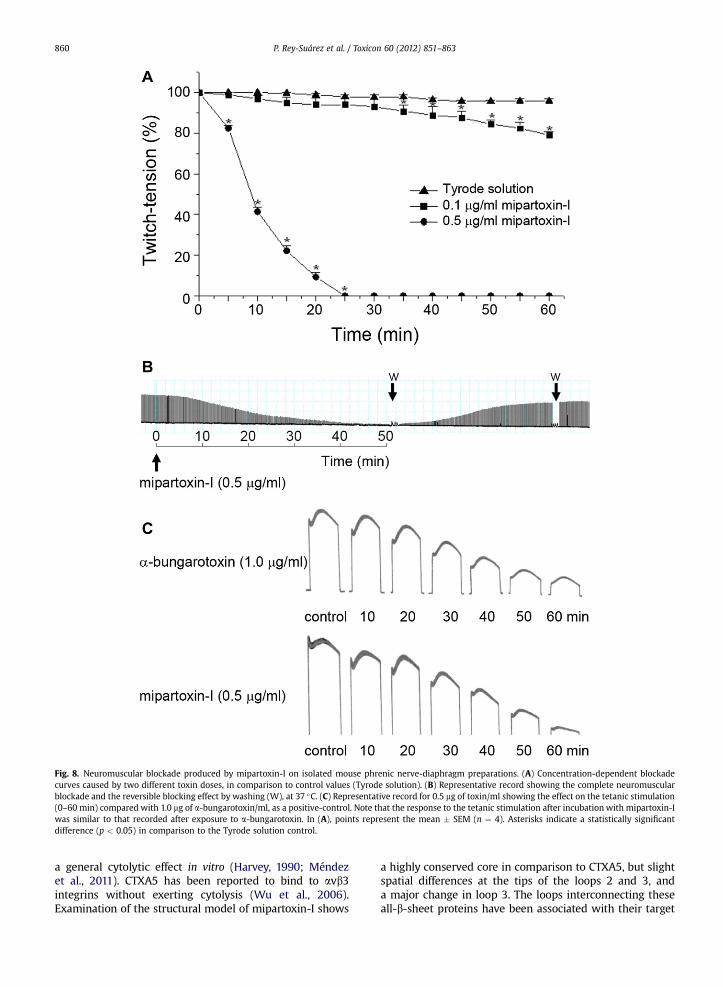
Fig. 8. Neuromuscular blockade produced by mipartoxin-I on isolated mouse phrenic nerve-diaphragm preparations. (A) Concentration-dependent blockadecurves caused by two different toxin doses, in comparison to control values (Tyrode solution). (B) Representative record showing the complete neuromuscularblockade and the reversible blocking effect by washing (W), at 37 �C. (C) Representative record for 0.5 mg of toxin/ml showing the effect on the tetanic stimulation(0–60 min) compared with 1.0 mg of a-bungarotoxin/ml, as a positive-control. Note that the response to the tetanic stimulation after incubation with mipartoxin-Iwas similar to that recorded after exposure to a-bungarotoxin. In (A), points represent the mean � SEM (n ¼ 4). Asterisks indicate a statistically significantdifference (p < 0.05) in comparison to the Tyrode solution control.
P. Rey-Suárez et al. / Toxicon 60 (2012) 851–863860
a general cytolytic effect in vitro (Harvey, 1990; Méndezet al., 2011). CTXA5 has been reported to bind to avb3integrins without exerting cytolysis (Wu et al., 2006).Examination of the structural model of mipartoxin-I shows
a highly conserved core in comparison to CTXA5, but slightspatial differences at the tips of the loops 2 and 3, anda major change in loop 3. The loops interconnecting theseall-b-sheet proteins have been associated with their target

Fig. 9. Lack of depolarization of the mouse hemidiaphragm muscle membrane resting potential by mipartoxin-I after 60 min, and significant change in theresponse to exogenous carbachol (Cch, 12.5 mg/ml) after 60 min compared to control preparations (T0). Points represent mean � SEM (n ¼ 4). Inset: effect ofmipartoxin-I (0.5 mg/ml) on the amplitude of MEPPs at room temperature before and at different times after exposure to toxin. Asterisks indicate a statisticallysignificant difference (p < 0.05) in comparison to control preparations.
P. Rey-Suárez et al. / Toxicon 60 (2012) 851–863 861
specificity (Dubovskii et al., 2001; Fry et al., 2003a,b; Kiniand Doley, 2010; Kini, 2011), and hence with their func-tional properties in vivo.
Considering the biochemical, structural, and phyloge-netic characteristics of mipartoxin-I, together with itshigh lethal action in mice (LD50 0.06 mg/g), its possibleneurotoxic activity was investigated. Mipartoxin-I causeda clear neuromuscular blockade on both avian andmammalian preparations. The biventer cervicis preparationwas more sensitive than the phrenic nerve-diaphragmpreparation, probably related to differences in the innerva-tion of these two preparations, with avian muscle havingboth focally- and multiply-innervated fibers that canrespond to electrical stimulation or exogenous nicotinicagonists (Hodgson and Wickramaratna, 2002). The inabilityof neostigmine and several washings to revert the toxin-induced inhibition in BC preparations agrees with the find-ings recently reported in preparations incubated with crudevenom of M. mipartitus (Renjifo et al., 2012). However,possible species differences in the affinity of this toxin forits receptors cannot be ruled out. Mipartoxin-I inhibitedcontractures to exogenous ACh, indicating a predominantlypost-synaptic action through the blockade of cholinergicnicotinic receptor, as also suggested for other studies usingMicrurus venoms (Goularte et al., 1995; Serafim et al., 2002;Abreu et al., 2008; Camargo et al., 2011; Renjifo et al., 2012).The post-synaptic action of mipartoxin-I was also indicatedby the absence of fade in the response to tetanic indirectstimulation at 70 Hz. This tetanic pattern was similar tothat produced by a-bungarotoxin, a non-depolarizing toxin(Gallacci and Oliveira, 1994; Serra and Oliveira, 2006;Camargo et al., 2011). These results were corroboratedusing carbachol to produce progressive depolarization of therestingmembrane potential, whereas the toxin had no effect
on this parameter, indicating an activity on the post-synapticnicotinic receptors.Moreover, the inability ofmipartoxin-I toalter the MEPPs frequency, in contrast with its efficiencyin decreasing MEPPs amplitude, suggests that, like othertypical a-neurotoxins, the toxin binds nicotinic receptorswithout interfering with the presynaptic mechanism ofneurotransmitter release. Taken together, these resultsindicate that mipartoxin-I causes neuromuscular blockadeby a post-synaptic action in neuromuscular preparations, i.e.a-neurotoxicity, without directly damaging muscle fibers.The small amounts of toxin available precluded furthercharacterization of its receptor subtype specificity. A recentstudy evaluated the neuromuscular activity of the crudevenom of M. mipartitus on the chick biventer cervicis prep-aration, and demonstrated a post-synaptic action (Renjifoet al., 2012), in full agreement with the present findingsusing its purified major neurotoxin, mipartoxin-I, and alsosupporting the conclusion that this protein plays a centralrole in the neurotoxicity of M. mipartitus venom.
The extraordinary biodiversity within the genus Micru-rus, with more than 70 species described (Castoe et al.,2007), seems to be also reflected in the wide divergenceemerging from the study of their venom toxins. Onlya handful of 3FTxs have been fully sequenced and func-tionally characterized from Micrurus venoms (Rosso et al.,1996; de Oliveira et al., 2000; Dokmetjian et al., 2009;Moreira et al., 2010). Mipartoxin-I, isolated from M. mipar-titus venom, is a novel member of the 3FTx superfamily witha potent post-synaptic a-neurotoxic effect on both avianand mammalian neuromuscular preparations, and lethalaction in mice. Since mipartoxin-I is the most abundant(28%) protein in the venom of this coral snake species, itwould be expected to play a major role in its toxicity.Therefore, mipartoxin-I should be a relevant target for the
P. Rey-Suárez et al. / Toxicon 60 (2012) 851–863862
development of a therapeutic antivenom. Together withits functional characterization, the structural informationreported in the present work might be useful in the prepa-ration of a synthetic or recombinant immunogen to over-come the scarcity of venom for immunization.
Conflicts of interest
None to declare.
Acknowledgments
We thank Dr Jean-Pierre Rosso (Laboratoire de Biochimie,Université de la Mediterranee, Marseille, France) andDr Juan J. Calvete (Laboratorio de Proteinómica Estructural,Instituto de Biomedicina de Valencia, Spain) for their expertsuggestions, and Gildo Bernardo Leite and Fabián Villaltafor technical assistance. Support was provided by a YoungResearcher Fellowship to P. Rey by COLCIENCIAS (1115-459-21441) and Universidad de Antioquia, Colombia, and byVicerrectoría de Investigación, Universidad de Costa Rica(741-A9-513). This study was performed as part of theM.Sc. thesis of P. Rey at the University of Antioquia.
References
Abascal, F., Zardoya, R., Posada, D., 2005. ProtTest: selection of best-fitmodels of protein evolution. Bioinformatics 21, 2104–2105.
Abreu, V.A., Leite, G.B., Oliveira, C.B., Hyslop, S., Furtado, M.F., Rodrigues-Simioni, L., 2008. Neurotoxicity of Micrurus altirostris (Uruguayancoral snake) venom and its neutralization by commercial coral snakeantivenom and specific antiserum raised in rabbits. Clin. Toxicol. 46,519–527.
Altschul, S.F., Gish, W., Miller, W., Myers, E.W., Lipman, D.J., 1990. Basiclocal alignment search tool. J. Mol. Biol. 215, 403–410.
Bolaños, R., Cerdas, L., Abalos, J.W., 1978. Venenos de las serpientes coral(Micrurus spp.): informe sobre un antiveneno polivalente para lasAméricas. Bol. Ofic. Sanit. PanAm. 84, 128–133.
Bolaños, R., Cerdas, L., Taylor, R., 1975. The production and characteristicsof a coral snake (Micrurus mipartitus hertwigi) antivenin. Toxicon 13,139–142.
Bülbring, E., 1946. Observations on the isolated phrenic-nerve diaphragmpreparation of the rat. Br. J. Pharmacol. 1, 38–61.
Calvete, J.J., Sanz, L., Angulo, Y., Lomonte, B., Gutiérrez, J.M., 2009. Venom,venomics, antivenomics. FEBS Lett. 583, 1736–1743.
Camargo, T.M., Roodt, A.R., Cruz-Höfling, M.A., Rodrigues-Simioni, L.,2011. The neuromuscular activity of Micrurus pyrrhocryptus venomand its neutralization by commercial and specific coral snake anti-venoms. J. Venom Res. 2, 24–31.
Castoe, T.A., Smith, E.N., Brown, R.M., Parkinson, C.L., 2007. Higher-levelphylogeny of Asian and American coralsnakes, their placement withinthe Elapidae (Squamata), and the systematic affinities of the enig-matic Asian coralsnake Hemibungarus calligaster (Wiegmann, 1834).Zool. J. Linnean Soc. 151, 809–831.
Ciscotto, P.H.C., Rates, B., Silva, D.A.F., Richardson, M., Silva, L.P.,Andrade, H., Donato, M.F., Cotta, G.A., Maria, W.S., Rodrigues, R.J.,Sanchez, E., De Lima, M.E., Pimenta, A.M.C., 2011. Venomic analysisand evaluation of antivenom cross-reactivity of South AmericanMicrurus species. J. Proteomics 74, 1810–1825.
Cohen, P., Berkeley, W.H., Seligmann, E.B., 1971. Coral snake venoms.In vitro relation of neutralizing and precipitating antibodies. Am. J.Trop. Med. Hyg. 20, 646–649.
Corrêa-Netto, C., Junqueira-de-azevedo, I., Silva, D., Ho, P.L., Leitão-de-Araújo, M., Alves, M.L., Sanz, L., Foguel, D., Zingali, R.B., Calvete, J.J.,2011. Snake venomics and venom gland transcriptomic analysisof Brazilian coral snakes, Micrurus altirostris and M. corallinus. J.Proteomics 74, 1795–1809.
de Oliveira, J.S., da Silva, A.R.B., Soares, M.B., Stephano, M.A., Dias, W.O.,Raw, I., Ho, P.L., 2000. Cloning and characterization of an a-neuro-toxin-type protein specific for the coral snake Micrurus corallinus.Biochem. Biophys. Res. Comm. 267, 887–891.
Dokmetjian, J.C., del Canto, S., Vinzón, S., Bonino, M.B.J., 2009. BiochemicalcharacterizationofMicrurus pyrrhocryptus venom. Toxicon 53, 375–382.
Dubovskii, P.V., Dementieva, D.V., Bocharov, E.V., Utkin, Y.N., Arseniev, A.S.,2001. Membrane binding motif of the P-type cardiotoxin. J. Mol. Biol.305, 137–149.
Fernández, J., Alape-Girón, A., Angulo, Y., Sanz, L., Gutiérrez, J.M.,Calvete, J.J., Lomonte, B., 2011. Venomic and antivenomic analysesof the Central American coral snake, Micrurus nigrocinctus (Elapidae).J. Proteome Res. 10, 1816–1827.
Fry, B.G., Vidal, N., Normamn, J.A., Vonk, F.J., Scheib, H., Ramjan, S.F.,Kuruppu, S., Fung, K., Hedges, S.B., Richardson, M.K., Hodgson, W.C.,Ignjatovic, V., Summerhayes, R., Kochva, E., 2006. Early evolution ofthe venom system in lizards and snakes. Nature 439, 584–588.
Fry, B.G., Wuster, W., Kini, R.M., Brusic, V., Khan, A., Venkataraman, D.,Rooney, A.P., 2003a. Molecular evolution and phylogeny of elapidsnake venom three-finger toxins. J. Mol. Evol. 57, 110–129.
Fry, B.G., Wüster, W., Ramjan, S.F.R., Jackson, T., Martelli, P., Kini, R.M.,2003b. Analysis of Colubroidea snake venoms by liquid chromatog-raphy with mass spectrometry: evolutionary and toxinologicalimplications. Rap. Comm. Mass Spectrom. 17, 2047–2062.
Gallacci, M., Oliveira, A.C., 1994. Pre- and postsynaptic mechanismsinvolved in fade induced by pancuronium in the isolated rat muscle.Pharmacology 49, 265–270.
Ginsborg, B.L., Warriner, J., 1960. The isolated chick biventer cervicis nervemuscle preparation. Br. J. Pharmacol. Chemother. 15, 410–411.
Goularte, F.C., Cruz-Höfling, M.A., Cogo, J.C., Gutiérrez, J.M., Rodrigues-Simioni, L., 1995. The ability of specific antivenom and low temper-ature to inhibit the myotoxicity and neuromuscular block induced byMicrurus nigrocinctus venom. Toxicon 33, 679–689.
Guex, N., Peitsch, M.C., 1997. SWISS-MODEL and the Swiss-PdbViewer: anenvironment for comparative protein modeling. Electrophoresis 18,2714–2723.
Hall, T.A., 1999. BioEdit: a user-friendly biological sequence alignmenteditor and analysis program for Windows 95/98/NT. Nucl. AcidsSymp. Ser. 41, 95–98.
Harvey, A.L., 1990. Cytolytic toxins. In: Shier, W.R., Mebs, D. (Eds.),Handbook of Toxinology. Marcel Dekker, New York, pp. 48–53.
Harvey, A.L., Barfaraz, A., Thomson, E., Faiz, A., Preston, S., Harris, J.B.,1994. Screening of snake venoms for neurotoxic and myotoxic effectsusing simple in vitro preparations from rodents and chicks. Toxicon32, 257–265.
Hodgson, W.C., Wickramaratna, J.C., 2002. In vitro neuromuscular activityof snake venoms. Clin. Exp. Pharmacol. Physiol. 29, 807–814.
Kiefer, F., Arnold, K., Künzli, M., Bordoli, L., Schwede, T., 2009. The SWISS-MODEL repository and associated resources. Nucl. Acids Res. 37,D387–D392.
Kini, R.M., 2011. Evolution of three-finger toxins – a versatile mini proteinscaffold. Acta Chim. Slov 58, 693–701.
Kini, R.M., Doley, R., 2010. Structure, function and evolution of three-fingertoxins-mini proteins with multiple targets. Toxicon 56, 855–867.
Laskowski, R.A., MacArthur, M.W., Moss, D., Thornton, J.M., 1993. PRO-CHECK: a program to check the stereochemical quality of proteinstructures. J. Appl. Cryst 26, 283–291.
Lomonte, B., Angulo, Y., Rufini, S., Cho, W., Giglio, J.R., Ohno, M., Daniele, J.J.,Geoghegan, P., Gutiérrez, J.M., 1999. Comparative study of the cytolyticactivity of myotoxic phospholipases A2 on mouse endothelial (tEnd)and skeletal muscle (C2C12) cells in vitro. Toxicon 37, 145–158.
Méndez, I., Gutiérrez, J.M., Angulo, Y., Calvete, J.J., Lomonte, B., 2011.Comparative study of the cytolytic activity of snake venoms fromAfrican spitting cobras (Naja spp., Elapidae) and its neutralization bya polyspecific antivenom. Toxicon 58, 558–564.
Ménez, A., 1985. Molecular immunology of snake toxins. Pharmac. Ther.30, 91–113.
Moreira, K.G., Prates, M.V., Andrade, F.A.C., Silva, L.P., Beirao, P.S.L.,Kushmerick, C., Naves, L.A., Bloch Jr., C., 2010. Frontoxins, three-fingertoxins from Micrurus frontalis venom, decrease miniature endplatepotential amplitude at frog neuromuscular junction. Toxicon 56, 55–63.
Nakashima, K.-I., Ogawa, T., Oda, N., Hattori, M., Sakaki, Y., Kihara, H.,Ohno, M., 1993. Accelerated evolution of Trimeresurus flavoviridisvenom gland phospholipase A2 isozymes. Proc. Natl. Acad. Sci. USA90, 5964–5968.
Olamendi-Portugal, T., Batista, C., Restano-Cassulini, R., Pando, V.,Villa-Hernandez, O., Zavaleta-Martínez-Vargas, A., Salas-Arruz, M.C.,Rodríguez de la Vega, R.C., Becerril, B., Possani, L., 2008. Proteomicanalysis of the venom from the fish eating coral snake Micrurussurinamensis: novel toxins, their function and phylogeny. Proteomics8, 1919–1932.
Oshima-Franco, Y., Leite, G.B., Belo, C.A., Hyslop, S., Prado-Franceschi, J.,Cintra, A.C., Giglio, J.R., Cruz-Höfling, M.A., Rodrigues-Simioni, L.,
P. Rey-Suárez et al. / Toxicon 60 (2012) 851–863 863
2004. The presynaptic activity of bothropstoxin-I, a myotoxin fromBothrops jararacussu snake venom. Basic Clin. Pharmacol. Toxicol. 95,175–182.
Otero, R., 1994. Manual de diagnóstico y tratamiento del accidente ofídico.Editorial Universidad de Antioquia, Medellín (Colombia), pp. 1–15.
Pergolizzi, R.G., Dragos, R., Ropper, A.E., Ménez, A., Crystal, R.G., 2005.Protective immunity against alpha-cobratoxin following a singleadministration of a genetic vaccine encoding a non-toxic cobratoxinvariant. Hum. Gene Ther. 16, 292–298.
Renjifo, C., Smith, E.N., Hodgson, W.C., Renjifo, J.M., Sanchez, A., Acosta, R.,Maldonado, J.H., Riveros, A., 2012. Neuromuscular activity of thevenoms of the Colombian coral snakes Micrurus dissoleucus andMicrurus mipartitus: an evolutionary perspective. Toxicon 59, 132–142.
Rey-Suárez, P., Núñez, V., Gutiérrez, J.M., Lomonte, B., 2011. Proteomic andbiological characterization of the venom of the redtail coral snake,Micrurus mipartitus (Elapidae), from Colombia and Costa Rica. J.Proteomics 75, 655–667.
Ronquist, F., Huelsenbeck, J.P., 2003. MrBayes 3: Bayesian phylogeneticinference under mixed models. Bioinformatics 19, 1572–1574.
Rosso, J.P., Vargas-Rosso, O., Gutierrez, J.M., Rochat, H., Bougis, P.E., 1996.Characterization of alpha-neurotoxin and phospholipase A2 activitiesfrom Micrurus venoms. Determination of the amino acid sequenceand receptor-binding ability of the major alpha-neurotoxin fromMicrurus nigrocinctus nigrocinctus. Eur. J. Biochem. 238, 231–239.
Serafim, F.G., Reali, M., Cruz-Höfling, M.A., Fontana, M.D., 2002. Action ofMicrurus dumerilli carinicauda coral snake venom on the mammalianneuromuscular junction. Toxicon 40, 167–174.
Serra, C.S.M., Oliveira, A.C., 2006. Cisatracurium: myographical andelectrophysiological studies in the isolated rat muscle. Fund. Clin.Pharmacol. 20, 291–298.
Trevors, J.T., 1986. A BASIC program for estimating LD50 values using theIBM-PC. Bull. Environ. Contam. Toxicol. 37, 18–26.
Wu, P.L., Lee, S.C., Chuang, C.C., Mori, S., Akakura, N., Wu, W.G., Takada, Y.,2006. Non-cytotoxic cobra cardiotoxin A5 binds to avb3 integrin andinhibits bone resorption. J. Biol. Chem. 281, 7937–7945.