Embed Size (px)
Citation preview

Int. J. Mol. Sci. 2014, 15, 14411-14426; doi:10.3390/ijms150814411
International Journal of
Molecular Sciences ISSN 1422-0067
www.mdpi.com/journal/ijms
Article
MiRNA-548ah, a Potential Molecule Associated with Transition from Immune Tolerance to Immune Activation of Chronic Hepatitis B
Tong-Jing Xing *, Hong-Tao Xu, Wen-Qing Yu, Bian Wang and Jing Zhang
Department of Infectious Diseases, Taizhou People’s Hospital, Taizhou 225300, Jiangsu Province,
China; E-Mails: [email protected] (H.-T.X.); [email protected] (W.-Q.Y.);
[email protected] (B.W.); [email protected] (J.Z.)
* Author to whom correspondence should be addressed; E-Mail: [email protected];
Tel./Fax: +86-523-8660-6305.
Received: 17 June 2014; in revised form: 24 July 2014 / Accepted: 31 July 2014 /
Published: 19 August 2014
Abstract: Objective: The present study aims to identify the differently expressed
microRNA (miRNA) molecules and target genes of miRNA in the immune tolerance
(IT) and immune activation (IA) stages of chronic hepatitis B (CHB). Methods: miRNA
expression profiles of peripheral blood mononuclear cells (PBMCs) at the IT and IA stages
of CHB were screened using miRNA microarrays and authenticated using a quantitative
real-time polymerase chain reaction (RT-PCR). Gene ontology (GO) and the Kyoto
encyclopedia of genes and genomes (KEGG) were used to analyze the significant functions
and pathways of possible target genes of miRNAs. Assays of the gain and loss of function
of the miRNAs were performed to verify the target genes in THP-1 cell lines. The
luciferase reporter test was used on 293T cells as direct targets. Results: Significantly
upregulated miR-548 and miR-4804 were observed in the miRNA microarrays and
confirmed by RT-PCR in PBMCs at the IT and IA stages of CHB. GO and KEGG analysis
revealed that MiR-548 and miR-4804 could be involved in numerous signaling pathways
and protein binding activity. IFNγR1 was predicted as a target gene and validated as the
direct gene of MiR-548. Significant negative correlation was found between the miR-548ah
and mRNA levels of IFN-γR1 in CHB patients. Conclusions: The abnormal expression
profiles of miRNA in PBMCs could be closely associated with immune activation of
chronic HBV infection. miR-548, by targeting IFN-γR1, may represent a mechanism that
can facilitate viral pathogenesis and help determine new therapeutic molecular targets.
OPEN ACCESS

Int. J. Mol. Sci. 2014, 15 14412
Keywords: chronic hepatitis B; microRNA; immune tolerance; immune activation
1. Introduction
The Hepatitis B virus (HBV) infection remains a serious public health problem worldwide despite
the wide use of the HBV vaccine. Over 350 million people have been chronically infected with HBV,
and approximately 20% to 40% of them will develop liver cirrhosis and liver cancer from chronic
episodes of inflammation [1]. Numerous studies suggest that HBV is not directly cytopathogenic for
infected hepatocytes. The host immune response, especially the virus-specific T cell response, plays an
important role in the viral clearance and pathogenesis of liver diseases [2,3]. Patients suffering from
chronic hepatitis B (CHB) have weak and limited T cell responses, whereas those with acute hepatitis
have vigorous and polyclonal T cell responses, which can help them completely clear the virus [4].
The lack of virus-specific T cell response in the former case might be attributed to various factors,
such as T cell deletion, anergy, exhaustion, ignorance, and T cell dysfunction [5], but the exact
mechanism is not yet clear. In addition, impairment of functions of dendritic cells, natural killer cells,
and natural killer T cells occur during chronic HBV infection [6,7].
MicroRNA (miRNA) is a small, endogenous, non-coding RNA that modulates gene expression at
the post-transcriptional level. MiRNA plays an important function in various activities of organisms
and is involved in the development of diseases such as infection and cancer [8,9]. Recent studies have
shown that miRNA molecules may have important functions in the development and differentiation of
immune cells as well as in the regulation of immune responses [10,11]. Certain miRNA molecules
operate in the negative feedback loops of the immune system, whereas other miRNAs help amplify the
immune response by repressing the response inhibitors [12,13]. MiRNAs exhibit spatial and temporal
specificities in the regulation of the immune system. For example, immune-related miRNA genes serve
as “fine-tuners” of gene expression rather than “switchers” [12]. Furthermore, regulation by miRNAs
is not antigen specific. Rather, they may target a specific gene locus in a specific cell type and affect its
performance in a specific time and space. Studying the hereditary, conditional resting, and activation
of miRNA genes might provide a foundation for immunological studies and contribute to development
of novel interventions for the immune-related disease [13].
Acquired chronic HBV infection can be divided into four phases according to the characteristics of
its immune response: immune tolerance (IT), immune activation (IA), immune control, and immune
reaction [14]. The IT phase is characterized by active HBV replication, positivity for HBeAg, and
normal alanine aminotransferase (ALT) levels. The IA phase is characterized by decreases in HBV
DNA and raised ALT levels. CHB activation may be associated with the enhancement of T helper 1
cells, increases in HBcAg-specific cytotoxic T lymphocytes, and decreases in HBcAg-specific
regulatory T cells [15]. Several studies have found that the abnormal expressions of certain miRNA
molecules (miR-155, miR-181, etc.) are related to the pathogenesis and disease progression of chronic
HBV infection [16,17]. However, whether or not exists a molecular mechanism of miRNA in the
transition from IT to IA of CHB is unclear. In the present study, miRNA expressions in peripheral
blood mononuclear cells (PBMCs) of the IT and IA phases of CHB were screened using miRNA
Int. J. Mol. Sci. 2014, 15 14413
microarrays and authenticated using quantitative real-time polymerase chain reaction (RT-PCR).
Target genes of the miRNAs associated with the IA of CHB were then predicted and identified.
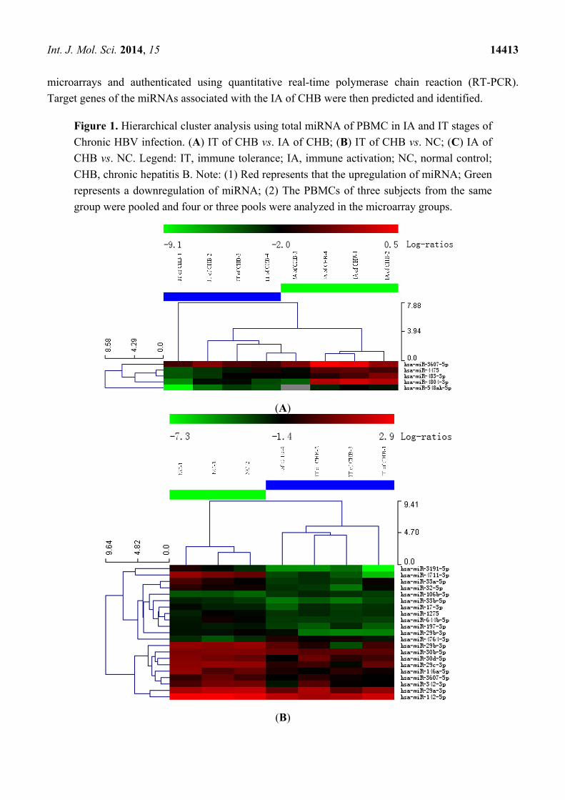
Figure 1. Hierarchical cluster analysis using total miRNA of PBMC in IA and IT stages of
Chronic HBV infection. (A) IT of CHB vs. IA of CHB; (B) IT of CHB vs. NC; (C) IA of
CHB vs. NC. Legend: IT, immune tolerance; IA, immune activation; NC, normal control;
CHB, chronic hepatitis B. Note: (1) Red represents that the upregulation of miRNA; Green
represents a downregulation of miRNA; (2) The PBMCs of three subjects from the same
group were pooled and four or three pools were analyzed in the microarray groups.
(A)
(B)

Int. J. Mol. Sci. 2014, 15 14414
Figure 1. Cont.
(C)
Int. J. Mol. Sci. 2014, 15 14415
2. Results
2.1. Different Expressions of miRNA Molecules in PBMC in IT and IA Stages of CHB
miRNA microarray was used to detect the miRNA expressions of PBMCs during the IT and IA
stages of CHB. Hierarchical cluster analysis was also performed. Results are shown in Figure 1A–C.
Compared with the normal controls, 2 significantly upregulated and 18 significantly downregulated
miRNA molecules were identified in PBMCs during the IT phase of CHB; 33 significantly
upregulated and 19 significantly downregulated miRNA molecules were also identified in PBMCs
during the IA phase of CHB. Compared with the IT phase of CHB, 5 significantly upregulated miRNA
molecules (hsa-miR-548ah-5p, hsa-miR-4804-3p, hsa-miR-483-3p, hsa-miR-3607-3p, hsa-miR-44475),
were identified in PBMCs during the IA phase of CHB. In particular, the expressions of
hsa-miR-548ah-5p and hsa-miR-4804-3p more significantly upregulated in the IA phase of CHB
(5.1 and 5.9 times, respectively).
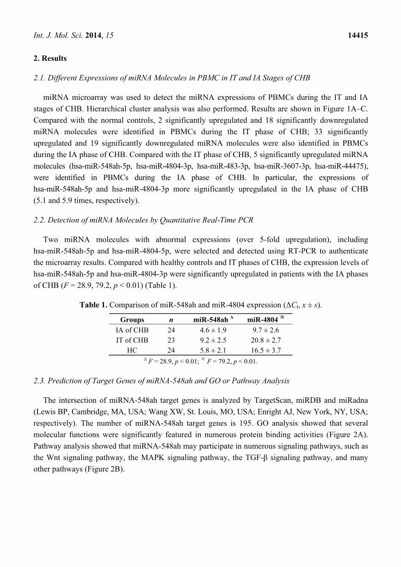
2.2. Detection of miRNA Molecules by Quantitative Real-Time PCR
Two miRNA molecules with abnormal expressions (over 5-fold upregulation), including
hsa-miR-548ah-5p and hsa-miR-4804-5p, were selected and detected using RT-PCR to authenticate
the microarray results. Compared with healthy controls and IT phases of CHB, the expression levels of
hsa-miR-548ah-5p and hsa-miR-4804-3p were significantly upregulated in patients with the IA phases
of CHB (F = 28.9, 79.2, p < 0.01) (Table 1).
Table 1. Comparison of miR-548ah and miR-4804 expression (ΔCt, x ± s).
Groups n miR-548ah Δ miR-4804 ※
IA of CHB 24 4.6 ± 1.9 9.7 ± 2.6 IT of CHB 23 9.2 ± 2.5 20.8 ± 2.7
HC 24 5.8 ± 2.1 16.5 ± 3.7 Δ F = 28.9, p < 0.01; ※ F = 79.2, p < 0.01.
2.3. Prediction of Target Genes of miRNA-548ah and GO or Pathway Analysis
The intersection of miRNA-548ah target genes is analyzed by TargetScan, miRDB and miRadna
(Lewis BP, Cambridge, MA, USA; Wang XW, St. Louis, MO, USA; Enright AJ, New York, NY, USA;
respectively). The number of miRNA-548ah target genes is 195. GO analysis showed that several
molecular functions were significantly featured in numerous protein binding activities (Figure 2A).
Pathway analysis showed that miRNA-548ah may participate in numerous signaling pathways, such as
the Wnt signaling pathway, the MAPK signaling pathway, the TGF-β signaling pathway, and many
other pathways (Figure 2B).

Int. J. Mol. Sci. 2014, 15 14416
Figure 2. Molecular function of Go analysis of miR-548ah target gene (A); Pathway
analysis of miR-548ah target gene (B).
(A)
(B)
2.4. IFN-γR1 Is Direct Target Gene of miR-548ah-5p
IFN-γR1 was predicted as a potential target gene of the miR-548ah-5p. The 3'-UTR of IFN-γR1
contained two complementary sites for the seed region of miR-548ah-5p. To validate whether IFN-γR1
is a direct target of miR-548ah-5p, the 3'-UTR fragment of IFNγR1 containing wild-type or mutant
miR-548ah-5p binding sequence was cloned downstream of the firefly luciferase reporter gene in
psiCHECK-2 plasmids. Among the 293 T cells cotransfected with the reporter plasmids and
miR-548ah-5p mimic or NC duplex, the luciferase activity of the reporter that contained wild-type 3'-UTR
was significantly suppressed by the miR-548ah-5p mimic; however the luciferase activity of the
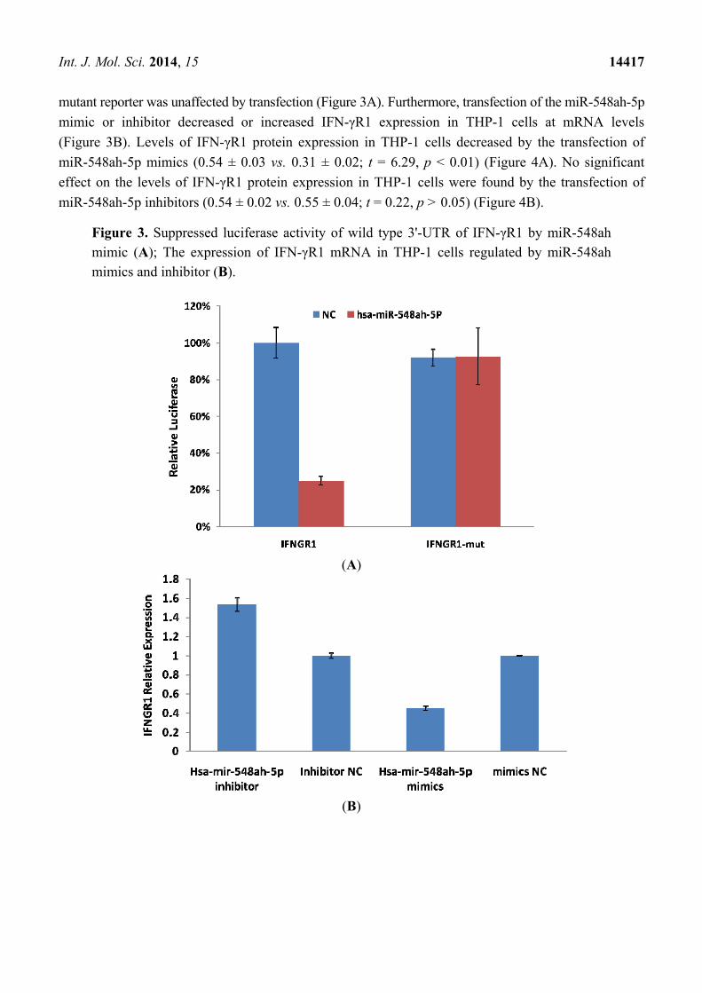
Int. J. Mol. Sci. 2014, 15 14417
mutant reporter was unaffected by transfection (Figure 3A). Furthermore, transfection of the miR-548ah-5p
mimic or inhibitor decreased or increased IFN-γR1 expression in THP-1 cells at mRNA levels
(Figure 3B). Levels of IFN-γR1 protein expression in THP-1 cells decreased by the transfection of
miR-548ah-5p mimics (0.54 ± 0.03 vs. 0.31 ± 0.02; t = 6.29, p < 0.01) (Figure 4A). No significant
effect on the levels of IFN-γR1 protein expression in THP-1 cells were found by the transfection of
miR-548ah-5p inhibitors (0.54 ± 0.02 vs. 0.55 ± 0.04; t = 0.22, p > 0.05) (Figure 4B).
Figure 3. Suppressed luciferase activity of wild type 3'-UTR of IFN-γR1 by miR-548ah mimic (A); The expression of IFN-γR1 mRNA in THP-1 cells regulated by miR-548ah mimics and inhibitor (B).
(A)
(B)

Int. J. Mol. Sci. 2014, 15 14418
Figure 4. Expression of endogenous IFN-γR1 in THP-1 cells by miR-548ah mimics (A)
and inhibitors (B). Glyceraldehyde-3-phosphate dehydrogenase (GAPDH) was used as an
internal control. Legend: mir-NC 1 to 3 and In-NC 1 to 3 are normal control of miR-548ah-5p;
mir1 to 3 are miR-548ah-5p mimic; inhibitor 1 to 3 are miR-548ah-5p inhibitors.
(A)
(B)
2.5. Correlation of miR-548ah with ALT, HBV DNA and IFN-γR1 mRNA of Patients with CHB
The 24 CHB patients had an ALT level of 2.3 ± 0.3 (logarithm values) and an HBV DNA level of
4.9 ± 1.9 (logarithm values). No significant correlation was found between miR-548ah, ALT, and
HBV DNA(r = 0.28, −0.11, p > 0.05) (Figure 5A,B). The mRNA levels of IFN-γR1 in IA phase
of CHB were 5.9 ± 2.0 (ΔCt). Significant negative correlation was found between the levels of
miR-548ah and mRNA levels of IFN-γR1 (r = −0.67, p < 0.01) (Figure 5C).
Figure 5. Scatter diagram of correlation between miR548ah (ΔCt) and three indicators
of CHB patients. (A) ALT (r = 0.28, p > 0.05); (B) HBVDNA (r = −0.11, p > 0.05);
(C) IFN-γR1 (ΔCt) (r = −0.67, p < 0.01).
(A) (B) (C)

Int. J. Mol. Sci. 2014, 15 14419
3. Discussion
Some studies have reported that the interaction between certain miRNAs and HBV has an important
function in the pathogenesis of chronic HBV infection [18,19]. Results from recent research has shown
that the miRNA expression of PBMC were associated with the severity of HBV-induced liver disease
and therapeutic outcome of IFN-α therapy in CHB patients [20,21]. Winther’s reports first indicated
the existence of a relationship between abundance of circulating miRNAs and immunological stages in
the natural course of CHB. Certain miRNAs may contribute to the establishment and maintenance of
CHB in children [22]. In this study, the expression profiles of miRNA molecules in the IT and IA
phases of CHB were detected using miRNA microarray. The results showed significantly different
expression profiles of miRNA molecules among the IT and IA phases of CHB and healthy controls.
Significant up- and downregulation of miRNA molecules were observed in the IA phase of CHB,
which may indicate partial recovery of immune function. The overall miRNA expressions were
downregulated in the IT phase of CHB. This may be attributed to inhibition of miRNA expression and
dysfunction of organism-specific immune response resulting from high HBV levels. Five upregulated
miRNAs, especially hsa-miR-548ah-5p and hsa-miR-4804-3p (>5 times), were found between the IA
and IT phases of CHB and verified through RT-PCR. Thus, our results first revealed that the abnormal
expression of miRNAs in PBMC are closely correlated with various immune states of CHB patients.
The abnormal expression of certain miRNA molecules may have important functions in the damage of
the immune homeostatic mechanisms of chronic HBV infection.
Human miR-548 is a large gene family that includes 69 members. Based on the predicted target
mRNAs of hsa-miR-548, functional enrichment analyses show that the miR-548 gene family functions
in multiple biological processes, including various human diseases [23]. Li et al. [24] found that
hsa-miR-548 family members are highly associated with the impaired IFN signaling of CHB.
Similarly, the expression level of hsa-miR-548ah-5p is significantly increased in the IA phase of CHB.
miRNA-548 may down-regulate host antiviral response via direct targeting of IFN-λ1 [25]. Pathway
analysis showed that miR-548ah may participate in numerous signaling pathways, such as the Wnt
signaling pathway, the MAPK signaling pathway, and the TGF-β signaling pathway. The significantly
upregulated expression of hsa-miR-548ah-5p may be related to changes in immune functions from the
IT phase to the IA phase of CHB. However, no correlation was found between the hsa-miR-548ah,
ALT and HBV DNA. Analysis of the reasons may be related to the following factors: (a) The number
of samples are small; (b) Different means of detection; (c) Affection of other abnormal expression of
miRNA molecules, etc. Hsa-miR-4804 is one of the newly discovered microRNAs and is located in
Chromosome 5. The function of hsa-miR-4804 and its role in the pathogenesis of chronic hepatitis B is
unclear at present and needs further study.
Interferon (IFN)-γ is an important cytokine that increases immune system activity. It is secreted by
active Th1 cells and by NK cells. Tasi et al., study have showed that the activation of Th1 immunity
accompanied by enhancement of CTL activity during therapy is a common immune mechanism for
successfully treating hepatitis B [26]. Functions of IFN-γ are completed upon binding to the receptors
of IFN-γ. The receptors of IFN-γ include two subunits: IFN-γR1 and IFN-γR2. IFN-γR1 was predicted
as a potential target gene of miR-548ah-5p. The mRNA and protein levels of endogenous IFN-γR1
could be inhibited by the mimics of miR-548ah-5p in the THP-1 cell lines. However, levels of

Int. J. Mol. Sci. 2014, 15 14420
endogenous IFN-γR1 protein did not increase from the transfection of miR-548ah-5p inhibitors into
THP-1 cells, which may be related to the lower expression of endogenous miR-548ah-5p. Luciferase
analysis showed that miR-548ah-5p could regulate the expression of human IFN-γR1 by directly
targeting IFN-γR1 mRNA. Significant negative correlation was found between the miR-548ah and
mRNA levels of IFN-γR1 in CHB patients. These results suggest that IFN-γR1, as a target gene of
miR-548ah-5p, influences the molecular function of IFN-γ through inhibition of IFN-γR1 expression.
This inhibition of miR-548ah-5p will reduce the function of IFN-γ and seems to be contradicted with
the immune activation of CHB patients. However, considering the control function of miRNA is the
“tuners” mechanism, rather than “switchers” mechanism, this way might be related with regulation of
hepatic inflammation activity degree in CHB patients. Furthermore, the interaction between other
miRNAs(miR-122, etc.) and IFN should be taken into account in this course [21,27].
In summary, miRNA expression profiles are significantly different in various PBMCs and may
be closely associated with immune activation of chronic HBV infection. miR-548ah, by targeting
IFN-γR1, may represent a mechanism that can facilitate viral pathogenesis and help determine novel
therapeutic molecular targets.
4. Methodology
4.1. Subjects
Blood samples were collected from 24 patients with chronic HBV infection as well as 9 healthy
people, who formed the control group, at the Taizhou People’s Hospital from 2011 to 2012. Among
the 24 chronic HBV infection patients, 12 had CHB in the IT phase and 12 had CHB in the IA phase.
The baseline data of the three groups are shown in Table 2. The quantitative PCR validation group
consisted of 47 patients with chronic HBV infection admitted at the Taizhou People’s Hospital from
2012 to 2013. 23 patients had CHB in the IT phase, while 24 had CHB in the IA phase. The control
group consisted of 24 healthy patients. The baseline data of the three groups are shown in Table 3.
Written informed consent was obtained from all subjects. The experimental protocol was approved by
the ethical commission of Taizhou People’s Hospital, and diagnostic criteria were based on the 2010
Chronic Hepatitis B Prevention Guide of China [28]. All patients were negative for antibodies against
hepatitis A, C, D, and E viruses as well as human immunodeficiency virus. All patients with a history
and clinical features of drug-induced liver injury, alcoholic hepatitis, and steatohepatitis as well as
those treated with nucleotide or nucleotide-analog antiviral or immunomodulatory drugs in the
previous six months were excluded from this study.
Table 2. Baseline data of group detected by microarray.
IT of CHB IA of CHB HC
Sex (m/f) 4/8 3/9 2/7 Age (year) 29 ± 7 33 ± 8 30 ± 6 ALT (U/L) 25.8 ± 7.1 216.5 ± 273.5 24.1 ± 6.2
HBV DNA (Log10copies/mL) 6.8 ± 1.4 5.7 ± 1.5 -

Int. J. Mol. Sci. 2014, 15 14421
Table 3. Baseline data of group detected by RT-qPCR.
IT of CHB IA of CHB HC
Sex (m/f) 14/9 19/5 15/9 Age (year) 31 ± 7 38 ± 10 29 ± 9 ALT (U/L) 23 ± 8 265 ± 188 16 ± 10
HBV DNA (Log10copies/mL) 7.1 ± 1.3 4.9 ± 1.9 -
4.2. PBMC Separation and miRNA Microarrays
4.2.1. PBMC Separation
PBMCs were separated by Ficoll-Hypaque gradient centrifugation. 5 × 10⁶ PBMCs were collected,
added 1 mL TRIzol (Invitrogen, Carlsbad, CA, USA) and frozen at −80 °C until examined. The
PBMCs of three subjects from the same group were pooled and four or three pools were analyzed in
the microarray groups.
4.2.2. RNA Extraction and Labeling
Total RNA was isolated using TRIzol (Invitrogen, Carlsbad, CA, USA) and miRNeasy mini kits
(QIAGEN, Duesseldorf, Germany) according to the manufacturer’s instructions. All RNA species,
including miRNA molecules, were efficiently recovered. RNA was determined and measured using a
nanodrop spectrophotometer (ND-1000, Nanodrop Technologies, Wilmington, DE, USA). RNA
integrity was determined by gel electrophoresis. A miRCURY™ Hy3™/Hy5™ Power labeling kit
(Exiqon, Vedbaek, Denmark) was used for miRNA labeling according to the manufacturer’s instructions.
4.2.3. Array Hybridization
Hy3™-labeled samples were hybridized by a miRCURY™ LNA array (v.18.0) kit (Exiqon,
Vedbaek, Denmark) according to the manufacturer’s instructions. Briefly, a mixture of 25 μL of
Hy3™-labeled samples and 25 μL of hybridization buffer was denatured for 2 min at 95 °C and
incubated on ice for 2 min. The mixture was then hybridized to the microarrays for 16–20 h at 56 °C in
a 12-bay hybridization system (Hybridization System, Nimblegen Systems, Inc., Madison, WI, USA).
This system provides an active mixing action and a constant incubation temperature to improve the
hybridization uniformity and enhance signals. After hybridization, the slides were washed several
times using a wash buffer (Exiqon, Vedbaek, Denmark) and then dried by centrifugation for 5 min
at 400 rpm. The slides were scanned using an Axon GenePix 4000B microarray scanner (Axon
Instruments, Foster City, CA, USA).
4.2.4. Data Analysis
The scanned images were imported into GenePix Pro 6.0 software (Axon Instruments, Foster City,
CA, USA) for grid alignment and data extraction. The replicated miRNA molecules were averaged,
and miRNA molecules with intensities of ≥50 in the samples were selected to calculate the
normalization factor. The expressed data were then normalized by median normalization. Significantly

Int. J. Mol. Sci. 2014, 15 14422
and differentially expressed miRNA molecules were identified by volcano plot filtering. Hierarchical
clustering was performed using MEV software (v4.6, TIGR, Rockville, MD, USA). There was a
significant upward or downward trend when the standard value of experimental group 1.5 times higher
than or 0.67 times lower than that of normal control group (p < 0.05).
4.3. Detection of miRNA Molecules and IFNγR1mRNA Using Real-Time Quantitative PCR
4.3.1. Synthesis of cDNA
Briefly, 2 μL of 10× reverse transcriptase (RT) buffer, 1.2 μL of 1 μM of RT-specific primers, 2 μL
of total RNA, and 0.2 μL of 200 U/μL Moloney murine leukemia virus RT were mixed in water and
diluted to a total volume of 20 μL. The reaction for RT-PCR amplification was performed as follows:
42 °C for 30 min and 85 °C for 10 min.
4.3.2. Quantitative Real-Time PCR
The primers were synthesized by Shanghai Invitrogen Biotechnology Co., Ltd. The primer
sequences of U6, hsa-miR-548ah-5p and hsa-miR-4804-3p are F: 5'ATTGGAACGATACAGAGA
AGATT3', R: 5'GGAACGCTTCACGAATTTG3'; GSP: 5'GAAGTTCCATCGAAAAGTGATTG3',
R: 5'TATGCTTGTTCTCGTCTCTGTGTC3'; GSP: 5'GCCCTGCTTAACCTTGCCCT3', R: 5'TGCA
GGGTCCGAGGT3'; respectively. Briefly, 10 μL of 2× Master Mix, 0.08 μL of 20 μM PCR specific
primer F and 0.08 μL of 20 μM PCR specific primer R, were mixed with water to a total volume of
18 μL. The mixture was then added to each hole of a 384-PCR plate corresponding followed by
addition of 2 μL of cDNA. The mixture was carefully glued using parafilm and briefly centrifuged.
The 384-PCR plate was placed on an RT-PCR instrument prior to the PCR process. U6 small
nuclear RNA molecule was used as internal controls. The reactions for U6, hsa-miR-548ah-5p and
hsa-miR-4804-3p were performed as follows: 95 °C for 3 min, followed by 40 cycles of 95 °C for 12 s
and 62 °C for 40 s. The primer sequences of IFN-γR1 (human NM_000416) are F: 5'AATAGCAGTA
TAAAAGGTTCTC3', R: 5'CTTACCACAGAGATCAAGGAC3'. Briefly, 0.4 μL ROX Reverse Dye
(50×), 10 µL SYBR premix ex taq, 0.4 μL of 10 μM PCR specific primer F, 0.4 μL of 10 μM PCR
specific primer R and 2 μL cDNA, were mixed with water to a total volume of 20 μL. Beta-Actin was
used as internal controls. The reaction for IFN-γR1 and Beta-Actin were performed as follows: 95 °C
for 30 s, followed by 40 cycles of 95 °C for 5 s and 62 °C for 34 s.
4.3.3. Data Analysis
Data were calculated using the 2−ΔΔCt method. ΔCt = miR548 (4804) Ct-U6 Ct. The higher the
ΔCt value, the lower the expression obtained.
4.4. Detection of the Main Clinical Indicators
Quantitative detection of HBV DNA was performed using an ABI7300-type quantitative PCR
instrument (Applied Biosystems, Carlsbad, CA, USA). HBV markers were detected using an
enzyme-linked immunosorbent assay (Beijing YuanPingHao Biological Company Limited, Beijing,

Int. J. Mol. Sci. 2014, 15 14423
China). Biochemical indicators were detected using an automatic biochemical analyzer (Hitachi Ltd.,
Tokyo, Japan).
4.5. Prediction of miRNA-548ah Target Gene
Possible target genes of miRNA-548ah were predicted by Target Scan [29], miRDB [30],
and miRadna [31]. The intersection of these three databases was taken. The accessed time was on
16 January 2014.
4.6. GO and KEGG Analysis of miRNA-548ah Possible Target Genes
miRNA targets were subjected to gene ontology (GO) [32] analysis to uncover the miRNA-gene
regulatory network on the basis of biological processes and molecular functions. Enrichment provided
a measure of the significance of the function. Pathway analysis was used to determine the significant
pathway of the differential genes according to the Kyoto encyclopedia of genes and genomes
(KEGG) [33]. Fisher’s exact and X2 test were used to select the significant pathway. The false
discovery rate (FDR) was calculated to correct the p value. p < 0.05 was considered to statistically
significant difference. The accessed time was on 20 January 2014.
4.7. Culture and Transfection of THP-1 Cell Lines
THP1 is a human monocytic cell line derived from an acute monocytic leukemia patient. THP-1
cell lines (obtained from Shanghai Institute for Biological Science, Shanghai, China) were cultured in
RPMI 1640 with 10% FBS, 100 U/mL penicillin, and 100 μg/mL streptomycin at 37 °C with 5%
carbon dioxide. The miR-548ah mimic, miR-548ah inhibitor, and unrelated sequence positive and
negative controls were purchased from GenePharma (Shanghai GenePharma Co., Ltd., Shanghai,
China). Cells were transfected using Lipofectamine Transfection Reagent (Life Technology, Carlsbad,
CA, USA) and were harvested 48 h later. The expression levels of miR-548ah and IFNγR1mRNA
were detected in THP-1 cells using qRT-PCR. The protein expression levels of IFNγR1 were measured
in the cells through Western blotting.
4.8. Western Blotting
Protein extracts from the THP-1 cells were prepared using a modified radioimmunoprecipitation
buffer with 0.5% sodium dodecyl sulfate in the presence of proteinase inhibitor cocktail. Protein
weighing 50 µg was electrophoresed in 10% sodium dodecyl sulfate-polyacrylamide gel electrophoresis
minigels and transferred onto polyacrylamide fluoride membranes. After blocking with 5% bovine
serum albumin, the membranes were incubated with mouse anti-IFNγR1 antibody (Santa Cruz
Biotechnology, Santa Cruz, CA, USA) or rabbit anti-glyceraldehyde-3-phosphate dehydrogenase
(GAPDH) antibody (Beyotime, Shanghai, China) at 4 °C overnight, followed by incubation with
horseradish peroxidase-conjugated goat anti-mouse antibody (Santa Cruz, CA, USA) or goat
anti-rabbit antibody (Beyotime, Nantong, China) for 1 h at room temperature. Signals were developed
using a chemoluminescent substrate (Amersham Biosciences Corp., Piscataway, NJ, USA) and
visualized through X-ray films. The gray value of each specific band on the images was digitized using
Int. J. Mol. Sci. 2014, 15 14424
Image J analysis software (NIH, Bethesda, MD, USA). Protein gray values were divided by the
internal reference GAPDH to correct errors. Results of a sample represent the relative protein content.
4.9. Construction and Transfection of Luciferase Reporter Plasmids
The 3'-untranslated region (UTR) fragment of IFNγR1 (138–359 nt, Genbank accession number
NM_000416), which contains two putative miR-548ah binding sequences (162–168 and 326–332 nt),
was amplified with the primers. The PCR products were cloned into the firefly luciferase
reporter vector psiCHECK-2 (Promega Corporation, Madison, WI, USA) called psiCHECK-IFNγR1.
Plasmids carrying the mutated sequence in the complementary sites for the seed regions of IFNγR1
were generated based on psiCHECK-IFNγR1 plasmids through site-specific mutagenesis called
psiCHECK-IFNγR1-mut. Transfection was carried out using Lipofectamine transfection reagent
(Invitrogen, Carlsbad, CA, USA) following the manufacturer’s protocol. In brief, 5 × 10⁴ 293T cells in
a 48-cell plate were transfected with the indicated miRNA mimic (Shanghai GenePharma, Shanghai,
China) or plasmid DNA and collected 48 h after transfection for assay. Luciferase activity was
measured using a dual-luciferase reporter assay kit (Promega Corporation, Madison, WI, USA) and
recorded using a multi-plate reader (GloMax, Promega, Madison, WI, USA).
4.10. Statistical Analysis
Data were expressed as mean ± standard deviation. The t-test was used for comparison between two
groups, while one-way ANOVA and SNK-q tests were used for multiple comparisons. The The X2 test
was used for count data. Pearson correlation was used between variables. Data analysis was performed
using SPSS17.0 statistical software (SPSS, Inc., Chicago, IL, USA). p < 0.05 was considered to
indicate statistically significant difference.
Acknowledgments
This work was supported by Department of Healthy of Jiangsu Province (H201362).
Author Contributions
Xing, T.-J. designed the research and drafted the article. Xu, H.-T. guided the research and revised
the article. Wang B collected the samples and performed the research. Yu, W.-Q. and Zhang, J.
performed the research.
Abbreviations
HBV, hepatitis B virus; miRNA, MicroRNA; CHB, chronic hepatitis B; IT, immune tolerance;
IA, immune activation; PCR, polymerase chain reaction.
Conflicts of Interest
The authors declare no conflict of interest.
Int. J. Mol. Sci. 2014, 15 14425
References
1. Lee, W.M.; Hepatitis, B. Virus infection. N. Engl. J. Med. 1997, 337, 1733–1745.
2. Chisari, F.V. Viruses, Immunity and Cancer: Lessons from hepatitis B. Am. J. Pathol. 2000, 156,
1117–1132.
3. Das, A.; Maini, M.K. Innate and adaptive immune responses in hepatitis B virus infection.
Dig. Dis. 2010, 28, 126–132.
4. Tsai, H.T.; Tsai, T.H.; Lu, T.M.; Yang, C.C. Immunopathology of hepatitis B virus infection.
Int. Rev. Immunol. 2008, 27, 427–446.
5. Maini, M.K.; Schurich, A. The molecular basis of the failed immune response in chronic HBV:
Therapeutic implications. J. Hepatol. 2010, 52, 616–619.
6. Revill, P.; Yuan, Z. New insights into how HBV manipulates the innate immune response to
establish acute and persistent infection. Antivir. Ther. 2013, 18, 1–15.
7. Ait-Goughoulte, M.L.; Lucifora, J.; Zoulim, F.; Durantel, D. Innate antiviral immune responses to
hepatitis B virus. Viruses 2010, 2, 1394–1410.
8. Dykxhoorn, D.M.; Lieberman, J. The silent revolution: RNA interference as basic biology,
research tool, and therapeutic. Annu. Rev. Med. 2005, 56, 401–423.
9. Esau, C.C.; Moniu, B.P. Theraeutic potential for micro-RNAs. Adv. Drug Deliv. Rev. 2007, 59,
101–114.
10. Li, Q.J.; Chau, J.; Ebert, P.J.; Sylvester, G.; Min, H.; Liu, G.; Braich, R.; Manoharan, M.;
Soutschek, J.; Skare, P.; et al. miR-181a is an intrinsic modulator of T cell sensitivity and
selection. Cell 2007, 129, 147–161.
11. Lu, L.F.; Boldin, M.P.; Chaudhry, A.; Lin, L.L.; Taganov, K.D.; Hanada, T.; Yoshimura, A.;
Baltimore, D.; Rudensky, A.Y. Function of miR-146a in controlling Treg cell- mediated
regulation of Th1 responses. Cell 2010, 142, 914–929.
12. Contreras, J.; Rao, D.S. MicroRNAs in inflammation and immune response. Leukemia 2012, 26,
404–413.
13. Dai, R.; Ahmed, S.A. MicroRNA, a new paradigm for understanding immunoregulation,
inflammation, and autoimmune diseases. Transl. Res. 2011, 157, 163–179.
14. Lok, A.S.; McMahon, B.J. Chronic hepatitis B. Hepatology 2007, 45, 507–539.
15. Feng, I.-C.; Koay, L.-B.; Sheu, M.-J.; Kuo, H.-T.; Sun, C.-S.; Lee, C.; Chuang, W.-L.; Liao, S.-K.;
Wang, S.-L.; Tang, L.-Y.; et al. HBcAg-specific CD4+CD25+ regulatory T cells modulate
immune tolerance and acute exacerbation on the natural history of chronic hepatitis B virus
infection. J. Biomed. Sci. 2007, 14, 43–57.
16. Wang, L.; Xie, Q.; Gong, B.D.; An, B.Y.; Xu, Y.M.; Cai, W.; Wang, H.; Zhou, H.; Guo, S.;
Yu, H.; et al. The expression of microRNAs in the peripheral blood mononuclear cells of the
patients with hepatitis B virus infection. Chin. J. Infect. Dis. 2010, 28, 473–479.
17. Zhang, J.; Shi, J.F.; Gao, C.; Jiang, M.; Wang, Y.L.; Gan, J.H.; Jiang, M.H.; Gu, G.H. The
significance and the change of miRNAs expression profile of peripheral blood mononuclear cell
in different period of HBV infection. Chin. J. Lab. Med. 2012, 35, 544–549.

Int. J. Mol. Sci. 2014, 15 14426
18. Wang, S.; Qiu, L.; Yan, X.; Jin, W.; Wang, Y.; Chen, L.; Wu, E.; Ye, X.; Gao, G.F.; Wang, F.;
et al. Loss of microRNA 122 expression in patients with hepatitis B enhances hepatitis B virus
replication through cyclin G(1)-modulated P53 activity. Hepatology 2012, 55, 730–741.
19. Jiang, X.; Kanda, T.; Wu, S.; Nakamura, M.; Miyamura, T.; Nakamoto, S.; Banerjee, A.;
Yokosuka, O. Regulation of microRNA by hepatitis B virus infection and their possible
association with control of innate immunity. World J. Gastroenterol. 2014, 20, 7197–7206.
20. Chen, L.; Li, C.Z.; Peng, Z.Q.; Zhao, J.X.; Gong, G.Z.; Tan, D.M. MiR-197 expression in
peripheral blood mononuclear cells from hepatitis B virus-infected patients. Gut Liver 2013, 7,
335–342.
21. Pan, X.B.; Ma, H.; Jin, Q.; Wei, L. Characterization of microRNA expression profiles associated
with hepatitis B virus replication and clearance in vivo and in vitro. J. Gastroenterol. Hepatol.
2012, 27, 805–812.
22. Winther, T.N.; Bang-Berthelsen, C.H.; Heiberg, I.L.; Pociot, F.; Hoqh, B. Differential plasma
microRNA profiles in HbeAg positive and HBeAg negative children with chronic hepatitis B.
PLoS One 2013, 8, e58236.
23. Liang, T.; Guo, L.; Liu, C. Genome-wide analysis of mir-548 gene family reveals evolutionary
and functional implications. J. Biomed. Biotechnol. 2012, 2012, 679563.
24. Li, N.; Chen, M.Q.; Cheng, Q.; Zhu, M.Q.; Shi, G.F. Identification of microRNA mediating type I
interferon pathway in chronic hepatitis B patients. Chin. J. Viral Dis. 2012, 2, 107–112.
25. Li, Y.; Xie, J.; Xu, X.; Wang, J.; Ao, F.; Wan, Y.; Zhu, Y. MicroRNA-548 down-regulates host
antiviral response via direct targeting of IFN-λ1. Protein Cell 2013, 4, 130–141.
26. Tsai, S.L.; Sheen, I.S.; Chien, R.N.; Chu, C.M.; Huang, H.C.; Chuang, Y.L.; Lee, T.H.; Liao, S.K.;
Lin, C.L.; Kuo, G.C.; et al. Activation of Th1 immunity is a common immune mechanism for the
successful treatment of hepatitis B and C: Tetramer assay and therapeutic implications. J. Biomed. Sci.
2003, 10, 120–135.
27. Hao, J.; Jin, W.; Li, X.; Wang, S.; Zhang, X.; Fan, H.; Li, C.; Chen, L.; Gao, B.; Liu, G.; et al.
Inhibition of alpha interferon (IFN-α)-induced microRNA-122 negatively affects the anti-hepatitis
B virus efficiency of IFN-α. J. Virol. 2013, 87, 137–147.
28. Chinese Liver Disease Association and Chinese Medical Association Infectious Disease Branch.
Chronic hepatitis B Prevention Guide (2010 Edition). Chin. J. Hepatol. 2011, 19, 13–24.
29. Target Scan Homepage. Available online: http://www.targetscan.org (accessed on 16 January, 2014).
30. miRDB Homepage. Available online: http://www.mirdb.org/ (accessed on 16 January 2014).
31. microRNA Homepage. Available online: http://www.microrna.org/microrna/home.do (accessed
on 16 January 2014).
32. Gene Ontology Homepage (GO). Available online: http://www.geneontology.org (accessed on
20 January 2014).
33. Kyoto Encyclopedia of Genes and Genomes (KEGG) Homepage. Available online:
http://www.genome.jp (accessed on 20 January 2014).
© 2014 by the authors; licensee MDPI, Basel, Switzerland. This article is an open access article
distributed under the terms and conditions of the Creative Commons Attribution license
(http://creativecommons.org/licenses/by/3.0/).