Embed Size (px)
Citation preview

129
R. Kazlauskas
Miscellaneous Projects in Sports Drug Testing at the National
Measurement Institute, Australia, 2005
Australian Sports Drug Testing Laboratory, National Measurement Institute (NMI),
1 Suakin St., Pymble, NSW 2073 Australia.
Background
This paper covers a collection of small projects that may be considered as topical and that has been
undertaken in our laboratory over the past year or so. The topics presented cover a rapid method
to distinguish between ephedrine and pseudoephedrine as well as some studies into supplements
that appeared within Australia and on the US market in 2005 and then rapidly disappeared as
authorities realised that many contravened the recently introduced steroid supplements laws.
Distinguishing ephedrine from pseudoephedrine
Within Australia ephedrine use by sports persons is very rare with pseudoephedrine making up a
large proportion of suspicious cases within our laboratory. In 2004 we had 135, in 2005 228, and
in the first 3 months of 2006 56, cases of pseudoephedrine detected in the stimulants screen. The
issue for anti-doping laboratories is that pseudoephedrine is not banned but ephedrine is banned
for use in sport. Similarly cathine is banned but phenylpropanolamine is not. Up to now we have
used the traditional “screen 1” using the GC/NPD and then had to perform the equivalent of a
confirmation using the pentafluorobenzoyl derivative (Kazlauskas et al 1999) to distinguish
whether the finding was due to pseudoephedrine or ephedrine. The time taken for this was
considerable and a simpler method was required.
A paper on the use of cyclohexanone (El-Haj et al 2003) to perform this differentiation was
published in 2003. This method was analogous to the use of formaldehyde in our laboratory to
provide sharper peaks in the GCNPD quantification but with no separation of the enantiomers, but
the larger cyclohexanone function provided a marked difference in retention time between the
various enantiomers. Thus the simple procedure of taking 200 µL of the t-butylmethylether layer
from the basic stimulants extraction and adding 50 µL of cyclohexanone provided the new
derivatives. Our normal protocol was to run any suspicious sample from the stimulants screen
In: W Schänzer, H Geyer, A Gotzmann, U Mareck (eds.) Recent Advances In Doping Analysis (14). Sport und Buch Strauß - Köln 2006

130
using GC/NPD on the GC/MS to determine the composition of the peaks. This included samples
that contained ephedrines to ensure the peak was not due to some other substance. The derivatised
samples were simply added to the end of the GCMS sequence and analysed in the same batch with
no extra work.
The reaction of cyclohexanone with ephedrine at room temperature was found to be much slower
than with pseudoephedrine. Cathine and phenylpropanolamine reacted almost immediately. Thus
by putting the samples at the end of the sequence time was allowed for the reactions to proceed to
an appreciable amount. The time course for the various ephedrines is shown in Figure 1. Heating
the sample to 50ºC greatly reduced the time for the reaction to go to completion.
Reaction with time
0.00
2.00
4.00
6.00
8.00
10.00
12.00
14.00
16.00
18.00
20.00
0.0 1.0 2.0 3.0 4.0 5.0 6.0 7.0 8.0
Time
Are
a P/
are
INSD
Pseudo
ppa
cathine
Ephedrine
Fig 1. Rates of the reaction of cyclohexanone with ephedrines relative to the internal standard. The ephedrine was at 10 ng/mL, pseudoephedrine at 25 ng/mL and the cathine at 5ng/ml and phenylpropanolamine (ppa) at 25 ng/mL.
The separation of the various derivatives was sufficient to ensure unambiguous identification using
relative retention times. A typical chromatogram is shown in Fig 2 and the mass spectral
fragmentation is shown in Fig 3. Note that the fragmentation is different for the nor – ephedrines
and this is reflected in the pseudoephedrine and ephedrine forming isoxazolidines and the cathine
and phenylpropanolamine forming simple Schiff’s bases.
The overall cost saving for us through the introduction of this method is substantial since we no
longer had to do the equivalent of 228 (in 2005) confirmations for a substance that was not
banned. The current procedure adds very little time to our analysis time.
In: W Schänzer, H Geyer, A Gotzmann, U Mareck (eds.) Recent Advances In Doping Analysis (14). Sport und Buch Strauß - Köln 2006
131
Fig 2. The TIC for the reaction showing the separation of the cyclohexanone derivatives of the four ephedrines.
Fig 3. Mass spectrum of each of the cyclohexanone derivatives.
Benzylpiperazine
Benzylpiperazine is used as substitute for amphetamines and its pharmacology is similar to
methamphetamine. It is restricted in most of Australia but is sold over the counter in New Zealand
where even the local shops have it available as a party pill.
A description of the detection of benzylpiperazine was presented in Cologne previously
(C.Manzoni et al 2001.) but it does not seem to have ever been reported as a banned substances
nor as an adverse analytical finding within sport. Recently in Australia and New Zealand seven
PSEUDOEPHEDRINE
EPHEDRINE
CATHINE
PHENYLPROPANOLAMINE
In: W Schänzer, H Geyer, A Gotzmann, U Mareck (eds.) Recent Advances In Doping Analysis (14). Sport und Buch Strauß - Köln 2006

132
weightlifters were found positive for benzylpiperazine and they were sanctioned for 2 years. An
investigation into this was carried out. The athletes claimed it was from a supplement to which
benzylpiperazine had been added. When the analytical finding was made we referred to WADA to
determine if benzylpiperazine was classified as a banned substance. They confirmed that they
considered it as a related substance but it did not appear on the 2007 draft as a named substance.
The detection of benzylpiperazine is very straight forward as it is volatile and is seen in the
stimulants screen “screen 1” with a RRT to DPA of 0.85. The typical TIC trace for a positive
sample is shown in Fig 4 and the full scan spectrum in Fig 5.
Fig 4. The TIC spectrum for the analysis of benzylpiperazine in a urine sample. The DPA is present at 200 ng/mL.
Fig 5. Mass spectrum of benzylpiperazine
showing good molecular ion and three
diagnostic peaks.
DPA
Benzylpiperazine
In: W Schänzer, H Geyer, A Gotzmann, U Mareck (eds.) Recent Advances In Doping Analysis (14). Sport und Buch Strauß - Köln 2006

133
Supplements
During 2004/early 2005 a number of steroid supplements were available on the web as nutritional
agents sold by bodybuilding sites. Several articles appeared in the Washington Post with
information provided by Professor D. Catlin (UCLA) showing that these were in fact steroids that
should not be available in this type of market. Many of these were rapidly withdrawn and are now
unavailable. We were able to purchase many of these from the websites before they were
withdrawn and have managed to look at the structures and metabolism.
Prostanazol
Prostanozol is sold by Anabolic Xtreme and Orastane-E by Gaspart Nutrition. It was labelled as
[3,2-c]-pyrazole-5alpha-etioallocholane-17beta-tetrahydropyranol 25 mg – again presumably in an
effort to hide its relationship to androstane.
The main component was isolated and recrystallised, and spectral data (MS, 1H-NMR) were
obtained. By GCMS and derivatisation with MSTFA a mass spectrum of the 17-hydroxy
compound was obtained, and no tetrahydropyranyl (THP) derivative was found. By LCMS we
found both the hydroxy compound and the THP derivative. The THP derivative appears to readily
hydrolyse even in methanol to give the hydroxy parent but hydrolyses much more readily in
methanol/acetic acid. All data obtained were consistent with it being analogous to the 17-
desmethyl-stanozolol structure.
An administration of prostanozol (100 mg) to a male volunteer was undertaken and urine samples
were collected for 48 h. Extraction of the urine samples using the routine steroid GCMS (as
derivatives with MSTFA/TMSI/Ethanethiol) screening process showed three main metabolites
under the corticosteroid area of the chromatogram. This made it very hard to detect them at low
levels.
Extraction of the metabolites using the routine stanozolol confirmation procedure (enzyme
hydrolysis at pH 7 with E. coli, BondElute SPE washed with water, 1M acetic acid, methanol,
drying the column and eluting the compounds with dichloromethane/isopropanol/ammonia) gave a
very clean extract. This showed four compounds with the diTMS/enolTMS derivative having
mass m/z 544 which changed to m/z 472 for the diTMS derivative on derivatisation with
MSTFA/TMSImidazole reagent, indicating that the oxygen function at C17 was a carbonyl. The
TMS/enolTMS derivatives are shown in Fig 9 and the corresponding spectra in Fig 10.
The GCMS data give m/z 254 for compounds I and II (see Fig 9) which indicates 3’-OH and 4-OH
derivatives. Metabolites III and IV give no m/z 254 in GCMS. These may be hydroxylated in
position 16 or other positions in rings C or D (Schänzer and Donike 1993)
In: W Schänzer, H Geyer, A Gotzmann, U Mareck (eds.) Recent Advances In Doping Analysis (14). Sport und Buch Strauß - Köln 2006

134
For further structural identification, as well as analysis with much increased sensitivity, the LCMS
and LCMSMS data were obtained for the extract from routine sample preparation for diuretics and
corticosteroids (Trout 2005). The LCMS was run using a Prevail column 2mm x 50mm on the
Quatro micro MSMS. This gives similar metabolites.
Fig 9. Extracted ions of the Prostanozol metabolites
Fig 10. Mass spectra of the metabolites and proposed structures.
III
III 16β-OH
3’-OH 4-OH
IV
I
II
IIIIV
In: W Schänzer, H Geyer, A Gotzmann, U Mareck (eds.) Recent Advances In Doping Analysis (14). Sport und Buch Strauß - Köln 2006

135
The LCMS chromatogram shows four metabolites with the same mass m/z 329 (M+H+)
corresponding to the TMS/enol-TMS derivatives seen in the GCMS analysis. This chromatogram
is shown in Fig 11.
Fig 11. LCMS Chromatogram showing the TIC and the extracted M+H+ ions at m/z 329.
The LCMSMS data from the fragmentation of the ion 329 for each of these compounds allow
confirmation of the GCMS structural data to be obtained by comparison to the data seen for the
hydroxylated stanozolol metabolites. The compound at 10.0 min gives fragmentation at m/z 85
and 97 similar to 3’-hydroxystanozolol and the compound at 9.77 min is small compared to the
others, but gives a complex spectrum with fragments at m/z 81 and 95 in a similar fashion to 4-
hydroxystanozolol. The two peaks at 9.42 and 9.05 min give similar simple spectra and may be
the 16-hydroxylated compounds again by analogy to the 16β-hydroxystanozolol (Mück and
Henion 1990). Thus tentative structures for these compounds are shown in Fig 12.
A sample of [3,2-c]-pyrazole-5a-androstan-16b-hydroxy-17-one O,N-diacetate was obtained from
BDG Synthesis (New Zealand) as an intermediate from their synthesis of 16β-hydroxystanozolol.
This diacetate was hydrolysed in methanol containing 5% hydrochloric acid at 60ºC for 2 days to
give the deacetylated compound which corresponded to the metabolite IV (20.55 min) in the
GCMS.
I
II
III/IV
In: W Schänzer, H Geyer, A Gotzmann, U Mareck (eds.) Recent Advances In Doping Analysis (14). Sport und Buch Strauß - Köln 2006

136
Fig 12 shows the LCMSMS spectra for each of the compounds arising from fragmentation of the
M+H+ ion m/z 329.
Fig 12. Structures for 3 of the metabolites from administration of prostanozol.
3’-OH
4-OH
16-OH
HN
N
H
OH
N
NH
H
HO
O
HN
N
HOH
O
HN
N
H
OH
O
Prostanozol
I II III/IV
In: W Schänzer, H Geyer, A Gotzmann, U Mareck (eds.) Recent Advances In Doping Analysis (14). Sport und Buch Strauß - Köln 2006
137
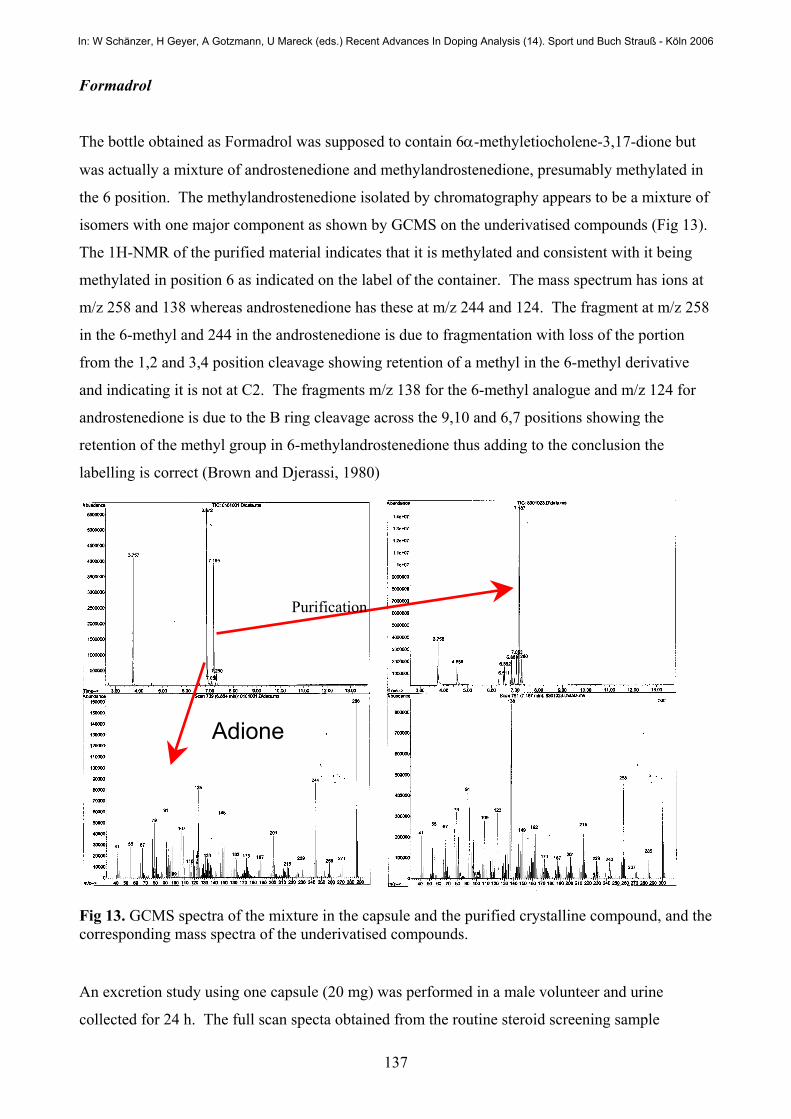
Formadrol
The bottle obtained as Formadrol was supposed to contain 6α-methyletiocholene-3,17-dione but
was actually a mixture of androstenedione and methylandrostenedione, presumably methylated in
the 6 position. The methylandrostenedione isolated by chromatography appears to be a mixture of
isomers with one major component as shown by GCMS on the underivatised compounds (Fig 13).
The 1H-NMR of the purified material indicates that it is methylated and consistent with it being
methylated in position 6 as indicated on the label of the container. The mass spectrum has ions at
m/z 258 and 138 whereas androstenedione has these at m/z 244 and 124. The fragment at m/z 258
in the 6-methyl and 244 in the androstenedione is due to fragmentation with loss of the portion
from the 1,2 and 3,4 position cleavage showing retention of a methyl in the 6-methyl derivative
and indicating it is not at C2. The fragments m/z 138 for the 6-methyl analogue and m/z 124 for
androstenedione is due to the B ring cleavage across the 9,10 and 6,7 positions showing the
retention of the methyl group in 6-methylandrostenedione thus adding to the conclusion the
labelling is correct (Brown and Djerassi, 1980)
Fig 13. GCMS spectra of the mixture in the capsule and the purified crystalline compound, and the corresponding mass spectra of the underivatised compounds.
An excretion study using one capsule (20 mg) was performed in a male volunteer and urine
collected for 24 h. The full scan specta obtained from the routine steroid screening sample
Adione
Purification
In: W Schänzer, H Geyer, A Gotzmann, U Mareck (eds.) Recent Advances In Doping Analysis (14). Sport und Buch Strauß - Köln 2006

138
preparation and derivatisation (MSTFA/TMSI/Ethanthiol) showed two closely eluting metabolites
for the 6-methyl derivative. It also showed large androsterone and etiocholanolone peaks from the
androstenedione present in the formulation. These are shown in Fig 14 as the chromatogram TIC
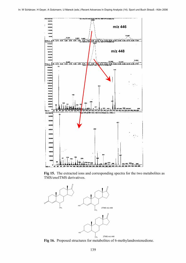
traces for the 0 and 6 h urine samples collected. The extracted ions and the corresponding spectra
are shown in Fig 15. The metabolites are very similar to those formed for drostanolone (the 2-
methyl isomer) and have almost the same retention times. Thus screening for drostanolone will
detect the metabolites of 6-methylandrostenedione but much care will be needed to distinguish
between the two positional isomers.
Fig 14. TIC spectra showing excretion of the metabolites of 6-methylandrostenedione.
By analogy with drostanolone the most likely metabolites are reduction at C3 and both reduction
at C3 and reduction of the double bond. In both metabolites only one carbonyl function is present
as derivatisation with MSTFA/TMS; Imidazole gives compounds with mass 72 less (m/z 374 and
376) indicating one carbonyl and one hydroxyl function in each. Thus the structures in Fig 16 are
most likely.
0 hr
6 hr m/z 448 in drostanolone window
In: W Schänzer, H Geyer, A Gotzmann, U Mareck (eds.) Recent Advances In Doping Analysis (14). Sport und Buch Strauß - Köln 2006
139
Fig 15. The extracted ions and corresponding spectra for the two metabolites as TMS/enolTMS derivatives.
Fig 16. Proposed structures for metabolites of 6-methylandrostenedione.
m/z 446
m/z 448
CH3
O
O
CH3
HO
O
CH3
HO
O
2TMS m/z 446
2TMS m/z 448
In: W Schänzer, H Geyer, A Gotzmann, U Mareck (eds.) Recent Advances In Doping Analysis (14). Sport und Buch Strauß - Köln 2006
140
Conclusions
There is still considerable work to be done to prove the structures of the supplements as it is not
trivial to work backwards from an unknown structure or even to confirm the identity of a
compound listed on the label. A process of isolation and spectral data gives some conclusions but
synthesis in an unambiguous manner is needed for final confirmations.
Much more study needs to be done to determine the structures of the metabolites but screening for
some of these products can be undertaken from the data obtained so far.
Acknowledgements
The staff at NMI needs to be acknowledged for sample preparation from the excretion studies and
obtaining GCMS and LCMSMS data. A. Lisi and K. Gazi performed the study of the
cyclohexanone derivatives of the ephedrines and benzylpiperazine, and Ben Parker and George
Wang, both Year in Industry students from the University of Sydney, did much of the isolation
work for the supplements. We acknowledge the Southern Cross University Ethics Committee for
approval of single dose studies.
References
Kazlauskas, R., Lisi, A., Trout, G., Chiral Derivatisation, Recent Advances in Doping Analysis (6), Proceedings of the Manfred Donike Workshop 15th Cologne Workshop on Dope Analysis, 15th to 20th March 1998, Sport und Book Strauss, Koln 1999, page 431-441. El-Haj, B.M., Al-Amri, A.M., Hassan, M.H., Ali, H.S., Bin Khadem, R.K., Forensic Science International, 2003, 135, 16-26. Manzoni,C., de Boer, D., Calcada, M.,. Povoa,I., dos Reys, L., Some Analytical Data Relevant for the Detection of 1 –Benzylpiperazine, a Pharmacological Alternative for Amphetamine, Recent Advances in Doping Analysis (9), Proceedings of the Manfred Donike Workshop 19th Cologne Workshop on Dope Analysis, 18th to 23rd March 2001, Sport und Book Strauss, Koln 2001, 239-243 Brown, F.J., Djerassi, C., “Elucidation of the course of the electron impact induced fragmentation of α,β-unsaturated 3-keto steroids”, J. Am. Chem. Soc., 102:2, 807-816, (1980). Mück, W.M., Henion, J.D., High-performance liquid chromatography/tandem mass spectrometry: Its use for the identification of stanozolol and its major metabolites in human and equine urine, Biomedical and Environmental Mass spectrometry 1990, 19:37-51. Schänzer, W., Donike M., Metabolism of anabolic steroids in man: synthesis and use of reference substances for identification of anabolic steroid metabolites, Analytica Chimica Acta, 1993, 275:23-48,
In: W Schänzer, H Geyer, A Gotzmann, U Mareck (eds.) Recent Advances In Doping Analysis (14). Sport und Buch Strauß - Köln 2006


![EMULGEL: A NOVEL APPROACH FOR …...Table 1: Classification of Topical drug delivery system [3] SOLID PREPARATION LIQUID PREPARATION SEMISOLID PREPARATION MISCELLANEOUS PREPARATION](https://img.pdfslide.net/doc/110x75/5e8d05ec0989714e041cdfea/emulgel-a-novel-approach-for-table-1-classification-of-topical-drug-delivery.jpg)