Embed Size (px)
Citation preview

RESEARCH Open Access
Gene-knockdown in the honey bee mite Varroadestructor by a non-invasive approach: studies ona glutathione S-transferaseEwan M Campbell1, Giles E Budge2, Alan S Bowman1*
Abstract
Background: The parasitic mite Varroa destructor is considered the major pest of the European honey bee (Apismellifera) and responsible for declines in honey bee populations worldwide. Exploiting the full potential of genesequences becoming available for V. destructor requires adaptation of modern molecular biology approaches tothis non-model organism. Using a mu-class glutathione S-transferase (VdGST-mu1) as a candidate gene weinvestigated the feasibility of gene knockdown in V. destructor by double-stranded RNA-interference (dsRNAi).
Results: Intra-haemocoelic injection of dsRNA-VdGST-mu1 resulted in 97% reduction in VdGST-mu1 transcript levels48 h post-injection compared to mites injected with a bolus of irrelevant dsRNA (LacZ). This gene suppression wasmaintained to, at least, 72 h. Total GST catalytic activity was reduced by 54% in VdGST-mu1 gene knockdown mitesdemonstrating the knockdown was effective at the translation step as well as the transcription steps. Althoughnear total gene knockdown was achieved by intra-haemocoelic injection, only half of such treated mites survivedthis traumatic method of dsRNA administration and less invasive methods were assessed. V. destructor immersedovernight in 0.9% NaCl solution containing dsRNA exhibited excellent reduction in VdGST-mu1 transcript levels(87% compared to mites immersed in dsRNA-LacZ). Importantly, mites undergoing the immersion approach hadgreatly improved survival (75-80%) over 72 h, approaching that of mites not undergoing any treatment.
Conclusions: Our findings on V. destructor are the first report of gene knockdown in any mite species anddemonstrate that the small size of such organisms is not a major impediment to applying gene knockdownapproaches to the study of such parasitic pests. The immersion in dsRNA solution method provides an easy,inexpensive, relatively high throughput method of gene silencing suitable for studies in V. destructor, other smallmites and immature stages of ticks.
BackgroundThe European honey bee, Apis mellifera, is vital to thepollination of agricultural and wild plants [1]. There iswidespread concern about the worldwide decline in theabundance of A. mellifera [2]. The ectoparasitic Varroamite (Varroa destructor) is the most important pest ofA. mellifera and plays a central role to honey bee losses[3]. V. destructor originally parasitized the Asian bee(A. cerana) where it nearly exclusively parasitized themale bees (drones), thus making little impact on the beecolony. However, in the 1950’s V. destructor shifted to
the European honey bee (A. mellifera) upon which itparasitizes both the drones and female bees (workers).This shift in parasitized caste is significant because theworkers make up the bulk of the adult bee populationwithin a colony [4]. V. destructor entered mainland Eur-ope in the 1970’s, the USA in 1987 and the UK in 1992and subsequently has been associated with the loss ofmillions of colonies [3]. The mite causes damage byfeeding on the haemolymph of both the developing beewithin brood cells and the adult bee. Mites also transmita variety of pathogens, most notably deformed wingvirus. In terms of both the number of enterprisesaffected and the impact of global food production, var-roasis is arguably the most serious disease of livestockin any species. Previous control of V. destructor by
* Correspondence: [email protected] of Biological Sciences (Zoology), University of Aberdeen, TillydroneAvenue, Aberdeen AB24 2TZ, UKFull list of author information is available at the end of the article
Campbell et al. Parasites & Vectors 2010, 3:73http://www.parasitesandvectors.com/content/3/1/73
© 2010 Campbell et al; licensee BioMed Central Ltd. This is an Open Access article distributed under the terms of the CreativeCommons Attribution License (http://creativecommons.org/licenses/by/2.0), which permits unrestricted use, distribution, andreproduction in any medium, provided the original work is properly cited.
chemical treatment is increasingly ineffective due to thedevelopment of widespread resistance in mites to thelimited available acaricides [5].There is an urgent need to harness modern molecular
techniques for research into the biology and, ultimately,the control of this non-model organism, V. destructor.RNA interference (RNAi) is a gene silencing techniquethat is becoming an ever more powerful tool in investi-gating the functional role of specific genes that may bepotential targets for chemotherapeutic intervention. TheRNAi mechanism involves the in vivo production ofsmall interfering RNA molecules (siRNAs) from largerintroduced double-stranded RNA (dsRNA). siRNAmolecules target and destroy specific mRNA, silencingthe target gene at the post-transcriptional stage. RNAihas been used successfully to investigate genes in arange of invertebrate species to date including gut pro-teins in tsetse flies [6]; moulting genes in Tribolium cas-taneum [7], aquaporins in aphids [8], a putativeprostaglandin E synthase in sealice [9] and extensively inticks (reviewed in [10]). In most cases, administration ofdsRNA to the invertebrate target is achieved by intra-haemocoelic injection, though immersion in solutionscontaining the dsRNA has been employed for nema-todes and sea lice.In this study, we assessed the feasibility of gene
knockdown in V. destructor using dsRNA and deter-mined the speed and persistence of the gene knockdowneffect. Additionally, we examined less invasive methodsof dsRNA administration to increase ease of experimen-tation and improve mite survivability. The chosen targetgene was a glutathione S-transferase of the mu class.GSTs were selected for study as they have been impli-cated in acaricide resistance in ticks [11,12] and scabiesmites [13,14] and were readily amenable to activityassays to assess the effect of gene knockdown at thetranslational step.
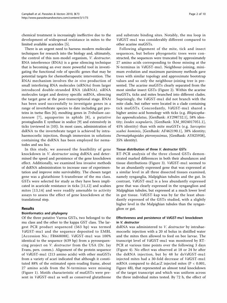
ResultsBioinformatics and phylogenyOf the three putative Varroa GSTs, two belonged to themu class and the other to the kappa GST class. The lar-gest PCR product sequenced (563 bp) was termedVdGST-mu1 and the sequence deposited to EMBL[Accession No.: FR668088]. VdGST-mu1 was 100%identical to the sequence (639 bp) from a pyrosequen-cing project on V. destructor from the USA (Dr. JayEvans, pers. comm.). Alignment of the protein sequenceof VdGST-mu1 (213 amino acids) with other muGSTsfrom a variety of acari indicated that although it consti-tuted 88% of the estimated open-reading frame, about27 amino acids from the N-terminus were missing(Figure 1). Motifs characteristic of muGSTs were pre-sent in VdGST-mu1 as well as conserved glutathione
and substrate binding sites. Notably, the mu loop inVdGST-mu1 was considerably different compared toother acarine muGSTs.Following alignment of the mite, tick and insect
sequences, but before phyogenetic trees were con-structed, the sequences were truncated by approximately27 amino acids corresponding to those missing at theN-terminus in VdGST-mu1. Neighbour-joining, mini-mum evolution and maximum parsimony methods gavetrees with similar topology and approximate bootstrapvalues and so only the neighbour-joining tree is pre-sented. The acarine muGSTs clearly separated from themost similar insect GSTs (Figure 2). Within the acarinemuGSTs, ticks and mites branched into different clades.Suprisingly, the VdGST-mu1 did not branch with themite clade, but rather were located in a clade containingtick muGSTs. Concordantly, VdGST-mu1 shared ahigher amino acid homology with ticks (e.g. Rhipicepha-lus appendiculatus, [GenBank: AY298732.1], 58% iden-tity; Ixodes scapularis, [GenBank: XM_0024017051.1],45% identity) than with mite muGSTs (e.g. Sarcoptesscabei hominis, [GenBank: AF462190.1], 38% identity;Dermatphagoides pteronyssimus, [GenBank: AY825938],35% identity).
Tissue distribution of three V. destructor GSTsRT-PCR analysis of the three cloned GSTs demon-strated marked differences in both their abundances andtissue distributions (Figure 3). VdGST-mu1 seemed tobe an abundantly expressed gene that was expressed ata similar level in all three dissected tissues examined,namely synganglia, Malpighian tubules and the gut. Incontrast, VdGST-mu2 is a less abundantly expressedgene that was clearly expressed in the synganglion andMalpighian tubules, but expressed at a much lower levelin gut tissue. VdGST-kap was by far the least abun-dantly expressed of the GSTs studied, with a slightlyhigher level in the Malpighian tubules than the syngan-glion or gut.
Effectiveness and persistence of VdGST-mu1 knockdownin V. destructordsRNA was administered to V. destructor by intrahae-mocoelic injection with a 20 nl bolus in distilled waterand the mites then allowed to feed on bee larvae. Thetranscript level of VdGST-mu1 was monitored by RT-PCR at various time points over the following 3 days(Figure 4). No effect was observed at 18 or 24 hr afterthe dsRNA injection, but by 48 hr dsVdGST-mu1injected mites had a 30-fold decrease of VdGST-mu1mRNA compared to dsLacZ injected mites (P < 0.001,Figure 4B), that represented an almost total knockdownof the target transcript and which was uniform acrossthe three individual mites tested. By 72 h, the effect of
Campbell et al. Parasites & Vectors 2010, 3:73http://www.parasitesandvectors.com/content/3/1/73
Page 2 of 10

the RNAi treatment at the transcript level was stillpresent (P < 0.001).
Effectiveness and mortality rates of different routes ofdsRNA administrationThough microinjection of dsRNA had very effectivelyknocked down VdGST-mu1 mRNA levels (96 ± 3.7%, n= 4, Figure 4B), such injection techniques were labor-ious, required specialised equipment and led to highmortality rates in the mites (50% survival, n = 62).Alternative, less invasive approaches to dsRNA adminis-tration were assessed (Table 1). A 1 μl droplet of 0.2%Triton-X100 (v/v) placed on mites immobilised on adhe-sive tape rapidly killed the mites (n = 6), whereas a 1 μldroplet of dsRNA in plain water had no mortality (100%survival, n = 14), but was totally ineffective in reducingVdGST-mu1 transcript levels. Mites completely sub-merged in 0.2% Triton-X100 died very quickly (n = 15),
whereas mites submerged in water died within a fewhours (n = 15). Increasing the osmolality of the soakingsolution by using 0.9% saline greatly increased survivalof mites (72% survival, n = 26) submerged for 14 h at4°C and then placed on bee pupae for a further 48 hr.This method effectively knocked down VdGST-mu1mRNA (87 ± 0.7%, n = 3; Figure 5). It should be notedthat mites removed from brood cells and placed on beepupae in Petri dishes but receiving no treatment at all,typically exhibited survival from 75 - 90% over a 48 hrperiod.
Phenotype of VdGST-mu1 knockdown mitesTo determine that the knockdown effect of treatment witheither dsRNA-lacZ or dsRNA-VdGST-mu1 was not sim-ply just at the mRNA (transcriptional) level, but was alsocarried through to the protein (translational) level, weassessed total GST catalytic activity in mites (Figure 6).
GHHH G H G GSarcoptes MSSKPTLGYWDLRGLGQSIRILLTYAGVDFVDKRYKIGSAPDFDRGEWLNDKF--NLGLD 58 Psoroptes MDTKPVLGYWDARGLGQAIRLLLTYADVDFIDKRYKVGPAPNYDRSEWLNDKY--NLGLD 58 Boophilus --MAPVLGYWDIRGLAQPIRLLLAHVDAKVDDKRYTCGPPPDFDRSSWLNEKT--KLGLE 56 Rhipicephalus --MAPILGYWNIRGLAQPFRLLLAHVDAKVEEKQYSCGPPPDFDKSYWLSEKP--KLGLD 56 Varroa -----------------------------FEERRYTTIAASDFDKSEWALEKANNKLNLA 31 . :::*. ...::*:. * :* :*.*
GGG GG GG HSarcoptes FPNLPYYIEGDVKLTQSIAILRYLGRKHKLDGQNEQEWRRITLCEQQIMDLLMALARICY 118 Psoroptes FPNCPYYLDGNVRLTQSIAILRYIARKYNLNGNNEMEKIRISLAEQQIVDLNQALSRVAY 118 Boophilus FPNLPYYIDGDVKLTQSMAILRYLARKHGLEGKTEAEKQRVDVSEQQFADFRMNWVRLCY 116 Rhipicephalus FPNLPYYIDGDVKLTQSMAILRYLARKYDLMGKTEAEKQRVDVVEQQLADFRVNWGRLCY 116 Varroa FPNLPYLVDGSVKLSQSHAIMRYLGRKFNLIGTTEIELAHCELVEQQIADLRTAFMKLCY 91 *** ** ::*.*:*:** **:**:.**. * * .* * : : ***: *: ::.*
Sarcoptes DPNFEKLKL--------------DLVAKLPDDLKLFSKFLGDHQFVAGTNISYIDFLVYE 164 Psoroptes NPNCEKLKP--------------EYLEKLPEQLELLSKFLNNQPFISGTNISYADFLLYE 164 Boophilus NPDFEKLKG--------------DYLKNLPASLKAFSDYLGTHKFFAGDNLTYVDFIAYE 162 Rhipicephalus SPDFEKLKG--------------DYLKDLPASLKAFSDYLGNRKFFAGDNLTYVDFIAYE 162 Varroa SPSFERLQEGTCSKADCLGVLNGGFIDRFAHMLQEISAFLGERKWFLNEKLTYVDFLAYE 151 .*. *:*: : :. *: :* :*. : :. . :::* **: **
Sarcoptes YLIRVKIFAPEIFTKFPNLNSYITRIESMPKISAYIKQQ--EPQLFNGPMAKWNTK------ 218 Psoroptes YLTKIKVLMPELFVKNDNLKKFHERIEALPRINEYIKKQ--QPKSFHGPTALWNGQYA–--- 220 Boophilus MLAQHLIFAPDCLKDFANLKAFVDRIEALPHVAAYLKSDKCIKWPLNGDMASFGSRLQKKP- 223 Rhipicephalus MLDQHLLFAPDCLKDFANLKAFVDRVAALPRVAAYLKSDKCIKWPLNGDMASFGSRLQKKP- 223 Varroa LLFQMYVWNSSVFKNVTNLTDFITRFEALPQISAYMKTDSYIKWPFNNIMASYGSRNQVCPF 213 * : : .. : . **. : *. ::*:: *:* : ::. * :. :
mu loop
Figure 1 Amino acid alignment of VdGST-mu1 with other acarine muGSTs. muGSTs from two mites, Sarcoptes scabiei type hominis[GenBank: AY825933] and Psoroptes ovis [GenBank: AM991140], and from two ticks Boophilus microplus [GenBank: AF077609] and Rhipicephalusappendiculatus [GenBank: AY298732] were aligned with VdGST-mu1. The boxed areas represent conserved mu-class GST motifs. Putativeglutathione (black highlighted “G”) and substrate (black highlighted “H”) binding sites are indicated. Homology across the five acarine sequencesis indicated “*” = residue identity; “:” = conserved residue and “.” = semi-conserved residue.
Campbell et al. Parasites & Vectors 2010, 3:73http://www.parasitesandvectors.com/content/3/1/73
Page 3 of 10

Mites injected with dsRNA-VdGST-mu1 exhibited signifi-cantly (P < 0.05) lower GST activity rates than thoseinjected with dsRNA-lacZ (28.1 ± 7.9 vs. 61.2 ± 12.9FLU min-1).
DiscussionGSTs are a large family of multifunctional enzymes [15]that catalyse the conjugation of reduced glutathione tothe electrophilic centres of lipophilic compounds
rendering them water soluble, less toxic products thatcan be rapidly excreted from the organism’s body. Assuch, GSTs play an important role in phase II detoxifi-cation of both endogenous and xenobiotic compoundssuch as pesticides. Specifically, GSTs have been shownto be involved in acaricide resistance in ticks [11,12],scabies mites [13,14] and plant mites [16]. In the presentstudy we demonstrated the presence and tissue expres-sion of two mu- and one kappa-class GST in V. destruc-tor. The largest sequence fragment termed, VdGST-mu1was 100% identical to the sequence present in anongoing pyrosequencing project (Dr. Jay Evans, personalcomm.) performed on V. destructor samples from theUSA. VdGST-mu1 was present in all mite tissuesassessed and was, by far, the most abundant of the threeGSTs examined. VdGST-mu1 contained conserved glu-tathione and substrate binding residues and motifs char-acteristic of mu-class GSTs. However, both the mu-loopand SNAIL/TRAIL motifs were notably different fromthose of other acarine muGSTs. Interestingly, VdGST-mu1 was more similar to tick muGSTs than that ofmites.VdGST-mu1 was used as a test gene to assess the fea-
sibility of dsRNAi gene knockdown in V. destructor.Essentially total suppression of VdGST-mu1 expressionwas achieved within 48 h after an intrahaemocoelic
Sarcoptes scabiei delta-GST
Tribolium castaneum
Pediculus humanus corporis
Apis mellifera
Drosophila melanogaster
Dermacentor variabilis
Boophilus microplus
Ixodes scapularis
Rhipicephalus appendiculatus
Varroa destructor
Dermatophagoides pteronyssinus-1
major allergen p Dp 15
Dermatophagoides pteronyssinus-2
Suidasia medanensis Sui m 8 allergen
Psoroptes ovis
100
57
44
100
92
51
34
41
80
98
100
100
74
mites
insectsticks
Lepidoglyphus destructor allergen
Sarcoptes scabiei type hominis
Sarcoptes scabiei type canis100
90
Figure 2 Phylogeny of the acarine muGST family. Phylogenetic tree constructed using amino acid sequences with neighbour-joining methodusing MEGA (4.1). Numbers are bootstrap values. Accession and database sequence identifiers are as follows: Sarcoptes scabiei type hominismuGST [GenBank: AY825933]; S. scabiei type canis muGST [GenBank: ACX33874.1]; Lepidoglyphus destructor allergen Lep, [GenBank: AY291572];Psoroptes ovis muGST [GenBank: AM991140]; Suidasia medanensis Sui m 8 allergen, [GenBank: AY800356]; Dermatophagoides pteronyssinus muGST-2 [GenBank: AY825939]; D. pteronyssinus major allergen Dp15 GST homolog [GenBank: S75286]; D. pteronyssinus muGST-1 [GenBank: AY825938];Rhipicephalus appendiculatus GST [GenBank: AY298732]; Ixodes scapularis GST [GenBank: XM_002401705]; Boophilus microplus GST [GenBank:AF077609]; Dermacentor variabilis [GenBank: EU551607]; Drosophila melanogaster GST-S1 [GenBank: NM_166217]; Apis mellifera GST [GenBank:FJ374871]; Pediculus humanus corporis GST [GenBank: XM_002426842]; Tribolium castaneum GST [GenBank: XM_962313]. The tree was rootedusing the S. canis var. delta-GST [GenBank: AY649788] as the outlier.
Actin
GST Mu
GST Mu2
GST Kap
Whole Syn. Mal. Gut.
Figure 3 Tissue specific expression of three GSTs in V.destructor. Expression of VdGST transcripts were determined by RT-PCR in different tissues of V. destructor using mu1, mu2 and kappaVdGST-specific primers and gel loading normalized to actin. Syn =synganglion; Mal = Malpighian tubules.
Campbell et al. Parasites & Vectors 2010, 3:73http://www.parasitesandvectors.com/content/3/1/73
Page 4 of 10

bolus injection of dsRNA and this suppression wasmaintained until at least 72 h. RNAi by injected dsRNAis an extensively employed approach for research inticks (reviewed in [10]) and total gene knockdown canbe maintained for up to, at least, 9 days [17]. However,V. destructor are considerably smaller (1 mm length ×1.5 mm width) than adult ticks and injection was onlypossible by pulled-glass capillary needles connected to arelatively sophisticated micro-injector system, ratherthan using manually operated micro-syringes as isthe case for ticks and most insects. Even though the
micro-injector system allowed us to demonstrate effec-tive dsRNAi in V. destructor, survival of mites under-going this very invasive and traumatic procedure wasonly 50% over a 48 h period.Various less invasive techniques of introducing dsRNA
into V. destructor were investigated that would improvespeed, ease of operation, increase throughput, could beperformed without expensive equipment and, mostimportantly, increase survivability of the treated groups.Topically applied dsRNA to immobilized mites for sev-eral hours failed to induce gene knockdown, presumably
Actin
GST Mu
18 24 48 72 18 24 48 72dsRNA-LacZ dsRNA-GST MuA.
B.S
Tm
u r
elat
ive
to A
ctin
40
60
80
100dsRNA-LacZdsRNA-GSTmu
******
Time (h)20 30 40 50 60 70
% G
S
0
20
Figure 4 Extent and persistence of dsRNA-VdGST-mu1 gene knockdown in adult V. destructor. (A) VdGST-mu1 transcipt levels weredetermined by RT-PCR up to 72 h post-dsRNA treatment and gel loading normalized to actin. (B) Semi-quantitative VdGST-mu1 RT-PCR bandintensities were determined by densitometry and the relative abundance to the within-sample actin band calculated. The extent of VdGST-mu1:actin knockdown is presented as a percentage for VdGST-mu1-dsRNA injected mites and control mites injected with LacZ-dsRNA at each giventime point. Data are means ± SEM (n = 3). Asterixes represent significant difference (P < 0.001) between treatments determined by Student’s t-test.
Table 1 Mortality of V. destructor and the degree of gene knockdown under different RNAi experimental conditions
Treatment Application Route Survival at 48 h (%) VdGSTMu knockdown (%)a
dsRNA in 0.9% NaCl topical droplet 79 (n = 14) 0% (n = 3)
dsRNA in 0.2% Triton-X100 topical droplet 0 (n = 6) n/d
distilled water immersion 0 (n = 15) n/a
0.2% Triton-X100 immersion 0 (n = 15) n/a
0.9% NaCl immersion 80 (n = 15) n/a
dsRNA in 0.9% NaCl immersion 76 (n = 21) 87 ± 0.7% (3)
dsRNA injection 50 (n = 62) 96 ± 3.7% (4)a n/d, not determined; n/a, not appropriate; data presented mean ± SEM
Campbell et al. Parasites & Vectors 2010, 3:73http://www.parasitesandvectors.com/content/3/1/73
Page 5 of 10

because the dsRNA was unable to cross the cuticle. Wetried the addition of the detergent Triton X-100 tobreak the surface tension of the water allowing thedsRNA solution to become more in contact with thecuticle rather than held off it by the fine hair layer(Figure 7). However, Triton X-100 caused rapid death ofthe mites. Next we assessed total immersion of mites asa possible route for dsRNA administration as was pre-viously successfully performed with salmon lice in sea-water [9]. Mites immersed in 0.2% Triton X-100 diedrapidly and, surprisingly, mites in distilled water diedwithin a few hours. We postulated that the hypo-osmo-larity of the distilled water caused an osmotic influx ofwater into the mite tissues and this was exacerbated in
the presence of Triton X-100 which may have allowedwater to enter through the spiracles more rapidly withthe reduced water tension.Increasing the osmotic concentration of the immer-
sion solution by using 0.9% NaCl allowed the mites toremain under solution overnight at 4°C with 75-80%
Actin
GST Mu
dsRNA-LacZ dsRNA-GST Mu
Figure 5 Effectiveness of gene-knockdown of VdGST-mu1 inV. destructor following immersion in dsRNA solution. Adult V.destructor were soaked overnight at 4°C in dsRNA in 0.9% NaCl.Mites (n = 3) were assayed by RT-PCR at 48 h post-treatment andgel loading normalized to actin.
Flu
ore
scen
ce u
nit
s
100
200
300
400
500
dsRNA-LacZdsRNA-GSTMu
Time (s)0 100 200 300 400
0
Figure 6 Total GST catalytic activity in V. destructor following VdGST-mu1 knockdown. GST activity of pairs of mites 48 h-post-injectionwith either VdGST-mu1 or LacZ-dsRNA measured by monitoring fluorescence of the substrate MCB over 10 min. Closed circles represent LacZ-dsRNA injected mites (n = 4) and open circles represent VdGST-mu1-dsRNA injected mites (n = 7). Data are means ± SEM.
b
a
Figure 7 Injection sites of V. destructor adult female. Arrowsindicate injection sites either between the soft tissue proximal tothe anal region and postcoxal plate (a), or in the soft tissue in coxalregion IV (b). Scale bar = 500 μm.
Campbell et al. Parasites & Vectors 2010, 3:73http://www.parasitesandvectors.com/content/3/1/73
Page 6 of 10

survival and achieved excellent gene silencing. It is notknown what is the site of dsRNA entry into the mite,but this is of considerable interest and importance. Wepresumed entry to be either by ingestion or though thetracheal system via the spiracles. Adult femaleV. destructor enter the prepupal cells 24 - 48 h beforethe cell is capped and they remain hidden from vigilantnurse bees by submerging themselves in the liquidbrood food [18] with the peritremes surrounding thespiracles protruding from the liquid enabling the mitesto breathe [19,20]. It may be possible to improve survi-vability and, possibly increase gene knockdown efficacy,if the osmolality of the immersion solution wereincreased to that of liquid brood food that is presumedto be greater than that of physiological saline (0.9%NaCl). Indeed, further optimisation of this approachcould be attempted, if thought necessary, by alteringimmersion temperature and time, but the method pre-sented here is a good starting point.Not only was gene knockdown achieved at the tran-
scriptional step (mRNA) but this was also apparent atthe translational step (protein) with decreased GST cata-lytic activity. The apparent discrepancy between a 97%reduction in VdGST-mu1 transcript level and the 54%reduction in GST enzyme activity is because the enzymeassay detects total GST activity not just that contributedby VdGST-mu1. There have been reports of so-called“off-target” effects of RNAi whereby unintended genessharing stretches of identical homology can be affected.A meta-analysis of dsRNAi studies in Drosophila high-throughput screens indicated that dsRNAs containing≥ 19 nucleotide perfect matches to unintended targetscan, in some instances, result in off-target effects [21].In the case of VdGST-mu1, the most similar gene weknow of in Varroa is VdGST-mu2 [EMBL AccessionNo.: FR675857] which shares 29% identity at the nucleo-tide level, but the greatest matching stretch possible isone of 12/19 nucleotides (Figure 8). Thus, it is likelythat the reduction in assayable total GST activity is dueto a specific knockdown of VdGST-mu1, but furtherstudies would be necessary to confirm this. Our datawould indicate that VdGST-mu1 is the predominantcatalytic GST in V. destructor and highlights the specifi-city advantage of gene knockdown over chemicalenzyme inhibitors which simultaneously affect multiplegene products.
ConclusionsIn conclusion, we have established dsRNA knockdownin the critically important bee parasite, V. destructor,using a mu-class GST as the candidate gene. A simple,inexpensive, high throughput method of dsRNA admin-istration was established with low mortality rates forV. destructor. This immersion approach may be applied
to other small acarine pests such as scabies mites, sheepscab mites, poultry mites and plant pest mites. Addi-tionally, the approach can be employed in ticks wherethe immersion approach is more convenient than injec-tions for adult ticks but can also be applied to the smal-ler larval and nymphal stages which are presently notincluded in such gene knockdown studies. The degreeof knockdown was essentially complete and persisted forseveral days and was apparent at both the transcript andprotein level. The dsRNAi approach will allow research-ers to make full use of the V. destructor gene sequencesbecoming available to investigate biological questions inthis important parasite, notably to develop control stra-tegies in the era when current suitable acaricides are nolonger effective due to widespread resistance. Peculiarlyto Varroa, because of the relatively close relatednessbetween the parasite and host (Varroa and bee) it isespecially difficult to develop pesticides effective againstthe Varroa, but innocuous to the bee. In this respect,the specificity of dsRNAi may allow targeting ofV. destructor genes as a control strategy, as suggestedfor other insect pests [22].
MethodsMite collection and husbandryVarroa destructor mites were collected from cappedbrood cells of Apis mellifera from hives in York, Eng-land that had purposefully been untreated for Varroacontrol. Prior to harvesting mites, the bee frames weremaintained in Aberdeen at 27ºC and 40% relativehumidity with a 15.5 h : 8.5 h, light:dark regime. Miteswere either removed from newly emerged adult bees orfrom pupae following uncapping of the brood cell andextraction of the pupae. Mites were collected, bisectedand placed in RNA ZR extraction buffer (ZymoResearch, Orange, California, USA) and stored at -80 ºCprior to initial total RNA isolation.
RT-PCR of GSTs from V. destructorSix adult (pharate and mobile) and 2 deutonymph miteswere homogenized in 600 μl extraction buffer and totalRNA prepared using a mini RNA Isolation II Kit (ZymoResearch), as per manufacturer’s instructions. ElutedRNA was co-precipitated with glycogen in 95% ethanoland resuspended in 10 μl of DEPC-treated water. Afterisolation, 2 μg total RNA was DNase treated with 2 μl(2U) RQ1-DNase (Promega, Southampton, UK) and 2 μlRQ1 buffer and incubated at 37°C for 30 min. DNase-treated total RNA (2 μg) was incubated at 70°C with 0.5μg of oligo d(T)15 (Promega) in a total volume of 10 μlfor 5 min. Material was snap-chilled on ice for 5 minprior to the addition of 5 μl 5×RT buffer, 1 μl dNTPs(25 mM each), 0.5 μl Bioscript-reverse transcriptase andDEPC water to 25 μl. The reaction was incubated at
Campbell et al. Parasites & Vectors 2010, 3:73http://www.parasitesandvectors.com/content/3/1/73
Page 7 of 10
42°C for 60 min prior to arrest by heating to 70°C for5 min.A V. destructor database from an ongoing pyrosequen-
cing project (Dr. Jay Evans, pers. comm.) was mined forputative V. destructor GST sequences by BLASTx analy-sis using GST sequences from the deer tick, Ixodes sca-pularis. Of 14 putative V. destructor GST partialsequences identified, we chose three to evaluate further:two mu-class GSTs (VdGST-mu1 and VdGST-mu2) andone kappa class (VdGST-kap). These Varroa transcriptswere isolated by RT-PCR using primers based on thesesequences. PCR reactions consisted of 1 μl cDNA tem-plate, 5 μl 10 × reaction buffer, 2 μl 50 mM MgCl2, 1 μldNTPs (25 mM each), 1 μl each primer (10 mM each),0.5 μl (1.25 U) Taq (Bioline, London,UK) and DEPC-treated water to give a 50 μl total volume. Primer pairsused were VdGST-mu-F1 (CAAGTTAAATCTTGCGTTTCC) and VdGST-mu1-R1 (CTGTCCGTCTTCATGTATGC), VdGST-mu2-F1 (ACGATTTATCCGTTTTGACG) and VdGST-mu2-R2 (CCAACTGATGTGTCGTTCC), VdGST-Kap-F1 (TACTGGTGGTCGTTTCAGG) and VdGST-Kap-R1 (CTTCATAGGC-CAAGAGATGC), generating products of 494, 396 and343 bp, respectively. PCR cycling conditions were as fol-lows: 1 cycle of 5 min at 94°C, followed by 35 cycles of1 min at 94°C, 1 min at 53°C and 45 s at 72°C followedby a final extension time of 15 min at 72°C. Productswere visualised on an agarose gel and specific bandsexcised and cloned into pCR4-TOPO TA vector (Invi-trogen). Purified plasmids were submitted for sequen-cing to Eurofins MWG (Ebersberg, Germany) fromflanking T7 and T3 promoter regions to confirm theoriginal sequence data.
BioinformaticsThe GST class of the different Varroa sequences wasdetermined by database searches and phylogenetic ana-lysis. For VdGST-mu1, protein sequences from variousacari were extracted from both non-redundant and EST
databases at GenBank by tBLASTx. Additionally, themost similar GST sequences in four model insect spe-cies for which complete genomes are available were alsoobtained. Protein sequences were aligned in CLUS-TALW2 [23]. These sequences were used to estimatephylogeny with neighbour-joining, minimum evolutionand maximum parsimony methods using MEGA version4.1 [24]. Phylogenetic trees were constructed with10,000 bootstrap replicates.
Expression of GST transcripts in tissues of V. destructorV. destructor mites, were removed from brood cells, asdescribed above, and dissected under ice-cold dissectionbuffer (20 mM TRIS, 5 mM EDTA, 0.9% NaCl, pH 7.4).Malpighian tubules, gut and synganglia and were dis-sected from 20, 25 and 98 mites respectively, washed infresh ice-cold dissection buffer and stored in 50 μlRNAlater (Sigma, Poole, UK) at -80 °C. Prior to RNAextraction, an additional 450 μl dissection buffer wasadded to sample tubes and centrifuged at 10,000 g for15 min at 4°C. Supernatant was removed and tissuewashed with fresh dissection buffer before a final spin at10,000 g for 15 min at 4°C. Supernatant was discardedand 600 μl RNA ZR extraction buffer added to each tis-sue sample. Total RNA extraction, DNase treatment andreverse transcription were as described above. VdGST-mu1, VdGST-mu2 and VdGST-kap transcripts in tissue-specific cDNA were assayed by PCR. PCR protocol andcycling conditions were carried out using the VdGST-mu1, VdGST-mu2 and VdGST-kap primers, asdescribed above over 30 cycles. Gel loading was normal-ized to V. destructor actin (VdActinF, CATCAC-CATTGGTAACGAG and VdActinR, CGATCCAGACGGAATACTT) generating a fragment of approxi-mately 195 bp.
Preparation of dsRNAVdGST-mu1-dsRNA was prepared using a BLOCK-iTRNAi TOPO transcription kit (Invitrogen), according to
Figure 8 Alignment of VdGST-mu1 and VdGST-mu2 indicating stretches of perfect identity. Nucleotide sequences of VdGST-mu1 [EMBL:FR668088] and VdGST-mu2 [EMBL: FR675857] were aligned using ClustalW2 and edited in JalView Vers 2.5.1 [28] with gaps not permitted andconsensus stretches shown.
Campbell et al. Parasites & Vectors 2010, 3:73http://www.parasitesandvectors.com/content/3/1/73
Page 8 of 10

the manufacturer’s instructions. LacZ-dsRNA was pre-pared and used as a negative control. Briefly, PCR wascarried out as described above using adult femaleV. destructor cDNA in conjunction with VdGST-mu1specific primers (VdGST-mu1 F1/R1), or with controlLacZ-plasmid and LacZ specific primers (LacZ-F2,ACCAGAAGCGGTGCCGGAAA and LacZ-R2, CCA-CAGCGGTGGTTCGGAT). Products were resolved onan agarose gel, excised and purified using a Qiagen gelextraction kit (Qiagen, Crawley, UK). TOPO-T7 linkerwas ligated to VdGST-mu1 and LacZ reactions before asecondary PCR was carried out to gain sense and anti-sense templates. T7-RNA polymerase was used in tran-scription reactions with target templates to generatesense and antisense RNA. Finally, RNA strands wereannealed and the resultant dsRNA purified and quanti-fied in a micro-spectrophotometer (Nanodrop Technol-ogy Ltd). dsRNA was ethanol precipitated andresuspended in DEPC-treated water to a working con-centration of 2.5 μg/μl and stored at -80°C.
Protocol of dsRNA injection and soakingAdult female V. destructor were removed from cappedbrood cells along with associated bee larvae. Microinjec-tions were carried out using pulled glass capillary nee-dles in conjunction with a Harvard micro-injectorsystem. Mites were placed on double-sided tape ventralside up, and injected with 20 nl (2.5 μg/μl) of eitherVdGST-mu1-dsRNA or LacZ-dsRNA in either the softtissue proximal to the anal region and postcoxal plate,or in the coxa IV region, as indicated in Figure 7. Nee-dles were left in each mite for 1 - 2 min to reduce theexpulsion of fluid from the wound and withdrawnslowly. Mites were left for 1 - 2 min to allow the injec-tion site to “seal” then returned to Petri dishes contain-ing 1 bee larvae per 4 mites. Dead or unhealthy lookingmites were removed after 1 hour and mortality wasmonitored over 72 h in LacZ-dsRNA, VdGSTmu1-dsRNA and non-injected mites.To assess non-invasive techniques for dsRNA delivery,
mites were either completely immersed in dsRNA orwere exposed to a droplet of dsRNA on their ventralcarapace. For soaking experiments, adult mites wereremoved from capped brood cells and placed in 500 μlmicrofuge tubes containing 20 μl VdGST-mu1-dsRNAor LacZ-dsRNA (2.5 μg/μl) supplemented with eithernothing, 0.9% NaCl, 0.2% Triton-X100 or both. Miteswere soaked at 4ºC overnight before being removed,dried and placed in Petri dishes at 27ºC, 95% relativehumidity with bee larvae. Alternatively, a sample ofmites was exposed to dsRNA by attaching them to dou-ble-sided tape and placing a 1 μl drop of VdGST-mu1-dsRNA or LacZ-dsRNA (2.5 μl/μg) supplemented with
either nothing, 0.9% NaCl, 0.2% Triton-X100 or both onthe ventral carapace. Mortality was monitored for 48 hprior to collection and validation of knockdown.
Validation of RNAiTo validate RNAi in injected, soaked and droplet-exposed adults, the total RNA was extracted from indi-vidual mites 48 h post-treatment. In addition, persis-tence of the RNAi effect was measured in injected mitesby harvesting at 18, 24, 48 and 72 h. Total RNA wasextracted from mites using mini-RNA isolation kit II,prior to DNase-treatment and reverse transcription, asdescribed previously. PCR was carried out using eitherVdGST-mu1-dsRNA or LacZ-dsRNA treated samplecDNA in conjunction with primers specific for actin orVdGST-mu1 using primers and cycling conditions, asdescribed above. Products were visualized on an agarosegel normalized to actin loading. To assess persistenceand approximate % knockdown, ImageJ software wasused to carry out semi-quantitative densitometric analy-sis on gel images.
Glutathione S-transferase activity in RNAi silenced mitesAn enzymatic assay was used to assess the effect ofsilencing VdGST-mu1 transcript on GST enzyme activ-ity in dsRNA-treated mites. GST activity was measuredby using monochlorobimane (MCB) as the substrate, aspreviously described for scabies mites [25] and plantmites [26], but here we adapted it for cuvettes ratherthan microplates. MCB forms a stable fluorescent conju-gate with reduced glutathione when catalyzed with GST.Mites injected with LacZ-dsRNA or VdGST-mu1-dsRNA were harvested 48 h post-treatment and frozenat -80ºC. Mites were grouped into pairs and homoge-nized by milling 2 × 30 s, with a single 3 mm tungstenbead in 175 μl of ice cold 0.05 M Tris buffer pH7.5 in amechanical tissue disruptor (TissueLyser, Qiagen). Aftermilling, samples were disrupted further in a sonicatingwater bath for 3 × 20 s with chilling in between burststhen centrifuged at 10,000 g for 5 min at 4ºC. Thesupernatant was retained, subjected to three freezethaw-cycles and sonicated for a further 2 × 20 s. Sam-ples were then centrifuged at 10,000 g for 5 min at 4ºCand 150 μl of supernatant from each sample used in theGST assay. Reduced glutathione was dissolved in 0.05 MTris pH 7.5 and MCB dissolved in methanol and addedto each reaction to a final concentration of 3 mM and0.3 mM, respectively, in a total reaction volume of200 μl. Mite homogenate and substrate were incubatedat 37ºC for 10 min with fluorescence of GSH-bimaneadduct measured every 2 s using a F4500 fluorimeter(Hitachi) with emission wavelength at 465 nm and exci-tation wavelength at 390 nm. All results presented were
Campbell et al. Parasites & Vectors 2010, 3:73http://www.parasitesandvectors.com/content/3/1/73
Page 9 of 10

corrected for non-enzymatic fluorescence obtained fromsamples substituting 150 μl 0.05 M Tris for the mitematerial.
AcknowledgementsWe are indebted to Dr. Jay Evans and Dr. Scott Cornman, USDA-ARS BeeResearch Laboratories, Beltsville, MD, USA for access to GST sequences fromtheir Varroa sequencing project prior to its collation, analysis and annotationand publication [27]. This project was sponsored by the Scottish BeekeepersAssociation. We gratefully acknowledge funding from a Genesis FaradaySPARK Award, funded as part of a Scottish Government SEEKIT project; theDepartment for Environment, Food and Rural Affairs and the WelshAssembly Government.
Author details1School of Biological Sciences (Zoology), University of Aberdeen, TillydroneAvenue, Aberdeen AB24 2TZ, UK. 2National Bee Unit, Food and EnvironmentResearch Agency, Sand Hutton, York, YO41 1LZ, UK.
Authors’ contributionsEMC participated in study conception and design, performed all theexperimental work and drafted the manuscript. GEB participated in studyconception, facilitated sample collection and helped draft and edit themanuscript. ASB conceived and designed the study, performed sequenceanalysis, statistical analysis, interpretation and drafted the manuscript. Allauthors read and approved the final manuscript.
Competing interestsThe authors declare that they have no competing interests.
Received: 26 July 2010 Accepted: 16 August 2010Published: 16 August 2010
References1. Klein A-, Vaissière BE, Cane JH, Steffan-Dewenter I, Cunningham SA,
Kremen C, Tscharntke T: Importance of pollinators in changinglandscapes for world crops. Proceedings of the Royal Society B: BiologicalSciences 2007, 274:303-313.
2. van Engelsdorp D, Meixner MD: A historical review of managed honeybee populations in Europe and the United States and the factors thatmay affect them. J Invertebr Pathol 2010, 103(SUPPL. 1):S80-S95.
3. Martin SJ: The role of varroa and viral pathogens in the collapse ofhoney bee colonies: A modelling approach. J Appl Ecol 2001,38:1082-1093.
4. Winston ML: The honey bee colony: Life history. The hive and the honeybee Hamilton Illinois: Dadant & SonsGraham JM , 10 1992.
5. Martin SJ: Acaricide (pyrethroid) resistance in Varroa destructor. Bee World2004, 85:67-69.
6. Walshe DP, Lehane SM, Lehane MJ, Haines LR: Prolonged geneknockdown in the tsetse fly Glossina by feeding double stranded RNA.Insect Mol Biol 2009, 18:11-19.
7. Minakuchi C, Namiki T, Shinoda T: Krüppel homolog 1, an early juvenilehormone-response gene downstream of Methoprene-tolerant, mediatesits anti-metamorphic action in the red flour beetle Tribolium castaneum.Dev Biol 2009, 325:341-350.
8. Shakesby AJ, Wallace IS, Isaacs HV, Pritchard J, Roberts DM, Douglas AE: Awater-specific aquaporin involved in aphid osmoregulation. InsectBiochem Mol Biol 2009, 39:1-10.
9. Campbell EM, Pert CC, Bowman AS: RNA-interference methods for gene-knockdown in the sea louse, Lepeophtheirus salmonis: Studies on aputative prostaglandin E synthase. Parasitology 2009, 136:867-874.
10. de la Fuente J, Kocan KM, Almazán C, Blouin EF: RNA interference for thestudy and genetic manipulation of ticks. Trends Parasitol 2007, 23:427-433.
11. Enayati AA, Asgarian F, Amouei A, Sharif M, Mortazavi H, Boujhmehrani H,Hemingway J: Pyrethroid insecticide resistance in Rhipicephalus bursa(Acari, Ixodidae). Pestic Biochem Physiol 2010, 97:243-248.
12. Enayati AA, Asgarian F, Sharif M, Boujhmehrani H, Amouei A, Vahedi N,Boudaghi B, Piazak N, Hemingway J: Propetamphos resistance inRhipicephalus bursa (Acari, Ixodidae). Vet Parasitol 2009, 162:135-141.
13. Molin EU, Mattsson JG: Effect of acaricides on the activity of glutathionetransferases from the parasitic mite Sarcoptes scabiei. Parasitology 2008,135:115-123.
14. Mounsey KE, Pasay CJ, Arlian LG, Morgan MS, Holt DC, Currie BJ, Walton SF,McCarthy JS: Increased transcription of glutathione S-transferases inacaricide exposed scabies mites. Parasites and Vectors 2010, 3:43.
15. Sheehan D, Meade G, Foley VM, Dowd CA: Structure, function andevolution of glutathione transferases: Implications for classification ofnon-mammalian members of an ancient enzyme superfamily. Biochem J2001, 360:1-16.
16. Stumpf N, Nauen R: Biochemical markers linked to abamectin resistancein Tetranychus urticae (Acari: Tetranychidae). Pestic Biochem Physiol 2002,72:111-121.
17. Campbell EM, Burdin M, Hoppler S, Bowman AS: Role of an aquaporin inthe sheep tick Ixodes ricinus: Assessment as a potential control target.Int J Parasitol 2010, 40:15-23.
18. Sammataro D, Gerson U, Needham G: Parasitic mites of honey bees: Lifehistory, implications, and impact. Annu Rev Entomol. 2000, 45:519-548.
19. Bruce WA, Delfinado-Baker M, Vincenta DL: Comparative morphology ofthe peritremes of Varroa and Euvarroa (Varroidae), parasites of honeybees (Apidae). Internat J Acarol 1997, 23:13-20.
20. Donzé G, Guerin PM: Time-activity budgets and space structuring by thedifferent life stages of Varroa jacobsoni in capped brood of the honeybee, Apis mellifera. J Insect Behav 1997, 10:371-393.
21. Kulkarni MM, Booker M, Silver SJ, Friedman A, Hong P, Perrimon N, Mathey-Prevot B: Evidence of off-target effects associated with long dsRNAs inDrosophila melanogaster cell-based assays. Nature Methods 2006,3:833-838.
22. Whyard S, Singh AD, Wong S: Ingested double-stranded RNAs can act asspecies-specific insecticides. Insect Biochem Mol Biol 2009, 39:824-832.
23. Larkin MA, Blackshields G, Brown NP, Chenna R, Mcgettigan PA,McWilliam H, Valentin F, Wallace IM, Wilm A, Lopez R, Thompson JD,Gibson TJ, Higgins DG: Clustal W and Clustal X version 2.0. Bioinformatics2007, 23:2947-2948.
24. Tamura K, Dudley J, Nei M, Kumar S: MEGA4: Molecular EvolutionaryGenetics Analysis (MEGA) software version 4.0. Mol Biol Evol 2007,24:1596-1599.
25. Pasay C, Arlian L, Morgan M, Gunning R, Rossiter L, Holt D, Walton S,Beckham S, McCarthy J: The effect of insecticide synergists on theresponse of scabies mites to pyrethroid acaricides. PLoS NeglectedTropical Diseases 2009, 3(1):e354, Epub 2009 Jan 6.
26. Nauen R, Stumpf N: Fluorometric microplate assay to measureglutathione S-transferase activity in insects and mites usingmonochlorobimane. Anal Biochem 2002, 303:194-198.
27. Cornman RC, Schatz MC, Johnston S, Chen YP, Pettis J, Hunt G, Bourgeois L,Elsik C, Grozinger CM, Evans JD: Genomic survey of the ectoparasitic miteVarroa destructor, a major pest of the honey bee Apis mellifera. BMCGenomics .
28. Waterhouse AM, Procter JB, Martin DMA, Clamp M, Barton GJ: JalviewVersion 2-A multiple sequence alignment editor and analysisworkbench. Bioinformatics 2009, 25:1189-1191.
doi:10.1186/1756-3305-3-73Cite this article as: Campbell et al.: Gene-knockdown in the honey beemite Varroa destructor by a non-invasive approach: studies on aglutathione S-transferase. Parasites & Vectors 2010 3:73.
Campbell et al. Parasites & Vectors 2010, 3:73http://www.parasitesandvectors.com/content/3/1/73
Page 10 of 10