Embed Size (px)
Citation preview

Mitochondrial Complex II Is Essential for GametophyteDevelopment in Arabidopsis1[W][OA]
Gabriel Leon2, Loreto Holuigue, and Xavier Jordana*
Departamento de Genetica Molecular y Microbiologıa, Facultad de Ciencias Biologicas, P. UniversidadCatolica de Chile, Casilla 114–D, Santiago, Chile
Mitochondrial complex II (succinate dehydrogenase [SDH]) is part of the tricarboxylic acid cycle and the respiratory electrontransport chain. Its flavoprotein subunit is encoded by two nuclear genes, SDH1-1 and SDH1-2, in Arabidopsis (Arabidopsisthaliana). The SDH1-2 gene is significantly expressed only in roots, albeit at very low level, and its disruption has no effect ongrowth and development of homozygous mutant plants. In contrast, SDH1-1 transcripts are ubiquitously expressed, withhighest expression in flowers. Disruption of the SDH1-1 gene results in alterations in gametophyte development. Indeed,heterozygous SDH1-1/sdh1-1 mutant plants showed normal vegetative growth, yet a reduced seed set. In the progeny of selfedSDH1-1/sdh1-1 plants, distorted segregation ratios were observed, and no homozygous mutant plants were obtained.Reciprocal test crosses with the wild type demonstrated that the mutated sdh1-1 allele is not transmitted through the malegametophyte and is only partially transmitted through the female gametophyte. Consistently, microscopic analysis showedthat mutant microspores develop normally until the vacuolated microspore stage, but fail to undergo mitosis I, and then cellstructures are degraded and cell content disappears. On the other hand, half the mutant embryo sacs showed arresteddevelopment, either at the two-nucleate stage or before polar nuclei fusion. Down-regulation of SDH1-1 by RNA interferenceresults in pollen abortion and a reduced seed set, as in the insertional mutant. Altogether, our results show that SDH1-1, andtherefore complex II, are essential for gametophyte development.
Succinate:ubiquinone oxidoreductase (succinate de-hydrogenase [SDH]; EC 1.3.5.1), commonly referred toas mitochondrial complex II, has a central role inmitochondrial metabolism as a member of both theelectron transport chain and the tricarboxylic acid(TCA) cycle. This membrane-associated complex cat-alyzes the oxidation of succinate to fumarate and thereduction of ubiquinone to ubiquinol, and has beencharacterized in bacteria and heterotrophic eukaryotes(Lemire and Oyedotun, 2002; Yankovskaya et al., 2003).In these organisms, complex II contains only four sub-units: two peripheral membrane proteins, a flavopro-tein (SDH1) and an iron-sulfur protein (SDH2), and twosmall integral membrane proteins (SDH3 and SDH4).The succinate-binding site is formed by the SDH1protein, which is covalently linked to a FAD moleculeacting as acceptor of a hydride ion at an early step of
succinate oxidation. This flavoprotein subunit interactswith the SDH2 subunit, which transfers the electronsto the membrane through its three nonheme iron-sulfur centers. The two integral membrane proteins,SDH3 and SDH4, anchor the SDH1-SDH2 subcomplexto the matrix side of the inner mitochondrial mem-brane and contain a b-type heme and the ubiquinone-binding site (Yankovskaya et al., 2003). Surprisingly, ithas been shown recently that plant complex II maycontain additional subunits of unknown function alongwith the four classical subunits (Millar et al., 2004).
SDH subunits are all encoded in the nuclear genomein Arabidopsis (Arabidopsis thaliana), just like complexII from heterotrophic eukaryotes (Scheffler, 1998;Figueroa et al., 2001, 2002; Millar et al., 2004). Surpris-ingly, several of the complex II subunits are encodedby more than one gene in Arabidopsis. For instance, wehave reported that three nuclear genes, named SDH2-1,SDH2-2 and SDH2-3, encode the iron-sulfur subunit,and two nuclear genes, designated SDH1-1 and SDH1-2,encode the flavoprotein (Figueroa et al., 2001, 2002).SDH2-1 and SDH2-2, which have similar structuresand encode nearly identical proteins, have similarexpression patterns (Figueroa et al., 2001; Elorza et al.,2004). These results are consistent with the fact thatSDH2-1 and SDH2-2 probably arose via a recent geneduplication event and are redundant. In contrast, wehave recently reported a highly tissue-specific expres-sion for SDH2-3: this gene, which is structurally dif-ferent and would have diverged for a long time fromSDH2-1 and SDH2-2, is highly expressed in the em-bryo during seed development and its transcript ac-cumulates in dry seeds (Elorza et al., 2006).
1 This work was supported by the Fondecyt grant number1060485 and Beca Apoyo Tesis Doctoral 2003 to G.L.
2 Present address: Nucleo Milenio de Biologıa Celular Vegetal,Centro de Biotecnologıa Vegetal, Universidad Andres Bello, Repub-lica 217, Santiago, Chile.
* Corresponding author; e-mail [email protected]; fax 56–2–2225515.
The author responsible for distribution of materials integral to thefindings presented in this article in accordance with the policydescribed in the Instructions for Authors (http://www.plantphysiol.org) is: Xavier Jordana ([email protected]).
[W] The online version of this article contains Web-only data.[OA] Open Access articles can be viewed online without a sub-
scription.www.plantphysiol.org/cgi/doi/10.1104/pp.106.095158
1534 Plant Physiology, April 2007, Vol. 143, pp. 1534–1546, www.plantphysiol.org � 2007 American Society of Plant Biologists www.plantphysiol.orgon June 8, 2018 - Published by Downloaded from
Copyright © 2007 American Society of Plant Biologists. All rights reserved.

SDH1-1 (At5g66760) and SDH1-2 (At2g18450) havesimilar structures and encode highly similar proteinsthat would be functional as complex II flavoproteinsince they are highly conserved when compared to theirhomologs in other organisms (Figueroa et al., 2002).Moreover, they contain the residues known to beinvolved in FAD binding and substrate binding, andin proton transfer during catalysis, and they are ac-tively imported into isolated plant mitochondria(Figueroa et al., 2002). However, SDH1-1 and SDH1-2appear to be expressed at very different levels: theSDH1-1 mRNA was easily detected by northern-blothybridization in all tissues examined, whereas SDH1-2transcripts were only detected in reverse transcription-PCR and 3# RACE experiments (Figueroa et al., 2002).
The highest SDH1-1 mRNA levels were found inflowers (Figueroa et al., 2002). Analogous enhancedexpression in flowers has been observed for otherplant nuclear genes encoding mitochondrial proteins,including SDH2-1 and SDH2-2, and for mitochondrialtranscripts (e.g. Huang et al., 1994; Smart et al., 1994;Heiser et al., 1996; Elorza et al., 2004). This is related tothe fact that pollen development is one of the highestenergy-requiring processes in plants. The most com-pelling evidence for an essential role of mitochondriaduring pollen development is the phenomenon ofcytoplasmic male sterility (CMS). CMS is a maternalinherited trait in which mutations in the mitochondrialgenome impair pollen development (Hanson andBentolila, 2004). Furthermore, an increase in the num-ber of mitochondria per cell has been reported inmaize (Zea mays) and tobacco (Nicotiana tabacum) an-thers during pollen development (Lee and Warmke,1979; Huang et al., 1994).
To gain insight into the physiological role of com-plex II and to explore the function of the multiplegenes encoding the same SDH subunit, our group hasundertaken a reverse genetic analysis of the SDHgenes. Here we report the analysis of T-DNA insertionalmutants in SDH1-1 and SDH1-2, the flavoprotein genes.Our results reveal that SDH1-2 is dispensable and thatthe sdh1-1 null allele behaves as a general gameto-phytic mutation, demonstrating that complex II playsan essential role in gametophyte development.
RESULTS
Isolation of T-DNA Insertion Mutants of ArabidopsisSDH1 Genes
Several different mutant lines carrying T-DNA in-sertions in the SDH1-2 gene were identified as de-scribed in ‘‘Materials and Methods.’’ One mutant wasisolated and further characterized: the T-DNAwas con-firmed to be in the eighth out of 15 exons, and inter-rupted codon 336. The precursor SDH1-2 polypeptidededuced from the gene sequence has 632 amino acidsand disruption at codon 336 that is upstream of severalresidues involved in FAD and substrate binding, and
in catalysis is expected to result in a null mutation.Homozygous sdh1-2 mutant plants were obtained andshowed no apparent phenotypic defects during vege-tative or reproductive growth when compared to wild-type plants, at least under the growth conditions used.
These results indicate that the loss of SDH1-2 has noimpact on Arabidopsis growth and development, a factconsistent with our previous expression data (Figueroaet al., 2002). Furthermore, data on SDH1-2 expres-sion in the existing large expression databases confirmthat SDH1-2 is not expressed or is expressed at avery low level in most tissues and developmentalstages (Supplemental Fig. S1; Schmid et al., 2005; seealso https://www.genevestigator.ethz.ch, Zimmermannet al., 2004 for microarray data; and http://mpss.udel.edu/at, Meyers et al., 2004 for Massively ParallelSignature Sequencing experiments). Significant ex-pression was only observed in roots, nevertheless itsexpression level is still less than 10% that of SDH1-1 inthe same tissue (Supplemental Fig. S1).
For SDH1-1, only one mutant line was identified: theT-DNA insertion was mapped to exon 9 and interruptscodon 317 (Fig. 1A). The SDH1-1 precursor polypep-tide has 634 amino acids and disruption at codon 317 isexpected to result in a null mutation. Genotyping of T2plants obtained from the seed pool sent by the Arabi-dopsis Biological Resource Center (ABRC) led to theidentification of several heterozygous SDH1-1/sdh1-1mutant plants (Fig. 1B). However, attempts to identifyhomozygous sdh1-1/sdh1-1 mutant plants were un-successful. Moreover, no homozygous mutant seed-lings were obtained in the progeny of selfed SDH1-1/sdh1-1 plants, suggesting that gametophyte and/orembryo development are altered and that SDH1-1 isan essential gene. Currently, there are no other mutantalleles available for SDH1-1, as confirmed by searchingthe Arabidopsis Insertion Data Base (http://atidb.org/cgi-perl/index).
Molecular Characterization of HeterozygousSDH1-1/sdh1-1 Mutant Plants
Northern-blot analysis was performed using RNAfrom three wild-type plants and six plants carrying thesdh1-1 mutated allele (Fig. 1C). As expected, the het-erozygous mutant plants showed a reduced steady-state level of the SDH1-1 mRNA (roughly 50%). Toassess whether this transcript decrease results in alower complex II activity, succinate:quinone reductase(SQR) activity was measured in three independentexperiments performed with mitochondrial fractionsprepared from wild-type and SDH1-1/sdh1-1 18-d-oldseedlings. Heterozygous mutant plants consistentlyshowed a 32% reduction in SQR activity (Fig. 1D).When 1 mM thenoyltrifluoroacetone, a known complexII inhibitor, was included in the assays, the activitywas completely abolished. Thus, these heterozygousmutant plants have a mild reduction in complex IIactivity, a result consistent with their normal pheno-type during sporophytic growth (see below).
Mitochondrial Complex II and Gametophyte Development
Plant Physiol. Vol. 143, 2007 1535 www.plantphysiol.orgon June 8, 2018 - Published by Downloaded from
Copyright © 2007 American Society of Plant Biologists. All rights reserved.

Since no other sdh1-1 mutant alleles are available toestablish a correlation between a phenotype (see be-low) and different mutations in SDH1-1, it was im-portant to characterize the mutated sdh1-1 locus and toanalyze the number of T-DNA insertions in the mutantgenome. PCR amplification of SDH1-1 gene/T-DNA
junctions first suggested that the insertion is complex.Indeed, both insertion sides were amplified using theleft-border primer (primer 1 in Fig. 1A) in combinationwith either an upstream (primer 2) or a downstream(primer 3) gene-specific primer. Sequencing of the PCRproducts demonstrated that the T-DNA left borderwas present at both ends of the insertion, suggestingthe presence of at least two T-DNA molecules. Moreimportantly, sequence analysis established that nomajor deletions or chromosomal rearrangements tookplace during the insertional event. Only a minor 23 bpdeletion occurred in SDH1-1 at the insertion site.
T-DNA copy number was determined through DNAgel-blot analysis, using a T-DNA noncutting enzyme(PstI). Hybridization of PstI-restricted DNA with aT-DNA-specific probe identified two DNA fragmentsin the heterozygous mutant plants (Fig. 2, b-glucuron-idase [GUS] probe). Although this result may sug-gest two independent insertions, all our attempts tosegregate them apart, for instance, by backcrossing towild-type plants, were unsuccessful. It is known thatinsertional events may be very complex and generatetandem arrays of T-DNA molecules in the same locus(De Neve et al., 1997; De Buck et al., 1999). To deter-mine whether this occurred in our mutant, we se-quentially hybridized the blot in Figure 2 with probesdirected to the SDH1-1 regions located upstream(probe 1) and downstream (probe 2) of the insertion(Fig. 2). Probe 1 (from a PstI site in SDH1-1 to the 5#T-DNA insertion site) identified in the mutant DNAthe 9 kb fragment also detected by the GUS probe,along with a 0.815 kb DNA fragment derived from theSDH1-1 wild-type allele. Probe 2 (from the 3# T-DNAinsertion site to a PstI site in SDH1-1) detected theother fragment (24 kb) identified by the GUS probeand the expected 0.815 kb DNA fragment of wild-typeSDH1-1. These results are summarized in the schemeshown in Figure 2: both DNA fragments hybridizingto the T-DNA probe are derived from the SDH1-1locus, clearly showing that no other loci were inter-rupted by T-DNA insertions in the SDH1-1/sdh1-1 het-erozygous mutant plants. Therefore, Southern-blotanalysis confirmed that these plants are heterozygousfor the sdh1-1 mutation and contained only one com-plex T-DNA insertion. It may be concluded that anyphenotypic alteration will be due to the presence of thesdh1-1 mutated allele.
Gametophytic Development Is Altered inSDH1-1/sdh1-1 Heterozygous Mutant Plants
All the heterozygous SDH1-1/sdh1-1 mutant plantswere phenotypically indistinguishable from the wildtype during vegetative development, at least under thegrowth conditions used. To examine flower develop-ment we used some parameters of the phenotypicanalysis platform described by Boyes et al. (2001) forArabidopsis. For instance, we did not find differencesbetween heterozygous mutant and wild-type plants inflowering time, flower number and size, number and
Figure 1. Identification of a mutant sdh1-1 allele and molecularcharacterization of heterozygous mutant plants. A, Genomic organi-zation of the SDH1-1 gene. Exons are presented as black boxes. TheT-DNA insertion site is indicated. Horizontal arrows show the positionof primers. The T-DNA insert is not drawn to scale. LB, T-DNA leftborder; P, genomic PstI sites flanking the T-DNA insert. B, Identificationof heterozygous mutant plants. Genotyping was performed by PCRamplification with primers 1 and 2 to identify the mutant allele (m), andprimers 2 and 3 to identify the wild-type allele (w). Six (1–6) hetero-zygous mutant plants were found. wt lanes correspond to a control withDNA from a wild-type plant, and lane 2DNA to a PCR control withouttemplate. C, Northern-blot analysis of SDH1-1 expression in fiveheterozygous mutant plants (lanes 1–5) and three wild-type plants(lanes wt). Each lane was loaded with 15 mg of total RNA isolated fromflowers. The blot was hybridized with specific SDH1-1 probe (derivedfrom the 3# UTR) and then with an actin probe as loading control. TheSDH1-1 transcript was present at a lower level in heterozygous mutantplants. D, Complex II activity is reduced in heterozygous mutant plants.SQR activity of wild-type and heterozygous mutant seedlings wasdetermined in the presence or absence of thenoyltrifluoroacetone(TTFA), a complex II inhibitor. Three independent experiments wereperformed and the actual values were 17.8, 14.6, and 15.3 nmol ofreduced DCIP min21 mg21 of protein for wild type seedlings, and 12.0,10.5, and 10.1 nmol of reduced DCIP min21 mg21 of protein for mutantseedlings. Error bars correspond to SDs.
Leon et al.
1536 Plant Physiol. Vol. 143, 2007 www.plantphysiol.orgon June 8, 2018 - Published by Downloaded from
Copyright © 2007 American Society of Plant Biologists. All rights reserved.

size of stamens, anther size, and anther dehiscence.However, microscopic analysis of mature pollen grainsshowed that significant numbers of abnormal and col-lapsed pollen grains were detected in the SDH1-1/sdh1-1 mutant (Fig. 3B). Furthermore, SDH1-1/sdh1-1siliques contained a reduced seed set and were shorterthan those of wild-type plants (Fig. 3C). Closer exam-ination of SDH1-1/sdh1-1 siliques revealed the pres-ence of small, white, unfertilized ovules (Fig. 3D). Noseed of an intermediate size indicative of abortion afterfertilization was observed. Seed set per silique wasquantitatively determined, using six T3 SDH1-1/sdh1-1plants and two wild-type plants grown hydroponi-cally in the same batch. The average seed set persilique was reduced by 33% in the SDH1-1/sdh1-1mutant plants (last bars, Fig. 3E), suggesting that only67% of the ovules were fertilized in these plants.Moreover, reduction of seed production in mutantplants was evenly distributed along the inflorescenceaxis, indicating that the overall seed set reduction inthe mutant is not due to the presence of flowers notproducing seeds along with flowers giving a normalseed number (Fig. 3E).
Altogether, these observations suggest that bothpollen and embryo sac development are compromisedin the heterozygous SDH1-1/sdh1-1 mutant plants, andthat the sdh1-1 mutation is gametophytic (Park et al.,1998; Drews and Yadegari, 2002; Johnson-Brousseauand McCormick, 2004; Niewiadomski et al., 2005).Indeed, the presence of abnormal pollen (along withnormal pollen) would not be sufficient to explain areduced seed set, since pollen is supposed to be ingreat excess with respect to ovules.
Genetic Analysis of the Mutated sdh1-1 Allele
The T-DNA insertion in the SDH1-1/sdh1-1 mutantplants confers kanamycin resistance (Kanr). Segrega-tion of Kanr was analyzed in the progeny of six selfedT2 heterozygous SDH1-1/sdh1-1 plants (SupplementalTable S1). From a total of 1,243 offspring, only 387seedlings were kanamycin resistant. This segregationratio of 0.5:1 for Kanr:Kans (kanamycin sensitive) is farbelow from the expected 3:1 segregation for a domi-nant resistant phenotype and from the 1:1 expectedratio for a fully penetrant mutation in either the maleor female gametophyte, and is indicative of a gameto-phytic defect affecting both the male and femaleparents (Howden et al., 1998). To test this, male andfemale transmission efficiencies (TEs) of the mutatedsdh1-1 allele were determined by performing recipro-cal test crosses between the heterozygous SDH1-1/sdh1-1 mutant and wild-type plants (Park et al., 1998).The F1 progenies were genotyped by PCR and ana-lyzed for Kanr or sensitivity (Supplemental Table S2).As expected, all Kanr seedlings carried the sdh1-1 mu-tated allele along with the SDH1-1 wild-type allele,and all Kans plants only bore the wild-type allele.When the heterozygous SDH1-1/sdh1-1 mutant wasused as the male parent, no seedlings bearing themutated sdh1-1 allele were found (male TE of 0%).When the heterozygous mutant was used as the fe-male parent, a female TE of 60% was measured (33instead of 55 seedlings bearing the mutated allele).Thus, the observed segregation ratio of 0.5:1 in selfedheterozygous plants was caused by defects in bothmale and female gametophytes, resulting in reducedTE of the mutated allele. Altogether, these resultsshowed that all the pollen grains and about half theovules bearing the mutated sdh1-1 allele were unableto transmit this allele.
Therefore, the sdh1-1 mutation is partially penetrantin the female gametophyte and fully penetrant in themale gametophyte, strongly suggesting that complexII activity is essential for pollen development andimportant for embryo sac development.
Pollen Development Is Perturbed in SDH1-1/sdh1-1Mutant Plants
Our genetic analyses demonstrated that the malegerm line was unable to transmit the sdh1-1 mutatedallele and prompted us to analyze pollen development
Figure 2. SDH1-1/sdh1-1 plants contain a unique complex T-DNAinsertion. Southern-blot analysis was performed to characterize theinsertion. Total DNA (6 mg) from wt (w) or mutant (m) plants wasdigested with PstI and hybridized sequentially with probes directed tothe T-DNA (GUS probe), the SDH1-1 upstream region (probe 1), andthe SDH1-1 downstream region (probe 2). The presence of a new PstIrestriction site (P) was inferred from the hybridization results and isshown in gray, in a sequence of unknown origin.
Mitochondrial Complex II and Gametophyte Development
Plant Physiol. Vol. 143, 2007 1537 www.plantphysiol.orgon June 8, 2018 - Published by Downloaded from
Copyright © 2007 American Society of Plant Biologists. All rights reserved.
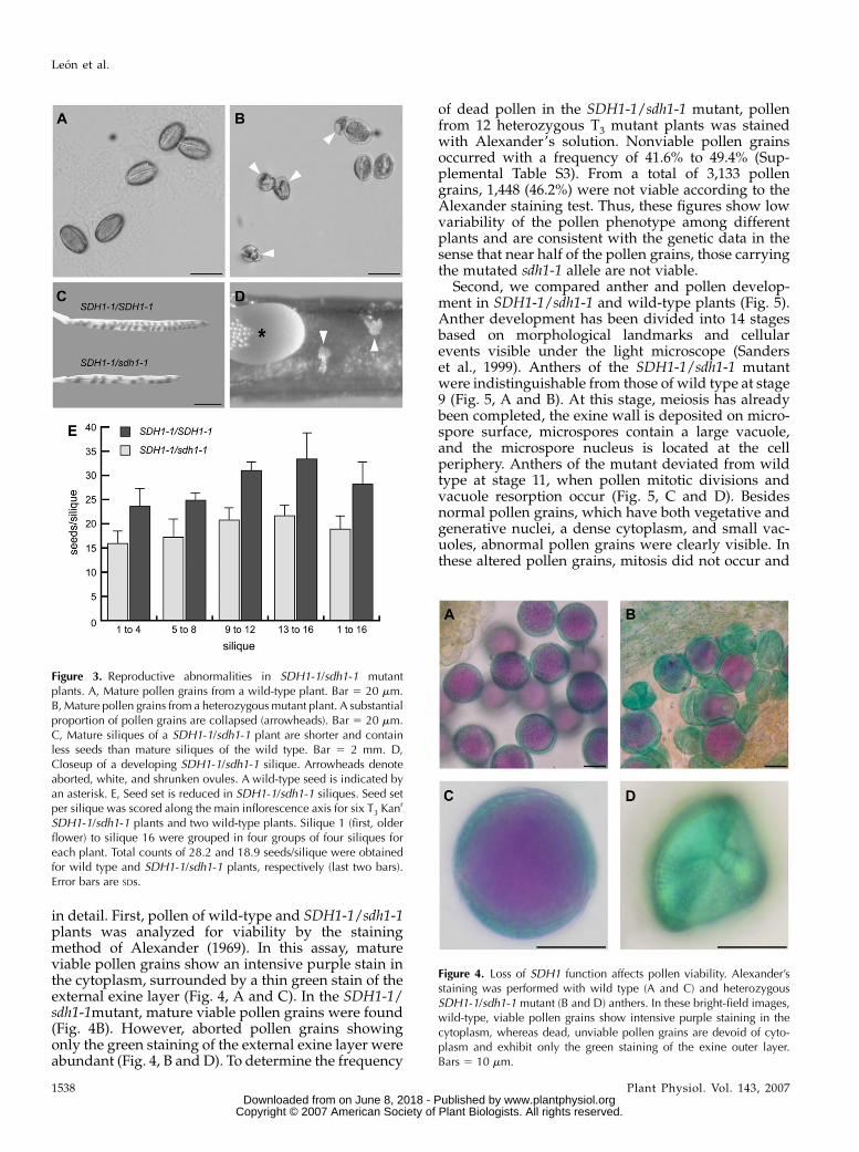
in detail. First, pollen of wild-type and SDH1-1/sdh1-1plants was analyzed for viability by the stainingmethod of Alexander (1969). In this assay, matureviable pollen grains show an intensive purple stain inthe cytoplasm, surrounded by a thin green stain of theexternal exine layer (Fig. 4, A and C). In the SDH1-1/sdh1-1mutant, mature viable pollen grains were found(Fig. 4B). However, aborted pollen grains showingonly the green staining of the external exine layer wereabundant (Fig. 4, B and D). To determine the frequency
of dead pollen in the SDH1-1/sdh1-1 mutant, pollenfrom 12 heterozygous T3 mutant plants was stainedwith Alexander’s solution. Nonviable pollen grainsoccurred with a frequency of 41.6% to 49.4% (Sup-plemental Table S3). From a total of 3,133 pollengrains, 1,448 (46.2%) were not viable according to theAlexander staining test. Thus, these figures show lowvariability of the pollen phenotype among differentplants and are consistent with the genetic data in thesense that near half of the pollen grains, those carryingthe mutated sdh1-1 allele are not viable.
Second, we compared anther and pollen develop-ment in SDH1-1/sdh1-1 and wild-type plants (Fig. 5).Anther development has been divided into 14 stagesbased on morphological landmarks and cellularevents visible under the light microscope (Sanderset al., 1999). Anthers of the SDH1-1/sdh1-1 mutantwere indistinguishable from those of wild type at stage9 (Fig. 5, A and B). At this stage, meiosis has alreadybeen completed, the exine wall is deposited on micro-spore surface, microspores contain a large vacuole,and the microspore nucleus is located at the cellperiphery. Anthers of the mutant deviated from wildtype at stage 11, when pollen mitotic divisions andvacuole resorption occur (Fig. 5, C and D). Besidesnormal pollen grains, which have both vegetative andgenerative nuclei, a dense cytoplasm, and small vac-uoles, abnormal pollen grains were clearly visible. Inthese altered pollen grains, mitosis did not occur and
Figure 4. Loss of SDH1 function affects pollen viability. Alexander’sstaining was performed with wild type (A and C) and heterozygousSDH1-1/sdh1-1 mutant (B and D) anthers. In these bright-field images,wild-type, viable pollen grains show intensive purple staining in thecytoplasm, whereas dead, unviable pollen grains are devoid of cyto-plasm and exhibit only the green staining of the exine outer layer.Bars 5 10 mm.
Figure 3. Reproductive abnormalities in SDH1-1/sdh1-1 mutantplants. A, Mature pollen grains from a wild-type plant. Bar 5 20 mm.B, Mature pollen grains from a heterozygous mutant plant. A substantialproportion of pollen grains are collapsed (arrowheads). Bar 5 20 mm.C, Mature siliques of a SDH1-1/sdh1-1 plant are shorter and containless seeds than mature siliques of the wild type. Bar 5 2 mm. D,Closeup of a developing SDH1-1/sdh1-1 silique. Arrowheads denoteaborted, white, and shrunken ovules. A wild-type seed is indicated byan asterisk. E, Seed set is reduced in SDH1-1/sdh1-1 siliques. Seed setper silique was scored along the main inflorescence axis for six T3 Kanr
SDH1-1/sdh1-1 plants and two wild-type plants. Silique 1 (first, olderflower) to silique 16 were grouped in four groups of four siliques foreach plant. Total counts of 28.2 and 18.9 seeds/silique were obtainedfor wild type and SDH1-1/sdh1-1 plants, respectively (last two bars).Error bars are SDs.
Leon et al.
1538 Plant Physiol. Vol. 143, 2007 www.plantphysiol.orgon June 8, 2018 - Published by Downloaded from
Copyright © 2007 American Society of Plant Biologists. All rights reserved.

the vacuole persisted. Moreover, the cytoplasm de-tached from the external exine layer and was progres-sively degraded (compare early stage 11 in Fig. 5Cwith late stage 11 in Fig. 5D). At stage 12, just prior todehiscence, the abnormal pollen grains showed vary-ing degrees of damage, including some in which thecellular material disappeared and the exine layercollapsed (Fig. 5E). It has to be pointed out that mutantSDH1-1/sdh1-1 anthers were not detectably differentfrom those of wild-type plants, except for these de-scribed pollen abnormalities. For instance, other an-ther tissues like the epidermis, endothecium, andvascular bundle were not affected. Furthermore, mu-tant and wild-type anthers underwent the same eventsat similar stages, including disappearance of the tape-tum and normal dehiscence.
Transmission electron microscopy was conductedon cross sections of wild-type and mutant SDH1-1/sdh1-1 anthers (Fig. 6). By the vacuolated microsporestage, mutant microspores were not detectably differ-ent from those of wild-type plants (Fig. 6, A and B;Owen and Makaroff, 1995). Numerous mitochondriaand starch-containing plastids are present throughoutthe cytoplasm, the exine is essentially complete andjust below it vesicles produced by the microspores addmaterial to the intine. While the nucleus is located nearthe side of the microspore, the asymmetric first mitoticdivision occurs, producing bicellular pollen grains(Fig. 6C). The generative cell is located at one end ofthe pollen grain and is surrounded by a wall that iscontinuous with the intine. At this stage, however, theSDH1-1 defect was evident in pollen sacs of SDH1-1/sdh1-1 anthers, since a mixture of wild-type-like (Fig.6C) and altered microspores (Fig. 6D) were foundwithin the locule. Abnormal pollen grains never un-derwent microspore mitosis, the large vacuole per-sisted, and the plasma membrane was withdrawnfrom the cell wall.
In SDH1-1/sdh1-1 anthers, wild-type-like pollengrains followed a normal developmental fate: gener-ative cell occupies a more central location in thecytoplasm of the vegetative cell, the large vacuole isresorbed (Fig. 6E), and lipid bodies accumulate in thevegetative cells at the surface of the generative cell(Fig. 6F). Later, the cytoplasm of the vegetative cellbecomes highly vacuolated (Fig. 6G). Neither of theselandmarks of normal pollen development was ob-served in altered pollen grains. In these grains, autol-ysis led to the disintegration of cellular structures andprogressive disappearance of cellular content (Fig. 6,H and I). Finally, pollen grains had collapsed com-pletely (Fig. 6J).
Embryo Sac Development Is Defective in
SDH1-1/sdh1-1 Mutant Plants
Reduced seed set, accompanied by the presence ofwhite, small senescing ovules, and reduced transmis-sion of the sdh1-1 mutation through the female game-tophyte, suggested defects in embryo sac maturation
and led us to analyze the phenotype of female game-tophytes. To do this, we allowed the female gameto-phytes within wild-type and heterozygous pistils toprogress to the terminal developmental stage, in theabsence of fertilization, and embryo sacs were exam-ined under Nomarski optics. In ovaries of wild-typeplants, all embryo sacs reached the terminal develop-mental stage, containing one secondary central cellnucleus, one egg cell nucleus, and two synergid cellnuclei (Fig. 7A). In mutant plants, only 72.75% (n 5291) of embryo sacs displayed the wild-type pheno-type (Fig. 7B). In addition, two abnormal phenotypeswere observed. In 14.75% (n 5 59) of the analyzedovules, embryo sacs were arrested at the two-nucleatestage (Fig. 7C), and in 12.5% of the embryo sacs (n 550) the overall morphology of the egg cell, central cell,and synergid cells was normal, yet polar nuclei failedto fuse (Fig. 7D). Therefore, 27% of the embryo sacs inSDH1-1/sdh1-1 plants were phenotypically mutant, avalue in good agreement with the seed set reduc-tion (33%).
Down-Regulation of SDH1-1 by RNA InterferenceResults in a Phenotype Similar to That of the
Insertional Mutant
Since no other mutant alleles were available forSDH1-1, an RNA interference approach was used.Figure 8A illustrates the construct: the 35S RNA pro-moter of the Cauliflower mosaic virus drives the tran-scription of partial SDH1-1 sequences cloned in senseand antisense orientations and separated by a plantintron. After formation of hairpin RNA structures(ihpRNA) and splicing, the resulting double-strandedRNA transcripts can cause posttranscriptional silenc-ing of endogenous gene activity (Smith et al., 2000).After floral-dipping transformation, only two inde-pendent transgenic T1 plants were obtained. Sincemature gametophytes or recently fertilized embryosare the likely targets of this transformation procedure(Clough and Bent, 1998), this low efficiency of trans-formation is consistent with our results showing thatgametophyte development is affected in the SDH1-1/sdh1-1 mutant. Moreover, northern-blot analysis ofthese primary transformants showed that SDH1-1mRNA levels are only partially reduced (roughly50%, data not shown), suggesting that a drastic reduc-tion in SDH1-1 expression may lead to unviable trans-formed embryos.
T2 seeds from one of these two lines were germi-nated on kanamycin and five resistant seedlings weretransferred to hydroponic medium. Northern-blotanalysis was performed using RNA from these fivetransgenic plants and two wild-type plants. The re-sults are shown in Figure 8B, with actin as a consti-tutive control. Compared to wild-type plants, alltransgenic plants showed a decrease in the SDH1-1mRNA levels. These RNAi plants were phenotypi-cally indistinguishable from the wild type duringsporophytic development. However, analysis of pollen
Mitochondrial Complex II and Gametophyte Development
Plant Physiol. Vol. 143, 2007 1539 www.plantphysiol.orgon June 8, 2018 - Published by Downloaded from
Copyright © 2007 American Society of Plant Biologists. All rights reserved.

viability by the staining method of Alexander showedvarying percentages, ranging from 13.5% to 66.8% ofdead, unviable pollen grains (Fig. 8, C and D). More-over, seed set was significantly reduced in the trans-genic plants bearing the RNAi construct (Fig. 8E).Interestingly, a correlation was found between thelevel of SDH1-1 mRNA expression and the degree ofseed set reduction and pollen abortion (for instance,plant no. 5 showed the strongest reduction in mRNA,pollen viability, and seed set).
In conclusion, plants in which SDH1-1 gene expres-sion is decreased by dsRNA showed the same pheno-type as the heterozygous insertional mutant plants, i.e.reduced seed set and pollen abortion.
DISCUSSION
SDH1 genes are interesting targets for reverse ge-netics analysis, to gain insights into the role of complexII in plants. Thus, we have characterized knockoutalleles of SDH1-1 and SDH1-2 genes. The SDH1-2 geneappears to be dispensable, at least under the growthconditions used, presumably because the SDH1-1 geneencodes the predominant flavoprotein. Our results donot rule out a SDH1-2 nonessential role in a restrictedgroup of cells or in a particular developmental stage,resulting in subtle beneficial characteristics. Such ahypothesis may provide an explanation for SDH1-2integrity and conservation when compared to SDH1-1.Alternatively, SDH1 duplication may be very recent,evolutionarily speaking, and either SDH1-2 inactiva-tion or SDH1-2 gain of a new function are under way.
Molecular and genetic characterization of heterozy-gous SDH1-1/sdh1-1 mutant plants permit us to con-clude that sdh1-1 is a gametophytic mutation affectingboth male and female gametophyte development.SDH1-1 is essential for pollen development and thefirst defect was evident in sdh1-1 microspores at pollenmitosis I, the asymmetric mitosis that results in bicel-lular pollen grains. Mutant microspores never under-went mitosis, the large vacuole was not resorbed, andcellular content progressively disappeared. These re-sults formally established that microspore mitochon-dria (and complex II) have an essential role in pollendevelopment.
During male gametophyte development, defined asdevelopment of the haploid microspores, pollen grainsshow a very high metabolic activity. For instance,respiration rates are 10 times higher in pollen grainsthan in green tissues (Tadege and Kuhlemeier, 1997).The uninucleate microspore develops to trinucleatepollen through two mitotic divisions, and simulta-neously starch, proteins, and other nutrients enrich thepollen of the vegetative cell (McCormick, 2004). Theseprocesses put demands on energetic and metabolicresources for the synthesis of proteins and other bio-molecules. In Arabidopsis, all the genes for complex IIcomponents, with the exception of SDH1-2 and SDH2-3,show enhanced expression in floral tissues (Figueroaet al., 2001, 2002). Furthermore, digital northern anal-ysis of developing pollen grains (Honys and Twell,2004) shows that these genes are expressed in the malegametophyte (Supplemental Fig. S2). Considering thatSDH is a component of the respiratory chain and theTCA cycle, loss of SDH1 in microspores would se-verely compromise both ATP production and provi-sion of carbon skeletons for biosynthesis, leading totheir abortion.
In addition to its essential role during pollen devel-opment, SDH1-1 is important for normal embryo sac
Figure 5. Pollen development in SDH1-1/sdh1-1 mutant plants. Anthersections were stained with Toluidine blue and photographed by bright-field microscopy. Abnormal pollen grains are indicated by arrowheads.Magnification is the same in all micrographs (Bars 5 20 mm). GN,Generative cell nucleus; N, nucleus; Tp, tapetum; V, vacuole; VN,vegetative cell nucleus. A, Anther from the wild type at the vacuolatedmicrospore stage. B, Anther from the SDH1-1/sdh1-1 mutant, at thevacuolated microspore stage. All microspores are indistinguishablefrom those of wild-type anthers. C, Anther from the SDH1-1/sdh1-1mutant after pollen mitosis I, showing normal pollen grains with twonuclei, and abnormal pollen grains (arrowheads) not entering mitosis.D, Anther from the SDH1-1/sdh1-1 mutant after pollen mitosis II,showing normal pollen grains with a dense cytoplasm, and abnormalpollen grains (arrowheads) containing cell debris. E, Anther just beforedehiscence, containing mature pollen grains and collapsed pollengrains devoid of cytoplasm (arrowheads).
Leon et al.
1540 Plant Physiol. Vol. 143, 2007 www.plantphysiol.orgon June 8, 2018 - Published by Downloaded from
Copyright © 2007 American Society of Plant Biologists. All rights reserved.

development. Although our data indicate that theSDH1-1 protein is essential for efficient megagameto-phyte development, a fraction of the gametophytesstill remains functional. Therefore, these mutant ga-metophytes cannot be completely energy deficient.The more plausible explanations for this observationare (1) a stochastic inheritance and persistence of wild-type mitochondria from the diploid megaspore mothercell, which would result in variable depletion rates ofSDH1-1 through cell division and protein turnoverduring embryo sac development, and/or (2) variableprovision of nutrients (ATP, carbon skeletons) to thefemale gametophyte from the surrounding sporo-phytic cells. In this context, it is tempting to speculatethat male mutant gametophytes have a more severephenotype because more nutrients are carried fromsporophytic tissues to the female than to the malegametophyte. These arguments may also explain whymore than one phenotype was found in the mutantembryo sacs (see below).
During megagametogenesis, a functional haploidmegaspore gives rise to the mature female gameto-phyte (Yadegari and Drews, 2004). This process com-prises three successive rounds of nuclear division,followed by cellularization. During cellularization, thetwo polar nuclei migrate to the center of the femalegametophyte and fused together in the central cell.The result is a seven-cell embryo sac that containsthree antipodal, two synergid, one egg, and one centralcell. Two abnormal phenotypes were observed insdh1-1 embryo sacs, developmental arrest at the two-nucleate stage, and failure of the two polar nuclei tofuse after their proper migration.
Large collections of gametophytic mutants defectivein female gametophyte development have been re-ported, and in some cases characterized with respectto the genes responsible for the observed defects (e.g.Christensen et al., 2002; Yadegari and Drews, 2004;Niewiadomski et al., 2005; Pagnussat et al., 2005;Portereiko et al., 2006). A wide variety of phenotypes
Figure 6. Transmission electron microscopy anal-ysis of anthers in the SDH1-1/sdh1-1 mutant. Ex,Exine layer; GN, generative nucleus; L, lipidbody; M, mitochondrion; N, nucleus; Nu, nucle-olus; S, starch granules within plastids; V, vacu-ole; VN, vegetative nucleus. Bar 5 5 mm in A, Cto E, and G to I; bar 5 1 mm in B, F, and J. A,Microspores at the vacuolated stage (anther stage9 according to Sanders et al., 1999). All micro-spores had wild-type morphology, with numerousmitochondria, starch granules within plastids, anda normal exine outer layer. B, Closeup of themicrospore shown in A. C, Bicellular pollen grainfrom SDH1-1/sdh1-1 anthers. The asymmetricfirst mitotic division has occurred (early antherstage 11). D, Same anther as in C. In addition topollen with wild-type morphology (C), abnormalpollen grains were present. They did not undergomitosis and cell content detached from the outerlayers. E and F, Later developmental stage of wild-type pollen grains from SDH1-1/sdh1-1 anthers.G, Mature wild-type pollen grain from SDH1-1/sdh1-1 anthers. H and I, Later developmentalstages of aberrant pollen grains from SDH1-1/sdh1-1 anthers. Cellular structures and contentprogressively disappeared and internal structureswere no longer distinguishable. J, Collapsed pol-len grain in the same anther as in G. The exinelayer, of sporophytic origin, remained wild-type like.
Mitochondrial Complex II and Gametophyte Development
Plant Physiol. Vol. 143, 2007 1541 www.plantphysiol.orgon June 8, 2018 - Published by Downloaded from
Copyright © 2007 American Society of Plant Biologists. All rights reserved.

were observed, including mutants arrested at the two-nucleate stage and mutants cellularized but with de-fects in fusion of the polar nuclei. Interestingly, amongthe mutants affected in fusion of polar nuclei, severalof the mutated genes encode mitochondrial proteins orpredicted mitochondrial proteins (Christensen et al.,2002; Portereiko et al., 2006). The best characterized areGFA2, a mitochondrial matrix chaperone of the DnaJprotein family (Christensen et al., 2002), RPL21M, amitochondrial 50S ribosomal subunit L21 (Portereikoet al., 2006), and now SDH1-1 (this work). Altogetherthese results strongly suggest a role for mitochondriain nuclear fusion (karyogamy). At this time, it is un-clear why these mutants have a nuclear fusion defect.However, the fact that mutations in a ribosomal pro-tein, a chaperone, and a complex II subunit lead to asimilar phenotype favors the hypothesis that the kar-yogamy defect is a consequence of impairment in basicmitochondrial functions, like electron transport, ATPproduction, and/or provision of carbon skeletons.
Few defects in the respiratory chain and TCA cyclehave been reported. For instance, a knockout mutation
in a subunit of the NAD1-dependent isocitrate dehy-drogenase has been described in Arabidopsis (Linet al., 2004). The homozygous mutant plants showedno detectable differences in vegetative growth andreproductive development compared to wild-typeplants. However, Arabidopsis contains multiple genes
Figure 7. Embryo sac development defects in SDH1-1/sdh1-1 mutantplants. Flowersofwild-type and SDH1-1/sdh1-1 plants were emasculated,and whole-mount preparations of ovules were analyzed by DIC micros-copy 48 h after emasculation. Embryo sac nuclei are indicated by arrows.A, Ovule from a wild-type Ws plant. The embryo sac is at the terminaldevelopmental stage and contains one secondary nucleus in the centralcell, one egg cell nucleus, and two synergid cell nuclei at the micropylarend. B, Ovule harboring a wild-type-like embryo sac in a SDH1-1/sdh1-1mutant plant. C, Ovule from the same pistil of the ovule shown in B. Theembryo sac displays a mutant phenotype, being arrested at the two-nucleate stage. D, Ovule from the same pistil of the ovule shown in B andC, but showing a second mutant phenotype. The two polar nuclei lie sideby side but failed to fuse. CC, Central cell nucleus; EC, egg cell nucleus; N,nuclei from an embryo sac arrested at the two-nucleate stage; PN, polarnuclei; SYN, synergid cell nuclei. Bars 5 50 mm.
Figure 8. Phenotype of SDH1-1 RNAi transgenic plants. A, Schematicdiagram of the construct used to down-regulate SDH1-1. The arrowsindicate the SDH1-1 sequence (first exon) cloned in both orientationsin the RNAi pHELLSGATE4 vector. Sense and antisense SDH1-1 tran-scribed sequences would be separated by the plant pyruvate orthophos-phate dikinase (PDK) intron, generating an ihpRNA. B, Northern-blotanalysis of SDH1-1 expression in five RNAi T2 plants and two wild-typecontrols. Each lane was loaded with 10 mg of total RNA isolated fromflowers. The blot was hybridized with a SDH1-1 specific probe andthen with an actin probe as loading control. RNAi plants had decreasedSDH1-1 mRNA levels. C and D, Pollen abortion in SDH1-1 RNAiplants. Alexander staining was performed on two wild-type and fiveRNAi T2 anthers. Sections C and D show the staining of one RNAi plantand one wild-type plant, respectively. Quantitative determination ofpollen abortion was done by counting viable, purple pollen grains, andaborted, green pollen grains, and the results obtained are shown underthe images. Bars 5 20 mm. E, Seed set is reduced in SDH1-1 RNAiplants. Seed set per silique was scored for the first eight siliques fromfive T2 RNAi plants (1–5) and two wild-type control plants (wt). Errorbars represent SDs.
Leon et al.
1542 Plant Physiol. Vol. 143, 2007 www.plantphysiol.orgon June 8, 2018 - Published by Downloaded from
Copyright © 2007 American Society of Plant Biologists. All rights reserved.

encoding catalytic and noncatalytic subunits of thisenzyme, and in mitochondrial extracts from homozy-gous mutant plants there was still half as much enzymeactivity as in wild-type plants. Antisense repression ofthe potato (Solanum tuberosum) mitochondrial citratesynthase gene resulted in a delaying flowering pheno-type and a specific disintegration of the ovary tissues,indicating an important role of the TCA cycle duringthe early stages of flower development and the for-mation of the sporophytic female tissues (Landschutzeet al., 1995). Additional efforts to analyze the functionof the TCA cycle have been performed in a mutant(Aco1) that exhibits a deficiency in expression of one oftwo isoforms of aconitase present in tomato (Lycoper-sicon esculentum; Carrari et al., 2003), and in antisensetransgenic plants exhibiting reduced activity of mito-chondrial malate dehydrogenase (Nunes-Nesi et al.,2005). However, the possible presence of multiplegenes has not been analyzed, these plants still main-tain mitochondrial aconitase and malate dehydrogen-ase activities, and their reproductive development isnot affected.
The best-characterized respiratory chain mutant is aNicotiana sylvestris mitochondrial mutant, CMS II. Inthis mutant, the mitochondrial nad7 gene encoding theNAD7 subunit of complex I is deleted, and mitochon-dria are impaired in complex I structure and function(Pla et al., 1995; Gutierres et al., 1997). The absence of acompetent complex I leads to impaired photosynthesisand slower growth, however plants attain biomasssimilar to that of wild-type plants and undergo repro-ductive development, although they are partially malesterile (Gutierres et al., 1997; Dutilleul et al., 2003a).Therefore, mutant plants were acclimated to this de-fect and this acclimation includes enhanced activity ofnonphosphorylating NAD(P)H dehydrogenases, whichbypass complex I (Sabar et al., 2000; Dutilleul et al.,2003a, 2003b). An Arabidopsis mutant lacking the 18 kDFe-S subunit of complex I has also been described andshown to be affected in cold acclimation (Lee et al.,2002). Homozygous mutant plants grow more slowlythan wild-type plants and a delay was observed inflowering. Nevertheless, these plants eventually reachheights similar to those of wild-type plants and werefully fertile. Altogether, data with these mutants indi-cate that a defect in complex I in plants is not lethal. Incontrast, loss of complex II leads to a more severephenotype, at least in gametophytic development, andthis is probably due to the role of SDH in the TCAcycle.
Deficiencies of any of the four subunits of SDH areassociated with a broad spectrum of human diseases,ranging from myo- and encephalopathies to aging andtumor formation (Rustin and Rotig, 2002). To ourknowledge, the first reported nuclear gene mutationcausing a respiratory chain defect in humans was amutation in the gene encoding the complex II flavo-protein subunit (Bourgeron et al., 1995). The mutationwas identified in two siblings presenting Leigh syn-drome (necrosis of specific brain territories, causing
encephalopathy) and SDH deficiency, and the patientswere homozygous for an amino acid substitution in aconserved domain of the protein. It has to be empha-sized that the available data suggest that complex II isessential in mammals and could not be completelylost. For instance, Piruat et al. (2004) reported the gen-eration of a SDH4 knockout mouse, the first mamma-lian model lacking a protein of the mitochondrialelectron transport chain, and showed that homozy-gous sdh4/sdh4 animals die at early embryonic stages.
Although development and function of gameto-phytes are crucial for plant reproduction, relativelylittle is known about the genes required for, and thepathways involved in gametophytic development inflowering plants. An important question is whetherplants exploit metabolism as a way to control devel-opment. We have found here that an apparent meta-bolic mutant shows developmental effects, raising thepossibility that some developmental regulators exerttheir effects through the modulation of basic metabolicprocesses. Metabolic regulation thus should be con-sidered as one possible effector pathway in models ofgenetic regulation of development.
MATERIALS AND METHODS
Plant Material and Growth Conditions
Arabidopsis (Arabidopsis thaliana) ecotype Wassilewskija (Ws) seeds were
cold treated for 48 h at 4�C in darkness and then germinated and grown
hydroponically at 20�C to 24�C, under a 16-h light/8-h dark cycle (Gibeaut
et al., 1997). To grow seedlings in vitro and for Kanr assays, seeds were surface
sterilized for 10 min with a solution of 10% (v/v) commercial bleach and 0.05%
(v/v) Tween 20. Then seeds were rinsed thoroughly with sterile distilled water
and sown on one-half-concentrated Murashige and Skoog medium solidified
with 0.8% (w/v) agar and supplemented or not with 50 mg L21 kanamycin.
Finally seeds were cold treated, germinated, and grown as described above.
Isolation of Insertional SDH1 Mutants
The Arabidopsis Knockout Facility (AKF) alpha and BASTA populations
(Sussman et al., 2000) were screened by PCR, using a primer specific for the
T-DNA left border (JL 202, 5# CATTTTATAATAACGCTGCGGACATCTAC 3#)
in combination with either a forward or a reverse gene-specific primer. For
SDH1-1, the forward primer was 5# GGAGTGAGAATAGGGGTAATTAAG-
GGAAT 3# and the reverse primer was 5# CTGGAGTAAAACGTAAACCC-
AGTTTCAGA 3#. For SDH1-2, forward and reverse primers were 5# TAT-
TTCACTGCTGCAACATTATGGGCTTT 3# and 5# GATGGACGGTCTCGAT-
TAAACGGTTAGAT 3#, respectively. Identification of DNA positive pools was
performed by DNA gel-blot analysis of the PCR products, using SDH1-1 or
SDH1-2 gene fragments as probes, and confirmed by sequencing of the
amplified DNA (see Sussman et al., 2000 and http://www.biotech.wisc.edu/
NewServicesAndResearch/Arabidopsis/OverviewIndex.html for methods).
Only one insertion in each SDH1-1 and SDH1-2 were found (both in the AKF
alpha mutant collection).
The Salk Institute collection (Alonso et al., 2003), the Torrey Mesa Research
Institute collection (Sessions et al., 2002), and the Wisconsin DsLox T-DNA
lines (http://www.hort.wisc.edu/krysan/DS-Lox) were searched in silico at
two sites: the Arabidopsis Insertion Database (http://atidb.org/cgi-perl/
index) and the T-DNAExpress Web sites (http://signal.salk.edu/cgi-bin/
tdnaexpress). Although several mutant alleles were identified for SDH1-2 (e.g.
SALK_015173 and SALK_087371), no T-DNA additional insertions were
found in the SDH1-1 gene. A line in the Wisconsin DsLox T-DNA population
(DsLox501H11) indexed as a SDH1-1 mutant was further characterized and
found to carry the T-DNA insertion downstream of the SDH1-1 3# untrans-
lated region (UTR; data not shown).
Mitochondrial Complex II and Gametophyte Development
Plant Physiol. Vol. 143, 2007 1543 www.plantphysiol.orgon June 8, 2018 - Published by Downloaded from
Copyright © 2007 American Society of Plant Biologists. All rights reserved.

Arabidopsis Ws seeds from the positive AKF pools containing the sdh1-1
and sdh1-2 mutant alleles were obtained from the ABRC (Ohio State Univer-
sity, Columbus, OH). These T2 seeds were stratified, germinated, and grown as
described above. To identify SDH1 mutant plants and eventually isolate
plants homozygous for a mutation, seedlings were genotyped by a PCR-based
approach, using total DNA extracted from one cotyledon or one small leaf.
Briefly, tissue was homogenized in 100 mL of TNE/SDS buffer (0.2 M Tris-HCl
pH 8.0, 0.25 M NaCl, 0.02 M EDTA, 0.5% [w/v] SDS), using a disposable pestle
adapted to a portable drill. After a 5 min centrifugation at 14,000g, the DNA in
the supernatant was precipitated with 1 volume of isopropanol, washed with
70% (v/v) ethanol, and resuspended in sterile water. The genotype of plants
was determined by PCR using primers flanking the insertion point for the
wild-type allele and a gene-specific and left border-specific (JL202) primer
pair for the insertional mutant alleles. For SDH1-1, the wild-type allele was
amplified with 5# CAACCTCAGCACATACATGCACAG 3# (primer 2 in Fig.
1A) and 5# CCATCTCATGCTTAACTCCACACA 3# (primer 3 in Fig. 1A), and
the mutant allele with JL202 (primer 1 in Fig. 1A) and primer 2. For SDH1-2,
the wild-type allele was amplified with 5# TATTTCACTGCTGCAACATT-
ATGGGCTTT 3# (primer sdha3) and 5# GATGGACGGTCTCGATTAAACG-
GTTAGAT 3# (primer sdha4), and the mutant allele with JL202 and sdha4.
Nucleic Acids Preparation and Analysis
Total DNA was prepared from green leaves according to Ausubel et al.
(1994). Total RNA was isolated from tissues harvested into liquid nitrogen
using TRIzol reagent and the manufacturer’s protocol (Invitrogen). Southern
and northern analyses were performed by standard procedures with32P-labeled probes (Ausubel et al., 1994). To obtain specific probes, DNA
fragments were PCR amplified using the following primer pairs: for a T-DNA
GUS probe (Fig. 2), 5# TCTACACCACGCCGAACACCTGG 3# and 5# TTC-
GGTGATGATAATCGGCTGATGC 3#; for probe 1 in Figure 2, 5# CTGCA-
GGTTTTCCACCTGTTTC 3# and 5# ACGTTCACCTTCACTGTTTCTA 3#; for
probe 2 in Figure 2, 5# AACGATATGCTCCTACAGCCAA 3# and 5# CCATCT-
CATGCTTAACTCCACACA 3#; for a SDH1-1 probe derived from the 3# UTR,
5# TGAGATTGGATTATAGGCCTGTT 3# and 5# CTGGAGTAAAACGTAA-
ACCCAGTTTCAGA 3#; and for an actin probe, 5# GCTATGTATGTCGC-
CATTCAAGC 3# and 5# CATCATATTCTGCCTTTGC(A/G)ATCC 3#.
Preparation of Mitochondria and SQR Activity Assay
A crude mitochondrial fraction was prepared from Arabidopsis in vitro-
grown 18-d-old seedlings. All steps were performed at 2�C to 4�C. The tissue
(1 g fresh weight) was chilled in 10 mL of ice-cold grinding buffer (0.03 M
MOPS/KOH pH 7.5, 0.3 M Mannitol, 0.001 M EDTA, 0.004 M L-Cysteine, 0.1%
[w/v] fatty acid-free bovine serum albumin [fraction V], 0.6% [w/v] Poly-
vinylpyrrolidone-40) and homogenized with a Dounce potter. The homoge-
nate was filtered through Miracloth and the filtrate was centrifuged at 1,000g
for 20 min. The supernatant was then centrifuged at 12,500g for 20 min and the
pellet was suspended in washing buffer (0.03 M MOPS/KOH pH 7.5, 0.3 M
Mannitol, 0.001 M EDTA). Both centrifugation steps were repeated. Finally, the
pellet (crude mitochondrial fraction) was resuspended in 200 mL of washing
buffer. Protein concentration was determined by the Bradford method
(Bradford, 1976) and SQR activity assayed immediately.
SQR activity was determined as described by Miyadera et al. (2003).
Mitochondrial fractions (25 mg protein) were assayed for activity spectropho-
metrically at 600 nm, in 1 mL of a reaction medium containing 0.05 M
potassium phosphate pH 7.5, 90 mM ubiquinone 2 (UQ2, Sigma-Aldrich),
55 mM dichlorophenolindophenol (DCIP; Merck; extinction coefficient 21 mM21
cm21), and 0.01 M sodium succinate (Merck). The reaction was performed at
30�C for 30 min, conditions in which DCIP reduction was proportional to
incubation time and protein concentration.
Microscopic Analysis
Alexander staining of pollen was performed by incubating anthers from
recently opened flowers in Alexander’s solution for 15 min (Alexander, 1969;
Johnson-Brousseau and McCormick, 2004). For anther structure analysis,
flowers were fixed overnight in 3% (v/v) glutaraldehyde, 0.1 M sodium
cacodylate, pH 7.2, dehydrated in acetone series to 100%, and embedded in
Embed812 resin according to the recommendations of the manufacturer
(EMS). Anther transverse sections (2 mm) were stained with 1% toluidine blue.
Alexander stained pollen and anther cross sections were viewed using a light
microscope (Optiphot-2, Nikon), and bright-field photographs were taken
with a Nikon Coolpix 4500 CCD digital camera.
For transmission electron microscopy, samples were fixed in 3% (v/v)
glutaraldehyde, 0.1 M sodium cacodylate, pH 7.2, and postfixed in 1% osmium
tetroxide for 1 h, dehydrated in a graduate acetone series, embedded in Epon
resin, and polymerized at 60�C for 24 h. Ultrathin sections (60 nm) were cut
with a diamond knife (Micro Star) on a Sorvall MT-5000 microtome and
mounted on 300 mesh copper grids. Sections were stained with 4% (w/v)
uranyl acetate and lead citrate in methanol (Reynolds, 1963) and viewed with
a Philips Tecnai 12 Bio Twin transmission electron microscope operating at
80 kV. Micrographs were taken using Kodak Electron Image SO163 film
(Kodak).
To analyze the terminal phenotype of ovules, flowers were emasculated
and ovules observed 48 h after emasculation. For this, wild-type and mutant
pistils were dissected longitudinally with hypodermic needles, fixed over-
night at room temperature in 50% (v/v) ethanol, 5% (v/v) glacial acetic acid,
10% (v/v) formaldehyde, and then cleared in chloral hydrate:glycerol:water
(8g:2 mL:1 mL). Single ovules were dissected and viewed with a light
microscope (Optiphot-2, Nikon) equipped for differential interference con-
trast (DIC). Images were captured using a Nikon Coolpix 4500 CCD digital
camera.
Generation of ihpRNA Lines
A 225-bp fragment containing the 5# UTR and the first exon of SDH1-1 was
PCR amplified using the forward primer 5# AAGCAGGCTACTAGTGTT-
CGCGTCTTCCT 3# and the reverse primer 5# CAAGAAAGCTGGGTAGAAC-
CAGTGGAGA 3# (partial attB sites in italics, SpeI restriction site underlined).
The resulting product, which contained at both ends partial attB recombina-
tion sites of the Gateway system (Invitrogen), was reamplified using prim-
ers attB1 (5# GGGGACAAGTTTGTACAAAAAAGCAGGCT 3#) and attB2
(5# GGGGACCACTTTGTACAAGAAAGCTGGGT 3#). The PCR product
(attB1:SDH1-1:attB2) was cloned into a pGEM-T easy vector (Promega) and
sequenced. An in vitro recombination reaction was performed according to
the manufacturer’s protocol, using BP Clonase (Invitrogen), the vector
pHELLSGATE4 (Helliwell et al., 2002), and gel-purified attB1:SDH1-1:attB2
DNA fragment. Spectinomycin-resistant Escherichia coli XL-1blue colonies
bearing the recombinant plasmid were selected. The resulting plasmid, con-
taining SDH1-1 sequences in both sense and antisense orientations separated
by a pyruvate orthophosphate dikinase intron and under the control of the
cauliflower mosaic virus 35S promoter, was transferred to Agrobacterium
tumefaciens C58. A. tumefaciens-mediated transformation of Arabidopsis
Columbia 0 plants was accomplished using the floral dip protocol (Clough
and Bent, 1998). Seeds of the T1 generation were selected for resistance
to kanamycin.
Supplemental Data
The following materials are available in the online version of this article.
Supplemental Figure S1. SDH1-1 and SDH1-2 expression in different
tissues and developmental stages.
Supplemental Figure S2. Expression of the complex II genes during
pollen development.
Supplemental Table S1. Segregation analysis of sdh1-1.
Supplemental Table S2. TE of sdh1-1 in reciprocal crosses.
Supplemental Table S3. Frequency of aborted pollen in SDH1-1/sdh1-1
plants.
ACKNOWLEDGMENTS
The authors are greatly thankful to Alejandro Araya, Simon Litvak, and
Laura Tarrago-Litvak for their constant encouragement. We also would like to
thank Hannetz Roschzttardtz and Rodrigo Tapia for critical reading of the
manuscript and constant discussion of results. Also, we thank to Bernard
Hausser for technical advice on DIC microscopy and Sheila Johnson-
Brousseau for experimental guidelines. We thank the AKF (University of
Wisconsin) for mutant screens and for making T-DNA insertion lines publicly
available through the ABRC.
Leon et al.
1544 Plant Physiol. Vol. 143, 2007 www.plantphysiol.orgon June 8, 2018 - Published by Downloaded from
Copyright © 2007 American Society of Plant Biologists. All rights reserved.

Received December 21, 2006; accepted February 14, 2007; published February
23, 2007.
LITERATURE CITED
Alexander MP (1969) Differential staining of aborted and non-aborted
pollen. Stain Technol 44: 117–122
Alonso JM, Stepanova AN, Leisse TJ, Kim CJ, Chen H, Shinn P, Stevenson
DK, Zimmerman J, Barajas P, Cheuk R, et al (2003) Genome-wide
insertional mutagenesis of Arabidopsis thaliana. Science 301: 653–657
Ausubel FM, Brent R, Kingston RE, Moore DD, Seidman JG, Smith JA,
Struhl K (1994) Current Protocols in Molecular Biology. Wiley Inter-
science, New York
Bourgeron T, Rustin P, Chretien D, Birch-Machin M, Bourgeois M,
Viegas-Pequignot E, Munnich A, Rotig A (1995) Mutation of a nuclear
succinate dehydrogenase gene results in mitochondrial respiratory
chain deficiency. Nat Genet 11: 144–149
Boyes DC, Zayed AM, Ascenzi R, McCaskill AJ, Hoffman NE, Davis KR,
Gorlach J (2001) Growth stage-based phenotypic analysis of Arabidop-
sis: a model for high throughput functional genomics in plants. Plant
Cell 13: 1499–1510
Bradford MM (1976) A rapid and sensitive method for the quantitation of
microgram quantities of protein utilizing the principle of protein-dye
binding. Anal Biochem 72: 248–254
Carrari F, Nunes-Nesi A, Gibon Y, Lytovchenko A, Ehlers Loureiro M,
Fernie AR (2003) Reduced expression of aconitase results in an en-
hanced rate of photosynthesis and marked shifts in carbon partition-
ing in illuminated leaves of wild species tomato. Plant Physiol 133:
1322–1335
Christensen CA, Gorsich SW, Brown RH, Jones LG, Brown J, Shaw JM,
Drews GN (2002) Mitochondrial GFA2 is required for synergid cell
death in Arabidopsis. Plant Cell 14: 2215–2232
Clough SJ, Bent AF (1998) Floral dip: a simplified method for Agrobacte-
rium-mediated transformation of Arabidopsis thaliana. Plant J 16: 735–743
De Buck S, Jacobs A, Van Montagu M, Depicker A (1999) The DNA
sequences of T-DNA junctions suggest that complex T-DNA loci are
formed by a recombination process resembling T-DNA integration.
Plant J 20: 295–304
De Neve M, De Buck S, Jacobs A, Van Montagu M, Depicker A (1997)
T-DNA integration patterns in co-transformed plant cells suggest that
T-DNA repeats originate from co-integration of separate T-DNAs. Plant
J 11: 15–29
Drews GN, Yadegari R (2002) Development and function of the angio-
sperm female gametophyte. Annu Rev Genet 36: 99–124
Dutilleul C, Driscoll S, Cornic G, De Paepe R, Foyer CH, Noctor G (2003a)
Functional mitochondrial complex I is required by tobacco leaves for
optimal photosynthetic performance in photorespiratory conditions
and during transients. Plant Physiol 131: 264–275
Dutilleul C, Garmier M, Noctor G, Mathieu C, Chetrit P, Foyer CH, De
Paepe R (2003b) Leaf mitochondria modulate whole cell redox homeo-
stasis, set antioxidant capacity, and determine stress resistance through
altered signaling and diurnal regulation. Plant Cell 15: 1212–1226
Elorza A, Leon G, Gomez I, Mouras A, Holuigue L, Araya A, Jordana X
(2004) Nuclear SDH2-1 and SDH2-2 genes, encoding the iron-sulfur
subunit of mitochondrial complex II in Arabidopsis, have distinct cell-
specific expression patterns and promoter activities. Plant Physiol 136:
4072–4087
Elorza A, Roschzttardtz H, Gomez I, Mouras A, Holuigue L, Araya A,
Jordana X (2006) A nuclear gene for the iron-sulfur subunit of mito-
chondrial complex II is specifically expressed during Arabidopsis seed
development and germination. Plant Cell Physiol 47: 14–21
Figueroa P, Leon G, Elorza A, Holuigue L, Araya A, Jordana X (2002) The
four subunits of mitochondrial respiratory complex II are encoded by
multiple nuclear genes and targeted to mitochondria in Arabidopsis
thaliana. Plant Mol Biol 50: 725–734
Figueroa P, Leon G, Elorza A, Holuigue L, Jordana X (2001) Three different
genes encode the iron-sulfur subunit of succinate dehydrogenase in
Arabidopsis thaliana. Plant Mol Biol 46: 241–250
Gibeaut DM, Hulett J, Cramer GR, Seemann JR (1997) Maximal biomass
of Arabidopsis thaliana using a simple, low-maintenance hydroponic
method and favorable environmental conditions. Plant Physiol 115:
317–319
Gutierres S, Sabar M, Lelandais C, Chetrit P, Diolez P, Degand H, Boutry
M, Vedel F, de Kouchkovsky Y, De Paepe R (1997) Lack of mitochon-
drial and nuclear-encoded subunits of complex I and alteration of the
respiratory chain in Nicotiana sylvestris mitochondrial deletion mutants.
Proc Natl Acad Sci USA 94: 3436–3441
Hanson MR, Bentolila S (2004) Interactions of mitochondrial and nuclear
genes that affect male gametophyte development. Plant Cell (Suppl) 16:
S154–169
Heiser V, Brennicke A, Grohmann L (1996) The plant mitochondrial 22
kDa (PSST) subunit of respiratory chain complex I is encoded by a
nuclear gene with enhanced transcript levels in flowers. Plant Mol Biol
31: 1195–1204
Helliwell CA, Wesley SV, Wielopolska AJ, Waterhouse PM (2002) High-
throughput vectors for efficient gene silencing in plants. Funct Plant Biol
29: 1217–1225
Honys D, Twell D (2004) Transcriptome analysis of haploid male game-
tophyte development in Arabidopsis. Genome Biol 5: R85
Howden R, Park SK, Moore JM, Orme J, Grossniklaus U, Twell D (1998)
Selection of T-DNA-tagged male and female gametophytic mutants by
segregation distortion in Arabidopsis. Genetics 149: 621–631
Huang J, Friedhelm S, Matzinger DF, Levings CS III (1994) Flower-
enhanced expression of a nuclear-encoded mitochondrial respiratory
protein is associated with changes in mitochondrion number. Plant Cell
6: 439–448
Johnson-Brousseau SA, McCormick S (2004) A compendium of methods
useful for characterizing Arabidopsis pollen mutants and gametophyti-
cally-expressed genes. Plant J 39: 761–775
Landschutze V, Willmitzer L, Muller-Rober B (1995) Inhibition of flower
formation by antisense repression of mitochondrial citrate synthase in
transgenic potato plants leads to a specific disintegration of the ovary
tissues of flowers. EMBO J 14: 660–666
Lee B, Lee H, Xiong L, Zhu J-K (2002) A mitochondrial complex I defect
impairs cold-regulated nuclear gene expression. Plant Cell 14: 1235–1251
Lee SLJ, Warmke HE (1979) Organelle size and number in fertile and
T-cytoplasmic male-sterile corn. Am J Bot 60: 141–148
Lemire BD, Oyedotun KS (2002) The Saccharomyces cerevisiae mitochon-
drial succinate: ubiquinone oxidoreductase. Biochim Biophys Acta 1553:
102–116
Lin M, Behal RH, Oliver DJ (2004) Characterization of a mutation in the
IDH-II subunit of the NAD1-dependent isocitrate dehydrogenase from
Arabidopsis thaliana. Plant Sci 166: 983–988
McCormick S (2004) Control of male gametophyte development. Plant Cell
(Suppl) 16: S142–S153
Meyers BC, Vu TH, Tej SS, Ghazal H, Matvienko M, Agrawal V, Ning J,
Haudenschild CD (2004) Analysis of the transcriptional complexity of
Arabidopsis thaliana by massively parallel signature sequencing. Nat
Biotechnol 22: 1006–1011
Millar HA, Eubel H, Jansch L, Kruft V, Heazlewood JL, Braun HP (2004)
Mitochondrial cytochrome c oxidase and succinate dehydrogenase
complexes contain plant specific subunits. Plant Mol Biol 56: 77–90
Miyadera H, Shiomi K, Ui H, Yamaguchi Y, Masuma R, Tomoda H,
Miyoshi H, Osanai A, Kita K, Omura S (2003) Atpenins, potent and
specific inhibitors of mitochondrial complex II (succinate-ubiquinone
oxidoreductase). Proc Natl Acad Sci USA 100: 473–477
Niewiadomski P, Knappe S, Geimer S, Fischer K, Schulz B, Unte US, Rosso
MG, Ache P, Flugge U-I, Schneider A (2005) The Arabidopsis plastidic
glucose 6-phosphate/phosphate translocator GPT1 is essential for pollen
maturation and embryo sac development. Plant Cell 17: 760–775
Nunes-Nesi A, Carrari F, Lytovchenko A, Smith AMO, Ehlers Loureiro M,
Ratcliffe RG, Sweetlove LJ, Fernie AR (2005) Enhanced photosynthetic
performance and growth as a consequence of decreasing mitochondrial
malate dehydrogenase activity in transgenic tomato plants. Plant
Physiol 137: 611–622
Owen HA, Makaroff CA (1995) Ultrastructure of microsporogenesis
and microgametogenesis in Arabidopsis thaliana (L.) Heynh. ecotype
Wassilewskija (Brassicaceae). Protoplasma 185: 7–21
Pagnussat GC, Yu H-J, Ngo QA, Rajani S, Mayalagu S, Johnson CS,
Capron A, Xie L-F, Ye D, Sundaresan V (2005) Genetic and molecular
identification of genes required for female gametophyte development
and function in Arabidopsis. Development 132: 603–614
Park SK, Howden R, Twell D (1998) The Arabidopsis thaliana gametophytic
mutation gemini pollen1 disrupts microspore polarity, division asymme-
try and pollen cell fate. Development 125: 3789–3799
Mitochondrial Complex II and Gametophyte Development
Plant Physiol. Vol. 143, 2007 1545 www.plantphysiol.orgon June 8, 2018 - Published by Downloaded from
Copyright © 2007 American Society of Plant Biologists. All rights reserved.

Piruat JI, Pintado CO, Ortega-Saenz P, Roche M, Lopez-Barneo J (2004)
The mitochondrial SDHD gene is required for early embryogenesis, and
its partial deficiency results in persistent carotid body glomus cell
activation with full responsiveness to hypoxia. Mol Cell Biol 24: 10933–
10940
Pla M, Mathieu C, De Paepe R, Chetrit P, Vedel F (1995) Deletion of the last
two exons of the mitochondrial nad7 gene results in lack of the NAD7
polypeptide in a Nicotiana sylvestris CMS mutant. Mol Gen Genet 248:
79–88
Portereiko MF, Sandaklie-Nikolova L, Lloyd A, Dever CA, Otsuga D,
Drews GN (2006) NUCLEAR FUSION DEFECTIVE1 encodes the Arab-
idopsis RPL21M protein and is required for karyogamy during female
gametophyte development and fertilization. Plant Physiol 141: 957–965
Reynolds ES (1963) The use of lead citrate at high pH as an electron-opaque
stain in electron microscopy. J Cell Biol 17: 208–212
Rustin P, Rotig A (2002) Inborn errors of complex II—unusual human
mitochondrial diseases. Biochim Biophys Acta 1553: 117–122
Sabar M, De Paepe R, de Kouchkovsky Y (2000) Complex I impairment,
respiratory compensations, and photosynthetic decrease in nuclear and
mitochondrial male sterile mutants of Nicotiana sylvestris. Plant Physiol
124: 1239–1250
Sanders PM, Bui AQ, Weterings K, McIntire KN, Hsu Y-C, Lee PY, Truong
MT, Beals TP, Goldberg RB (1999) Anther developmental defects in
Arabidopsis thaliana male-sterile mutants. Sex Plant Reprod 11: 297–322
Scheffler IE (1998) Molecular genetics of succinate:quinone oxidoreductase
in eukaryotes. Progr Nucleic Acid Res Mol Biol 60: 267–315
Schmid M, Davison TS, Henz SR, Pape UJ, Demar M, Vingron M,
Scholkopf B, Weigel D, Lohmann JU (2005) A gene expression map of
Arabidopsis thaliana development. Nat Genet 37: 501–506
Sessions A, Burke E, Presting G, Aux G, McElver J, Patton D, Dietrich B,
Ho P, Bacwaden J, Ko C, et al (2002) A high-throughput Arabidopsis
reverse genetics system. Plant Cell 14: 2985–2994
Smart CJ, Moneger F, Leaver CJ (1994) Cell-specific regulation of gene
expression in mitochondria during anther development in sunflower.
Plant Cell 6: 811–825
Smith NA, Singh SP, Wang M-B, Stoutjesdijk PA, Green AG, Waterhouse
PM (2000) Total silencing by intron-spliced hairpin RNAs. Nature 407:
319–320
Sussman MR, Amasino RM, Young JC, Krysan PJ, Austin-Phillips S
(2000) The Arabidopsis knockout facility at the University of Wisconsin-
Madison. Plant Physiol 124: 1465–1467
Tadege M, Kuhlemeier C (1997) Aerobic fermentation during tobacco
pollen development. Plant Mol Biol 35: 343–354
Yadegari R, Drews GN (2004) Female gametophyte development. Plant
Cell (Suppl) 16: S133–S141
Yankovskaya V, Horsefield R, Tornroth S, Luna-Chavez C, Miyoshi H,
Leger C, Byrne B, Cecchini G, Iwata S (2003) Architecture of succinate
dehydrogenase and reactive oxygen species generation. Science 299:
700–704
Zimmermann P, Hirsch-Hoffmann M, Hennig L, Gruissem W (2004)
Genevestigator: Arabidopsis microarray database and analysis toolbox.
Plant Physiol 136: 2621–2632
Leon et al.
1546 Plant Physiol. Vol. 143, 2007 www.plantphysiol.orgon June 8, 2018 - Published by Downloaded from
Copyright © 2007 American Society of Plant Biologists. All rights reserved.