Embed Size (px)
Citation preview

3,250+OPEN ACCESS BOOKS
106,000+INTERNATIONAL
AUTHORS AND EDITORS112+ MILLION
DOWNLOADS
BOOKSDELIVERED TO
151 COUNTRIES
AUTHORS AMONG
TOP 1%MOST CITED SCIENTIST
12.2%AUTHORS AND EDITORS
FROM TOP 500 UNIVERSITIES
Selection of our books indexed in theBook Citation Index in Web of Science™
Core Collection (BKCI)
Chapter from the book Toxicology Studies - Cells , Drugs and EnvironmentDownloaded from: http://www.intechopen.com/books/toxicology-studies-cells -drugs-and-environment
PUBLISHED BY
World's largest Science,Technology & Medicine
Open Access book publisher
Interested in publishing with InTechOpen?Contact us at [email protected]
Chapter 3
Mitochondrial Targeting for Drug Development
Jalal Pourahmad, Ahmad Salimi andEnayatollah Seydi
Additional information is available at the end of the chapter
http://dx.doi.org/10.5772/59719
1. Introduction
What is the reason that mitochondrion is so attractive target for pharmacotherapy? Theproblem raised is a bit complex. Although the appearance of mitochondria in animal cellsstarted more than one million years ago, when the proteobacteria (in particular, Rickettsialesor close relatives) entered and practiced coexistence inside eukaryotic cells via endocymbiosis,But the biological science started to pay attention on mitochondria in the middle of 19th century,the time that mitochondria were discovered in tissue section of liver and flight muscle. Theearliest definition of this organelle in cells was written by Richard Altmann on 1890, when henamed them as ‘‘bioblasts’’ and hypothesized that they were ‘‘elementary organisms’’ livinginside cells with ‘‘vital functions’’ [1]. After 1890, the progress in understanding the structureand function of mitochondria was quite steady, but we can single out major milestones in everydecade. In the 1950s, investigators analyzed mitochondria by electron microscopy andcharacterized that they are the sites of respiration, Oxidative Phosphorylation (OXPHOS) andfatty acid oxidation. In the 1960s, investigators found out mitochondrial DNA (mtDNA) anddescribed chemiosmotic theory. In the 1980s, the first consummate sequence of mammalianmtDNA and the first molecular identification of a cause of mitochondrial diseases werereported. A great incrementation in interest on mitochondria occurred in 1996, when research‐ers demonstrated that the organelles are associated with programming cell death or apoptosis[2]. After this manifestation, mitochondrial study commenced growing as a biomedical field.Owing to the unique structure and function of mitochondria, several clinically used drugs canimprove or damage their bioenergetics. These drugs can act via the regulation of: (a) perme‐ability transition pores (PTPs), (b) fatty acid uptake or oxidation, (c) the electron transportchain (ETC), (d) cardiolipin (CL) content (e) ion channels and transporters, (f) Adenosinetriphosphate (ase) (ATPase) and (g) mtDNA and protein synthesis [3]. Mitochondria are sub-cellular organelles that play pivotal roles essential for energy (ATP) production, metabolism,
© 2015 The Author(s). Licensee InTech. This chapter is distributed under the terms of the Creative CommonsAttribution License (http://creativecommons.org/licenses/by/3.0), which permits unrestricted use, distribution,and reproduction in any medium, provided the original work is properly cited.
and homeostasis. In addition, mitochondria orchestrate some survival and cell death signaling.The reason why the mitochondria is considered as a potential drug target for the treatment ofhyperproliferative and metabolic disorders. Unsimilarities in the reduction/oxidation condi‐tion of tumor versus non-tumor cells may be beneficial to get selective cytotoxic and anti-colonygenic effect on tumor cell populations. It was shown that pro-oxidant drugs, includingElesclomol and Trisenox have therapeutic benefits in the treatment of cancer. Findingsobtained with Bz-423 in mouse demonstrate the potential for mitochondria-targeted drugs tocontrol disorders of immune function. Investigation associating an elevated oxidant state withmitochondrial damage, aging dictates, and degenerative disease the need for a better under‐standing of how and when pharmacological manipulation of mitochondrial function preparesmost therapeutic benefit [4].
Mitochondria carry out vital biochemical functions essential for cells such as homeostasiscalcium, cell death and survival, in addition to ATP production. They represent a convergencepoint for death signals triggered by both intracellular and extracellular cues. Not surprisinglyit's incoherent, therefore, mitochondria additionally offer targets for xenobiotics to exert eitherdetrimental or therapeutic effects on cell survival and function. Efforts to harness mitochon‐drial targets for therapeutic benefit have focused largely on cancer, although treatments forischemia, metabolic diseases and neurodegenerative diseases also are being explored. Thischapter will describe current thinking and recent advances in the discovery of small moleculedrugs acting on targets in the mitochondrion [5].
2. Why we choose mitochondria for drug targeting
The mitochondrion is as a respiratory organelle exists in almost all eukaryotic nucleated cells.Its unique structure is consisted of four distinct sub-structures with different specific functions:the mitochondrial matrix, the inner mitochondrial membrane (IMM), the outer mitochondrialmembrane (OMM) and the intermembrane space (IMS). The structure of the inner mitochon‐drial membrane (IMM), is extensively folded and compartmentalized. The numerous invagi‐nations of the membrane are called cristae, which house the 4 complexes of the mitochondrialrespiratory chain and ATP synthase, controlling the vital levels of cellular bioenergetics. Thisprimary function of the mitochondrion is responsible for supplying cellular energy, the reasonwhy we call it power plant of the cell.’’ However, it is not the only important function ofmitochondria in the cell [6]. Adenosine triphosphate (ATP) production through the oxidativephosphorylation (OXPHOS) process requires a continuous flow of electrons. As such, mito‐chondria are the major so are the major source of reactive oxygen species (ROS, i.e. superoxideand H2O2), generated as byproducts of the ETC. ROS reflect the level of cellular oxidative stress,causing severe damage to macromolecules when overproduced. Consequently, according tothe Harman’s oxidative stress theory, they have been linked to aging, age-related pathologies,and death. However, when produced in a controlled amount, ROS may also play importantsignaling roles in various redox-dependent processes, including apoptosis, cell proliferationand hypoxia. Furthermore, mitochondria are active players in cellular calcium homeostasis.Mitochondrial Ca2+ accumulation regulates functions as diverse as aerobic metabolism andinduction of cell death. Finally, mutations in mitochondrial DNA (mtDNA) are responsible
Toxicology Studies - Cells, Drugs and Environment62
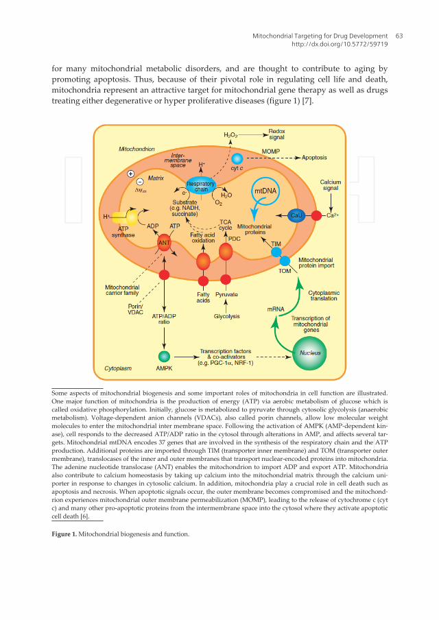
for many mitochondrial metabolic disorders, and are thought to contribute to aging bypromoting apoptosis. Thus, because of their pivotal role in regulating cell life and death,mitochondria represent an attractive target for mitochondrial gene therapy as well as drugstreating either degenerative or hyper proliferative diseases (figure 1) [7].
Some aspects of mitochondrial biogenesis and some important roles of mitochondria in cell function are illustrated.One major function of mitochondria is the production of energy (ATP) via aerobic metabolism of glucose which iscalled oxidative phosphorylation. Initially, glucose is metabolized to pyruvate through cytosolic glycolysis (anaerobicmetabolism). Voltage-dependent anion channels (VDACs), also called porin channels, allow low molecular weightmolecules to enter the mitochondrial inter membrane space. Following the activation of AMPK (AMP-dependent kin‐ase), cell responds to the decreased ATP/ADP ratio in the cytosol through alterations in AMP, and affects several tar‐gets. Mitochondrial mtDNA encodes 37 genes that are involved in the synthesis of the respiratory chain and the ATPproduction. Additional proteins are imported through TIM (transporter inner membrane) and TOM (transporter outermembrane), translocases of the inner and outer membranes that transport nuclear-encoded proteins into mitochondria.The adenine nucleotide translocase (ANT) enables the mitochondrion to import ADP and export ATP. Mitochondriaalso contribute to calcium homeostasis by taking up calcium into the mitochondrial matrix through the calcium uni‐porter in response to changes in cytosolic calcium. In addition, mitochondria play a crucial role in cell death such asapoptosis and necrosis. When apoptotic signals occur, the outer membrane becomes compromised and the mitochond‐rion experiences mitochondrial outer membrane permeabilization (MOMP), leading to the release of cytochrome c (cytc) and many other pro-apoptotic proteins from the intermembrane space into the cytosol where they activate apoptoticcell death [6].
Figure 1. Mitochondrial biogenesis and function.
Mitochondrial Targeting for Drug Developmenthttp://dx.doi.org/10.5772/59719
63
3. Mitochondrial diseases
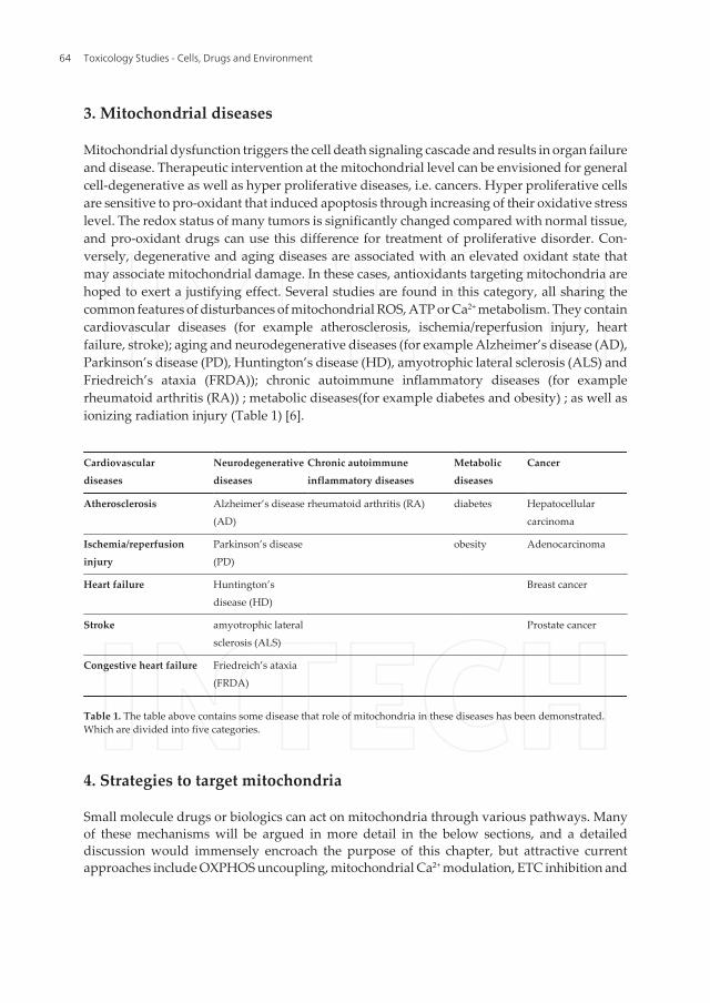
Mitochondrial dysfunction triggers the cell death signaling cascade and results in organ failureand disease. Therapeutic intervention at the mitochondrial level can be envisioned for generalcell-degenerative as well as hyper proliferative diseases, i.e. cancers. Hyper proliferative cellsare sensitive to pro-oxidant that induced apoptosis through increasing of their oxidative stresslevel. The redox status of many tumors is significantly changed compared with normal tissue,and pro-oxidant drugs can use this difference for treatment of proliferative disorder. Con‐versely, degenerative and aging diseases are associated with an elevated oxidant state thatmay associate mitochondrial damage. In these cases, antioxidants targeting mitochondria arehoped to exert a justifying effect. Several studies are found in this category, all sharing thecommon features of disturbances of mitochondrial ROS, ATP or Ca2+ metabolism. They containcardiovascular diseases (for example atherosclerosis, ischemia/reperfusion injury, heartfailure, stroke); aging and neurodegenerative diseases (for example Alzheimer’s disease (AD),Parkinson’s disease (PD), Huntington’s disease (HD), amyotrophic lateral sclerosis (ALS) andFriedreich’s ataxia (FRDA)); chronic autoimmune inflammatory diseases (for examplerheumatoid arthritis (RA)) ; metabolic diseases(for example diabetes and obesity) ; as well asionizing radiation injury (Table 1) [6].
Cardiovasculardiseases
Neurodegenerativediseases
Chronic autoimmuneinflammatory diseases
Metabolicdiseases
Cancer
Atherosclerosis Alzheimer’s disease(AD)
rheumatoid arthritis (RA) diabetes Hepatocellularcarcinoma
Ischemia/reperfusioninjury
Parkinson’s disease(PD)
obesity Adenocarcinoma
Heart failure Huntington’sdisease (HD)
Breast cancer
Stroke amyotrophic lateralsclerosis (ALS)
Prostate cancer
Congestive heart failure Friedreich’s ataxia(FRDA)
Table 1. The table above contains some disease that role of mitochondria in these diseases has been demonstrated.Which are divided into five categories.
4. Strategies to target mitochondria
Small molecule drugs or biologics can act on mitochondria through various pathways. Manyof these mechanisms will be argued in more detail in the below sections, and a detaileddiscussion would immensely encroach the purpose of this chapter, but attractive currentapproaches include OXPHOS uncoupling, mitochondrial Ca2+ modulation, ETC inhibition and
Toxicology Studies - Cells, Drugs and Environment64
control of oxidative stress through increase or decrease of mitochondrial ROS accumulation.The inhibition of the ETC can happen through direct inhibition of a protein subunit of one ormore of the enzyme complexes or via reception of electrons current across the ETC instead ofthe natural receiver cytochrome c or ubiquinone. In the Oxidative Phosphorylation (OXPHOS)uncoupling occurrence, protons are shifted from the mitochondrial matrix to the intermem‐brane space (IMS) and do not avert across the F1F0-ATPase and back to the matrix, but insteadmigrate directly across the inner mitochondrial membrane (IMM). This bypass results in lackof ATP formation but heat production. Typical instances for agents that elevate OXPHOSuncoupling are weak bases and weak acids, which can be protonated in the IMS and carryprotons across the IMM. Interestingly, compounds affecting the activity of inner membraneuncoupling proteins (UCPs) can inhibit cell death. An important occurrence starting theapoptotic cascade is the mitochondrial membrane permeabilization (MMP), which begins thecollapse of the mitochondrial potential (∆Ψ), the release of cyt c and other protease andnuclease activators. The inhibition of this process can be attained with inhibitors of themitochondrial permeability transition pore (mPTP) complex, openers of the mitochondrialATP-regulated (mitoKATP) or inhibitors of the mitochondrial Na+-Ca2+ exchange orCa2+activated (mitoKCa) potassium channels. Modulation of mitochondrial Ca2+ can also beenvisioned by interference with mitochondria-specific Ca2+ transporters. Additional strategiesfor drug-induced perturbation of mitochondrial biochemistry include the inhibition of the cytc-catalyzed peroxidation of the mitochondria-specific phospholipid CL, and the targeting ofother specific mitochondrial proteins via inhibition of kinases, F1F0-ATPase, enzymes of theKrebs cycle, or members of the anti-apoptotic Bcl-2 family. It has been known for a while thatinhibition of the oxidative cellular damage through a decrease of mitochondrial ROS accu‐mulation can be attained by the delivery of antioxidants acting as radical and/or electronscavengers. Many compounds are able to inhibit the β-oxidation of unsaturated fatty acids,causing cellular accumulation of fat. Alternatively, anti-apoptotic agents could be designedvia inhibition of the cyt c-catalyzed peroxidation of CL. Finally, the mitochondrial biochem‐istry is also severely derailed by mtDNA binding/oxidation or inhibition of mtDNA synthesis,or modulation of mitochondrial fission/fusion. Chemical agents that bind to mtDNA oftenresult in inhibition of DNA synthesis. If adequate selectivity in the binding process can beacceded, this mechanism f action may display an attractive strategy to block the expression ofmutated mtDNA accountable for genetic mitochondrial disarrays. Lately, compounds thatmodulate mitochondrial fission/fusion have been suggested as a valuable replacement intreatment of neurodegenerative diseases (figure 2) [6]. While the OMM is relatively permeabledue to the abundance of the VDAC protein, the IMM is extremely impermeable and acts as astiff barrier to the passive propagation of all types of molecules. It is also wealthy in the unusualphospholipid cardiolipin (CL), and keeps a strong negative internal potential of −180 mVneeded for the ETC function. A widely used strategy for targeting mitochondria takes benefitof this considerable biophysical membrane nature, since cationic molecules are attracted toand accumulate preferentially within the negatively charged mitochondrial matrix. Anotherstrategy is based on the access of an agent to mitochondrial membrane components, particu‐larly to the phospholipid cardiolipin CL which is particularly found in the IMM. Moreover theformer more specific properties, adequate lipophilicity is also needed to achieve an adequateenrichment in mitochondrial compartments. A rising approach to the selective delivery ofbioactive cargo molecule into mitochondria uses a carrier of short peptide sequences with
Mitochondrial Targeting for Drug Developmenthttp://dx.doi.org/10.5772/59719
65

specific physicochemical properties. For example, Horton et al. newly reported such mito‐chondria-penetrating peptides with changing cationic and hydrophobic residues. Othervariants have been based on an oligomeric carbohydrate scaffold, always attaching keyguanidinium moieties due to their delocalized cationic form. Finally, the tethering of activemolecules to mitochondrial targeting sequences (MTSs) has also been successively utilized.Mitochondrial targeting sequences (MTSs) are peptides applied by cells for the delivery ofnuclear-encoded mitochondrial proteins, comprising structural motifs recognized by themitochondrial import machinery. Another class of mitochondrial delivery vectors, appropriatefor the import of impermeable or big molecules, is the vesicle-based transporter system. Thetargeted agent is encapsulated in a cationic liposome, which undergoes cellular internalizationand subsequent fusion with the OMM. In summary, by the utilization of a wide range ofvarious delivery systems, the targeting of mitochondria for therapeutic advantages can beemployed to enrich both pro-oxidants as well as antioxidants in mitochondrial compartments.Antioxidants are of preliminary interest for their antiaging properties, with some of the mainapplications centered neurodegenerative and cardioprotection diseases, while cytotoxic andpro-oxidant agents are under research for cancer therapy [6].
Figure 2. Pharmacological targeting of mitochondria of possible sites of drug action.
Respiration:tamoxifen, acetaminophen, amiodarone,
barbiturates, propofol, halothane, resveratrol,…
Protein biosynthesis and replication: vit k3, tamoxifen, acetaminophen, zidovndie, abacare, flaluridine,…..
Krebs cycle:nicotinamide, carnitine, reveratol, dichoroacetate,…
Antioxidants: co Q10, mito Q10, lipoicacid,
vitE,mito E
Permeability transition:
carnitine, arsenites,
bongkrekicacid, acetaminophen, diclofenac,
amiodarone, CsA,…..
Apoptotic regulator protein: Gossypol, antimycine A,
obaloctax,….
TSPO
Translocator protein:ROS-4684, TRO4303303, SSR180575,…
Figure 2. Pharmacological targeting of mitochondria of possible sites of drug action.
4.1. Targeting the mitochondrial electron transport chain
Mitochondria are unusual organelles. They act as the power plants of the cell, are surroundedby two membranes, and have their own genome. The mitochondrion consists a matrixencircled by two membranes, the MOM and the MIM. The MIM comprises several invagina‐
Toxicology Studies - Cells, Drugs and Environment66
tions called cristae and is very impermeable to ions and small molecules, which need specifictransport proteins to exit or enter the mitochondrial matrix. Under aerobic statuses, theproteins of the ETC, placed in the MIM, reduce oxygen to water through a series of steps alongthe electron transport chain that use NADH and FADH2 derived from the glycolysis andtricarboxylic acid cycle. These reductions effectively efflux protons (H+) through the MIM suchthat they accumulate in the IMS creating a pH gradient across the MIM that contribute to anoverall electrochemical gradient (DC). This gradient as a source of energy to drive the synthesisof ATP from ADP and phosphate is applied by the mitochondrial F1F0-ATPase. This succes‐sion of chemical stages is collectively known as OXPHOS.
Small amounts of ROS are generated as a result of incomplete oxygen reduction in duringnormal OXPHOS. ROS comprise superoxide, the result of partial oxygen reduction, hydroxylradicals and the subsequently formed hydrogen peroxide, each of which displays differentchemistry. A high NADH: NAD+ ratio (as may arise owing to high rates of glycolysis) canincrease ROS production, as does state 4 respiration in which electron transport occurs in theloss of ATP synthesis, for instance, when ADP levels are low [8]. Inhibitors of the ETC and ofthe F1F0- ATPase can also enhance mitochondrial ROS production. ROS act as secondarymessengers with important signaling roles, but in addition ROS contribute to oxidativedamage of cellular macromolecules. It is also remarkable that the production of ROS has beenrecognized as a widespread mechanism for the bactericidal effect of many widely usedantibiotics including drugs targeting DNA, the cell wall and protein synthesis [9]. Thus, ROSperform both a destructive role and necessary role in cells. Inhibitors of the electron transportchain are useful tools for furthering our understanding of this essential bioenergetics process[10]. Inhibitors of complex I (NADH ubiquinone oxidoreductase) include the photochemicalAnnonaceousacet ogenins that have been attributed with antimicrobial and anticancerproperties and rotenone used as a rodenticide. The widely used diabetes drug metformininhibits complex I and has been shown to induce AMP-activated protein kinase-dependentand p53 increase in glycolysis to countervail for modulation of the respiratory chain, whicheffectively increases glucose utilization. Succinate–ubiquinone oxidoreductase (Complex II) isone proposed target of redox-silent vitamin E analogs such as α-tocopheryl succinate.Cytochrome c oxidoreductase (complex III) is inhibited by the natural product myxothiazoleand by antimycin A (the active constituent of the piscicide Fintrol).Cytochrome c oxidase(complex IV) is a target of cyanide. Complex I and complex III are the main sources ofmitochondria derived ROS in vitro, although the synthesis of superoxide by complex III isconsidered to be more physiologically related. The electron transport chain provides the H+
gradient that is necessary for the mitochondrial F1F0- ATPase to function. The relatedmacrolide apoptolidin and Oligomycin, a natural product that blocks the proton channel areboth inhibitors of the F1F0-ATPase.Apoptolidins display remarkably selective cytotoxicitytoward a subset of tumor cell lines in vitro, suggesting that inhibition of the ATPase is notexactly cytotoxic. Other compounds reported to bind to the F1F0-ATPase include aurovertin,resveratrol, PK1119, Bz-423, and diindolyl methane (DIM) [11]. The benzodiazepine derivativeBz-423 was identified as a lead for the treatment of autoimmune diseases.Bz-423 reducesdisease in murine models of lupus, psoriasis, and arthritis and has cytotoxic and anti-prolif‐erative effects on tumor cells in vitro. Bz-423 is an uncompetitive inhibitor of the F1F0-ATPase,
Mitochondrial Targeting for Drug Developmenthttp://dx.doi.org/10.5772/59719
67
deceleration the ATPase without causing a significant drop in cellular ATP levels. Thetherapeutic effects of this compound are moderated by the induction of superoxide O2
-.Resveratrol, a constituent of grape skins, increases longevity in rodents and has been attributedwith beneficial effects against inflammation, heart disease, and cancer. Notwithstanding theexistence of a crystal structure of resveratrol bound to the F1F0-ATPase, this protein is one ofseveral reported targets for resveratrol and related compounds, including the protein deace‐tylase, sirtuin [5].
The ETC and the F1F0-ATPase proteins can be decoupled by uncoupling proteins that promotethe leakage of protons back through the MIM. The resulting drop in membrane potentialreduces ROS production and represents a natural protective mechanism against inhibition ofrespiration. This is a natural process that results in thermogenesis. F1F0-ATPase inhibitors,without affecting ATP synthesis, specifically block ATP hydrolysis have been described: suchcompounds should be effective under ischemic conditions when the ATPase can operate inthe reverse of its normal direction leading to a catastrophic drop in ATP levels that causes celldeath [12]. This premise has not been tested clinically. Mammalian and bacterial ATP synthasesexhibit substantial differences in structure and intracellular location presenting the opportu‐nity for species selective ATP synthase modulation [5]. The mycobacterial ATP synthaseinhibitor, R207910, is currently in Phase III trials for the treatment of tuberculosis [13].
4.2. Targeting transporters and channels in mitochondria
It is well recognized that the totality of the mitochondrial membrane is crucial for mitochon‐drial function. Not only are the inner and outer membranes targeted by drugs, but, in addition,many of the ion channels, proteins, and transporters embedded within the lipid membraneare also targeted. Among the main drug targets are: 1) lipophilic cations targeting the IMM(e.g., rhodamine-123) 2)cardiolipin(CL) (e.g., 10-N-alkyl-arcine orange), 3) carnitine palmi‐toyltransferase- 1 (CPT-1) inhibitors (e.g., oxfenicine, perhexiline, and etomoxir), 4) Na+/ Ca+2
exchanger regulators, 5) B-cell lymphoma 2 (Bcl-2) protein inhibitors (e.g., gossypol) 6) IMMpotassium channel regulators (e.g., glibencamide and diazoxide), and 7) MPT pore complexregulators (e.g., CsA).We can activate permeabilization of the mitochondrial membrane or canprotect membrane integrity. Among the best mitochondrial protein targets for many drugs area group of proteins that form the PTP complex across the OMM and IMM. This complex isresponsible for mitochondrial permeability transition and plays a crucial role in both survivaland death signaling pathways. Depending on the pharmacological strategy, MPT poreactivation stimulates apoptosis and prevents the differentiation of many tumor cells. Strategiesto induce this effect typically involve direct action against the MPT pore protein complex orindirect action via depleting endogenous inhibitors of MPT pore or increasing ROS andcalcium ions in the cytoplasm. Various MPT pore complex inhibitors, in anticancer therapeuticapproaches, are used, including: 1) hexokinase modulators such as glucose-6-phosphate andglucose 2)creatine kinase modulators such as cyclocreatine and creatine; 3) cyclophilin D(CypD) -affecting drugs such as sanglipherin A and CsA; 4) voltage dependent ion channelmodulators such as arsenic trioxide; 5) benzodiazepine receptor modulators such as Ro-54846and PK11195; and 6) adeninenucleotide translocase modulators such as CD437, PENAO (4-(N
Toxicology Studies - Cells, Drugs and Environment68
(Spenicillaminylacetyl) amino) phenylarsonous acid), lonidamide, betulinic acid, clotrane, andbongkrekic acid, GSAO (4-[N-[S-glutathionylacetylamino] phenylarsenoxide)[14], GSAO andPENAO are tumor-metabolism inhibitors that target ANT of the inner-mitochondrial mem‐brane. Both the compounds are currently being appraised in trials in patients with solidtumors. The trivalent arsenical moiety reacts with the two matrix-facing cysteine residues ofANT, inactivating the transporter. This leads to tumor-supporting cells and death andproliferation arrest of tumor cells [14].Above-mentioned drugs grouping although usefulappears to be synthetic, and surely will be modified. According to some authors MPT poremay consist of quite different proteins. Recent investigation on MPT pore molecular identityhas to redefine a new context on described interaction.CL, a negatively charged phospholipid,is almost exclusively localized in the mitochondrial inner membrane. CL maintains architec‐ture and membrane potential. A loss of CL content has been associated with mitochondrialdamage in multiple tissues in a variety of pathological conditions, including aging, heartfailure, and ischemia. It was reported that preadministration of NAO (10-N-alkyl-arcineorange), that is a dye associated specifically with CL, decreased the release of cytochrome c, acomponent of the ETC in mitochondria, released in response to pro-apoptotic stimuli [15].Another drug target example is CPT-1, an enzyme located in the OMM and responsible forthe transport of long-chain fatty acids across the membrane by binding them to carnitine.Perhexiline and etomoxir (antianginal agents) act by inhibiting CPT-1 and protect heart fromfatty acid-induced ischemic injury [16].
In contrast to the MIM, the mitochondrial outer membrane is more permeable to smallmolecules so that the IMS resembles cytosol in its small molecule composition. In addition,however, the IMS sequesters proteins such as apoptosis inducing factor (AIF), smac/ Diablo(second mitochondria derived activator of caspases), and cyt c that when released into thecytosol activate caspases and induce apoptosis. One process for the release of these deathinducing protein factors involves swelling of the mitochondrion so that the outer membraneruptures producing MPT. These events are mediated by the MPT, a channel that comprisesmultiple proteins including the VDAC located in the MOM, ANT located in the MIM, as wellas the peripheral benzodiazepine receptor (PBR), CypD, hexokinase, and possibly alsoBax.andBcl-2 Inhibitors of the MPTP have been reviewed elsewhere as have inhibitors of Bcl-family proteins. High affinity ligands of the PBR have been associated with immunothera‐peutic and anticancer properties. The relationship of these effects to physiological functionsof the PBR requires more study. Newly, VDAC ligands identified in cell-based screens wereshown to be cytotoxic toward cells bearing oncogenic Ras protein [4].
4.2.1. Targeting mitochondrial Adenine nucleotide translocator (ANT)
Adenine nucleotide transporter interacts with Voltage dependent anion channel (VDAC) andcyclophilin D and other proteins to make the mitochondrial permeability transition pore(mPTP) at locations where IM contacts OM [17]. Adenine nucleotide transporter (ANT), thekey IM protein of mPTP, exchanges ATP and ADP. ANT can form mitochondrial permeabilitytransition pores (mPTP) which induces other membrane leakiness of mitochondria andsubsequent swelling of matrix. This event happens following the surface area of the IM (withits folded cristae) exceeds that of the OM. Quite in contrast, the conformation of ANT is
Mitochondrial Targeting for Drug Developmenthttp://dx.doi.org/10.5772/59719
69
modulated by ANT ligands [18] and sensitive interaction of cyclosporine A with cyclophilinD, indirectly blocks VDAC activity. When reconstructed into planar lipid bilayers or intoliposomes or into planar lipid bilayers, ANT can form nonspecific channels in response toproapoptotic agents such as the HIV-1 viral protein R (Vpr), Ca2+, lonidamine and atractyloside.Moreover, ANT channel formation is inhibited by Bcl-2 and enhanced by Bax. However, mouseknockout studies led to the conclusion that ANT would not (always) be required for apoptoticMPT. Recent evidence suggests that some ANT isoforms (ANT1, ANT3) are apoptogenic whileothers are not (ANT2)[19]. In 2005, a fourth ANT isoform (ANT4) has been identified in mouseand man by means of two independent experimental approaches. ANT2, which are overexpressed in cancer cells, help to stabilize mitochondrial membranes and survival cell. Indeed,it was suggested that in cancer cells, small interfering RNA (siRNA) that down regulate ANT2may constitute a valid strategy for the selective induction of tumor cell apoptosis [20].
4.2.2. Targeting mitochondrial cyclophilin D
CypD is a nuclear-encoded mitochondrial isoform of cyclophilin, with a molecular mass of 18kDa. It enters mitochondria using a targeting sequence that is cleaved following translocationinto the matrix. At present, extensive data have been obtained in favor of Cyclophilin D as anessential component and key regulator of MPT pore using various pharmacological inhibitorsand genetic manipulations. The first evidence for the involvement of Cyclophilin D in MPTpore formation came from studies showing an inhibitory effect of CsA, extensively used intissue and organ transplantation, as an immunosuppressant, on pore opening. Other docu‐ment for the essential role of Cyp D in MPT pore formation has been reported by severalindependent groups in reports with Cyp D knockout mice in which mitochondria isolatedfrom these animals displayed a low sensitivity to Ca2+and, as a result, a delayed MPT poreopening. The inhibitory effect of CsA and its analogs involves interaction with Cyp D thatreduces sensitivity of pore opening to Ca2+. Cyp D favors MPT pore opening by facilitating theCa2+triggered conformational change. Most probable, interaction of CypD and Ca2+, P and thepore is a multifaceted process that also includes enhancement of susceptibility of the MPT poreproteins to oxidative stress [21].
Several studies have shown that Cyp D is up-regulated in many human tumors and canfunction as an apoptosis repressor. Growing number of evidence demonstrated that the anti-apoptotic regulation of Cyp D might be associated with the stabilization of hexokinase IIbinding to mitochondria. Inactivation of CypD with cyclosporine A or knock- down of theexpression using siRNA was shown to release hexokinase II from mitochondria. Because CypD is a mitochondrial matrix protein, an intermediate in the IMM between the OMM and matrixis necessary for its modulation of hexokinase II binding to VDAC. ANT in the IMM could playthis intermediation role.However, study showed the opposing pro-apoptotic role of Cyclo‐philin D in apoptosis. They demonstrated that hexokinase II detachment-triggering apoptosismight be associated with a disruption of the interaction of Cyp D with ANT. Furthermore,inhibition of CyP-D was shown to prevent the onset of the MPT pore [22].
The MPT pore, a critical mediator of cell death, has appeared as a serious therapeutic targetfor limiting acute ischemia reperfusion injury. The genetic amputation and pharmacological
Toxicology Studies - Cells, Drugs and Environment70
inhibition of mitochondrial Cyp D, a key mediator of apoptosis signaling, has emerged as animportant therapeutic target for minimizing acute hypoxic/ischemic injury. The geneticablation and biological inhibition of mitochondrial cyclophilin-D (CypD), a regulatorycomponent of the mitochondrial permeability transition pore (mPTP), has been reported todecrease myocardial infarction progression in in vivo studies. However, it is note worthy thatCypD-deficient hearts are still susceptible to mPTP opening and cell death signaling occurredthrough mechanisms which are not dependent on CypD. Very recently, cyclosporin-A (CsA),an immunosuppressive therapeutic agent and biological inhibitor of CypD has been shown toreduce myocardial infarction progression and improve left ventricular function in ST-elevatedMI patients undergoing primary percutaneous coronary surgery, given at reperfusion [23].
Animals lacking CypD display increased resistance to ischemic insults, muscular dystro‐phies, multiple sclerosis (MS), ALS, and AD, and the CypD inhibitor CsA and its analogshave displayed neuroprotective effects in several animal models of acute neurologicaldamage and chronic neurodegenerative disease. Preserving the integrity of mitochondrialmembranes through inhibition of mPT has been put forward as the central mechanism forthe neuroprotective and cardioprotective effects of CsA, even though the drug has severalpharmacological targets. It has also been suggested that CypD is downregulated in neuronsduring development, which would decrease the sensitivity of the MPT pore to calcium, andprohibit the use of CypD as a pharmacological target in disorders of the adult centralnervous system (CNS) [24].
4.2.3. Targeting mitochondrial peripheral benzodiazepine receptor (PBR)
The elaborate structure of mitochondria is important for the normal performance of theorganelle and as a potential therapeutic target. Two specialized membranes embed eachmitochondrion, dividing the organelle into an arrow IMS restricted by the OMM and the innerIMM. The OMM comprises many channels formed by the protein porin that makes themembrane relatively permeable. One of the membrane proteins is the peripheral benzodiaze‐pine receptor (PBR). PBR is a small evolutionarily conserved protein involved in steroidsynthesis and cholesterol transport; it is also a regulator of apoptosis. The PBR is also involvedin OMM permeabilization by interaction with the pro-apoptotic Bcl family of proteins.However, OMM permeability maybe independent of MPT pore opening because blocking PBRwith 4’-chlorodiazepam (CDZ) prevents against ischemia-induced cytochrome c releaseindependent of damage to the IMM;4’-chlorodiazepam (CDZ)also reduces ischemia-inducedarrhythmias. PBR is found in close association with the VDAC and additional components ofthe mitochondrial contact site. This close association also suggests that PBR-VDAC may serveas a target for modulating apoptosis and may have implications for drug design to treat suchdisorders as cancer and neurodegenerative diseases [20].
4.2.4. Voltage-dependent anion channel (VDAC)
VDACs, also known as mitochondrial porins that show 68% similarity between mice andhumans. Among three VDAC isoforms, VDAC1 is the most widely expressed in mammalsfollowed by VDAC2 and then VDAC3. Studies have found that VDACs are highly conserved.
Mitochondrial Targeting for Drug Developmenthttp://dx.doi.org/10.5772/59719
71
Three isoforms of VDAC: VDAC1, VDAC2 and VDAC3 are reported. The additional exon inVDAC2 is believed to encode part of the 5′-UTR region. VDAC1 and VDAC2 are expressed inthe skeletal muscles, heart, liver, and brain. There is also very low level expression of VDAC1but only in the testes. VDAC3 is expressed in the spleen, lung, adrenal, ovary, liver, testiculartissue and kidney muscles. Voltage dependent anion channel (VDAC) function functions inthe cell, including regulating mitochondrial shape and structural changes, regulating ATPtransport, regulating calcium transport, regulating apoptosis signaling, regulating hexokinaseinteractions with mitochondria, regulating cell survival, growth, and fertility and maintainingsynaptic plasticity through mitochondrial permeability in the transition pore. These functionshave been found to be altered in cells from patients with mitochondrial and neurodegenerativediseases, leading to mitochondrial dysfunction. As well as, increasing evidence suggests thatVDAC interacts with several cytoplasmic proteins, changes channel activity and VDACclosure and reduces VDAC channel conductance. It is believed that VDAC is constantly openin metabolic state. However, recent evidence suggests that VDAC closes intelligibly duringapoptosis in unhealthy neurons. As a result, with its pores closed, mitochondria may not beable to uptake ADP, inorganic phosphate and respiratory substrates from the cytoplasm andto release ATP into the cytoplasm. The pro-apoptotic protein tBid has been found to promotethe pore closure whereas anti-apoptotic proteinBcl2-XL has been found to prevent VDACclosure. VDAC displays to be involved in both anti - and pro – apoptosis aspects of mitochon‐dria. VDAC channel conductance may be impaired in a couple different ways. (1)Phosphory‐lated VDAC may also interact with cytoplasmic proteins, leading to the blockade ofmitochondrial pores. Recently, in a study of brain tissue from postmortem brains of patientswith AD, Reddy and Manczak found that VDAC interacted with mutant AD proteins, whichin turn blocked mitochondrial pores and interrupted the flow of ADP, ATP, respiratorysubstrates and inorganic phosphate substrates between mitochondria and the cytoplasm,ultimately leading to mitochondrial dysfunction. (2) In neurons from mitochondrial diseases,VDAC may interact with cytoskeletal and mutant proteins that may have accumulated duringdisease progression and may have blocked the mitochondrial pores [25].
A lot of literature testes the role of VDAC in the regulation of cell death. VDAC is being studiedas a cancer-specific target because tumor cells have increased VDAC expression and glycolysis.The role of VDAC1, VDAC2 and VDAC3, in cell death is intricate, but importantly, in vivoevidence shows that in cancer cells, the association of VDAC1with HK prevents againstmitochondrial-mediated apoptosis. Therefore, disruption of the VDAC1-hexokinase (HK)complex exhibits an attractive therapeutic cancer target. Over expression of HK1, 2 and theirconnection with VDAC are notable characteristics of glycolytic cancer cells. It was found thatthe VDACs expression has been elevated in cancerous cells compared with normal cells andcould be altered with chemotherapy. Increased VDAC concentration is an unfavorableprognostic factor; moreover, RNA interference induced VDAC down regulation inhibitscancer growth. This evidence seems in contrast with the finding that over expression of VDACinduces apoptosis, but it illustrates how the context may influence the functional meaning ofa biological parameter. In cancer up regulation of VDAC goes hand in with HK2 up-regulationand can be considered a component of glycolytic up-regulation. HK2 binding to VDAC, whichallows for ATP transport out of mitochondria, leads to a cancer cell metabolic advantage
Toxicology Studies - Cells, Drugs and Environment72
(termed the Warburg effect), and it antagonizes cell death through the inhibition of Bax-induced cytochrome c release and/or inhibition of the MPT pore [26].
4.2.5. Changes in the configuration of MPT pore as a target in treatment
Cellular redox potential can be changed by function of OXPHOS proteins as well as by theproliferative state. Elevations in intracellular oxidant potential can have discrete chemicalconsequences: for example, a pair of cysteine thiols in the ANT becomes oxidized to a disulfidelinkage that results in opening of the MPT pore. Thus, manipulating cellular redox representsan approach to altering mitochondrial function. Arsenic trioxide is currently marketed for thetreatment of acute promyelocytic leukemia. Its mechanism of action is undoubtedly multifac‐torial but is understood to involve the formation of disulfide linkages in mitochondrialproteins, including members of the MPT pore leading to their inhibition and the productionof ROS [27]. Elesclomol (STA-4783), an injectable drug currently undergoing Phase III clinicalevaluation for the treatment of metastatic melanoma, selectively kills cancer cells throughapoptosis as a result of an increase in their already raised oxidant level [28].
4.3. Superoxide dismutase (SOD) as a target in mitochondria
Superoxide dismutase (SOD) represents a group of enzymes that use as cofactor zinc andcopper, or nickel, iron, or manganese ions. There are three major families of superoxidedismutase, depending on the metal cofactor: The Ni type, which binds nickel (only in prokar‐yotes) and Cu/Zn (which binds both copper and zinc), Fe and Mn types (which bind eitheriron or manganese). SOD1 is located in the cytoplasm, SOD2 in the mitochondria, and SOD3is extracellular. The first is a dimer, whereas the others are tetramers (four subunits).SOD2,the mitochondrial enzyme, has manganese in its reactive site whereas SOD1 and SOD3 containcopper and zinc. [28]
A key role in oxidative stress protection is played by the manganese containing SOD2 inmitochondria. This enzyme is also critical for fetus growth and viability n many eukaryoticorganisms, since complete loss of the enzyme results in neonatal lethality in mice. In additionto oxidative tress nitrosative stress can completely inactivate mitochondrial Mn-SOD as well,possibly through nitration of a single tyrosine residue (Tyr-34). Consequently, this favorsperoxynitrite generation in mitochondrion. Tyrosine nitration induced Mn-SOD inactivationbeing identified in more than 50 human diseases including ischemia/reperfusion, inflamma‐tion, human kidney allograft rejection and human pancreatic ductal adenocarcinoma [29].
The renal ischemia-reperfusion injury is one of the most important clinical xamples in whichMn-SOD represents the main antioxidant protective mechanism. A significant increase insuperoxide production is usually associated with Ischemia/reperfusion conditions whichleads to a rapid depletion of SOD. Therefore, any external therapeutic involvement needsthe sufficient amount of SOD to overcome the superoxide radical byproduct of ischemia-reperfusion conditions. Any therapeutic administration of exogenous SOD fails due to shorthalf-life of the enzyme in plasma. A way to ensure a continuous production of SOD isentering SOD gene in renal tissue which guarantees protection from renal ischemia-
Mitochondrial Targeting for Drug Developmenthttp://dx.doi.org/10.5772/59719
73
reperfusion injury. The effective gene delivery without toxic side effects was established byintravenous injection of the gene vectors during experiments on animal models before theischemic insult. A significant progress in the area of kidney biology, especially in heredita‐ry kidney disease and inflammatory and fibrotic diseases was achieved by the use ofadenovirus as a vector for kidney-directed gene therapy [30]. Although some advantagesmake adenoviral vectors suitable for gene transfer into complex organs such as the kidney.But in contrast some disadvantages downgrade these vectors. For instance, the expressionof the transfected gene is limited to weeks or months in this technique, because adenovi‐rus does not integrate into the host genome. Secondly, the adenovirus can elicit immunolog‐ical responses, therefore vector cannot be administered repeatedly. During emergencysituations in other inflammatory renal disease states, the SOD gene therapy with adenovi‐ral vector is recommended, however, occurrence of harmful effects maximum within a weekis expected (e.g., post-transplant acute renal failure) [29].
ALS a neurodegenerative disease leads to paralysis, muscle wasting, and death, usually within2 - 3 years of symptom onset due to death of motor neurons. The central mechanism by whichmotor neuron death occurs in familial ALS is oxidative stress which is due to the mutations inthe antioxidant enzyme SOD1gene. Many hypotheses studied so far using ALS mouse models.Some of these studies showed that SOD1 mutants have very low benefits (3, 35). One of themost important pharmacological outcomes obtained in ALS mouse models was increasingexpression of either growth factors such glial cell-derived neurotrophic factor, IGF-1, andVEGF (11–13) or RNAi molecules by the delivery of viral vectors (14–16) to silence SOD1mutant gene expression. In gene therapy the primary cause of toxicity (i.e. mutant SOD1proteins) is targeted, unlike drug therapy which usually acts on cell survival or deleteriouspathways [29].
Reduction of myocardial reperfusion injury through an effective immunization with SOD andcatalase has also been hypothesised. Indeed, the cardioprotective effect of intracoronaryinfusion of SOD may further increase with coadministration of catalase. It is proven thatcalcium antagonists, rennin-angiotensin system antagonists, Na+/H+ exchanger inhibitors,nitric oxide donors and adenosine induce cardioprotective effects during primary angioplastyfor the management of acute myocardial infarction. When these reagents were administratedusing intracoronary infusion, their efficiency has increased. Another way to attenuate myo‐cardial ischemia-reperfusion injury is anterograde intracoronary and intravenous adminis‐tration of anti- P-selectin and anti- ICAM-1 antibodies. The ideal injection route for theseantibodies is retrograde intracoronary infusion, which has direct access to postcapillaryvenules [29].
Application of inhibitors of cellular redox maintaining proteins which reduce intracellular ROSis complementary to the use of pro-oxidant molecules, for example, administeration of catalaseor SOD in association with various peroxidases. 2-Methoxyestradiol by increasing cellular ROSformation due to its inhibition of SOD, enhances the cytotoxic effects of apoptotic agents anddisplays anti-leukemic activity in culture. On the other hand it has been hypothesized,continuous mitochondrial ROS formation leading to oxidative stress and mitochondrialdamage has link to degenerative diseases and aging. Based on the ROS etiology of aging the
Toxicology Studies - Cells, Drugs and Environment74
ROS inhibition should have therapeutic benefit. Administration of antioxidants manganese(III) tetrakis (4-benzoic acid) porphyrin (MnTBAP) or N-acetylcysteine also improved glucosehomoeostasis and insulin sensitivity in obese insulin-resistant mice [31]. MitoQ, a coenzymeQ analog is currently in trial for the treatment of Parkinson’s Disease due to its potentialmitochondrial ROS inhibition. Knowing the beneficial effects of ROS shouldn’t underscore theimportance of a detailed knowledge of pathological conditions under which ROS formationis happening, as well as the identity and biological half-life of the ROS produced.
Free radicals are generally involved in many pathological processes. The injuring mechanismof reactive radical species is concentration dependent, which finally damages all cellularconstituents. Any insufficiency or functional failure in the body antioxidant systems results inthe shortening of the lifespan. Therefore, the first therapeutic approach is restoring the normalfunction of the antioxidant enzymes like SOD.
4.4. Mitochondrial KATP channels target for therapy
Potassium channel openers (KCOs) are agents, discovered in the early 1980s, that act bystimulating ion flux through K+ channels. Many drugs such as, diazoxide, nicorandil, andcromakalim have been identified as KCOs. KCOs act on two types of ion channels: Ca2+
activated K+ channels (BK channels) and ATP-regulated K+ channels (KATP channels). KCOswere first identified by their antihypertensive or antianginal mode of action. Now, they are atvarious stages of development as and cardioprotective agents. Preclinical and clinical evidencealso supports the therapeutic role of KCOs in vascular and pulmonary hypertension, and thetreatment of overactive bladder. Until recently, it was believed that the effects of KCOs wereentirely attributed to the modulation of K+ channels in cell plasma membranes. However, itis now proven, that new targets for KCOs exist in intracellular membranes including those ofmitochondria, zymogen granules, and sarcoplasmic reticulum. It seems that Mitochondria areparticularly very important targets for KCOs, because the interaction of these compounds withmitochondria appears to mediate the cardioprotection of KCOs. The protective role ofmitochondrial ion channels was recently summarized and mitochondrial targets for anti-ischemic drugs were recently described [32].
4.4.1. Potassium channel openers and mitochondrial K+ channels
A small-conductance potassium channel, with properties similar to those of the KATP channelfrom the plasma membrane, in the inner membrane of rat heart and liver mitochondria anddesignated the mitoKATP. The mitoKATP channel was blocked not only by ATP, but also,similarly to the plasma membrane KATP channel, by antidiabetic sulfonylureas. These obser‐vations raised the question whether the mitoKATP channel could be activated by KCOs. In fact,an increased influx of K+ and depolarization of liver mitochondria in the presence of KCOswas observed. Also, other KCOs were shown to activate potassium ion transport into mito‐chondria. KCOs such as levcromakalim, cromakalim, and pinacidil have been shown todepolarize cardiac mitochondria. KCO-induced membrane depolarization was associatedwith an increase in the rate of mitochondrial respiration and decreased ATP synthesis.More‐over, KCOs released cytochrome cand calcium ions from cardiac mitochondria. Despite the
Mitochondrial Targeting for Drug Developmenthttp://dx.doi.org/10.5772/59719
75

effect on K+ transport, diazoxide also exhibits a direct effect on mitochondrial energy metab‐olism by inhibition of respiratory chain complex II in liver mitochondria. Recently, mitoKATP
channel opener BMS-191095 with no peripheral vasodilator activity was described.Usingisolated mitochondria or proteoliposomes reconstituted with partly purified mitoKATP channeland measuring potassium flux demonstrated that heart and liver liver mitochondrial KATP
channels have some pharmacological similarities with the cell membrane KATP channel, i.e.,both channels are activated by KCOs. Mitochondrial KATP channels are 1000 times moresensitive to diazoxide than that of cell membrane KATP channels. This document concludedthat the interaction of mitochondrial KATP channels with KCOs plays a key role in cardiopro‐tection [32].
4.4.2. Mitochondrial KATP channel: A novel target for cardioprotection
Mitochondrial KATP channel: A Novel target for Cardioprotection. KCOs mimic hypoxic/ischemic preconditioning in the absence of ischemia in the heart myocardial cells, the reasonwhy antagonists of KATP channel, like 5-hydroxydecanoic acid and glibenclamide, amelioratethe positive effects of short time hypoxic/ischemic conditions on the heart myocardium. Theprimary postulation to justify these events includes cell membrane KATP channels. Newly, itwas shown that in fact KCOs including diazoxide affect the mitoKATP channel in mitochondria.In a complementary approach, it was shown that diazoxide did not activate plasma membraneKATP channels, but induced oxidation of mitochondrial flavoproteins, due to the activation ofmitoKATP channel. These findings established the fact that the target for the diazoxide protec‐tive effects in heart myocytes is the mitochondrial KATP channel rather than the cell membraneKATP channel. It is also note worthy that evidence for mitochondrial KATP channels as effectorsof cardiac myocardial preconditioning has also been proven in human subjects. The initialobservations on the cardioprotective action of KCOs on mitochondria were further confirmedand developed in a series of reports. It has been shown that other KCOs such as nicorandil,cromakalim, and pinacidil modulate mitochondrial Ca2+ uptake, respiration, mitochondrialmembrane potential, ATP generation, and mitochondrial Ca2+ uptake. The main questionremains how the opening of the mitoKATP channel could protect cells against ischemic damage.1) Opening of the mitoKATP channel followed by mitochondrial swelling could improvemitochondrial ATP handling and/or production. 2) The protective effect of mitoKATP activationcould be mediated by lowering Ca2+ overloading of mitochondria. In fact, it was found thatdiazoxid preserves mitochondrial function in ischaemic rat cardiomyocyte. It is now proventhat hypoxia approximately decreases mitochondrial oxygen consumption rate to 40% of thenormal value, and administration of diazoxide maintains the prehypoxic mitochondrialoxygen consumption rate during hypoxia/ischemia. Cardiac ATP concentration was signifi‐cantly raised following diazoxide treatment. Secondly, by lowering Ca2+ overloading ofmitochondria the protective effect of mitochondrial KATP activation could be induced. It wasshown that the opening of the mitochondrial KATP channel may increase mitochondrialreactive oxygen species (ROS) formation. This increase could lead to protein kinase C activa‐tion, which is known to be necessary for the cardioprotection. Besides, it seems that mito‐chondrial KATP channel is enrolled in delayed preconditioning because of an alteration inexpression of "protective" proteins (3). It was that pretreatment of hippocampal neurons with
Toxicology Studies - Cells, Drugs and Environment76
cromakalim and diazoxide increases the expression level of Bcl-2 and Bcl-XL which areinvolved in the control of apoptosisBcl-2 [32].
5. Mitochondria as a biosensor for drug development
Extensive study over the last 50 years indicates that many medications can induce mitochon‐drial damage [33]. Medication- induced dysfunctions include the alteration of mitochondrialcomponents and metabolic pathways. These dysfunctions are a major challenge and problemfor drug development. There is mounting evidence of the mitotoxicity (table 2).
Interestingly knowledge of the mechanisms that trigger drug-induced mitochondrial damagewill be helpful in the development of strategies to decrease the potentially toxic effects ofmedications. Additional, these issues affect the most aerobically poised organs such as heartand kidneys or organs exposed to higher concentrations of the drug for example liver. Recentlyusing mitochondria as a biosensor for determination safety of drug development has in‐creased. The reasons are as follows: A) in general, mitochondria control many of the pro-deathand anti-death cell signals; B) a number of reports describe an association between patientsreceiving medication and effects on mitochondrial metabolism 3) drug safety has become apriority of many pharmaceutical companies [4].
It is quite obvious that mitochondria are key elements of cell life which several well knowndrugs induce toxic effects on them in several non-target and target organs. As soon as possibleby improvement of preliminary drug safety assessment the possibility of drug toxic reactionsduring clinical practice will be avoided. Depending on the targeted organ, severe in vitromitochondrial impairment may be sufficient to ban an efficient drug in the market or prevent‐ing a promising drug candidate from further clinical trials. Drug companies now have a newdilemma, which is to realize how much of the evaluated mitochondrial toxicity is a keypredictor of the drug pharmacological or adverse effects. Pharmaceutical suppliers have alsonow a difficult problem which is to know how much of the supposedly mitochondrialimpairment is a component of the therapeutic effect. On the other hand, it may be a toughchoice to remove dispensing drugs showing a certain degree of mitochondrial toxicity invitro evaluations but with a very unique significant therapeutic effect. Despite showingmitochondrial toxicity, sometimes pharmaceutical companies may decide to push the leadcandidate molecule forward for further in vivo assays in order to also clarify ways of reducingadverse mitochondrial toxicity [3]. Some pharmacological strategies could be used to decreasemitochondrial toxicity. For example in the cardiac toxicity of doxorubicin (DOX), Onepossibility is to improve drug targeting, decreasing the amount of drug that reach non-targetorgans. One successful example of this strategy is the use of pegylated liposomal DOX, whichhas a quite different pharmacokinetic profile including an increased circulation time and adecreased volume of distribution [34]. Another strategy is the co-administration of protectiveagents, one example of which being the preventive role of the beta-blocker carvedilol on DOXinduced cardiac mitochondrial impairment.
Mitochondrial Targeting for Drug Developmenthttp://dx.doi.org/10.5772/59719
77
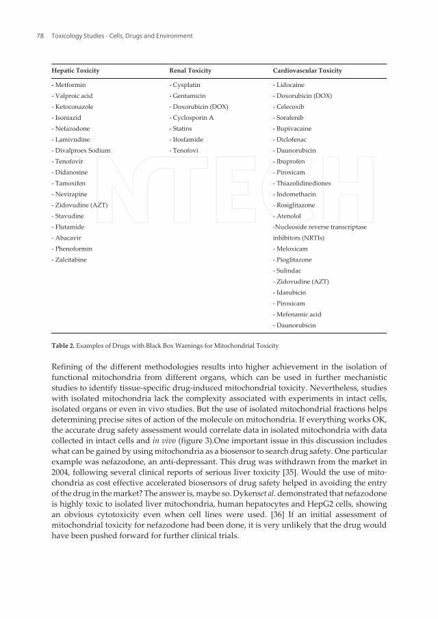
Hepatic Toxicity Renal Toxicity Cardiovascular Toxicity
- Metformin- Valproic acid- Ketoconazole- Isoniazid- Nefazodone- Lamivudine- Divalproex Sodium- Tenofovir- Didanosine- Tamoxifen- Nevirapine- Zidovudine (AZT)- Stavudine- Flutamide- Abacavir- Phenoformin- Zalcitabine
- Cysplatin- Gentamicin- Doxorubicin (DOX)- Cyclosporin A- Statins- Ifosfamide- Tenofovi
- Lidocaine- Doxorubicin (DOX)- Celecoxib- Sorafenib- Bupivacaine- Diclofenac- Daunorubicin- Ibuprofen- Piroxicam- Thiazolidinediones- Indomethacin- Rosiglitazone- Atenolol-Nucleoside reverse transcriptaseinhibitors (NRTIs)- Meloxicam- Pioglitazone- Sulindac- Zidovudine (AZT)- Idarubicin- Piroxicam- Mefenamic acid- Daunorubicin
Table 2. Examples of Drugs with Black Box Warnings for Mitochondrial Toxicity
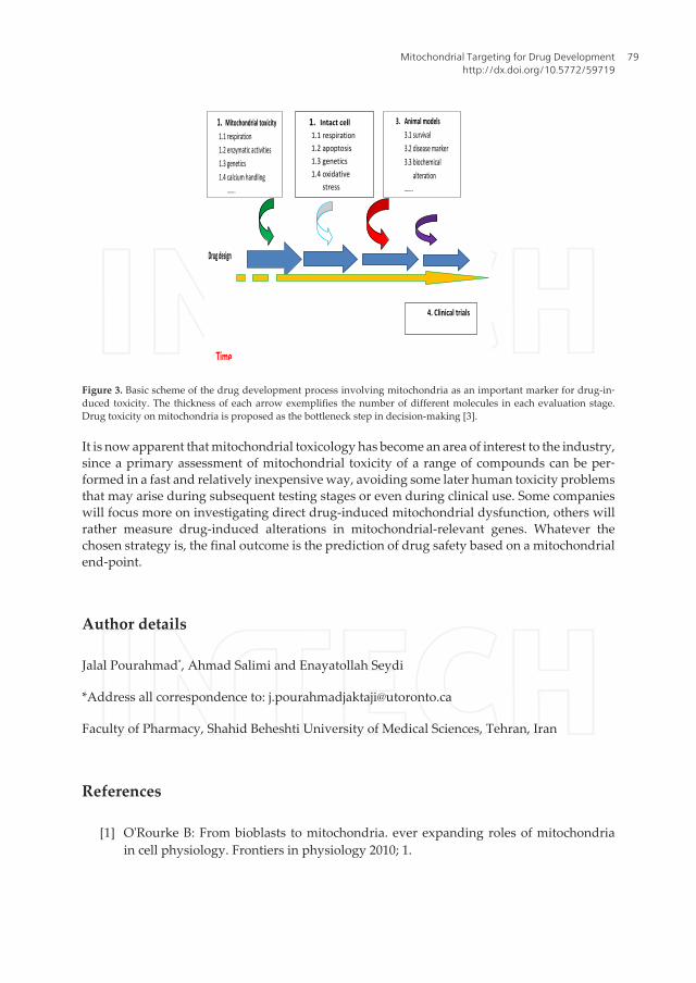
Refining of the different methodologies results into higher achievement in the isolation offunctional mitochondria from different organs, which can be used in further mechanisticstudies to identify tissue-specific drug-induced mitochondrial toxicity. Nevertheless, studieswith isolated mitochondria lack the complexity associated with experiments in intact cells,isolated organs or even in vivo studies. But the use of isolated mitochondrial fractions helpsdetermining precise sites of action of the molecule on mitochondria. If everything works OK,the accurate drug safety assessment would correlate data in isolated mitochondria with datacollected in intact cells and in vivo (figure 3).One important issue in this discussion includeswhat can be gained by using mitochondria as a biosensor to search drug safety. One particularexample was nefazodone, an anti-depressant. This drug was withdrawn from the market in2004, following several clinical reports of serious liver toxicity [35]. Would the use of mito‐chondria as cost effective accelerated biosensors of drug safety helped in avoiding the entryof the drug in the market? The answer is, maybe so. Dykenset al. demonstrated that nefazodoneis highly toxic to isolated liver mitochondria, human hepatocytes and HepG2 cells, showingan obvious cytotoxicity even when cell lines were used. [36] If an initial assessment ofmitochondrial toxicity for nefazodone had been done, it is very unlikely that the drug wouldhave been pushed forward for further clinical trials.
Toxicology Studies - Cells, Drugs and Environment78
Running Title
27
1 2
3
4
5
6
7
8
9
10
11
12
13
14
15
16
17 Figure 3. Basic scheme of the drug development process involving mitochondria 18 as an important marker for drug-induced toxicity. The thickness of each arrow 19 exemplifies the number of different molecules in each evaluation stage. Drug 20 toxicity on mitochondria is proposed as the bottleneck step in decision-making [3]. 21
22
23
It is now apparent that mitochondrial toxicology has become an area of 24
interest to the industry, since a primary assessment of mitochondrial toxicity 25
of a range of compounds can be performed in a fast and relatively 26
inexpensive way, avoiding some later human toxicity problems that may 27
arise during subsequent testing stages or even during clinical use. Some 28
companies will focus more on investigating direct drug-induced 29
mitochondrial dysfunction, others will rather measure drug-induced 30
alterations in mitochondrial-relevant genes. Whatever the chosen strategy is, 31
the final outcome is the prediction of drug safety based on a mitochondrial 32
end-point. 33
34
35
1. Mitochondrial toxicity
1.1 respiration
1.2 enzymatic activities
1.3 genetics
1.4 calcium handling
…….
1. Intact cell1.1 respiration
1.2 apoptosis
1.3 genetics
1.4 oxidative
stress
3. Animal models
3.1 survival
3.2 disease marker
3.3 biochemical
alteration
…….
Drug design
4. Clinical trials
Time
Figure 3. Basic scheme of the drug development process involving mitochondria as an important marker for drug-in‐duced toxicity. The thickness of each arrow exemplifies the number of different molecules in each evaluation stage.Drug toxicity on mitochondria is proposed as the bottleneck step in decision-making [3].
It is now apparent that mitochondrial toxicology has become an area of interest to the industry,since a primary assessment of mitochondrial toxicity of a range of compounds can be per‐formed in a fast and relatively inexpensive way, avoiding some later human toxicity problemsthat may arise during subsequent testing stages or even during clinical use. Some companieswill focus more on investigating direct drug-induced mitochondrial dysfunction, others willrather measure drug-induced alterations in mitochondrial-relevant genes. Whatever thechosen strategy is, the final outcome is the prediction of drug safety based on a mitochondrialend-point.
Author details
Jalal Pourahmad*, Ahmad Salimi and Enayatollah Seydi
*Address all correspondence to: [email protected]
Faculty of Pharmacy, Shahid Beheshti University of Medical Sciences, Tehran, Iran
References
[1] O'Rourke B: From bioblasts to mitochondria. ever expanding roles of mitochondriain cell physiology. Frontiers in physiology 2010; 1.
Mitochondrial Targeting for Drug Developmenthttp://dx.doi.org/10.5772/59719
79
[2] Liu X, Kim CN, Yang J, Jemmerson R, Wang X. Induction of apoptotic program incell-free extracts: requirement for dATP and cytochrome c. Cell 1996; 86(1):147-157.
[3] Pereira CV, Moreira AC, Pereira SP, Machado NG, Carvalho FS, Sardão VA, OliveiraPJ. Investigating drug-induced mitochondrial toxicity: a biosensor to increase drugsafety? Current Drug Safety 2009; 4(1):34-54.
[4] Olszewska A, Szewczyk A. Mitochondria as a pharmacological target: magnumoverview. IUBMB life 2013; 65(3):273-281.
[5] Toogood PL. Mitochondrial drugs. Current opinion in chemical biology 2008; 12(4):457-463.
[6] Smith RA, Hartley RC, Cocheme HM, Murphy MP. Mitochondrial pharmacology.Trends in pharmacological sciences 2012; 33(6):341-352.
[7] Frantz MC, Wipf P. Mitochondria as a target in treatment. Environmental and molec‐ular mutagenesis 2010; 51(5):462-475.
[8] Adam-Vizi V, Chinopoulos C. Bioenergetics and the formation of mitochondrial re‐active oxygen species. Trends in pharmacological sciences 2006;27(12):639-645.
[9] Kohanski MA, Dwyer DJ, Hayete B, Lawrence CA, Collins JJ. A common mechanismof cellular death induced by bactericidal antibiotics. Cell 2007; 130(5):797-810.
[10] Dias N, Bailly C. Drugs targeting mitochondrial functions to control tumor cellgrowth. Biochemical pharmacology 2005; 70(1):1-12.
[11] Cleary J, Johnson KM, Opipari AW, Glick GD. Inhibition of the mitochondrial F 1 F0-ATPase by ligands of the peripheral benzodiazepine receptor. Bioorganic & medic‐inal chemistry letters 2007; 17(6):1667-1670.
[12] Atwal KS, Wang P, Rogers WL, Sleph P, Monshizadegan H, Ferrara FN, Traeger S,Green DW, Grover GJ. Small molecule mitochondrial F1F0 ATPase hydrolase inhibi‐tors as cardioprotective agents. Identification of 4-(N-arylimidazole)-substituted ben‐zopyran derivatives as selective hydrolase inhibitors. Journal of medicinal chemistry2004; 47(5):1081-1084.
[13] Koul A, Dendouga N, Vergauwen K, Molenberghs B, Vranckx L, Willebrords R, Ris‐tic Z, Lill H, Dorange I, Guillemont J. Diarylquinolines target subunit c of mycobacte‐rial ATP synthase. Nature chemical biology 2007; 3(6):323-324.
[14] Neuzil J, Dong L-F, Rohlena J, Truksa J, Ralph SJ. Classification of mitocans, anti-can‐cer drugs acting on mitochondria. Mitochondrion 2013;13(3):199-208.
[15] Biasutto L, Dong L-F, Zoratti M, Neuzil J. Mitochondrially targeted anti-canceragents. Mitochondrion 2010;10(6):670-681.
[16] Hamann LG, Ding CZ, Miller AV, Madsen CS, Wang P, Stein PD, Pudzianowski AT,Green DW, Monshizadegan H, Atwal KS.Benzodiazepine-based selective inhibitors
Toxicology Studies - Cells, Drugs and Environment80
of mitochondrial F 1 F 0 ATP hydrolase. Bioorganic & medicinal chemistry letters2004; 14(4):1031-1034.
[17] Marzo I, Brenner C, Zamzami N, Susin SA, Beutner G, Brdiczka D, Rémy R, Xie Z-H,Reed JC, Kroemer G. The permeability transition pore complex: a target for apoptosisregulation by caspases and Bcl-2–related proteins. The Journal of experimental medi‐cine 1998;187(8):1261-1271.
[18] Brustovetsky N, Brustovetsky T, Jemmerson R, Dubinsky JM. Calcium‐induced Cyto‐chrome c release from CNS mitochondria is associated with the permeability transi‐tion and rupture of the outer membrane. Journal of neurochemistry 2002; 80(2):207-218.
[19] Galluzzi L, Larochette N, Zamzami N, Kroemer G. Mitochondria as therapeutic tar‐gets for cancer chemotherapy. Oncogene 2006;25(34):4812-4830.
[20] Camara AK, Lesnefsky EJ, Stowe DF. Potential therapeutic benefits of strategies di‐rected to mitochondria. Antioxidants & redox signaling 2010; 13(3):279-347.
[21] Javadov S, Karmazyn M, Escobales N. Mitochondrial permeability transition poreopening as a promising therapeutic target in cardiac diseases. Journal of Pharmacolo‐gy and Experimental Therapeutics 2009; 330(3):670-678.
[22] Suh DH, Kim M-K, Kim HS, Chung HH, Song YS. Mitochondrial permeability transi‐tion pore as a selective target for anti-cancer therapy. Frontiers in oncology 2013; 3.
[23] Lim SY, Hausenloy DJ, Arjun S, Price AN, Davidson SM, Lythgoe MF, Yellon DM.Mitochondrial cyclophilin‐D as a potential therapeutic target for post‐myocardial in‐farction heart failure. Journal of cellular and molecular medicine 2011; 15(11):2443-2451.
[24] Hansson MJ, Morota S, Chen L, Matsuyama N, Suzuki Y, Nakajima S, Tanoue T, OmiA, Shibasaki F, Shimazu M. Cyclophilin D-sensitive mitochondrial permeability tran‐sition in adult human brain and liver mitochondria. Journal of neurotrauma 2011;28(1):143-153.
[25] Reddy PH. Is the mitochondrial outermembrane protein VDAC1 therapeutic targetfor Alzheimer's disease? Biochimica et Biophysica Acta (BBA)-Molecular Basis ofDisease 2013; 1832(1):67-75.
[26] Leanza L, Venturini E, Kadow S, Carpinteiro A, Gulbins E, Becker KA.Targeting amitochondrial potassium channel to fight cancer. Cell calcium 2014.
[27] Dilda PJ, Hogg PJ. Arsenical-based cancer drugs. Cancer treatment reviews 2007;33(6):542-564.
[28] Berkenblit A, Eder JP, Ryan DP, Seiden MV, Tatsuta N, Sherman ML, Dahl TA, De‐zube BJ, Supko JG. Phase I clinical trial of STA-4783 in combination with paclitaxel inpatients with refractory solid tumors. Clinical cancer research 2007; 13(2):584-590.
Mitochondrial Targeting for Drug Developmenthttp://dx.doi.org/10.5772/59719
81
[29] Cristiana F, Elena A, Nina Z: Superoxide Dismutase. Therapeutic Targets in SOD Re‐lated Pathology. Health 2014; 2014.
[30] Yin M, Wheeler MD, Connor HD, Zhong Z, Bunzendahl H, Dikalova A, Samulski RJ,Schoonhoven R, Mason RP, Swenberg JA. Cu/Zn-superoxide dismutase gene attenu‐ates ischemia-reperfusion injury in the rat kidney. Journal of the American Society ofNephrology 2001; 12(12):2691-2700.
[31] Houstis N, Rosen ED, Lander ES. Reactive oxygen species have a causal role in multi‐ple forms of insulin resistance. Nature 2006; 440(7086):944-948.
[32] Szewczyk A, Wojtczak L. Mitochondria as a pharmacological target. Pharmacologicalreviews 2002; 54(1):101-127.
[33] Neustadt J, Pieczenik SR. Medication‐induced mitochondrial damage and disease.Molecular nutrition & food research 2008; 52(7):780-788.
[34] Solomon R, Gabizon AA. Clinical pharmacology of liposomal anthracyclines: focuson pegylated liposomal Doxorubicin. Clinical Lymphoma and Myeloma 2008; 8(1):21-32.
[35] Lucena MI, Carvajal A, Andrade RJ, Velasco A. Antidepressant-induced hepatotoxic‐ity. Expert opinion on drug safety 2003; 2(3):249-262.
Toxicology Studies - Cells, Drugs and Environment82