Embed Size (px)
Citation preview
MITOSIS
1
Dr. Susan MaskelWestern CT State University

Background Information
CHROMOSOMES
DNA proteins
deoxyribonucleic acid interspersed with DNA
stores genetic info controls processes
2
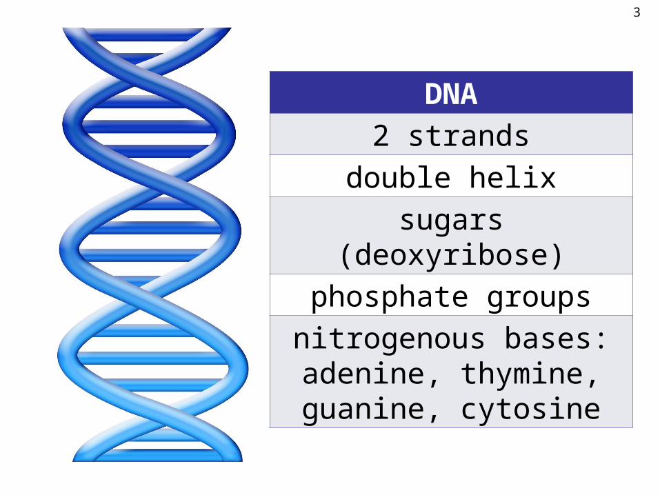
DNA
2 strands
double helix
sugars (deoxyribose)
phosphate groups
nitrogenous bases:adenine, thymine,guanine, cytosine
3
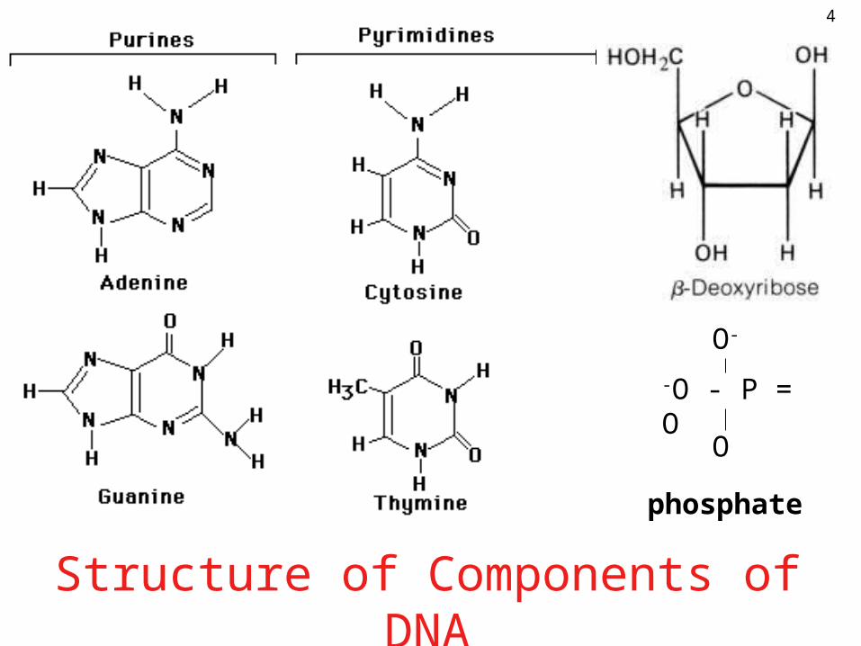
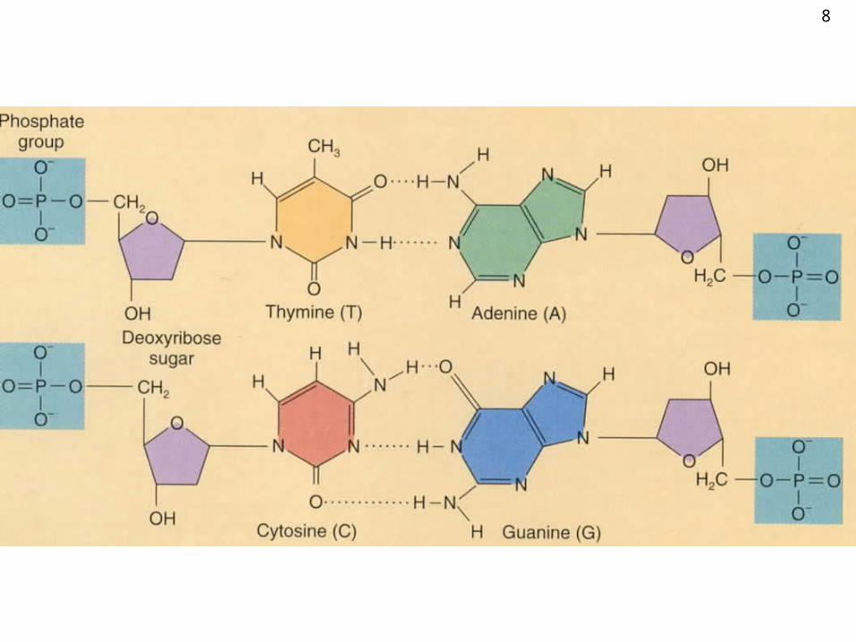
Structure of Components of DNA
-O - P = O
O-
O
phosphate
4

DNA
S
S
S
S
P
P
P
B
B
B
B
B
B
B
B
S
S
S
S
P
P
P
PP
S B B S
Key:
S = sugar
P = phosphate
B = base
5

sugar-phosphate backbone
nitrogenous bases form “rungs of ladder”
6
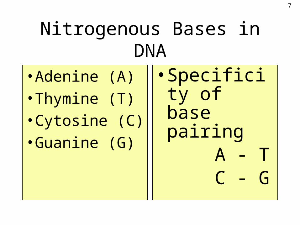
Nitrogenous Bases in DNA
• Adenine (A)
• Thymine (T)
• Cytosine (C)
• Guanine (G)
• Specificity of base pairing A - T C - G
7
8

DNA
9
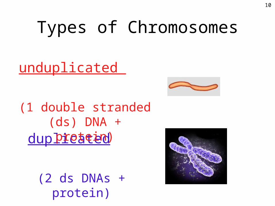
Types of Chromosomes
duplicated
(2 ds DNAs + protein)
unduplicated
(1 double stranded (ds) DNA + protein)
10
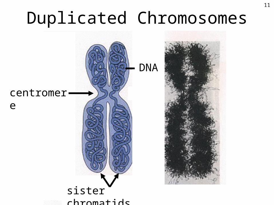
Duplicated Chromosomes
DNA
sister chromatids
centromere
11
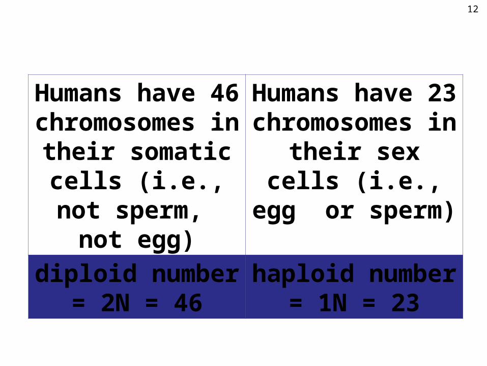
Humans have 46 chromosomes in
their somatic cells (i.e., not sperm,
not egg)
Humans have 23 chromosomes in
their sex cells (i.e., egg or sperm)
diploid number = 2N = 46
haploid number = 1N = 23
12
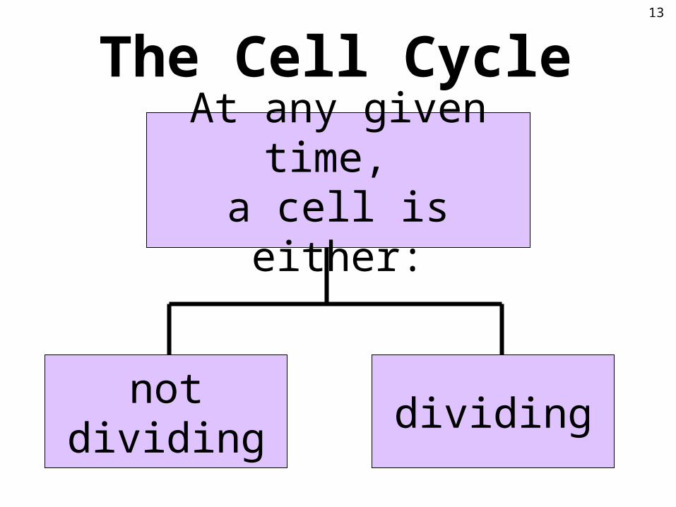
not dividing dividing
At any given time, a cell is either:
The Cell Cycle13
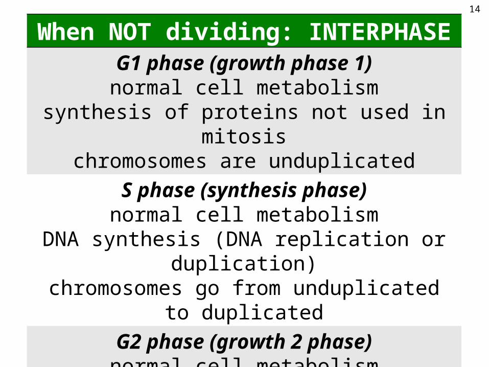
When NOT dividing: INTERPHASEG1 phase (growth phase 1)
normal cell metabolismsynthesis of proteins not used in mitosis
chromosomes are unduplicated
S phase (synthesis phase)normal cell metabolism
DNA synthesis (DNA replication or duplication)chromosomes go from unduplicated to duplicated
G2 phase (growth 2 phase)normal cell metabolism
synthesis of proteins needed for mitosischromosomes are duplicated
14
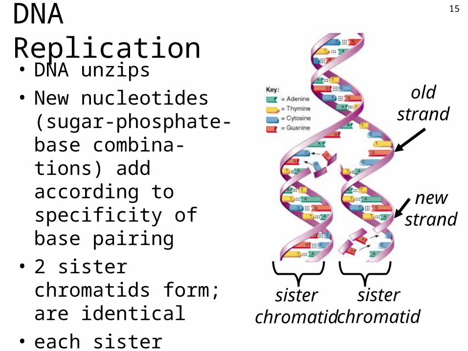
DNA Replication• DNA unzips
• New nucleotides (sugar-phosphate-base combina-tions) add according to specificity of base pairing
• 2 sister chromatids form; are identical
• each sister chromatid has 1 new & 1 old DNA strand
• semiconservative replication
sister chromatid
old strand
new strand
sister chromatid
15

When cells ARE dividing:MITOSIS
division of the nucleus of somatic cells; 1 divisionmaintains the chromosome number
1 mother cell with 46 chromosomes givesrise to 2 daughter cells, each with 46 chromosomes
MEIOSISdivision of the nucleus of sex cells; 2 divisions
halves the chromosome number1 mother cell with 46 chromosomes gives
rise to 4 daughter cells, each with 23 chromosomes
CYTOKINESISdivision of cytoplasm
occurs during mitosis & meiosis
16

CELL CYCLEINTERPHASE
G1: unduplicated chromosomes
S: chromosomes duplicateG2: duplicated chromosomes
MITOSIS (M phase)start with duplicated
chromosomes; end with unduplicated chromosomes
4 phases:Prophase
MetaphaseAnaphaseTelophase
(cytokinesis occurs mainly during telophase)
17
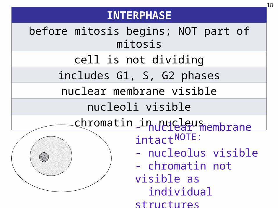
INTERPHASE
before mitosis begins; NOT part of mitosis
cell is not dividing
includes G1, S, G2 phases
nuclear membrane visible
nucleoli visible
chromatin in nucleus
- nuclear membrane intact- nucleolus visible- chromatin not visible as individual structures
NOTE:
18
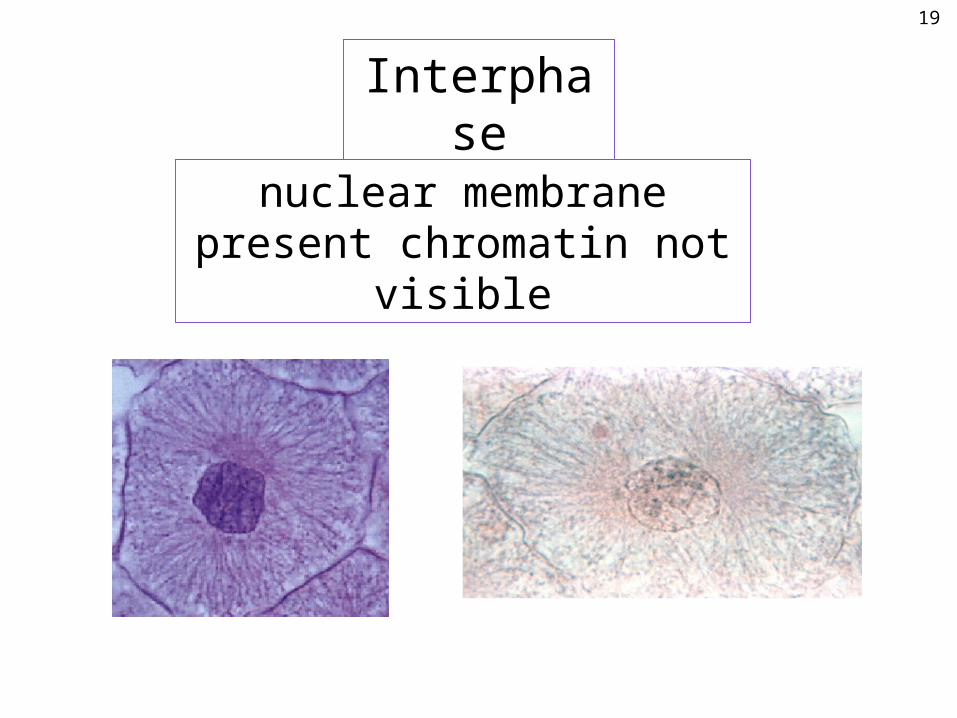
Interphase
nuclear membrane present chromatin not visible
19
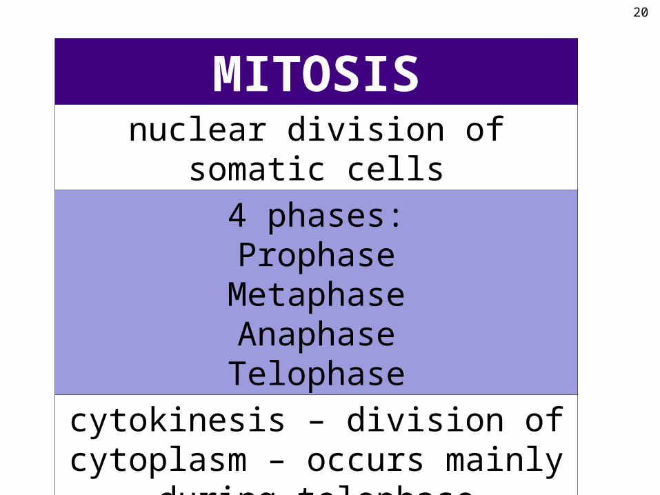
MITOSISnuclear division of somatic cells
4 phases:Prophase
MetaphaseAnaphaseTelophase
cytokinesis – division of cytoplasm – occurs mainly during telophase
20
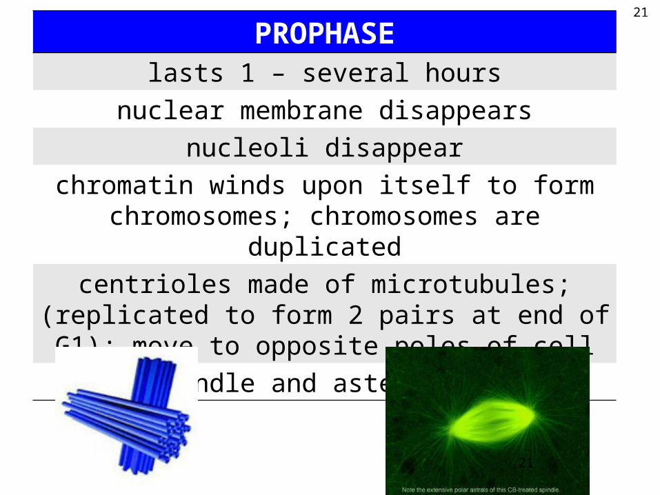
PROPHASElasts 1 – several hours
nuclear membrane disappears
nucleoli disappear
chromatin winds upon itself to form chromosomes; chromosomes are duplicated
centrioles made of microtubules; (replicated to form 2 pairs at end of G1); move to opposite poles of cell
spindle and asters form
21
21
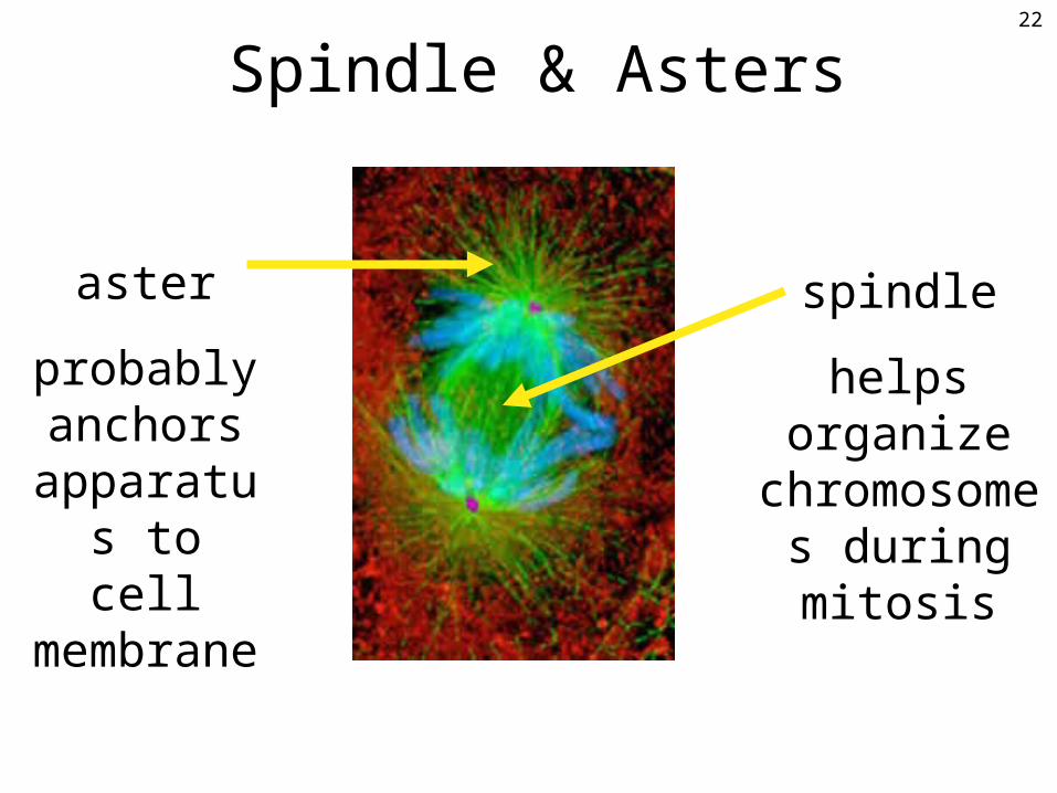
Spindle & Asters
aster
probably anchors
apparatus to cell
membrane
spindle
helps organize chromosomes during mitosis
22

Prophase
In this hypothetical cell, 2 N = 4. In humans, 2 N = 46.
nuclear membrane disappears nucleolus disappearschromatin chromosomesspindle & asters form
NOTE:
23

Prophase
24

METAPHASE5 – 15 minutes
chromosomes line up in single file along center of spindle
chromosomes are attached to spindle fibers in area of centromere
In this hypothetical cell, 2 N = 4. In humans, 2 N = 46.
chromosomes lined up in single file in center of spindle
NOTE:
25

Metaphase26
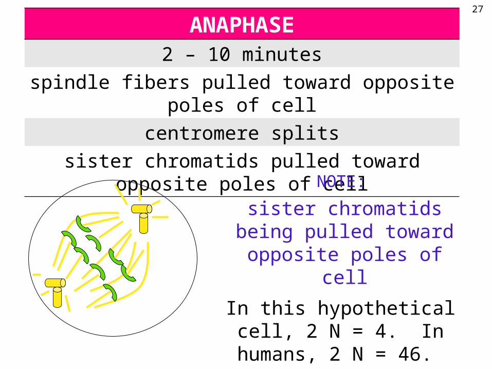
ANAPHASE2 – 10 minutes
spindle fibers pulled toward opposite poles of cell
centromere splits
sister chromatids pulled toward opposite poles of cell
In this hypothetical cell, 2 N = 4. In humans, 2 N = 46.
NOTE:
sister chromatids being pulled toward opposite poles
of cell
27

Anaphase28

TELOPHASE
10 – 30 minutes
cleavage furrow forms & deepens until cell divides into 2 daughter cells (cytokinesis)
opposite of prophase occurs:nuclear membrane & nucleoli reappear
chromosomes uncoil into chromatinspindle & asters disappear
In this hypothetical cell, 2 N = 4. In humans, 2 N = 46.
NOTE:
cleavage furrow forms;2 daughter cells will form; the
opposite of prophase will occur
29

Telophase30
31
32

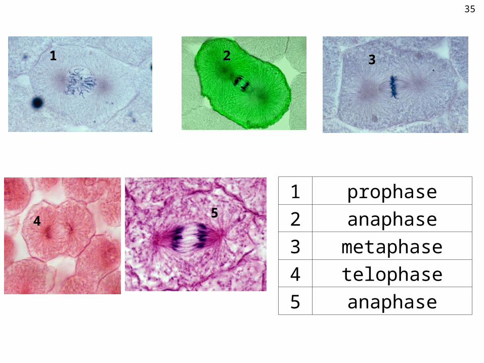
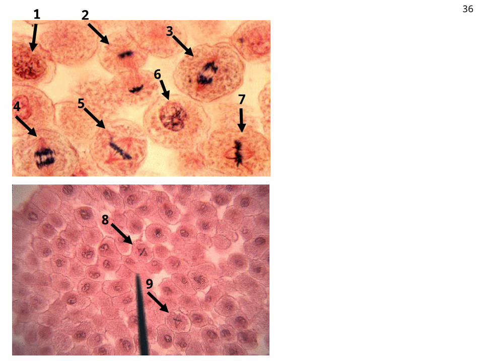
TEST YOUR KNOWLEDGE:Name the phase of mitosis seen on
the following slides.
33
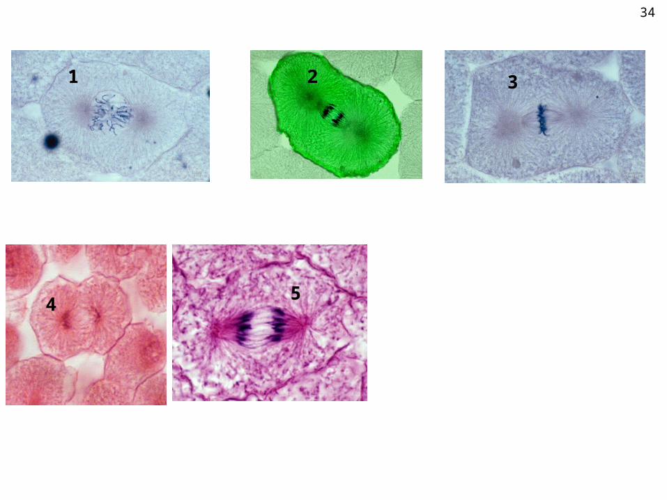
1 2 3
45
34
1 2 3
4
1 prophase
2 anaphase
3 metaphase
4 telophase
5 anaphase
5
35
1 23
4 5
6
7
8
9
36

1 23
4 5
6
7
8
9
1 prophase
2 telophase
3 anaphase
4 anaphase
5 metaphase
6 prophase
7 metaphase
8 metaphase
9 metaphase
37

1
2
3
4
5
6
78
9
1011
12
13 14
38
1
2
3
4
5
6
78
9
1011
12
13 14
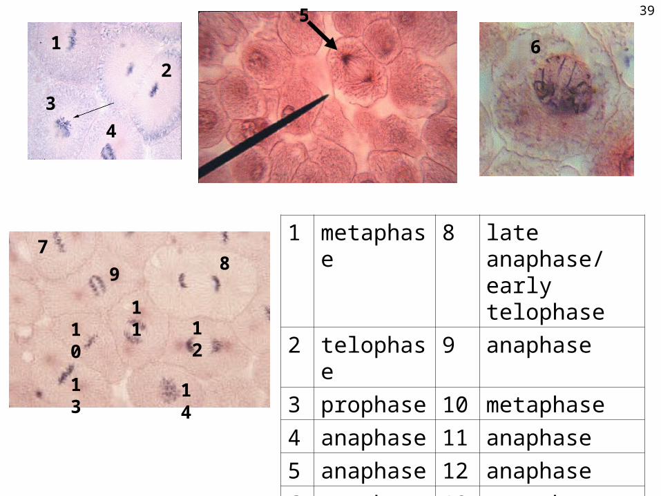
1 metaphase 8 late anaphase/ early telophase
2 telophase 9 anaphase
3 prophase 10 metaphase
4 anaphase 11 anaphase
5 anaphase 12 anaphase
6 prophase 13 metaphase
7 metaphase 14 prophase
39
Now it’s YOUR turn to identify stages of mitosis with a
microscope!
40