Embed Size (px)
Citation preview

Mitosis/Meiosis Diagrams
Ch. 12 and 13

Figure 12.7
G2 of Interphase Prophase Prometaphase
Centrosomes(with centriole pairs)
Chromatin(duplicated)
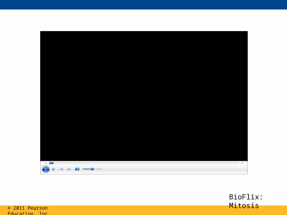
Nucleolus Nuclearenvelope
Plasmamembrane
Early mitoticspindle
AsterCentromere
Chromosome, consistingof two sister chromatids
Fragments of nuclearenvelope
Nonkinetochoremicrotubules
Kinetochore Kinetochoremicrotubule
Metaphase
Metaphase plate
Anaphase Telophase and Cytokinesis
Spindle Centrosome atone spindle pole
Daughterchromosomes
Cleavagefurrow
Nucleolusforming
Nuclearenvelopeforming
10
m
© 2011 Pearson Education, Inc.
BioFlix: Mitosis

Figure 12.6
INTERPHASE
G1
G2
S(DNA synthesis)
MITOTIC(M) PHASE
CytokinesisM
itosi
s

Figure 12.9b
Chromosomemovement
Microtubule
Motor protein
Chromosome
Kinetochore
Tubulinsubunits
CONCLUSION
© 2011 Pearson Education, Inc.
Animation: Cytokinesis Right-click slide / select ”Play”
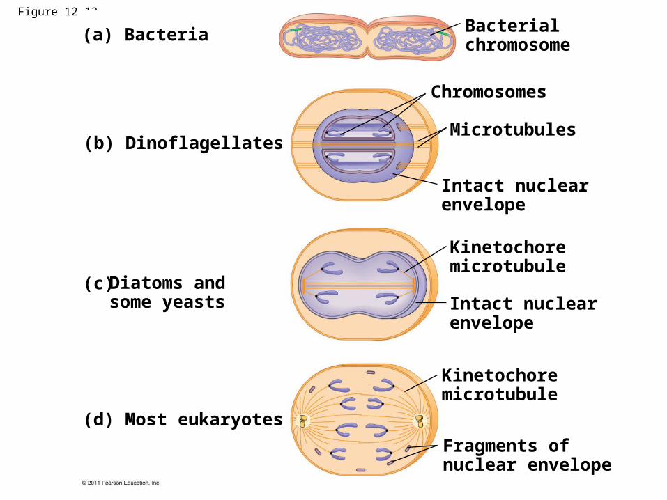
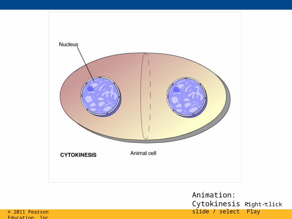
Figure 12.13
(a) Bacteria
(b) Dinoflagellates
(d) Most eukaryotes
Intact nuclearenvelope
Chromosomes
Microtubules
Intact nuclearenvelope
Kinetochoremicrotubule
Kinetochoremicrotubule
Fragments ofnuclear envelope
Bacterialchromosome
(c) Diatoms andsome yeasts
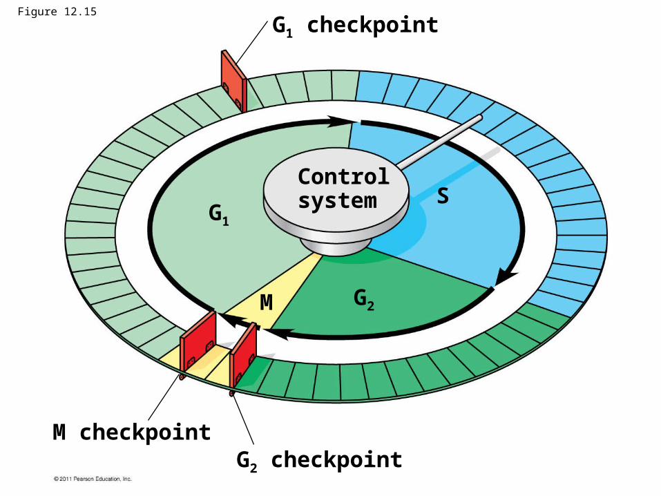
G1 checkpoint
G1
G2
G2 checkpointM checkpoint
M
SControlsystem
Figure 12.15

Figure 12.17
(a) Fluctuation of MPF activity and cyclin concentration during the cell cycle
(b) Molecular mechanisms that help regulate the cell cycle
MPF activityCyclinconcentration
Time
M M MS SG1G2 G1
G2 G1
Cdk
Degradedcyclin
Cyclin isdegraded
MPF
G2checkpoint
Cdk
Cyclin
M
S
G1
G 2

Figure 12.18
A sample of humanconnective tissue iscut up into smallpieces.
Enzymes digestthe extracellularmatrix, resulting ina suspension offree fibroblasts.
Cells are transferred toculture vessels.
Scalpels
Petridish
PDGF is addedto half thevessels.
Without PDGF With PDGF
10 m
1
2
3
4

Figure 12.19
Anchorage dependence
Density-dependent inhibition
Density-dependent inhibition
(a) Normal mammalian cells (b) Cancer cells
20 m 20 m
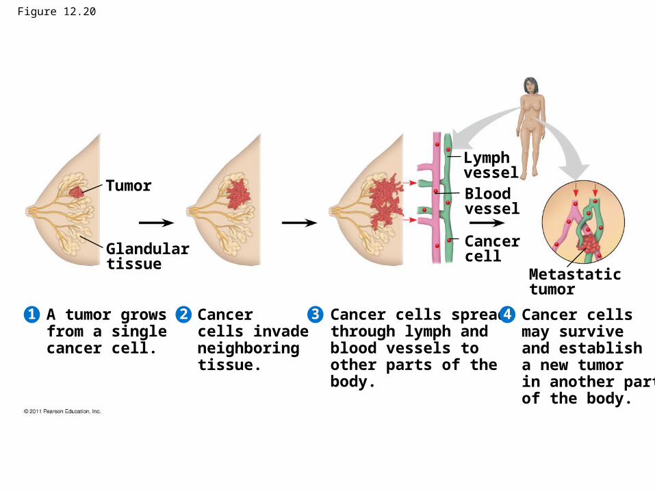
Figure 12.20
Glandulartissue
Tumor
Lymph vesselBloodvessel
Cancercell
Metastatictumor
A tumor growsfrom a singlecancer cell.
Cancer cells invade neighboringtissue.
Cancer cells spreadthrough lymph andblood vessels to other parts of the body.
Cancer cells may survive and establisha new tumor in another part of the body.
4321
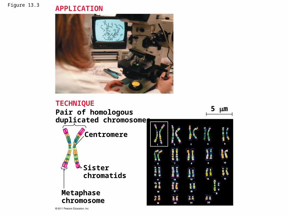
Figure 13.3
Pair of homologousduplicated chromosomes
Centromere
Sisterchromatids
Metaphasechromosome
5 m
APPLICATION
TECHNIQUE

Figure 13.3b
Pair of homologousduplicated chromosomes
Centromere
Sisterchromatids
Metaphasechromosome
5 m

Figure 13.4
Sister chromatidsof one duplicatedchromosome
Key
Maternal set ofchromosomes (n 3)Paternal set ofchromosomes (n 3)
Key
2n 6
Centromere
Two nonsisterchromatids ina homologous pair
Pair of homologouschromosomes (one from each set)

Figure 13.5
Key
Haploid (n)Diploid (2n)
Egg (n)
Haploid gametes (n 23)
Sperm (n)
Ovary Testis
Mitosis anddevelopment
Diploidzygote(2n 46)
Multicellular diploidadults (2n 46)
MEIOSIS FERTILIZATION
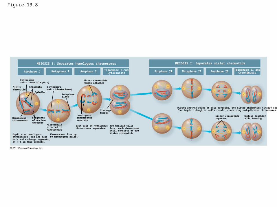
Figure 13.8
MEIOSIS I: Separates homologous chromosomes
Prophase I Metaphase I Anaphase I Telophase I andCytokinesis
Centrosome(with centriole pair)
Sisterchromatids
Chiasmata
Spindle
Homologouschromosomes
Fragmentsof nuclearenvelope
Duplicated homologouschromosomes (red and blue)pair and exchange segments;2n 6 in this example.
Centromere(with kinetochore)
Metaphaseplate
Microtubuleattached tokinetochore
Chromosomes line upby homologous pairs.
Sister chromatidsremain attached
Homologouschromosomesseparate
Each pair of homologous chromosomes separates.
Cleavagefurrow
Two haploid cellsform; each chromosomestill consists of twosister chromatids.
MEIOSIS I: Separates sister chromatids
Prophase II Metaphase II Anaphase IITelophase II and
Cytokinesis
Sister chromatidsseparate
Haploid daughtercells forming
During another round of cell division, the sister chromatids finally separate;four haploid daughter cells result, containing unduplicated chromosomes.
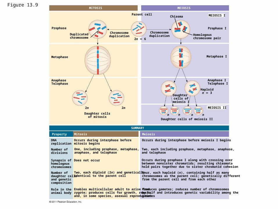
Figure 13.9
Prophase
Duplicatedchromosome
MITOSIS
Chromosomeduplication
Parent cell
2n 6
Metaphase
AnaphaseTelophase
2n 2n
Daughter cellsof mitosis
MEIOSIS
MEIOSIS I
MEIOSIS II
Prophase I
Metaphase I
Anaphase ITelophase I
Haploidn 3
Chiasma
Chromosomeduplication Homologous
chromosome pair
Daughter cells of
meiosis I
Daughter cells of meiosis II
n n n n
SUMMARY
Property Mitosis Meiosis
DNAreplication
Number ofdivisions
Synapsis ofhomologouschromosomes
Number of daughter cellsand geneticcomposition
Role in the animal body
Occurs during interphase beforemitosis begins
One, including prophase, metaphase,anaphase, and telophase
Does not occur
Two, each diploid (2n) and geneticallyidentical to the parent cell
Enables multicellular adult to arise fromzygote; produces cells for growth, repair,and, in some species, asexual reproduction
Occurs during interphase before meiosis I begins
Two, each including prophase, metaphase, anaphase,and telophase
Occurs during prophase I along with crossing overbetween nonsister chromatids; resulting chiasmatahold pairs together due to sister chromatid cohesion
Four, each haploid (n), containing half as manychromosomes as the parent cell; genetically differentfrom the parent cell and from each other
Produces gametes; reduces number of chromosomesby half and introduces genetic variability among the gametes
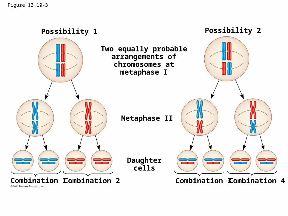
Figure 13.10-3
Possibility 1 Possibility 2
Two equally probablearrangements ofchromosomes at
metaphase I
Metaphase II
Daughtercells
Combination 1 Combination 2 Combination 3 Combination 4
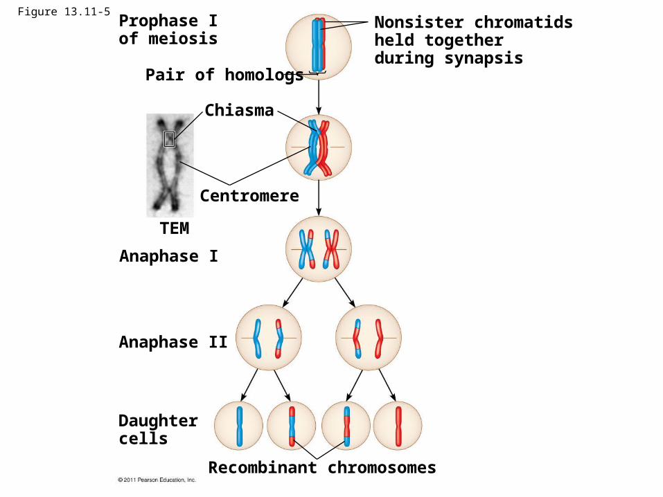
Figure 13.11-5Prophase Iof meiosis
Nonsister chromatidsheld togetherduring synapsis
Pair of homologs
Chiasma
Centromere
TEM
Anaphase I
Anaphase II
Daughtercells
Recombinant chromosomes

© 2011 Pearson Education, Inc.
Animation: Genetic Variation Right-click slide / select “Play”