Embed Size (px)
Citation preview
Mitotic chromosome condensationmediated by the retinoblastomaprotein is tumor-suppressive
Courtney H. Coschi,1,2 Alison L. Martens,1,2 Kieran Ritchie,2,3 Sarah M. Francis,1,2
Subrata Chakrabarti,4 Nathalie G. Berube,3,4,5 and Frederick A. Dick1,3,4,5,6
1London Regional Cancer Program, University of Western Ontario, London, Ontario N6A 4L6, Canada; 2Department ofBiochemistry, University of Western Ontario, London, Ontario N6A 4L6, Canada; 3Children’s Health Research Institute,University of Western Ontario N6A 4L6, London, Ontario N6A 3K7, Canada; 4Department of Pathology, University of WesternOntario, London, Ontario N6A 4L6, Canada; 5Department of Pediatrics, University of Western Ontario, London, Ontario N6A4L6, Canada
Condensation and segregation of mitotic chromosomes is a critical process for cellular propagation, and, inmammals, mitotic errors can contribute to the pathogenesis of cancer. In this report, we demonstrate that theretinoblastoma protein (pRB), a well-known regulator of progression through the G1 phase of the cell cycle, playsa critical role in mitotic chromosome condensation that is independent of G1-to-S-phase regulation. Using genetargeted mutant mice, we studied this aspect of pRB function in isolation, and demonstrate that it is an essentialpart of pRB-mediated tumor suppression. Cancer-prone Trp53�/� mice succumb to more aggressive forms ofcancer when pRB’s ability to condense chromosomes is compromised. Furthermore, we demonstrate that defectivemitotic chromosome structure caused by mutant pRB accelerates loss of heterozygosity, leading to earlier tumorformation in Trp53+/� mice. These data reveal a new mechanism of tumor suppression, facilitated by pRB,in which genome stability is maintained by proper condensation of mitotic chromosomes.
[Keywords: Cohesion; condensation; chromosome instability; cell cycle; chromatin]
Supplemental material is available at http://www.genesdev.org.
Received February 17, 2010; revised version accepted May 6, 2010.
The compaction of the mammalian genome into mitoticchromosomes, and their faithful segregation to recipientdaughter cells in mitosis, is a critical event for eukaryoticcells. This stage of the cell division cycle carries consider-able risk, as there is no opportunity to reverse the effectsof missegregated or damaged chromosomes in daughtercells. For these reasons, understanding mitosis is of vitalimportance, because changes in genome integrity can leadto cancer.
The processes of chromosome condensation and segre-gation are intimately linked, as insufficient condensationof chromosome arms can prevent their proper separationin anaphase (Belmont 2006). Of chief importance in fa-cilitating proper chromosome structure is the mitoticchromosome scaffold, upon which mitotic chromosomesare condensed. These scaffolds are composed largely oftopoisomerase 2 and condensins (Maeshima and Laemmli2003). While the enzymatic activity of topoisomerases
suggests a mechanism by which they participate in chro-mosome compaction, the precise role of condensins hasbeen less clear. There are two types of condensin com-plexes (I and II) that are very similar structurally. Each iscomprised of an SMC2 and SMC4 subunit that interact toform the coiled-coil arms of their ring-like structures(Losada and Hirano 2005). In addition, each condensincomplex also contains distinct subunits: CAP-H, CAP-D2,and CAP-G in condensin I, and CAP-H2, CAP-D3, andCAP-G2 in condensin II (Losada and Hirano 2005). Surpris-ingly, depletion of individual components of these com-plexes by RNAi does not prevent outright chromosomecondensation (Ono et al. 2003; Hirota et al. 2004). Instead,each complex appears to offer a unique contribution to thearchitecture of mitotic chromosomes. This may be due inpart to differences in the timing of their loading ontochromosomes, with Condensin II being present on chro-matin in interphase, and Condensin I being loaded onlyafter nuclear envelope breakdown (Ono et al. 2003; Hirotaet al. 2004). Additionally, differences in subunit composi-tion imply that they may have different functional orregulatory properties (Losada and Hirano 2005). Impor-tantly, defects and delays in chromatid condensation
6Corresponding author.E-MAIL [email protected]; FAX (519) 685-8616.Article published online ahead of print. Article and publication date areonline at http://www.genesdev.org/cgi/doi/10.1101/gad.1917610.
GENES & DEVELOPMENT 24:1351–1363 � 2010 by Cold Spring Harbor Laboratory Press ISSN 0890-9369/10; www.genesdev.org 1351
Cold Spring Harbor Laboratory Press on June 10, 2018 - Published by genesdev.cshlp.orgDownloaded from

are manifested as lagging chromosomes during ana-phase that impede mitotic progression, resulting in an-euploidy (Hirota et al. 2004; Ono et al. 2004; Samoshkinet al. 2009). However, there is limited evidence connectingcondensin proteins and chromosome condensation tocancer (Ham et al. 2007; Lapointe et al. 2008).
In addition to condensation defects leading to segrega-tion errors, faithful chromosome segregation can also beaffected by mitotic spindle abnormalities. This includesthe mitotic spindle checkpoint, which serves to detectunattached kinetochores at metaphase (Nasmyth 2005;Musacchio and Salmon 2007). Signals that emanate froma complex containing MAD2 and BUBR1 at unattachedkinetochores prevent the E3 ubiquitin ligase complexAPC/C from triggering the degradation of cyclin B1 andsecurin. Once this checkpoint is satisfied, mitotic cyclin-dependent kinase activity drops, and a securin-associatedprotease called separase is free to cleave cohesins, allowingsister chromatid separation (Nasmyth 2005; Musacchioand Salmon 2007). Experimental models in which spindleassembly checkpoint components are misexpressed showerrors in chromosome segregation (Pei and Melmed 1997;Hernando et al. 2004; Vader and Lens 2008). Thus, defectsin either chromosome condensation or spindle assemblycheckpoints can lead to segregation errors and aneuploidy,and potentially contribute to cancer pathogenesis. A num-ber of examples of spindle assembly checkpoint defectscontributing to cancer incidence in genetically modifiedmice have been reported (for review, see Schvartzman et al.2010). Conversely, even though defects in chromosomecondensation cause similar mitotic errors, there are no re-ports using gene targeted mouse models to confirm a rolefor condensins as tumor suppressors.
The retinoblastoma tumor suppressor protein (pRB) isbest known for its role in regulating the G1-to-S-phasetransition early in the cell cycle (for review, see Burkhartand Sage 2008). Its ability to negatively regulate tran-scription of DNA replication machinery through E2Ftranscription factors creates a mechanism by which itcan inhibit cell cycle entry. More recently, pRB has beenshown to influence both chromosome condensation andmitotic checkpoint function (Longworth and Dyson 2010;Schvartzman et al. 2010). Components of the spindleassembly checkpoint, such as MAD2, are regulated byE2F transcription factors (Ren et al. 2002; Hernando et al.2004). For this reason, loss of E2F regulation by pRB,which is almost ubiquitous in cancer, leads to genomeinstability (Mayhew et al. 2007; Schvartzman et al. 2010).Conversely, the ability of pRB to influence mitotic chro-mosome condensation has emerged as an E2F-independentfunction, and loss of pRB function can influence chromo-some loss irrespective of proliferation (Zheng et al. 2002;Gonzalo et al. 2005; Isaac et al. 2006; Longworth et al.2008). To date, evidence for pRB’s involvement in chro-mosome condensation has been largely genetic. Mousefibroblasts deficient for all pRB family proteins or a knock-in mutation that partially inactivates pRB have abnormalcentromeric heterochromatin, leading to chromosomefusions and aneuploidy (Gonzalo et al. 2005; Isaac et al.2006). Further analyses that combine the use of Drosophila
genetics and mammalian cell culture suggest that pRB caninteract with the condensin II subunit CAP-D3, and thatthis interaction is necessary for chromosome compactionin mitosis (Longworth et al. 2008). While studies of spindlecheckpoint components such as MAD2 have offeredexplanations for why their deregulation in cancer can betraced back to pRB function (Schvartzman et al. 2010), theimportance of pRB’s role in chromosome condensation isless well understood, and has not yet been connected toa cancer phenotype.
In this study, we investigate the mechanism used bypRB to facilitate mitotic chromosome condensation. Werely on a viable, gene targeted mouse strain in which pRBis mutated to block LXCXE-dependent interactions, suchas those with viral oncoproteins and chromatin remodel-ing enzymes like histone deacetylases (Isaac et al. 2006).Cells from these mice have limited proliferative controldefects, except for the responsiveness to transforminggrowth factor b (TGF-b) and senescence-inducing stimuli(Francis et al. 2009; Talluri et al. 2010). We demonstratethat pRB interacts with the Condensin II complex toestablish proper chromosome structure. Our experimentsreveal that condensation defects caused by a deficiency inpRB–LXCXE interactions occur before metaphase, andare unrelated to the ability to regulate G1-to-S-phaseprogression. We used Rb1DL/DL; Trp53�/� mice as well asTrp53�/� controls—both of which are uniformly defectivein their response to G1 arrest stimuli such as DNAdamage- and oncogene-induced senescence—to studypRB’s role in chromosome condensation in isolation.Rb1DL/DL; Trp53�/� mice succumb to much more aggres-sive forms of cancer than p53-deficient controls, and theirtumors are characterized by elevated levels of chromo-some instability. Furthermore, we demonstrate that de-fective chromosome condensation caused by mutant pRBcan accelerate loss of heterozygosity and cancer onset inTrp53+/� mice. This study reveals that participation inmitotic chromosome condensation is an integral aspectof pRB’s function as a tumor suppressor protein.
Results
Aberrant chromosome condensation and segregationin Rb1DL/DL mutant cells
A number of recent reports have indicated that cells de-ficient for pRB family proteins have chromosomal abnor-malities (Gonzalo et al. 2005; Longworth et al. 2008), andwe demonstrated centromere fusions and lagging anaphasechromosomes in cells from Rb1DL/DL mice (Isaac et al.2006). One interpretation of altered chromosome numbersin these cells is that they are a consequence of deregulatedE2F transcriptional control in G1 leading to inappropriateproliferation. In this way, alterations in transcriptionalcontrol or commitment to enter S phase early in the cellcycle are manifested in subsequent mitotic errors. To in-vestigate this possibility with Rb1DL/DL mutant cells, wecompared chromosomal abnormalities found in homozy-gous mouse embryonic fibroblasts (MEFs) with embryonicstem cells (ESCs). Since MEFs have a pronounced G1 phaseregulated by pRB (Herrera et al. 1996; Harrington et al.
Coschi et al.
1352 GENES & DEVELOPMENT
Cold Spring Harbor Laboratory Press on June 10, 2018 - Published by genesdev.cshlp.orgDownloaded from

1998), and ESCs lack the ability to arrest in G1 (Aladjemet al. 1998), we reasoned that chromosomal abnormalitiesfound in both are unlikely to be a consequence of un-regulated cell cycle advancement. Figure 1A shows repre-
sentative chromosome spreads from each cell type for bothwild-type and Rb1DL/DL mutants. Chromosomes werestained with DAPI and a major satellite DNA probe tovisualize contacts between centromeres from different
Figure 1. Rb1DL/DL cells display aberrant chromosome condensation and segregation. (A) Metaphase chromosome spreads wereprepared from MEFs or ESCs of the indicated genotypes. Chromosomes were stained with DAPI (blue) and a probe for major satelliterepeats (green) to mark centromeres. Red arrows indicate contact between centromeres from different chromosomes, and yellow arrowsindicate centromere contact involving three or more chromosomes. Inlays highlight expanded views of select chromosomes. Bars,25 mm. (B) The frequency of centromere interactions per mitosis is plotted for each genotype (Rb1DL/DL and Rb1+/+) and cell type (MEFand ESC). (Right graph) In addition, the number of centromeres involved in each interaction was determined for ESC metaphasespreads, and is displayed as the frequency of multiple chromosome interactions. (C) Video microscopy was performed on MEFs ex-pressing an H2B-GFP reporter by capturing phase-contrast and GFP images every 3 min over a 15-h time course. The images shownbegin with the onset of chromatin condensation in prophase as the left-most panel. The last image of the metaphase plate before theonset of anaphase is indicated along with the elapsed time since the onset of prophase (in minutes). The right-most image showsresolved daughter (or binucleated) cells. The numbers in the left-most image correspond to references in the Supplemental Material andSupplemental Movies. Bars, 50 mm.
Chromosome condensation by pRB
GENES & DEVELOPMENT 1353
Cold Spring Harbor Laboratory Press on June 10, 2018 - Published by genesdev.cshlp.orgDownloaded from

chromosomes. In both cell types, a statistically significantincrease in centromere interactions (Fig. 1A, inlays) wasdetected in the mutant Rb1DL/DL genotype, suggesting anincrease in chromosome fusions (x2 test, P < 0.05 foreach comparison in Fig. 1B). Interestingly, there was alsoan increase in the number of centromeres interacting inRb1DL/DL ESCs compared with wild type (x2 test, P < 0.05)(Fig. 1A [inlay], B [right]). In addition to its contribution tocentromere structure, pRB is also known to silence tran-scription at nearby rDNA repeats (Cavanaugh et al. 1995;Hannan et al. 2000; Ciarmatori et al. 2001). Therefore, wealso investigated their involvement in these fusions, asthey are found on the p-arms of mouse chromosomes 12,15, 16, 17, 18, and 19, and are therefore in close proximityto the centromere (Supplemental Fig. 1). These chromo-somes are not overrepresented in Rb1DL/DL fusion events,further suggesting that loss of transcriptional regulationearly in the cell cycle does not contribute to this phenotype(Supplemental Fig. 1). These data support a specific role forpRB in regulating chromatin structure at the centromericrepeat sequences of mitotic chromosomes that is indepen-dent of pRB’s ability to regulate the G1-to-S-phase transi-tion in these cells.
In order to better understand the origin of defectivechromosomal compaction in Rb1DL/DL mutant cells and itseffect on mitosis, we established a video microscopy assayto observe cell division in MEFs. Early passage MEFs weretransduced with an H2B-GFP-expressing retrovirus. Thislead to equivalent expression of the H2B-GFP fusion inboth genotypes. Importantly, expression of this fusion pro-tein was very low and did not lead to a detectable increasein total H2B levels in these cells (Supplemental Fig. 2A).H2B-GFP-expressing cells were monitored microscopicallyunder phase-contrast and fluorescent optics to visualizechromosome condensation, metaphase alignment, ana-phase, and cytokinesis. Images from three representativemovies are shown in Figure 1C. Typically, mutant cellstook longer to progress from the onset of chromosomecondensation to the point at which the metaphase plate ismost tightly aligned (Table 1). Furthermore, the metaphaseplate in mutant mitoses was less compact than wild type(Table 1). To determine whether these phenotypes areassociated with defects later in mitosis, we observed sisterchromatid separation in anaphase, and found that lagging
chromosomes occurred more frequently in mutant cells(Table 1). Lagging chromosomes resulted in a prolongedanaphase that was often resolved abruptly, suggestingeither chromosomal breaks or missegregation of wholechromosomes (Fig. 1C, row 2). Alternatively, some cellsfailed to complete mitosis and became binucleated(Fig. 1C, row 3). In summary, mitosis in Rb1DL/DL cellsis characterized by delayed chromosome condensation,an abnormal metaphase plate, and lagging chromosomesthat lead to aneuploidy.
The preceding experiments are consistent with pre-vious reports of defects in condensation and/or cohesionof mitotic chromosomes (Ono et al. 2004). For thesereasons, we investigated the levels of the condensin andcohesin protein complexes that were present on chroma-tin in Rb1DL/DL cells. Protein extracts were prepared fromcell cultures that were synchronized in S phase, released,and harvested at their maximal mitotic index (M-phase-enriched), or that were asynchronous. SDS-PAGE andWestern blotting were used to analyze the chromatinfractions from these lysates using histone proteins asa marker for this fraction (Fig. 2A; Suppplemental Fig. 2B).Since Cohesins and Condensins are multiprotein com-plexes, we chose representative subunits to measure theirpresence on chromatin. The SMC1 subunit was used asa surrogate for the levels of the Cohesin complex; CAP-Hand CAP-D2 were used similarly for Condensin I, andCAP-H2 and CAP-D3 were used to detect Condensin II.This analysis revealed reduced levels of Condensin II onchromatin derived from Rb1DL/DL cells, while the overalllevels of Condensin I and Cohesin were unchanged. Toensure that the reduction in Condensin II loading onchromatin is not due to overall reduction of the protein,we probed for levels in whole-cell extracts and deter-mined that Condensin II protein is expressed at wild-typelevels in Rb1DL/DL fibroblasts (Supplemental Fig. 2C).
Depletion of the Condensin II subunit CAP-D3 byRNAi delays progression from the onset of condensationto anaphase and results in lagging chromosomes in pRB-deficient HeLa cells (Hirota et al. 2004). Furthermore,a recent study indicates that GST-RB can bind to Con-densin II complexes using its LXCXE-binding cleft region(Longworth et al. 2008). For this reason, we investigatedthe role of CAP-D3 in more detail using video microscopy
Table 1. Summary of mitotic phenotypes observed in video microscopy experiments
Genotypea N-valueLagging
chromosomesb N-valueAverage time from onset of
condensation to onset of anaphaseAverage metaphase
plate widthc
Rb1+/+ 57 14 43 27.95 min 4.99 mmRb1DL/DL 37 19d 27 33.89 mine 6.22 mme
Rb1+/+ shLuc 38 11 37 33.92 min 4.77 mmRb1+/+ sh63 41 34d 43 107.07 mine 5.04 mmRb1+/+ sh64 10 8d 11 186.5 mine 7.96 mme
Rb1+/+ sh66 27 18d 27 121.4 mine 7.03 mme
aFor all statistical tests, Rb1DL/DL is compared with wild type, and all shRNAs directed against CAP-D3 were compared with shLuc.bIncludes mitoses where the metaphase plate never visually divided, chromatin decondensed, and cells became tetraploid.cFive equally spaced cross-sections for each metaphase plate were measured from the last image before the initiation of anaphase.dA difference from controls that is above 95% confidence interval (x2 test, P < 0.05).eAbove 95% confidence interval (t-test, P < 0.05).
Coschi et al.
1354 GENES & DEVELOPMENT
Cold Spring Harbor Laboratory Press on June 10, 2018 - Published by genesdev.cshlp.orgDownloaded from
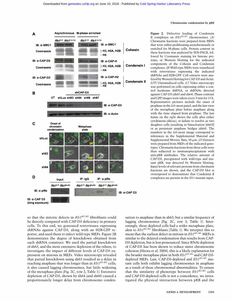
so that the mitotic defects in Rb1DL/DL fibroblasts couldbe directly compared with CAP-D3 deficiency in primarycells. To this end, we generated retroviruses expressingshRNAs against CAP-D3, along with an H2B-GFP re-porter, and used them to infect wild-type MEFs. Figure 2Bdemonstrates the degree of knockdown obtained fromeach shRNA construct. We used the partial knockdownof sh63, and the more extensive depletion of the others, toinvestigate the impact of different levels of CAP-D3 ex-pression on mitosis in MEFs. Video microscopy revealedthat partial knockdown using sh63 resulted in a delay inreaching anaphase that was longer than in Rb1DL/DL cells;it also caused lagging chromosomes, but little wideningof the metaphase plate (Fig. 2C, row 2; Table 1). Extensivedepletion of CAP-D3, shown by sh64 (and sh66) caused aproportionately longer delay from chromosome conden-
sation to anaphase than in sh63, but a similar frequency oflagging chromosomes (Fig. 2C, row 3; Table 1). Inter-estingly, these depleted cells had a wider metaphase plate,akin to Rb1DL/DL fibroblasts (Table 1). We interpret this tomean that the earliest defect in mitosis in Rb1DL/DL MEFs issimilar to the delayed condensation that results from CAP-D3 depletion, but is less pronounced. Since RNAi depletionof CAP-D3 has been shown to reduce sister chromosomecohesion (Hirota et al. 2004), this is a likely explanation forthe broader metaphase plate in both Rb1DL/DL and CAP-D3-depleted MEFs. Last, CAP-D3-depleted and Rb1DL/DL mu-tant cells both exhibit lagging chromosomes in anaphaseas a result of these chromosomal abnormalities. To ensurethat the similarity of phenotype between Rb1DL/DL cellsand CAP-D3-depleted cells is not a coincidence, we inves-tigated the physical interaction between pRB and the
Figure 2. Defective loading of CondensinII complexes on RB1DL/DL chromosomes. (A)Chromatin fractions were prepared from MEFsthat were either proliferating asynchronously orenriched for M-phase cells. Protein content inthese fractions was analyzed by SDS-PAGE, fol-lowed by Coomassie staining for histone pro-teins, or Western blotting for the indicatedcomponents of the Cohesin and Condensincomplexes. (B) Wild-type MEFs were transducedwith retroviruses expressing the indicatedshRNAs and H2B-GFP. Cell extracts were ana-lyzed by Western blotting for CAP-D3 and Actin.(UT) Untransduced cells. (C) Video microscopywas performed on cells expressing either a con-trol luciferase shRNA, or shRNAs directedagainst CAP-D3 (sh63 and sh64). Phase-contrastand GFP images were taken every 3 min for 15 h.Representative pictures include the onset ofprophase in the left-most panel, and the last viewof the metaphase plate before anaphase alongwith the time elapsed from prophase. The lastframe on the right shows the cells after eithercytokinesis (shLuc), or failure to resolve as twodaughter cells (resulting in binucleation [sh63])or as persistent anaphase bridges (sh64). Thenumbers in the left-most image correspond toreferences in the Supplemental Material andSupplemental Movies. Bars, 50 mm. (D) Extractswere prepared from MEFs of the indicated geno-types. Chromatin fractions from these cells werethen subjected to immunoprecipitation withanti-pRB antibodies. The relative amount ofCAP-D3, precipitated with wild-type and mu-tant pRB, was detected by Western blotting.Input levels of relevant proteins from chromatinfractions are shown, and the CAP-D3 blot isoverexposed to demonstrate that Condensin IIcomplexes are present in the Rb1 mutant input.
Chromosome condensation by pRB
GENES & DEVELOPMENT 1355
Cold Spring Harbor Laboratory Press on June 10, 2018 - Published by genesdev.cshlp.orgDownloaded from
Condensin II complex. Immunoprecipitation of pRB fol-lowed by Western blotting for CAP-D3 reveals that pRBinteracts with the Condensin II complex in the chromatinfraction of wild-type but not Rb1DL/DL MEFs (Fig. 2D).Western blots reveal the input levels of each of therelevant proteins, and that CAP-D3 is present in thisfraction. Furthermore, control immunoprecipitations wereperformed to detect pRB–E2F3 interactions to confirmthe specificity of this interaction defect (SupplementalFig. 2E).
Therefore, these experiments reveal a role for pRB outsideof the regulation of E2F target genes in the G1 phase of thecell cycle. We demonstrate that endogenous pRB interactswith Condensin II to compact mitotic chromosomes. Fur-thermore, a deficiency in this process causes a specific de-fect in condensation during prophase that manifests aslagging anaphase chromosomes in a primary cell culturesystem.
The Rb1DL mutation exacerbates tumorigenesisin Trp53�/� mice
Since chromosome instability is commonly thought tocontribute to tumorigenesis by facilitating the acquisitionof malignant characteristics, we sought to investigate howthe Rb1DL mutation impacts cancer pathogenesis. Since weshowed that the Rb1DL mutation compromises G1 cellcycle arrest in response to negative growth signals fromDNA damage and oncogene-induced senescence (Talluriet al. 2010), we chose to cross Rb1DL/DL mice into a Trp53�/�
background, as Trp53�/�mice are known to be defective forthe G1 arrest response from DNA breaks and senescence(Lowe et al. 1993; Serrano et al. 1997; Braig et al. 2005; Postet al. 2010). Consequently, comparing Rb1DL/DL; Trp53�/�
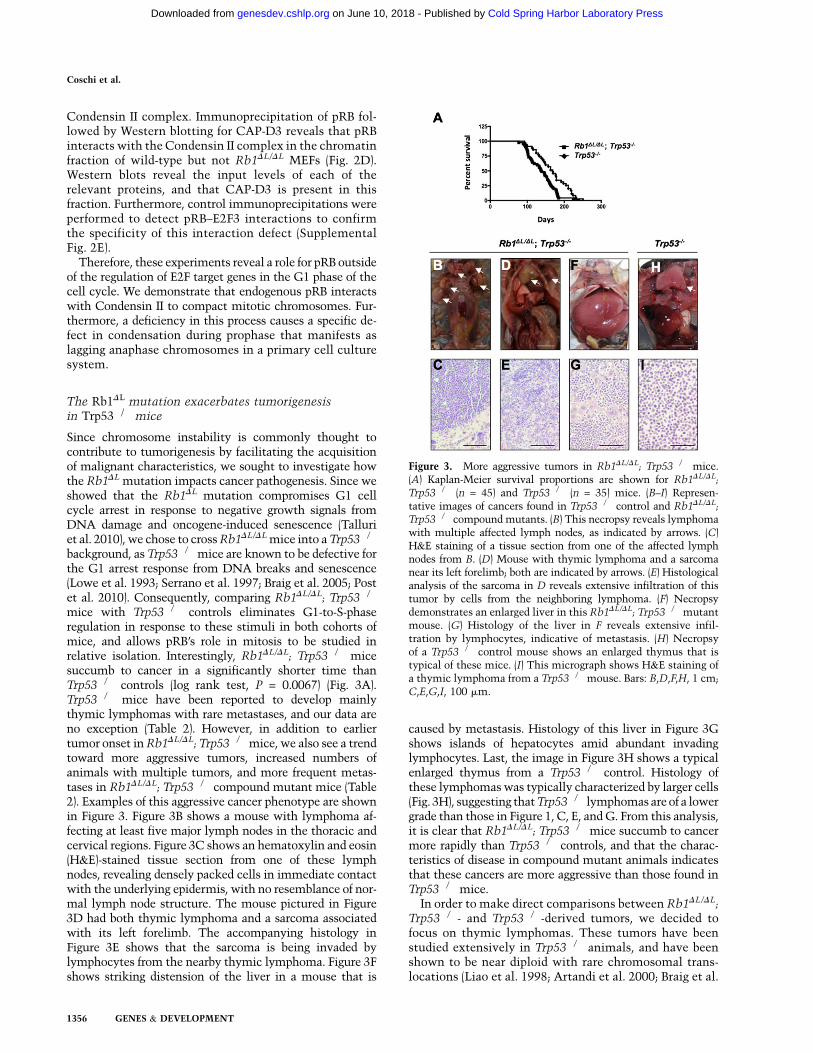
mice with Trp53�/� controls eliminates G1-to-S-phaseregulation in response to these stimuli in both cohorts ofmice, and allows pRB’s role in mitosis to be studied inrelative isolation. Interestingly, Rb1DL/DL; Trp53�/� micesuccumb to cancer in a significantly shorter time thanTrp53�/� controls (log rank test, P = 0.0067) (Fig. 3A).Trp53�/� mice have been reported to develop mainlythymic lymphomas with rare metastases, and our data areno exception (Table 2). However, in addition to earliertumor onset in Rb1DL/DL; Trp53�/�mice, we also see a trendtoward more aggressive tumors, increased numbers ofanimals with multiple tumors, and more frequent metas-tases in Rb1DL/DL; Trp53�/� compound mutant mice (Table2). Examples of this aggressive cancer phenotype are shownin Figure 3. Figure 3B shows a mouse with lymphoma af-fecting at least five major lymph nodes in the thoracic andcervical regions. Figure 3C shows an hematoxylin and eosin(H&E)-stained tissue section from one of these lymphnodes, revealing densely packed cells in immediate contactwith the underlying epidermis, with no resemblance of nor-mal lymph node structure. The mouse pictured in Figure3D had both thymic lymphoma and a sarcoma associatedwith its left forelimb. The accompanying histology inFigure 3E shows that the sarcoma is being invaded bylymphocytes from the nearby thymic lymphoma. Figure 3Fshows striking distension of the liver in a mouse that is
caused by metastasis. Histology of this liver in Figure 3Gshows islands of hepatocytes amid abundant invadinglymphocytes. Last, the image in Figure 3H shows a typicalenlarged thymus from a Trp53�/� control. Histology ofthese lymphomas was typically characterized by larger cells(Fig. 3H), suggesting that Trp53�/� lymphomas are of a lowergrade than those in Figure 1, C, E, and G. From this analysis,it is clear that Rb1DL/DL; Trp53�/� mice succumb to cancermore rapidly than Trp53�/� controls, and that the charac-teristics of disease in compound mutant animals indicatesthat these cancers are more aggressive than those found inTrp53�/� mice.
In order to make direct comparisons between Rb1DL/DL;Trp53�/�- and Trp53�/�-derived tumors, we decided tofocus on thymic lymphomas. These tumors have beenstudied extensively in Trp53�/� animals, and have beenshown to be near diploid with rare chromosomal trans-locations (Liao et al. 1998; Artandi et al. 2000; Braig et al.
Figure 3. More aggressive tumors in Rb1DL/DL; Trp53�/� mice.(A) Kaplan-Meier survival proportions are shown for Rb1DL/DL;
Trp53�/� (n = 45) and Trp53�/� (n = 35) mice. (B–I) Represen-tative images of cancers found in Trp53�/� control and Rb1DL/DL;Trp53�/� compound mutants. (B) This necropsy reveals lymphomawith multiple affected lymph nodes, as indicated by arrows. (C)H&E staining of a tissue section from one of the affected lymphnodes from B. (D) Mouse with thymic lymphoma and a sarcomanear its left forelimb; both are indicated by arrows. (E) Histologicalanalysis of the sarcoma in D reveals extensive infiltration of thistumor by cells from the neighboring lymphoma. (F) Necropsydemonstrates an enlarged liver in this Rb1DL/DL; Trp53�/�mutantmouse. (G) Histology of the liver in F reveals extensive infil-tration by lymphocytes, indicative of metastasis. (H) Necropsyof a Trp53�/� control mouse shows an enlarged thymus that istypical of these mice. (I) This micrograph shows H&E staining ofa thymic lymphoma from a Trp53�/�mouse. Bars: B,D,F,H, 1 cm;C,E,G,I, 100 mm.
Coschi et al.
1356 GENES & DEVELOPMENT
Cold Spring Harbor Laboratory Press on June 10, 2018 - Published by genesdev.cshlp.orgDownloaded from
2005). In this way, they offered an ideal starting point forinvestigating the effects caused by the Rb1DL mutation onchromosome instability. To ensure that the comparisonbetween these tumors was appropriate, we sought toinvestigate whether the Rb1DL mutation affects thymicdevelopment in a way that could bias this analysis. First,defects caused by the Rb1DL mutation alone are not suf-ficient to cause cancer in this or any other organ in thesemice (Supplemental Fig. 3). Second, gross histologicalanalysis of thymi from Rb1DL/DL and Rb1DL/DL; Trp53�/�
animals did not reveal any obvious abnormalities (Supple-mental Fig. 4A). Furthermore, analysis of CD4- and CD8-positive cells revealed no alterations in T-cell develop-ment, and rates of proliferation were unaltered by theRb1DL mutation (Supplemental Fig. 4B,C). Finally, E2Ftarget gene expression was not deregulated in thymi fromthese mice (Supplemental Fig. 4D). From these experi-ments, we conclude that differences in Rb1DL/DL; Trp53�/�
and Trp53�/� thymic lymphomas are unlikely to be ex-plained by differences in either development, or the basalproliferation rate of cells in this organ.
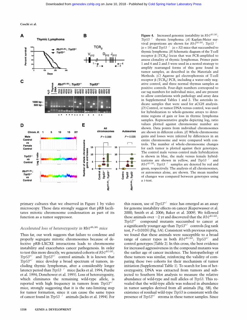
Examining the survival proportions of the animals thatsuccumbed to thymic lymphoma alone revealed that theRb1DL/DL; Trp53�/� mice have a shorter latency beforetumor formation compared with Trp53�/� controls (logrank test, P = 0.0198) (Fig. 4A). One possibility to explainthe difference in aggressiveness of these tumors is thatcompound mutant lymphomas may arise from manyinitiating thymocytes, resulting in a polyclonal tumor,whereas the Trp53�/� controls may be mono- or oligoclo-nal. To address this question, we used a PCR assay todetect individual T-cell receptor recombination events toestimate the number of individual thymocytes that havebecome transformed and populate each lymphoma (Fig.4B,C). This revealed that tumors of both genotypes wererarely monoclonal, and both showed a similar range ofclonality, suggesting that clonality does not bias our com-parison between the thymic lymphomas found in animalsof these two genotypes.
To investigate the effects of the Rb1DL mutation onchromosome instability, we used array comparative ge-nomic hybridization (aCGH) to compare the genomes ofthymic lymphoma cells from Rb1DL/DL; Trp53�/� andTrp53�/� mice. Figure 4D shows representative log2 ratioplots from male versus female control hybridizations, aswell as from selected Rb1DL/DL; Trp53�/� and Trp53�/�
tumors hybridized against same sex control DNA. Using
male versus male and male versus female control hybrid-izations as a guide for normal copy number and whole-chromosome gains, we inferred changes in chromosomecopy number present in these tumors. Since these lym-phomas are polyclonal, the gain or loss of a single chro-mosome in one clone can be a relatively modest changewhen the whole thymus is analyzed as one. For thisreason, we searched for chromosomes that were statisti-cally different than control, and did not try to distinguishif these represent single or multiple chromosome gains.Satisfyingly, the average number of gains and losses in ourTrp53�/� lymphoma controls (4.2) was similar to thefrequency of chromosome number changes reported bykaryotyping in other studies of Trp53-deficient lympho-mas in which these cancers typically have chromosomecounts in the low 40s (Liao et al. 1998; Artandi et al. 2000;Braig et al. 2005). Our analysis revealed that whole-chromosome gains or losses were more prevalent inRb1DL/DL; Trp53�/� lymphomas than in Trp53�/� controls(Fig. 4E). Because not all tumors originate from mice of thesame sex, and this creates unequal opportunities to gainand lose sex chromosomes, we displayed these data ingraphs for all chromosomes and for autosomes. Thissuggests that whole-chromosome instability may be theunderlying mechanism that increases cancer susceptibil-ity in Rb1DL/DL; Trp53�/� animals compared with Trp53�/�
mice, an interpretation that is consistent with centromerefusions observed in metaphase spreads (Fig. 1A) and lag-ging chromosomes in anaphase that were observed in ourvideos (Fig. 1C). We note that the quantity of copy numbersegments (local regions of gain or loss relative to adja-cent chromosomal sequences) is also elevated in mostRb1DL/DL; Trp53�/� thymic lymphomas (SupplementalFig. 5). This implies that smaller genomic rearrangementsalso take place, and this is consistent with resolution oflagging chromosomes in Figure 1C, row 2, occurring bychromosomal breakage. Therefore, both forms of chromo-somal instability may be caused by the Rb1DL mutation,and contribute to the increase in cancer susceptibility thatwe observe.
Our analysis of the Rb1DL allele’s effects on cancerreveal that it causes a dramatic increase in susceptibility,and Rb1DL/DL; Trp53�/� mice are characterized by moreaggressive tumors than the Trp53�/� controls. Further-more, characterization of genomic abnormalities found inRb1DL/DL tumors demonstrates that they are consistentwith the chromosomal and mitotic abnormalities in
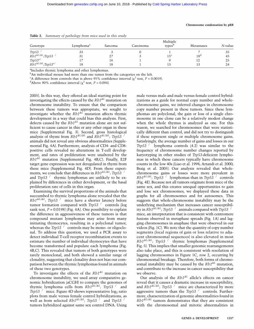
Table 2. Summary of pathology from mice used in this study
Genotype Lymphomaa Sarcoma CarcinomaMultiple
typesb Metastases N-value
Trp53�/� 33 3 0 1 7 35Rb1DL/DL;Trp53�/� 31 27 3 15c 18d 45Trp53+/� 17 16 1 9 12 25Rb1DL/DL;Trp53+/� 18 18 1 13 13 24
aIncludes thymic lymphoma and other lymphomas.bAn individual mouse had more than one tumor from the categories on the left.cA difference from controls that is above 95% confidence interval (x2 test, P = 0.0019).dAbove 90% confidence interval (x2 test, P = 0.094).
Chromosome condensation by pRB
GENES & DEVELOPMENT 1357
Cold Spring Harbor Laboratory Press on June 10, 2018 - Published by genesdev.cshlp.orgDownloaded from
primary cultures that we observed in Figure 1 by videomicroscopy. These data strongly suggest that pRB facili-tates mitotic chromosome condensation as part of itsfunction as a tumor suppressor.
Accelerated loss of heterozygosity in Rb1DL/DL mice
Thus far, our work suggests that failure to condense andproperly segregate mitotic chromosomes because of de-fective pRB–LXCXE interactions leads to chromosomeinstability and exacerbates cancer pathogenesis. In orderto test this more directly, we generated cohorts of Rb1DL/DL;Trp53+/� and Trp53+/� control animals. It is known thatTrp53+/� mice develop a broad spectrum of tumors, in-cluding thymic lymphomas, after a considerably longerlatency period than Trp53�/�mice (Jacks et al. 1994; Purdieet al. 1994; Donehower et al. 1995). Loss of heterozygosity,which eliminates the remaining wild-type allele, isreported with high frequency in tumors from Trp53+/�
mice, strongly suggesting that it is the rate-limiting stepfor tumor formation, since it can cause the same typesof cancer found in Trp53�/� animals (Jacks et al. 1994). For
this reason, use of Trp53+/� mice has emerged as an assayfor genome instability effects on cancer (Kuperwasser et al.2000; Smith et al. 2006; Baker et al. 2009). We followedthese animals over ;2 yr and discovered that the Rb1DL/DL;Trp53+/� compound mutants succumbed to cancer ata significantly younger age than Trp53+/� controls (log ranktest, P = 0.0105) (Fig. 5A). Consistent with previous reports,we found that these animals were susceptible to a broadrange of cancer types in both Rb1DL/DL; Trp53+/� andcontrol genotypes (Table 2). In this cross, the best evidencefor increased aggressiveness in the compound mutants wasthe earlier age of cancer incidence. The histopathology ofthese tumors was similar, reinforcing the validity of com-paring these two cohorts for their mechanism of tumorinitiation (Supplemental Table 1). To search for loss of het-erozygosity, DNA was extracted from tumors and sub-jected to Southern blot analysis to measure the relativeabundance of wild-type and null alleles of Trp53. This re-vealed that the wild-type allele was reduced in abundancein tumor samples derived from all animals (Fig. 5B); theexistence of residual wild-type Trp53 is consistent with thepresence of Trp53+/� stroma in these tumor samples. Since
Figure 4. Increased genomic instability in Rb1DL/DL;
Trp53�/� thymic lymphoma. (A) Kaplan-Meier sur-vival proportions are shown for Rb1DL/DL; Trp53�/�
(n = 18) and Trp53�/� (n = 32) mice that succumbed tothymic lymphoma. (B) Schematic diagram of the T-cellreceptor b (TCRb) locus that was PCR-amplified toassess clonality of thymic lymphomas. Primer pairs1 and 4 and 2 and 3 were used in a nested strategy toamplify rearranged forms of this gene found intumor samples, as described in the Materials andMethods. (C) Agarose gel electrophoresis of T-cellreceptor b (TCRb) PCR, including a water-only neg-ative control, and three normal thymus samples aspositive controls. Four-digit numbers correspond toear tag numbers for individual mice, and are presentto allow correlations with pathology and array datain Supplemental Tables 1 and 2. The asterisks in-dicate samples that were used for aCGH analysis.(D) Control, or tumor DNA versus control, was usedfor hybridization to whole-genome arrays to deter-mine regions of gain or loss in thymic lymphomasamples. Representative graphs depicting log2 ratiovalues plotted against chromosome number areshown. Data points from individual chromosomesare shown in different colors. (E) Whole-chromosomegains and losses were inferred by differences in anentire chromosome and were compared with con-trols. The number of whole-chromosome changesfor each tumor is plotted against their genotypes.The control male versus control male hybridizationis shown in blue, the male versus female hybrid-izations are shown in yellow, and Trp53�/� andRb1DL/DL; Trp53�/� samples are denoted by red andgreen, respectively. The analysis of all chromosomes,or autosomes alone, are shown. The mean numberof changes was compared between genotypes usinga t-test.
Coschi et al.
1358 GENES & DEVELOPMENT
Cold Spring Harbor Laboratory Press on June 10, 2018 - Published by genesdev.cshlp.orgDownloaded from

these data reveal loss of the wild-type Trp53 locus in all ofthe tumors we analyzed, and Rb1DL/DL; Trp53+/� mice de-velop cancer earlier, this suggests that the rate-limiting stepfor tumor formation—namely, loss of heterozygosity—hastaken place more rapidly and was facilitated by the Rb1DL
mutation in these mice (Fig. 5C).This analysis of loss of heterozygosity in tumors from
Rb1DL/DL; Trp53+/�mice offers evidence that cells bearingthe Rb1DL mutation are more prone to chromosomal in-stability. Based on this experiment, and the analysis ofRb1DL/DL; Trp53�/� lymphomas, our study reveals that
chromosome condensation mediated by pRB is likely animportant component of its role as a tumor suppressor.
Discussion
This study investigates the role of the pRB in mitoticchromosome condensation. Our data indicate that this isa mechanism by which pRB acts as a tumor suppressor.The novelty of this tumor-suppressive mechanism reliesextensively on the ability to separate the mitotic-specificfunctions of pRB from cell cycle entry control in ourcancer-prone mice. The analysis of thymic lymphomas inRb1DL/DL; Trp53�/�mice allows us to distinguish betweenthe effects of the Rb1DL mutation at these different pointsin the cell cycle. First, the response to DNA damage, orother stress-inducing stimuli that activate p53, leads toincreased p21/CIP1 expression; this in turn inhibitscyclin-dependent kinases, and leads to pRB activationduring G1 and cell cycle arrest (Campisi and d’Adda diFagagna 2007). Since we demonstrated previously thatRb1DL/DL cells are defective for a G1 arrest in responseto g-irradiation or oncogene-induced senescence (Talluriet al. 2010), including p53 deficiency in both cohorts ofour tumor study prevents these Rb1DL defects from con-founding our interpretations. In addition to these defects,we also determined that Rb1DL/DL cells are resistant to thegrowth inhibitory effects of TGF-b (Francis et al. 2009).This growth inhibitory cytokine has been shown to playa key role in peripheral T-cell regulation. In particular,transgenic mice expressing a dominant-negative TGF-btype II receptor in CD8-positive T cells are prone to de-velop lymphoproliferative disease and, ultimately, lym-phoma (Lucas et al. 2000, 2004). Interestingly, the phe-notype of these animals is very different from Rb1DL/DL;Trp53�/�mice, with extensive expansion of T cells in theperiphery but not the thymus. In fact, these studies suggestthat TGF-b signaling may have very little function in thethymus. Since Rb1DL/DL mice do not display any lympho-proliferative characteristics in their lifetime (Supplemen-tal Fig. 4), it is unlikely that defective TGF-b growthcontrol can explain the cancer phenotype of Rb1DL/DL;Trp53�/� mice. There is no evidence of aberrant prolifer-ation or alterations in thymic development or mophologyin Rb1DL/DL; Trp53�/�mice. Furthermore, E2F target genesare regulated normally in this tissue. Because there is evi-dence of elevated chromosomal instability in these thymiclymphomas, our conclusion that pRB can function as atumor suppressor by facilitating chromosome condensa-tion is the most appropriate interpretation of these data.
The physical interaction between pRB and CondensinII offers a logical explanation for how pRB can participatein mitotic chromosome condensation. The phenotypesseen in video microscopy experiments of Rb1DL/DL cellssuggest an acute defect in condensation during prophase.However, the extensive reduction in Condensin II levelson chromatin in asynchronously proliferating Rb1DL/DL
MEFs suggests that pRB participates in chromatin loadingearlier in the cell cycle, as these cultures contain onlya small proportion of mitotic cells. In addition, CondensinII is known to be present on chromatin in interphase nuclei
Figure 5. Accelerated loss of heterozygosity in Rb1DL/DL; Trp53+/�
mice. (A) Kaplan-Meier survival proportions are shown for Rb1DL/DL;Trp53+/� (n = 24) and Trp53+/� (n = 25) mice that succumbed todetectable cancers. (B) Southern blot analysis of tumors fromRb1DL/DL; Trp53+/� and Trp53+/� mice was performed to assessthe relative abundance of wild-type and null Trp53 alleles. Four-digit numbers correspond to ear tags for individual mice toallow correlation with pathology data in Supplemental Table 1.The ratio of mutant to wild-type allele abundance was deter-mined by phosphorimaging, and is displayed below each lane.(C) Model of the Rb1DL mutation’s role in cancer susceptibilityof these mice. Prior reports establish that Trp53+/�mice succumbto cancer after a long latency, and that it is accompanied by lossof heterozygosity at the Trp53 locus. The age at which cancerinitiates in Rb1DL/DL; Trp53+/� mice and the loss of the wild-typeTrp53 locus in these tumors suggests that chromosome instabil-ity, caused by the Rb1DL mutation, induces loss of heterozygositymore rapidly, causing an earlier onset of cancer.
Chromosome condensation by pRB
GENES & DEVELOPMENT 1359
Cold Spring Harbor Laboratory Press on June 10, 2018 - Published by genesdev.cshlp.orgDownloaded from
(Hirota et al. 2004). These observations suggest that pRB’srole in chromosome condensation may take place earlierin the cell cycle, perhaps in G1 where it is relativelyunphosphorylated and already thought to regulate chro-matin structure. Immunoprecipitation and Western blot-ting experiments demonstrate that wild-type pRB inter-acts with Condensin II in chromatin fractions, but thisinteraction was absent from Rb1DL/DL chromatin. BecauseCondensin II is underrepresented in this fraction in thefirst place, the lack of interaction may not reflect a need forthe pRB–LXCXE-binding cleft to mediate physical contactwith Condensin II, but may indicate that this aspect of pRBis more important for Condensin II to be loaded on chro-matin. It will be important in future studies to determineprecisely how pRB uses LXCXE-type interactions to exertits regulatory role over Condensin II function. At this point,we have no evidence to indicate that it must be direct.Similar studies from the Dyson and te Riele laboratories(Manning et al. 2010; van Harn et al. 2010) show thatpRB may also participate in chromosome cohesion at thecentromere. In those studies, Cohesin complexes are re-duced at centromeres. We cannot rule out that a similarbiochemical defect may be present in Rb1DL/DL cells.However, condensin complexes are also well known tobe concentrated at centromeric heterochromatin (Onoet al. 2004; Oliveira et al. 2005; Vagnarelli et al. 2006).Taken together, this suggests that future studies to un-derstand pRB’s role in mitosis will need to focus moreclosely on its ability to regulate chromatin at centromericregions, as this is likely where it acts to ensure properchromosome architecture and segregation in mitosis.
This study reveals a novel mechanism of tumor sup-pression by pRB. While other reports have indicated thatdefective pRB is associated with chromosomal abnormal-ities (Hernando et al. 2004; Gonzalo et al. 2005; Iovinoet al. 2006; Isaac et al. 2006; Longworth et al. 2008; Amatoet al. 2009), our work demonstrates that this manifests inmore rapid tumor formation. This raises the question:How important is this aspect of tumor suppression by pRBrelative to its well-characterized role in regulating E2Ftranscription factors and entry into S phase? The lack ofspontaneous tumors in our Rb1DL/DL mice may suggestthat it is less important. We favor a more cautious view ofthis question. The inability to arrest proliferation in G1because of a pRB deficiency is also accompanied by de-regulation of activator E2Fs, and this creates an intrinsicprogrowth signal. This is inherently a stronger oncogenicevent than diminished chromosome condensation becauseit combines the loss of negative growth regulation withthe gain of a growth-promoting signal. Defective chromo-some condensation on the other hand, creates the oppor-tunity for genetic change that can contribute to cancerpathogenesis, but does not provide an inappropriate growth-promoting signal. For these reasons, experiments designedto equalize the loss of safeguards with gain of proliferativeadvantages will be necessary to appropriately comparethese aspects of tumor suppression by pRB. Only throughthis type of investigation will it be possible to fully com-prehend what makes the Rb gene such a critical factor incell cycle regulation and cancer.
Materials and methods
Cell culture, viral infections, and microscopy
Primary MEFs were prepared and cultured according to standardmethods as reported previously (Hurford et al. 1997). ESCs wereprepared by intercrossing Rb1DL/+ mice and harvesting day 3.5blastocysts for culture. The inner cell mass was dissociated andcolonies were picked and expanded for genotyping as described(Hogan et al. 1994). Mitotic chromosome spreads were preparedfrom MEFs by treating cells with 50 ng/mL colcemid for 3 hbefore harvesting, swelling, and fixing. ESC chromosome spreadswere generated similarly after treating cultures with 10 mg/mLcolchicine for 3 h. Chromosome spreads were stained with amajor satellite pericentromeric probe as before (Isaac et al. 2006).Staining of rDNA repeats and the probes used were as described(Grummt et al. 1979; Romanova et al. 2006). Fluorescent mi-croscopic images were captured on a Zeiss Axioskop 40 micro-scope using a Spotflex camera and EyeImage software.
To introduce H2B-GFP into MEFs, we created a pBABE retrovi-ral vector that expresses H2B-GFP by cloning the gene from pBOS-H2B-GFP. Short hairpin retroviral vectors targeting CAP-D3 werepurchased from Open Biosystems, and were cloned into the pLMPplasmid along with the H2B-GFP gene. Viral vectors were pack-aged into ecotropic retroviruses using Bosc23 cells, and were sub-sequently used to infect MEFs as described (Pear et al. 1993).
Live cell microscopy was carried out by plating early passage,H2B-GFP-transduced MEFs onto glass-bottom tissue culturedishes (MatTek) containing phenol-free DMEM and 5% FBS sup-plemented with penicillin, streptomycin, and glutamine. Duringimaging, cells were maintained at 37°C with 5% CO2 usinga stage-mounted environment chamber (Neue Biosciences), andwere monitored with an automated inverted microscope (DMI6000b, Leica). Phase-contrast and fluorescent images were col-lected every 3 min over a 15-h time course using OpenlabSoftware automation (Ritchie et al. 2008). Full-length movies foreach example shown in Figures 1 and 2 are available in the Sup-plemental Material. Measurements of metaphase plate dimen-sions were made using Volocity software.
Stained tissue sections were examined microscopically on aZeiss Axioskop 40 microscope, and were photographed using aSpotflex camera and EyeImage software.
Antibodies and protein detection
The following antibodies were used to detect or precipitateproteins in this study: rabbit anti-histone H2B (07-371, UpstateBiotechnologies), goat anti-GFP (G095, Clontech), rabbit anti-CAP-H (a kind gift of Kyoko Yokomori, University of Californiaat Irvine) (Heale et al. 2006), rabbit anti-CAP-H2 (a generous giftof Tatsuya Hirano, Riken, Japan) (Ono et al. 2003), rabbit anti-SMC1 (A300-055A, Bethyl Laboratories), anti-pRB (G3-245, BD-Pharmingen), anti-BubR1: C-20 (sc-16195, Santa Cruz Biotech-nology), anti-PCNA: PC10 (sc-56, Santa Cruz Biotechnology),anti-MCM7: 141.2 (sc-9966, Santa Cruz Biotechnology), anti-E2F3: C-18 (sc-878, Santa Cruz Biotechnology), and anti-E2F3clone PG37 (05-551, Upstate Biotechnologies). Antibodies toCAP-D3 were raised against a GST fusion protein containingamino acids 1243–1506 of CAP-D3. CAP-D2 antibodies wereraised against a GST fusion containing amino acids 943–1132.Rabbits were immunized in a commercial facility (PTG Labora-tories). Antibodies were affinity-purified by adsorbing to a His-tagged version of the same protein coupled to a Sulfolink column(Pierce), eluted in low pH, and neutralized.
Cell extracts for Western blotting were prepared by freeze–thawlysis as described previously (Hurford et al. 1997). Chromatin
Coschi et al.
1360 GENES & DEVELOPMENT
Cold Spring Harbor Laboratory Press on June 10, 2018 - Published by genesdev.cshlp.orgDownloaded from
fractions were prepared by low-salt wash of nuclei followedby nuclease treatment to solubilize chromatin-bound proteins(Mendez and Stillman 2000). MEFs were synchronized using atwo-step method of confluence arrest and aphidicolin, followed bywashout and collection of cells when the mitotic index is greatest(Isaac et al. 2006). Nuclease-prepared chromatin fractions wereused for immunoprecipitation experiments where they were pre-cleared with mouse IgG and protein-G magnetic beads before pre-cipitation using anti-pRB antibodies. Proteins in all experimentswere resolved by SDS-PAGE followed by Western blotting usingstandard techniques.
Mice
The Rb1DL mutant strain carries three amino acid substitutionsin its Rb1-encoded protein (I746A, N750A, and M754A) thatdisrupt interactions with LXCXE motif-containing proteins, butnot E2F transcription factors (Dick et al. 2000; Isaac et al. 2006);details of its construct and initial characterization have beenpublished previously (Isaac et al. 2006; Francis et al. 2009; Talluriet al. 2010). The Trp53�/� mice were purchased from JacksonLaboratories and were intercrossed with Rb1DL mutants toproduce the required genotypes for this study. All animals weremaintained in a mixed 129/B6 background. Mice were housedand maintained according to the guidelines of the CanadianCouncil on Animal Care. Animals were followed throughouttheir lives for signs of tumor burden, and were euthanized whentumors became visible or the animal experienced sudden weightloss or became lethargic. All animals were subjected to a thoroughnecropsy, and abnormal tissues, organs, or tumors were fixed informalin and processed for histological assessment. Portions oftumors were also snap-frozen and used to prepare genomic DNA.Tissues were embedded, sectioned, and stained with H&E accord-ing to standard methods, and were photographed as describedabove. See Supplemental Table 1 for synopsis of histopathology forall animals used in this study.
Thymic development
To investigate thymic development and proliferation, we iso-lated thymuses from 6- to 8-wk-old animals. Tissue was eitherfixed and frozen for cryosectioning, or dissociated and stained forflow cytometry. Fluorescently labeled antibodies against CD4(553650) and CD8 (553032) were purchased from BD Pharmin-gen. CD4/CD8 flow cytometry was carried out essentially asdescribed (Bruins et al. 2004). To detect proliferation, mice wereinjected with 200 mL of 16 mg/mL BrdU in DMEM 1 h beforeeuthanasia. BrdU-labeled thymuses were fixed, sectioned, andstained with anti-BrdU antibodies (347580, BD Biosciences), andwere photographed under fluorescent optics as described above.
PCR, Southern blotting, and aCGH
High-molecular-weight DNA was extracted from frozen tumorsamples using standard DNA isolation procedures and were usedin the following analyses: DNA from thymic lymphomas wasanalyzed for T-cell receptor V(D)J recombination patterns usingnested PCR. In brief, the Db12 and Jb12 region was characterizedusing primer pairs and PCR conditions as described by Whitehurstet al. (1999). Loss of heterozygosity of Trp53 in tumors fromTrp53+/� mice was determined by Southern blotting. GenomicDNA was digested with EcoRI and StuI, and was resolved, trans-ferred, and hybridized using standard methods. The probe wasa genomic DNA clone encompassing intron 7 to intron 9. Bandintensities were measured on a Molecular Dynamics Storm
scanner PhosphorImager and densitometry analysis was pre-formed using AlphaEase FC software.
For aCGH experiments, DNA was extracted from livers offive male and female wild-type animals to create pools of controlDNA. Same sex control versus control, control male versus con-trol female, and tumor DNA versus the appropriate sex controlhybridizations were performed by NimbleGen on a mouse whole-genome array (design 2006-07-26-MM8-WG-CGH). Segmentationanalysis described by Olshen et al. (2004) was performed, and wasused to infer changes in copy number. A segment was defined asa group of adjacent data points whose log2 ratio values were notstatistically different. To determine cutoff values, indicating nor-mal copy number, the mean log2 ratio for all data points of indi-vidual chromosomes from the control hybridization was deter-mined. A range of plus or minus one standard deviation from thismean was determined to encompass the log2 ratio from 99.8% ofall segments from the control hybridization, suggesting that it wasa reliable range that captures the vast majority of normal copynumber chromosomal segments. This range was calculated foreach individual chromosome from the control hybridization.Segments from individual chromosomes from each tumor hybrid-ization were then compared with the appropriate range for theirchromosome to determine if they were of a normal copy num-ber. This allowed us to assess, on a chromosome-by-chromosomebasis, the copy number status of each segment. Using this ap-proach, we interpreted whole-chromosome gains or losses basedon the chromosomal locations of each constituent segment, andtheir associated copy number status (see Supplemental Fig. 6 foran illustrated example). See Supplemental Table 2 for raw seg-ment data for aCGH experiments.
Acknowledgments
We are grateful to our colleagues in the Dyson and te Rielelaboratories for communicating unpublished results. We thankmany members of the London Regional Cancer Program andChildren’s Health Research Institute for advice during the courseof this work, in particular, Francisca Aidoo and Christian Isaac forhelp during the initial stages of this work. C.C. acknowledgesfellowship support from NSERC and OGS, and is a member of theCaRTT training program. K.R. is supported by OGS. S.M.F. is therecipient of a Translational Breast Cancer Research Studentshipfrom the London Regional Cancer Program and acknowledgespast support from CBCF Ontario and OGSST. N.G.B. is a CIHRNew Investigator. F.A.D. is a Research Scientist of the CanadianCancer Society, and this work was funded by MOP-64253 fromthe CIHR.
References
Aladjem MI, Spike BT, Rodewald LW, Hope TJ, Klemm M,Jaenisch R, Wahl GM. 1998. ES cells do not activate p53-dependent stress responses and undergo p53-independentapoptosis in response to DNA damage. Curr Biol 8: 145–155.
Amato A, Lentini L, Schillaci T, Iovino F, Di Leonardo A. 2009.RNAi mediated acute depletion of retinoblastoma protein(pRb) promotes aneuploidy in human primary cells viamicronuclei formation. BMC Cell Biol 10: 79.
Artandi SE, Chang S, Lee SL, Alson S, Gottlieb GJ, Chin L,DePinho RA. 2000. Telomere dysfunction promotes non-reciprocal translocations and epithelial cancers in mice.Nature 406: 641–645.
Baker DJ, Jin F, Jeganathan KB, van Deursen JM. 2009. Wholechromosome instability caused by Bub1 insufficiency drivestumorigenesis through tumor suppressor gene loss of hetero-zygosity. Cancer Cell 16: 475–486.
Chromosome condensation by pRB
GENES & DEVELOPMENT 1361
Cold Spring Harbor Laboratory Press on June 10, 2018 - Published by genesdev.cshlp.orgDownloaded from
Belmont AS. 2006. Mitotic chromosome structure and conden-sation. Curr Opin Cell Biol 18: 632–638.
Braig M, Lee S, Loddenkemper C, Rudolph C, Peters AH,Schlegelberger B, Stein H, Dorken B, Jenuwein T, SchmittCA. 2005. Oncogene-induced senescence as an initial barrierin lymphoma development. Nature 436: 660–665.
Bruins W, Zwart E, Attardi LD, Iwakuma T, Hoogervorst EM,Beems RB, Miranda B, van Oostrom CT, van den Berg J, vanden Aardweg GJ, et al. 2004. Increased sensitivity to UVradiation in mice with a p53 point mutation at Ser389. Mol
Cell Biol 24: 8884–8894.Burkhart DL, Sage J. 2008. Cellular mechanisms of tumour
suppression by the retinoblastoma gene. Nat Rev Cancer 8:671–682.
Campisi J, d’Adda di Fagagna F. 2007. Cellular senescence:When bad things happen to good cells. Nat Rev Mol CellBiol 8: 729–740.
Cavanaugh AH, Hempel WM, Taylor LJ, Rogalsky V, Todorov G,Rothblum LI. 1995. Activity of RNA polymerase I transcrip-tion factor UBF blocked by Rb gene product. Nature 374:177–180.
Ciarmatori S, Scott PH, Sutcliffe JE, McLees A, Alzuherri HM,Dannenberg JH, te Riele H, Grummt I, Voit R, White RJ.2001. Overlapping functions of the pRb family in theregulation of rRNA synthesis. Mol Cell Biol 21: 5806–5814.
Dick FA, Sailhamer E, Dyson NJ. 2000. Mutagenesis of the pRBpocket domain reveals that cell cycle arrest functions areseparable from binding to viral oncoproteins. Mol Cell Biol20: 3715–3727.
Donehower LA, Harvey M, Vogel H, McArthur MJ, MontgomeryCAJ, Park SH, Thompson T, Ford RJ, Bradley A. 1995. Effectsof genetic background on tumorigenesis in p53-deficient mice.Mol Carcinog 14: 16–22.
Francis SM, Bergsied J, Isaac CE, Coschi CH, Martens AL,Hojilla CV, Chakrabarti S, Dimattia GE, Khoka R, WangJY, et al. 2009. A functional connection between pRB andtransforming growth factor b in growth inhibition andmammary gland development. Mol Cell Biol 29: 4455–4466.
Gonzalo S, Garcia-Cao M, Fraga MF, Schotta G, Peters AH, CotterSE, Eguia R, Dean DC, Esteller M, Jenuwein T, et al. 2005.Role of the RB1 family in stabilizing histone methylation atconstitutive heterochromatin. Nat Cell Biol 7: 420–428.
Grummt I, Soellner C, Scholz I. 1979. Characterization ofa cloned ribosomal fragment from mouse which containsthe 18S coding region and adjacent spacer sequences. Nucleic
Acids Res 6: 1351–1369.Ham MF, Takakuwa T, Rahadiani N, Tresnasari K, Nakajima H,
Aozasa K. 2007. Condensin mutations and abnormal chro-mosomal structures in pyothorax-associated lymphoma.Cancer Sci 98: 1041–1047.
Hannan KM, Hannan RD, Smith SD, Jefferson LS, Lun M,Rothblum LI. 2000. Rb and p130 regulate RNA polymeraseI transcription: Rb disrupts the interaction between UBF andSL-1. Oncogene 19: 4988–4999.
Harrington EA, Bruce JL, Harlow E, Dyson N. 1998. pRB playsan essential role in cell cycle arrest induced by DNA damage.Proc Natl Acad Sci 95: 11945–11950.
Heale JT, Ball AR Jr, Schmiesing JA, Kim JS, Kong X, Zhou S,Hudson DF, Earnshaw WC, Yokomori K. 2006. Condensin Iinteracts with the PARP-1–XRCC1 complex and functions inDNA single-strand break repair. Mol Cell 21: 837–848.
Hernando E, Nahle Z, Juan G, Diaz-Rodriguez E, Alaminos M,Hemann M, Michel L, Mittal V, Gerald W, Benezra R, et al.2004. Rb inactivation promotes genomic instability by uncou-pling cell cycle progression from mitotic control. Nature 430:797–802.
Herrera RE, Sah VP, Williams BO, Makela TP, Weinberg RA,Jacks T. 1996. Altered cell cycle kinetics, gene expression,and G1 restriction point regulation in Rb-deficient fibro-blasts. Mol Cell Biol 16: 2402–2407.
Hirota T, Gerlich D, Koch B, Ellenberg J, Peters JM. 2004.Distinct functions of condensin I and II in mitotic chromo-some assembly. J Cell Sci 117: 6435–6445.
Hogan B, Beddington R, Costantini F, Lacy E. 1994. Manipulat-
ing the mouse embryo: A laboratory manual. Cold SpringHarbor Press, Plainview, NY.
Hurford R, Cobrinik D, Lee M-H, Dyson N. 1997. pRB and p107/p130 are required for the regulated expression of differentsets of E2F responsive genes. Genes Dev 11: 1447–1463.
Iovino F, Lentini L, Amato A, Di Leonardo A. 2006. RB acuteloss induces centrosome amplification and aneuploidy inmurine primary fibroblasts. Mol Cancer 5: 38. doi: 10.1186/1476-4598-5-38.
Isaac CE, Francis SM, Martens AL, Julian LM, Seifried LA,Erdmann N, Binne UK, Harrington L, Sicinski P, Berube NG,et al. 2006. The retinoblastoma protein regulates pericentricheterochromatin. Mol Cell Biol 26: 3659–3671.
Jacks T, Remington L, Williams BO, Schmitt EM, Halachmi S,Bronson RT, Weinberg RA. 1994. Tumor spectrum analysisin p53-mutant mice. Curr Biol 4: 1–7.
Kuperwasser C, Hurlbut GD, Kittrell FS, Dickinson ES, LauciricaR, Medina D, Naber SP, Jerry DJ. 2000. Development ofspontaneous mammary tumors in BALB/c p53 heterozygousmice. A model for Li-Fraumeni syndrome. Am J Pathol 157:2151–2159.
Lapointe J, Malhotra S, Higgins JP, Bair E, Thompson M, SalariK, Giacomini CP, Ferrari M, Montgomery K, Tibshirani R,et al. 2008. hCAP-D3 expression marks a prostate cancersubtype with favorable clinical behavior and androgen sig-naling signature. Am J Surg Pathol 32: 205–209.
Liao MJ, Zhang XX, Hill R, Gao J, Qumsiyeh MB, Nichols W,Van Dyke T. 1998. No requirement for V(D)J recombinationin p53-deficient thymic lymphoma. Mol Cell Biol 18: 3495–3501.
Longworth MS, Dyson NJ. 2010. pRb, a local chromatin orga-nizer with global possibilities. Chromosoma 119: 1–11.
Longworth MS, Herr A, Ji JY, Dyson NJ. 2008. RBF1 promoteschromatin condensation through a conserved interactionwith the Condensin II protein dCAP-D3. Genes Dev 22:1011–1024.
Losada A, Hirano T. 2005. Dynamic molecular linkers of thegenome: The first decade of SMC proteins. Genes Dev 19:1269–1287.
Lowe SW, Schmitt EM, Smith SW, Osborne BA, Jacks T. 1993.p53 is required for radiation-induced apoptosis in mousethymocytes. Nature 362: 847–849.
Lucas PJ, Kim SJ, Melby SJ, Gress RE. 2000. Disruption of T cellhomeostasis in mice expressing a T cell-specific dominantnegative transforming growth factor b II receptor. J Exp Med191: 1187–1196.
Lucas PJ, McNeil N, Hilgenfeld E, Choudhury B, Kim SJ,Eckhaus MA, Ried T, Gress RE. 2004. Transforming growthfactor-b pathway serves as a primary tumor suppressor inCD8+ T cell tumorigenesis. Cancer Res 64: 6524–6529.
Maeshima K, Laemmli UK. 2003. A two-step scaffolding modelfor mitotic chromosome assembly. Dev Cell 4: 467–480.
Manning AL, Longworth MS, Dyson NJ. 2010. Loss of pRBcauses centromere dysfunction and chromosomal instability.Genes Dev (this issue). doi: 10.1101/gad.1917310.
Mayhew CN, Carter SL, Fox SR, Sexton CR, Reed CA, SrinivasanSV, Liu X, Wikenheiser-Brokamp K, Boivin GP, Lee JS,et al. 2007. RB loss abrogates cell cycle control and genome
Coschi et al.
1362 GENES & DEVELOPMENT
Cold Spring Harbor Laboratory Press on June 10, 2018 - Published by genesdev.cshlp.orgDownloaded from
integrity to promote liver tumorigenesis. Gastroenterology
133: 976–984.Mendez J, Stillman B. 2000. Chromatin association of human
origin recognition complex, cdc6, and minichromosome main-tenance proteins during the cell cycle: Assembly of prerepli-cation complexes in late mitosis. Mol Cell Biol 20: 8602–8612.
Musacchio A, Salmon ED. 2007. The spindle-assembly check-point in space and time. Nat Rev Mol Cell Biol 8: 379–393.
Nasmyth K. 2005. How do so few control so many?Cell 120:739–746.
Oliveira RA, Coelho PA, Sunkel CE. 2005. The condensin Isubunit Barren/CAP-H is essential for the structural integ-rity of centromeric heterochromatin during mitosis. MolCell Biol 25: 8971–8984.
Olshen AB, Venkatraman ES, Lucito R, Wigler M. 2004. Circularbinary segmentation for the analysis of array-based DNAcopy number data. Biostatistics 5: 557–572.
Ono T, Losada A, Hirano M, Myers MP, Neuwald AF, Hirano T.2003. Differential contributions of condensin I and conden-sin II to mitotic chromosome architecture in vertebrate cells.Cell 115: 109–121.
Ono T, Fang Y, Spector DL, Hirano T. 2004. Spatial and temporalregulation of Condensins I and II in mitotic chromosomeassembly in human cells. Mol Biol Cell 15: 3296–3308.
Pear WS, Nolan GP, Scott ML, Baltimore D. 1993. Production ofhigh-titre helper-free retroviuses by transient transfection.Proc Natl Acad Sci 90: 8392–8396.
Pei L, Melmed S. 1997. Isolation and characterization ofa pituitary tumor-transforming gene (PTTG). Mol Endocrinol
11: 433–441.Post SM, Quintas-Cardama A, Terzian T, Smith C, Eischen CM,
Lozano G. 2010. p53-dependent senescence delays Emu-myc-induced B-cell lymphomagenesis. Oncogene 29: 1260–1269.
Purdie CA, Harrison DJ, Peter A, Dobbie L, White S, Howie SE,Salter DM, Bird CC, Wyllie AH, Hooper ML, et al. 1994.Tumour incidence, spectrum and ploidy in mice with a largedeletion in the p53 gene. Oncogene 9: 603–609.
Ren B, Cam H, Takahashi Y, Volkert T, Terragni J, Young RA,Dynlacht BD. 2002. E2F integrates cell cycle progressionwith DNA repair, replication, and G(2)/M checkpoints.Genes Dev 16: 245–256.
Ritchie K, Seah C, Moulin J, Isaac C, Dick F, Berube NG. 2008.Loss of ATRX leads to chromosome cohesion and congres-sion defects. J Cell Biol 180: 315–324.
Romanova L, Korobova F, Noniashvilli E, Dyban A, Zatsepina O.2006. High resolution mapping of ribosomal DNA in earlymouse embryos by fluorescence in situ hybridization. Biol
Reprod 74: 807–815.Samoshkin A, Arnaoutov A, Jansen LE, Ouspenski I, Dye L,
Karpova T, McNally J, Dasso M, Cleveland DW, StrunnikovA. 2009. Human condensin function is essential for centro-meric chromatin assembly and proper sister kinetochoreorientation. PLoS One 4: e6831. doi: 10.1371/journal.pone.0006831.
Schvartzman JM, Sotillo R, Benezra R. 2010. Mitotic chromo-somal instability and cancer: Mouse modelling of the humandisease. Nat Rev Cancer 10: 102–115.
Serrano M, Lin AW, McCurrach ME, Beach D, Lowe SW. 1997.Oncogenic ras provokes premature senescence associatedwith accumulation of p53 and p16. Cell 88: 593–602.
Smith AP, Henze M, Lee JA, Osborn KG, Keck JM, Tedesco D,Bortner DM, Rosenberg MP, Reed SI. 2006. Deregulatedcyclin E promotes p53 loss of heterozygosity and tumorigen-esis in the mouse mammary gland. Oncogene 25: 7245–7259.
Talluri S, Isaac CE, Ahmad M, Henley SA, Francis SM, MartensAL, Bremner R, Dick FA. 2010. A G1 checkpoint mediated
by the retinoblastoma protein that is dispensable in terminaldifferentiation but essential for senescence. Mol Cell Biol 30:948–960.
Vader G, Lens SM. 2008. The Aurora kinase family in celldivision and cancer. Biochim Biophys Acta 1786: 60–72.
Vagnarelli P, Hudson DF, Ribeiro SA, Trinkle-Mulcahy L, SpenceJM, Lai F, Farr CJ, Lamond AI, Earnshaw WC. 2006. Con-densin and Repo-Man-PP1 co-operate in the regulation ofchromosome architecture during mitosis. Nat Cell Biol 8:1133–1142.
van Harn T, Foijer F, van Vugt M, Banerjee R, Yang F, Oostra A,Joenje H, te Riele H. 2010. Loss of RB proteins causesgenomic instability in the absence of mitogenic signaling.Genes Dev (this issue). doi: 10.1101/gad.580710.
Whitehurst CE, Chattopadhyay S, Chen J. 1999. Control of V(D)Jrecombinational accessibility of the D b 1 gene segment atthe TCRb locus by a germline promoter. Immunity 10: 313–322.
Zheng L, Flesken-Nikitin A, Chen PL, Lee WH. 2002. Deficiencyof retinoblastoma gene in mouse embryonic stem cells leadsto genetic instability. Cancer Res 62: 2498–2502.
Chromosome condensation by pRB
GENES & DEVELOPMENT 1363
Cold Spring Harbor Laboratory Press on June 10, 2018 - Published by genesdev.cshlp.orgDownloaded from
10.1101/gad.1917610Access the most recent version at doi: originally published online June 15, 201024:2010, Genes Dev.
Courtney H. Coschi, Alison L. Martens, Kieran Ritchie, et al. protein is tumor-suppressiveMitotic chromosome condensation mediated by the retinoblastoma
Material
Supplemental
http://genesdev.cshlp.org/content/suppl/2010/06/08/gad.1917610.DC1
Related Content
Genes Dev. July , 2010 24: 1329-1333
Julien Sage and Aaron F. StraightRB's original CIN?
References
http://genesdev.cshlp.org/content/24/13/1351.full.html#related-urls
Articles cited in:
http://genesdev.cshlp.org/content/24/13/1351.full.html#ref-list-1This article cites 61 articles, 23 of which can be accessed free at:
License
ServiceEmail Alerting
click here.right corner of the article or
Receive free email alerts when new articles cite this article - sign up in the box at the top
Copyright © 2010 by Cold Spring Harbor Laboratory Press
Cold Spring Harbor Laboratory Press on June 10, 2018 - Published by genesdev.cshlp.orgDownloaded from


![SDG2-Mediated H3K4me3 Is Crucial for Chromatin ... · SDG2-Mediated H3K4me3 Is Crucial for Chromatin Condensation and Mitotic Division during Male Gametogenesis in Arabidopsis1[OPEN]](https://img.pdfslide.net/doc/110x75/5bc9db1c09d3f2df158b48a6/sdg2-mediated-h3k4me3-is-crucial-for-chromatin-sdg2-mediated-h3k4me3-is.jpg)